Note: Descriptions are shown in the official language in which they were submitted.
CA 02658003 2008-12-03
WO 2007/145901 PCT/US2007/013167
COMPOSITIONS ENRICHED IN NEOPLASTIC STEM CELLS AND METHODS
COMPRISING SAME
BACKGROUND OF THE INVENTION
[001] To date, methods of analysis of neoplastic cells are neither efficient
nor uniform enough for
research purposes. Neoplastic stem cells isolated from tissue samples by
various fractionation procedures
consisted of mixed cell types. Efficient isolation of neoplastic stem cells
provides a means of exploring
basic mechanisms in cancer cell biology and disease. Methods for specifically
anci efficiently isolating
and propagating a cell subpopulation to provide a large neoplastic stem cell
population for in-vitro and in-
vivo studies are desirable.
[002] Identification of stem cell markers in neoplastic cells provides
valuable infonnation that is
useful for a variety of applications in both clinical and basic research
settings. The identification and
subsequent isolation of neoplastic stem cells (NSCs) from a particular tumor
or metastatic lesion is
useful, for example, in diagnosing a pathology and/or developing a rational
therapeutic treatment that
targets a developing pathology. In some instances, isolation and/or enrichment
of NSCs is desirable for
further in-vitro studies exploring physiological and molecular mechanisms,
wherein in other instances,
these cells can be used to inoculate a test animal for further studies of
cancer progression or therapy.
[003] NSCs can be sorted using techniques such as FACS (Fig. 10) or antibiotic
selection assays that
do not distinguish between sub-populations of cells based on their biological
activity and/or physiological
function. The assays, moreover, preclude recovery of native non-antibiotic-
expressing or treated stem
cells. Other methods of cellular identification and subsequent isolation
and/or enrichment such as gel
electrophoresis, fail to probe pure populations, suffer from contamination
and/or compromise cell
viability.
SUMMARY OF THE INVENTION
[004] This invention relates, in another embodiment, to a neoplastic stem cell
population enriched for
expression of OCT4.
[005] In another embodiment, this invention provides a method of identifying
neoplastic stem cells,
comprising the steps of (i) contacting neoplastic cells with an agent which
specifically interacts with
OCT4; and (ii) identifying cells with which the agent specifically interacts.
[006] In another embodiment, this invention provides a method of isolating
neoplastic stem cells,
comprising the steps of (i) contacting neoplastic cells with an agent which
specifically interacts with
OCT4; and (ii) isolating cells with which the agent specifically interacted.
[007] In another embodiment, this invention provides a rnethod of enriching a
neoplastic cell
population for neoplastic stem cells, comprising the steps of (i) contacting a
m:ixed cell population
I
CA 02658003 2008-12-03
WO 2007/145901 PCT/US2007/013167
comprising a plurality of cancerous cells with a vector comprising an
antibiotic resistance gene
operatively linked to an Oct4 promoter ; and (ii) culturing the mixed cell
population in the presence of an
antibiotic.
[008] In another embodiment, this invention provides a method of inducing
cancer comprising
introducing a neoplastic stem cell population enriched for expression of OCT4
to a mammal.
[009] In another embodiment, this invention provides a method of analyzing
cancer progression
and/or pathogenesis in-vivo, comprising the steps of (i) transplanting OCT4h'
neoplastic stem cells into an
animal; and (ii) analyzing cancer progression and/or pathogenesis in the
animal.
[0010] In another embodiment, this invention provides a method of abrogating,
or inhibiting cancer
comprising the step of: contacting neoplastic cells with an agent that
inhibits OCT4 expression or
function in said neoplastic cells.
[0011] In another embodiment, this invention provides a method of preventing,
abrogating, or
inhibiting tumor growth comprising the step of: contacting neoplastic cells
with an agent that inhibits
OCT4 expression or function in a tumor.
[0012] In another embodiment, this invention provides a method of preventing,
abrogating, or
inhibiting cell metastasis comprising the step of: contacting neoplastic cells
with an agent that inhibits
OCT4 expression or function in said neoplastic cells.
BRIEF DESCRIPTION OF THE DRAWINGS
[0013] The subject matter regarded as the invention is particularly pointed
out and distinctly claimed
in the concluding portion of the specification. The invention, however, both
as to organization and
method of operation, together with objects, features, and advantages thereof,
may best be understood by
reference to the following detailed description when read with the
accompanying drawings in which:
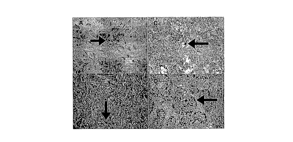
[0014] Fig. I presents light micrographs of serial sections of solid tumors,
probed with polyclonal
anti-OCT4 antibodies and control sections. Fig. lA is a cross section of a
chondrosarcoma tumor; Fig. IB
is a cross section of an osteosarcoma tumor; Fig. 1 C is a cross section of a
glioblastoma multiforme
(GBM) tumor; and Fig. ID is a cross section of fetal human testis used for
positive control. Arrows
indicate OCT4 positive nuclei.
[0015] Fig. 2A demonstrates the results of semi-quantitive RT-PCR probing for
OCT4, STAT3 and
Nanog mRNA expression wherein 0-tubulin and GFAP served as normalized controls
in representative
glioblastoma primary cultures (MT917, MT926, MT928, MT1231) and cell lines
(LN18, LN229, LN428,
U251). Fig. 2B demonstrates results of a Western blot analysis of OCT4, STAT3
and Nanog protein
expression wherein R-tubulin and GFAP served as normalized controls in cell
lines (LN18, LN229,
LN319, LN428, D247, U251, U373, T98G). Blots were probed with anti-OCT4, anti-
phospho-STAT3
and anti-Nanog.
2
CA 02658003 2008-12-03
WO 2007/145901 PCT/US2007/013167
[0016] Fig. 3 plots the results of 2-D quantitative PCR probing OCT4 and nanog
gene expression in
adherent cell cultures and floating osteosarcoma-derived spheres. Substrate-
attached cultures showed
significantly (p<0.05) lower expression of OCT4 and Nanog. Correlation of OCT4
(X axis) and Nanog
(Y axis) expression in sarcospheres is significantly (p<0.05) higher than in
substrate-attached cultures.
[0017] Fig. 4 illustrates clone-forming potential of glioblastoma-derived
cells suppressed by OCT4
siRNA. Fig. 4A demonstrates results of a Western blot analysis wherein
suppression of exogenous OCT4
protein in a transfected cell culture was achieved by treatment with specific
OCT4 siRNA comprising the
DNA sequence: TTGATCCTCGGACCTGGCTAA. Fig. 4B plots the frequency of clone-
formation by
selected glioblastoma cells (MT317, LN-229, MT-917). Cells were co-transfected
with eGFP (Green
Fluorescent Protein) and OCT4 siRNA. Experiments were perfomzed in triplicate,
bars represent standard
errors.
[0018] Fig. 5A is a light micrograph image (x200) of suspended mammaspheres
derived from an
MCF-7R breast cancer cell line and cultured in methylcellulose. Fig. 5B is a
fluorescent micrograph
(x200) of a mammasphere transferred from methylcellulose, attached to
substratum and immunostained
for OCT4 (white) and pancytokeratine (gray) expression.
[0019] Fig.6 presents light micrographs (x200) of immunohistochemically
stained breast cancer
tumors probed for OCT4 expression. Dark punctuate staining are OCT4 positive
nuclei. Fig. 6A is a cross
section of ductal carcinoma tumor, and Fig. 6B is a cross section of breast
cancer metastasis to brain.
Arrows indicate OCT4 positive nuclei.
[0020] Fig. 7 is a fluorescent microscope (x200) image of an OCT4-EGFP
transfected glioblastoma
cell in methyl cellulose after first division (Fig. 7A). A glioblastoma
floating neurosphere (clone) of
OCT-EGFP transfected cells after several rounds of divisions is shown in Fig.
7B.
[0021] Fig. 8 is a fluorescent microscope image (x200) of' cultured breast
cancer cells (A),
osteosarcoma (B), and glioblastoma multiforme cells (C) expressing EGFP
through an OCT4 responsive
promoter.
[0022] Fig. 9 is a fluorescent microscope image of the cultured glioblastoma
cell line Ln428 (x200)
(A) and osteosarcoma OS521 (x100) (B) expressing EGFP through Nanog responsive
promoter.
[0023] Fig. 10 illustrates FACS flow isolation graphs of subpopulations of
tumor cells expressing
OCT4 from cultured glioblastoma cell line Ln428. M2 gate represents OCT4
positive cells. Fig. IOA
represents the initial FACS sorting for OCT4 positive cells of a mixed clonal-
OCT4 cell population. Fig.
IOB represents isolated OCT4 positive cells after two passages (roughly 2
weeks) followed by FACS
analysis (B) to detennine their purity. This population was found to be 96.16
% pure for OCT4 protein
expression.
[0024] Fig. 11 is a graph illustrating the tumor forming potential of OCT4
positive and OCT4 negative
MDA MB 231 breast cancer cells transfected with OCT4-EGFR.
3
CA 02658003 2008-12-03
WO 2007/145901 PCT/US2007/013167
[0025] Fig. 12 schematically depicts the procedure for obtaining OCT4 enriched
tumor stem cells
from any tumor tissue for cancer related studies including drug discovery
studies. Stage A: Preparation of
Cells: 1) Surgical removal of tumor 2) Mincing and preparations to create a
single cells suspension. Stage
B: Stable labeling of tumor stem cells with OCT4 responsive promoter: 1) Tumor
stem cell culture in a
single cell suspension for expantion and selection for tumor stem cells under
the appropriate conditions 2)
transfection with a plasmid comprising an EGFP gene under the control of an
OCT4 responsive promoter
(stage C). Stage C: Creation of a highly pure tumor stem cell cultures: EGFP
expressing cells are further
selected via FACS and re-cultured for expansion resulting in bulk culture
quantities. Stage D: D1, tumor
stem cells are further studied using rigorous cell and molecular biology
techniques. D2, tumor stem cells
are exposed to a vast variety of drugs Stage E: Isolated tumor stem cells are
inoculated into
immunodeficient mice to create xenograft tumor models followed by basic
efficacy, safety (or lack of
toxicity), and outcomes studies generating final drug lists.
[0026] Fig. 13 is a microscope image (x200) of mammosphere cultures derived
from an MDA-MB-
435 melanoma cell line, biomarked for the presence of NSC expressing Oct-3/4.
Fig. 13A is a light
micrograph of suspended tumor-derived spheres cultured in methylcellulose.
Fig. 13B is a fluorescent
micrograph of the suspended tumor-derived spheres shown in Fig. 13A. Fig. 13C
is a light micrograph of
tumor spheres after attachment to the substratum. Fig. 13D is a fluorescent
micrograph of the attached
tumor spheres shown in Fig. 13C.
~[0027] It will be appreciated that for simplicity and clarity of
illustration, elements shown in the
figures have not necessarily been drawn to scale. For example, the dimensions
of some of the elements
may be exaggerated relative to other elements for clarity. Further, where
considered appropriate, reference
numerals may be repeated among the figures to indicate corresponding or
analogous elements.
DETAILED DESCRIPTION OF THE PRESENT INVENTION
[0028] In the following detailed description, numerous specific details are
set forth in order to provide
a thorough understanding of the invention. However, it will be understood by
those skilled in the art that
the present invention may be practiced without these specific details. In
other instances, well-known
methods, procedures, and components have not been described in detail so as
not to obscure the present
invention.
[0029] While certain features of the invention have been illustrated and
described herein, many
modifications, substitutions, changes, and equivalents will now occur to those
of ordinary skill in the art.
It is, therefore, to be understood that the appended claims are intended to
cover all such modifications and
changes as fall within the true spirit of the invention.
[0030] In another embodiment, the invention comprises a neoplastic stem cell
(NSC) population
enriched for expression of OCT4, Nanog, STAT3 or combinations thereof. In
another embodiment, NSCs
represent a subpopulation of cells within a population comprising neoplastic
cells, which is capable of
4
CA 02658003 2008-12-03
WO 2007/145901 PCT/US2007/013167
initiating and maintaining cancer following a prolonged period of time. In
another embodiment, NSCs
drive the formation and growth of tumors (Fig. 11). In another embodiment, the
term drive as used herein
refers to guide, control, direct, initiate, go through, penetrate or
combinations thereof.
[0031] In another embodiment, NSCs comprise properties such as longevity, self-
renewal and
quiescence. In another embodiment, NSCs comprise enhanced invasive capacity.
In another embodiment,
NSCs are multipotent, self-renewing and are able to produce proliferating
sarcospheres from sarcomas,
neurospheres from brain tumors or mammaspheres from breast cancers (Fig. 5).
In another embodiment,
NSCs are capable of keeping their self-renewal potential during 1-100 passages
of in-vitro cultivation. In
another embodiment, NSCs are capable of keeping their self-renewal potential
during 1-90 passages of
in-vitro cultivation. In another embodiment, NSCs are capable of keeping their
self-renewal potential
during 20-60 passages of in-vitro cultivation. In another embodiment, NSCs
express genes involved in
the specific functions and/or in self-renewal of NSCs, such as OCT4, Nanog,
STAT3 or combinations
thereof.
[0032] In another embodiment, the present invention provides that solid cancer
represents a
population of cells derived from a common founder cell, or NSC. In another
embodiment, the present
invention provides that tumors represent a population of cells derived from a
common founder cell, or
NSC. In another embodiment, the present invention provides that NSC phenotype
is similar in many ways
to that of normal stem cells. In another embodiment, the present invention
provides that NSC phenotype
is quite different to that of normal stem cells leading to the irregularities
with respect to abnormal
developmental profile. In another embodiment, the present invention provides
that NSC phenotype is
quite different from that of normal stem cells leading to the irregularities
with respect to lack of key
proliferation controls.
[0033] In another embodiment, the present invention provides that NSC
population comprises a mix
of true or mother NSCs and the progenitors neoplastic cells derived from NSCs.
In another embodiment,
the present invention provides that progenitors derived from NSCs are
different in key ways from mother
NSCs. In another embodiment, the present invention provides that different in
key ways comprise high
proliferation kinetics. In another embodiment, the present invention provides
that NSCs are typically
present in very low percentages relative to the total cancer cell population,
correlating roughly to the
"hostility" of the enviroment (i.e., a natural environment such as a breast
NSC in its primary breast tissue
location versus a breast NSC located in a metatstatic and/or foreign location
such as the brain.
[0034] In another embodiment, thepresent invention provides that NSCs comprise
about 0.001 to 1%
of the parental primary cancer population. In another embodiment, the present
invention provides that
NSCs comprise about 0.005 to 1% of the parental primary cancer population. In
another embodiment, the
present invention provides that NSCs comprise about 0.01 to 0.1% of the
parental primary cancer
population. In another embodiment, the present invention provides that NSCs
cornprise about 0.05 to
CA 02658003 2008-12-03
WO 2007/145901 PCT/US2007/013167
0.1% of the parental primary cancer population. In another embodiment, the
present invention provides
that NSCs comprise about 0.005 to 0.01 % of the parental primary cancer
population.
[0035] In another embodiment, the present invention provides that NSCs
comprise about I to 80% of
the cell population in permanent cancer cell lines parental. In another
embodiment, the present invention
provides that NSCs comprise about I to 10% of the cell population in permanent
cancer cell lines
parental. In another embodiment, the present invention provides that NSCs
comprise about 7 to 14% of
the cell population in permanent cancer cell lines parental. In another
embodiment, the present invention
provides that NSCs comprise about 15 to 25% of the cell population in
permanent cancer cell lines
parental. In another embodiment, the present invention provides that NSCs
comprise about 10 to 30% of
the cell population in permanent cancer cell lines parental. In another
embodiment,l:he present invention
provides that NSCs comprise about 30 to 50% of the cell population in
permanent cancer cell lines
parental. In another embodiment, the present invention provides that NSCs
comprise about 20 to 40% of
the cell population in ,permanent cancer cell lines parental. In another
embodiment, the present invention
provides that NSCs comprise about 50 to 80% of the cell population in
permanent cancer cell lines
parental. In another embodiment, the present invention provides that NSCs
comprise about 1 to 5% of the
cell population in permanent cancer cell lines parental. In another
embodiment, the present invention
provides that NSCs comprise about 5 to 10% of the cell population in permanent
cancer cell lines
parental. In another embodiment, the present invention provides that NSCs
comprise about 3 to 8% of the
cell population in permanent cancer cell lines parental. In another
embodiment, the present invention
provides that NSCs comprise about 7 to 10% of the cell population in permanent
cancer cell lines
parental.
[0036] In another embodiment, the present invention provides that NSCs
comprise about 1 to 100% of
the parental metastatic cancer population cell. In another embodiment, the
present invention provides that
NSCs comprise about 1 to 10% of the parental metastatic cancer population
cell. In another embodiment,
the present invention provides that NSCs comprise about 10 to 30% of the
parental metastatic cancer
population cell. In another embodiment, the present invention provides that
NSCs comprise about 30 to
50% of the parental metastatic cancer population cell. In another embodiment,
the present invention
provides that NSCs comprise about 50 to 75% of the parental metastatic cancer
population cell. In
another embodiment, the present invention provides that NSCs comprise about 75
to 100% of the
parental metastatic cancer population cell. In another embodiment, the present
invention provides that
NSCs comprise about 30 to 80% of the parental metastatic cancer population
cell. In another
embodiment, the present invention provides that NSCs comprise about 20 to 90%
of the parental
metastatic cancer population cell. In another embodiment, the present
invention provides that NSCs
comprise about 10 to 100% of the parental metastatic cancer population cell.
In another embodiment, the
present invention provides that NSCs comprise about 20 to 40% of the parental
metastatic cancer
population cell.
6
CA 02658003 2008-12-03
WO 2007/145901 PCT/US2007/013167
[0037] In another embodiment, the present invention provides that bulk cancer
cells (BCCs) comprise
the rnajority of the cancer cell population from a primary solid tumor. In
another embodiment, the present
invention provides that BCCs comprise the majority of the cancer cell
population from a permanent
cultured cell lines derived from cancers. In another embodiment, the present
invention provides that a
BCC population lacks stem cell characteristics. In another embodiment, the
present invention provides
that a BCC population lacks OCT-4 expression.
[0038] In another embodiment, the methods of the present invention provides
isolation of NSCs from
cancer tissue biopsies and permanent cancer cell lines.by selection of NSCs
previously manipulated and
biomarked to allow for detection. In another embodiment, the methods of the
present invention provide
stably transfecting NSCs with DNA vectors which expresses fluorescent or
luminescent proteins
regulated by an Oct-4 responsive promoter (Figure 12). In another embodiment,
the methods of the
present invention provides separating NSCs from the total cancer cell
population resulting in cultures of
high purity using FACS sorting of fluorescent biomarkers. In another
embodiment, the methods of the
present invention provides separating NSCs from the total cancer cell
population resulting in cultures of
high purity using FACS sorting of those cells that express GFP (Green
Fluorescent :Protein) driven by an
Oct4 promoter (Figure 9).
[0039] In another embodiment, the sequence of the Oct-4 CDNA of the present
invention comprises
the sequence:
tcccttcgcaagccctcatttcaccaggcccccggcttggggcgccttccttccccatggcgggacacctggcttcgga
tttcgccttctcgccc
cctccaggtggtggaggtgatgggccaggggggccggagccgggctgggttgatcctcggacctggctaagcttccaag
gccctcctggagggccag
gaatcgggccgggggttgggccaggctctgaggtgtgggggattcccccatgccccccgccgtatgagttctgtggggg
gatggcgtactgtgggccc
caggttggagtggggctagtgccccaaggcggcttggagacctctcagcctgagggcgaagcaggagtcggggtggaga
gcaactccgatggggcc
tccccggagccctgcaccgtcacccctggtgccgtgaagctggagaaggagaagctggagcaaaacccggaggagtccc
aggacatcaaagctctgc
agaaagaactcgagcaatttgccaagctcctgaagcagaagaggatcaccctgggatatacacaggccgatgtggggct
caccctgggggttctatttgg
gaaggtattcagccaaacgaccatctgccgctttgaggctctgcagcttagcttcaagaacatgtgtaagctgcggccc
ttgctgcagaagtgggtggagg
aagctgacaacaatgaaaatcttcaggagatatgcaaagcagaaaccctcgtgcaggcccgaaagagaaagcgaaccag
tatcgagaaccgagtgag
aggcaacctggagaatttgttcctgcagtgcccgaaacccacactgcagcagatcagccacatcgcccagcagcttggg
ctcgagaaggatgtggtccg
agtgtggttctgtaaccggcgccagaagggcaagcgatcaagcagcgactatgcacaacgagaggattttgaggctgct
gggtctcctttctcaggggga
ccagtgtcctttcctctggceccagggccccattttggtaccccaggctatgggagccctcacttcactgcactgtact
cctcggtccctttccctgaggggg
aagcctttccccetgtctccgtcaccactctgggctctcccatgcattcaaactgaggtgcctgcccttctaggaatgg
gggacagggggaggggaggag
ctagggaaagaaaacctggagtttgtgccagggtttttgggattaagttcttcattcactaaggaaggaattgggaaca
caaagggtgggggcaggggag
tttggggcaactggttggagggaaggtgaagttcaatgatgctcttgattttaatcccacatcatgtatcacttttttc
ttaaataaagaagcctgggacacagt
aaaaaaaaaaaaaaaaaaaaaaaaaaaaa (SEQ. ID NO: 1). In another embodiment, the Oct-
4 CDNA the present
invention comprises a nucleic acid sequence homologous to SEQ. ID. NO: 1. In
another embodiment, the
Oct-4 CDNA sequence is a Hoino sapiens Oct-4 CDNA sequence. In another
embodiment, the Oct-4
7
CA 02658003 2008-12-03
WO 2007/145901 PCT/US2007/013167
CDNA sequence is from a non-human species. Each possibility represents a
separate embodiment of the
present invention.
[0040] In another embodiment, the sequence of the Oct-4 CDNA of the present
invention comprises
the sequence:
gtagtcctttgttacatgcatgagtcagtgaacagggaatgggtgaatgacatttgtgggtaggttatttctagaagtt
aggtgggcagcttgg
aaggcagaggcacttctacagactattccttggggccacacgtaggttcttgaatcccgaatggaaaggggagattgat
aactggtgtgtttatgttcttaca
agtcttctgccttttaaaatccagtcccaggacatcaaagctctgcagaaagaactcgagcaatttgccaagctcctga
agcagaagaggatcaccctggg
atatacacaggccgatgtggggctcaccctgggggttctatttgggaaggtattcagccaaacgaccatctgccgcttt
gaggctctgcagcttagcttcaa
gaacatgtgtaagctgcggcccttgctgcagaagtgggtggaggaagctgacaacaatgaaaatcttcaggagatatgc
aaagcagaaaccctcgtgca
ggcccgaaagagaaagcgaaccagtatcgagaaccgagtgagaggcaacctggagaatttgttcctgcagtgcccgaaa
cccacactgcagcagatc
agccacatcgcccagcagcttgggctcgagaaggatgtggtccgagtgtggttctgtaaccggcgccagaagggcaagc
gatcaagcagcgactatgc
acaacgagaggattttgaggetgctgggtctectttctcagggggaccagtgtcctttectctggccccagggccccat
tttggtaccccaggctatgggag
ccetcacttcactgcactgtactcctcggtccctttccctgagggggaagcctttccccctgtctccgtcaccactctg
ggctctcccatgcattcaaactgag
gtgcctgcccttctaggaatgggggacagggggaggggaggagetagggaaagaaaacctggagtttgtgccagggttt
ttgggattaagttcttcattca
ctaaggaaggaattgggaacacaaagggtgggggcaggggagtttggggcaactggttggagggaaggtgaagttcaat
gatgctcttgattttaatccc
acatcatgtatcacttttttcttaaataaagaagcctgggacacagtaaaaaaaaaaaaaaaaaaaaaaaaaaaaa
(SEQ. ID NO: 2). In
another embodiment, the Oct-4 CDNA the present invention comprises a nucleic
acid sequence
homologous to SEQ. ID. NO: 2. In another embodiment, the Oct-4 CDNA sequence
is a Homo sapiens
Oct-4 CDNA sequence. In another embodiment, the Oct-4 CDNA sequence is from a
non-human species.
Each possibility represents a separate embodiment of the present invention.
[0041] In another embodiment; the sequence of the OCT-4 protein of the present
invention comprises
the sequence: MAGHLASDFAFSPPPGGGGDGPGGPEPGWVDPRTWLSFQGPPGGPGIGPGVGPG
SEV WGIPPCPPPYEFCGGMAYCGPQVGVGLVPQGGLETSQPEGEAGVGVESNSDGASPEPCTVT
PGAV KLEKE KLEQNPEES QDIKALQKELEQFAKLLKQKRITLGYTQAD V GLTLG V LFG K V FS QT
TICRFEALQLSFKNMCKLRPLLQKW VEEADNNENLQEICKAETLVQARKRKRTSIENRVRGNLE
NLFLQCPKPTLQQISHIAQQLGLEKDV V RV WFCNRRQKGKRSS SDYAQREDFEAAGSPFS GGPV
SFPLAPGPHFGTPGYGSPHFTALYSSVPFPEGEAFPPVSVTTLGSPMHSN (SEQ. ID NO: 3). In
another embodiment, the OCT-4 protein of the present invention comprises an
amino acid sequence
homologous to SEQ. ID. NO: 3. In another embodiment, the OCT-4 protein is a
Homo sapiens OCT-4
protein. In another embodiment, the OCT-4 protein is from a non-human species.
Each possibility
represents a separate embodiment of the present invention.
[0042] In another embodiment, the sequence of the OCT-4 protein of the present
invention comprises
the sequence:
MCKLRPLLQKWVEEADNNENLQEICKAETLVQARKRKRTSIENRVRGNLENLFLQCPKPT
LQQISHIAQQLGLEKDV VRVWFCNRRQKGKRSSSDYAQREDFEAAGSPFSGGPVSFPLAPGPHF
GTPGYGSPHFTALYSSVPFPEGEAFPPVSVTTLGSPMHSN (SEQ. ID NO: 4). In another
8
CA 02658003 2008-12-03
WO 2007/145901 PCT/US2007/013167
embodiment, the OCT-4 protein of the present invention comprises an amino acid
sequence homologous
to SEQ. ID. NO: 4. In another embodiment, the OCT-4 protein is a Homo sapiens
OCT-4 protein. In
another embodiment, the OCT-4 protein is from a non-human species. Each
possibility represents a
separate embodiment of the present invention.
n another embodiment, the sequence of the Oct-4 responsive promoter of the
present invention comprises
the cacccaggggcggggccagaggtcaaggctagagggtggg (SEQ. ID NO: 5). In another
embodiment, the Oct-4
responsive promoter of the present invention comprises a nucleic acid sequence
homologous to SEQ. ID.
NO: 5. In another embodiment, the Oct-4 responsive promoter sequence is a
murine Oct-4 responsive
promoter sequence. In another embodiment, the Oct-4 responsive promoter
sequence is from a Homo-
sapiens. In another embodiment, the Oct-4 responsive promoter sequence is from
a non-human species.
Each possibility represents a separate embodiment of the present invention.
[0043] In another embodiment, the Oct-4 DNA sequence of the present invention
is at least 60%
homologous to anyone SEQ. ID NOs: 1-2. In another embodiment, the Oct-4 DNA
sequence of the
present invention is at least-70% homologous to anyone SEQ. ID NOs: 1-2. In
another embodiment, the
Oct-4 DNA sequence of the present invention is at least 80% homologous to
anyone SEQ. ID NOs: 1-2.
In another embodiment, the Oct-4 DNA sequence of the present invention is at
least 90% homologous to
anyone SEQ. ID NOs: 1-2. In another embodiment, the Oct-4 DNA sequence of the
present invention is
at least 95% homologous to anyone SEQ. ID NOs: 1-2.
[0044] In another embodiment, the Oct-4 responsive promoter DNA sequence of
the present invention
is at least 60% homologous to anyone SEQ. ID NOs: 5. In another embodiment,
the Oct-4 responsive
promoter DNA sequence of the present invention is at least 70% homologous to
anyone SEQ. ID NOs: 5.
In another embodiment, the Oct-4 responsive promoter DNA sequence of the
present invention is at least
80% homologous to anyone SEQ. ID NOs: 5. In another embodiment, the Oct-4
responsive promoter
DNA sequence of the present invention is at least 90% homologous to anyone
SEQ. ID NOs: 5. In
another embodiment, the Oct-4 responsive promoter DNA sequence of the present
invention is at least
95% homologous to anyone SEQ. ID NOs: 5.
[0045] In another embodiment, the Oct-4 protein sequence of the present
invention is at least 60%
homologous to anyone SEQ. ID NOs: 3-4. In another embodiment, the Oct-4
protein sequence of the
present invention is at least 70% homologous to anyone SEQ. ID NOs: 3-4. In
another embodiment, the
Oct-4 protein sequence of the present invention is at least 80% homologous to
anyone SEQ. ID NOs: 3-4.
In another embodiment, the Oct-4 protein sequence of the present invention is
at least 90% homologous
to anyone SEQ. ID NOs: 3-4. In another embodiment, the Oct-4 protein sequence
of the present invention
is at least 95% homologous to anyone SEQ. lD NOs: 3-4.
[0046] In another embodiment, the methods of the present invention provide a
highly pure biomarked
NSC population. In another embodiment, the methods of the present invention
provides that a highly
9
CA 02658003 2008-12-03
WO 2007/145901 PCT/US2007/013167
pure biomarked NSC population is studied in numerous ways by taking advantage
of their fluorescent
properties (Figures 7 - 9).
[0047] In another embodiment, the methods of the present invention provide
that NSCs can be
passaged without loosing their NSC phenotype for at least 5 passages. In
another embodiment, the
methods of the present invention provide that NSCs can be passaged without
loosing their NSC
phenotype for at least 8 passages. In another embodiment, the methods of the
present invention provide
that NSCs can be passaged without loosing their NSC phenotype for at least 10
passages. In another
embodiment, the methods of the present invention provide that NSCs can be
passaged without loosing
their NSC phenotype for at least 15 passages. In another embodiment, the
methods of the present
invention provide that NSCs can be passaged without loosing their NSC
phenotype for at least 20
passages. In another embodiment, the methods of the present invention provide
that NSCs can be
passaged without loosing their NSC phenotype for at least 25 passages. In
another embodiment, the
methods of the present invention provide that NSCs can be passaged without
loosing their NSC
phenotype for at least 30 passages. In another embodiment, the methods of the
present invention provide
that NSCs can be passaged without loosing their NSC phenotype for at least 35
passages. In another
embodiment, the methods of the present invention provide that NSCs can be
passaged without loosing
their NSC phenotype for at least 40 passages. In another embodiment, the
methods of the present
invention provide that NSCs can be passaged without loosing their NSC
phenotype for at least 45
passages. In another embodiment, the methods of the present invention provide
that NSCs can be
passaged without loosing their NSC phenotype for at least 50 passages.
[0048] In another embodiment, the methods of the present invention provide
that NSCs can be
passaged and retain Oct-4 expression for at least 5 passages. In another
embodiment, the methods of the
present invention provide that NSCs can be passaged and retain Oct-4
expression for at least 10 passages.
[0049] In another embodiment, the methods of the present invention provide
that NSCs can be
passaged and retain Oct-4 expression for at least 15 passages. In another
embodiment, the methods of the
present invention provide that NSCs can be passaged and retain Oct-4
expression for at least 20 passages.
In another embodiment, the methods of the present invention provide that NSCs
can be passaged and
retain Oct-4 expression for at least 25 passages. In another embodiment, the
methods of the present
invention provide that NSCs can be passaged and retain Oct-4 expression for at
least 30 passages. In
another embodiment, the methods of the present invention provide that NSCs can
be passaged and retain
Oct-4 expression for at least 35 passages. In another embodiment, the methods
of the present invention
provide that NSCs can be passaged and retain Oct-4 expression for at least 40
passages. In another
embodiment, the inethods of the present invention provide that NSCs can be
passaged and retain Oct-4
expression for at least 45 passages. In another embodiment, the methods of the
present invention provide
that NSCs can be passaged and retain Oct-4 expression for at least 50
passages.
CA 02658003 2008-12-03
WO 2007/145901 PCT/US2007/013167
[0050] In another embodiment, the methods of the present invention provide
that NSC populations are
expanded into large volume mass cultures for extended periods of time without
losing their desired pure
NSC phenotype. In another embodiment, the methods of the present invention
provide that NSC
populations are expanded into large volume mass cultures for extended periods
of time without losing
their Oct-4 expression.
[0051] In another embodiment, NSCs are enriched for a stem cell marker. In
another embodiment, the
stem cell marker is OCT4, Nanog, STAT3 or combinations thereof. In another
embodiment, the stem cell
marker is a transcription factor such as OCT4. In another embodiment, OCT4 is
differentially expressed
in NSCs. In another embodiment, immunological methods of enriching for OCT4
expressing cells based
on their affinity to surface antigens are used. In another embodiment, NSCs
are enriched by an
immunomagnetic based cell separation technique. In another embodiment, NSCs
are enriched by the
electrophoretic cell separation technique based on the electrophoretic
mobility reduction via incubation
with antibodies specific to surface antigen. In another embodiment, the
reduction in electrophoretic
mobility by incubation with surface antigen specific antibodies is performed
under non-capping
conditions. In another embodiment, NSCs are further enriched through
fluorescence-activated cell sorter
(FACS), immunomagnetic beads, or magnetic-activated cell sorter (MACS).
[0052] In another embodiment, mixed populations of cancerous cells are grown
under nonadherent
cell culture conditions, wherein NSCs form spherical clusters of cells
("spheres") from which OCT4
positive NSCs can be enriched. In another embodiment, cells derived from free
floating spheres express
higher levels of OCT4 and Nanog mRNA than equivalent, adherent cell cultures
as shown in Fig. 3. In
another embodiment, the cells comprising the spheres are free floating. In
another embodiment, in-vitro
enrichment of NSCs from breast tumor specimens is carried out using a
nonadherent mammasphere cell
culture system. In another embodiment, in-vitro enrichment of NSCs from bone
sarcoma tumor cells is
carried out using a nonadherent sarcosphere cell culture system. In another
embodiment, in-vitro
enrichment of NSCs from brain tumor cells is carried out using a nonadherent
neurosphere cell culture
system. In another embodiment, in-vitro enrichment of NSCs from brain tumor
cells is carried out using
free floating spheres.
[0053] In another embodiment, the NSC-enriched subpopulation of cancerous
cells is at least 60%
positive for OCT4 expression. In another embodiment, the NSC-enriched
subpopulation of cancerous
cells is at least 70% positive for OCT4 expression. In another embodiment, the
NSC-enriched
subpopulation of cancerous cells is at least 80% positive for OCT4 expression.
In another embodiment,
the NSC-enriched subpopulation of cancerous cells is at least 80% positive for
Nanog expression. In
another embodiment, the NSC-enriched subpopulation of cancerous cells is at
least 80% positive for
STAT3 expression. In another embodiment, the NSC-enriched subpopulation of
cancerous cells is at least
80% positive for the expression of OCT4, STAT 3, Nanog or combinations
thereof. In another
embodi-nent, the NSC-enriched subpopulation of cancerous cells is at least 90%
positive for OCT4
11
CA 02658003 2008-12-03
WO 2007/145901 PCT/US2007/013167
expression. In another embodiment, the NSC-enriched subpopulation of cancerous
cells is at least 90%
positive for Nanog expression. In another embodiment, the NSC-enriched
subpopulation of cancerous
cells is at least 90% positive for STAT3 expression. In another embodiment,
the NSC-enriched
subpopulation of cancerous cells is at least 90% positive for the expression
of OCT4, STAT 3, Nanog or
combinations thereof. In another embodiment, the NSC-enriched subpopulation of
cancerous cells is at
least 95% positive for OCT4 expression. In another embodiment, the NSC-
enriched subpopulation of
cancerous cells is at least 95% positive for Nanog expression. In another
embodiment, the NSC-enriched
subpopulation of cancerous cells is at least 95% positive for STAT3
expression. In another embodiment,
the NSC-enriched subpopulation of cancerous cells is at least 95% positive for
the expression of OCT4,
STAT 3, Nanog or combinations thereof.
[0054] In another embodiment, the invention provides that the level of NSC-
enriched subpopulation
of cancerous cells is determined by FACS analysis (Fig. 10), in-situ
hybridization, inimunohistochemistry
or a combination thereof, as described in the material and methods section.
[0055] In another embodiment, the NSC-enriched population is characteri zed by
OCT4h' expression.
In another embodiment, OCT4h' expression is at least twice as high as 0-actin
expression. In another
embodiment, OCT4h' expression is at least four times as high as (3-actin
expression. In another
embodiment, the NSC-enriched population is further characterized by high
expression of Nanog, STAT3,
or combinations thereof.
[0056] In another embodiment, the expression level of OCT4, Nanog or STAT3 is
determined by the
mRNA transcription level. In another embodiment, the transcription levels are
deterrnined by quantitative
or semi-quantitative PCR or RT-PCR methods as shown in Fig. 2A and described
in the materials and
methods section. In another embodiment, the expression level of OCT4, Nanog or
STAT3 is detennined
by the protein expression level. In another embodiment, the protein expression
level is determined by
western blot analysis as shown in Fig. 2B and described in the materials and
methods section. In another
embodiment, protein expression level is determined indirectly by using a
reporter gene. In another
embodiment, the reporter gene comprises an EGFP construct. In another
embodiment, the OCT4
expression level in an OCT4-EGFP transfected glioblastoma cell culture (Figs.
7 and 8) is determined as
described in the materials and methods section.
[0057] In another embodiment, the NSC subpopulation is eiu-iched from "soft"
or "hard" tumors. In
another embodiment, "hard" tumors include all tumors except leukemia,
lymphomas, melanomas, and
multiple myeloma, which, in another embodiment, are classified as "soft." In
another embodiment, the
NSC subpopulation is enriched from isolated metastatic cells. In another
embodiment, the NSC
subpopulation is'enriched from a tissue culture comprising cells derived from
a tumor-derived cell line.
[0058] In another embodiment, the subject invention comprises a composition
comprising a
population of NSCs enriched for expression of OCT4. In another embodiment, the
iiivention comprises a
population of NSCs enriched for expressioti of OCT4h'. In another embodiment,
the composition further
12
CA 02658003 2008-12-03
WO 2007/145901 PCT/US2007/013167
comprises an appropriate environment, such as those described herein, wherein,
a NSC can be induced to
proliferate and generate NSC progeny. In another embodiment, the term
environment in which NSC
progeny are placed, refers to the combination of external or extrinsic
physical and/or chemical conditions
that affect and influence the growth and development of NSCs. In another
embodiment, the environment
can be ex-vivo or in-vivo. In another embodiment, the circulatory system
(blood and lymphatic) can serve
as an in-vivo environment that induces NSCs to generate progeny. In another
embodiment, the
environment is ex-vivo and comprises NSCs placed in cell culture medium in an
incubator.
[0059] In another embodiment, the environment further comprises cell culture
medium comprising
DMEM/F12. In another embodiment, the cell culture medium further comprises
methylcellulose in a final
concentration of less than 3%, more preferably, less than 1.5%. In another
embodiment, the medium is
supplemented with 8-20% fetal bovine serum (FBS), 30-70% media derived from
cultures of primary
human foreskin fibroblasts, or a combination thereof. In another embodiment,
the medium further
comprises screening agents which bind OCT4. In another embodiment, the medium
further comprises
screening agents which interact with an OCT4 responsive element.
[0060] In another embodiment, the medium is further supplemented with 5-5OnM
of progesterone, 5-
500 M putrescine, 2-100ng/ml recombinant EGF, 20-4OnM sodium selenit, 10-40
g/ml transferring, 5-
50 pg/ml insulin. 2-100ng/ml recombinant FGF2 or a combination thereof. In
another embodiment, the
medium is supplemented with 8-20% fetal bovine serum (FBS), 30-70% media
derived from cultures of
primary human foreskin fibroblasts, or a combination thereof. In another
embodiment, the medium
comprises nucleic acids. In another embodiment, the medium comprises a plasmid
DNA. In another
embodiment, the plasmid DNA comprises an OCT4 responsive promoter. In another
embodiment, the
OCT4 responsive promoter is linked to a reporter gene (Fig. 8). In another
embodiment, the OCT4
responsive promoter is linker to an antibiotic resistance gene. In another
embodiment, the medium
comprises siRNA. In another embodiment, the siRNA antisense encodes for anti-
OCT4, anti-Nanog,
anti-STAT3 or combinations thereof. In another embodiment, the anti-OCT4 siRNA
inhibits clone
formation (Fig. 4B) by inhibiting de-novo production of OCT4 protein (Fig.
4A).
[0061] In another embodiment, cells are plated in ultra low attachment plates.
In another embodiment,
the cells are kept in an incubator maintaining a temperature at 36-42 C. In
another embodiment, the
incubator furiher maintains 4-8% CO2. In another embodiment, the incubator
maintains 90-100%
humidity. In another embodiment, cells are plated in a final density of 1 x
102-1 x 10⁶ cells/cm2.
[0062] In another embodiment, NSCs of the present invention are derived from a
cell line. In another
embodiment, NSCs of the present invention are derived from a primary cell
culture. In another
embodiment, the primary cell culture comprising NSCs is derived from a tumor
or cell metastasis. In
another embodiment, the invention comprises tumors and cell metastasis which
comprise NSCs. In
another emboditnent, tumors and cell metastasis are derived from but not
limited to: adrenocortical
carcinoma, anal cancer, bladder cancer, brain tumor, brain stem glioma, brain
tumor, cerebellar
13
CA 02658003 2008-12-03
WO 2007/145901 PCT/US2007/013167
astrocytoma, cerebral astrocytoma, ependymoma, medulloblastoma, supratentorial
primitive
neuroectodermal, pineal tumors, hypothalamic glioma, breast cancer, carcinoid
tumor, carcinoma,
cervical cancer, colon cancer, endometrial cancer, esophageal cancer,
extrahepatic bile duct cancer,
ewings family of tumors (pnet), extracranial germ cell tumor, eye cancer,
intraocular melanoma,
gallbladder cancer, gastric cancer, germ cell tumor, extragonadal gestational
trophoblastic tumor, head
and neck cancer, hypopharyngeal cancer, islet cell carcinoma, laryngeal
cancer, leukemia, acute
lymphoblastic, leukemia, oral cavity cancer, liver cancer, lung cancer, small
cell, lymphoma, AIDS-
related, lymphoma, central nervous system (primary), lymphoma, cutaneous T-
cell, lymphoma, hodgkin's
disease, non-hodgkin's disease, malignant mesothelioma, melanoma, merkel cell
carcinoma, metasatic
squamous carcinoma, multiple myeloma, plasma cell neoplasms, mycosis
fungoides, myelodysplastic
syndrome, myeloproliferative disorders, nasopharyngeal cancer, neuroblastoma,
oropharyngeal cancer,
osteosarcoma, ovarian epithelial cancer, ovarian germ cell tumor, ovarian low
malignant potential tumor,
pancreatic cancer, exocrine, pancreatic cancer, islet cell carcinoma,
paranasal sinus and nasal cavity
cancer, parathyroid cancer, penile cancer, pheochromocytoma cancer, pituitary
cancer, plasma cell
neoplasm, prostate cancer, rhabdomyosarcoma, rectal cancer, renal cell cancer,
salivary gland cancer,
sezary syndrome, skin cancer, cutaneous T-cell lymphoma, skin cancer, kaposi's
sarcoma, skin cancer,
melanoma, small intestine cancer, soft tissue sarcoma, soft tissue sarcoma,
testicular cancer, thymoma,
malignant, thyroid cancer, urethral cancer, uterine cancer, sarcoma, unusual
cancer of childhood, vaginal
cancer, vulvar cancer, or wilms' tumor_
[0063j In another embodiment, the invention provides a method of identifying
NSCs, comprising the
steps of contacting neoplastic cells with an agent which specifically
interacts with OCT4 through its
employment to a cell culture comprising primary cell culture or a cell line
culture. In another
embodiment, NSCs subpopulation is identified in "soft or hard" tumor. In
another embodiment, "Hard"
tumors include all tumors except leukemia, lymphomas, melanomas, and multiple
myeloma, which are
classified as "soft." In another embodiment, NSCs are identified among
metastatic cells.
[0064] In another embodiment, the invention provides a method of identifying
NSCs, comprising the
steps of contacting neoplastic cells with an agent which specifically
interacts with OCT4 and identifying
the cells with which the agent specifically interacted, as described herein.
In another embodiment, the
agent identifying OCT4 interacts with=the cell membrane. In another
embodiment, the agent interacts with
the POU5F1 gene encoding OCT4 or a fragment thereof. In another embodiment,
the agent interacts with
the mRNA encoding OCT4 or a fragment thereof. In another embodiment, the agent
interacts with the
OCT4 protein or a fragment thereof. In another embodiment, the agent interacts
with a specific post
translational form of OCT4 such as, but not limited to, the phosphorylated
OCT4 protein.
[0065] In another embodiment, the invention provides a method of identifying
NSCs using a DNA
probe that specifically interacts with OCT4 mRNA in a DNA-RNA heteroduplex. In
another
embodiment, the method of identifying NSCs utilizes an RNA probe that
specifically interacts with
14
CA 02658003 2008-12-03
WO 2007/145901 PCT/US2007/013167
OCT4 mRNA in an RNA-RNA homoduplex. In another embodiment, the method of
identifying NSCs
utilizes a peptide nucleic acid (PNA) probe that specifically interacts with
OCT4 mRNA in a PNA-RNA
heteroduplex. In another embodiment, the nucleic acid probe or PNA further
comprises a label which can
be readily identified. In another embodiment, the methods utilize a specific
probe comprising a nucleic
acid that enables selective identification of OCT4 expressing cells.
[0066] In another embodiment, the invention provides a method for
identification of NSCs comprising
a ligand that specifically interacts with OCT4 protein or a fragment thereof.
In another embodiment, the
invention provides a method for identification of NSCs comprising a ligand
that specifically interacts
with OCT4 protein or a fragment thereof. In another embodiment, a monoclonal
or polyclonal anti-OCT4
antibody is utilized to detect OCT4.
[0067] In another embodiment, the invention provides a method of detecting
OCT4 expressing cells.
In another embodiment, the detection method is direct, wherein a radioactive
label is used, which in
another embodiment comprises a radioactive compound such as 32P or 1251. In
another embodiment, direct
labeling comprises a fluorescent, chemiluminescent, or gold label. In another
embodiment, the detection
method is indirect comprising a nucleic acid probe similar to
immunohistochemical probes as known to
one skilled in the art. In another embodiment, probes may be labeled with
hapten or biotin used to bring
an enzyme which creates the detectable event (e.g., chemiluminescent,
colorirnetric or fluorescent) to the
site of hybridization. In another embodiment, wherein amplification of the
detection signal is required, a
secondary labeled antibody specifically identifying the primary antibody is
utilized. In another
embodiment, the methods utilizing a specific probe comprising an antibody
enable selective identification
of OCT4 expressing cells.
[0068] In another embodiment, a heterogeneous cell population for OCT4
expression is transfected
with a plasmid comprising an OCT4 responsive promoter controlling the
expression of an identifiable,
reporting gene product. In another embodiment, the identifiable gene product
comprises green
fluorescent proteins such as but not limited to: GFP, Emerald, Azami Green, or
ZsGreenl; blue
fluorescent proteins such as but not limited to: EBFP or Sapphire; cyan
fluorescent proteins such as but
not limited to: Cerulean, ECFP, AmCyanl or Midoriishi-Cyan; yellow fluorescent
proteins such as but
not limited to: ZsYellowl, PhiYFP, Citrine, or Venus; orange fluorescent
proteins such as but not limited
to: Kusabira-Orange or mOrange; red fluorescent proteins such as but not
limited to:, DsRed, HcRed,
mPlum, mRaspbeny, mTomato, mStrawberry or green-to-red fluorescent Dendra. In
another
embodiment, the identifiable gene product serves as a distinguishable marker
between cells expressing
OCT4 and cells not expressing OCT4 (Fig. 8).
[0069] In another embodiment, the invention provides a method of identifying
NSCs expressing
OCT4, which comprises visualizing the probed NSCs. In another embodiment,
visualization of NSCs
expressing OCT4 is carried out by exposing the labeled specinien to a film. In
another einbodiment,
visualization of NSCs expressing OCT4 can be perfonned with a fluorescent
microscope. In another
CA 02658003 2008-12-03
WO 2007/145901 PCT/US2007/013167
embodiment, visualization of NSCs eicpressing OCT4 can be performed with a
confocal microscope. In
another embodiment, visualization of NSCs expressing OCT4 can be performed
with an electron
microscope. In another embodiment, a light microscope is used for
visualization of NSCs expressing
OCT4, while in another embodiment, the signal is detectable using the naked
eye. In another
embodiment, the results of the above mentioned visualization methods can be
further recorded and/or
visualized on a CCD camera.
[0070] In another embodiment, the invention provides a method of isolating
neoplastic stem cells,
comprising the steps of contacting neoplastic cells with an agent which
specifically interacts with OCT4.
In another embodiment, a cell culture comprising primary cell culture or a
cell line culture is employed.
[0071] In another embodiment, the invention provides cell separation methods
which include cell
isolation methods. In another embodiment, tissue dissociation techniques are
utilized prior to cell
separation methods. In another embodiment, enzymes such as liberase, trypsin,
elastase, dispase,
collagenase or combinations thereof are employed for effective tissue
dissociation. In another
embodiment, further trituration with a pipette tip to break apart the cell
aggregates is needed.
[0072] In another embodiment, the invention provides a method of isolating
neoplastic stem cells,
comprising the steps of contacting neoplastic cells with an agent which
specifically interacts with OCT4
and isolating the cells with which the agent specifically interacts, as
described. In another embodiment,
the methods described previously for identification of neoplastic stem cells,
particularly the steps of
contacting neoplastic cells with an agent which specifically interacts with
OCT4 protein or mRNA, are
also used for isolation of NSCs.
[0073] In another embodiment, the invention provides a heterogeneous cell
population transfected
with a plasmid comprising an Oct4 responsive promoter controlling the
expression of an identifiable
and/or selectable gene product (Fig. 8). In another embodiment, the methods
described previously for
identification of neoplastic stem cells comprising the use of various
identifiable fluorescent protein
sequences are also employed for cell separation methods. In another
embodiment, the identifiable gene
product is used selectively to isolate OCT4 expressing cells resulting in a
uniform OCT4 expressing
NSCs.
[0074] In another embodiment, NSCs expressing OCT4 are separated in
chromatography columns in
which antibodies specific to OCT4 that are attached to the column bind OCT4
expressing NSCs and
thereby separate them. In another embodimetit, an agent that is covalently
bound to magnetic particles
and that specifically interacts with OCT4 is employed to retain OCT4
expressing NSCs in a magnetic
field. In. another embodiment, sorting of OCT4 expressing NSCs labeled with
antibodies comprising a
fluorescent label, through a FACS is used to separate NSCs from a
heterogeneous population of cells as
shown in Fig. 10. In another embodiment, the separation methods as described
herein results in an
isolated population of OCT4 expressing cells.
16
CA 02658003 2008-12-03
WO 2007/145901 PCT/US2007/013167
[0075] In another embodiment, the invention provides methods of enriching NSCs
expressing OCT4.
In another embodiment, a primary cell culture is enriched for OCT4 expressing
cells. In another
embodiment, the primary cell culture for which methods for enriching OCT4
expressing NSCs is
employed is derived from a soft tumor, a hard tumor, or a metastatic cell
population. In another
embodiment, the OCT4 expressing NSC subpopulation is enriched from a tissue
culture comprising cells
derived from a cell line.
[0076] In another embodiment, the invention provides methods of enriching OCT4
expressing NSCs
which comprise transfection of a heterogeneous cell population with a plasmid
comprising an Oct4
responsive promoter controlling the expression of a selectable gene product
(Fig. 8). In another
embodiment, the selectable gene encodes an antibiotic resistance protein. In
another embodiment, the cell
enrichment methods further comprise the selecting agent. In another embodiment
the selecting agent is an
antibiotic which selectively eradicates non-OCT4 expressing cells resulting in
an enriched OCT4
expressing NSC cell population.
[0077] In another embodiment, the invention provides a method of inducing
cancer comprising
introducing a neoplastic stem cell population enriched for expression of OCT4
to a mammal. In another
embodiment, the method of inducing cancer comprises promoting cell growth that
leads to cancer. In
another embodiment, the method of inducing cancer comprises providing
metastatic cells that induce
cancer.
[0078] In another embodiment, NSCs of the invention isolated from mamaspheres,
sarcospheres or
neurospheres are used as cancer inducers. In another embodiment, an animal is
inoculated with NSCs. In
another embodiment, NSCs are injected intravenously. In another embodiment,
NSCs are injected into
the bone_ In another embodiment, NSCs are injected into an animal
intradermally, intramuscularly or
intraperitoneally. In another embodiment, NSCs are injected directly to the
mammary gland of a model
animal. In another embodiment, inoculation comprises injection of NSCs into
the fat pads of a model
animal.
[0079] In another embodiment, the invention provides methods of inducing
cancer. In another
embodiment, the methods of inducing cancer as described herein are performed
in immunodeficient
rodents. In another embodiment, the immunodeficient rodent is a nude mouse or
rat. In another
embodiment, the inununodeficient rodent is a SCID mouse. In another
embodiment, the immunodeficient
rodent is an NIH-Ill mouse.
[0080] In another embodiment, the invention provides a method of inducing
tumors or metastases
comprising introducing a neoplastic stem cell population enriched for
expression of OCT4 to a mammal.
In another embodiment, orthotopical or ectopical tumors are being induced
(Fig. 11). In another
embodiment, metastases take place through the lymphatic system, through the
bloodstream, by spreading
through body spaces, or through implantation.
17
CA 02658003 2008-12-03
WO 2007/145901 PCT/US2007/013167
[0081] In another embodiment, the'invention provides a method of analyzing
cancer progression
and/or pathogenesis in-vivo comprising transplanting OCT4h' neoplastic stem
cells into an animal; and
analyzing cancer progression and/or pathogenesis in an animal. In another
embodiment, cancer comprises
carcinoma, sarcoma, lymphoma, leukemia, or myeloma.
[0082] In another embodiment, NSCs of the invention are labeled by
transfecting OCT4h' neoplastic
stem cells with a fluorescent protein. In another embodiment, the identifiable
gene product comprises
various fluorescent proteins as described hereinabove. In another embodiment,
the identifiable gene
product comprises a luminescent protein. In another embodiment, the
luminescent protein is luciferase.
In another embodiment, isotopes are used for tracking the transplanted OCT41"
neoplastic stem cells in
the animal model. In another embodiment, the isotopes comprise 32P, 125I1i2al,
123I, 14C,'09Cd, 51Cr , 67Cu,
179Ta, "In, i$F, or combinations thereof. In another embodiment a magnetic
label is used for cell
detection.
[0083] In another embodiment, the transplanted labeled cells of the invention
were tracked with a
single-photon emission-computed tomographic (SPECT) scanner, a positron
emission tomography (PET)
scanner, or single photon emission commuted tomography. In another embodiment,
wherein cells are
labeled magnetically, MRI is used for detection. In another embodiment, a back-
illuminated, cooled,
charge-coupled device (CCD) camera is used for luminescent detection. In
another embodiment, LED
flashlights with excitation filter and an emission filter are used for
detection of fluorescently labeled cells.
In another embodiment, light box with fiber-optic lighting at about 490 nm and
filters, placed on top of
the light box, are used to image large tumors. In another embodiment, small
tumors and metastases are
visualized using a fluorescence dissecting microscope that incorporates a
light source and filters for
excitation at about 490 nm. In another embodiment, color CCD cameras as well
as dual-photon lasers are
used for ultra-high-resolution in-vivo imaging of fluorescent protein
expression.
[0084] In another embodiment, the invention provides a method of analyzing
cancer progression
and/or pathogenesis in-vivo including determining cell metastasis. In another
embodiment, analysis of
cell metastasis comprises determination of progressive growth of cells at a
site that is discontinuous from
the primary tumor. In another embodiment, the site of cell metastasis analysis
comprises the route of
neoplastic spread. In some embodiment, cells can disperse via blood
vasculature, lymphatics, within body
cavities or combinations thereof. In another embodiment, cell metastasis
analysis is performed in view of
cell migration, dissemination, extravasation, proliferation or combinations
thereof.
[0085] In another embodiment, the invention provides a method of analyzing
cancer progression
and/or pathogenesis in-vivo. In another embodiment, analysis of cancer
progression and/or pathogenesis
in-vivo comprises determining the extent of tumor progression. In another
elnbodiment, analysis
comprises the identification of the tumor (Fig. 11). In another embodiment,
analysis of tumor progression
is perfornied on the original tumor or "primary tumor". In another embodiment,
analysis is performed
over time depending on the type of cancer as known to one skilled in the art
(Fig. 11). In another
18
CA 02658003 2008-12-03
WO 2007/145901 PCT/US2007/013167
embodiment, further analysis of secondary tumors originating from
metastasizing cells of the primary
tumor is analyzed in-vivo. In another embodiment, the size and shape of
secondary tumors are analyzed.
In some embodiment, further ex-vivo analysis is performed. In another
embodiment, the frequency of
OCT4 expressing cells in chondrosarcoma or oteosarcoma tumors is assessed as
shown in Fig. I.
[0086] In another embodiment, the terms assessed, screened, evaluated and
analyzed are used
interchangeably.
[0087] In another embodiment, pathological samples of metastasis or tumors are
evaluated at specific
points in time, as known to one skilled in the art. In another embodiment,
quantitative or qualitative
methods assessing tumor suppressor genes, oncogenes, apoptotic genes, signal
transduction genes,
receptors, transcription factors, ligands or combinations thereof comprising:
PCR, western-blot, northern
blot, southern blot, immunohistochemical or in situ hybridization analysis are
further employed.
[0088] In another embodiment, tumor or metastatic cells are isolated from
pathological samples for
further analysis. In another embodiment, tumor or metastatic cells are
isolated from pathological samples
and grown in culture. In another embodiment, the cell proliferation potential
of the primary tumor cell
culture is assessed. In another embodiment, OCT4 positive cells are isolated
and/or enriched from the
pathological sample comprising tumor or metastatic cells according to the
methods described
hereinabove. In another embodiment, the OCT4 positive cells isolated from a
tumor are further analyzed.
In some embodiment, various agents are further employed to the tumor or
metastasis primary cell culture.
In another embodiment, the agent is a carcinogen. In another embodiment. The
agent is a pro-apoptotic
agent or a differentiating agent.
[0089] In another embodiment, the invention provides a method of assessing the
effect of a carcinogen
on a primary cell culture. In another embodiment, the carcinogen comprises,
but is not limited to,
carcinogenic substances in categories 1 through 3 of the International Agency
for Research on Cancer
(IARC).
[0090] In another embodiment, the invention provides a method of assessing the
effect of a therapeutic
agent on a primary cell culture derived from a tumor or a metastasis. In
another embodiment, therapeutic
agents are screened ex-vivo, on a tumor-. or metastasis-derived primary cell
culture. In another
embodiment, the therapeutic agents. comprise interferons, interleukins, colony-
stimulating, alkylating
agents, nitrosoureas, antimetabolites, antitumor antibiotics, plant (vinca)
alkaloids, steroid hormones or
combinations thereof. In another embodiment, the therapeutic agent is a
chemotherapy agent. In another
embodiment, the chemotherapy agent is non-specific and hence may kill a
cancerous cell during any
phase of the cell-cycle. In another embodiment, the chemotherapy agent is
specific and is thus able to kill
a cancerous cell during a specific phase of the cell-cycle.
[0091] In another embodiment, the present invention provides that
heterogeneous cancer cell
populations derived from clinical tunior specimens (whether primary or
metastatic) or from permanent
tumor cell lines can be manipulated to allow for the isolation and propagation
of their respective cancer
19
CA 02658003 2008-12-03
WO 2007/145901 PCT/US2007/013167
stem cell populations. In another embodiment, the present invention provides
methods for the
identification, sorting and stable maintenance in culture subsets of NSCs
based on their ability to maintain
the expression of fluorescent (or luminescent) proteins driven by the promoter
of the Oct3/4 transcription
factor. In another embodiment, the present invention provides that Oct3/4
transcription factoi in concert
with SOX-2, Nanog and STAT3, are the regulators of norn7al stem cell phenotype
in the context of
embryonic development including the process of self-renewal.
[0092] In another embodiment, the present invention provides that in the
context of cancer, NSCs do
not have the appropriate proliferation controls allowing the process of self-
renewal to go unchecked
resulting in dysplastic tissue mass at site of proliferation. In another
embodiment, the methods of the
present invention provide the use of biomarkers that are regulated in parallel
to the molecular machinery
mentioned above. In another embodiment, these regulated biomarkers monitor the
"stemness" of a given
cancer cell.
[0093] In another embodiment, the present invention allows for the monitoring
of the relative viability
and "stemness" of the NSC population.
[0094] In another embodiment, the present invention provides that NSCs are
responsible for
metastasis to systemic organs. In another embodiment, the present invention
provides that NSCs are
required for metastasis to systemic organs. In another embodiment, the present
invention provides that
NSCs are responsible for recurrent cancer growth in the primary location after
attempts at treatment (i.e.,
surgery, radiation, and chemotherapy). In another embodiment, the methods of
the present invention
provide a platform which allows for the identification of drugs that target
those NSCs with metastatic
potential.
[0095] In another embodiment, the method of the present invention is carried
out using cells cultured
in miniaturized format. In another embodiment, cells cultured in miniaturized
format of the present
invention comprise multi-well plates. In another embodiment, a multi-well
plate of the present invention
comprises 96 wells. In another embodiment, a multi-well plate of the present
invention comprises 384
wells (example 2). In another embodiment, a multi-well plate of the present
invention comprises 1536
wells. In another embodiment, a multi-well plate of the present invention
comprises from 2-5000 wells.
In= another embodiment, a multi-well plate of the present invention comprises
froin 20-3000 wells. In
another embodiment, a multi-well plate of the present invention comprises from
96-2000 wells.
[0096] In another embodiment, the invention provides a method of evaluating
the effect of
photodynamic therapy (PDT) on tumor derived primary cell culture. In another
embodiment, the effect of
radiation therapy or radiofrequency ablation alone or in combination 'with any
other form of a therapeutic
agent on tumor primary cell culture is further assessed. In another
embodiment, the effect of
chemoembolization on tumor derived primary cell culture is analyzed. In
another embodiment, the effect
of local hyperthermia on tumor derived primary cell culture is analyzed. In
some embodiment, the in-vivo
effect of various agents and conditions is desired.
CA 02658003 2008-12-03
WO 2007/145901 PCT/US2007/013167
[0097] In another embodiment, the invention provides a method wherein an agent
of interest is further
adn-iinistered in-vivo to an animal that has been transplanted with OCT4
expressing NSCs. In another
embodiment, the OCT4 expressing NSCs express OCT4h`. In another embodiment,
administration of an
agent is according to procedures known to one skilled in the art. In another
embodiment, single or
multiple administrations of an agent or agents are required, as known to one
skilled in the art. In another
embodiment, the agent or agents are administered over a period of days to
weeks or over a period of
months to years, depending on cancer progression and/or regression, as known
to one skilled in the art. In
another embodiment, the agent is a carcinogen which in another embodiment is a
carcinogenic substance
in categories 1 through 3 of the International Agency for Research on Cancer
(IARC). In another
embodiment, the agent is a therapeutic agent.
[0098] In another embodiment, the invention provides a means of exploring the
effects of a
therapeutic agent on cancer progression (Fig. 12). In another embodiment, the
effects of a therapeutic
agent on cell metastasis potential are evaluated. In another embodiment, the
effects of a therapeutic agent
on a soft tumor are evaluated. In another embodiment, the effects of a
therapeutic agent on a hard tumor
are evaluated. In another embodiment, the effect of a therapeutic agent on
primary and/or secondary
tumor growth is evaluated.
[0099] In another embodiment, the therapeutic agent or agents administered in-
vivo to an animal
transplanted with OCT4 or OCT4h' neoplastic stem cells (Fig. 12) comprise:
interferons, interleukins,
colony-stimulating, alkylating agents, nitrosoureas, antimetabolites,
antitumor antibiotics, plant (vinca)
alkaloids, steroid hormones or combinations thereof. In another embodiment,
the therapeutic agent is a
chemotherapy agent. In another embodiment, the chemotherapy agent is non-
specific and therefore has
the potential to kill a cancerous cell during any phase of the cell-cycle. In
another embodiment, the
chemotherapy agent is specific and thus is able to kill cancerous cells during
a specific cell cycle phase.
[00100] In another embodiment, the in-vivo effect of PDT on an animal
transplanted with OCT4h`
neoplastic stem cells is evaluated. In another embodiment, the in-vivo effects
of radiation therapy or
radiofrequency ablation alone or in combination with any other form of a
therapeutic agent in-vivo are
further assessed. In another embodiment, the in-vivo effects of
chemoembolization or local hyperthermia,
on cancer progression and/or regression are evaluated.
[00101] In another embodiment, the in-vivo effect of biological therapy on an
animal transplanted with
OCT4h' neoplastic stem cells derived from tumor or primary cell culture is
analyzed. In another
embodiment, biological therapies comprise immunotherapy. In another
embodiment, immunotherapy
comprises the use of a vaccine comprising immunogenic fragments derived from
Nanog, STAT3, OCT4,
or combinations thereof, as described hereinabove. In another embodiment, the
effect of nonspecific
immunomodulating agent or agents is assessed. In another embodiment, the
nonspecific
immunomodulating agent is bacillus Calmette-Gueriri (BCG) or levamisole.
21
CA 02658003 2008-12-03
WO 2007/145901 PCT/US2007/013167
[00102] In another embodiment, OCT4 modifiers are screened in-vivo for cancer
progression or
regression in an animal transplanted with OCT4h' neoplastic stem cells (Fig.
12). In another embodiment,
OCT4 monoclonal antibodies are screened in-vivo. In another embodiment,
intrabodies specific to an
OCT4 protein are screened in-vivo. In another embodiment, PNAs, aptamers, or
antisense siRNA are
further evaluated in-vivo as shown in Fig. 4.
[00103] In another embodiment, the invention provides a method of preventing,
treating, abrogating, or
inhibiting cancer, tumor growth, cell metastasis or combinations thereof
comprising the step of contacting
neoplastic cells with an agent that inhibits OCT4 expression or function. In
another embodiment, OCT4
is inhibited transiently. In other embodiments, OCT4 is inhibited
constitutively.
[00104] In another embodiment, the invention provides a method of inhibiting
OCT4 comprising
targeting OCT4 expression at the DNA level and thus inhibiting or abrogating
OCT4 transcription. In
another embodiment, inhibition of OCT4 at the DNA level is accomplished via
the formation of DNA
triple-stranded structures. In another embodiment, the triple helix inhibition
complex, is designed as an
OCT4 gene-specific oligonucleotide and thus inhibits OCT4 transcription.
[00105] In another embodiment, the invention provides a method of inhibiting
OCT4 comprising
targeting OCT4 expression at the RNA level and thus inhibiting OCT4
expression. In another
embodiment, RNA is mRNA. In another embodiment, antisense based therapeutics
are used to inhibit
OCT4. In another embodiment, synthetic oligonucleotides are designed to be
complementary in sequence
to a specific OCT4 mRNA sequence and thus inhibit OCT4 expression.
[00106] In another embodiment, the invention provides peptide nucleic acids
(PNAs) that are
artificially constructed to hybridize to an OCT4 rnRNA sequence and thus
inhibit OCT4 expression. In
another embodiment, the binding agent is a specifically engineered ribozyme,
which cleaves OCT4
mRNA transcripts and subsequently inhibits OCT4 expression.
[00107] In another embodiment, the method of inhibiting OCT4 function
comprises targeting OCT4
protein. In another embodiment, inhibition of OCT4 function is achieved
through the specific binding of
an antibody to an OCT4 protein and thus inhibiting or abrogating OCT4 protein
binding to an OCT4
responsive DNA element. In another embodiment, intrabody or antibodies raised
subsequent to OCT4
immunotherapy, inhibit or abrogate OCT4 function.
[00108] In another embodiment, the invention provides an intrabody specific to
OCT4 protein. In
another embodiment, intrabodies comprise a single chain of a coupled variable
domain of the heavy chain
to the variable domain of the light chain through a peptide linker and are
used to interfere with the
binding of the OCT4 protein to an OCT4 DNA responsive element. In another
embodiment, intrabodies
are directed to the cell nucleus where they inhibit or abrogate binding of an
OCT4 protein to an OCT4
DNA responsive element. In another embodiment, the intrabodies target the OCT4
protein DNA binding
domain on the OCT4 protein and hence, inhibit or abrogate OCT4 protein binding
to an OCT4 DNA
responsive element.
22
CA 02658003 2008-12-03
WO 2007/145901 PCT/US2007/013167
[00109] In another embodiment, the invention provides a vaccine comprising an
OCT4 peptide. In
another embodiment, the OCT4 peptide elevates OCT4 specific antibodies. In
another embodiment, the
peptide consists of the full length OCT4 gene. In another embodiment, the
peptide is a mutated form of
OCT4. In another embodiment, a 4-18 amino acid long OCT4 peptide is used_ In
another embodiment,
the vaccine comprises OCT4 peptides of uniform length and sequence. In another
embodiment, the
vaccine comprises a mixture of OCT4 peptides that differ in both length and
sequence.
[00110] In another embodiment, oligonucleotide aptamers are used to bind
specific OCT4 protein
sequence and thus inhibit or abrogate OCT4 protein binding to an OCT4 DNA
responsive element.
[00111] In another embodiment, the invention provides a mutated OCT4 protein.
In another
embodiment, the mutated OCT4 protein is used to block the transcription of
downstream OCT4
responsive genes. In another embodiment, the mutated OCT4 protein used to
inhibit or abrogate OCT4
responsive gene expression and the wild type OCT4 protein have similar
affinities to the OCT4 DNA
responsive element. In another embodiment, the mutated OCT4 protein used to
inhibit or abrogate OCT4
responsive genes expression have a higher affinity to the OCT4 DNA responsive
element compared to the
wild type OCT4 protein.
Materials and Methods
Immunohistochemical identification of OCT4 STAT3 or Nanog positive cells in
tissue
[00112] Immunohistochemical staining for OCT4, STAT3 or Nanog histological
analyses of tumor
biopsies from human and from mice were preformed as follows: Formalin fixed
paraffin embedded tissue
sections (5 m) were sequentially deparaffinized, rehydrated and blocked for
endogenous peroxidase
activity following a 95 C degree, 25 minute antigen retrieval in Trilogy
unmasking solution (Cell
Marque, Hot Springs AR). Slides were biotin blocked, serum blocked and
immunostained using a goat
ABC Elite Kit (Vector Labs, Burlingame, CA) Antibodies to OCT 3/4, STAT3 and
Nanog (R&D
Systems, Minneapolis, MN) were applied at 1:50 dilution for one hour at room
temperature. Positive
staining was detected with DAB (3,3'-Diaminobenzidene) and light green SF
yellowish or hematoxylin
(Sigma, St. Louis, MO) was used as counterstain. Alternatively, primary breast
cancer foci and the brain
and lung were harvested for sectioning by cryostat. Tissues were cut on a
cryostat at 16 m to generate
sets that are in the axial plane (breast and lung) and coronal plane (brain).
Hematoxylin-eosin (H&E)
staining was performed on one set, and immunohistochemistry on a second set.
The
immunohistochemistry was perforrned using the following procedures. The frozen
breast, lung, and brain
sections were (1) incubated in 2% non-fat milk and 0.3% Triton-X in PBS for 1
hour; (2) incubated in
OCT4, STAT3 or Nanog antibodies in 3% donkey serum and 0.1 % Triton-X
overnight at room
temperature; (3) washed with PBS for 3 times; (4) incubated with secondary
antibody for 4 hours in dark
at room temperature; (5) washed with PBS for 3 times; and (6) dehydrated
through graded ethanol,
cleared with xylene, and coverslipped with DPX mounting medium (44581, Fluka
Biochemika).
23
CA 02658003 2008-12-03
WO 2007/145901 PCT/US2007/013167
Immunoreactivity was visualized with a Bio-Rad confocal microscope and images
collected on a
computer for later analysis.
LnmunoWochemical identification of OCT4, STAT3 or Nanog positive cellculture
[00113] Immunohistochemical staining for identification of OCT4, STAT3 or
Nanog positive cells in
primary culture of tumor biopsies from human and mice was carried out by
mincing the specimens into
small particles in DMEM/F12 medium digested with 300 U/ml Collagenase Type II
( Gibco BRL
Invitrogen Corporation, Grand Island, NY, USA) for 3-6 hours and passed
through a 70 pm Cell Strainer
(Becton Dickinson Lab Ware, Franklin Lakes, NJ USA) to prepare single-cell
suspension. Cells were
then drained of all medium rinsed with PBS,suspended in culture medium and
plated. After cells
attachment fixation solution containing fresh 4% formaldehyde solution with
0.1% Triton X-100 was
applied. Cells were blocked for endogenous peroxidase activity following
biotin blocked, serum blocked
and immunostained using a goat ABC Elite Kit (Vector Labs, Burlingame, CA)
Antibodies to OCT 3/4,
STAT3 and Nanog (R&D Systems, Minneapolis, MN) were applied at 1:200 for 30
minutes at room
temperature. Positive staining was detected with DAB (3,3'-Diaminobenzidene)
and hematoxylin (Sigma,
St. Louis, MO) was used as the counterstain.
The OCT4-EGFP and Nanog- EGFP constructs
[00114] The OCT4-EGFP and.'Nanog- EGFP constructs were engineered using
strategies and
techniques previously described (Gerrard et al., 2005). A plasmid containing
the EGFP reporter
(pEGFPl, BD Biosciences) and the selectable marker G418 under the control of
the OCT4 and Nanog
promoter was used. The promoter fragment of human OCT4 spans from base -3917
to base +55 of the
OCT4 gene (hOCT4pr, from 67539 to 71490 in the human DNA sequence), and
contains two appropriate
regulatory elements which drove developmentally specific EGFP expression. The
prornoter fragment of
human Nanog spans from base -132 to base +300 (from base 697969 to base 701269
in the human
genomic DNA sequence of Chromosome 12).
Expression vectors used to create biomarked cells
Oct4hP-eGFP plasmid was constructed using the human Oct-4 responsive prom ter
(Oct4hP, from 67539
to 71490 in human DNA sequence with accession number AP000509)_that was
amplified by polymerase
chain reaction with primers Oct4hP-F (5'-TT CCC ATG TCA AGT AAG TGG GGT GG-3')
and
Oct4hP-R (5'-CGA GAA GGC AAA ATC TGA AGC CAG G-3') using human genomic DNA
(Promega G3041) as a template. The fraginent was cloned into a TOPO vector
(Invitrogen) and the
fidelity of the DNA sequence was confirmed with bi-directional DNA sequencing.
Oct4hP was then
cloned into the expression vector pEGFPI (Clontech Cat # 6086-1, Genbank
Accession # U55761) by
insertion into the HindIII and BamHl sites upstream of eGFP (Figure 12A).
24
CA 02658003 2008-12-03
WO 2007/145901 PCT/US2007/013167
Modification of the neurosphere culture system for isolation and analyses of
NSCs from ulioblastoma,
bone sarcomas, and breast cancer
[00115] The neurosphere culture system proposed by Weiss and Raynolds (1992)
was modified by
inhibiting the potential of cells for substrate attachment by exploiting
pleiotropical growth factors - EGF,
FGF2 and insulin in semi-solid methylcellulose (MC). In this experiment NSCs
were transfected with
EGFP reporter plasmid having the EFGP and G418 genes under the human promoter
of OCT4 as shown
in Fig. 7.
Lesions and foci dissections
[00116] The lesions and foci were dissected from their respective tissue
locations using a Leica
MZ16FA dissecting microscope with a GFP3 filter (for fluorescence capability)
and Q-imaging Retiga
EXi monochrome digital camera with RGB filter for in-vitro studies and
molecular analysis. This tumor
material was further studied by using different assays.
RNA isolation and target cDNA amplification
[00117] Total RNA was isolated using the RNeasy Mini Kit (and treated with
RNAase-Free DNase Set
(Qiagen Sciences, MD, USA), by using DNAase treatment. A SuperScript II RNase
H+ Reverse
Transcriptase first-strand synthesis system (InVitrogen Life Technologies,
Carlsbad, CA) was used to
synthesize the cDNA, from 1.5 Rg of total RNA by priming with Oligo(dT)12_1$
(Invitrogen Life
Technologies, Carlsbad, CA). The target cDNA was amplified by using Platinum
TaqDNA Polymerase
(InVitrogen Life Technologies, Carlsbad, CA) and 37 cycles of PCR. Primers for
human beta tubulin III
amplified 1356-1497 bp product transcribed from the non-translated 3' UTR-
region of the Hbeta4 gene as
described in (Kavallaris et al., 1997). The primers for human OCT3/4
(Accession # Zl 1898), Nanog (NM
024865), Stat3 (NM 139276), Gata-4 (NM 002052), -6 (NM 005257), AFP (NM
001134), Runx 1(NM
001754), were originally generated by using the Oligo5.1 program. All primers
are provided in Table 1.
Table 1. Primers used for gene expression anal s~y RT PCR
CA 02658003 2008-12-03
WO 2007/145901 PCT/US2007/013167
Gene Forward Primer Reverse Primer Product lengt
(bp)
ATA4 GCCCAAGAACCTGAATA AGACATCGCACTGACTGAGAACG 208
AATCTAAG TC
TTCCCCCACAACACAACC GTAGAGCCCATCTTGACCCGAAT
ATA6 TACAG AC 118
GGTGTAGCGCTGCAAAC AATTTAAACTCCCAAAGCAGCAC
FP 210
GATG GA
CAT3 GGGTGGAGAAGGACATC GCCGACAATACTTTCCGAATGC 198
AGCGGTAA
JNXI CTCAGGTTTGTCGGTCGA CCGCAGCTGCTCCAGTTCAC 216
AGTGGAA
ano GCTGAGATGCCTCACAC TCTGTTTCTTGACTGGGACCTTGT 163
g GGAG C
CT 3A/4 TGGAGAAGGAGAAGCTG GGCAGATGGTCGTTTGGCTGAAT 186
GAGCAAAA A
ESTIN CAGCTGGCGCACCTCAA AGGGAAGTTGGGCTCAGGACTGG 209
GATG
111 TUBULIN CC7T GCTCGCAGCTGGAGT CATAAATACTGCAGGAGGGC 141
GCTCGATCAACTCACCGC
FAP CAACA GACCTGACAGACGCTGCTGCCC 207
Western blot analysis
[00118) Cells were dissolved in lysing buffer containing 50 mM Tris-HCI, 150
mM NaCI, 1mM
EDTA, 1% NP40, 0.1% SDS, 1%Na-deoxycholate, 1mM Na-vanadate, and protease
inhibitors: 5 g/ml
pepstatin, 1mM phenylmethylsulphonylfluoride, 10 g/ml leupeptin, 1mM NaF (
Sigma Chemical Co.,
St. Louis, MO) for at least 1 hour on ice. After centrifugation (12,000 g for
10 min at 4 C), the protein
concentration of the supernatant was measured by BCA Protein Assay kit
(Pierce, Rockford, IL) using
Benchmark Microplate Reader (Bio Rad Laboratories, Hercules, CA USA). Lysates
were mixed (1:1)
with Laemmli Buffer (Sigma Chemical Co., St. Louis, MO). 15 g of protein was
loaded per lane of 8-
16% or 10-20% Tris-HCI Ready Gels (Bio Rad Laboratories, Hercules, CA) and
separated by
electrophoresis. The nitrocellulose membranes (Sigma Chemical Co., St. Louis,
MO) with transferred
proteins was blocked as the manufacture recommended and incubated over night
with shaking at 4 C
with the corresponding primary antibodies against: STAT3 (R&D Systems),
phospho- Tyr705 Stat3, (Cell
Signaling Technology, IL), beta-III tubulin (BAbCO, Berkeley,CA), beta-actin
(Signia Chemical Co., St.
Louis, MO), alpha fetoprotein (Santa Cruz Biotech, CA), OCT-3/4 (Santa Cruz
Biotech., CA, Nanog
from (R&D systems), diluted in solution containing 5% bovine albumin, (Sigma
Chemical Co., St. Louis,
MO) in Tris-Buffered Saline (TBS) and 0.1 % Tween-20 (Bio-Rad Laboratories,
Hercules, CA USA).
After washing in TBS with 0.1% Tween-20, the blots were incubated with
secondary peroxidase-
conjugated goat antibodies to mouse or rabbit IgG (Cell Signaling Technology,
IL.) or rabbit antibodies to
26
CA 02658003 2008-12-03
WO 2007/145901 PCT/US2007/013167
goat IgG (Jackson Irnrnuno Research Laboratories, West Grove, PA).
Immunoreactive bands were
detected by ECL+ Westem Blotting Detection Reagents (Amersham Biosciences,UK)
for 60 seconds or
more and exposed to X-ray films.
Pre and post transfection Cell culture media
[00119] Cells were cultured in DMEM/F12 medium supplemented with 10%
(volume/volume) of
characterized fetal bovine serum (FBS) (HyClone, Logan, Utah USAUSA) at 37 C,
7.0% CO2_ All cells
were tranfected by -electroporation with a Gibco Cell Porator pulser. The cell
suspension was
supplemented with 10 g of plasmid DNA, and transfected according to protocols
known to one skilled
in the art (Fig. 11C). After electroporation, a whole suspension was plated at
a density of 60,000
cells/2ml/well in DMEM/F12 with 0.8% of MC, supplemented with progesterone
(20nM), putrescine
(100 M), sodium selenite (30nM), transferrin (25 g/ml), insulin (20 g/ml)
(Sigma Chemical Co., St.
Louis, MO USA) and the growth factors EGF (lOng/ml) and recombinant FGF2
(IOng/ml).
Selection of transfected EGFP- mammasphere clones
[00120] To select transfected EGFP- mammasphere clones, G418 was added after 3
days of culturing
(200mg/ml). In plates with G418, only green EGFP-positive clones were
generated and collected for
further manipulations. The generated EGFP- positive mammasphere were used for
establishing EGFP-
subpopulation with stable integration of EGFP. The green mammasphere
expressing EGFP under the
control of the Oct4 promotei- grew as un-attached suspended mammasphere (Fig.
7B). EGFP cells were
isolated directly under fluorescent microscope (Fig. 11 D).
Example 1:
The freguency of OCT4 expressing cells in tumors
[00121] In order to better explore the frequency of OCT4 expressing cells in
tumors,
immunohistochemical analysis was performed on serial sections derived from:
osteosarcoma tumor,
glioblastoma tumor, and ductal carcinoma. The results in Fig. 1 indicate that
OCT4 positive nuclei are
present in osteosarcoma and glioblastoma tumors; furthermore, the results in
Fig. 6A-B indicate that
OCT4 positive nuclei are also present in ductal carcinoma and breast cancer
metastasis to the brain,
respectively. Although, OCT4-positive cells were observed in both ductal
carcinoma and breast cancer
metastasis, more frequent OCT4 positive nuclei were indicated in breast cancer
metastasis to the brain
(Fig. 6B), compared to primary breast cancer embodied in ductal carcinoma
(Fig. 6A). Both tumors and
metastasis comprise OCT4-positive cells, thus the methods as described herein
provide that these OCT4-
positive cells are valid target in cancer therapy.
Example 2:
OCT4 Nanog and STAT3 are expressed in concert in glioblastomas clinical
specimens and cell lines
[00122] In order to determine whether OCT4, Nanog and STAT3 are co-expressed
in glioblastomas
clinical specimens and ceil lines LN18, LN229, LN428 and U251 semi-
quantitative RT-PCR analysis
27
CA 02658003 2008-12-03
WO 2007/145901 PCT/US2007/013167
followed by western blot analysis was performed. The results as indicated in
Fig 2A show a moderate to
high mRNA expression of OCT4, Nanog and STAT3. The protein expression levels
were in correlation
with the mRNA expression levels as shown in Fig. 2B, wherein, moderate to high
protein expression
levels of OCT4, Nanog and STAT3 are exhibited.
Example 3-
OCT4 expression in tumor cells grown attached or unattached to a substrate
[00123] In order to test the impact of cell-substrate attachment on OCT4
expression in tumor cells,
bone sarcoma and mammary tumor cells were grown attached to a substrate or un-
attached as
sarcospheres or mammasphere, respectively. The results as shown in Fig. 3
indicate that hi OCT4 and
Nanog expression is dependent on cell attachment and thus, tumor cells grown
unattached in sarcospheres
and mammasphere highly expresses OCT4 and Nanog, in contrast to their
suppression in substrate-
attached tumor cells wherein the expression of Nanog and OCT4 is relatively
low.
Example 4-
OCT4 role in clone generation from gliorna-derived tumor stem cells cultured
in a neurosphere system
[00124] To test the functional role of OCT4 gene in maintenance of self-
renewal in a model culture
system, siRNA silencing of POU5F1/OCT4 gene was assessed (Fig. 4). Toward this
end, tumor cells
derived from three representatives of glioblastoma cell types, including two
cell lines and a primary tumor
culture isolated from a patient, were co-transfected with EGFP and OCT4 siRNA,
and plated in a
neurosphere culture system. In a set of parallel control experiments, cells
were co-transfected with EGFP
and scrambled control siRNA. Clones transfected with EGFP and siRNAs became
visible on the 7th day
after plating (Fig. 4A). The proportion of clones with detectable EGFP, formed
by cells transfected with
OCT4-siRNA, fell by more than 60% when compared to controls (Figure 4B). These
findings
demonstrated that down regulation of endogenous OCT4 by siRNA in plated tumor-
derived cells under
growth constraining conditions of a neurosphere culture system results in a
significant decrease of clone-
formation by tumor-derived EGFP-containing cells.
Example 5-
Tagging MDA MB 231 breast cancer cell line with EGFP to asses NSCs involvement
in orthotopic tumor
formation
[00125] The potential of neoplastic stem cells derived from breast cancer cell
line-MDA MB 231 stably
expressing EGFP under Oct4 promoter in nude mice was assessed through NSCs
involvement in
orthotopic tumor formation in nude mice. This assay is based on the
presumption that EGFP expression
correlates with endogenous OCT4 gene expression. OCT4 positive and OCT4
negative MDA MB 231
breast cancer cells transfected with OCT4-EGFR construct and sorted by FACS
were inoculated into fat
pad of twelve nude mice which were grouped in four groups each group
comprising 3 animal: Group I
was inoculated with 5,000 OCT4 positive cells/animal, Group 2 was inoculated
with 50,000 OCT4
positive cells/animal, Group 3 was inoculated with 500,000 OCT4 positive
cells/animal, Group 4 was
28
CA 02658003 2008-12-03
WO 2007/145901 PCT/US2007/013167
inoculated with 500,000 OCT4 negative cells/animal. The graph in Fig. 11 shows
that animals inoculated
with OCT4-positive cells began developing tumors on the 37th day post
inoculation; however, animals
inoculated with OCT4-negative cells began developing tumors on the 50"' day.
The results also indicate
that the isolated EGFP-positive cells expressed OCT4, EGFP, CD44, AC133 and ES
cell marker SSEA4
as shown in Table 2.
Table 2. FACS analysis for stem cell markers in MDA-MB 231 breast cancer cells
and control stem
cells positive (results are given in percentage)
Markers Oct3/4 CD44 AC133 SSEA-4
Cell s
MB 231 89.67 97.02 20.04 64.80
Control stem cells 91.05 30.31 25.13 77.70
Example 6
Fluorescence biomarker driven by the Oct3/4 promoter is strong and specific
for cancer stem cells
[00126] Mammosphere cultures were derived from an MDA-MB-435 melanoma cell
line and
biomarked for the presence of cancer stem cells expressing Oct-3/4. MDA-MB-435
cells were stably
transfected with Oct4hP-eGFP and CMV-mRFP. The cells were then FACS sorted for
GFP expressing
cells to create a highly pure cancer stem cell population for further studies.
Fluorescent micrograph of
suspended tumor-derived spheres shown in Figure 13B demonstrated that the
fluorescence biomarker
driven by the Oct3/4 promoter is strong and specific for cancer stem cells.
Furthermore, fluorescent
micrograph of the attached tumor spheres shown (Figure 13D) exhibited a
similar phenomenon. These
findings demonstrate that fluorescence driven by the Oct3/4 promoter is strong
and specific for cancer
stem cells biomarked for study as therapeutic targets in various systems.
29