Note: Descriptions are shown in the official language in which they were submitted.
CA 02658082 2009-01-16
NNC-T701
= - 1 -
DESCRIPTION
PHYSIOLOGICALLY ACTIVE POLYPEPTIDE- OR PROTEIN-
ENCAPSULATING POLYMER MICELLES, AND
METHOD FOR PRODUCTION OF THE SAME
Technical Field
. The present invention relates to polymer micelles
containing physiologically active polypeptides or
proteins at a high content, and which can be biologically
administered and are stable in vivo.
Background Art,
Advances in genetic engineering techniques have
allowed numerous physiologically active polypeptides and
proteins to be provided in a stable manner by cell
culturing methods, for application in the treatment or
prevention of diseases. Such polypeptides, however,
generally have a short half-life in vivo due to their
extremely rapid enzymolysis, metabolism and the like, and
in most cases it has not been possible to obtain
satisfactory effects when they are administered as drugs.
A great deal of research has been conducted to date
toward solving this issue, with focus on modification of
the polypeptides and proteins with polymers or their
sustained-release formulations.
For example, polyethylene glycolation is a polymer
modification technique currently used for clinical
purposes. Extension of in vivo half-life has been
achieved for interferon and the like, thus allowing some
degree of sustained effect. This has resulted in less
frequent administration and thus reduced burden on
patients, but such polymer-modified proteins generally
exhibit lower activity due to the modification, and it
has been difficult to control the modification sites and
modification rates in a reproducible manner.
Microcapsules are also currently used in the clinic
CA 02658082 2009-01-16
- 2 -
as a sustained-release technology. This technology is
implemented by employing in vivo-degradable polylactic
acid or polylactic acid/glycolic acid copolymer as the
base for inclusion of a drug into fine particles.
However, the particle size is usually in the micrometer
range and is not suitable for intravenous administration.
Microcapsules with particle sizes reduced to nanosize
have been reported, which are subjected to surface
modification to control their uptake into the
reticuloendothelial system of the liver or spleen
following intravenous administration (Adv. Drug Deliv.
Rev. 17, 31-48 (1995)). However, the particle sizes
obtained by such methods are at minimum a few hundred
nanometers (Int. J. Pharm. 149, 43-49 (1997)), while the
surface modification is laborious and it has also been
difficult to control the organ distribution in a
reproducible manner.
Liposomes using phospholipids may also be mentioned
as examples of sustained-release technology currently
used in the clinic (Pharm. Tech. Japan 19, 99-110
(2003)). The advantage of liposomes is their low
toxicity and antigenicity, because phospholipids are
biological substances, and the fact that altering the
lipid composition allows encapsulation of numerous
bioactive substances such as water-soluble drugs, fat-
soluble drugs, macromolecules, proteins, nucleic acids
and the like. However, such liposomes do not necessarily
have adequate drug retention properties. Specifically,
the amounts of drugs that can be encapsulated per unit
liposome formulation are currently inadequate and more
efficient methods are desired. In addition, the problems
such as insufficient stability in vivo and difficulty of
industrial production have still not been satisfactorily
overcome.
Polymer micelles may be mentioned as a sustained-
release technology that is currently being investigated
in the clinic as a means of solving these problems (Br.
CA 02658082 2009-01-16
- 3
J. Cancer 93, 678-697 (2005), Br. J. Cancer 92, 1240-1246
(2005)). Polymer micelles can be produced using block
copolymers composed of hydrophilic polymers and
hydrophobic polymers. In water, these block copolymers
generally form polymeric micelles with the core
comprising of hydrophobic segments, and therefore exhibit
excellent properties in terms of fat-soluble drug
encapsulation, solubilization and sustained release.
(Japanese Patent Publication No. 2777530).
Such polymer micelles are also studied for
encapsulation and sustained release of water-soluble
drugs. For example, one method of encapsulating
adriamycin as a water-soluble compound into polymer
micelles involves chemical linkage of the drug to the
side chains of the hydrophobic polymer (Japanese Patent
Publication No. 2694923). Other alternative methods have
also been disclosed for efficient encapsulation by
introducing electrostatic interaction between polymer
micelles and a peptide, such as a method in which
negatively charged functional groups are introduced into
the side chains of hydrophobic segments in a block
copolymer, for drugs with chargeable substances such as a
positively charged basic peptides (Japanese Patent
Publication No. 2690276), or a method in which a
biodegradable polymer with a carboxyl group, such as
polylactic acid or poly(lactic-co-glycolic acid), is
added (W02005/023230). However, these cannot be applied
for water-soluble drugs with large molecular weights, and
especially proteins and polypeptides. Japanese Patent
Publication No. 2690276 discloses examples of
encapsulating proteins into micelles. However, the
micelles themselves are poorly stable and, when actually
administered to the body, are believed to undergo an
immediate breakdown, because they have no hydrophobic
portions and form only under electrical charge.
A method for stabilizing micelles encapsulating
polyelectrolytes has been disclosed, wherein polyion
CA 02658082 2009-01-16
-4...
,
complex micelles with a core-shell structure, formed of a
polyeleotrolyte and a block copolymer containing
hydrophilic and electrically charged segments, have at
least one thiol group carried on the electrically charged
segments forming the core so that stability is enhanced
by crosslinking with disulfide bonds between the
electrically charged segments, via the thiol groups they
carry (Japanese Unexamined Patent Publication (Kokai) No.
2001-146556). During actual use, however, after
administration by intravenous injection, the micelles
dissociate due to dilution or interaction with serum
proteins or undergo interaction with proteins having S$
bonds in the molecules. These interactions lead to
inactivation of the proteins and destabilization of the
micelles. Therefore, this method cannot be applied for
most proteins or polypeptides.
In order to increase the therapeutic effects of
physiologically active polypeptides and proteins it is
= necessary to provide polymer micelles that stably and
efficiently encapsulate the physiologically active
polypeptides and proteins while allowing their release in
a controlled manner, as explained above, but at the
current time no such micelles exist that elicit a low
immune response and that can be applied to a wide range
of physiologically active polypeptides and proteins.
The following techniques have also been proposed to
date in an attempt to fulfill the specifications
mentioned above, in order to increase the therapeutic
effects of physiologically active polypeptides and
proteins, but not all of them have been successful.
(A) Japanese Unexamined Patent Publication (Kohyo)
No. 2004-525939 relates to a colloidal suspension of
nanoparticles, based on polyamino acid blocks and
polyalkylene glycol-type hydrophilic polymer blocks, such
as polyethylene glycol (PEG). Since formation of drug
(protein or polypeptide) nanoparticles is based on
adsorption of the drug onto nanoparticles, the protein or
CA 02658082 2009-01-16
- 5 -
polypeptide is present on the nanoparticle surfaces.
Specifically, it is believed that attack by digestive
enzymes in the body causes rather rapid decomposition of
the protein or other substance on the nanoparticle
surfaces, resulting in its inactivation. In addition,
since the isoelectric points of the proteins and
polypeptides that are to be encapsulated are not
considered in forming the nanoparticles, the release may
be relatively rapid, making it impossible to obtain a
long-lasting effect.
(B) European Patent Publication No. EP1084172B1
relates to delivery of nucleic acids, in particular,
using palmitoyl poly-L--lysine polyethylene glycol or
palmitoyl poly-L-ornithine polyethylene glycol, in the
presence of cholesterol. The particle sizes of the fine
particles obtained by this technique are a few hundred
nanometers at the smallest, and since they rapidly
accumulate in the reticuloendothelial system after
intravenous administration, they cannot easily produce
long-lasting effects.
(C) Japanese Unexamined Patent Publication (Kokai)
No. 11-269097 relates to fine particles with functions
such as organ directivity and sustained release, of which
the base is a block copolymer comprising a biodegradable
polymer as hydrophobic segments and polyamino acid as
hydrophilic segments. This strategy is characterized by
using biodegradable polyamino acid as the hydrophilic
segments, but compared to polyethylene glycol., it is
expected to have higher immunogenicity and increased
interaction with serum proteins after intravenous
administration, leading to shorter retention in blood
circulation, making it impossible to obtain a long-
lasting effect.
(D) USP6090925 discloses a method in which an
acetate or phosphate buffering solution containing
polyethylene glycol and polyvinylpyrrolidone is added to
an aqueous solution of a low molecular compound or
CA 02658082 2009-01-16
- 6
peptide which is to be encapsulated, and then a polymer
such as serum albumin having an isoelectric point near
the pH of the buffering solution is added thereto and
microparticles are formed by heating and cooling steps.
Because this method includes a heating step at about 70 C,
it is considered poorly suitable for heat labile
proteins.
Disclosure of the Invention
Besides the numerous basic physiologically active
polypeptides and proteins there also exist a large number
of polypeptides and proteins with weakly acidic to
neutral isoelectric points, such as interferon-a, G-CSF
and insulin. At the current time there does not exist a
polymer micelle composition that can be applied for such
a wide range of physiologically active proteins or
peptides, that allow them to be stably and efficiently
encapsulated and released at a controlled rate. It is
therefore an object of the present invention to provide a
block copolymer composition satisfying the conditions
mentioned above, as well as a method for its production.
As a result of much diligent research conducted in
light of the current circumstances explained above, the
present inventors have discovered that physiologically
active polypeptides and proteins can be efficiently
encapsulated in polymer micelles by using a block
copolymer comprising hydrophilic segments composed of
polyethylene glycol, and hydrophobic segments composed of
a polyamino acid selected the group consisting of acidic
amino acids, hydrophobic derivatives thereof and mixtures
of said acidic amino acids and said hydrophobic
derivatives. Furthermore, by adjusting the pH used for
preparation of the polymer micelles in consideration of
the isoelectric points of the physiologically active
polypeptides and proteins, we successfully accomplished
more efficient encapsulation. The present inventors have
completed this invention upon finding that this method
CA 02658082 2009-01-16
- 7 -
can be applied for numerous physiologically active
polypeptides and proteins regardless of their acidity or
basicity, that modifying the block copolymer can
contribute to hydrophobic interaction between the
hydrophobic segments and the polypeptide or protein, thus
allowing the encapsulation efficiency and release rate of
the drug to be improved, and in particular that the
structure of the hydrophobic side-chains in the block
copolymer contributes significantly to the drug release
rate.
The present invention encompasses the following
aspects.
(13 A polymer micelle composition encapsulating
physiologically active polypeptides or proteins and being
composed of a block copolymer comprising hydrophilic
segments composed of polyethylene glycol, and hydrophobic
segments composed of a polyamino acid selected from among
acidic amino acids, their hydrophobic derivatives and
mixtures of acidic amino acids and their hydrophobic
derivatives.
[2] A composition according to [1] above, wherein the
hydrophobic derivatives of acidic amino acids are acidic
amino acid alkyl esters or acidic amino acid alkylamides.
[3] A composition according to [1] above, wherein the
acidic amino acid is aspartic acid or glutamic acid.
[4] A composition according to [1] above, wherein the
block copolymer has the following formula (I) or (II):
--(0c1i2C1-12) _______ ri LI ____ (COCHNH) x ____________________
(COR7CHNH)--- Ry
(CH2) y C=0 (I)
C=0 R5
R5 R6
R6
or
CA 02658082 2009-01-16
- 8 -
R3 OCH2CH2)77--L2 iNHCHCO) (NHCHR7CO)n, ____ R4
(CH2) y , C=0 (II)
C=0 R5
Rs R6
R6
wherein, RI and R3 each independently represent
hydrogen or a lower alkyl group either unsubstituted or
substituted with an optionally protected functional
group, R2 represents hydrogen, a saturated or unsaturated
C1-C29 aliphatic carbonyl group or an arylcarbonyl group,
R4 represents hydroxyl, a saturated or unsaturated C1-C30
aliphatic oxy or aryl-lower alkyloxy group, R5 represents
-0- or -NH-, R6 represents hydrogen, phenyl, -(CH)4-
phenyl, C4-C16 alkyl either unsubstituted or substituted
with an amino group or carboxyl group, or benzyl, R7
represents methylene, n represents an integer of 10-2500,
x represents an integer of 10-300, in represents an
integer of 0-300, with the proviso that when m is
present, the (COCHNH) units and (COR7CHNH) units are
random, R6 may be selected for each amino acid unit in one
block copolymer and is randomly present, but hydrogen as
R6 constitutes less than 60% of the total R6, y represents
an integer of 1 or 2, L1 represents a linking group
selected from the group consisting of -NH-, -0-, -0-Z-
NH-, -CO-, -CH2-, -0-Z-S-Z- and -000-Z-NH-, where each Z
independently represents a C1-C6 alkylene group, and L2
represents a linking group selected from the group
consisting of -000-Z-00- and -NHCO-Z-CO-, where Z is a C1-
C6 alkylene group.
[5] A composition according to [4] above, wherein the
block copolymer has a polyamino acid side chain
esterification or amidation rate of 40-100%.
[6] A composition according to any one of [1]-[5] above,
wherein the isoelectric point (p1) of the protein or
CA 02658082 2009-01-16
- 9
=
polypeptide is 3-11.5.
[7] A method of preparing a polymer micelle composition
according to any one of [1)-[6] above, characterized by
comprising a step of mixing the block copolymer with the
physiologically active polypeptide or protein, and
adjusting the pH of the mixture to a pH different from
the isoelectric point (pI) of the physiologically active
polypeptide or protein to encapsulate the physiologically
active polypeptide or protein into the hydrophobic core
region of micelles composed of the block copolymer,
wherein the pI of the physiologically active
polypeptide or protein, the isoelectric point (ply) of
the acidic amino acid and/or its derivative in the
hydrophobic segments of the block copolymer, and the pH
that is different from the pI, are in the relationship:
pI > pH > pI'
such that the hydrophobic segments of the block copolymer
are negatively charged at the pH while the
physiologically active polypeptide or protein is
positively charged.
[8] A method according to [7] above, wherein the pH has a
difference of at least 1 from the pI of the water-soluble
macromolecular drug.
The invention affords the advantage of allowing
efficient encapsulation of high-molecular-weight drugs
such as physiologically active polypeptides and proteins
in polymer micelles, while also permitting control of
their release rate.
Brief Description of the Drawings
Fig. 1 shows a time-course of IgG release from
different igG-encapsulating polymer micelles.
Fig. 2 shows a time-course of interferon-a plasma
concentration after in vivo administration of different
interferon-a-encapsulating polymer micelles.
Fig. 3 shows a time-course of FITC-labeled lysozyme
plasma concentration after intravenous administration of
CA 02658082 2009-01-16
- 10 -
FITC-labeled lysozyme-encapsulating polymer micelles or
FITC-labeled lysozyme solution to rats.
Fig. 4 shows a time-course of interferon-a plasma
concentration after intravenous administration of
interferon-a-encapsulating polymer micelles or
interferon-a solution to rats.
Fig. 5 shows a time-course of interferon-a plasma
concentration after intravenous administration of
interferon-a-encapsulating polymer micelles or
interferon-a solution to rats.
Fig. 6 shows a time-course of human granulocyte
colony stimulating factor plasma concentration after
intravenous administration of human granulocyte colony
stimulating factor-encapsulating polymer micelles or
human granulocyte colony stimulating factor solution to
rats.
Fig. 7 shows a time-course of human granulocyte
colony stimulating factor plasma concentration after
intravenous administration of human granulocyte colony
stimulating factor-encapsulating polymer micelles or
human granulocyte colony stimulating factor solution to
rats.
Best Mode for Carrying Out the Invention
According to a preferred mode of the invention, it
is possible to efficiently encapsulate physiologically
active polypeptides or proteins in polymer micelles by
using a block copolymer comprising hydrophilic segments
composed of polyethylene glycol, and hydrophobic segments
composed of a polyamino acid selected from the group
consisting of acidic amino acids, hydrophobic derivatives
thereof and mixtures of said acidic amino acids and said
hydrophobic derivatives.
According to another preferred mode of the
invention, it is possible to more efficiently encapsulate
physiologically active polypeptides or proteins into the
CA 02658082 2009-01-16
, - 11 -
hydrophobic core regions of polymer micelles comprising a
block copolymer, by adjusting the pH during preparation
of the polymer micelles based on the isoelectric point
(pI) of the physiologically active polypeptide or protein
to be encapsulated.
For more efficient encapsulation of physiologically
active polypeptides or proteins into the polymer
micelles, the pH during preparation of the polymer
micelles is preferably adjusted to a value different from
. the pI of the polypeptides or proteins. The pH during
preparation of the polymer micelles differs, and more
specifically it preferably differs by at least 1, from
the pI of the physiologically active polypeptide or
protein, within a range such that the physiologically
active polypeptide or protein is not denatured. For even
more efficient encapsulation of the physiologically
active polypeptide or protein, the physiologically active
polypeptide or protein preferably has the opposite
electrical charge from the hydrophobic segments of the
block copolymer, i.e. the sections forming the core of
the polymer micelles, at the pH during preparation of the
polymer micelles. For example, if the physiologically
active polypeptide or protein is positively charged at
the pH during preparation of the polymer micelles, the
hydrophobic segments of the block copolymer are
preferably negatively charged, and if the physiologically
active polypeptide or protein is negatively charged, the
hydrophobic segments of the block copolymer are
preferably positively charged. According to a preferred
mode, for example, if the pI of the physiologically
active polypeptide or protein, the isoelectric point
(pI') of the acidic amino acid and/or its derivative in
the hydrophobic segments of the block copolymer and the
pH during preparation of the polymer micelles are in the
relationship:
pI > pH > pI'
the hydrophobic segments of the block copolymer will be
CA 02658082 2009-01-16
- 12 -
,
negatively charged and the physiologically active
polypeptide or protein will be positively charged, at
that pH. The isoelectric points of the acidic amino
acids aspartic acid and glutamic acid are 2.77 and 3.22,
respectively.
According to another preferred mode of the
invention, specifying the pi of the physiologically
active polypeptide or protein allows to select the
hydrophobic segments in the block copolymer and to
determine appropriately the pH during preparation of the
polymer micelles, as suitable for the conditions, so that
different physiologically active polypeptides and
proteins with a wide range of pI values can be applied.
For example, when it is desired to encapsulate a
polypeptide or protein having a basic pi, a block
copolymer is selected so that the pI' of the acidic amino
acid and/or its derivative in the hydrophobic segments is
further acidic than that pi, and a pH is appropriately
selected between the pI and pi' values for formation of
the micelles at that pH, in order to accomplish efficient
encapsulation. Conversely, when it is desired to
encapsulate a polypeptide or protein having an acidic pI,
a block copolymer is selected so that the pI' is further
acidic or further basic than that pI, and a pH is
appropriately selected between the pi and pI' values for
formation of the micelles at that pH, in order to
accomplish efficient encapsulation. Preferably, the
hydrophobic segments of the block copolymer of the
invention have functional groups that are negatively
charged in a neutral range, such as pH 5-8. By using
such a block copolymer, it is possible to select a pH in
a neutral range as the pH during micelle formation, and
to avoid exposure of the polypeptide or protein to an
extreme acidic or basic millieu.
When considering the pH during preparation of the
polymer micelles, the pi of the physiologically active
polypeptide or protein and the pi of the acidic amino
CA 02658082 2009-01-16
- 13 -
,
acid and/or its derivative in the hydrophobic segments,
encapsulation of the physiologically active polypeptide
or protein into the polymer micelles may be accomplished
by preparing an aqueous mixture of the block copolymer
that will form the polymer micelles and the
physiologically active polypeptide or protein that is to
be encapsulated, and the pH of the mixture is adjusted to
a pH that is appropriately selected based on the pI of
the drug and the pI' of the acidic amino acid and/or its
derivative in the hydrophobic segments of the block
copolymer, as explained above.
According to a preferred mode, the block copolymer
is dissolved in an appropriate organic solvent, for
example, a non-water-miscible organic solvent such as
dichloromethane, chloroform, diethyl ether, dibutyl
ether, ethyl acetate or butyl acetate, a water-miscible
organic solvent such as methanol, ethanol, propyl
alcohol, isopropyl alcohol, dimethyl sulfoxide,
dimethylformamide, dimethylacetamide, acetonitrile,
acetone or tetrahydrofuran, or a mixture thereof.
Optionally, the solution may be air-dried to a solid film
under a nitrogen gas stream, for example, and the organic
solvent removed if necessary by drying under reduced
pressure. An aqueous solution of the water-soluble
macromolecular drug that is to be encapsulated is then
added to and mixed with the block copolymer that has been
treated. Finally, the pH of the mixture is slowly
adjusted to the desired pH to form polymer micelles while
encapsulating the physiologically active polypeptide or
protein therein.
The polymer micelles may be formed, for example, by
stirring a mixture of the block copolymer and the
physiologically active polypeptide or protein. Formation
of the polymer micelles is preferably carried out with
application of energy such as sonication. When
sonication is used, the formation may be accomplished
using a Biodisruptor (Nippon Seiki Co., Ltd.), for
ak 02658082 2009-01-16
- 14 -
,
example, at Level 4, while cooling on ice. The exposure
time is not particularly restricted so long as the
physiologically active polypeptide or protein is not
denatured, and may be at 1 second intermission for 5
seconds-10 minutes, and preferably 5 seconds-2 minutes.
According to another preferred mode, the dried block
copolymer may be worked into a homogeneous powder with a
mortar or the like and the physiologically active
polypeptide or protein in powder form, or the
physiologically active polypeptide or protein dissolved
in a small amount of solution, may be added thereto and
gently mixed therewith, after which a suitable buffering
solution may be added and mixed therewith for between 2
and 24 hours prior to ultrasonic treatment.
According to yet another preferred mode, empty
micelles are first prepared and then the physiologically
active polypeptide or protein is added, with stirring or
stationing, to obtain polymer micelles encapsulating the
physiologically active polypeptide or protein.
Specifically, a suitable buffering solution may be added
to the block copolymer and subjected to ultrasonic
treatment to prepare empty micelles as mentioned above,
and then the physiologically active polypeptide or
protein dissolved in the same buffering solution or the
physiologically active polypeptide or protein diluted
with the buffering solution may be added thereto and the
mixture gently stirred with a stirrer or stationed. The
period of time for stirring or stationing is preferably
between 2 and 24 hours, and the temperature is preferably
from 4 C to 30 C and most preferably 4 C. This method is
advantageous from the standpoint of stability of the
physiologically active polypeptide or protein, since the
physiologically active polypeptide or protein is not
subjected to ultrasonic treatment. In any case, the
suitable buffering solution is preferably one that
satisfies the aforementioned relationship between pI and
pH.
CA 02658082 2009-01-16
- 15 -
According to the method of the invention, there are
no particular restrictions on physiologically active
polypeptides or proteins that can be efficiently
encapsulated in the polymer micelles, but preferably they
are physiologically active polypeptides or proteins that
are water-soluble and have molecular weights of at least
1,500 and preferably at least 2,000. As examples of
physiologically active polypeptides and proteins there
may be mentioned interferon-a, p and y, erythropoietin, G-
CSF, growth hormone, interleukins, TNF, granular
leukocyte-macrophage colony-stimulating factor,
macrophage colony-stimulating factor, hepatocyte growth
factor, the TGF-P superfamily, EGF, FGF, IGF-I and the
like. This also naturally includes derivatives of the
aforementioned proteins, such as those having one or more
amino acid substitutions, additions or deletions, so long
as their activity is not compromised.
The physiologically active polypeptide or protein
will have different isoelectric points, even with the
same protein, depending on the presence of sugar chains
or its higher-order structure, especially when it is
produced by gene recombination. Therefore, when
preparing polymer micelles in consideration of the pH
during preparation of the polymer micelles, the pI of the
physiologically active polypeptide or protein and the pi'
of the acidic amino acid and/or its derivative in the
hydrophobic segments, it is preferred to set the pH
during encapsulation after determining the isoelectric
point of the protein or polypeptide that is to be
encapsulated, using isoelectric point electrophoresis.
The amount of physiologically active polypeptide or
protein used for micellation is not particularly
restricted, but will generally be 0.01-50% by weight and
preferably 0.1-10% by weight relative to the weight of
the water-soluble macromolecular drug with respect to the
block copolymer.
Polymers that may be used to form drug-encapsulating
ak 02658082 2009-01-16
= - 16 -
polymer micelles according to the invention are block
copolymers comprising hydrophilic segments composed of
polyethylene glycol, and hydrophobic segments composed of
a polyamino acid selected from the group consisting of
acidic amino acids, hydrophobic derivatives thereof and
mixtures of said acidic amino acids and said hydrophobic
derivatives, of which one type may be a hydrophobic
segment having a charged functional group. A
"hydrophobic segment having a charged functional group"
means that the segment as a whole has the hydrophobicity
necessary to form the core of the polymer micelles
composed of the block copolymer, and that the
hydrophobicity is due to hydrophobic sections randomly
present in the segments, with negatively charged portions
also present in the segment.
The hydrophobic segments of the block copolymer of
the invention are capable of firmly holding the
macromolecular drug which is to be encapsulated by
hydrophobic interaction, and when the hydrophobic
segments are charged, they can hold the macromolecular
drug through electrostatic interaction as well. The
present inventors have found that the structure of the
hydrophobic groups in the hydrophobic segments of the
block copolymer can control hydrophobic interaction
between the encapsulated physiologically active
polypeptide or protein and the block copolymer, thus
allowing the release rate to be controlled. While it is
not our intention to be limited to any particular theory,
it is believed that, as will be demonstrated by the
examples that follow, the physiologically active
polypeptide or protein is held more firmly in the micelle
cores if the structure of the hydrophobic groups
introduced into the hydrophobic segments of the block
copolymer that form the micelles is a linear structure of
alkyl groups rather than a planar structure such as
benzyl or phenyl, and it is therefore released over a
longer period of time. In other words, by modifying the
CA 02658082 2009-01-16
- 17 -
,
structuxe of the hydrophobic groups introduced into the
hydrophobic segments of the block copolymer, it is
possible to adjust the release rate of the
physiologically active polypeptide or protein. For
example, when it is desired to obtain a higher drug
release rate, the introduction of hydrophobic groups with
a planar structure such as benzyl or phenyl may be
increased, and if it is desired to obtain a lower drug
release rate, the introduction of hydrophobic groups with
a linear structure such as alkyl groups may be increased.
When an intermediate release rate is desired, the ratio
of introduction of hydrophobic groups with a planar
structure such as benzyl or phenyl and hydrophobic groups
with a linear structure such as alkyl groups may be
varied to appropriately adjust the release rate.
The following block copolymers are examples of
useful block copolymers for the invention.
The hydrophilic segments are composed of
poly(ethylene glycol) [or poly(ethylene oxide)], and may
optionally include segments derived from polysaccharides,
poly(vinylpyrrolidone), poly(vinyl alcohol),
poly(acrylamide), poly(acrylic acid),
poly(methacrylamide), poly(metbacrylic acid),
poly(methacrylic acid esters), poly(acrylic acid esters),
polyamino acids or derivatives thereof, although this is
not meant to be restrictive. The polysaccharides
referred to here include pullulan, dextran, fructan and
galactan.
The hydrophobic segments, on the other hand, may be
acidic amino acids, and especially poly(aspartic acid)
and/or its derivatives or poly(g1utamic acid) and/or its
derivatives. Specific but not exclusive examples include
poly(acidic amino acid) derivatives such as poly(0-benzy1
aspartate), poly(P-benzyl aspartate-co-aspartic acid),
poly(0-alkyl aspartate), poly(P-alkyl aspartate-co-
aspartic acid), poly(Vally1 aspartate), poly(0-ally1
CA 02658082 2009-01-16
- 18 -
=
aspartate-co-aspartic acid), poly(P-ally1 aspartate),
poly(D-aralkyl aspartate-co-aspartio acid), poly(p-aralkyl
aspartate), poly(y-benzyl glutamate), poly(y-benzyl
glutamate-co-glutamic acid), poly (y-alkylglutamate),
poly(y-alkyl glutamate-co-glutamic acid), poly(y-aralkyl
glutamate), poly(y-aralkyl glutamate-co-glutamic acid),
poly(-alkyl aspartamide-co-aspartic acid) and poly(y-
aralkylglutamide-co-glutamic acid) segments.
The hydrophobic segments are hydrophobic due to
hydrophobic side-chains. As examples of such hydrophobic
side-chains there may be mentioned benzyl, phenyl, alkyl,
C4-C16 alkyl either unsubstituted or substituted with an
amino or carboxyl group, and -(CH2)4-phenyl, as well as
any desired combinations thereof. As explained above,
since the release rate of the encapsulated drug is
adjusted by the structure of hydrophobic side-chains
introduced into the poly(amino acid derivative) segments,
the hydrophobic side-chains are preferably phenyl or
benzyl when a rapid release rate is desired, and the
hydrophobic.side-chains are preferably alkyl, such as C4-
C16 alkyl groups, when a slower release rate is desired.
Such poly(amino acid derivative) segments may be
modified forms of known polyethylene glycol-co-
polyaspartic acid benzyl ester or polyethylene glycol-co-
polyglutamic acid benzyl ester. Polyethylene glycol-co-
polyaspartic acid benzyl ester or polyethylene glycol-co-
polyglutamic acid benzyl ester can be prepared by using
polyethylene glycol having one end protected and an amino
group at the other end, e.g., Me0:-PEG-CH2CH2CH2-NH2, as the
initiator, and adding N-carboxy-P-benzyl-L-aspartate
(LA-NCA) or N-carboxy-y-benzyl-L-glutamate (BLG-NCA) to
the desired polymerization degree (number of amino acid
units) in a dewatered organic solvent for reaction.
After acetylating the ends of the obtained block
copolymer with acetyl chloride or acetic anhydride, the
benzyl groups are removed by alkaline hydrolysis to form
CA 02658082 2009-01-16
- 19 -
polyethylene glycol-co-polyaspartic acid or polyethylene
glycol-co-polyglutamic acid, and then benzyl alcohol is
added to the desired esterification ratio in an organic
solvent and reaction is conducted in the presence of a
condensation agent such as N-N'-dicyclohexylcarbodiimide
(DCC) or N-N'-diisopropylcarbodiimide (DTPCI), to obtain
a block copolymer having benzyl ester portions.
Reaction with 1-octanol, for example, instead of
benzyl alcohol will yield polyethylene glycol-co-
polyaspartic acid octyl ester and polyethylene glycol-co-
polyglutamic acid octyl ester, while using 1-dodecanol
will likewise yield polyethylene glycol-co-polyaspartic
acid dodecyl ester and 1-hexadecanol will yield
polyethylene glycol-co-polyaspartic acid hexadecyl ester.
When the hydrophobic side-chains are to be
introduced by amide bonds, a hydrophobic side-chain with
an amino group may be reacted with the carboxyl group of
polyethylene glycol-co-polyaspartic acid benzyl ester or
polyethylene glycol-co-polyglutamic acid benzyl ester
that has been acetylated as described above and then had
the benzyl group removed by alkaline hydrolysis, or
polyethylene glycol-co-polyaspartic acid benzyl ester may
be reacted with a compound containing a primary amine,
utilizing aminolysis for conversion of the ester bond an
amide bond.
Alternatively, 1-octylamine or the like may first be
added to polyethylene glycol-co-polyaspartio acid benzyl
ester in an organic solvent to the desired amidation rate
and reaction conducted for a prescribed time period, and
then 1,8-diaminooctane or the like added in excess of the
unconverted benzyl ester, to obtain poly(amino acid
derivative) segments having a combination of hydrophobic
side-chains with the hydrophobic group ends substituted
with amino groups and hydrophobic side-chains without
amino group substitution. The rate of esterification or
amidation is 40%-100% with respect to the total number of
amino acid units. Aspartic acid and glutamic acid may be
CA 02658082 2009-01-16
- 20 -
in optically active forms or mixtures thereof. The
hydrophilic segments and hydrophobic segments may be
linked by known linking groups, such as ester bonds,
amide bonds, imino groups, carbon-carbon bonds or ether
bonds.
Block copolymers that are easily produced and can be
conveniently used for the invention include those
represented by the following formulas (I) and (II).
CA 02658082 2009-01-16
- 21 -
R3. -(OCH2CH2) L1 (COCHNH)), (C0R7CHN1) ___ n2
(CH2) y C=0 ( I )
CO R5
R5 R6
R6
or
R3 --(OCH2CH2) L2 ______ (NHCHCO)x _____ (NHCHR7C0)7r----Ra
(CH2) yY C=0 (II)
CO R5
R5 R6
R6
In the formulas, R1 and R3 each independently
represent hydrogen or a lower alkyl group either
unsubstituted or substituted with an optionally protected
functional group, R2 represents hydrogen, a saturated or
unsaturated C1-C29 aliphatic carbonyl group or an
arylcarbonyl group, R4 represents hydroxyl, a saturated or
unsaturated C1-C30 aliphatic oxy or aryl-lower alkyloxy
group, R5 represents -0- or -NH-, R represents hydrogen,
phenyl, -(C142)4-phenyl, C4-C16 alkyl either unsubstituted
or substituted with an amino group or carboxyl group, or
benzyl, R, represents methylene, n represents an integer
of 10-2500, x represents an integer of 10-300, m
represents an integer of 0-300, with the proviso that
when m is present, the (COCHNH) units and (COR7CHNH) units
are random, R6 may be selected for each amino acid unit in
one block copolymer and is randomly present, but. hydrogen
as R6 constitutes less than 60% of the total R6, y
represents an integer of 1 or 2, L1 represents a linking
group selected from the group consisting of -NH-, -0-, -
O-Z-NH-, -CO-, -CH2-, -0-Z-S-Z- and -0C0-Z-NH-, where each
Z independently represents a C1-C6 alkylene group, and L2
CA 02658082 2009-01-16
= - 22 -
represents a linking group selected from the group
consisting of -000-Z-00- and -NHCO-Z-CO-, where Z is a C1-
C6 alkylene group.
As optionally protected functional groups there may
be mentioned hydroxyl, acetal, ketal, aldehyde, sugar
residues, maleimide, carboxyl, amino, thiol and active
ester groups. Hydrophilic segments wherein R1 and R3
represent lower alkyl groups substituted with optionally
protected functional groups may be obtained by the
methods described in W096/33233, W096/32434 and
W097/06202, for example. A lower alkyl group is a C7 or
lower and preferably C4 or lower straight-chain or
branched alkyl group, examples of which include methyl,
ethyl, propyl, isopropyl, butyl and isobutyl.
The polymer micelles may be formed, for example, by
dissolving the block copolymer and the physiologically
active polypeptide or protein in a suitable buffering
solution and stirring the mixture, as explained above.
Formation of the empty micelle is preferably carried out
with application of energy such as sonication. When
sonication is used, the formation may be accomplished
using a Biodisruptor (Nippon Seiki Co., Ltd.), for
example, at Level 4, while cooling on ice. The exposure
time is not particularly restricted so long as the
physiologically active polypeptide or protein is not
denatured, and may be at 1 second intermission for 5
seconds-10 minutes, and preferably 5 seconds-2 minutes.
As a different method, the dried block copolymer may be
worked into a homogeneous powder with a mortar or the
like and the physiologically active polypeptide or
protein in powder form, or the physiologically active
polypeptide or protein dissolved in a small amount of
solution, may be added thereto and gently mixed
therewith, after which a suitable buffering solution may
be added and mixed therewith for between 2 and 24 hours
at 4 C prior to ultrasonic treatment while cooling on ice.
As yet another method, a suitable buffering solution
CA 02658082 2009-01-16
= - 23 -
may be added to the block copolymer and the mixture
subjected to ultrasonic treatment to prepare empty
micelles as mentioned above, and then the physiologically
active polypeptide or protein dissolved in the same
buffering solution or the physiologically active
polypeptide or protein diluted with the buffering
solution may be added thereto and the mixture gently
stirred with a stirrer or stationed. The time for
stirring or stationing is preferably between 2 hours and
5 days, and the temperature is preferably from 4 C to 30 C
and most preferably 4 C. This method is advantageous from
the standpoint of stability of the physiologically active
polypeptide or protein, since the physiologically active
polypeptide or protein is not subjected to ultrasonic
treatment. In any case, the suitable buffering solution
is preferably one that satisfies the aforementioned
relationship between pI and pH.
The particle size of the physiologically active
polypeptide or protein-encapsulating polymer micelles
. prepared in this manner is not particularly restricted so
long as it is a size permitting in vivo administration,
but it is preferably not larger than 10 pm and more
preferably not larger than 5 pm. Particularly for
intravenous administration use, it is preferably not
larger than 500 nm and more preferably not larger than
300 run. If necessary, an aqueous solution containing the
physiologically active polypeptide or protein-
encapsulating polymer micelles may be filtered with a
hydrophilic filter having a desired pore size.
When the physiologically active polypeptide or
protein-encapsulating polymer micelles of the invention
are to be administered in vivo, the route of
administration may be any desired one such as intravenous
administration, subcutaneous administration,
intramuscular administration, intraarticular
administration, intraperitoneal administration or
CA 02658082 2009-01-16
= - 24 -
intraocular administration. As a preferred mode of the
invention, the production method may include a step in
which various saccharides and/or various polyethylene
glycols (e.g. Macrogol) are added to the drug-
encapsulating polymer micelle aqueous solution (or the
aqueous solution) prior to sterile filtration.
Saccharides that may be used include maltose, trehalose,
xylitol, glucose, sucrose, fructose, lactose, mannitol,
dextrin and the like, and polyethylene glycols that may
be used include those with molecular weights of about
1000 to about 35,000, such as Macrogol 1000, 1540, 4000,
6000, 20,000 and 35,000, although these examples are not
limitative.
A physiologically active polypeptide or protein-
encapsulating polymer micelle formulation of the
invention may be lyophilized so long as this does not
affect the stability of the encapsulated physiologically
active polypeptide or protein. When lyophilized, the dry
formulation may be redissolved or reconstituted into a
physiologically active polypeptide or protein-
encapsulating polymer micelle-containing solution using
water or an aqueous solution.
For lyophilization, a saccharide may be added to the
solution prior to lyophilization to a final concentration
of 0.1-15% (w/v), or polyethylene glycol may be added to
a final concentration of 0.5-10% (w/v). The proportion
of the block copolymer to the saccharide or polyethylene
glycol will normally be 1:1-1:10 or 1:0.5-1:10 by weight.
According to a preferred mode, the ends of the
hydrophilic segments may have functional groups capable
of bonding to a targetable molecule. As functional
groups capable of bonding targetable molecules there may
be mentioned hydroxyl, acetal, ketal, aldehyde, carboxyl,
maleimide, amino, thiol and active ester groups, without
any particular restrictions, and such functional groups
may also be protected. As targetable molecules there may
be mentioned ligands, antibodies or their functional
CA 02658082 2009-01-16
= - 25 -
fragments, proteins, peptides and the like, without any
particular restrictions. When a targetable molecule is
to be bonded to the functional group, it may be bonded by
any known method appropriately selected according to the
structure of the molecule.
Examples and comparative examples will now be
presented for a more detailed description of the
invention.
In the following description, for example, a block
copolymer with a PEG average molecular weight of 12,000,
a polyamino acid average unit of 40 residues and a benzyl
ester introduction ratio of approximately 65% is denoted
by 12-40(65) after the name of the block copolymer, while
the same with an octyl ester or other introduction ratio
of approximately 65% is denoted by 12-40(65). The term
"approximately 65%" means about 62%-68%.
Examples
1) Measurement of encapsulation efficiency
example 1 (Human IgG-encapsulating micelle preparation 1)
The block copolymer used was polyethylene glycol-co-
polyaspartic acid benzyl ester (hereinafter, PEG-PBLA.
In this polymer, the aspartic acid residues without
benzyl esters are of general formula (I) wherein R5 is -0-
and R6 is hydrogen (same hereunder). Also, all of the
following block copolymers are of general formula (I)
wherein RI. is CH3. 1,1 is -OiC1-12}3Ni4 and R2 is COCH3. After
precisely weighing out 10 mg of PEG-PBLA 12-50(65) into a
glass vial, 1 mL of dichloromethane was added for
dissolution. The solution was dried into a film under a
nitrogen gas stream, and then further dried for about 1
hour under reduced pressure. To this there was added 62
L of a 20 mM phosphate buffer (pH 6, 16.5 mg/mL)
solution containing purified human igG (MP Biomedicals
Co.) (pI: approximately 8), and then 1.938 mL of 20 mM
phosphate buffer (pH 6) or 20 mM TAPS buffer (pH 8) was
slowly added while gently stirring at 4 C. After stirring
CA 02658082 2009-01-16
= ¨ 26
overnight at 4 C, a Biodisruptor (High Power Unit, product
of Nissei Corp.) was used for sonication for about 10
seconds (1 second intermission, output: Low) while
cooling on ice, and the mixture was subjected to gel
filtration (Sepharose6 CL-4B, Sigma-Aldrich Corp., -20 X
30 cm). The IgG concentration of each recovered fraction
(eluent: 20 mM phosphate buffer (pH 7.4), flow rate: 1.0
mL/min, fraction volume: 1 mL) was assayed using a BCA
Protein Assay (Pierce Corp.). The encapsulation
efficiency was calculated by the following formula.
(Protein content in micelle
Encapsulation fraction) X 100
efficiency (%) = Total protein content in
all fractions
The encapsulation efficiency was 94% with
preparation at pH 6, and the encapsulation efficiency was
41% with preparation at pH 8. The results indicate that
the protein was more efficiently encapsulated by
preparing the micelles under pH conditions different from
the isoelectric point of the encapsulated protein, than
preparing them at near the isoelectric point, based on
electrostatic interaction between the protein and PEG-
PBLA 12-50(65).
Example 2 (Human IgG-encapsulating micelle preparation 2)
The block copolymer used was polyethylene glycol-co-
acid benzyl ester (hereinafter, PEG-PBLG.
In this polymer, the glutamic acid residues without
benzyl esters are of general formula (I) wherein Rs is -0-
and R6 is hydrogen (same hereunder)). After precisely
weighing out 10 mg of PEG-PBLG 12-40(65) into a glass
vial, 1 mL of dichloromethane was added for dissolution.
The solution was dried into a film under a nitrogen gas
stream, and then further dried for about 1 hour under
reduced pressure. To this there was added 62 L of a
CA 02658082 2009-01-16
- 27 -
phosphate buffer solution (pH 6, 16.5 mg/mL) containing
purified human IgG (MP Biomedicals Co.) (pI:
approximately 8), and then 1.938 mL of 20 mM phosphate
buffer (pH 6) or 20 mM TAPS buffer (pH 8) was slowly
added while gently stirring at 4 C. After stirring
overnight at 4 C, a Biodisruptor (High Power Unit, product
of Nissei Corp.) was used for sonication for about 10
seconds (1 second intermission, output: Low), and the
mixture was subjected to gel.filtration (Sepharose CL-4B,
Sigma-Aldrich Corp., -24 x 30 cm). The IgG concentration
of each recovered fraction (eluent: 20 mM phosphate
buffer (pH 7.4), flow rate: 1.0 mL/min, fraction volume:
1 mL) was assayed using a BOA Protein Assay (Pierce
Corp.). The encapsulation efficiency was calculated by
the following formula.
= (Protein content in micelle
Encapsulation fraction) X 100
efficiency (%) = Total protein content in
all fractions
The encapsulation efficiency was 83% with
preparation at pH 6, and the encapsulation efficiency was
63% with preparation at pH B. The results indicate that
the protein was more efficiently encapsulated by
preparing the micelles under pH conditions different from
the isoelectric point of the encapsulated protein, than
preparing them at near the isoelectric point, based on
electrostatic interaction between the protein and PEG-
PBLG 12-40(65).
Example 3 (Human IgG-encapsulating micelle preparation 3)
The block copolymer used was polyethylene glycol-co-
acid octyl ester (hereinafter, PEG-POLA. In
this polymer, the aspartic acid residues without octyl
esters are of general formula (I) wherein R5 is -0- and R6
is hydrogen (same hereunder)). After precisely weighing
CA 02658082 2009-01-16
- 28 -
,
out 10 mg of PEG-POLA 12-40(65) into a glass vial, 1 mL
of dichloromethane was added for dissolution. The
solution was dried into a film under a nitrogen gas
stream, and then further dried for about 1 hour under
reduced pressure. To this there was added 62 AL of a 20
mM phosphate buffer (pH 6, 16.5 mg/mL) solution
containing purified human IgG (MP Biomedicals Co.) (pI:
approximately 8), and then 1.938 mL of 20 mM phosphate
buffer (pH 6) or 20 mM TAPS buffer (pH 8) was slowly
added while gently stirring at 4 C. After stirring
overnight at 4 C, a Biodisruptor (High Power Unit, product
of Nissei Corp.) was used for sonication for about 10
seconds (1 second intermission, Low) while cooling on
ice, and the mixture was subjected to gel filtration
(Sepharose CL-4B, Sigma-Aldrich Corp., -20 x 30 cm). The
IgG concentration of each recovered fraction (eluent: 20
mM phosphate buffer (pH 7.4), flow rate: 1.0 mL/min,
fraction volume: 1 mL) was assayed by amino acid analysis
(AccQ-Tag", Waters Co.). The encapsulation efficiency
was calculated by the following formula.
(Protein content in micelle
Encapsulation fraction) x 100
efficiency (%) Total protein content in
all fractions
The encapsulation efficiency was 97% with
preparation at pH 6, and the encapsulation efficiency was
24% with preparation at pH 8. The results indicate that
the protein was more efficiently encapsulated by
preparing the micelles under pH conditions different from
the isoelectric point of the encapsulated protein, than
preparing them at near the isoelectric point, based on
electrostatic interaction between the protein and PEG-
POLA 12-40(65).
Comparative Example 1 (Human IgG-encapsulating micelle
CA 02658082 2009-01-16
- 29 -
,
preparation 4)
After precisely .weighing out 10 mg of PEG-PBLA 12-
50(100) into a glass vial, 1 mL of dichloromethane was
added for dissolution. The solution was dried into a
film under a nitrogen gas stream, and then further dried
for about 1 hour under reduced pressure. To this there
was added 62 gL of a 20 mM phosphate buffer (pH 6, 16.5
mg/mL) solution containing purified human IgG (MP
Biomedicals Co.) (pI: approximately 8), and then 1.938 mL
of 20 mM phosphate buffer (pH 6) or 20 mM TAPS buffer (pH
8) was slowly added while gently stirring at 4 C. After
stirring overnight at 4 C, a Biodisruptor (High Power
Unit, product of Nissei Corp.) was used for sonication
for about 10 seconds (1 second intermission, output: Low)
while cooling on ice, and the mixture was subjected to
gel filtration (Sepharose CL-4B, Sigma-Aldrich Corp., -2+
x 30 cm). The IgG concentration in the recovered
fractions (eluent: 20 mM phosphate buffer (pH 7.4), flow
rate: 1.0 mL/min, fraction volume: 1 mL) was assayed
using a BCA Protein Assay (Pierce Corp.) The
encapsulation efficiency was calculated by the following
formula.
(Protein content in micelle
Encapsulation fraction) x 100
efficiency (%) = Total protein content in
all fractions
The encapsulation efficiency was 22% with
preparation at pH 6, and the encapsulation efficiency was
65% with preparation at pH 8. The results indicate that
when using PEG-PBLA 12-50(100) which lacks carboxyl
groups, electrostatic interaction did not play a part
even when the micelles were prepared under pH conditions
different from the isoelectric point of the encapsulated
protein, and therefore it was difficult to achieve
efficient encapsulation of the protein. The protein was
CA 02658082 2009-01-16
- 30
encapsulated based on hydrophobic interaction when
preparation was at near the isoelectric point.
Comparative Example 2 (Human FITC-labeled IgG-
encapsulating micelle preparation)
Three block copolymers were used, a polyethylene
glycol-polyaspartic acid (average number of residues:
approximately 50, non-esterified) block copolymer, PEG-
PBLA 12-50(65) and PEG-POLA 12-40(65). After precisely
weighing out 20 mg of each polymer into a glass vial, 2
m1 of dichloromethane was added for dissolution. The
solution was dried into a film under a nitrogen gas
stream, and then further dried for about 1 hour under
reduced pressure. To this there was added 100 ILL of an
FITC-labeled human immunoglobulin (FITC-IgG) phosphate
buffer solution (Sigma-Aldrich Corp., 20 mg/mL), and then
1.9 mL of 20 mM phosphate buffer (pH 6) was slowly added
while gently stirring at After further stirring
overnight at 4 C, a Biodisruptor (High Power Unit, product
of Nissei Corp.) was used for sonication for about 10
seconds (1 second intermission, output: Low) while
cooling on ice, and the mixture was subjected to
ultracentrifugation (30,000 rpm, 1 hour, 4 C, MLA-130
Rotor by Beckman Coulter). The micelle fraction
recovered as precipitation was suspended in 20 mM
phosphate buffer (pH 6) and then subjected to gel
filtration (Sepharose CL-4B, Sigma-Aldrich Corp., -2(I)
cm). The FITC-IgG concentration in each recovered
fraction (eluent: 20 mM phosphate buffer (pH 7.4), flow
30 rate: 1.0 mL/min, fraction volume: 1 mL) was assayed
using a plate reader (PowerScan HT, Dainippon Sumitomo
Pharma Co., Ltd.) (excitation wavelength: 485 nm 20 nm,
emission wavelength: 528 nm 20 nm), and the
encapsulation efficiency was calculated by the following
formula.
CA 02658082 2009-01-16
- 31
(Protein content in micelle
Encapsulation fraction) x 100
efficiency (%) = Total protein content in
all fractions
The encapsulation efficiency was 6% when a
polyethylene glycol-polyaspartic acid (average number of
residues: 50, non-esterified) block copolymer was used.
When PEG-PBLA 12-50(65) and PEG-POLA 12-40(65) were used,
the encapsulation efficiency was 51% and 60%,
respectively. The results indicate that protein can be
more efficiently encapsulated by using a polymer
comprising overall hydrophobic segments with both
hydrophobic substituents and electrically charged groups,
suggesting that hydrophobic interaction is also
responsible for encapsulation, in addition to
electrostatic interaction. These results, when
considered in light of the results of Examples 1-3,
indicate that the structure of the hydrophobic groups in
the hydrophobic segments of the block copolymer do not
play a major role in encapsulation efficiency.
Example 4 (Preparation of FITC-labeled bovine serum
albumin-encapsulating micelles)
After precisely weighing out 40 mg of PEG-PBLA 12-
50(65) into a glass vial, 2 mL of dichloromethane was
added for dissolution. The solution was dried into a
film under a nitrogen gas stream, and then further dried
for about 1 hour under reduced pressure. To this there
was added 200 'AL of an FITC-labeled bovine serum albumin
(Sigma-Aldrich Corp.) (pl: approximately 5) aqueous
solution (20 mg/mL), and then 3.8 mL of 50 mM citrate
buffer (pH 3.5) or 20 mM phosphate buffer (pH 6) was
gradually added while gently stirring at 4 C. After
further stirring overnight at 4 C, a Biodisruptor (High
Power Unit, product of Nissei Corp.) was used for
sonication for about 10 seconds (1 second intermission,
CA 02658082 2009-01-16
= - 32 -
output: Low) while cooling on ice, and the mixture was
subjected to ultracentrifugation (30,000 rpm, 1 hour, 4 C,
MLA-130 Rotor by Beckman Coulter). The FITC-labeled
bovine serum albumin concentration in the supernatant was
assayed using a plate reader (PowerScan HT, Dainippon
Sumitomo Fharma Co., Ltd.) (excitation wavelength: 485 nm
20 nm, emission wavelength: 528 nm 20 nm), and the
encapsulation efficiency was calculated by the following
formula.
(Protein content before
ultracentrifugation -
Encapsulation protein content of
efficiency (%) = supernatant) x 100
Protein content before
ultracentrifugation
The encapsulation efficiency was 16% with
preparation at pH 3.5, and the encapsulation efficiency
was 7% with preparation at pH 6. The results indicate
that the protein was more efficiently encapsulated by
preparing the micelles under pH conditions different from
the isoelectric point of the encapsulated protein, than
preparing them at near the isoelectric point, based on
electrostatic interaction between the protein and PEG-
PBLA 12-50(65).
Example 5 (Preparation of bovine hemoglobin-encapsulating
micelles)
After precisely weighing out 20 mg of PEG-PBLA 12-
50(65) into a glass vial, 2 mL of dichloromethane was
added for dissolution. The solution was dried into a
film under a nitrogen gas stream, and then further dried
for about 1 hour under reduced pressure. To this there
was added 100 11L of a bovine hemoglobin (Sigma-Aldrich
Corp.) (pI: approximately 7) aqueous solution (20 mg/mL),
and then 1.9 mL of 20 mM phosphate buffer (pH 6.0) or 20
mM phosphate buffer (pH 7.4) was gradually added while
CA 02658082 2009-01-16
- 33 -
gently stirring at 4 C. After stirring overnight at 4 C,
a Biodisruptor (High Power Unit, product of Nissei Corp.)
was used for sonication for about 10 seconds (1 second
intermission, output: Low) while cooling on ice, and the
mixture was subjected to gel filtration (Sepharose CL-4B,
Sigma-Aldrich Corp., -24) x 30 cm). The hemoglobin
concentration in each recovered fraction (eluent: 20 mM
phosphate buffer (pH 7.4), flow rate: 1.0 mL/min,
fraction volume: 1 mL) was assayed using a plate reader
(PowerScae HT, Dainippon Sumitomo Pharma Co., Ltd.). The
encapsulation efficiency was calculated by the following
formula.
(Protein content in micelle
Encapsulation fraction) x 100
efficiency (%) = Total protein content in
all fractions
The encapsulation efficiency was 19% with
preparation at pH 6.0, and the encapsulation efficiency
was 10% with preparation at pH 7.4. The results indicate
that the protein was more efficiently encapsulated by
preparing the micelles under more acidic conditions than
the isoelectric point of the encapsulated protein, than
preparing them under more alkaline conditions than the
isoelectric point, based on electrostatic interaction
' between the protein and PEG-PBLA 12-50(65).
Example 6 (Preparation of recombinant human interferon-a--
encapsulating micelles)
On the one hand, after precisely weighing out 7.0 mg
of PEG-PBLA 12-50(65) into a glass vial, 0.7 mL of
dichloromethane was added for dissolution. The solution
was dried into a film under a nitrogen gas stream, and
then further dried for about 1 hour under reduced
pressure. To this there was added a recombinant human
interferon-a (pI: approximately 6.0) PBS solution (IFN-a,
ak 02658082 2009-01-16
- 34 -
=
PBL Biomedical Laboratories) (0.2 mg/mL 35 'IL), and then
200 gL of 0.1 M MES buffer (pH 5.0) was added. The
mixture was gently mixed at 4 C to essentially total
dissolution of the polymer, and then 20 mM MES buffer (pH
5.0) was added to a total volume of 1.4 mL and stirring
was continued overnight at 4 C. Upon completion of the
stirring, the mixture was subjected to
ultracentrifugation (30,000 rpm, 1 hour, 4 C, MLA-130
Rotor by Beckman Coulter) and the IFN-a concentration of
the supernatant was assayed using an ELISA kit (PBL
Biomedical Laboratories).
On the other hand, after precisely weighing out 2.6
mg of PEG-PBLA 12-50(65) into a glass vial, 0.26 mL of
dichloromethane was added for dissolution. The solution
was dried into a film under a nitrogen gas stream, and
then further dried for about 1 hour under reduced
pressure. To this there was added the same recombinant
human interferon-a PBS solution (0.2 mg/mL 12.5 gL) as
before, and then 100 gL of 0.1 M TAPS buffer (pH 8.0) was
added. The mixture was gently mixed at 4 C to essentially
total dissolution of the polymer, and then 400 gl of 20
mm TAPS buffer (pH 8.0) was added and stirring was
continued overnight at 4 C. Upon completion of the
stirring, the mixture was subjected to
ultracentrifugation (30,000 rpm, 1 hour, 4 C, MLA-130
Rotor by Beckman Coulter) and the IFN-a concentration of
the supernatant was assayed using an ELISA kit (PBL
Biomedical Laboratories). The encapsulation efficiency
was calculated by the following formula based on the
measured values obtained from each test.
CA 02658082 2009-01-16
- 35
(Protein content at
preparation - protein
Encapsulation content of supernatant) x
efficiency (%) = 100
Protein content at
preparation
The encapsulation efficiency was 100% with
preparation at pH 5.0, and the encapsulation efficiency
was 36% with preparation at pH 8Ø The results indicate
that the protein was more efficiently encapsulated by
preparing the micelles under more acidic conditions than
the isoelectric point of the encapsulated protein, than
preparing them under more alkaline conditions than the
isoelectric point, based on electrostatic interaction
between the protein and PEG-PBLA 12-50(65).
Example 7 (Preparation of papain-encapsulating micelles)
After precisely weighing out 20 mg of PEG-PBLA 12-
40(65) into a glass vial, 2 mL of dichloromethane was
added for dissolution. The solution was dried into a
film under a nitrogen gas stream, and then further dried
for about 1 hour under reduced pressure. To this there
was added 100 L of a papain (Sigma-Aldrich Corp.) (pI:
approximately 8.8) aqueous solution (20 mg/mL), and then
1.9 mL of 20 mM phosphate buffer (pH 6.0) or 20 mM TAPS
buffer (pH 8.0) was gradually added while gently stirring
at 4 C. After stirring overnight at 4 C, a Biodisruptor
(High Power Unit, product of Nissei Corp.) was used for
sonication for about 10 seconds (1 second intermission,
output: Low) while cooling on ice, and the mixture was
subjected to gel filtration (Sepharose CL-4B, Sigma-
Aldrich Corp., -20 x 30 cm). The papain concentration in
each recovered fraction (eluent: 20 mM phosphate buffer
(pH 7.4), flow rate: 1.0 mL/min, fraction volume: 1 mL)
was assayed using a BCA Protein Assay (Pierce Corp.) The
encapsulation efficiency was calculated by the following
formula.
CA 02658082 2009-01-16
- 36
(Protein content in micelle
Encapsulation fraction) x 100
efficiency (%) = Total protein content in
all fractions
The encapsulation efficiency was 27% with
preparation at pN 6.0, and the encapsulation efficiency
was 9% with preparation at pH 8Ø The results indicate
that protein can be more efficiently encapsulated by
preparing micelles under pH conditions different from the
isoelectric point of the encapsulated protein, than
preparing them at near the isoelectric point, based on
electrostatic interaction between the protein and PEG-
PBLA 12-40(65).
Moreover, the results of the examples described
above demonstrate that a wide range of proteins can be
efficiently encapsulated in polymer micelles according to
the invention.
2) Evaluation of protein release ,rate from micelles
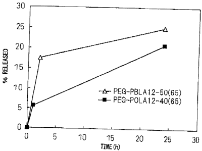
Example 8 (Evaluation of release from human FITC-labeled
IgG-encapsulating micelles)
The block copolymer used was PEG-PBLA 12-50(65) or
PEG-POLA 12-40(50). After precisely weighing out 20 mg
of each polymer into a vial, 2 mL of dichloromethane was
added for dissolution. The solution was dried into a
film under a nitrogen gas stream, and then further dried
for about 1 hour under reduced pressure. To this there
was added 100 jiJ. of FITC-labeled human immunoglobulin
(FITC-IgG) (Sigma-Aldrich Corp., 20 mg/mL), and then 3.9
mL of 20 mM phosphate buffer (pH 6.0) was slowly added
while gently stirring. After further stirring overnight
at 4 C, a Biodisruptor (High Power Unit, product of Nissei
Corp.) was used for sonication for about 10 seconds (1
second intermission, output: Low) while cooling on ice,
and the mixture was subjected to ultracentrifugation
CA 02658082 2009-01-16
- 37 -
(30,000 rpm, 4 C, 1 hour) to obtain micelles. The
recovered micelles were suspended in 20 mM phosphate
buffer (pH 6) and added to bovine serum [final bovine
serum concentration: 50% (v/v)], and then incubated at
37 C. In order to evaluate release of the encapsulated
FITC-IgG, 1 mL of sample was subjected to gel filtration
(Sepharose CL-4B, Sigma-Aldrich Corp., -2(1) x 30 cm) after
a predetermined incubation time. The FITC-IgG
concentration in each recovered fraction (eluent: 20 mM
phosphate buffer (pH 7.4), flow rate: 1.0 mL/min,
fraction volume: 1 mL) was assayed using a plate reader
(PowerScane' HT, Dainippon Sumitomo Fharma Co., Ltd.)
(excitation wavelength: 485 nm 20 nm, emission
wavelength: 528 nm i20 nm), and the release rate was
calculated by the following formula.
Protein content of FITC-
labeled human IgG fraction
x 100
Release rate (%)
Protein content of micelle
fraction recovered by gel
filtration applied
immediately after mixture
with buffering solution
(The buffering solution was 20 mM phosphate buffer (pH
6).)
The time-course of the release is shown in Fig. 1.
These results indicate that the protein encapsulated in
the micelles exhibited prolonged release without initial
burst, even in the presence of serum. The release rate
was thus shown to be dependent on the structure of the
hydrophobic groups. Without being constrained by any
particular theory, it is believed that a macromolecular
drug is held more firmly in the micelle cores if the
structure of the hydrophobic groups introduced into the
hydrophobic segments of the block copolymer that form the
CA 02658082 2009-01-16
- 38 -
,
micelles is a linear structure of alkyl groups rather
than a planar structure such as benzyl, such that the
release occurs in a more controlled manner.
Example 9 (Interferon-a intravenous administration test)
The block copolymer used was PEG-PBLA 12-50(65) or
PEG-POLA 12-40(65). After precisely weighing out 10 mg
of the polymer into a vial, 1 mL of dichloromethane was
added for dissolution. The solution was dried into a
film under a nitrogen gas stream, and then further dried
for about 3 hours under reduced pressure. To this there
was added a recombinant human interferon-a PBS solution
(IFN-a, PBL Biomedical Laboratories) (0.2 mg/mL, 46 gL),
and then 200 gL of 0.2 M MES buffer (pH 5.0) was added.
The mixture was gently stirred at 4 C to essentially total
dissolution of the polymer, and then 20 mM MES buffer (pli
5.0) was added to a total volume of 2 mL and stirring was
continued for a full day at 4 C. The sample was subjected
to ultracentrifugation (30,000 rpm, 1 hour, 4 C, MLA-130
Rotor by Beckman Coulter), and the non-encapsulated IFN-a
was removed while recovering the precipitated micelles.
The micelles were suspended in a 5% glucose aqueous
solution and provided for the following animal
experiment.
Six-week-old Wistar male rats were divided into
groups of 2 rats each, and the test solution was
administered through the tail vein at a dosage of 1 x 10⁶
TU/kg. 5 minutes and 1, 3, 6, 9 and 24 hours after
administration, about 0.2 mL of blood was collected from
the cervical vein using a heparin-coated syringe. The
blood was immediately centrifuged at 13,800 rpm, 4 C (EF-
1300, ECO-Fugem, Tomy Seiko Co., Ltd.) and the plasma was
harvested and stored at -30 C until analysis. The plasma
concentration of interferon-a was determined by a human
interferon-a misA kit (PBL Biomedical Laboratories).
CA 02658082 2009-01-16
- 39 -
,
The results are shown in Fig. 2. Encapsulation of
interferon-a in polymeric micelles improved the retention
of plasma concentration. The pharmacokinetic parameters
calculated according to a non-compartment model are shown
below.
CA 02658082 2009-01-16
- 40
PEG-POLA PEG-PBLA
Fharmacokinetic 0.9% NaC1
12-40(65) 12-
50(65)
parameters solution
micelles micelles
AUCinf (%
0.20 7.0 3.5
dose/mL.h)
T112 (h) 0.11 10.4 1.3
Cl (mL/h/body) 491 14.4 28.9
MRTinf (h) 0.2 3.2 0.4
Vss (mL/body) 81 46 12
Micellation increased the AUC by 17-fold to 35-fold.
These results indicate that the protein-encapsulating
micelles stay in the blood circulation for a long time
without initial burst. Also these results show that the
protein release rate in vivo is dependent on the
hydrophobic group structure of the block copolymer,
similar to the in vitro results.
Example 10 (Rat intravenous administration test with
FITC-labeled lysozyme-encapsulating micelles)
1) FITC labeling of lysozyme
After dissolving 100 mg of lysozyme (from egg white)
(Sigma-Aldrich Corp.) in 2 mL of a 100 mM boric acid
buffer (pH 8.5), 170 1,tIJ of a 50 mg/mL DMS0 solution
containing FITC (PIERCE) was added. After stirring at
room temperature for 1 hour, the unreacted FITC was
removed by gel filtration (PD-10, product of GE
Healthcare Bioscience) (eluent: 20 mM sodium phosphate
buffer, pH 7.4). After subsequent dialysis against water
at 4 C, it was purified by additional gel filtration
(Sepharose CL-4B, Sigma-Aldrich Corp.) (eluent: 20 mM
sodium phosphate buffer, pH 7.4).
2) Preparation of FITC-labeled lysozyme-encapsulating
micelles and rat PK test
After precisely weighing out 40 mg of the block
copolymer PEG-POLA 12-40(65) into a glass vial, 4 mg of
FITC-labeled lysozyme (29.2 mg/mL, 137 I1L) and then 500
CA 02658082 2009-01-16
- 41
gL of 20 mM sodium phosphate buffer (pH 6.0) were added.
After further stirring overnight at 4 C, a Biodisruptor
(High Power Unit, product of Nissei Corp.) was used for
sonication for about 10 seconds (1 second intermission,
output: Low) while cooling on ice. The micelle fraction
recovered as a precipitate from ultracentrifugation
(80,000 rpm, 1 hour, 4 C, MLA-80 Rotor by Beckman Coulter)
was suspended in 20 mM sodium phosphate buffer (pH
7.4)/5% glucose, and then, washed by the same
ultracentrifugation procedure, resuspended in the same
buffer solution and provided for the following rat
administration test.
Six-week-old Wistar male rats were divided into
groups of 3 rats each, and the test solution was
administered through the tail vein at a dosage of 10
mg/kg of FITC-labeled lysozyme. 5 minutes and 1, 3, 6, 9
and 24 hot.ix's after administration, about 0.2 mL of blood
was collected from the cervical vein using a heparin-
coated syringe. The blood was immediately centrifuged at
4 C (EF-l300, ECO-Fuge7", Tomy Seiko Co., Ltd.) and the
plasma was harvested and stored at -30 C until analysis.
A FITC-labeled lysozyme solution (20 mM sodium phosphate
buffer, pH 7.4/5% glucose) was also tested in the same
manner. The blood plasma concentration was measured by
HPLC the following HPLC conditions.
System: Waters Alliance System
Column: Tosoh TSK-gel Super SW3000 (4.6+ x 300 mm)(30 C)
Mobile phase: 20 mM sodium phosphate buffer (pH 7.4)
Flow rate: 0.25 mL/min
Detection: Fluorescence (Ex: 492 nm, Em: 520 nm)
Injection volume: 10 gL
The results are shown in Fig. 3. The ADC increased
approximately 15-fold by micellation compared to
administration of the solution alone. These results
indicate that the protein encapsulated in the micelles
stay in the blood circulation for a long time without
CA 02658082 2009-01-16
- 42 -
initial burst.
Example 11 (Intravenous administration test with
interferon-a-encapsulated micelles 2)
The block copolymer used was polyethylene glycol-co-
polyaspartic acid dodecyl ester (hereinafter, PEG-PDLA.
In this polymer, the aspaxtic acid residues without
dodecyl esters are of general formula (I) wherein R5 is -
0- and R6 is hydrogen (same hereunder)), or polyethylene
glycol-co-polyaspartic acid hexadecyl ester (hereinafter,
PEG-PHLA. The aspartic acid residues without hexadecyl
esters are of the same general formula wherein R5 is -0-
and R6 is hydrogen (same hereunder)). After precisely
weighing out 200 mg of PEG-PDLA 12-40(65) or PEG-PHLA 12-
40(65) into a glass vial, 10 mL of 20 mM MES buffer (pH
5.0) was added and the mixture was vigorously stirred
overnight at 4 C. The polymer dispersion was subjected to
ultrasonic treatment for about 15 minutes (1 second
intermission, output: Low) using a Biodisruptor (High
Power Unit, product of Nissei Corp.) while cooling on
ice, to obtain an empty micelle solution with a polymer
concentration of 20 mg/mL. A 0.65 mL portion of the
empty micelle solution was transferred to a microtube
(Ieda Chemicals Co., Ltd.), and then 65 L of a
recombinant human interferon-a PBS solution (IFN-a, PBL
Biomedical Laboratories) and 50 I, of 0.1 M MES buffer
(pH 5.0) were added and the mixture was carefully
pipetted and then allowed to stand at 4 C for 4 days. It
was then rinsed with a 5% glucose solution using an
ultrafiltration unit [AMICONe ULTRA-4 by Millipore
(molecular cutoff: 100,000)] and concentrated for use in
the following animal experiment.
Six-week-old Wistar rats were divided into groups of
3 rats each, and the test solution was administered
through the tail vein at a dosage of 1 x 10⁶ IU/kg. 5
minutes and 1, 3, 6 and 24 hours after administration,
ak 02658082 2009-01-16
- 43 -
=
about 0.2 ml of blood was collected from the cervical
vein using a heparin-coated syringe. The blood was
immediately centrifuged at 13,800 rpm, 4 C (EF-1300, ECO-
FugeTM, Tomy Seiko Co., Ltd.) and the plasma was harvested
and stored at -30 C until analysis. The plasma
concentration of interferon-a was determined by a human
interferon-a ELISA kit (PBL Biomedical Laboratories).
Fig. 4 shows time-course of plasma concentration of
interferon-a after administration of interferon-a-
encapsulating polymer micelles. The RUC was enhanced by
32-fold when using PEG-PDLA 12-40(65) polymeric micelles
and 27-fold when using PEG-PHLR 12-40(65) polymeric
micelles compared with the solution alone.
Example 12 (intravenous administration test with
interferon-a--encapsulated micelles 3)
The block copolymer used was polyethylene glycol-co-
polyglutamic acid octyl ester (hereinafter, PEG-POLG. In
this polymer, the glutamic acid residues without octyl
esters are of general formula (I) wherein R5 is -0- and R6
is hydrogen (same hereunder)). After precisely weighing
out 150 mg of PEG-POLG 12-40(65) into a glass vial, 5 ml
of 20 mM MS buffer (pH 5.0) was added and the mixture
was vigorously stirred overnight at 4 C. The polymer
dispersion was subjected to ultrasonic treatment for
about 15 minutes (1 second intermission, output: Low)
using a Biodisruptor (High Power Unit, product of Nissei
Corp.) while cooling on ice, to obtain an empty micelle
solution with a polymer concentration of 30 mg/mL. A 0.6
mL portion of the empty micelle solution was transferred
to a Cryovial (Ieda Chemicals Co., Ltd.), and then 90 pL
of a recombinant human interferon-a solution (IFN-a, PBL
Biomedical Laboratories) and 110 L of 0.1 M MES buffer
(pH 5.0) were added and the mixture was carefully
pipetted and then allowed to stand at 4 C for 3 days. A
CA 02658082 2009-01-16
- 44 -
400 L portion thereof was then injected into a
microtube, 20 mM MES buffer (pH 5.0) was added to 500 L,
and the mixture was rinsed with a 5% glucose solution
using an ultrafiltration unit [AMICONR ULTRA-4 by
Millipore (molecular cutoff: 100,000)] and concentrated
for use in the following animal experiment.
Six-week-old Wistar rats were divided into groups of
2 rats each, and the test solution was administered
through the tail vein at a dosage of 1 x 10⁶ IU/kg. 5
minutes and 1, 3, 6 and 24 hours after administration,
about 0.2 mL of blood was collected from the cervical
vein using a heparin-coated syringe. The blood was
immediately centrifuged at 13,800 rpm, 4 C (EF-1300, ECO-
FugeTm, Tomy Seiko Co., Ltd.) and the plasma was harvested
and stored at -30 C until analysis. The plasma
concentration of interferon-a was determined by a human
interferon-a ELISA kit (PBL Biomedical Laboratories).
Fig. 5 shows the time-course of plasma concentration
of interferon-a after administration of interferon-a-
encapsulating polymer micelles. micellation increased
the AUC by 57-fold.
Example 13 (Preparation of mellitin-encapsulating
micelles)
The block copolymer used was PEG-PoLA 12-40(65) or
PEG-POLG 12-40(65). After precisely weighing out 150 mg
of polymer into a glass vial, 5 mL of 20 mM sodium
phosphate buffer (pH 7.4) was added and the mixture was
vigorously stirred at 4 C. The polymer dispersion was
subjected to ultrasonic treatment for about 15 minutes (1
second intermission, output: Low) using a Biodisruptor
(High Power Unit, product of Nissei Corp.) while cooling
on ice, to obtain an empty micelle solution with a
polymer concentration of 30 mg/mL. After then adding 20
mM sodium phosphate buffer (pH 7.4) for adjustment to 10
mg/mL (1 mL), the basic polypeptide mellitin (Sigma-
CA 02658082 2009-01-16
- 45 -
,
Aldrich Corp.) (1 mg) was added, and the mixture was
allowed to stand overnight at 4 C and then subjected to
gel filtration (Sepharose CL-4B, GE Healthcare
Bioscience, 14 x 30 cm). The mellitin concentration in
each recovered fraction (eluent: 20 mM sodium phosphate
buffer (pH 7.4), flow rate: 1.0 mL/min, fraction volume:
1 mL) was assayed using a BCA Protein Assay (Pierce
Corp.) The encapsulation efficiency was calculated by
the following formula.
(Protein content in micelle
Encapsulation fraction) x lop
efficiency (%) = Total protein content in
all fractions
The encapsulation efficiency was 71% for PEG-POLA
12-40(65) and 48% for PEG-POLG 12-40(65). The results
indicate that polypeptides with molecular weights of
approximately 2,800 can be efficiently encapsulated.
Example 14 (Rat intravenous administration test with
human granulocyte colony stimulating factor-encapsulating
micelles 1)
The block copolymer used was polyethylene glycol-co-
polyaspartic acid ootylamide (hereinafter, PEG-PONLA.
This polymer comprises 50% of an amino acid residue of
general formula (I) wherein R5 is -NH- and R6 is an octyl
group, and 50% of an amino acid residue wherein R5 is -NH-
and R6 is an amino group-substituted octyl group (same
hereunder)). After precisely weighing out 30 mg of PEG-
PONLA 12-40(50) into a glass vial, 2 mL of 5% glucose-
containing 20 mM phosphate buffer (pH 7.4) was added, and
then a Biodisruptor (High Power Unit, product of Nissei
Corp.) was used for sonication for about 10 minutes (1
second intermission, output: Low) while cooling on ice,
to prepare empty micelles. Recombinant human granulocyte
colony stimulating factor (G-CSF, Green Cross) (pI:
CA 02658082 2009-01-16
- 46
approximately 6.0) (300 pg/mL, 1 mL) was added and the
mixture was allowed to stand for a full day at 4 C. The
non-encapsulated G-CSF was then removed using an
ultrafiltration unit [AMICON ULTRA-4 by Millipore
(molecular cutoff: 100,000)] to prepare micelles for the
following animal experiment.
Six-week-old Wistar male rats were divided into
groups of 3 rats each (6 rats per group for the solution
alone), and the test solution was administered through
the tail vein at a dosage of 100 1.1.g/kg of the test
solution. 5 minutes and 1, 3, 6 and 24 hours after
administration, about 0.2 mL of blood was collected from
the cervical vein using a heparin-coated syringe. The
sample was immediately centrifuged at 13,800 rpm, 4 C (EF-
1300, ECO-FugeTM, Tomy Seiko Co., Ltd.) and the plasma was
harvested and stored at -30 C until analysis. A RayBioR
Human G-CSF ELISA kit (RayBiotech Inc.) was used for
plasma concentration assay.
The assay results are shown in Fig. 6, and the
pharmacokinetic parameters calculated according to a non-
compartment model are shown in Table 2. Micellation
increased the AUC by 3-fold, and prolonged the half-life
by 2-fold.
CA 02658082 2009-01-16
- 47 -
Table 2 Pharmacokinetic parameters for human granulocyte
colony stimulating factor-encapsulating polymer micelles
or human granulocyte colony stimulating factor solution
after intravenous administration to rats.
Pharmacokinetic PEG-PONLA 12-
Solution
parameters 40(50) micelles
Aucinf (% dose/mL.h) 7.4 22
T112 (h) 2.6 4.1
Cl (mL/h/body) 13 4.4
MRTizif (h) 1.9 2.8
Vss (mL/body) 25 13
Example 15 (Rat intravenous administration test with
human granulocyte colony stimulating factor-encapsulating
micelles 2)
The block copolymer used was PEG-POLG. After
precisely weighing out 30 mg of PEG-POLG 12-40(65) into a
glass vial, 2 mL of 20 mM MES buffer (pH 5.0) was added,
and then a Biodisruptor (High Power Unit, product of
Nissei Corp.) was used for sonication for about 10
minutes (1 second intermission, output: Low) while
cooling on ice, to prepare empty micelles. A recombinant
human G-CSF solution (Green Cross) (300 lg/mL, 1 mL) was
added and the mixture was allowed to stand for a day at
4 C. The non-encapsulated G-CSF was then removed using an
ultrafiltration unit [AMICON6 ULTRA-4 by Millipore
(molecular cutoff: 100,000)] and diluted with 5% glucose-
containing 20 mil phosphate buffer (pH 7.4) for use as
micelles for an animal experiment. The micelles were
intravenously administered to the rats at a dosage of 100
VIII', in terms of G-CSF, in the same manner as Example
12. Blood was collected and the plasma concentration was
assayed in the same manner.
The results are shown in Fig. 7, and the
pharmacokinetic parameters calculated according to a non-
compartment model are shown in Table 3. Encapsulation in
polymeric micelles of human granulocyte increased the AUC
CA 02658082 2009-01-16
- 48 -
by 15-fold and prolonged the half-life by 5-fold compared
with the solution, and the plasma concentration was still
detectable even 120 hours after administration. These
results indicate that the protein-encapsulating micelles
stay in the blood circulation for a long time without
initial burst.
CA 02658082 2013-11-26
- 49 -
Table 3 Pharmacokinetic parameters for human granulocyte
colony stimulating factor-encapsulating polymer micelles
or human granulocyte colony stimulating factor solution
after intravenous administration to rats.
PharmacokineticPEG-POLG 12-
Solution
parameters 40(65) micelles
AUCinf (% dose/mL.h) 7.4 111
T112 (h) 2.6 14
Cl (mL/h/body) 13 0.90
MRTinf (h) 1.9 12
Vss (mL/body) 25 11
The scope of the claims should not be limited by
the preferred embodiments set forth in the examples,
but should be given the broadest interpretation
consistent with the description as a whole.