Note: Descriptions are shown in the official language in which they were submitted.
DEMANDES OU BREVETS VOLUMINEUX
LA PRESENTE PARTIE DE CETTE DEMANDE OU CE BREVETS
COMPREND PLUS D'UN TOME.
CECI EST LE TOME DE _2
NOTE: Pour les tomes additionels, veillez contacter le Bureau Canadien des
Brevets.
JUMBO APPLICATIONS / PATENTS
THIS SECTION OF THE APPLICATION / PATENT CONTAINS MORE
THAN ONE VOLUME.
THIS IS VOLUME 1 OF 2
NOTE: For additional volumes please contact the Canadian Patent Office.
CA 02658290 2009-12-30
64005-623F(S)
1
MONITORING HYBRIDIZATION DURING PCR USING FLUORESCENT DYE
SPECIFIC TO DOUBLE-STRANDED DNA
This application is a divisional application of
Canadian patent application serial number 2,591,550 filed
July 4, 2007, which is a divisional application of Canadian
patent application serial number 2,257,109, entitled
"Monitoring Hybridization during PCR", which entered the
national phase in Canada December 3, 1998 and has an
effective filing date of June 4, 1997.
BACKGROUND OF THE INVENTION
This invention relates generally to observing
fluorescence signals resulting from hybridization in
conjunction with the polymerase chain reaction. More
specifically, the present invention relates to observing
hybridization with fluorescence during and/or immediately
after PCR and using this information for product
identification, sequence alteration detection, and
quantification.
The polymerase chain reaction (PCR) is fundamental
to molecular biology and is the first practical molecular
technique for the clinical laboratory. Despite its
usefulness and popularity, current understanding of PCR is
not highly advanced. Adequate conditions for successful
amplifications must be found by trial and error and
optimization is empirical. Even those skilled in the art
are required to utilize a powerful technique without a
comprehensive or predictive theory of the process.
PCR is achieved by temperature cycling of the
sample, causing DNA to denature (separate), specific primers
to attach (anneal), and replication to occur (extend). One
CA 02658290 2009-02-04
64005-623F
la
cycle of PCR is usually performed in 2 to 8 min. requiring 1
to 4 hours for a 30-cycle amplification. The sample
temperature response in most PCR instrumentation is very
slow compared to the times required for denaturation,
annealing and extension. The physical (denaturation and
annealing) and enzymatic (extension) reactions in PCR occur
very quickly. Amplification times for PCR can be reduced
from hours to less than 15 min.
Rapid cycling techniques are made possible by the
rapid temperature response and temperature homogeneity
possible for samples in high surface area-to-volume sample
containers such as capillary tubes. For further
information, see also: C.T. Wittwer, G.B. Reed, and
K.M. Ririe, Rapid cycle DNA amplification, in K.B. Mullis,
F. Ferre, and R.A. Gibbs. The polymerase chain reaction,
Birkhauser, Boston, 174-181, (1994). Improved temperature
homogeneity allows the time and temperature requirements of
PCR to be better defined and understood. Improved
temperature homogeneity also increases the precision of any
analytical technique used to monitor PCR during
amplification.
CA 02658290 2009-02-04
64005-623
Fluorimetry is a sensitive and versatile technique with many applications in
molecular biology. Ethidium bromide has been used for many years to visualize
the
size distribution of nucleic acids separated by gel electrophoresis. The gel
is usually
transilluminated with ultraviolet liuht and the red fluorescence of double
stranded
nucleic acid observed. Specifically, ethidium bromide is commonly used to
analyze the
products of PCR after amplification is completed. Furthermore, EPA 0 640 828 A
I to
Higuchi & Watson discloses using ethidium
bromide during amplification to monitor the amount of double stranded DNA by
measuring the fluorescence each cycle. The fluorescence intensity was noted to
rise
and fall inversely with temperature, was greatest at the annealing/extension
temperature (50=C), and least at the denaturation temperature (94=C), Maximal
fluorescence was acquired each cycle as a measure of DNA amount. The Higuchi &
Watson application does not teach using fluorescence'to monitor hybridization
events,
nor does it suggest acquiring, fluorescence over different temperatures to
follow the
extent of hybridization. Moreover, Higuch & Watson fails to teach or suggest
using
the temperature dependence of PCR product hybridization for identification or
quantification of PCR products.
The Higuchi & Watson application, however, does mention using other
fluorophores, including dual-labeled probe systems that generate flourescence
when
hydrolyzed by the 5'-exonuclease activity of certain DNA polymerases, as
disclosed in
US Patent No. 5,210,015 to Gelfand et al. The fluorescence observed from these
probes primarily depends on hydrolysis of the probe between its two
fluorophores. The
amount of PCR product is estimated by acquiring fluorescence once each cycle.
Although hybridization of these probes appears necessary for hydrolysis to
occur, the
fluorescence signal primarily results from hydrolysis of the probes, not
hybridization,
wherein an oligonucleotide probe with fluorescent dyes at opposite ends
thereof
provides a quenched probe system useful for detecting PCR product and nucleic
acid
hybridization, K.J. Livak et al., 4 PCR Meth. Appl. 357-362 (1995). There is
no
suggestion of following the temperature dependence of probe hybridization with
fluorescence to identify sequence alterations in PCR products.
The specific hybridization of nucleic acid to a complementary strand for
identification has been exploited in many different formats. For example,
after
CA 02658290 2009-02-04
-3-
restriction enzyme digestion, genomic DNA can be size fractionated and
hybridized to
probes by Southern blotting. As another example. singe base mutations can be
detected by "dot blots" with allele-specific oligonucleotides. Lsually.
hybridization is
performed for minutes to hours at a single temperature to achieve the
necessary
discrimination Alternately, the extent of hybridization can he dynamically
monitored
while the temperature is changing by using fluorescence techn'iues. For
example,
fluorescence melting curves have been used to monitor hybridization. L.E.
Morrison
L.M. Stols, Sensitive fluorescence-based thermodynamic and kinetic
measurements
of DNA hybridization in solution. 32 Biochemistry 3095-3104, 1993) The
temperature scan rates are usually 10=C/hour or less, partly because of the
high thermal
mass of the fluorimeter cuvette
Current methods for monitoring hybridization require a lot of time. If
hybridization could be followed in seconds rather than hours. hybridization
could be
monitored during PCR amplification, even during rapid cycle PCR. The many uses
of
monitoring hybridization durinu PCR, as will be full' disclosed herein,
include, product
identification and quantification. sequence alteration detection. and
automatic control
of temperature cycling parameters by fluorescence feedback
The prior art, as explained above, carries out temperature cycling slowly and
empirically. When analysis of PCR products by hybridization is needed.
additional
time consuming steps are required. Thus, it would be a great advance in the
art to
provide methods for monitoring hybridization during PCR and analyzing the
reaction
while it is taking place. that is. durinu or immediately after temperature
cycling without
manipulation of the sample. By monitoring hybridization during PCR, the
underlying
principles that alloy PCR to work can be followed and used to analyze and
optimize
the reaction during amplification
BRIEF SLt.vL\IARY OF THE INVENTION
It is an object of the present invention to provide a double-strand-specific
DNA
dye for monitoring product hybridization during PCR
It is another object of the invention to provide a system for identifying PCR-
amplified products by their fluorescence meltinu curves
CA 02658290 2009-02-04
-4-
It is also an object of the invention to provide a method for improving the
sensitivity of PCR quantification with double-strand-specific DNA dyes.
It is still another objection of the invention for determining the amount of
specific product amplified by PCR by melting curves to correct for nonspecific
amplification detected with the double-strand-specific DNA dye.
It is a further object of the invention to provide a method of relative
quantification of different PCR products with double-strand-specific dyes.
It is yet another object of the invention to provide a method of product
quantification by the reannealing Kinetics of the product in the presence of a
double-
strand-specific DNA dye.
It is a still further object of the invention to provide a novel resonance
energy
transfer pair to monitor primer and/or probe hybridization.
It is still another object of the invention to provide a method of product
quantification by the reannealing kinetics of a probe to the product using a
resonance
energy transfer pair.
It is also an object of the present invention to provide a method to determine
initial template copy number by following the fluorescence of a hybridization
probe or
probes each cycle during PCR amplification.
It is another object of the invention to provide a system for homogeneous
detection of PCR products by resonance energy transfer between two labeled
probes
that hybridize internal to the PCR primers.
It is still another object of the invention to provide a system for
homogeneous
detection of PCR products by resonance energy transfer between one labeled
primer
and one labeled probe that hybridizes internal to the PCR primers.
It is vet another object of the invention to provide a system for detection of
sequence alterations internal to PCR primers by resonance energy transfer and
probe
melting curves.
It is a further object of the invention to provide a system for relative
quantification of different PCR products by probe melting curves
It is yet another object of the invention to provide methods to determine the
initial template copy number by curve fitting the fluorescence vs cycle number
plot.
CA 02658290 2009-02-04
It is still another object of the invention to provide a system and method for
performing PCR rapidly and also continuously monitoring the reaction and
adjusting
the reaction parameters while the reaction is ongoing.
It is another object of the invention to replace the nucleic acid probes by
synthetic nucleic acid analogs or derivatives, e.g. by peptide nucleic acids
(PNA),
provided that they can also be labeled with fluorescent compounds.
These and other objects and advantages of the invention ,vill become more
fully
apparent from the description and claims which follow, or may be learned by
the
practice of the invention.
The present invention particularly decreases the total time required for PCR
amplification and analysis over prior art techniques while at the same time
allowing the
option of significantly increasing the quality of the reaction by optimizing
amplification
conditions.
fhe present invention provides methods and applications for continuous
fluorescence monitoring of DNA amplification. Required instrumentation
combines
optical components with structures to provide rapid temperature cycling to
continuously monitor DNA amplification by a variety of different fluorescence
techniques. In one illustrative embodiment, fluorescence is acquired
continuously from
a single sample or alternately from multiple samples on a rotating carousel
with all of
the samples being simultaneously subjected to rapid thermal cycling. Further
information on associated instrumentation can be found in the L.S. patent
applications
referenced above.
In accordance with one aspect of the present invention, fluorescence during
DNA amplification was monitored by. 1) the double strand-specific dye SYBR
Green
1, and 2) resonance energy transfer of fluorescein to CYST" or Cy5.5T''' with
hybridization probes. Fluorescence data acquired once per cycle allow
quantification
of initial template copy number.
Furthermore. in contrast to measuring fluorescence once per cycle.
embodiments of'the present invention are disclosed which monitor temperature.
time
and fluorescence continuously throughout each cycle thus producing a 3-
dimensional
spiral. This 3-dimensional spiral can be reduced to temperature vs. time,
fluorescence
AMENDED SHEET
CA 02658290 2009-02-04
64005-623E
-6-
vs. time, and fluorescence vs. temperature plots. Fluorescence vs. temperature
plots of
the fluorescence from hybridization probes can be used to detect sequence
alterations
in the product. These sequence alterations may be natural, as in mutations or
polymorphisms, or artificial, as in an=en~gineered alternative template for
quantitative
PCR.
In accordance with another aspect of the present invention, fluorescence
monitoring is used to acquire product melting curves during PCR by
fluorescence
monitoring with double-strand-specific DNA specific dyes. Plotting
fluorescence as a
function of temperature as the thermal cycler heats through the dissociation
temperature of the product gives a PCR product melting curve. The.shape and
position of this DNA melting curve is a function of GC/AT ratio, length, and
sequence,
and can be used to differentiate amplification products separated by less than
2=C in
melting temperature. Desired products can be distinguished from undesired
products,
including primer dimers. Analysis of melting curves can be used to extend the
dynamic
range of quantitative PCR and to differentiate different products in multiplex
amplification. Using double strand dyes, product denaturation, Teannealing,
and
extension can be followed within each cycle. Continuous monitoring of
fluorescence
allows acquisition of melting curves and product, annealing curves during
temperature
cycling.
The present invention provides reagents and methods for rapid cycle PCR with
combined amplification and analysis by fluorescence monitoring in under thirty
minutes, more preferably in under fifteen minutes, and most preferably in
under ten
minutes.
CA 02658290 2011-08-12
6-1005-623F(S)
6a
Thus in one aspect, the present invention provides a method for
detecting a target nucleic acid sequence in a biological sample during
amplification
comprising the steps of: (a) adding a thermostable polymerase, a double-strand-
specific fluorescent DNA binding dye and primers configured for amplification
of the
target nucleic acid sequence to the biological sample; (b) amplifying the
target nucleic
acid sequence by polymerase chain reaction in the presence of the dye, the
polymerase chain reaction comprising thermally cycling the biological sample
between at least a denaturation temperature and an elongation temperature
through
a plurality of amplification cycles; (c) illuminating the biological sample
comprising the
amplified target nucleic acid sequence with light at a wavelength absorbed by
the
dye; and (d) detecting a fluorescent emission from the dye, while the
biological
sample is heated, using a 520-580 nm band pass filter, wherein the fluorescent
emission is related to the quantity of the amplified target nucleic acid
sequence in the
sample; and (e) generating a melting curve and conducting melting curve
analysis for
the amplified target nucleic acid sequence.
In another aspect, the present invention provides a method of analyzing
nucleic acid hybridization comprising the steps of (a) providing a mixture
comprising a
nucleic acid sample to be analyzed and a double-strand-specific fluorescent
DNA
binding dye; (b) monitoring fluorescence while changing temperature at a rate
of >_ 0.1 C/second; and (c) generating a melting curve and conducting melting
curve
analysis for the nucleic acid sample to be analyzed.
In another aspect, the present invention provides a method for detecting
a target nucleic acid sequence in a biological sample during amplification
comprising
the steps of: (a) adding a thermostable polymerase and primers
CA 02658290 2010-08-19
64005-623F(S)
6b
configured for amplification of the target nucleic acid
sequence to the biological sample; (b) amplifying the target
nucleic acid sequence by polymerase chain reaction in the
presence of a fluorescent dye which is pico green*, the
polymerase chain reaction comprising thermally cycling the
biological sample between at least a denaturation
temperature and an elongation temperature through a
plurality of amplification cycles using a rapid temperature
cycling profile wherein 30 amplification cycles are
completed in 10 to 30 minutes; (c) illuminating the
biological sample comprising the amplified target nucleic
acid sequence with light at a wavelength absorbed by the
fluorescent dye; (d) detecting a fluorescent emission from
the fluorescent dye related to the quantity of the amplified
target nucleic acid sequence in the sample; and (e)
generating a melting curve and conducting melting curve
analysis for the amplified target nucleic acid.
In another aspect, the present invention provides
a method of monitoring the amplification of a nucleic acid
in a biological sample during PCR amplification, comprising
the steps of: (a) forming an amplification mixture
comprising the biological sample, pico green* as a
fluorescent entity capable of producing a fluorescent signal
related to the amount of nucleic acid present in the sample,
a thermostable polymerase, and primers for the nucleic acid;
(b) amplifying the target sequence by thermally cycling the
amplification mixture through a plurality of thermal cycles,
(c) illuminating the sample and monitoring the fluorescent
signal from the fluorescent entity during amplification; and
(d) generating a melting curve and conducting melting curve
analysis for the amplified target nucleic acid sequence.
*Trade-mark
CA 02658290 2010-08-19
64005-623F(S)
6c
In another aspect, the present invention provides
a kit for analysis of a nucleic acid sequence during
amplification, the kit comprising: an amplification solution
comprising a fluorescent dye which is pico green*; a
thermostable DNA polymerase; and deoxynucleoside
triphosphates.
In another aspect, the present invention provides
a method for detecting a target nucleic acid sequence in a
biological sample during amplification comprising the steps
of: (a) adding a thermostable polymerase and primers
configured for amplification of the target nucleic acid
sequence to the biological sample; (b) amplifying the target
nucleic acid sequence by polymerase chain reaction in the
presence of a fluorescent dye which is pico green*, the
polymerase chain reaction comprising thermally cycling the
biological sample between at least a denaturation
temperature and an elongation temperature through a
plurality of amplification cycles; (c) illuminating the
biological sample comprising the amplified target nucleic
acid sequence with light at a wavelength absorbed by the
fluorescent dye; (d) detecting a fluorescent emission from
the fluorescent dye related to the quantity of the amplified
target nucleic acid sequence in the sample; wherein during
each of the plurality of amplification cycles the sample is
held no more than 60 seconds at the elongation temperature
and held less than 1 second at the denaturation temperature;
and (e) generating a melting curve and conducting melting
curve analysis for the amplified target nucleic acid
sequence.
*Trade-mark
CA 02658290 2009-02-04
64005-623
-7-
A method for analyzing a target DN.A sequence of a biological sample
comprises
amplifying the target sequence by polymerase chain reaction in the
presence of two nucleic acid probes that hybridize to adjacent regions of the
target
sequence. one of the probes being labeled with an acceptor fluorophore and the
other
probe labeled with a donor fluorophore of a fluorescence energy transfer pair
such that
upon hybridization of the two probes with the target sequence, the donor and
acceptor
fluorophores are within 25 nucleotides of one another, the polyymerase chain
reaction
comprising the steps of adding a thermostable polymerase and primers for the
targeted
nucleic acid sequence to the biological sample and thermally cycling the
biological
sample between at least a denaturation temperature and an elongation
temperature;
exciting the sample with light at a wavelength absorbed by the donor
fluorophore; and
monitoring the temperature dependent fluorescence from the
fluorescence energy transfer pair.
A method of real time monitoring of a polymerase chain reaction amplification
of a target nucleic acid sequence in a biological sample comprises
(a) adding to the biological sample an effective amount of two
nucleic acid primers and a nucleic acid probe, wherein one of the primers and
the probe
are each labeled with one member of a fluorescence energy transfer pair
comprising an
acceptor fluorophore and a donor fluorophore, and wherein the labeled probe
hybridizes to an amplified copy of the target nucleic acid sequence within 15
nucleotides of the labeled primer;
(b) ampliR-inu the target nucleic acid sequence by polymerase chain
reaction,
(c} illuminating the biological sample with light of a selected
wavelength that is absorbed by said donor fluorophore: and
CA 02658290 2009-02-04
-8-
(d) detecting the fluorescence e nission of the sample.
An improved method of amplifying a target nucleic acid sequence of a
biological sample comprises
(a) adding to the biological sample an effective amount of a
nucleic-acid-binding fluorescent entity;
(b) amplifying the target nucleic acid sequence using polymerase
chain reaction, comprising thermally cycling the biological sample using
initial
predetermined temperature and time parameters, and then
(i) illuminating the biological sample with a selected
wavelength of light that is absorbed by the fluorescent entity during the
polymerase chain reaction;
(ii) monitoring fluorescence from the sample to determine
the optimal temperature and time parameters for the polymerase chain reaction;
and
(iii) adjusting the initial temperature and time parameters in
accordance with the fluorescence.
In one illustrative embodiment, the fluorescent entity comprises a double
strand
specific nucleic acid binding dye. and in another illustrative embodiment the
fluorescent
entity comprises a fluorescently labeled oligonucleotide probe that hybridizes
to the
targeted nucleic acid sequence.
A method for detecting a target nucleic acid sequence of a biological sample
comprises
(a) adding to the biological sample an effective amount of a Pair of
oligonucleotide probes that hybridize to the target nucleic acid sequence, one
of the
probes being labeled with an acceptor fluorophore and the other probe labeled
with a
donor fluorophore of a fluorescence energy transfer pair, wherein an emission
spectrum of the donor fluorophore and an absorption spectrum of the acceptor
fluorophore overlap less than 25 .0, the acceptor fluorophore has a peak
extinction
coefficient greater than 100,000 M"cm"' and upon hybridization of the two
probes, the
donor and acceptor fluorophores are within 25 nucleotides of one another,
(b) illuminating, the biological sample with a selected wavelength of
light that is absorbed by said donor fluorophore; and
f1M~ ;D D s,; t
CA 02658290 2009-02-04
-9-
(c) detecting the emission of the biological sample
An illustrative resonance energy transfer pair comprises fluorescein ai the
donor and
Cy5 or Cv 5.5 as the acceptor.
A method of real time monitoring of a polymerase chain reaction amplification
of a target nucleic acid sequence in a biological sample comprises
ampliRing the target sequence by polymerase chain reaction in the
presence of two nucleic acid probes that hybridize to adjacent regions of the
target
sequence, one of the probes being labeled with an acceptor fluorophore and the
other
probe labeled with a donor fluorophore of a fluorescence energy transfer pair
such that
upon hybridization of the two probes with the target sequence, the donor and
acceptor
fluorophores are within 25 nucleotides of one another. the polymerase chain
reaction
comprising the steps of adding a thermostable polymerise and primers for the
targeted
nucleic acid sequence to the biological sample and thermally cycling the
biological
sample between at least a denaturation temperature and an elongation
temperature;
exciting the biological sairiple with light at a wavelength absorbed by
the donor fluorophore and detecting the emission from the biological sample:
and
monitoring the temperature dependent fluorescence from the
fluorescence energy transfer pair.
A method of real time monitoring of a polymerase chain reaction amplification
of a target nucleic acid sequence in a biological sample comprises
amplifying the target sequence by polymerase chain reaction in the
presence of S1BRT`1 Green 1, the polymerase chain reaction comprising the
steps of
adding a thermostable polymerase and primers for the targeted nucleic acid
segt. nce
to the biological sample and thermally cycling the biological sample between
at least a
23 denaturation temperature and an elongation temperature;
exciting the biological sample with light at a wavelength absorbed by
the SlBRTN Green I and detecting the emission from the biological sample: and
monitoring the temperature dependent fluorescence from the SYBRT"r
Green I Preferably, the monitoring step comprises determining a melting
profile of the
amplified target sequence-
A method for analyzing a target DNA sequence of a biological sample
comprises
CA 02658290 2009-02-04
-10-
(a) 1adding to the biological sample an effective amount of two
nucleic acid primers and a nucleic acid probe, wherein one of the primers and
the probe
are each labeled with one member of a fluorescence energy transfer pair
comprising an
acceptor fluorophore and a donor fluorophore, and wherein the labeled probe
hybridizes to an amplified copy of the target nucleic acid sequence within 15
nucleotides of the labeled primer;
(b) amplifying the target nucleic acid sequence by polymerase chain
reaction;
(c) illuminating the biological sample with light of a selected
wavelength that is absorbed by said donor fluorophore and detecting the
fluorescence
emission of-the sample. In another illustrative embodiment, the method further
comprises the step of monitoring the temperature dependent fluorescence of the
sample, preferably by determining a melting profile of the amplified target
sequence.
A method of detecting a difference at a selected locus in a first nucleic acid
as
compared to a second nucleic acid comprises
(a) providing a pair of oli__onucleotide primers configured for amplifying
by polymerase chain reaction. a selected segment of the first nucleic acid and
a
corresponding segment of the second nucleic acid, wherein the selected segment
and
corresponding segment each comprises the selected locus, to result in
amplified
products containing a copy of the selected locus;
(b) providing a pair of oligonucleotide probes, one of the probes being
labeled with an acceptor fluorophore and the other probe being labeled with a
donor
fluorophore of a fluorogenic resonance energy transfer pair such that upon
hybridization of the two probes with the amplified products the donor and
acceptor are
in resonance energy transfer relationship, wherein one of the probes is
configured for
hybridizing to the amplified products such that said one of the probes spans
the
selected locus and exhibits a melting profile when the difference is present
in the first
nucleic acid that is distinguishable from a melting profile of the second
nucleic acid;
(c) amplifying the selected segment of first nucleic acid and the
corresponding segment of the second nucleic acid by polymerase chain reaction
in the
presence of effective amounts of probes to result in an amplified selected
segment and
an amplified corresponding segment. at least a portion thereof having both the
probes
CA 02658290 2009-02-04
-I1-
hybridized thereto with the fluorogenic resonance energy transfer pair in
resonance
energy transfer relationship;
(d) illuminating the amplified selected segment and the amplified
corresponding segment with the probes hybridized thereto with a selected
wavelength
of light to elicit fluorescence by the fluorogenic resonance energy transfer
pair;
(e) measuring fluorescence emission as a function of temperature to
determine in a first melting profile of said one of the probes melting. from
the amplified
selected segment of first nucleic acid and a second melting profile of said
one of the
probes melting from the amplified corresponding segment of second nucleic
acid, and
(f) comparing the first melting profile to the second melting profile,
wherein a difference therein indicates the presence of the difference in the
sample
nucleic acid.
A method of detecting a difference at a selected locus in a first nucleic acid
as
compared to a second nucleic acid comprises
(a) providing a pair of oligonucleotide primers configured for amplifying.
by polymerase chain reaction, a selected segment of the first nucleic acid and
a
corresponding segment of the second nucleic acid, wherein the selected segment
and
corresponding segment each comprises the selected locus, to result in
amplified
products containing a copy of the selected locus;
(b) providing an oligonucleotide probe, wherein one of the primers and the
probe are each labeled with one member of a fluorescence energy transfer pair
comprising an donor fluorophore and an acceptor fluorophore, and wherein the
labeled
probe and labeled primer hybridize to the amplified products such that the
donor'and
acceptor are in resonance energy transfer relationship, and wherein the probe
is
configured for hybridizing to the amplified products such that said probe
spans the
selected locus and exhibits a melting profile when the difference is present
in the first
nucleic acid that is distinguishable from a melting profile of the second
nucleic acid;
(c) amplifying the selected se~_ment of first nucleic acid and the
corresponding se<_ment of the second nucleic acid by polymerase chain reaction
in the
presence of effective amounts of primers and probe to result in an amplified
selected
segment and an amplified corresponding segment, at least a portion thereof
having the
CA 02658290 2009-02-04
-12-
labled primer and probe hybridized thereto with the fluorogenic resonance
energy
transfer pair in resonance energy transfer relationship;
(d) illuminating the amplified selected segment and the amplified
corresponding segment with the labeled primer and probe hybridized thereto
with a
S selected wavelength of light to elicit fluorescence by the fluorogenic
resonance energy
transfer pair;
(e) measuring fluorescence emission as a function of temperature to
determine in a first melting profile of said probe melting from the amplified
selected
segment of first nucleic acid and a second melting profile of said probe
melting from
the amplified corresponding segment of second nucleic acid; and
(f) comparing the first melting profile to the second melting profile,
wherein a difference therein indicates the presence of the difference in the
sample
nucleic acid.
A method of detecting heterozygosity at a selected locus in the genome of an
individual, wherein the genome comprises a mutant allele and a corresponding
reference allele, each comprising the selected locus, comprises
(a) obtaining sample genomic DNA from the individual;
(b) providing a pair of oligonucleotide primers configured for amplifying,
by polymerase chain reaction, a first selected segment of the mutant allele
and a second
selected segment of the corresponding reference allele wherein both the first
and
second selected segments comprise the selected locus;
(c) providing a pair of olivonucleotide probes, one of the probes being
labeled with an acceptor fluorophore and the other probe being labeled with a
donor
fluorophore of a fluorogenic resonance energy transfer pair such that upon
hybridization of the two probes with the amplified first and second selected
segments
one of the probes spans the selected locus and exhibits a first melting
profile with the
amplified first selected segment that is distinguishable from a second melting
profile
with the amplified second selected segment;
(d) amplifying the first and second selected segments of sample genomic
DNA by polymerase chain reaction in the presence of effective amounts of
probes to
result in amplified first and second selected segments, at least a portion
thereof having
CA 02658290 2009-02-04
-13-
both the probes hybridized thereto with the fluorogenic resonance energy
transfer pair
in resonance energy transfer relationship.
(e) illuminating the amplified first and second selected segments having the
probes hybridized thereto with a selected wavelength of light to elicit
fluorescence by
the donor and acceptor;
(f) measuring a fluorescence emission as a function of temperature to
determine a first melting profile of said one of the probes melting from the
amplified
first selected segment and a second melting profile of said one of the probes
melting
from the amplified second selected segment; and
(g) comparing the first melting profile to the second melting profile,
wherein distinguishable melting profiles indicate heterozygosity in the sample
genomic
DNA.
A method of detecting heterozygosity at a selected locus in the genome of an
individual, wherein the genome comprises a mutant allele and a corresponding
1 S reference allele, each comprising the selected locus, comprises
(a) obtaining sample genomic DNA from the individual:
(b) providing a pair of oligonucleotide primers configured for amplifying,
by polymerase chain reaction, a first selected segment of the mutant allele
and a second
selected segment of the corresponding, reference allele wherein both the first
and
second selected segments comprise the selected locus;
(c) providing an oligonucleotide probe, wherein one of the primers and the
probe are each labeled with one member of a fluorescence energy transfer pair
comprising an donor fluorophore and an acceptor fluorophore. and wherein the
labeled
probe and labeled primer hybridize to the amplified first and second selected
segments
such that one of the probes spans the selected locus and exhibits a first
melting profile
with the amplified first selected segment that is distinguishable from a
second melting
profile with the amplified second selected segment;
(d) amplifying the first and second selected segments of sample genomic
DNA by polvmerase chain reaction in the presence of effective amounts of
primers and
probe to result in amplified first and second selected segments. at least a
portion
thereof having both the labeled primer and probe hybridized thereto with the
fluorogenic resonance energy transfer pair in resonance energy transfer
relationship:
CA 02658290 2009-02-04
-14-
(e) illuminating the amplified first and second selected segments having the
labeled primer and probe hybridized thereto with a selected wavelength of
light to elicit
fluorescence by the donor and acceptor;
(f) measuring a fluorescence emission as a function of temperature to
determine a first melting profile of said probe melting from the amplified
first selected
segment and a second melting profile of said probe melting from the amplified
second
selected segment; and
(g) comparing the first melting profile to the second melting profile,
wherein distinguishable melting profiles indicate heterozygosity in the sample
genomic
DNA.
A method of determining completion of a polymerase chain reaction in a
polymerase chain reaction mixture comprising (1) a nucleic acid wherein the
nucleic
acid or a polymerase-chain-reaction-amplified product thereof consists of two
distinct
complementary strands, (2) two oligonucleotide primers configured for
amplifying by
polymerase chain reaction a selected segment of the nucleic acid to result in
an
amplified product, and (3) a DNA polymerase for catalyzing the polymerase
chain
reaction, comprises
(a) adding to the mixture (1) an effective amount of an oligonucleotide
probe labeled with a resonance energy transfer donor or a resonance energy
transfer
acceptor of a fluorogenic resonance energy transfer pair, wherein the probe is
configured for hybridizing to the amplified product under selected condition'.
of
temperature and monovalent ionic strength. and (2) an effective amount of a
reference
oligonucleotide labeled with the donor or the acceptor, with the proviso that
as
between the probe and reference oligonucleotide one is labeled with the donor
and the
other is labeled with the acceptor, wherein the reference oligonucleotide is
configured
for hybridizing to the amplified product under the selected conditions of
temperature
and monovalent ionic strength such that the donor and the acceptor are in
resonance
energy transfer relationship when both the probe and the reference
oligonucleotide
hybridize to the amplified product;
(b) amplifying the selected segment of nucleic acid by polymerase chain
reaction to result in the amplified product. at least a portion thereof having
both the
AMENDED SHEET
CA 02658290 2009-12-30
64005-623F(S)
-t;-
probe and the reference oligonucleotide hybridized thereto with the
fluorogenic
resonance energy transfer pair in resonance energy transfer relationship. and
(c) illuminating the amplified product having the probe and reference
olujonucleotide hybridized thereto with a selected wavelength of light for
eliciting
fluorescence by the fluorogenic resonance energy pair and monitoring
fluorescence
emission and determining a cycle when the fluorescence emission reaches a
plateau
phase, indicating the completion of the reaction.
A method of determining completion of a polymerase chain reaction in a
polymerase chain reaction mixture comprising (1) a nucleic acid wherein the
nucleic
acid or a polymerase-chain-reaction-amplified product thereof consists of two
distinct
complementary strands.. (2) two oligonucleotide primers configured for
amplifying by
polymerase chain reaction a selected segment of the nucleic acid to result in
an
amplified product, and (3) a DNA polymerase for catalyzing the polymerase
chain
i ea:tion, comprises
(a) adding to the mixture an effective amount of a nucleic-acid-binding
fluorescent dye;
(b) amplifying the selected segment of nucleic acid by polymerase chain.
reaction in the mixture to which the nucleic-acid-binding fluorescent dye has
been
added to result in the amplified product with nucleic-acid-binding fluorescent
dye
bound thereto; and
(c) illuminating amplified product with nucleic-acid-binding fluorescent dye
bound thereto with a selected wavelength of light for elicitin* fluorescence
therefrom
and monitoring fluorescence emission and determining a cycle when the
fluorescence
emission reaches a plateau phase, indicating the completion of the reaction.
Preferably,
the nucleic-acid-binding fluorescent dye is a member selected from the group
consisting of SYBRT"' GREEN 1, ethidium bromide. pico green` acridine orange,
thiazole orange. YO-PRO-1, and chromomvcin A3. and more preferably- is SYBRTe
I
GREEN 1.
A method of controlling temperature cycling parameters of a polvmerase chain
reaction comprising repeated cycles of annealing. extension, and denaturation
phases
of a polymerase chain reaction mixture comprising a double-strand-specific
fluorescent
*Trade-mark
CA 02658290 2009-02-04
-16-
dye, wherein the parameters comprise duration of the annealing phase, duration
of the
denaturation phase, and number of cycles, comprises
(a) illuminating the reaction with a selected wavelength of light for
eliciting
fluorescence from the fluorescent dye and continuously monitoring fluorescence
during
the repeated annealing, extension, and denaturation phases,
(b) determining at least
(i) duration for fluorescence to stop increasing during the extension
phase, or
(ii) duration for fluorescence to decrease to a baseline level during
the denaturation phase, or
(iii) a number of cycles for fluorescence to reach a preselected level
during the extension phase; and
(c) adjusting the length of the extension phase according to the length of
time for fluorescence to stop increasing during the extension phase, the
length of the
denaturation phase according to the length of time for fluorescence to
decrease to the
baseline level during the denaturation phase, or the number of cycles
according to the
number of cycles for fluorescence to reach the preselected level during the
extension
phase.
A method of determining a concentration of an amplified product in a selected
polymerase chain reaction mixture comprises
(a) determining a second order rate constant for the amplified product at a
selected temperature and reaction conditions by monitoring rate of
hybridization of a
known concentration of the amplified product;
(b) determining rate of annealing for an unknown concentration of the
amplified product; and
(c) calculating the concentration of the amplified product from the rate of
annealing and the second order rate constant.
Preferably, the rate of annealing is determined after multiple cycles of
amplification.
One illustrative method of determining the second oder rate constant comprises
the
steps of
raising the temperature of a first polymerase chain reaction mixture
comprising a known concentration of the amplified product and an effective
amount of
AMP:!DED SHEE:
CA 02658290 2009-02-04
-17-
a double-strand specific fluorescent dye, above the denaturation temperature
of the
amplified product to result in a denatured amplified product;
rapidly reducing the temperature of the first polymerase chain reaction
mixture comprising the known amount of denatured amplified product to a
selected
temperature below the denaturation temperature of the amplified product while
continuously monitoring the fluorescence of the first polymerase chain
reaction
mixture as a function of time;
plotting fluorescence as a function of time for determining maximum
fluorescence, minimum fluorescence, the time at minimum fluorescence, and a
second
order rate constant for the known concentration of amplified product from the
equation
F a. - F.n
F = Fõ k(t-l0)[DNA) + I
wherein F is fluorescence, Fm,~ is maximum fluorescence, F,,,;,, is minimum
fluorescence,
k is the second order rate constant, t,, is the time at Fmk, and [DNA) is the
known
concentration of the amplified product.
A method of determining a concentration of a selected nucleic acid template by
competitive quantitative polymerase chain reaction comprises the steps of
(a) in a reaction mixture comprising:
(i) effective amounts of each of a pair of oligonucleotide primers
configured for amplifying, in a polymerase chain reaction, a selected segment
of
the selected template and a corresponding selected segment of a competitive
template to result in amplified products thereof,
(ii) an effective amount of an oligonucleotide probe labeled with a
resonance energy transfer donor or a resonance energy transfer acceptor of a
fluorogenic resonance energy transfer pair, wherein the probe is configured
for
2D~ hybridizing to the amplified products such that the probe melts from the
amplified product of the selected template at a melting temperature -that is
distinguishable from the melting temperature at which the probe melts from the
amplified product of the competitive template,
(iii) an effective amount of a reference oligonucleotide labeled with
the donor or the acceptor, with the proviso that as between the probe and
CA 02658290 2009-02-04
-IS-
transfer oligonucleotide one is labeled with the donor and the other is
labeled
with the acceptor, wherein the reference oligonucleotide is configured for
hybridizing to the amplified products such that the donor and the acceptor are
in resonance energy transfer relationship when both the probe and the
reference
oligonucleotide hybridize to the amplified products;
amplifying, by polymerase chain reaction, an unknown amount of the selected
template
and a known amount of the competitive template to result in the amplified
products
thereof;
(b) illuminating the reaction mixture with a selected wavelength of light for
eliciting fluorescence by the fluorogenic resonance energy transfer pair and
determining a fluorescence emission as a function of temperature as the
temperature of
the reaction mixture is changed to result in a first melting curve of the
probe melting
from the amplified product of the selected template and a second melting curve
of the
probe melting from the competitive template;
(c) converting the first and second melting curves to first and second
melting melting peaks and determining relative amounts of the selected
template and
the competitive template from such melting peaks; and
(d) calculating the concentration of the selected template based on the
known amount of the competitive template and the relative amounts of selected
template and competitive template.
A fluorogenic resonance energy transfer pair consists of fluorescein and Cy5
or
Cy5.5.
A method of determining a concentration of a selected nucleic acid template in
a polymerase chain reaction comprises the steps of.
(a) in a reaction mixture comprising:
(1) effective amounts of each of a first pair of oligonucleotide
primers configured for amplifying, in a polvrrrerase chain reaction, a
selected
first segment of the selected template to result in an amplified first product
thereof,
(ii) effective amounts of each of a second pair of oligonucleotide
primers configured for amplifying, in a polymerase chain reaction. a selected
CA 02658290 2009-02-04
-19-
second segment of a reference template to result in an amplified second
product thereof,
(iii) an effective amount of a nucleic-acid-bindint, fluorescent dye,
amplifying. by polymerase chain reaction, an unknown amount of the selected
template
i to result in the amplified first product and a known amount of the reference
template
to result in the amplified second product thereof,
(b) illuminating the reaction mixture with a selected wavelenuth of light for
eliciting fluorescence by the nucleic-acid-binding fluorescent dye and
continuously
monitoring the fluorescence emitted as a function of temperature to result in
a melting,
curve of the amplified products wherein the first product and second product
melt at
different temperatures:
(c) converting the melting curves to melting melting peaks and determining
relative amounts of the selected template and the reference template from such
melting
peaks; and
(d) calculating the concentration of the selected template based on the
known amount of the reference template and the relative amounts of selected
template
and reference template.
A method of monitoring amplification of a selected template in a polymerase
chain reaction that also comprises a positive control template comprises the
steps of.
(a) in a reaction mixture comprising:
(i) effective amounts of each of a first pair of oligonucleotide
primers configured for amplifying, in a polymerase chain reaction. a selected
first segment of the selected template to result in an amplified first product
thereof..
(ii) effective amounts of each of a second pair of oliuonucleotide
primers configured for amplifying, in a polymerase chain reaction, a selected
second segment of the positive control template to result in an amplified
second
product thereof,
(iii) an effective amount of a nucleic-acid-binding, fluorescent dye;
subjecting the selected template and the positive control template to
conditions for
amplifying the selected template and the positive control template by
polymerase chain
reaction: and
CA 02658290 2009-02-04
-20-
(b) illuminating the reaction mixture with a selected wavelength of light for
elicitinu fluorescence by the nucleic-acid-binding fluorescent dve and
continuously
monitoring the fluorescence emitted as a function of temperature during an
amplification cycle of the polymerase chain reaction to result in a first
melting peak of
the amplified first product, if the selected template is amplified, and a
second melting
peak of the amplified second product, if the positive control template is
amplified;
wherein obtaining of the second melting curve indicates that the polymerase
chain reaction was operative, obtaining the first melting curve indicates that
the
selected first segment was amplifiable, and absence of the first melting curve
indicates
that the selected first segment was not amplifiable.
A method of detecting the factor V Leiden mutation in an individual, wherein
the factor V Leiden mutation consists of a single base chance at the factor V
Leiden
mutation locus as compared to wild type, comprises the steps of
(a) obtaining sample genomic DNA from the individual,
(b) providing wild type genomic DNA as a control;
(c) providing a pair of oligonucleotide primers configured for amplifying by
polymerase chain reaction a selected segment of the sample genomic DNA and of
the
wild type genomic DNA wherein the selected segment comprises the factor V
Leiden
mutation locus to result in amplified products containing a copy of the factor
V Leiden
mutation locus;
(d) providing an oligonucleotide probe labeled with a resonance energy
transfer donor or a resonance energy transfer acceptor of a fluorogenic
resonance
energy transfer pair, wherein the probe is configured for hybridizing to the
amplified
products such that the probe spans the mutation locus and exhibits a melting
profile
when the factor V Leiden mutation is present in the sample genomic DNA that is
differentiable from a melting profile of the wild type genomic DNA;
(e) providing a transfer oligonucleotide labeled %%Ith the resonance energy
transfer donor or the resonance energy transfer acceptor, with the proviso
that as
between the probe and transfer oligonucleotide one is labeled with the
resonance
enemy transfer donor and the other is labeled with the resonance energy
transfer
acceptor, wherein the transfer oligonucleotide is configured for hybridizing
to the
amplified products such that the resonance energy transfer donor and the
resonance
CA 02658290 2009-02-04
-21-
energy transfer acceptor are in resonance energy transfer relationship when
both the
probe and the transfer oligonucleotide hybridize to the amplified products;
(f) amplifj=ing the selected segment of sample genomic DNA and wild type
genomic DNA by polymerase chain reaction in the presence of effective amounts
of
olivonucleotide probe and transfer oligonucleotide to result in amplified
selected
segments, at least a portion thereof having both the probe and the transfer
oligonucleotide hybridized thereto with the fluorogenic resonance energy
transfer pair
in resonance energy transfer relationship;
(fig) determining fluorescence as a function of temperature during an
amplification cycle of the polymerase chain reaction to result in a melting
profile of the
probe melting from the amplified segment of sample genomic DNA and a melting
profile of the probe melting from the amplified segment of wild type genomic
DNA;
and
(h) comparing the melting profile for the sample _enomic DNA to the
melting profile for the wild type genomic DNA, wherein a difference therein
indicates
the presence of the factor V Leiden mutation in the sample genomic DNA
A method of analyzing; nucleic acid hybridization comprises the steps of
(a) providing a mixture comprising a nucleic acid sample to be analyzed
and a nucleic acid binding fluorescent entity; and
(b) monitoring fluorescence while changing temperature at a rate of ?
0. 1 'C-second
A method of quantitating an initial copy number of a sample containing an
unknown amount of nucleic acid comprises the steps of.
(a) amplifying by polymerase chain reaction at least one standard of known
concentration in a mixture comprising the standard and a nucleic acid binding
fluorescent entity;
(b) measuring fluorescence as a function of cycle number to result in a set
of data points,
(c) fitting the data points to a given predetermined equation describing
fluorescence as a function of initial nucleic acid concentration and cycle
number;
CA 02658290 2009-02-04
(d) amplifying the sample containing the unknown amount of nucleic acid
in a mixture comprising the sample and the nucleic acid binding fluorescent
entity and
monitoring fluorescence thereof, and
(e) determining initial nucleic acid concentration from the equation
determined in step (c).
A fluorescence resonance energy transfer pair is disclosed wherein the pair
comprises a donor fluorophore having an emission spectrum and an acceptor
fluorophore having an absorption spectrum and an extinction coefficient
greater than
100,000 W cm", wherein the donor fluorophore's emission spectrum and the
acceptor
fluorophore's absorption spectrum overlap less than 25%. One illustrative
fluorescence resonance energy transfer pair described is where the donor
fluorophore
is fluorescein and the acceptor fluorophore is Cy5 or C_y5.5.
A method for analyzing a target DNA sequence of a biological sample
comprises
amplifying the target sequence by polymerase chain reaction in the
presence of a nucleic acid binding fluorescent entity, said polymerase chain
reaction
comprising the steps of adding a thermostable polymerase and primers for the
targeted
nucleic acid sequence to the biological sample and thermally cycling the
biological
sample between at least a denaturation temperature and an elongation
temperature;
exciting the sample with light at a wavelength absorbed by the nucleic
acid binding fluorescent entity; and
monitoring the temperature dependent fluorescence from the nucleic
acid binding fluorescent entity as temperature of the sample is changed.
Preferably, the
nucleic acid binding fluorescent entity comprises a double stranded nucleic
acid binding
fluorescent dye, such as SYBRT"' Green 1. The temperature dependent
fluorescence
can be used to identify the amplified products, preferably by melting curve
analysis.
Relative amounts fo two or more amplified products can be determined by
analysis of
melting curves. For example, areas under the melting curves are found by non-
linear
least squares regression of the sum of multiple gaussians.
BRIEF DESCRIPTION OF THE SEVERAL VIEWS OF THE DRAWINGS
CA 02658290 2009-02-04
-2 3-
Figures I A&B are graphs representing an equilibrium PCR paradigm (A) and a
kinetic PCR paradigm (B).
Figure 2 illustrates useful temperature v. time segments for fluorescence
hybridization monitoring.
Figure 3 is a temperature y. time chart exemplary of rapid temperature cycling
for PCR.
Figure 4 shows the results of four different temperature/time profiles (A-D)
and their resultant amplification products after thirty cycles (inset).
Figures 5A, B & C illustrate the mechanism of fluorescence generation for
three different methods of fluorescence monitoring of PCR: (A) double-stranded
DNA
dye, (B) hydrolysis probe, and (C) hybridization probes.
Figure 6 shows the chemical structure of the monovalent N-
hydroxvsuccinimide ester of Cy5TM.
Figure 7 shows the chemical structure of the monovalent N-
hydroxysuccinimide ester of Cy5.5Tn1
Figure 8 shows the emission spectrum of fluorescein (solid line) and the
excitation spectrum of Cy5 (broken line).
Figure 9 shows resonance energy transfer occurring between fluorescein- and
Cy5-labeled adjacent hybridization probes at each cycle during PCR.
Figure 10 shows the effect of varying the ratio of the Cy-5 probe to the
fluorescein probe on the resonance energy transfer signal generated during
PCR.
Figure 11 shows the effect of varying the probe concentration at a given probe
ratio on the resonance energy transfer signal generated during PCR.
Figure 12 shows the effect of spacing between the labeled oligonucleotides on
the resonance energy transfer signal generated during PCR.
Figure 13 shows the time course of adjacent probe hybridization by
fluorescence energy transfer immediately after 30 cycles of amplification with
Taq
DNA polymerase (exo solid line) and the Stoffel fragment of Taq DNA polymerase
(exo', dotted line) of temperature cycling and the type of polymerase on
fluorescence
development during PCR with adjacent hybridization probes: temperature is
shown
with a bold line.
CA 02658290 2009-02-04
-24-
Figure 14 is a fluorescence ratio v. cycle number plot for amplification with
Taq DNA polymerase (exo-; solid line). Stoffel fragment of Taq DNA polymerase
(exo-; broken line), and KlenTaq DNA polymerase (exo dotted line): top panel -
cycling is between 94 C and 60'C with a 20 second hold at 60 C; middle panel -
cycling is between 94 C and 60'C with a 120 second hold at 60 C; bottom panel -
cycling is between 94 C and 60`C with a slow increase from 60 C to 75 C.
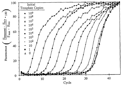
Figure 15 is a fluorescence v. cycle number plot for a number of different
initial
template copy reactions monitored with SybrT"' Green 1: 0, (o); 1, (a), 10, (-
); 102
,
101, (+); 101, 105, (0)I 106, (X).
101, (= ); 101, (0); 109, (4) =
Figure 16 is a fluorescence ratio v. cycle number plot for a number of
different
initial template copy reactions monitored with a dual-labeled hydrolysis
probe: 0, (-);
1, (=); 10, (0); 102, (*)); 101'(0) ; 101, (0), los, (+); 106, (^), 10', (0);
10k, (" 109,
(4).
Figure 17 is a fluut essence ratio v. cycle number plot for a number of
different
initial template copy reactions monitored with adjacent hybridization probes:
0, (-), 1,
(A); 10, (0); 102, (*)), 10 , (=): 101, (D). ]O5, (+), 106, (U), 10', (0);
101, 109, (4)=
Figure 18 is a fluorescence ratio v. cycle number plot distinguishing two
hybridization probe designs monitored by resonance energy transfer: (0) two
hybridization probes labeled respectively with fluorescein and Cy5; and (=)a
primer
labeled with Cy5 and a probe labeled with fluorescein.
Figures 19A-C provide a comparison of three fluorescence monitoring
techniques for PCR, including the double-strand specific DNA dye SYBR Green I
(A),
a dual-labeled fluorescein/rhodamine hydrolysis probe (B), and a fluorescein-
labeled
hybridization probe with a Cy5-labeled primer (C); Figure 19D shows the
coefficient
of variation for the three monitoring techniques represented in Figures 19A-C.
Figure 20 shows a typical log fluorescence vs cycle number plot of a standard
amplification monitored with SYBR Green I.
Figure 21 shows an expontial curve fit to cycles 20-27 of the data from Figure
20.
Figure 22 shows an exponential In to an unknown to determine initial copy
number from amplification data.
CA 02658290 2009-02-04
-25-
Figure 23 shows a typical fluorescence v. cycle number plot of five standards
monitored each cycle with adjacent hybridization probes, wherein initial copy
numbers
are represented as follows: 10`, (0); 10{, (0); 105, (= ); 10', (0); 10', (=
).
Figure 24 shows a curve fit to the standard data of Figure 23.
Figure 25 shows a typical fluorescence vs cycle number plot of five standards
monitored each cycle with a hydrolysis probe, wherein initial copy numbers are
represented as follows: 1.5, (0); 15, (0), 150, (=); 1500, (0); 15,000, (4).
Figure 26 shows a curve fit to the standard data of Figure 25.
Figure 27 shows a typical log fluorescence vs cycle number plot of three
standard amplifications monitored with SYBR Green I, wherein: (a); (a); (=).
Figure 28 shows different curve fit to the standard data of Figure 27.
Fissures 29A&B show plots of (A) time v. fluorescence and (B) time v.
temperature demonstrating the inverse relationship between temperature and
fluorescence.
Figure 30 is a chart showing 2D plots of temperature v. time, fluorescence v.
time, and fluorescence vv. temperature, also shown as a 3D plot, for the
amplification of
a 180 base pair fragment of the hepatitis B genome in the presence of SYBR
Green 1.
Figure 31 is a fluorescence v. temperature projection for the amplification of
a
536 base pair fragment of the human beta-globin gene in the presence of SYBR
Green
1.
Figures 32A&B provide a plot showing (A) a linear changge in fluorescence
ratio with temperature for hydrolysis probes, and (B) a radical change with
temperature for hybridization probes.
Figure 33 shows a fluorescence ratio v. temperature plot of amplification with
'1 5 an exo' polymerase in the presence of adjacent hybridization probes.
Figure 34 shows a fluorescence ratio v. temperature plot of amplification with
an exo' polymerase in the presence of adjacent hybridization probes
Figure 35 shows a 3-dimensional plot of temperature, time and fluorescence
during amplification with an exo" polymerase in the presence of adjacent
hybridization
probes.
CA 02658290 2009-02-04
-26-
Figure 36 shows a 3-dimensional plot of temperature, time, and fluorescence
during amplification with an exo- polymerase in the presence of adjacent
hybridization
probes.
Figure 37 shows melting curves for PCR-amplified products of hepatitis B
virus (.; 50% GC, 180 bp), beta-globin (=, 53.29,0 GC, 536 bp); and prostate
specific
antigen (X; 603% GC, 292 bp).
Figure 38 shows melting curves for PCR-amplified product of hepatitis B virus
at heating rates of 0.1 C to 5.0 C.
Figure 39 shows melting curves for PCR-amplified product of hepatitis B virus
at various SYBRT I Green I concentrations.
Figures 40A&B show (A) melting curves and (B) electrophoretically
fractionated bands of products of a beta-globin fragment amplified with (a) no
added
template, (b) 106 copies of added template under low stringency conditions,
and (c)
106 copies of added template under higher stringency conditions.
is Figures 41 A&B show (A) melting curves and (B) inelting peaks of hepatitis
B
virus fragment (HBV), (3-globin, and a mixture thereof.
Figures 42A-D show (A) a relative fluorescence v. cycle number plot for PCR
amplified products from various amounts of 3-globin template, (B) melting
peaks and
(C) electrophoretic bands of the various products, and (D) corrected
fluorescence of
the data of (A).
Figures 43A&B show (A) melting curves and (B) melting peaks from PCR
amplified products of a mixture of the cystic fibrosis gene and the c-erbB-2
oncogene.
Figure 44 show melting peaks at various cycle numbers for the cystic fibrosis
gene (CFTR) and c-erbB-2 (neu).
Figure 45 shows a graph of integrated melting peaks of CFTR and neu PCT
products.
Figures 46A&B show (A) melting curves and (B) melting peaks for PCR
products of a person heterozygous for the factor V Leiden mutation (solid
line),
homozygous for the factor V Leiden mutation (dotted line), homozygous wild
type
(broken line), and no DNA control (alternating dot and dash).
Figure 47 shows a fluorescence ratio v. temperature plot of continuous
monitoring during cycle 40 of PCR products of a sample homozygous for the
factor \'
CA 02658290 2009-02-04
-27-
Leiden mutation (solid line), heterozygous for the factor V Leiden mutation
(dotted
line). and homozygous wild type (alternating dot and dash).
Figure 48 shows melting peaks of a homozygous mutant of the
methylenetatrahydrofolate gene (solid line), homozygous wild type (broken
line),
heterozygous mutant (dotted line), and no DNA control (alternating dot and
dash).
Figure 49 shows the shape of reannealing curves of amplified (3-globin PCR
products from various initial template amounts.
Figure 50 shows the determination of a second order rate constant for
determining initial template concentration.
Figure 51 shows a block diagram for controlling thermal cycling from
fluorescence data.
Figures 52A&B show (A) a temperature v. time plot acquired after 20 cycles
and (B) a fluorescence v. time plot acquired after 25 cycles wherein thermal
cycling
was controlled from fluorescence data.
Figures 53-105 are progammingdiagrams used in accordance with the present
invention
DETAILED DESCRIPTION
Before the present methods for monitoring hybridization during PCR are
disclosed and described, it is to be understood that this invention is not
limited to the
particular configurations, process steps, and materials disclosed herein as
such
configurations. process steps, and materials may vary somewhat. It is also to
be
understood that the terminology employed herein is used for the purpose of
describing
particular embodiments only and is not intended to be limiting since the scope
of'the
present invention will be limited only by the appended claims and equivalents
thereof.
It must be noted that. as used in this specification and the appended claims,
the
singular forms "a," "an," and "the" include plural referents unless the
context clearly
dictates otherwise.
In describing and claiming the present invention, the following terminology
will
be used in accordance with the definitions set out below.
As used herein. "nucleic acid," "DNA," and similar terms also include nucleic
acid analogs, i.e. analogs having other than a phosphodiester backbone. For
example,
the so-called "peptide nucleic acids," which are known in the art and have
peptide
CA 02658290 2009-02-04
-2s-
bonds instead of phosphodiester bonds in the backbone, are considered within
the
scope of the present invention.
As used herein, "continuous monitoring" and similar terms refer to monitoring
multiple times during a cycle of PCR, preferably during temperature
transitions, and
more preferably obtaining at least one data point in each temperature
transition.
As used herein, "cycle-by-cycle" monitoring means monitoring the PCR
reaction once each cycle, preferably during the annealing phase of PCR.
As used herein, "fluorescence resonance energy transfer relationship" and
similar terms refer to adjacent hybridization of an oligonucleotide labeled
with a donor
fluorophore and another oligonucleotide labeled with an acceptor fluorophore
to a
target nucleic acid such that the donor fluorophore can transfer resonance
energy to
the acceptor fluorophore such that the acceptor fluorophore produces a
measurable
fluorescence emission. If the donor fluorophore and acceptor fluorophore are
spaced
apart by too great a distance, then the donor fluorophore cannot transfer
resonance
energy to the acceptor fluorophore such that the acceptor fluorophore emits
measurable fluorescence, and hence the donor fluorophore and acceptor
fluorophore
are not in resonance energy transfer relationship. Preferably, when the two
labeled
oligonucleotides are both probes and neither functions as a PCR primer ("probe-
probe"), then the donor and acceptor fluorophores are within about 0-25
nucleotides,
more preferably within about 0-5 nucleotides, and most preferably within about
0-2
nucleotides. A particularly preferred spacing is I nucleotide. When one of the
labeled
oligonucleotides also functions as a PCR primer ("probe-primer"), then the
donor and
acceptor fluorophores are preferably within about 0-1 5 nucleotides and more
preferably within about 4-6 nucleotides.
As used herein, "effective amount' means an amount sufficient to produce a
selected effect. For example, an effective amount of PCR primers is an amount
sufficient to amplify a segment of nucleic acid by PCR provided that a DNA
polymerase, buffer, template, and other conditions, including temperature
conditions,
known in the art to be necessary for practicing PCR are also provided.
PCR requires repetitive template denaturation and primer annealing. These
hybridization transitions are temperature- dependent. The temperature cycles
of PCR
that drive amplification alternately denature accumulating product at a high
CA 02658290 2009-02-04
64005-623
-29-
temperature and anneal primers to the product at a Tower temperature. The
transition
temperatures of product denaturation and primer annealing depend primarily on
GC
content and length. if a probe is designed to hybridize internally to the PCR
product,
the melting temperature of the probe also depends on GC content, length, and
degree
of complementarity to the target. Fluorescence probes compatible with PCR can
monitor hybridization during amplification.
In accordance with the present invention, which
is preferably used in connection with rapid cycling
(fully described in the PCT publication No. WO 97/46707
entitled System And Method For Monitoring PCR Processes),
5 a kinetic paradigm for PCR is
appropriate. Instead of thinking about PCR as three reactions (denaturation,
annealing, extension) occurring at three different temperatures for three time
periods
(Figure 1 A), a kinetic paradigm for PCR is more useful (Figure 1 B). With
a,kinetic
paradigm, the temperature vs. time curve consists of continuous transitions
between
overlapping reactions. Denaturation and annealing are so rapid that no holding
time at
a particular temperature is necessary. Extension occurs over a range of
temperatures
at varying rates. A complete analysis would require knowledge of all relevant
rate
constants over all temperatures. If rate constants of all reactions were
known, a
"physicochemical description of PCR" could be developed. Determining these
rates
wouid require precise sample temperature control and is greatly simplified by
reaction
monitoring during temperature cycling.
Figure 2 illustrates useful temperature v. time segments for fluorescence
- hybridization monitoring. Product melting curves are obtained during a slow
temperature increase to denaturation. By quickly lowering the temperature
after
denaturation to a constant temperature, product, probe, or primer annealing
can
optionally be followed. Probe melting curves are obtained by slowly heating
through
temperatures-around the probe Tm. The embodiment represented in Figure 2
provides
all analysis during temperature cycling with immediate real time display.
Fluorescent
probes are included as part of the amplification solution for continuous
monitoring of
primer. probe, or product hybridization during temperature cycling.
CA 02658290 2009-02-04
30-
The fluorescence hybridization techniques contained herein are based on rapid
cvcling, with its advantages in speed and specificity.
A sample temperature profile during rapid cycle PCR is shown in Figure 3.
Denaturation and annealing appear as temperature "spikes" on these figures, as
opposed
to the broad plateaus of conventional temperature cycling for PCR, e.g. Figure
IA.
Rapid temperature cycling is contrasted to conventional temperature cycling in
Figure
4, wherein it is shown that 30 cycles of amplification can be completed in 15
minutes
and the resulting PCR products contain many fewer side products. Thus, with
rapid
cycling the required times for amplification are reduced approximately 10-
fold, and
specificity is improved.
Example I
Figure 4 shows the results of four different temperature/time profiles (A-D)
and
their resultant amplification products after thirty cycles (A-D). The profiles
A and B in
Figure 4 were obtained using a prior art heating block device using a prior
art microfuge
tube. As can be seen in Figure 4, the transitions between temperatures are
slow and many
nonspecific bands are present in profiles A and B. Profile B shows improvement
in
eliminating some of the nonspecific bands (in contrast to profile A) by
limiting the time
each sample remains at each temperature, thus indicating that shorter times
produce more
desirable results.
Profiles C and D were obtained using a rapid temperature cycler. As can be
seen
in Figure 4, amplification is specific and, even though yield is m::-Kimal
with 60-second
elongation times (C), it is still entirely adequate with 10-second elongation
times (D)..
The optimal times and temperatures for the amplification of a 536 bp fragment
of
beta-globin from human genomic DNA were also determined. Amplification yields
and
product specificity were optimal when denaturation (93 C) and annealing (55 C)
were less
than I second. No advantage was found to longer denaturation or annealin
times. The
yield increased with longer elongation times at 77`C, but there was little
change with
elongation times longer than 10-20 seconds. These unexpected results indicate
that the
previously available devices used for DNA amplification are not maximizing the
conditions
needed to optimize the physical and enzymatic requirements of the reaction.
CA 02658290 2009-02-04
64005-623
-31-
Further information can be obtained from: C.T. Wittwer et al.. Rapid Cycle
Allele-Specific Amplification with Cystic Fibrosis delta F(508) Locus, 39
Clinical
Chemistn' 804 (1993) and C.T. Wittwer et al., Rapid DNA Amplification, THE
POLYMERASE CHAIN REACTION 174 (199-1)_.
As indicated earlier, the polymerase chain reaction can be performed rapidly.
In
addition to facilitating rapid heat transfer, the use of optically clear
capillary tubes allows
for continuous fluorescence monitoring of DNA amplification in accordance with
the
present invention.
Fluorescent probes can be used to detect and monitor DNA amplification. Useful
probes include double-stranded-DNA-specific dyes and sequence-specific probes.
Three
different fluorescence techniques for following DNA amplification are compared
in Figure
5. In Figure 5A, fluorescence depends on the hybridization of PCR product as
detected
with a double-strand-specific DNA dye. In figure 5B, fluorescence depends on
the
hydrolysis of a 5'-exonuclease quenching probe, which is well known in the art
as discussed
above. Figure SC diagrams a hybridization scheme based on resonance energy
transfer
between fluorophores on two adjacent probes. The method of Figure 5A is not
sequence
specific, although product specificity can be determined by melting, curves,
one aspect of
the current invention. Both Figures SB and SC are sequence specific. However,
the
hybridization method also allows analysis with melting curves, another aspect
of the
current invention.
In monitoring fluorescence from reactions involving hydrolysis probes as in
Figure
5B and from reactions involvins; hybridization probes as in Figure 5C, it is
advantageous
to measure fluorescence emitted by both the donor fluorophore and the acceptor
fluorophore. In practice, the majority of the fluorescence emitted by
hydrolysis probes is
from the donor fluorophore, and the majority of the fluorescence emitted by
hybridization
probes is from the acceptor fluorophore.
Double-strand-specific DNA dye selection. Those skilled in the art will be
familiar with the use of ethidium bromide in fluorescence techniques. When a
double
strand-specific fluorescent dye is present during amplification, fluorescence
generally
CA 02658290 2009-02-04
64005-623
-32-
increases as more double stranded product is made, see R. Higuchi et al.,
Simultaneous
amplification and detection of specific DNA sequences, 10 Bio/Technology 413-
417
(1992). A fluorescence PCR assay for hepatitis C RNA using the intercalater,
YO-PRO-1
is also known in the art. See T. Ishiguro et al., Homogeneous quantitative
assay of
hepatitis C virus RNA by polymerase chain reaction in the presence of a
fluorescent
intercalater, 229 Anal. Biochem. 207-213 (1995). It is preferred that SYBRTM
Green I,
which is well known in the art and available from Molecular Probes of Eugene,
Oregon,
be used as a double-strand-specific dye. The molecular structure of this dye
is a trade
secret, but it is recommended by the manufacturer as a more sensitive
double-strand-specific dye for DNA detection on gels. SYBRTMM Green I is heat
labile,
however, and thus is not useful for fluorescence monitoring of PCR according
to
conventional methods where the temperature of the reaction mixture is
maintained at
melting temperatures for extended periods of time. Because of this heat
lability, it was
unexpected to discover that SYBRT"I Green I can be used to monitor PCR
reactions when
melting temperatures are not maintained for extended periods, i.e. when PCR is
carried out
by rapid cycling according to the kinetic paradigm described above.
Example 2
Different double-strand-specific DNA dyes were compared by monitoring the
amplification of a 110 base pair fragment from the PCO3/PCO4 primer pair of
the human
beta-globin gene from 10,000 template copies. Primers were synthesized by
standard
phosphoramidite chemistry as known in the art, namely, using Pharmacia Biotech
Gene
Assembler Plus (Piscataway, New Jersey). The human beta-Tobin primers
PC03/PC04
(110 base pairs) are described in C.T. Wittwer et al., Automated polymerase
chain reaction
in capillary tubes with hot air, 17 Nucl. Acids. Res. 4353-4357 (1989)_
DNA amplification was performed in 50 mM Tris, pH
8.5 (25 C), 3 mM MgCI,, 500 uo/ml bovine serum albumin, 0.5 .M of each primer,
0.2
mM of each deoxynucleoside triphosphate and 0.2 U of Taq polymerase per 5 Pi
sample
unless otherwise stated in the following examples. Purified amplification
product was used
as DNA template and was obtained by phenol/chloroform extraction and ethanol
precipitation, see D.M. Wallace, Large- and small-scale phenol extractions and
precipitation of nucleic acids, 152 Methods in Enzymology 33-48 (1987),
followed by
CA 02658290 2009-02-04
-33-
removal of primers by repeated washing through a Centricon 30
microconcentrator
(Amicon, Danvers, Massachusetts). Template concentrations were determined by
absorbance at .260 nm. A(260):A(280) ratios of templates were greater than
1.7.
SYBRT^I Green I (Molecular Probes, Eugene, Oregon) was used at a l :10,000
dilution, ethidium bromide was at 5 ug/ml, and acridine orange was at 3 ug/ml.
These
concentrations were determined to be optimal concentrations for maximizing the
fluorescence signal observed during amplification for each dye. Excitation was
through
a 450-490 rim interference filter from a xenon arc source, except for ethidium
bromide,
where a 520-550 nm excitation was used. For SYBRTM Green I, the emmision at
520-550
was monitored. Ethidium bromide fluorescence was observed through a 580-620 nm
bandpass. The acridine orange signal was taken as the ratio of green (520-550
rim) to red
(>610 nm) fluorescence. The fluorescence of the sample before amplification
was
compared to the fluorescence after 35 cycles (94 C max, 60 C for 20 sec) at 60
C. The
fluorescence increase was 5.3-fold for SYBRT111 Green I, 1.7-fold for ethidium
bromide,
and 1.2-fold for acridine orange. In separate experiments, the fluorescence
from SYBRT"f
Green I was stable for greater than 30 min at 70 C. It is also conveniently
excited with
visable light and is claimed to be less of a mutagen than ethidium bromide.
Background
fluorescence in all cases arose primarily from the primers.
SYBRThf Green I is a preferred double-strand-specific dye for fluorescence
monitoring of PCR, primarily because of superior sensitivity, arising from
greater
discrimination between double stranded and single stranded nucleic acid.
S'YBRT" Green
I can be used in any amplification and is inexpensive. In addition, product
specificity can
be obtained by analysis of melting cunves, as will be described momentarily.
Resonance energy transfer dye selection for hybridization probes.
Fluorescence resonance energy transfer can occur between 2 fluorophores if
they are in
physical proximity and the emission spectrum of one fluorophore overlaps the
excitation
spectrum of the other. Introductory theory on fluorescence resonance energy
transfer can
be found in many recent review articles. The rate of resonance energy transfer
is:
(8.785E-5)(t-')(k2)(n-4)(QD)(R'6)(JDI), where:
AMENDED SHEE1I
CA 02658290 2009-02-04
-34-
t = excited state lifetime of the donor in the absence of the acceptor:
k` = is an orientation factor between the donor and acceptor;
n = refractive index of visible light in the intervening medium,
qõ = quantum efficiency of the donor in the absence of the acceptor:
R = distance between the donor and acceptor (in angstroms):
J,,,, = the integral of (Fõ)(e \(W') with respect to W at all overlapping
wavelengths
with.
Fõ = peak normalized fluorescence spectrum of the donor,
e, = molar absorption coefficient of the acceptor (M''cm'), and
W = wavelength (nm).
For any given donor and acceptor, a distance where 50'/-o resonance energy
transfer
occurs can be calculated and is abbreviated R,,. Because the rate of resonance
energy
transfer depends on the 6th power of the distance between donor and acceptor.
resonance
energy transfer changes rapidly as R varies from Ro. At 2R0, very little
resonance energy
transfer occurs, and at 0.5R0, the efficiency of transfer is nearly complete,
unless other
forms of de-excitation predominate (i.e., collisional quenching). R, values
for many
different donor and acceptor pairs have been compiled and vary between 22 and
72
angstroms.
In double helical DNA, 10 bases are separated by about 34 angstroms. By
labeling
the bases of DNA with donor and acceptor fluorophores, resonance energy
transfer has be
used as a spectroscopic ruler to observe the helical geometry of DNA and
analyze the
structure of a four-way DNA junction. Resonance energy transfer can also be
used as a
monitor of hybridization. If a labeled oligonucleotide is hybridized to a
labeled template
strand, R can be brought from much greater than Ro to well below R0,
increasing resonance
energy transfer dramatically. Alternately, 2 labeled probes can be hybridized
to the same
template strand for a similar change in fluorescence energy transfer.
The practical use of resonance energy transfer to monitor hybridization
depends on
the sensitivity required and how much time is available. Using a competitive
hybridization
technique with I nM labeled probes, PCR-amplified DNA was detected after 15
min at
C. Faster signal generation is desirable. If only seconds were required for
hybridization. PCR products could conveniently be quantified each cycle of
amplification.
1~l1+11'.:ii c .
01,
CA 02658290 2009-02-04
-35-
Even further, the extent of probe hybridization could be monitored within a
temperature
cycle.
Hybridization is a second order process (see B. Young & M. Anderson,
Quantitative analysis of solution hybridization, In: Nucleic Acid
Hybridization: A Practical
Approach 47-71, (B. Harries, S. Higgins eds., 1985). When the concentration of
the
probe is much greater than the concentration of the target, the hybridization
rate is
inversely proportional to concentration of probe. For example, by doubling the
probe
concentration, the hybridization time is cut in half. High probe
concentrations would be
necessary for cycle-by-cycle monitoring during PCR, because hybridization must
occur
before the hybridization site is covered by polymerase extension.
The high probe concentrations required for hybridization monitoring during PCR
require a resonance energy transfer pair with unique characteristics. Consider
excitation
of a donor (D) and an acceptor (A) pair with light. The number of fluorophores
of D and
A directly excited will be proportional to the extinction coefficient (e) of
each fluorophore
at the excitation wavelength, or:
Number of D molecules directly excited = (K)(e0)
Number of A molecules directly excited = (K)(e.,)
where K is a proportionality constant. De-excitation of the donor will occur
by
fluorescence, resonance energy transfer, and other processes summarized as
thermal
quenching. If pr = probability of resonance energy transfer, and p.m =
probability of donor
thermal quenching, then the probability of donor fluorescence is: =
-15 1-PF-Pro
and the number of fluorescing donor molecules is:
(K)(e0)(I-Pr-PTD)
CA 02658290 2009-02-04
36-
if the probability of detecting a donor emission in the donor emission window
(for
example. a bandpass filter window) is pnu, then the number of observed donor
emissions
is
(Pn:),)(K)(eL))( I-PF-Prn)
Now. the number of excited acceptor fluorophores is the sum of those directly
excited and
those excited through resonance energy transfer:
i0 (K)(e .) - (K)(eo)(PF)
if p, = the probability of thermal quenching of the acceptor, then the
probability of
acceptor fluorescence is:
15 1-Pr;
and the number of fluorescing acceptor molecules is:
[(K)(e) + (K)(eu)(Pr)] [1-(PTA)]
If the probability of detecting an acceptor emission in the acceptor emission
window is
then the number of observed acceptor emissions is:
(P:,,)[(K)(e.{) + (K)(eu)(P1)] [1-(Pra)]
Finally. if the probability of observing a donor emission in the acceptor
emission window
is p,,.,. then the number of observed emissions (both D and A) in the acceptor
emission
window is:
(P.,.,)[(K)(e_,) ` (K)(eo)(pF)} [1-(pr:,)] - (Pn.,)(K)(en)(1-Pr-Pru)
CA 02658290 2009-02-04
-37-
Since fluorescence measurements are relative and K is present in all terms, if
we remove
K and rearrange, the observed intensity at the donor window is proportional to
(donor
excitation) - (energy transfer)-
I) (e0)(P0D)(1-Prn) - (eo)(Pnu)(Pr)
and the observed intensity at the acceptor window is proportional to (acceptor
excitation)
+ (energy transfer) - ( donor emission in the acceptor window):
2) (e:,)(PA.1)(1-PrA) + (en)(Pnn)(Pr)(l -Pr.a) + (e0)(P0.)(I -Pm'PF)
As resonance energy transfer increases, the donor signal decreases and the
acceptor
signal increases. The percent signal change depends on the background light
intensity in
each window. With high concentrations of probes, this background light
intensity is high.
During PCP, when varying target (product) concentrations need to be monitored,
it is not
possible to match the donor concentration to the target concentration. The
excess donor
molecules contribute to the background light intensity in both the donor and
acceptor
windows and partially mask the energy transfer phenomena. Background light in
the
acceptor window comes from not only donor emission in the acceptor window, but
also
from direct excitation of the acceptor. This background can be minimized with
a low e_.,
and a low PD.a.
The fluorescein/rhodamine fluorescence energy transfer pair, commonly used for
nucleic acid detection. has high background fluorescence. Both direct acceptor
excitation
(e:.', ca. 10% eõ ~y) and emission of the donor at wavelengths used to detect
acceptor
emission (p,.,, ca. 20% peak emission) are high. This pair can be used to
monitor
hybridization if the probe concentration is near to the target concentration
and enough time
is allowed for complete hybridization. It is not a useful pair of fluorophores
for continuous
monitoring of PCR because high probe concentrations are required and the
template
concentration in PCR is continually changing.
CA 02658290 2009-02-04
-38-
Monitoring product concentration during PCR by hybridization has not been
possible in the past because an acceptable resonance energy transfer pair had
not been
found. There have been few attempts to use resonance energy transfer for
direct
"noncompetitive" detection of hybridization. For example, U.S. Patent No.
5,565.322
states the observed energy transfer efficiency in terms of re-emission by the
acceptor was
relatively low " At probe concentrations that are high enough for significant
hybridization
to occur in seconds. the background fluorescence is too NO.
Fluorescein is perhaps the most widely used fluorophore. Its extinction
coefficient
and quantum efficiency are high and it is extensively used in microscopy,
immunoassays.
and flow cvtometn=. It is the donor in a commonly used resonance energy
transfer pair
with rhodamine. CO is a popular red-emitting fluorophore with a very high
extinction
coefficient. The structure of the N-hydroxysuccinimide ester of Cv5 is shown
in Figure 6.
and the structure of the related dye. Cy5.5, is shown in Figure 7. These dyes
are
indodicarbocyanine dyes that are used commonly in flow cytometry and automated
IS fluorescence sequencers and are available from Amersham (Pittsburg, PA).
Both
fluorescein and Cy5 are commercially available as amidites for direct,
automated
incorporation into oligonucleotides. However, Cy5 has never been reported as a
resonance
energy transfer pair with fluorescein. Intuitively, fluorescein emission and
Cy5 absorption
do not overlap enough for resonance energy transfer to be considered. The
emission
spectrum of fluorescein and absorption spectrum of Cy5 attached to
oligonucleotides are
shown in Figure 8. When the areas under the curves are normalized, the overlap
from the
technical spectra is 19%. Cy5.5 excitation is shifted to the red by about 25
nm, further
decreasing the overlap with fluorescein emission to about 15%. Working in the
red infrared region of the spectrum is advantageous when choosing optical
components for
instrumentation Laser diodes can be used for illumination, photodiode
detectors have
excellent sensitivity. and most materials have minimal autofluorescence in the
pertinent
spectral region.
Despite low spectral overlap, it has been discovered that fluorescein and
either Cy5
or Cv5.5 make an excellent resonance energy transfer pair for hybridization
monitoring
during PCR.
Example 3
CA 02658290 2009-02-04
-39-
A 110 bp beta-globin fragment was amplified from 50 ng human genomic DNA
according to the procedure of Example 2 with the internal probes CAAACAGACA
CCATGGTGC.A CCTGACTCCT GAGGA-fluorescein (SEQ ID NO:3) and
Cy5-G.-V-AGTCTGCC GTTACTGCCC TGTGCrGGCAA G-p (SEQ.ID NO: 18) at 0.2 pN1
each and 0.8 U KlenTaq I polymerase (a 5'-exonuclease deficient variant of Taq
polymerase
- U.S. Patent No 5,436, 149) in a 10 p1 reaction. The probes hybridized
internal to the
primers on the same strand and were immediately adjacent without any
intervening bases.
Probes and primers were synthesized by standard phosphoramidite chemistry as
known in the art, using a Pharmacia Biotech Gene Assembler Plus (Piscataway,
New
Jersev) The 3'- fluorescein-labeled probe was synthesized on a fluorescein-
labeled CPG
cassette (Glen Research. Sterling, VA) with the final trityl-ON to assist with
C 18 reverse
phase HPLC purification. The late eluting peak was collected and the trityl
group was
removed on a PolyPack (Glen Research). The fluorescein-labeled oligo was
eluted with
50% acetonitrile and again purified by C 18 reverse phase HPLC. The 5'-CyS-
labeled probe
was synthesized with a chemical phosphorvlation agent on the 3'-end (Glen
Research) and
adding a Cy5 amidite (Pharmacia) to the 5'-end during trityl-OFF synthesis.
Failure
sequences were removed by C18 reverse phase I-IPLC. Probe purity was checked
with
polyacrvlamide electrophoresis and the absorbance of the dye and the oligo.
HPLC was perfumed on a 4 x 250 mm Hypersil ODS column (Hewlett Packard)
with a 0. I M triethanolamine:acetate mobile phase and an acetonitrile
gradient at I ml/min.
The eluate was monitored for both absorbance and fluorescence (490 nm
excitation.
520 nm emission for fluorescein and 650 nm excitation, 670 nm emission for
Cy5).
Tritylated- and fluorescein-labeled oligonucleotides were eluted with a 10-20%
acetonitrile
gradient, and Cy5-labeled oligonucleotides eluted over a 10-40% acetonitrile
gradient.
Temperature cycling, was 94 C for 0 sec with a programmed approach rate of
20--C/sec, 60 C for 20 sec with an approach rate of 20 C/sec, and 75 C for 0
sec with an
approach rate of 1 C/sec in a capillary fluorescence rapid temperature
cycler. During
temperature cycling, fluorescein and CyS fluorescence were acquired each cycle
at the end
of the annealing/extension segment. Resonance energy transfer was observed as
both a
decrease in fluorescein fluorescence, and an increase in Cy5 fluorescence
beginning around
cycle 26 of amplification (Figure 9). In general. observing the fluorescence
ratio of Cy5
to fluorescein fluorescence is perferred.
CA 02658290 2009-02-04
-40-
The unexpectedly good results with the fluorescein/Cy5 pair can at least
partly be
rationalized The overlap integral, JD., depends not only on spectral overlap,
but also on
the extinction coefficient of the acceptor (Cv5 has an extinction coefficient
of 250,000
NI''cm' at 650 nni), and on the 4th power of the wavelength. Both of these
factors will
favor a high Jõ., for CyS, even given low spectral overlap. Recently,
phycoerythrin and
CO t.ere shown to be an effective tandem probe for immunofluorescence, despite
low
spectral overlap. In a later example, the utility of fluorescein and Cy5.5 as
labels on
hybridization probes is demonstrated. Fluorescence resonance energy transfer
can be used
to monitor nucleic acid hybridization even when the interacting dyes have low
spectral
overlap. The use of fluorescein with Cy5, Cy5.5 and other red or infrared
emitting dyes
as resonance energy transfer pairs for monitoring hybridization has not been
previously
recounized. Fluorescein has a long emission "tail" that goes out to 600 nm,
700 nm and
beyond that can be used to excite these far red and infrared dyes. The rate of
energy
transfer is dependent on the overlap integral, but is also effected by the 6th
power of the
distance between the fluorophores. If the probes are designed so that the
resonance energy
transfer dyes are in close proximity, the transfer rate is high. At least with
fluorescein/Cy-5,
fluorescein/Cy-5.5 and like pairs, resonance energy transfer appears to
predominate over
collisional quenching and other forms of energy loss when the fluorophores are
close
together, as in the above example where the fluorophores are attached to
adjacent probes
with no interveninu bases.
The potential usefulness of a resonance energy transfer pair for hybridization
probes
can be judged by observing the change in the ratio of light intensity in the
donor and
acceptor windows at minimal and maximal resonance energy transfer. One way to
obtain
minimal and maximal transfer is to attach both fluorophores to the same
oligonucleotide
and measure fluorescence ratio before and after digestion with
phospodiesterase.
Example 4
The dual-labeled fluoresceintCy5 probe Cy5-CTGCCG-F-TACT GCCCTGTGGG
,0 GCAAGGp (SEQ ID NO:19) was synthesized by standard phosphoramidite
chemistry.
\,,here p is a terminal 3'-phosphate (chemical phosphorviation reagent, Glen
Research), F
is a fluorescein residue introduced as an amidite with a 2-aminobutvl-1,3-
propanediol
CA 02658290 2009-02-04
-41-
backbone to maintain the natural 3-carbon internucleotide phosphodiester
distance
(ClonTech. Palo Alto, CA). and Cvv5 is added as the amidite (Pharmacia). The
ratio of Cy5
to fluorescein fluorescence in 0, 1 M1 Tris, pH 3.0 was obtained before and
after exhastive
hydrolysis with phosphodiesterase (Sigma. St. Louis, MO). The change in the
fluorescence
ratio was 220-fold after hydrolysis. A dual-labeled fluorescein/rhodamine
probe
F-ATGCCCT*CCC CCATGCCATC CTGCGTp (SEQ ID NO:20) was purchased from
Perkin Elmer (Foster City, CA), where F is fluorescein and * is a rhodamine
attached to
a modified T residue by an amino-linker arm. The change in the fluorescence
ratio
(rhodamine to fluorescein) was 22-fold after hydrolysis with
phosphodiesterase.
The potential signal from the fluorescein/Cy5 pair was 10-fold that of the
fluorescein/rhodamine pair.
Example 5
The effect of the ratio, concentration, and spacing of fluorescein and Cy5-
labeled
adjacent hybridization probes during PCR was studied. Amplification of the
beta globin
locus and probe pair of Example 3 was used and the maximum change in the
fluorescence
ratio of Cy5 to fluorescein was observed. The maximal signal occurred when the
ratio of
Cy5 to fluorescein-labeled probes was 2:1 (Figure 10). At this 2:1 ratio, the
best signal
occurred at a fluorescein probe concentration of 0.2 M and a Cy5-labeled
probe
concentration of 0.4 jN1 (Figure 11). The optimal number of intervening bases
between
adjacent hybridization probes during PCR was also determined. Several probes
of the
same length but slightly shifted in their hybridization position were
synthesized according
to Example 3 so that when they hybridized to the beta globin target, 0, 1, 2,
3, 4, or 6
bases remained between the probes. The highest signal during PCR occurred with
one
intervening base (Figure 12). Although some resonance energy transfer was
detected at
a spacing of 15 and even 25 bases, much better transfer occurred at 0-5 bases.
Heller et al. (U.S_ Patent No. 4.996,143), found that energy transfer
efficiency
decreased as the number of nucleotides between fluorophores decreased from 4
to 0 units.
In contrast. the best energy transfer with the fluorescein/Cy5 pair was seen
at 0 to 2
intervening nucleotides.
CA 02658290 2009-02-04
-42-
Hybridization probe method. If 2 probes are synthesized that hybridize
adjacently on
a target and each is labeled with one fluorophore of a resonance energy
transfer pair. the
resonance eneruv transfer increases when hybridization occurs (Figure 5C). The
fluoresceinirhodamine pair is most commonly used for nucleic acid detection.
One aspect of this invention is to provide a sequence-specific homogeneous
hybridization method for detection of PCR products. It is not obvious how to
achieve this.
Using hybridization probes during amplification is counterintuitive. It does
not seem that
both probe hybridization and polymerase extension can occur. To get sequence
specific
fluorescence. the probes must be hybridized, but the probes cannot be
hybridized if the
polymerase is to complete primer extension and exponentially amplifi' DNA.
One solution to this problem is to use a dual-labeled single probe and utilize
the
exonuclease activity of common heat stable DNA polymerases to cleave the probe
durinurt extension, thereby separating the 2 fluorophores. In this case, the
fluorescence
signal arises from separation of the resonance energy transfer pair upon piube
hydrolysis
(Figure 5B). rather than approximation of the fluorophores by adjacent
hybridization
(FiL,ure 5C). However, dual-labeled probes are difficult to make, requiring
manual addition
of at least one fluorophore to the oligo and usually require extensive
purification. The
probes are expensive, and two dual-labeled probes are necessary for
competitive
quantification of a target or for mutation detection. A further concern is
that the observed
fluorescence depends on the cumulative amount of probe hydrolyzed, not
directly on the
amount of product present at any given cycle. This results in a continued
increase in
fluorescence even after the PCR plateau has been reached. Finally and most
importantly.
probe hydrolysis does not always occur during polymerase extension, an effect
that is not
well understood. For example, the dual-labeled fluorescein/Cy5 probe of
Example 4
showed very poor hydrolysis during PCR when it was flanked by primers. Indeed,
several
dual-labeled fluorescein/Cy5 probes, including those with terminal labels,
were made and
all showed poor hydrolysis and signal generation during amplification.
Homogeneous detection of PCR products with adjacent hybridization probes would
solve many of the problems of the 5'-exonuclease system. Synthesis of adjacent
hybridization probes is relatively simple because amidites for both
fluorescein and Cy5 are
available for direct incorporation during automated synthesis and dual
labeling of one
probe is not required- Because their fluorescence results from hybridization,
not
CA 02658290 2009-02-04
-43-
hydrolysis. the temperature dependence of probe fluorescence could be used for
mutation
detection and quantification. However, use of adjacent hybridization probes
for
homogeneous detection of PCR products has not been reported previously.
Surprisingly,
both hybridization for signal generation and amplification by polymerase
extension through
the area blocked by the probes can occur.
Example
A 110 bp beta-globin fragment was amplified from genomic DNA with adjacent
fluorescein- and Cy5-labeled probes as described in Example 3. Either 0.4 U
(Taq) or 0.8
U (Stoffel fragment. Perkin Elmer, or KlenTaq 1) of enzyme was used in 10 Al
reactions.
Unless indicated otherwise, temperature cycling was 94 C for 0 sec with a
programmed
approach rate of 20'C/sec, 60 C for 20 sec with an approach rate of 20 C/sec,
and 75 C
for 0 sec with an approach rate of 1'C/sec. Figure 13 shows the development of
fluorescence by 2 adjacent hybridization probes immediately after the template
was
amplified for 30 cycles. After a brief denaturation at 94 C, the temperature
was lowered
to 60=C and fluorescence increased for about 20 sec. The magnitude of the
signal is
greater with an exonuclease deficient polymerase (Stoffel fragment) than with
native Taq
polymerase that includes a 5'-exonuclease activity. After about 20 sec., the
fluorescence
drops as the polymerase displaces and/or hydrolyzes the probes. The relative
decrease in
fluorescence is slightly faster when the polymerase has 5'-exonuclease
activity (Taq DNA
polymerase) then when it lacks this activity (Stoffel fragment).
In Figure 14 (top panel), the temperature is cycled between 94 C and 60 C with
a 20 sec hold at 60 C. Fluorescence is acquired at the end of the 20 sec when
fluorescence
is maximal. Good amplification occurs with Taq (exo-), but not with Stoffel
fragment
(exo') as verified by both fluorescence development and agarose gels (gels not
shown).
However, if the time at 60 C is increased from 20 sec to 120 sec (Figure 14,
middle panel),
the exo" polymerase amplifies well. The slower rate of probe displacement with
an exo"
polymerase apparently requires more time at 60 C for efficient amplification
than the exo-
polymerase. The time required by exo" polymerases can be reduced by slowly
increasing
the temperature from 60'C to 75 C (Figure 14, bottom panel). The polymerase
stalls when
it reaches the probe. However, at the probe melting temperatures, the probes
melt off the
template and the polymerase continues unencumbered to complete polymerization
of the
CA 02658290 2009-12-30
64005-623F(S)
-44-
strand. Polymerization is completed as long as the temperature is not raised
too quickly
afier probe melting. Figure 14 (bottom panel) shows one exo- polymerase (Taq)
and two
exo- polvmerases (Stoffel fragment and KlenTaq 1)
When exonuclease activity is present, some of the probe is hydrolyzed each
cycle
as evidenced by an the decrease in fluorescence with extensive amplification.
This is
observed in Figures 13 and 14 (middle and bottom panels), but is does not
occur with exo'
polvmerases. Because the fluorescence is stable on extensive amplification,
exo'
polymerases such as KlenTaq I are preferred.
The success of using adjacent hybridization probes to monitor PCR depends on
several factors. Resonance energy transfer is maximized when there is either 0
to 2
intervening bases between adjacent hybridization probes. To increase the
fraction of
strands that hybridize to the probes before the primer extends through the
area of probe
hybridization. the probe melting temperatures should be greater than the
primer melting
temperatures (preferably >S C).
Cycle-by-cycle fluorescence. Conventional endpoint analysis of DNA
amplification by gel electrophoresis identifies product size and estimates
purity. However,
because amplification is at first stochastic, then exponential. and finally
stagnant, the utility
'0 of endpoint analysis is limited for quantification. One aspect of the
present invention
includes cycle-by-cycle monitoring for quantification of'
nitial template copy number with
hybridization probes. As will be appreciated by those skilled in the art, once-
per-cycle
monitoring of multiple samples undergoing DNA amplification is a powerful
quantitative
tool. Cycle-by-cycle monitoring is achieved by acquiring fluorescence during
the extension
or combined annealing/extension phase of each cycle and relating the
fluorescence to
product concentration.
Example 7
Cycle-by-cycle monitoring of PCR was performed by three different fluorescence
techniques. Fluorescence was monitored by (i) the double-strand-specific dye
S113RT"
Green 1. (ii) a decrease in fluorescein quenching by rhodamine after
exonuclease cleavage
of a dual-labeled hydrolysis probe and (iii) resonance energy transfer of
fluorescein to Cv5
*Trade-mark
CA 02658290 2009-02-04
64005-623
-45-
by adjacent hybridization probes. Amplification reagents and conditions were
as described
in Example 2. The human beta-globin primers RS42/GM29 (536 base pairs) and
PC03/PC04 (110 base pairs) are described in C.T. Wittwer et al., Automated
polymerase
chain reaction in capillary tubes with hot air, 17 Nucl. Acids. Res. 4353-4357
(1989).
Temperature cycling for beta-globin was
95 C maximum, 61 C minimum, 15 sec at 72 C and an average rate between
temperatures
of 5.2 C/sec. The beta-actin primers and fluorescein/rhodamine dual probe were
obtained
from Perkin Elmer (no. N808-0230). Temperature cycling for beta-actin was 94 C
maximum, 60 C for 15 sec with an average rate between temperatures of 6:2
C/sec. The
single labeled probes 5'-C..ACAGACA CCATGGTGCA CCTGACTCCT
GAGGA-fluorescein-3' (SEQ ID NO:3) and 5'-Cy5-AAGTCTGCCG TTACTGCCCT
GTGGGGC.kAGp ($EQ ID NO:4) were synthesized as in Example 3. These adjacent
probes hybridize internal to the PCO3/PCO4 beta-globin primer pair on the same
DNA
strand and are separated by one base pair. Temperature cycling was 94 C
maximum, 59 C
for 20 sec with an average rate between temperatures of 7.0 C/sec.
Hybridization probes
(beta-actin and beta-lobin) were used at 0.2 MM each.
When multiple samples are monitored once each cycle with SYBRTnt Green I, a
10'-10" range of initial template concentration can be discerned as
represented in Figure
15. This amplification is of a 536 base pair fragment of the beta-globin gene,
with
SYBRT^' Green I as the double-strand specific dye. When the data were
normalized as the
percent maximal fluorescence of each sample, one hundred initial copies were
clearly
separated from ten copies. However, the difference between one and ten copies
was
marginal, and no difference was observed between zero and one average copies
per sample.
In contrast, sequence-specific probes have a similar dynamic range but, appear
to
discriminate even a single initial template copy from negative controls.
Signal generation
With 5'-exonuclease probes (beta-actin fragment, Figure 16) is dependent not
only on DNA
synthesis, but requires hybridization and hydrolysis between the fluorophores
of the
dual-labeled probe. This hydrolysis reduces quenching and the fluorescence
ratio of
fluorescein to rhodamine emission increases. Whereas the fluorescence from
double strand
dyes levels off with excess cycling (Figure 15), the signal from exonuclease
probes
continues to increase with each cycle (Figure 16). Even though no net product
is being
synthesized, probe hybridization and hydrolysis continue to occur. As the
template copy
CA 02658290 2009-02-04
-46-
number decreases below 10', signal intensity decreases, but low copy numbers
can still be
quantified because the negative control signal is stable-
in Figure 17, amplification is monitored using adjacent hybridization probes
and is
expressed as a ratio of Cy5 to fluorescein fluorescence. The change in
fluorescence ratio
is lar rely due to an increase in Cy5 fluorescence from resonance energy
transfer (Figure
9) In contrast to dual-labeled hydrolysis probes. the fluorescence signal of
hybridization
probes decreases at high cycle numbers if the polymerase contains an
exonuclease activity
(see also Figure 14).
The present invention's feasibility using two different methods for resonance
energy
transfer detection of hybridization during PCR will now be demonstrated. The
first method
uses two adjacent hybridization probes. one labeled 3' with fluorescein and
the other
labeled 5' with Cy5. As product accumulates during PCR. the probes hybridize
next to
each other during the annealing segment of each cycle. The second method uses
a primer
labeled with Cv5 and a single hybridization probe. The labeled primer is
incorporated into
the PCR product during amplification and only a single hybridization is
necessary.
Example 8
Cycle-by-cycle monitoring of PCR was performed by resonance energy transfer
between a Cy5-labeled primer and a fluorescein-labeled hybridization probe.
This was
compared to monitoring with adjacent Cy5/fluorescein hybridization probes. The
Cy5-labeled primer was CAACTTCATC CACGT*TCACC (SEQ ID NO:2 1) where T*
is a modified T base with Cy5 attached and the corresponding probe was
GTCTGCCGTT
ACTGCCCTGT GGGGCAA-fluorescein (SEQ ID NO:22). The adjacent hybridization
probes were CCTCAAACAG ACACCATGGT GCACCTGACT CC-fluorescein (SEQ ID
NO:23) and Cv5-GAAGTCTGCC GTTACTGCCC TGTGGGGCAAp (SEQ ID NO:24).
The hvbridizaton probes were synthesized according to Example 3 and used at
0.2 NI.
The Cy-5-labeled primer was synthesized in two steps. Automated synthesis was
used to
incorporate an amino-modifier C6dT (Glen Research) at the desired T position.
Then, the
monovalent N-hvdroxvsuccinimide ester of Cy5 (Fivure 6) was manually
conjugated to the
CA 02658290 2009-02-04
-47-
amino linker according to the manufacturer's instructions (Amersham). HPLC
purifification was as described in Example 3_
The Cv5-labeled primer (0.5 W) was used instead of PCO4 to amplify the 110
base pair beta-globin fragent from human genomic DNA as in Example 3, except
that 0.4
U of Taq pol}.merase was used per 10 scl. The-adjacent hybridization probes
also
monitored amplification of the same beta-globin fragment. Temperature cycling
was done
at 94 C for 0 sec and 60 C for 20 sec. The fluorescence was monitored once
each cycle
at the end of the annealing/extension segment. In both methods, fluorescence
energy
transfer to Cy5 increases with hybridization and is plotted as a ratio of Cy5
to fluorescein
fluorescence (Figure 18).
In additional experiments, the number of bases separating the Cy5-label and
the
fluorescein label were varied. The best fluorescence resonance energy transfer
was
observed with about 4-6 bases between the fluorophores, although a signal was
detectable
up to at least 15 bases.
In contrast to hydrolysis probes, the fluorescence signal from hybridization
probes
is not cumulative and develops anew during each annealing phase. The
fluorescence is a
direct measure of product concentration because the hybridization is a pseudo-
first order
reaction. Because the concentration of probe is much greater than the product,
the fraction
of product hybridized to probe is independent of product concentration. These
characteristics indicate that using a single hybridization probe along with a
labeled primer
will provide a superior monitor of product accumulation for quantification.
The inherent
variance of different fluorescence techniques during cycle-by-cycle monitoring
is also
important for quantification.
Example 9
DNA amplification was perfonied according to Example 2 for each of three
different fluorescence monitoring methods. S1BRT"' Green I was used at a
1:10,000
dilution in the amplification of a 205 base pair human beta-lobin fragment
from primers
KM29 and PCO4. The hydrolysis probe and conditions are those specified in
Example 7.
The hybridization probe, TCTGCCGTTA CTGCCCTGTG GGGCAAG-fluorescein (SEQ
ID NO:5) was used with K,%129 and the Cy5-labeled primer
CA 02658290 2009-02-04
--t8-
CA.ACTTC.ATCCACGTT"CACC (SEQ ID NO.6) where T" was a Cy5-labeled T base
synthesized as in example S. All amplifications were performed in ten
replicates with
15.000 template copies (50 ng of human genomic DN.-J10 il). The temperature
cycles
ere 3 1 sec lone, (94--C maximum, 60 C for 20 sec, average rate between
temperatures
6 2'C.'sec) Fluorescence was acquired for each sample between seconds 15 and
20 of the
annealing/extension phase.
FWure 19 allows comparison of three fluorescence monitoring techniques for
PCR.
The fluorescence probes are the dsDNA dye SlBRTM Green I (Figure 19A), a
dual-labeled fluorescein/rhodamine hydrolysis probe (Figure 19B), and a
fluorescein-labeled hybridization probe with a Cy5-labeled primer (Figure
19C). All probes
had nearly the same sensitivity with detectable fluorescence occurring around
cycle 20.
With extended amplification. the signal continued to increase with the
hydrolysis probe,
was level with SYBRT`I Green 1, and slightly decreased with the hybridization
probe. The
precision of the three fluorescence monitoring techniques are compared in
Figure 19D.
I The mean -/- standard deviations are plotted for each point. The data are
plotted as the
coefficient of variation (standard deviation/mean) of the fluorescence ratio
above baseline
(taken as the average of cycles 11-15).
Although the chang=e in fluorescence ratio from the hydrolysis probe is
greater than
that from the hybridization probe (Figures 19B and 19C). the coefficient of
variation of
fluorescence from the hydrolysis probes is greater (Figure 19D). That is, the
fluorescence
resulting from the hybridization probe method is more precise than using a
hydrolysis
probe, even though the absolute signal levels are lower. This is an unexpected
advantage
of hybridization probes over the more usual dual-labeled hydrolysis probes.
Quantification of initial template copy number. Quantitative PCR has become
an important technique. in both biomedical research and in the clinical
laboratory. The
process of quantification often includes running a standard curve of samples
containing
known copy numbers of the target sequence. The copy number of an unknown
sample is
determined by extrapolation between the known values. When a complete
amplification
curve is monitored cycle-bv-cycle using, fluorescence. radioactivity or any
other method
that gives a signal proportional to the amount of DNA present. many data
points are
CA 02658290 2009-02-04
-49-
available for analysis and it is not obvious which value to choose to
represent a standard
or unknown.. Prior art is to choose a "threshold value" of the signal and then
use the cycle
number when the standard or unknown crosses that threshold as the
representative value
(see Higuchi & Watson. EPA 0 640 828 A]). This approach uses a very small
amount of
the available data in an amplification curve. In addition, the assignment of
the threshold
value is highly subjective and is subject to conscious or unconscious bias.
More of the
available data could be used objectively by applying non-linear curve fitting
techniques to
the data in an amplification curve. Preferably, equations could be found that
describe the
shape of the amplification curves by modeling factors of the underlying
process.
A number of different equations could be used to fit the data produced during
amplification. DNA amplifications typically have a log linear segment and the
data in this
se~_ment can be fit to an equation that describes an exponential increase like
that expected
in a DNA amplification. The log-linear portion of a DNA amplification can be
described
by the equation:
y = A*[DNA]*(1+E)"
wherein A is a scaling factor that converts units of signal to units of DNA;
[DNA] is the
starting concentration of DNA in the reaction;
E is the efficiency of the reaction; and n is the cycle number.
A quantification process would involve: (1) fitting the known standards to
this
equation allowing the parameters A and E to float, and (2) fitting the unknown
samples
to the equation using the values of A and E from the standards and allowing
[DNA] to
float. This technique uses much more of the data and uses the portion of the
data, the
log-linear portion. that is likely to be most informative. Figures 20, 21 and
22-show an
example of this approach. Ten-fold dilutions of a purified PCR product were
amplified as
a standard curve and an "unknown" human genomic DNA standard was used. Figure
20
shows that the log-linear portion is easily identified either by the user or
by software.
Figure 21 shows a fit of the equation y=A*[DNA]*(1+E)" to the 10' copy
standard.
Figure 22 uses average values from several standards for A and E and fits
[DNA). The fit
value of 16.700 is very close to the theoretical value for a single copy gene
in genomic
DNA (15,000 copies).
Using all the data in an amplification curve would include the background
level and
the plateau value. %Vhile at high copy number the plateau is uninformative, at
low copy
CA 02658290 2009-02-04
number it is often proportional to starting copy number. The background level
could be
useful in determining the first point that shows a significant increase in
signal. At this time
all the factors involved in the shape of the DNA amplification curve are not
known, so one
approach is to describe the shape of the curve. Figure 23 shows amplification
curves using
fluorescent hybridization probes to detect a five order of magnitude range of
DNA
template concentrations. Each curve is fit to the equation:
v=((as"x ab)-(ds*x+db))/(l+(x/c)^b)+(ds*x-+-db)
wherein "as' is the background of the slope line, "ab" is the y intercept of
the background
line. " ds- is the slope of the plateau line, "db" is the y intercept of the
slope line, "c" is
cycle number where the reaction is half way from background to plateau (A,,,),
and "b" is
the slope of the log-linear portion of the amplification.
This equation Lives good fits to this amplification data, and Figure 24 shows
that
the value of the A;,, correlates well with the log of the starting copy number
across seven
orders of magnitude. Figure 25 shows the same equation fit to data from
amplifications
that used a hydrolysis probe to detect DNA template over a 5 order of
magnitude range.
This equation Lives good fits to this amplificatiuu data, and Figure 26 shows
that the value
of the A, correlates well with the log of the starting copy number This
demonstrates the
flexibility of the full curve fit approach as the equation has given good fits
to both the sharp
plateaus of the hybridization probe amplification curves and the steadily
increaseing
"plateaus" of the hydrolysis probe curves.
Total curve fits are not limited to this equation. Figure 27 shows an
amplification
of three concentrations of DNA template fit to the equation:
} -(((as*x+ab)-(dmax*x/dd+x)) / (1+(xlc)^b))+(dmax*x/dd+x), which is similar
to the first
6 parameter equations except that the plateau is defined by a hyperbolic curve
rather than
by a line. Figure 28 shows that the A. for this equation correlates -w ell to
the starting copy
number.
\t'hile the A;,, has been used in these examples and level between the
background
and the plateau could be chosen if a particular technique is more robust lower
or higher in
the amplification profile. For example a series of amplification standard
curves are
evaluated for the best correlation between the starting copy number and the
the A,,,.
the A,,, the A,,, and the A,,,. The level of amplification that best
correlates with the known
starting copy number is detemuned. This,,,,-ill be different for different
detections systems.
CA 02658290 2009-02-04
-51-
Figure 19 shows that coefficient of variation for various detection systems.
The level of
amplification that is the best predictor is likely to be the level with the
lowest coefficient
of variation.
As the DNA amplification reaction itself is better understood, other equations
that
have parameters that reflect physical processes could be used. The plateau of
the DNA
amplification curve has different causes in different reactions. It is often
due to the inability
of the primers to compete with product reannealing in the latter cycles. This
effect could
be captured with a parameter that is dependent on the square of the
concentration of
product in the reaction (as reannealing rate is proportional to the square of
the product
concentration). Another cause of the plateau can be the depletion of the
primers. Primer
limited reactions have a characteristic shape, they have a very sharp plateau
that can be
recognized. Primer limited reaction fits will include parameters that define
this sharp top.
Enzyme limited reactions have a very rounded plateau that can be fit
accordingly.
Weighting factors can be devised that reflect the known coefficients of
variation for the
given system to more heavily weight the more reliable data points. By fitting
more of the
points in an amplification profile, more accurate and robust estimates of
starting copy
number can be obtained. One or more of the parameters of these fits can be
used to
estimate the starting copy number of unknown samples.
Continuous fluorescence monitoring of PCR. The present invention's feature
of continuous monitoring, that is, monitoring many times within each PCR
cycle, will now
be discussed. While fluorescence monitoring during PCR can be done once each
cycle at
a constant temperature, the present invention provides the important advantage
of
providing continuous monitoring throughout the PCR cycle. Temperature is
important
because fluorescence changes as the temperature changes. Figures 29A&B
demonstrate
the inverse relationship between temperature and fluorescence for SYBTh" Green
I. This
is a confounding effect during temperature cycling that is usually eliminated
by considering
fluorescence once per cycle at a constant extension temperature. However, in
accordance
with the present invention, monitoring fluorescence during temperature changes
is very
informative Prior to the present invention, continuous fluorescence monitoring
within
each cycle, as opposed to once each cycle, has not been carried out. In
accordance with
the present invention. time. temperature and fluorescence is acquired every
sec, every 200
msec, every 100 msec or even at a greater frequency. Such data can reveal fine
details of
CA 02658290 2009-02-04
-52-
product denaturation. reannealing and extension, and probe annealing and
melting during
rapid cycling not available in previously available methods.
Example 10
A ISO-base-pair fragment of the hepatitis B-surface antigen gene was amplified
from 10' copies of purified PCR product using primers 5'-CGTGGTGGAC
TTCTCTC.&AT-3' (SEQ ID N0: 1), and 5'-AGAAGATGAG GCATAGCAGC-3' (SEQ
ID NO.2)(Genbank sequence I-I\,I-IEPB). The amplification conditions of
Example 2 were
followed except that the reaction contained a 1:20,000 dilution of SYBRT"1
Green I and
2 m,\1 MgCL. Each temperature cycle was 27 sec long (92 C maximum, 59 C
minimum,
5 sec at 70 C, average rate between temperatures 3.0 C/sec). Time,
temperature, and 2,
channels of fluorescence were acquired every 200 msec and continuously
displayed as
fluorescence v. cycle number and fluorescence v. temperature plots. Figure 30
shows a
3D trace of temperature, time and fluorescence for cycles 10 through 34. This
3D curve
is also projected in Figure 30 as 2D plots of temperature v. time,
fluorescence v. time, and
fluorescence v. temperature. The temperature v. time projection of Figure 30
repeats each
cycle and provides essentially the same information as set forth in Figure 3.
Because
fluorescence varies inversely with temperature. the fluorescence v. time
projection shown
in Figure 30 at early cycles is a scaled mirror image of the temperature v.
time plot (see
Figure 29). As product accumulates, the fluorescence increases at all
temperatures with
double stranded product. However at denaturation temperatures, fluorescence
returns to
baseline since only single stranded DNA is present. The fluorescence v.
temperature
projection of double stranded dyes shown in Figure 30 eliminates the time axis
and shows
the temperature dependence of strand status during DNA amplification.
Example 11
A 536 base pair fragment of the human beta-globin gene was amplified from 25
ng
of uenomic DNA and a 1:10,000 dilution of SYBRT`t Green I in a volume of 5 ul.
Each
temperature cycle was 28 sec lone (95CC maximum. 61`C minimum, 15 sec at 72 C
with
an average rate between temperatures of 5.2 C/sec). Other conditions are the
same as
those described in Figure 30. Cycles 15-40 are displayed. The temperature
dependence
CA 02658290 2009-02-04
-53-
of product strand status during PCR is revealed by fluorescence v. temperature
plots using
as shown in Figure 31. Early cycles represented appear identical, with a
nonlinear increase
in fluorescence at lower temperatures. As amplification proceeds, temperature
cycles
appear as rising loops between annealing and denaturation temperatures. As the
sample
is heated, fluorescence is high until denaturation occurs. As the sample
cools, fluorescence
increases, reflecting product reannealing. When the temperature is constant
during
extension, increasing fluorescence correlates with additional DNA synthesis.
As will be appreciated by an understanding of this disclosure, continuous
monitoring within a cyce can provide insight into DNA amplification mechanics
not
previously available in the art. Using the present invention, many aspects of
DNA
amplification that have heretofore been little understood are discernable. For
example,
rapid cycle amplification claims to denature the product in less than one
second, while the
prior art uses ten seconds to one minute of denaturation. Observing product
melting by
real time fluorescence monitoring with double strand dyes in accordance with
the present
invention (Figures 30 and 31) shows that use of the shorter denaturation times
is effective.
As another example, many causes of the known "plateau effect" have been
proposed, but
few data are available to cistinguish between alternatives. As shown in Figure
31, product
reannealing is very rapid. In fact, during later cycles of amplification, a
majority of product
is reannealed each cycle during cooling before the primer annealing
temperature is reached.
This occurs with cooling rates of 5-10 C/sec in rapid cycle instrumentation.
The product
reannealing with slower, prior art temperature cyclers will be more extensive
and this
undesirable effect will be greater. Product reannealing appears to be a major,
and-perhaps
the sole, cause of the "plateau effect."
Now consider continuous monitoring of sequence specific probes. As will be
appreciated by an understanding of this disclosure, continuous monitoring
within a cycle
can identify the nature cf probe fluorescence.
Example 12
Continuous monitoring of amplification every 200 msec was performed with a
dual-labeled hydrolysis probe (beta-actin) and adjacent hybridization probes
(beta-globin)
as in Example 7. In Figure 32A, cycles 20-45 of a reaction monitored with the
hydrolysis
CA 02658290 2009-02-04
-54-
probe is shown. Hydrolysis probes show a linear change in fluorescence ratio
with
temperature and a parallel increase in fluorescence as more probe is
hydrolyzed. In
contrast, the fluorescence ratio from hybridization probes varies radically
with temperature
(Future 32B. cycles 20-40). During the annealin /extension phase, the probes
hybridize
to single stranded product and the fluorescence ratio (Cy5/fluorescein)
increases. During
heating to product denaturation temperatures, the probes dissociate around 70
C, returning
the fluorescence ratio to background levels.
Example 13
A 110 base pair beta-globin fragment was amplified from 50 ng of ;enomic DNA
in a volume of 10 g1. The amplification conditions and adjacent hybridization
probes of
Example 3 were followed with either 0.4 U of Taq polymerase or 0.8 U of
KlenTaq 1.
Fluorescence was monitored each 100 msec. Fluorescence v. temperature plots
using
KlenTaq I (Figure 33) and Taq (Figure 34) demonstrate melting of the probes at
about
70 C. The maximal signal with KlenTaq I is greater than that with Taq, because
of the
exonuclease activity of the latter. At later cycles with Taq, the fluorescence
each cycle
begins to decrease as the concentration of intact probe decreases. Three
dimensional plot
of temperature, time, and fluorescence are shown in Figure 35 (KlenTaq i) and
Figure 36
(Taq).
The present invention's combination of (1) continuous fluorescence monitoring
within each temperature cycle and (2) analysis of the temperature and time
dependence of
hybridization provides advantages not otherwise obtainable. Figure 2 shows
that
information that was previously unobtainable can be extracted by continuous
monitoring
throughout the cycle. Continuous fluorescence monitoring during the product
melting
phase of the cycle provides useful information on the purity, identity, and
quantity of DNA
present during that cycle.
As a PCR reaction is heated from the extension temperature to the denaturation
temperature, any DNA in the sample is melted to single strands. This
denaturation can be
observed as a drop in the fluorescence of SYBRT"I Green 1. For small PCR
products, the
melting transition occurs over a narrow temperature range and the midpoint of
that melting
CA 02658290 2009-02-04
-55-
range is referred to as the Tm. Similar to sizing by gel electrophoresis,
melting peak
analysis measures a fundamental characteristic of DNA and can be used to
identify
amplified products. Unlike gel electrophoresis, melting curve analysis can
distinguish
products of the same length but different GC/AT ratio. In addition, two
products with the
same length and GC content, but differing in their GC distribution (for
example, equally
distributed vs. a GC clamp on one end) would have very different melting
curves.
The temperature at which PCR products melt varies over a large range. Using
empirical formulas known in the art, the effect of GC content on the melting
temperature
(Tm) of DNA predicts that a 0% GC duplex would melt 41 - C lower than a 100%
GC
duplex. Given the same GC content, a 40 base pair primer dimer would melt 12-C
below
a 1000 bp product. Hence, the range of Tm for potential PCR products spans at
least
50=C. This wide range allows most PCR products to be differentiated by melting
curves.
Thus, the combination of fluorescence monitoring of PCR with melting curve
analysis
provides simultaneous amplification, detection, and differentiation of PCR
products.
Example 14
DNA melting curves for three different PCR products were acquired on a
microvolume fluorimeter integrated with a 24-sample thermal cycler with optics
for
SYBRT"I Green I fluorescence (LightCycler LC24, Idaho Technology, Idaho Falls,
Idaho).
The primers for the 180 base pair hepatitis B surface antigen gene
amplification were 5'-
CGTGGTGGAC TTCTCTCAAT-3' (SEQ ID NO:]) and 5'-AGAAGATGAG
GCATAGCAGC-3'(SEQ ID NO:2). The primers for the 292 base pair prostate
specific
antigen (PSA) gene amplification were 5'-CTGTCCGTGA CGTGGATT-3'=(SEQ ID
NO:7) and 5'-AAGTCCTCCG AGTATAGC-3'(SEQ ID NO:8). The 536 base pair human
beta-Tobin gene amplification was done as in Example 7. PCR was performed as
described in Example 2. Amplification products were purified by
phenoUchloroform
extraction and ethanol precipitation, followed by repeated washing through a
Centri con 30
microconcentrator (available from Amicon of Danvers, Massachusetts). Template
concentrations were determined by absorbency at 260 nm and had A(260)/A(280)
ratios
greater than 1.7,
Fifty ng of purified DNA in 50 mM Tris, pH 8.5, 2 mM MgCl,, and 250 pg/ml
bovine serum albumin and a 5 ul volume were pipetted into the open plastic
reservoir of
CA 02658290 2009-02-04
-56-
composite glass/plastic reaction tubes, centrifuged at low speed to place the
sample at the
tip of the glass capillary, and sealed inside with a plastic plug.
Fluorescence data for
melting curves was acquired by integrating the signal over 0.25 - 2.0 seconds
during a
linear temperature transition to 95=C at 01-10.0=C/second. The fluorescence
was
continuously acquired and displayed at fluorescence v, temperature plots in
the LabView
programming environment (National Instrument, Austin, TX). Figure 37 shows the
melting curves of the three purified PCR products.
The Tm's of three products in Figure 37 span only 6 degrees and two of the
curves
are separated by only 2 degrees. This small separation is ample to allow easy
differentiation of the products. The importance of GC percentage over length
on Tm is
illustrated by the 292 bp PSA product melting at a higher temperature than the
longer 536
bp beta-globin fragment. Melting curves are often obtained at rates of
0.5=C/minute to
ensure equilibrium. Moreover, as the heating rate decreases, the melting curve
shifts to
lower temperatures and becomes sharper (Figure 38, hepatitis B fragment). Note
however,
that the melting curves of Figure 37 were obtained during a heating rate of
0.2=C/sec
(12=C/minute) and can differentiate products differing in Tm by 2=C or less.
The apparent Tm of PCR products is also dependent on double-strand-specific
DNA dve concentration (Figure 39. hepatitis B fragment). Higher concentrations
of dye
increase the stability of the DNA duplex and the observed Tm.
For monitoring of melting curves with SYBRT`1 Green I, the preferred
conditions
are 1:7,000-1:30,000 fold dilution of SYBR Green I with heating rates of 0.1-
0.5=C/second. These conditions allow easy differentiation of products that
differin Tm by
2= C.
More precise temperature control and software for melting peak analysis will
reduce the detectable difference in Tm to a fraction of a degree. This will
allow the
differentiation of most PCR products. Not all products can be differentiated
by Tm
however, just as it is possible to misread electrophoresis results because of
comigration of
two or more products, it is possible that some of the product melting in the
expected range
may not be the intended product. However, if no DNA melts in the range of the
expected
product, it can conclusively be said that none of the expected product is
present.
CA 02658290 2009-02-04
-57-
Another form of product differentiation available with melting curve analysis
is the
distinctive patterns of domain melting seen in longer PCR products. While
short products
(<300 bp) usually melt in one transition, longer products can have internal
melting domains
that give melting curves of a complex, distinctive shape. These complex
melting curves
can be used as a fingerprint for product identification.
Melting curve analysis can be used to differentiate intended product from
nonspecific products such as primer dimers. Primer dimers melt over a wide
range of low
temperatures: very different from the sharp melting curves of specific PCR
amplification
products. Larger heterogeneous products which resulted from running many
cycles at low
annealing stringency have lower and broader melting curves when compared with
pure
PCR product.
Example 5
Amplification of the 536 beta-globin gene fragment was performed as in Example
7 with a 1:30.000 dilution of SYBRT`t Green I except that the conditions were
varied. In
reaction A (Figure 40), no template was added and the reaction was cycled at
94=C for 0
sec. 60=C for 0 sec, and 72=C for 10 sec for 30 cycles to produce small
nonspecific
amplification products. In B, amplification of 106 initial copies of purified
template at low
stringency (94=C for 0 sec, 50=C for 0 sec, and 72=C for 10 sec) for 55 cycles
showed a
broad size range of amplification products on gel electrophoresis and melts
across a wide
temperature range. In C, 106 initial copies of purified template were cycled
at 94=C for
0 sec. 60=C for 0 sec, and 72=C for 10 sec for 30 times and shows a single
bright band and
melts in a sharp transition. The temperature transition rate was 0.2=C/sec.
XHind III
digest of;. phage DNA (M) is used as a marker.
Figure 40 shows how melting curves accurately reflect the specificity of a PCR
reaction. The sharp, high temperature melting curve C corresponds to a single
band on a
=el. The low temperature, broad melting, curve A comes from analysis of a no
template
control that shows only primer dimers. Over-amplification of the product in C
gives the
intermediate melting curve B, still clearly differentiable from the specific
product.
AMENDED SHEET
CA 02658290 2009-02-04
-58-
The melting curves seen, for example, in Figure 37, can be better quantified
by first
taking the derivative of fluorescence (F) with respect to temperature (T).
This derivative
is plotted as -dF/dT v. T and converts the melting curves to melting peaks.
Example 16
The purified hepatitis B and beta-globin gene fragments of Example 14 were
melted
individually and together with a temperature transition rate of 0.2=C/sec and
other
conditions as specified in Example l4 (Figure 41). The somewhat subjective
determination
of Tm from the melting curves (top) is easily called by eye from the melting
peaks
(bottom). The area under the melting peaks can also be quantified by
integration of the
area under the curves. The fluorescence baseline was first subtracted from the
-dF/dT v.
T plot assuming that the magnitude of the baseline varies as the area under
the curve. Then
the peaks were fit by nonlinear least squares regression to gaussians with the
mean,
standard deviation, and height of the peak as the fit parameters. The area
under each
~gaussian was taken as the peak area. All calculations were performed in the
LabView
programming environment (National Instruments, Austin, TX). Figure 41 shows an
example of this conversion of melting curves to melting peaks. The code for
these
calculations is included as appendix A.
The ability to distinguish specific product from primer dimer and other
reaction
artifacts improves the use of double-strand-specific DNA dyes in the
quantification of low
initial copy numbers. Relatively large initial template copy numbers have been
quantified
using ethidium bromide (Hiauchi & Watson, supra).. However, at low initial
copy
numbers, the background amplification of primer dimers and other amplification
artifacts
interferes with the specific amplification signal. With the present
invention's ability to
differentiate specific products from non-specific artifacts, double-strand-
specific DNA dyes
can be used to quantify low initial template copy numbers. This is
advantageous because
of the simplicity of using these dyes. The double-strand-specific DNA dyes can
be used
in any amplification and custom fluorescently-labeled oligonucleotides are not
necessary.
Quantification of very low copy numbers with double-strand-specific DNA dyes
requires
very good amplification specificity or, as provided by the present invention,
a means to
differentiate the desired product from nonspecific amplification.
AMENDED SHEET
CA 02658290 2009-02-04
-59-
Example 17
The present invention's approach to product purity determination was used to
improve quantitative PCR based on once-per-cycle monitoring of double-strand-
specific
DNA dye fluorescence. Fluorescence was acquired once each cycle after
polymerase
extension of the product for a series of reactions varying in the initial
concentration of
purified beta-globin template (see Figure 42A). The beta globin template and
amplification
conditions were as given in Example 7. The log-linear increase above
background
fluorescence began at a cycle number dependent on initial template
concentration. The
plots of the five reactions ranging from 10' to 102 copies per reaction were
separated by
about four cycles. The sample with an average 102 copies per reaction showed a
decrease
in reaction efficiency, and reactions with initial copy number below 100 gave
fluorescence
profiles that were less useful. The fluorescence profiles for the reactions
containing 10 and
I (average) copies rise in reverse order, and the negative control showed
amplification
after about 30 cycles. This is due to amplification of primer dimers and other
nonspecific
amplification products that cannot be distinguished from the intended product
by once-per-
cycle fluorescence monitoring of double-strand-specific DNA specific dyes.
Melting peaks were acquired for each sample (Figure 42B) and these were found
to correlate well with electrophoresis results (Figure 42C). The reaction
containing zero
and one average initial template copies produced no discernible
electrophoresis band at the
expected 536 base pair location. The reactions containing 10 and 100 initial
copies of
template showed weak electrophoresis bands. This agreed well with the melting
peak
analysis, which showed no DNA melting in the ranee of the intended product (90
- 92=C)
for the reactions containing zero and one initial copies and small peaks in
this temperature
range for 10 and 100 copies. Strong electrophoresis bands for the reactions
containing 10'
- 106 initial copies correlate well with large melting peaks in the expected
90 - 92-C range.
The ratio of intended product to total product, determined by melting peak
integration, ranged from 0.28 for 105 copies to 0.0002 for zero initial
template copies.
Each fluorescence value in Figure 41 A was multiplied by the appropriate ratio
to give the
corrected plot (designated "corrected fluorescence" in Figure 42D). This
procedure
extended the effective dynamic range of quantitation to between 10 and I
initial template
copies.
CA 02658290 2009-12-30
64005-623F(S)
-60-
Meltingg, peaks can distinguish specific products from non-specific products
(Figure
40) and they can distinguish two purified PCR products mixed together (Figure
41) so they
should also be useful for distinguishing two specific products amplified
together in a single
reaction tube. Melting curves obtained by continuous monitoring of PCR
reactions
according to the present invention are useful in multiplex PCR.
Example 18
In this example, two gene fragments were simultaneously amplified from genomic
DNA and monitored with SYBRT" Green I fluorescence. During each amplification
cycle,
different amplification products denature at melting temperatures dependent on
the length
of the product, GC ratio, and other factors well known in the art. The
temperature at
which each product melts can be monitored with the double-strand-specific dye,
SYBRT"I
Green I. At 81 base pair fragment from the cystic fibrosis gene was amplified
using the
primers described herein as SEQ ID NO: 14 and SEQ ID NO:15 along with a 98
base pair
fragment of the c-erbB-2 (HER2/neu) oncogene using the primers described
herein as SEQ
ID NO:16 and SEQ ID NO:17.
Amplification reactions were comprised of 50 mM Tris-HCI, pH 8.3, 3 mM MgCI,,
500 pg/ml of bovine serum albumin, 200 pM of each dNTP, and 0.5 pM of the
cystic
fibrosis primers, 0.3 pM of the HER2/neu primers, a 1:30,000 dilution of
SYBRT" Green
1, 1 U AmpliTaq Gold DNA polymerase (Perkin Elmer, Foster City, CA), and 50 nu-
of
human genomic DNA in 10 pl.
After activation of the polymerase at 95=C for 30 minutes, the samples were
cycled
at 94=C for 0 seconds (slope = 20), 55=C for 0 seconds (slope = 20), and 70=C
for 10
seconds (slope = 20) for 35 cycles. The samples were cooled to 70'C, and the
fluorescence was continuously acquired during a 0.2=C/sec ramp to 94=C.
Melting curves
(Figure 43) clearly showed two distinct products melting at 78=C (CFTR) and
88=C (neu).
The two products differ in Tin by approximately 10=C and are easily
distinguishable.
Multiplex amplification is useful in cases where an internal control is needed
during
amplification. For example, many translocations are detectable by PCR by
placing primers
on each side of the breakpoint. If no amplification occurs, the translocation
is not present
as long as the DNA is intact and no inhibitor is present. These possibilities
can be ruled
*Trade-mark
CA 02658290 2009-02-04
-61-
out by amplifying a positive control locus in the same reaction mixture. Such
control
amplifications are best done as internal controls with simultaneous
amplification and
detection.
Example 19
in this example, the procedure of Example 18 was followed except that after
activation of the polymerase at 95=C for 30 minutes, the samples were cycled
at 94=C for
0 seconds (slope = 20), 55=C for 0 seconds (slope = 20), and 70=C for 10
seconds (slope
= 20) for 20 cycles, followed by 94=C for 0 seconds (slope = 1), 55-C for 0
seconds (slope
= 20). and 70-C for 20 seconds (slope = 20) for 15 cycles. For cycles 26-31,
fluorescence
was continuously acquired during each 1=C/sec transition from 70=C to 94=C.
The melting
curves were converted to melting peaks and displayed (Figure 44).
Note that the amplification efficiency of the CFTR fragment appears greater
than the neu
fragment. The amplification efficiency can be rigorously determined by
integrating the
melting peak data as in Example 16.
This kind of quantitative data referenced to a control has many applications.
For
instance. certain oncogenes, such as HER2/neu, are amplified in vivo in many
tumors.
That is, the genes are replicated in genomic DNA, sometimes many fold. Often,
the clinical
behavior of the tumor depends on the degree of oncogene replication.
Amplification of the
oncogene and a control template allows quantitative assessment of the relative
copy
number. As a further example, quantification of viral load in patients
infected with HIV
or hepatitis C is important in prognosis and therapy. Using a control template
and
monitoring the efficiency of amplification of both control and natural
templates during
amplification, accurate quantification of initial template copy number is
achieved.
The present invention's feature of using melting curves for relative
quantification
will now be explained. In accordance with the present invention, an additional
use for
melting curves is quantitative PCR. Figure 42 showed there was a correlation
between the
area under the melting peak and the amount of specific product. Relative
quantification
of two PCR products would be possible if the two products were amplified with
similar
efficiency (or if the differing efficiencies were known and compensated for).
Relative
CA 02658290 2009-02-04
-62-
quantification of two products by integrating melting peak areas (see Example
16) is an
aspect of the current invention.
Example 20
The cystic fibrosis and HER-2-neu gene fragments of Example 18 were amplified.
purified as in Example 2. and adjusted to 175 ug/m1. The samples were mixed in
various
ratios (total 8 ul) and added to buffer (1 ul) and SYBRT"t Green I (1 ul).
Final
concentrations were 50 m'v1 Tris. pH 8.3, 3 mM MgCl,, 250 g/ml bovine serum
albumin,
and a 1:30,000 dilution of SYBRTA1 Green I. Melting curves were acquired at
0.2-C/sec,
background fluorescence subtracted and the peaks integrated as described in
Example 16.
The results are displayed in Figure 45. Excellent correlation was found
between the
relative areas under melting peaks and the relative amounts of the two
products.
Relative quantification of two PCR products is important in many quantitative
PCR
applications. Multiplex amplification of two or more products followed by
integration of
the areas under the melting peaks will be extremely useful in these areas.
mR1NA is often
quantified relative to the amount of mRNA of a housekeeping gene.
Another important use of relative quantification is in competitive
quantitative PCR.
Typically a competitor is synthesized that has the same priming sites, but
differs in length
from the original target sequence. Known amounts of the competitor are spiked
into an
unknown sample and relative quantitation is performed. Competitors can be made
that
differ from the target sequence in Tm rather than length. The relative amounts
of the
products can be quantified by comparing the areas under their melting peak's.
As the
amount of one of the products is known, the quantity of the original target
can be obtained.
Using the melting peak method is significantly easier than the currently used
methods
which involve running multiple tubes for each unknown sample and often pulling
tubes at
various cycle numbers during the reaction to find the log-linear portion of
the reaction.
The relative amounts of the two products must then be determined. Usually this
is done
by labeling one of the dNTPs with a radioisotope and then quantifying the
amount of label
incorporated into each band after agarose gel electrophoresis. In comparison.
the current
invention allows the reaction to be monitored continuously so the log-linear
portion of the
CA 02658290 2009-02-04
64005-623
-63-
amplification can be easily identified. Relative quantification can be done
quickly by
integration of melting peaks. An all day process is reduced to less than an
hour.
From the foregoing discussion. it will be appreciated that fluorescence
monitoring
during DNA amplification is an extraordinarily powerful analytical technique.
When
sequence-specific detection and quantification are -desired, resonance energy
transfer
probes can be used instead of double-strand-specific DNA dyes. The Tm of
hybridization
probes shifts about 4-8=C if a single base mismatch is present. If a
hybridization probe is
placed at a mutation site, single base mutations are detectable as a shift in
the probe
melting temperature.
Example 21
The factor V Leiden mutation is a single base change (G to A) that substitutes
a
glutanune residue for an arginine residue at amino acid residue 506 (R506Q).
For further
information. see R.M. Bertina et at., Mutation in Blood Coagulation Factor V
Associated
with Resistance to Activated Protein C, 369 Nature 64-67 (1994) and J.
Voorberg et al.,
Association of Idiopathic Venous Thromboemboli.sm with a Single Point-Mutation
at
Arktr ' of Factor V, 343 Lancet 1535-36 (1994).
As used herein, "factor V Leiden mutation locus" means the nucleotide position
in the factor V gene at which a guanine base in the wild type is replaced by
an adenine base
in the factor V Leiden mutant. SEQ ID NO:9 shows a portion of the wild type
factor V
gene, and SEQ ID NO: 10 shows the corresponding portion of the factor V Leiden
gene.
with the relevant nucleotide at position 31 in each case. The complete
nucleotide sequence
of the factor V gene is described at R.J. Jenny et at., Complete -cDNA and
Derived Amino
Acid Sequence of Human Factor V, 84 Proc, Nat'l Acad. Sci. USA 4846-50 (1987),
and sequences can also be obtained at Genbank locus HURT 10.
The amino acid change in the mutant factor V protein makes this clotting
factor resistant
to deuradation and increases the tendency to clotting and thrombosis. As the
most
common cause of inherited thrombophilia, this mutation is the target of a
common
laboratory test done in clinical molecular genetics laboratories.
The standard method of analysis for the factor V Leiden mutation is to amplify
the
gene se,,,ment by PCR. digest the resulting amplified products with a
restriction
endonuclease that cuts the wild type sequence but not the mutant, and
distinguish digested
CA 02658290 2009-02-04
64005-623
-64-
wild type and undigested mutant products by gel electrophoresis. R.M. Bertina
et al.,
milm-cl. This is a method well known in the art for analysis for defined
mutations. Such
a test usually requires about 4 hours, including PCR amplification (2 hours),
enzyme
digestion (1 hour), and electrophoresis (1 hour). Post-amplification steps
include opening,
the sample tube, adding the enzyme, and transferring the digested sample to
the
electrophoresis apparatus. Post-amplification processing increases the risk of
end product
contamination, and manual handling requires care to prevent mislabeling of
samples. A
method that simultaneously amplifies and analyzes for point mutations would
eliminate
these concerns.
A method for complete amplification and analysis of the factor V Leiden
mutation
within 30 min in the same instrument comprises asymmetrically amplifying a
portion of a
human genomic DNA. sample containing the mutation locus, followed by obtaining
and
analyzing a melting curve for the amplified DNA. Genomic DNA is prepared
according
to methods well known in the art, e.g. I Sambrook et at., Molecular Cloning: A
Laboratory Manual (2d ed., 1989), Preferably, the
melting curve is obtained by the resonance energy transfer methodology
described above
with a fluorogenic hybridization probe. Such an assay easily discriminates
between
homozygous wild type, homozygous mutant, and heterozygous,genotypes. In a
preferred
embodiment, the oligonucleotide probe is 3'-labeled with fluorescein and
designed to
hybridize on the amplified DNA near to a Cy5-labeled primer for resonance
energy
transfer. This method can be applied to any defined mutation.
The probe oligonucleotide is preferably about 15-40 nucleotide residues in
length.
The probe could conceivably contain as few as about 10 nucleotide residues,
however,
possible disadvantages of such short oligonucleotides include low specificity,
low melting
temperature, and increased background. Oligonucleotides larger than 40
residues could
also be used, but would be unnecessarily expensive. Thus, the limits on the
size of the
probe oligonucleotide are only those imposed by functionality. The probe
oligonucleotide
should span the mutation, but the mutation preferably does not correspond to
either the 5'-
or 3'-terminal nucleotide residue of the probe. Since the present invention is
based on
melting, curves. and lack of base pairing at the termini is known to have less
of an effect on
melting properties than at internal sites, the probe should be designed such
that the
mutation occurs at an internal position.
CA 02658290 2009-02-04
-65-
The oligonucleotide primers for amplification of the selected mutation locus
are
preferably about 15 to 30 residues in length. Primers shorter than the
preferred ranee
could be used but may not be as specific as would be desired. Similarly,
primers longer
than the preferred range could be used, but would be unnecessarily expensive.
Thus, the
limits on the sizes of the PCR primers are only those imposed by
functionality.
The distance between the resonance energy transfer pair is also important for
the
proper functioning of the invention. The optimum distance between the
resonance energy
transfer pair is about 5 nucleotides. A distance of about 2 to 8 nucleotides
is preferred,
although a distance of up to about 10-15 nucleotides is functional. Having the
resonance
energy transfer pair on adjacent nucleotides is not necessarily beneficial
because the
distance between the resonance energy transfer pair is effected by the
position on the DNA
helix.
In this example. PCR amplification was carried out in 10 ml reaction mixtures
comprising 50 mM=t Tris. pH 8.3, 3 mM MgCI2, 500 p /ml bovine serum albumin,
200 aM
each dNTP, 0.5 gM Cy5-labeled primer (SEQ ID NO:11), 0.2 uM unlabeled opposing
primer (SEQ ID NO: 12), 0.1 ,uhi fluorescein-labeled probe (SEQ ID NO: 13),
0.4 U Taq
polymerase. and fifty ng human genomic DNA. Four different samples of DNA were
tested: human genomic DNA from an individual homozygous for the factor \'
Leiden
mutation. human genomic DNA from a heterozygous individual; human genomic DNA
from an individual homozygous for the wild type factor V allele; and a
negative control
without DNA_ The orientation of the Cy5-labeled primer, the fluorescein-
labeled probe.
and the mutation site (identified by asterisk) are shown below:
CA 02658290 2009-02-04
-66-
Cv5
5'-TAATCTGTAAGAGCAGATCC-3' (SEQ ID NO: 11)
TAATCTGTAAGAGCAGATCCCTGGACAGGCGAGGAATACAGGTATT (SEQIDNO:9)
ATTAGACATTCTCGTCTAGGGACCTGTCCGCTCCTTATGTCCATAA
3'-CTGTCCGCTCCTTATGTCCATAA-5'(SEQ ID NO: 13)
1
Fluorescein
The sequence of the unlabeled opposing primer was TGTTATCACACTGGTGCTAA
(SEQ ID NO: 12) and the amplified product was 186 base pairs in length. The
Cy5-labeled
primer was obtained as in Example 8. Cycling conditions were 94=C for 0 sec
(slope=20), 50=C for 10 sec (slope=20), and 72=C for 0 sec (slope-- 1) for. 50
cycles,
followed by cooling to 45=C and continuous fluorescence monitoring at a slope
of
0.2=C/sec to 94=C for the melting curve. The highest quality melting curves
were obtained
at the end of amplification with a slow temperature transition rate (0.2=C/sec
- Figure 46),
.20 although monitoring during each cycle at 1=C/sec between 50=C and 94=C
also provided
clear genotype identification (Figure 47). The melting curves are easiest to
visualize by
plotting the negative derivative of fluorescence with respect to temperature
vs temperature
(-dF/dT vs T). Such a plot allows facile visual identification of all possible
genotypes from
1
the raw fluorescence data.
The closer the Cy5 label is to the primer's 3'-end, the greater the resonance
energy
transfer signal. However, the 3'-end must have a free 3'-hydroxyl for
polymerase
extension, and placing the Cy5 too close to the 3'-end (either on the 3' or
penultimate base)
may inhibit polymerase attachment and extension. The 3'-fluorescein probe
should
hybridize as close to the primer as possible (minor overlap of 1-3 bases can
be tolerated)
CA 02658290 2009-02-04
-67-
and the mutation site should be near the middle of the probe. A 5-base
separation between
the hybridized fluorophores and a mutation at base 8 of a 23-mer probe gave a
melting
curve shift of S=C between mutant and wild type sequences (Figure 46).
Mutation detection by probe melting can also be performed with 2 labeled
probes
instead of one labeled probe and one labeled primer. In this embodiment, one
probe is
labeled 5 with Cy5 and the other probe is labeled 3' with fluorescein. Since
both these
fluorescent probes can be synthesized directly from the amidites, a manual
synthesis step
is not required as it is in the primer/probe system. The fluorescein-labeled
probe should
be designed such that the mutation locus is near the center of the fluorescein-
labeled probe.
The length of the CO-labeled probe should be designed such that it melts at a
higher
temperature (>5=C) than the fluorescein-labeled probe which spans the mutation
locus.
Because background from fluorescein is more troublesome than that from Cy5,
the
concentration of the Cy5-labeled probe should preferably be 2-5 fold that of
the
fluorescein-labeled probe. The two probes should hybridize to the same strand
of genomic
DNA. and the resonance energy transfer pair should be separated by about 0 to
5
nucleotide residues. Alternately, the probe that spans the mutation site can
be labeled with
Cy5 and the other probe labeled with fluorescein.
It will be appreciated that the particular probes and primers disclosed herein
for
detection of the factor V Leiden mutation are merely illustrative, and that a
person of
ordinary skill in the art will be able to design other probes and primers for
detection of
mutations without undue experimentation by following the principles and
guidelines set
forth herein. It should also be recognized that although the invention is
described with
respect to detection of a single base mutation in genomic DNA, the same
principles can be
applied to detection of a mutation in cDNA. Preparation of the cDNA requires
extra
CA 02658290 2009-02-04
-68-
process steps and time, as is well known in the art, thus it is preferred to
use genomic DNA
because of the advantages of speed and lower cost. Further, the same technique
can be
used to detect insertions and deletions by designing the hybridization probe
so that it
melting temperature changes when the mutation or polymorphism is present. The
invention can be used to detect any known mutation where a probe can be
designed to
differ in melting temperature when hybridized to mutant vs wild type.
Although fluorescein and Cy5 were used as resonance energy transfer labels in
the
example above, other acceptors, such as Cy5.5, can also be used with
fluorescein.
Example 22
The factor V locus of Example 21 was amplified as before except that the
primer
was labeled with Cy5.5 instead of Cy5. Cy5.5 emission was observed through a
68") rim
long pass dichroic and a 683-703 nm bandpass interference filter. The Cy5.5 to
fluorescein
ratio increased above background at about cycle 30 and the ratio approximately
doubled
by 50 cycles of asymmetric amplification. When amplified with wild type DNA,
the probe
Tm was 65-66=C as judged by melting peaks.
Another example for detecting single base mutations will now be given.
Example 3
There is a common point mutation in the methylenetetrahydrofolate reductase
(MTHFR) gene (C6i,T) that converts an alanine to a valine residue and results
in a
thermolabile enzyme. This mutation can reduce MTHFR activity and lead to
elevated
homocvsteine plasma levels which has been implicated as an independent risk
factor for
early vascular disease and thrombosis as is well known in the art. One of the
primers was
CA 02658290 2009-02-04
-69-
labeled with Cy5 (TGA-4GGAGAAGGTGTCT'GCGGG.-A) (SEQ ID NO.25) where T'
represents a modified T residue linked to Cy5 (see Example 9 for synthesis and
purification). The probe sequence was fluorescein-
CCTCGGCTA.AATAGTAGTGCGTCGA (SEQ ID NO:-'6) and the other primer was
AGGACGGTGCGGTGAGAGTG (SEQ ID NO:27). A 198 base pair fragment of the
MTHFR gene was amplified from 50 ng of human genomic DNA in 50 mM Tris. pH
8.3.
2 mM McisCI,, 500 ug ml bovine serum albumin, 0.2 mM1 of each dNTP, 0.5 jM of
the
C6-labeled primer. 0. 1 uM of the opposing primer. 0.1 .~\1 of the fluorescein-
labeled
probe, and 0.4 U Taq DNA polymerase per 10 pl. Each cycle was 30 sec lone and
consisted of denaturation at 94=C followed by a 20 sec combined
annealin`T'extension step
at 60=C The temperature transition rate between steps was 20=C/sec. After 60
cycles, a
melting curve was acquired as follows: heating from 50-65=C at 0.5=C/sec, 65-
75=C at
0.1 =C/sec. and 75-94=C at 0.5=C/sec. After baseline subtraction and
conversion to meltin<tt
peaks, all possible genotypes were easily distinguished (Figure 48).
1s
The discriminatory power of hybridization probes is also used to great
advantage
in multiplex or competitive PCR. For example, an artificial template is
designed with a
single internal base change and a hybridization probe designed to cover the
base change
as in Examples 21 and 23. Relative amplification of the competitor and natural
template
are determined by acquiring and integrating melting peaks as in Example 16.
Alternately,
if multiple detection probes are used that sequentially melt off different
targets at different
temperatures, relative quantification is achieved by the same analysis. In
general, any
quantitative technique described previously for double-strand-specific DNA
dyes can be
made sequence specific with hybridization probes.
CA 02658290 2009-02-04
'64005-623
-70-
Absolute Product Concentration by Product Reannealing Kinetics. Product
concentration determinations are also advantageously carried out using the
present
invention. Continuous monitoring of double stranded DNA formation allows DNA
quantification at any cycle of amplification by reannealing kinetics. The
sample
temperature is quickly dropped from the denaturation temperature and held
constant at a
lower temperature that is still high enough to prevent primer annealing
(Figure 2). The rate
of product reannealing follows second order kinetics (see B. Young & M.
Anderson,
Quantitative analysis of solution hybridization, In: Nucleic Acid
Hybridization: A Practical
Approach 47-71 (B. Hames & S. Higgins, eds., (1985).
For any given PCR product and temperature, a second order rate constant
can be measured. Once the rate constant is known, any unknown DNA
concentration can
be.determined from experimental reannealing data. Cooling is never
instantaneous, and
some reannealing occurs before a constant temperature is reached. Rapid
cooling will
maximize the amount of data available for rate constant and DNA concentration
determination. The technique requires pure PCR product, but such can be
assured by
melting curves also obtained during temperature cycling using the present
invention.
This method of quantification by the present invention is advantageously
independent of any signal intensity variations between samples.
Example 24
A 536 base pair fragment of the beta-globin gene was amplified from human
genomic DNA (Example 7) and purified (see Example 2). Different amounts of the
purified DNA were mixed with a 1:30:000 dilution of SYBRTM Green I in 5 ul of
50 mM
Tris. pH 8.3 and 3rru\'I MgCl,. The samples were denatured at 94=C and then
rapidly
CA 02658290 2009-02-04
-71-
cooled to 85=C. The fluorescence at 520-550 nm was monitored at 85=C over
time. When
different concentrations of DNA were tested, the shape of the reannealing
curve was
characteristic of the DNA concentration (See Figure 49). For any given PCR
product and
temperature. a second order rate constant can be determined. Figure 50 shows
the
determination of a second order reannealing rate constant for 100 ng of the
536 base pair
fraL,ment in 5 ~l at 85=C. The curve was fit by non-linear least squares
regression with
F,,,;, tõ and k as the floating parameters using the second order rate
equation shown
in Figure 50. Analysis programs for this kind of curve fitting are well known
in the an (for
example. the user defined curve fit of Delta Graph. DeltaPoint, Inc, Monteray,
CA). Once
the rate constant is known. an unknown DNA concentration can be determined
from
experimental reannealin , data.
With the rate constant (k) defined, DNA concentrations are determined on
unknown samples. The fluorescence vs time curves of unknown samples are fit by
non-
linear least squares regression, preferably during temperature cycling in real
time (for
example, using the nonlinear Levenberg-Marquardt method described in the
LabView
programming environment, National Instruments, Austin, TX). For this fit,
Fm,,, F;,,;,,, tõ
and [DNA] are the floating parameters and k is constant.
Since some fluorescent dyes affect reannealing in a concentration dependent
manner, the assumption of second order kinetics for product reannealing is
checked by
determining the rate constant at different standard DNA concentrations. The
relationship
is defined and alternate formula for fitting incorporated as necessary.
Also within the scope of the present invention is to use probe annealing rates
to
determine product concentrations. The rate of fluorescence resonance energy
transfer is
CA 02658290 2009-02-04
-72-
followed over time after a quick drop to a probe annealing temperature that is
greater than
the primer annealing temperature (Figure 2). For the case of amplification
with a labeled
primer and one labeled probe, the rate of annealing (and fluorescence
generation) is second
order. When using two labeled probes, the rate of fluorescence development is
third order.
These two arrangements are shown in Figure 18. When the concentration of the
probe(s)
is much greater than the product concentration, pseudo-first order and pseudo-
second
order equations are adequate to describe the possibilities. The appropriate
rate equations
for these different conditions are well known in the art (see Young, B. and
Anderson, M.,
supra). For the purposes of this invention, it is adequate that the prior art
suggests
appropriate rate equations that are tested experimentally and corrected if
necessary.
When probe annealing rates are used to determine product concentrations,
possible
interfering effects include product reannealing (with probe displacement by
branch
migration) and primer annealing and extension through the probe. The later is
minimized
when the probe Tm's are higher than the primer Tm's and a probe annealing
temperature
is chosen to minimize primer annealing. Figure 13 shows that even if extension
occurs, the
fluorescence increases with time for about 20 sec_ During this period, the
fluorescence
increase depends on product concentration.
Probe annealing rates are used to determine product concentration similar to
the
method described above for determining product concentration by product
reannealing.
The steps are summarized as follows: (1) choosing the appropriate rate
equation for the
system, (2) running known DNA standards to determine the rate constant, (3)
checking the
validity of the rate equation by comparing different rate constants derived
from different
concentrations, and (4) using the rates constants to determine the DNA
concentration of
unknowns from their probe annealing data.
CA 02658290 2009-02-04
-73-
Fluorescence Feedback for Control of Temperature Cycling. In contrast to
endpoint
and cycle-by-cycle analysis, the present invention can also monitor
fluorescence throughout
each temperature cycle. Continuous fluorescence monitoring can be used to
control
temperature cycling parameters. The present invention uses fluorescence
feedback for real
time control and optimization of amplification. Continuous fluorescence
monitoring of
PCR samples containing a double-strand-specific DNA dye or fluorescently
labeled
oli<<onucleotide probes can be used to monitor hybridization and melting
during individual
amplification cycles. This information can be used by the temperature control
algorithms
within the temperature cycling apparatus to improve and customize thermal
cycling
conditions. Conventional PCR is performed by programming all cycling
parameters before
amplification With continuous monitoring, determination of temperature cycling
requirements can occur during amplification, based on continuous observation
of
annealing, extension, and denaturation. The potential benefits of using
hybridization
information to control temperature cycling include:
1. Ensuring complete denaturation of the PCR product each cycle while:
a. Minimizing exposure to excessively high denaturation temperatures thus
avoiding heat-induced damage to the amplification producis and polymerase.
Limiting the
time product is exposed to denaturation temperatures is especially useful for
amplification
of long products.
b. Increasing reaction specificity by minimizing the denaturation temperature.
This selects against products with a Tm higher than the intended amplification
product.
CA 02658290 2009-02-04
-74-
2. Maximizing the amplification efficiency by ensuring adequate time for
primer
annealing each cycle while:
a. Minimizing the amount of time required for amplification by allowing no
longer than is needed to reach a certain efficiency of primer annealing.
b. Enhancing reaction specificity by minimizing the time at the annealing
temperature.
3. Maximizing the amplification efficiency by ensuring adequate time for
product
extension each cycle while:
a. Minimizing the amount of time required for amplification by allowing no
longer than needed to complete product extension.
b. Enhancing reaction specificity by selecting against products longer than
the
intended amplification product. These would require longer than the allotted
time to
complete product extension.
4. initiating thermal cycling changes dependent on the level of fluorescence
obtained
or the current efficiency of_amplification. For example, over-amplification
and nonspecific
reaction products can be minimized by terminating thermal cycling when the
efficiency
drops to a certain level. As another example, temperature cycling can be
modified to
initiate slower temperature ramps for melting curve acquisition when the
fluorescence
becomes detectable. This saves time because the slower ramps need not be used
on earlier
cycles. Other desirable changes may become evident on continued practice of
the
invention.
CA 02658290 2009-12-30
64005-623F(S)
-7 5-
Control is based on an estimate of reaction parameters from the fluorescence
data
The ori'iinal tluorescence data is either acquired as a change in fluorescence
over time
(temperature specific rates of denaturation. annealing, and extension), a
change in
fluorescence over temperature (product or probe Trill, or a change in extent
of
S amplification (amplification yield and efficiency). These rates, Tm's and
their first and
second derivatives are used to determine optimal reaction parameters that
include
denaturation temperature and time. primer annealing temperature and time,
probe annealing
temperature and time, enzyme extension temperature and time, and number of
cycles.
Double-strand-specific DNA dyes are used for the control of denaturation,
control
of extension. and to initiate thermal cycling changes at a certain
amplification level or
efficiency Resonance energy transfer dyes are used for the control of
annealing as will be
described after the following example.
Example 25
1 S A commercial fluorescence monitoring thermal cycler (LC24 LightCvcler
Idaho
Technology Inc., Idaho Fails, Idaho) was modified so that the software is no
longer
programmed with temperature/time setpoints, but is programmed to acquire
fluorescence
values. then to use these values for thermal cycler control.
As depicted in the Functional Block Diagram (Figure S 1), the Run-Time Program
communicates through serial and DAQ-board interfaces with the LightCycler.
This allows
high level access to either temperature or fluorescence data and either can be
used by the
Board-level Software for temperature control. However, in the current
embodiment of the
instrument. only the temperature data is converted into digital form at the
Controller
Hardware level. The fluorescence data is sent in analog form through the
Digital
*Trade-mark
CA 02658290 2009-02-04
-76-
acquisition board interface, is analyzed by the Run-time Program, and is sent
back to the
Board-level software via the serial interface.
Product melting control.
A melting peak was acquired for the intended PCR product and a baseline
fluorescence was acquired for the sample containing the reaction cocktail at
the
temperature at which the product was completely melted.
Each cycle of the reaction then used this fluorescence value as a target. The
approach to product denaturation was made in two stages to overcome the time-
lag due
to the requirement of sending the fluorescence value to a remote computer for
analysis.
then returning the instruction that the value had been reached. With each
product melting
step, the temperature was increased until the fluorescence reached an
intermediate value,
then the heating power was reduced so that a temperature ramp rate of roughly
3=isec gave
the computer time to analyze the fluorescence and signal the thermal cycler
that product
denaturation had occurred.
The resulting temperature/time plot (Fitz 52) shows a characteristic increase
in the
melting temperature after cycle 20 as the concentration of amplification
product increases.
Product Tm is a function of product concentration.
'0 Product annealing extension:
During an extended hold at a combined annealing/extension temperature, the
fluorescence was monitored and this information was used to ensure that
adequate, but not
excessive time had been allowed for product extension. The fluorescence was
monitored
at 10 second intervals, if the fluorescence increased more than a settable
ratio (typically
CA 02658290 2009-02-04
-77-
1.00 - 1.05 ). then the annealing/extension step was continued. Otherwise, the
next
product melting step was initiated. The interval of 10 seconds was chosen to
give a
minimum of 20 seconds at the combined annealing/extension temperature.
The resultins4 fluorescence/time plot (Fig 52) shows a characteristic increase
in. the
dwell time at the combined annealing/extension temperature as the
concentration of
amplification product grows. As the primer concentration and polymerase become
limiting. more time is needed to complete product extension with each cycle.
Amplification plateau:
At the end of each annealing/extension step, the fluorescence value was
acquired
and stored. When this value had increased to 1.2 times the lowest end-cycle
fluorescence
value and had subsequently stopped increasing below a user settable ratio
(typically 1.00 -
1.02), the thermal cycling was terminated. Alternately, a melting-curve
acquisition step
was initiated by entering a slow 0,1 - 0 2=C/second temperature ramp through
the product
Tin and monitoring the fluorescence of the sample continuously.
The resulting fluorescence/time plot (Fig 52) shows that after twenty-fi\ve
cycles
of amplification the ratio of cycle-by-cycle fluorescence growth fell below
1.00 and the
reaction was terminated.
CA 02658290 2009-02-04
-78-
In one embodiment of the present invention, detection of the amplification
plateau
is used to acquire a high-resolution melting curves for each sample in a
multiple sample run
at the optimal temperature cycle for each sample. As a sample reaches its
amplification
plateau. a melting-curve is acquired for that sample; then regular temperature
cycling is
resumed until another reaction reaches its amplification plateau.
Real time monitoring and control of annealing distinct from extension is also
provided by the present invention. If one of the primers is 3'-labeled with
Cy5, no
extension can occur. However, if labeled primer (1-10%) is mixed with
unlabeled primer
(90-99%), amplification efficiency will be slightly decreased, but annealing
is observable
as fluorescence energy transfer from a double-strand-specific dye to Cy5. The
primer with
the lowest Tm (as determined by nearest neighbor thermodynamics as'known in
the art)
is labeled with Cy5 and SYBRT`I Green I is included as a double-strand-
specific dye.
Alternately, primer annealing can be monitored indirectly with equivalent
complementary
oliuonucleotides. An oligonucleotide of the same length and Tm as the lowest
melting
primer is designed with no complementarity to the amplified sequence. This
oli,1onucleotide is 5'-labeled with Cy5 and its complement is 3'-labeled with
fluorescein or
some other resonance energy transfer pair. Hybridization of these
oligonucleotides is
followed by resonance energy transfer. The concentration of one probe is made
the same
as the concentration of the lowest Tm primer and the concentration of the
other probe is
made much less than this in order to obtain pseudo-first-order kinetics that
approximates
the pseudo-first-order kinetics of primer annealing to product. The efficiency
of annealing
is monitored and used to control annealing temperature and times by one of
these methods.
AMENDED SHEET
CA 02658290 2009-02-04
-79-
It is also within the scope of the present invention to entirely replace
temperature
and time setpoints with fluorescence feedback control. For example, three
samples are
placed in a fluorescence temperature cycler with feedback capacity. The
samples are:
1. A non-reacting sample containing amplified product and SYBRTM Green 1.
2. A non-reacting sample containing complementary fluorescently labeled
primers
with a Tm equal to the lowest Tm primer and concentrations as noted above.
3 The sample to be amplified and SYBRTM Green I.
With each cycle of amplification. product denaturation is ensured by
monitoring sample I
as the temperature is increased. A melting curve is determined in real-time
and when the
sample has denatured, the transition to the annealing step is begun. Primer
annealing is
monitored indirectly through the hybridization of two complementary primers in
sample
2 One of the primers is 3' labeled with fluorescein and the other is 5'
labeled with Cy5 or
similar dye. The temperature is decreased until sample 2 shows primer
hybridization as
indicated by an increase in the ratio of fluorescence at 670 nm / 540 nm. This
ratio
increases due to resonance energy transfer between the fluorophores when they
are
approximated by hybridization. Product extension is followed by monitoring the
fluorescence of one or more of the actual samples as demonstrated in Example
25.
Summary. From the foregoing discussion, it will be appreciated that continuous
fluorescence monitoring during DNA amplification to monitor hybridization is
an
AMENDED SHEET
CA 02658290 2009-02-04
-80-
extraordinarily powerful analytical technique. Using the methods described
herein and
depending on the number of initial template copies present, product
identification and
quantification can be achieved in five to twenty minutes after temperature
cycling has
begun. The present invention achieves several advantages not heretofore
available in the
art. For example, the present invention provides single-color fluorescence
methods to
monitor product purity, relative quantitation by multiplex PCR or competitive
PCR
absolute product quantification by reannealing kinetics, and an improved
method for initial
template quantification by fluorescence vs cycle number plots. The present
invention also
provides dual-color, sequence-specific methods for sequence variation
detection, relative
quantitation by multiplex PCR or competitive PCR, product quantification by
probe
annealing kinetics, and initial template quantification by fluorescence vs
cycle number plots.
The following table compares double-strand-specific DNA dyes, hydrolysis
probes,
and hybridization probes useful in continuous monitoring of PCR. The
fluorescence of
double-strand-specific DNA dyes depends on the strand status of the DNA. The
dual-
labeled hydrolysis probes are quenched while intact and donor fluorescence
increases when
the probe is hydrolyzed. Hybridization probes depend on increased resonance
energy
transfer when hybridization brings 2 fluorophores closer together.
AfUI~~,~~J SHEET
CA 02658290 2009-02-04
-81-
Summary of Fluorescent Probes for Continuous Monitoring of PCR
Fluorescent Probe
dsDNA dye Hvdrolysis Hybridization
Mechanism Strand status Quenching Transfer
Probe Synthesis Unnecessary Difficult Simple
Specificity Product Tm Sequence Sequence
Meltins-, Analysis Yes No Yes
Multicolor Analysis No Yes Yes
In accordance with the present invention, time, temperature and fluorescence
are
acquired I -10 times every sec and fine details of product and/or probe
hybridization are
observed during temperature cycling. With double-strand-specific DNA dyes, the
hybridization of product with respect to temperature is used to identify
products by melting
curves. In addition, relative product quantification is achieved by multiplex
amplification
of two or more different products that differ in Tm. Further, competitive; PCR
is
performed by altering the sequence internal to the common primers so that two
or more
products have different Tm's. Absolute product quantification is obtained by
rapidly
cooling the denatured product and observing reannealing kinetics. The
sensitivity of initial
ternplate quantification with fluorescence vs cycle number plots is increased
by analysis of
product melting curves to control for nonspecific amplification and curve
fitting algorithms.
Finally. immediate fluorescence feedback for control of denaturation
conditions, elongation
AMENDED S is k
CA 02658290 2009-02-04
= r = r .
-82-
times and product yield are obtained by monitoring product strand status with
double-
strand-specific DNA dyes.
The ability to monitor probe hybridization with fluorescence during
temperature
cycling is a powerful tool. The present invention provides dual-color
fluorescence methods
that depend on probe hybridization (not hydrolysis) for sequence-specific
detection and
quantification during PCR. The annealing kinetics and melting of hybridization
probes
provides information not available with probes that rely on exonuclease
hydrolysis between
fluorophores. Continuous monitoring of sequence-specific probe hybridization
can be
followed over temperature changes by resonance energy transfer. Probe melting
occurs
at a characteristic temperature determined by its sequence and complementarity
to the
product. Two schemes have been detailed by the present invention, (1) two
adjacent
hybridization probes. and (2) one labeled probe that hybridizes to a single
stranded PCR
product that incorporates a labeled primer. The melting temperature of
sequence-specific
probes identifies and discriminates products during PCR. DNA polymorphisms or
mutations, including single base mutations. are detected by probe Tm shifts.
In addition,
relative product quantification is achieved by multiplex amplification of at
least two
different products with one or more probes that melt from their respective
products at
different temperatures. Further. competitive PCR is performed by altering the
sequence
internal to the primers so that one or more probes hybridize to the competitor
and the
natural template at different Tm's. Alternately, relative or competitive PCR
are performed
by multicolor analysis with probes labeled with different fluorophores, such
as Cy5 and
Cy5.5. Absolute product concentration is determined by analysis of probe
annealing
kinetics. Initial template copy number is determined from fluorescence vs
cycle number
plots by curve fitting algorithms.
-A 1L.'T
CA 02658290 2009-02-04
-83-
When multiplex analysis in one PCR reaction is desired, it is common practice
to
use different fluorescent labels with distinguishable emission spectra to
identify the multiple
products. The analysis is complicated by the limited number of fluorophores
available and
the overlapping emission spectra of those fluorophores that are available (see
HM Shapiro.
supra). Analysis of product or probe hybridization with melting curves is
another method
to distinguish multiple PCR products. By following hybridization during
temperature
cycling. the number of probes and/or spectral colors needed to distinguish
multiple
products can be minimized. The present invention may be embodied in other
specific
forms without departing from its spirit or essential characteristics. The
described
embodiments are to be considered in all respects only as illustrative and not
restrictive.
The scope of the invention is. therefore, indicated by the appended claims
rather than by
the foregoing description. All changes which come within the meaning and range
of
equivalency of the claims are to be embraced within their scope.
Programming code for carrying out melting curve and other analyses is found in
Figures 53-105.
DEMANDES OU BREVETS VOLUMINEUX
LA PRESENTE PARTIE DE CETTE DEMANDE OU CE BREVETS
COMPREND PLUS D'UN TOME.
CECI EST LE TOME 1 DE 2
NOTE: Pour les tomes additionels, veillez contacter le Bureau Canadien des
Brevets.
JUMBO APPLICATIONS / PATENTS
THIS SECTION OF THE APPLICATION / PATENT CONTAINS MORE
THAN ONE VOLUME.
THIS IS VOLUME 1 OF 2
NOTE: For additional volumes please contact the Canadian Patent Office.