Note: Descriptions are shown in the official language in which they were submitted.
CA 02658407 2009-03-16
VARIABLE CAPACITIVE ELECTRODE PAD
BACKGROUND
Technical Field
The present disclosure relates to electrosurgical apparatuses, systems and
methods. More
particularly, the present disclosure is directed to electrosurgical systems
utilizing one or more
capacitive return electrode pads configured to monitor contact quality
thereof.
Background of Related Art
Energy-based tissue treatment is well known in the art. Various types of
energy (e.g.,
electrical, ultrasonic, microwave, cryogenic, heat, laser, etc.) are applied
to tissue to achieve a
desired result. Electrosurgery involves application of high radio frequency
electrical current to a
surgical site to cut, ablate, coagulate or seal tissue. In monopolar
electrosurgery, the active
electrode is typically part of the surgical instrument held by the surgeon and
applied to the tissue
to be treated. A patient return electrode is placed remotely from the active
electrode to carry the
current back to the generator and safely disperse current applied by the
active electrode.
The return electrodes usually have a large patient contact surface area to
minimize heating
at that site. Heating is caused by high current densities which directly
depend on the surface
area. A larger surface contact area results in lower localized heat intensity.
Return electrodes are
typically sized based on assumptions of the maximum current utilized during a
particular surgical
procedure and the duty cycle (i.e., the percentage of time the generator is
on).
The first types of return electrodes were in the form of large metal plates
covered with
conductive jelly. Later, adhesive electrodes were developed with a single
metal foil covered with
conductive jelly or conductive adhesive. However, one issue with these
adhesive electrodes was
1
CA 02658407 2009-03-16
that if a portion peeled from the patient, the contact area of the electrode
with the patient
decreased, thereby increasing the current density at the adhered portion and,
in turn, increasing
the heating at the tissue. This risked burning the patient in the area under
the adhered portion of
the return electrode if the tissue was heated beyond the point where
circulation of blood could
cool the skin.
To address this problem various return electrodes and hardware circuits,
generically
called Return Electrode Monitors (REMs) and Return Electrode Contact Quality
Monitors
(RECQMs), were developed. Such systems relied on measuring impedance at the
return
electrode to calculate a variety of tissue and/or electrode properties. These
systems were only
configured to measure changes in impedance of the return electrodes to detect
peeling. Further,
the systems were only designed to work with conventional resistive return
electrode pads. Still,
other systems are configured to detect a fault (e.g., defect) in the return
electrode pad (e.g., in the
dielectric material) based on a detected phase difference between the current
and the voltage of
the electrosurgical energy. One such system is disclosed in U.S. Patent
Application Serial No.
61/037,210 entitled "SYSTEM AND METHOD FOR DETECTING A FAULT IN A
CAPACITIVE RETURN ELECTRODE FOR USE IN ELECTROSURGERY," which is being
filed with the United States Patent and Trademark Office concurrently
herewith.
SUMMARY
The present disclosure relates to an electrosurgical variable capacitive
return electrode
pad. The variable capacitive pad includes one or more pairs of split return
electrodes arranged in
a capacitive configuration. This allows a generator having a return electrode
monitoring system
2
CA 02658407 2009-03-16
to measure capacitance between the split return electrodes and map the initial
capacitance with
full adherence of the variable capacitive pad to the patient. The return
electrode monitoring
system then monitors the capacitance between the split return electrodes and
correlates the
change therein with the contact quality of the variable capacitive pad based
on the initial
mapping.
According to one aspect of the present disclosure an electrosurgical system is
disclosed.
The system includes one or more variable capacitive pads including a pair of
split electrodes
arranged in a capacitive configuration, wherein the pair of split electrodes
is configured to return
electrosurgical energy to a generator. The system also includes a return
electrode monitoring
system coupled to the at least one pair of split electrodes and is configured
to map an initial
capacitance between the at least one pair of split electrodes with
substantially full adherence of
the at least one variable capacitive pad to the patient and determines an
adherence factor of the
variable capacitance pad as a function of a change in capacitance between the
at least one pair of
split electrodes with respect to the map of the initial capacitance indicative
of substantially full
adherence.
According to another aspect of the present disclosure a return electrode
monitoring
system is disclosed. The system includes a variable capacitive pad having a
pair of split
electrodes arranged in a capacitive configuration, wherein the pair of split
electrodes is
configured to return electrosurgical energy to a generator. The system also
includes a detection
circuit coupled to the pair of split electrodes and configured to measure
capacitance
therebetween. The detection circuit also maps initial capacitance between the
at least one pair of
split electrodes with substantially full adherence of the variable capacitive
pad to the patient and
3
CA 02658407 2009-03-16
determines contact quality of the variable capacitive pad based on the change
in capacitance
between the pair of split electrodes.
A method for monitoring a variable capacitive pad is also contemplated by the
present
disclosure. The method includes the step of providing a monitor signal
waveform to a variable
capacitive pad having a pair of split electrodes arranged in a capacitive
configuration, wherein
the pair of split electrodes is configured to return electrosurgical energy to
a generator. The
method also includes the steps of measuring a property of the monitor signal
waveform and
determining capacitance between the pair of split electrodes based on the
property of the monitor
signal waveform.
BRIEF DESCRIPTION OF THE DRAWINGS
Various embodiments of the present disclosure are described herein with
reference to the
drawings wherein:
Fig. I is a schematic block diagram of an electrosurgical system according to
the present
disclosure;
Fig. 2 is a schematic block diagram of a generator according to one embodiment
of the
present disclosure;
Figs. 3A - 3D are top cross-sectional views of multi-sectioned capacitive
return electrode
pads in accordance with the present disclosure; and
Fig. 4 is a flow chart diagram of a method according to one embodiment of the
present
disclosure.
4
CA 02658407 2009-03-16
DETAILED DESCRIPTION
Particular embodiments of the present disclosure are described hereinbelow
with
reference to the accompanying drawings. In the following description, well-
known functions or
constructions are not described in detail to avoid obscuring the present
disclosure in unnecessary
detail.
A capacitive return electrode pad can safely return more current than a return
electrode
pad incorporating a resistive design. However, conventional capacitive return
electrode pads are
not configured to couple with a return electrode monitoring ("REM") system.
The REM system
monitors the adherence of the return electrode pad to the patient by measuring
the impedance
and/or current between one or more split pads. Split pad designs have been
incorporated into
resistive return electrode pads but previously were not included in capacitive
return electrode
designs due to the increased impedance of these electrode pads.
The present disclosure provides for a variable capacitive return electrode pad
incorporating capacitive and return electrode monitoring technologies. More
specifically, the
capacitive return pad according to embodiments of the present disclosure
includes a plurality of
split electrodes that create a capacitance therebetween. When the capacitive
return pad is in
contact with the patient, capacitance between the split electrodes increases.
This capacitance,
and any changes therein, is used to monitor the contact area between the
patient and the
capacitive return pad.
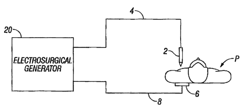
Fig. I is a schematic illustration of an electrosurgical system according to
one
embodiment of the present disclosure. The system includes an electrosurgical
instrument 2
having one or more electrodes for treating tissue of a patient P. The
instrument 2 is a monopolar
5
CA 02658407 2009-03-16
instrument including one or more active electrodes (e.g., electrosurgical
cutting probe, ablation
electrode(s), etc.). Electrosurgical RF energy is supplied to the instrument 2
by a generator 20
via an electrosurgical cable 4, which is connected to an active output
terminal, allowing the
instrument 2 to coagulate, ablate and/or otherwise treat tissue. The energy is
returned to the
generator 20 through a return electrode pad 6 via a return cable 8. The system
may include a
plurality of return electrodes pads 6 that are arranged to minimize the
chances of tissue damage
by maximizing the overall contact area with the patient P. In addition, the
generator 20 and the
return electrode 6 may be configured for monitoring so-called "tissue-to-
patient" contact to
insure that sufficient contact exists therebetween to further minimize chances
of tissue damage.
The generator 20 includes input controls (e.g., buttons, activators, switches,
touch screen,
etc.) for controlling the generator 20. In addition, the generator 20 may
include one or more
display screens for providing the user with variety of output information
(e.g., intensity settings,
treatment complete indicators, etc.). The controls allow the user to adjust
power of the RF
energy, waveform, and other parameters to achieve the desired waveform
suitable for a particular
task (e.g., coagulating, tissue sealing, intensity setting, etc.). The
instrument 2 may also include a
plurality of input controls that may be redundant with certain input controls
of the generator 20.
Placing the input controls at the instrument 2 allows for easier and faster
modification of RF
energy parameters during the surgical procedure without requiring interaction
with the generator
20.
Fig. 2 shows a schematic block diagram of the generator 20 having a controller
24, a high
voltage DC power supply 27 ("HVPS") and an RF output stage 28. The HVPS 27
provides high
voltage DC power to an RF output stage 28, which then converts high voltage DC
power into RF
energy and delivers the RF energy to the active electrode. In particular, the
RF output stage 28
6
CA 02658407 2009-03-16
generates sinusoidal waveforms of high RF energy. The RF output stage 28 is
configured to
generate a plurality of waveforms having various duty cycles, peak voltages,
crest factors, and
other suitable parameters. Certain types of waveforms are suitable for
specific electrosurgical
modes. For instance, the RF output stage 28 generates a 100% duty cycle
sinusoidal waveform
in cut mode, which is best suited for ablating, fusing and dissecting tissue,
and a 1-25% duty
cycle waveform in coagulation mode, which is best used for cauterizing tissue
to stop bleeding.
The controller 24 includes a microprocessor 25 operably connected to a memory
26,
which may be volatile type memory (e.g., RAM) and/or non-volatile type memory
(e.g., flash
media, disk media, etc.). The microprocessor 25 includes an output port that
is operably
connected to the HVPS 27 and/or RF output stage 28 that allows the
microprocessor 25 to
control the output of the generator 20 according to either open and/or closed
control loop
schemes. The microprocessor 25 may be substituted by any suitable logic
processor (e.g., control
circuit) adapted to perform the calculations discussed herein.
The generator 20 may include a sensor circuit (not explicitly shown) having
suitable
sensors for measuring a variety of tissue and energy properties (e.g., tissue
impedance, tissue
temperature, output current and/or voltage, etc.) and to provide feedback to
the controller 24
based on the measured properties. Such sensors are within the purview of those
skilled in the art.
The controller 24 then signals the HVPS 27 and/or RF output stage 28, which
then adjust DC
and/or RF power supply, respectively. The controller 24 also receives input
signals from the
input controls of the generator 20 or the instrument 2. The controller 24
utilizes the input signals
to adjust power outputted by the generator 20 and/or performs other control
functions thereon.
Referring now to Fig. 3A, the return electrode pad 6 is embodied as a variable
capacitive
pad ("VCP") 50 for providing a return path for electrosurgical current and
monitoring surface
7
CA 02658407 2009-03-16
impedance and capacitance according to the present disclosure. While the VCP
50 is depicted as
having a general rectangular shape, it is within the scope of the disclosure
for the VCP 50 to have
any suitable regular or irregular shape.
VCP 50 includes a carrier layer 53 having one or more layers, such as, a
backing layer 52,
a heat distribution layer 54, a passive cooling layer 55, and an attachment
layer 56. An active
cooling layer (not explicitly shown) or any other suitable insulating layer or
combination above
layers 53, 54, 55, and 56 may also be included.
The attachment layer 56 is disposed on a patient-contacting surface of the VCP
50 and
may be formed from an adhesive material (not explicitly shown) which may be,
but is not limited
to, a polyhesive adhesive, a Z-axis adhesive, a water-insoluble, hydrophilic,
pressure-sensitive
adhesive, or any combinations thereof, such as POLYHESIVETM adhesive
manufactured by
Valleylab, a division of Tyco Healthcare of Boulder, Colorado. The adhesive
may be conductive
or dielectric. The attachment layer 56 ensures an optimal surface contact area
between the
electrosurgical return electrode pad 6 and the patient "P," which minimizes
the risk of damage to
tissue. In another embodiment, the VCP 50 may be reusable and have a
sufficiently large surface
area so that the VCP 50 may be used without the attachment layer 56, allowing
the VCP 50 to be
cleaned and sanitized between uses.
The backing layer 52 supports a pair of split return electrodes 52a and 52b
for positioning
under a patient during electrosurgery. The backing layer 52 may be made of
cloth, cardboard,
non-woven or any suitable material. In one embodiment, the backing layer 52
may be formed
from a dielectric material such as flexible polymer materials to enhance
capacitive properties of
the VCP 50. The polymer materials may be polyimide film sold under a trademark
KAPTONTM
and polyester film, such as biaxially-oriented polyethylene terephthalate
(boPET) polyester film
8
CA 02658407 2009-03-16
sold under trademarks MYLARTM and MELINEXTM. In another embodiment the backing
layer
52 may act as an insulating layer between the pair of split return electrodes
52a and 52b and the
attachment layer 56.
The split return electrodes 52a and 52b may be made from materials that
include
aluminum, copper, mylar, metalized mylar, silver, gold, stainless steel or
other suitable
conductive material and may be of various shapes and may be arranged in
various configurations
and orientations. The split configuration of the split return electrodes 52a
and 52b create a
measurable capacitance therebetween which may be measured by generator 20 to
determine
adherence of the VCP 50 to the patient "P."
More specifically, capacitive coupling between the split return electrodes 52a
and 52b
increases upon the initial placement of the VCP 50 in contact with the patient
"P." This
capacitance corresponds to full adherence of the VCP 50 to the patient. During
the procedure,
the VCP 50 may peel from the patient "P," thereby decreasing the adherence
factor thereof. The
decrease in adherence directly affects the capacitance between the split
return electrodes 52a and
52b. Measuring the change in capacitance between the split return electrodes
52a and 52b,
therefore, provides an accurate measurement of adherence of the VCP 50 to the
patient "P." The
amount of capacitance coupling or the change in capacitance coupling then may
be used to insure
positive patient contact or to determine adequate patient coverage of the VCP
50.
With returned reference to Fig. 2, the generator 20 includes a return
electrode monitoring
("REM") system 70 having a detection circuit 22 which is coupled to the split
return electrodes
52a and 52b. The VCP 50 is in contact with the patient "P" and returns the
electrosurgical
energy to the generator 20 via the split return electrodes 52a and 52b that
are coupled to leads 41
and 42 respectively. In one embodiment, the VCP 50 may include a plurality of
pairs of split
9
CA 02658407 2009-03-16
electrode pads which are coupled to a corresponding number of leads. The leads
41 and 42 are
enclosed in a return cable 8 and are terminated at a secondary winding 44 of a
transformer 43.
The leads 41 and 42 are interconnected by capacitors 45 and 46. A return lead
48 is coupled
between the capacitors 45 and 46 and is adapted to return the electrosurgical
energy to the RF
output stage 28. The transformer 43 of the REM system 70 also includes a
primary winding 47
which is connected to the detection circuit 22.
Coinponents of the REM system 70, e.g., the transformer 43, the split return
electrodes
52a and 52b, the capacitors 45 and 46, along with the detection circuit 22
form a resonant system
which is adapted to resonate at a specific interrogation frequency from the
controller 24.
Namely, the controller 24 provides an interrogation signal at a specific
interrogation frequency to
the detection circuit 22. The detection circuit 22 then rectifies the
interrogation signal to
generate a monitor signal. The monitor signal is a constant, physiologically
benign waveform
(e.g., 140 kHz, 2 mA) which the detection circuit 22 applies to the split
return electrodes 52a and
52b. The monitor signal thereafter passes through the patient and is returned
to the circuit 22 via
the split return electrodes 52a and 52b.
The returning monitor signal is modified by the capacitance of the split
return electrodes
52a and 52b. More specifically, as the capacitance between the split return
electrodes 52a and
52b changes due to peeling of the VCP 50 from the patient, the resonance of
the detection circuit
22 with respect to other components changes as well. The change in the
resonance, in turn,
affects the change in amplitude of the monitor signal. Thus, the detection
circuit 22 determines
the magnitude of the capacitance between the split return electrodes 52a and
52b by monitoring
changes in amplitude of the monitor signal waveform. The detection circuit 22
then supplies the
capacitance measurement to the controller 24 which determines whether the
capacitance is within
CA 02658407 2009-03-16
a predetermined range. Initially, the controller 24 may determine an initial
capacitance value
corresponding to full adherence of the VCP 50. The initial capacitance value
may be used as a
baseline measurement of capacitive coupling between the split return
electrodes 52a and 52b to
determine the contact area between the patient "P" and the VCP 50. Additional
measurements
may be made after the VCP 50 is placed in contact with the patient "P" and
prior to initiating the
delivery of electrosurgical energy to patient tissue. Subsequent measurements
may be made after
commencement of electrosurgical energy delivery to determine any degradation
in contact quality
or change in a characteristic of patient contact. If the capacitance is out of
range, thereby
indicating excessive peeling of the return electrode pad 6, the generator 20
issues an alarm (e.g.,
audibly, visually, etc. via the controller 24) and/or the controller 24
adjusts the output of the
generator 20 (e.g., terminates RF supply).
Figs. 3B-3D illustrate additional embodiments of the VCP 150, 250, and 300,
respectively, and the corresponding arrangement of the split return
electrodes. More specifically,
Figs. 3B and 3C illustrate that the split return electrodes 152a, 152b and
252a, 252b may be of
various shapes and sizes designed to maximize capacitive coupling between the
VCP 150, 250
and the patient "P." In Fig. 3B, the VCP 150 includes L-shaped split return
electrodes 152a and
152b arranged in a reverse interlocking configuration to maximize contact area
with the patient
"P." In Fig. 3C, the VCP 250 includes oval shaped split return electrodes 252a
and 252b.
In another embodiment illustrated in Fig. 3D, the VCP 350 may include a
plurality of
split return electrodes arranged in an interweaving pattern. The VCP 350
includes three (3) split
return electrodes for each pair, namely, 352ai,352a2, and 352a3 and 352b1,
352a2, and 352b3.
The split return electrodes 350ai - 350a3 are coupled to the return lead 41
and the split return
electrodes 350b1 - 350b3 are coupled to the return lead 42 with each pair of
the split electrodes
11
CA 02658407 2009-03-16
being arranged in a sequential manner. Having multiple pairs of return
electrode pads within the
VCP 350 allows for fault protection in case one or more pairs of the return
electrode pads fail.
Fig. 4 illustrates a method for monitoring adherence of the VCP 50 to the
patient "P." In
step 100, the controller 24 supplies a interrogation signal to the REM system
70. The controller
24 rectifies the interrogation signal and supplies a monitor signal waveform
across the split
return electrodes 52a and 52b. In step 102, the detection circuit 22 measures
the current and
voltage of the monitor signal waveform, which are used by the controller 24 to
determine the
phase of the monitor signal waveform with respect to frequency. In step 104,
the controller 24
determines the reactance as a function of the voltage, current and phase
values with respect to
frequency. The phase of the monitor signal waveform may be determined by
sweeping the
interrogation signal across the resonance range of the REM system 70. This
allows for
correlation of phase responses with respect to multiple frequency
interrogation signals.
The reactance is used to determine the capacitance of the split return
electrodes 52a and
52b. In step 106, prior to the start of the electrosurgical procedure, the
capacitance is mapped
with respect to full adherence of the return electrode pad 6. In step 108, as
the procedure
commences, the capacitance is monitored and is used to determine the adherence
of the VPC 50.
Other methods for monitoring contact quality of the return pad to the patient
include
utilizing a sensor to communicate parameters such as capacitance, to the
electrosurgical
generator. One such method is disclosed in United States Patent Application
Serial No.
11/800,687 entitled "CAPACITIVE ELECTROSURGICAL RETURN PAD WITH CONTACT
QUALITY MONITORING," filed May 7, 2007.
While several embodiments of the disclosure have been shown in the drawings
and/or
discussed herein, it is not intended that the disclosure be limited thereto,
as it is intended that the
12
CA 02658407 2009-03-16
disclosure be as broad in scope as the art will allow and that the
specification be read likewise.
Therefore, the above description should not be construed as limiting, but
merely as
exemplifications of particular embodiments. Those skilled in the art will
envision other
modifications within the scope and spirit of the claims appended hereto.
13