Note: Descriptions are shown in the official language in which they were submitted.
CA 02658934 2009-01-26
WO 2008/013763 PCT/US2007/016528
1
PACKED DEIVIINERALIZED CANCELLOUS TISSUE FORMS FOR DISC NUCLEUS
AUGMENTATION, RESTORATION, OR REPLACEMENT AND METHODS OF
IlVII'LANTATION
RELATED APPLICATIONS
This application claims priority from Provisional Application Number
60/832,956 filed
July 25, 2006.
FIELD OF THE INWNTION
The present invention generally relates to tissue forms used for augmentation,
restoration or replacement of intervertebral discs.
A healthy intervertebral disc facilitates motion between pairs of vertebrae
while
absorbing and distributing compression forces and torque forces. The disc is
composed of two
parts; naraely a tough outer ring (the annulus fibrosis) which holds and
stabilizes a soft cenftl
core material (the nucleus pulposus) that bears the majority of the load
forces.
The tissue form of the present invention is dense cancellous tissue be derived
from
proximal and distal femur, proximal and distal tibia, talus, calcaneus,
proximal humerus,
patella, or ilium that is first fully demineralized, cleaned, treated such
that the bone is non-
osteoinductive, and then processed into small uniform geometries that may be
either cuboidal,
disc-shaped ("mini-discs"), or spherical. The relative sizes of these shapes
are on the order of
1.0 mm to 4.0 mm in either length or diameter. The tissue forms, following
demineralization,
are compressible such that individual units can be packed tightly together in
a confined space
and when packed together behave mechanically as one coherent unit while
providing suitable
load-bearing properties in the disc nucleus. When confined in the disc space
under normal
loading conditions, this plurality of shaped units functions as an elastic
body, which is
deformable, yet resilient under dynamic loading and has intrinsic shape-memory
properties.
CA 02658934 2009-01-26
WO 2008/013763 PCT/US2007/016528
2
BACKGROUND OF THE IlVVENTION
Minimally invasive surgery that is aimed to treat degenerative disc disease
and. preserve
motion in the spine is currently under investigation. Since the onset of the
degenerative cascade
in the disc is typically associated with dehydration and volume loss in the
nucleus pulposus, a
potential early intervention step may involve adding mechanically suitable
material to the
nucleus pulposus at the first observable signs of discomfort and loss of disc
height. This
procedure would restore nuclear volume and pressure against the inner wall of
the annulus
fibrosus. In certain embodiments, a degree of decompression or "lift" between
the adjacent
vertebrae may be possible with this technique. In effect, the result would be
the "re-inflation"
of the annulus fibrosus, the annular "tire". Desirable outcomes of the
procedure would be
motion preservation, pain relief, and maintenance or restoration of disc
height. I:.ong-term re-
modeling of the biological allograft-based implant into fibrous tissue or disc-
like tissue would
also provide favorable clinical outcomes.
At present, there are no nucleus pulposus replacement devices or augmentation
technologies available for clinical usage in the United States. The Prosthetic
Disc Nucleus
(PDN), which is manufactured by Raymedica, was the first implant designed for
nucleus
replacement with the intention of attempting to restore natural mechanics in
the spine. This
implant is an acrylic-based hydrogel encased in a polyethylene jacket. While
this technology
has been implanted in over 3000 patients in Europe, significant issues
regarding implant
migration and implant hardening have been encountered. Other drawbacks in the
design of this
implant include the requirement of a substantial annulotomy and total
nucleotomy as well as the
inability of the implant to fill the entire nuclear ca.vity. In addition, the
limited ability of the
implant to swell inside the disc nucleus leads to high extrusion rates and
inadequate load
transfer of compressive forces in the disc nucleus to tensile forces on the
annulus fibrosus.
Generally speaking artificial disc replacements falls into two general
categories, total
disc replacement and nuclear displacement. Total disc replacement devices have
a number of
CA 02658934 2009-01-26
WO 2008/013763 PCT/US2007/016528
3
problems; namely that they are large and non-compressible, require the removal
of a large
portion of the annulus and require a highly invasive surgical approach in
order to be implanted.
If these disc replacement devices do not remain firmly attached to the
vertebral bodies, these
implants can extrude or migrate from their intended position, cause
significant complications
and are very difficult to revise. The second category of disc replacement is
nuclear replacement
which is a form of partial disc replacement. Various types of methods and
devices have been
used to attempt to accomplish successful disc replacement.
United States Patent Number 6,652,593 issued November 25, 2003 is directed
toward an
osteoinductive implant comprising demineralized cancellous bone, which
comprises a non-
particulate bone. A unitary bone block is compressed into a smaller
configuration such as a
pellet and then hardened via drying. Upon re-hydration, the pellet will expand
and assume its
original shape inside a cavity. The implant is capable of being compressed and
hardened into a
first shape and then capable of expanding into a second shape larger than the
first shape when
re-softened and permitted to expand. The `593 implant is designed to be
supplied either in
geometries that fill correspondingly sized voids or in compressed initial
geometries that are
used to expand and fill any given shape smaller than or equal to their
expanded size.
United States Patent Publication 2006/0030948 filed September 21, 2005 is
directed
toward an osteogenic implant having a predetermined shaped formed of an
aggregate of
different sized elongate (possessing a high median length to median thickness
ratio) bone
particles.
United States Patent Publication Number 2004/0054414 filed on September 18,
2002 is
directed toward a method of augmenting an intervertebral disc nucleus by the
surgical addition
of a particulate collagen-based material. The collagen-based material, having
a mean particle
size ranging from 0.05 mm to 5 mm, may be injected in either a wet or dry
state and may be
supplemented with growth factors, proteoglycans, and cells. The `414
publication notes the use
of demineralized bone matrix particles with sizes ranging from between 0.05 mm
and 3 mm and
CA 02658934 2009-01-26
WO 2008/013763 PCT/US2007/016528
4
the use of elongated cylindrical plugs. The plugs are described to be
dehydrated and
compressed in the radial direction and are inserted into delivery cannula for
delivery into the
disc space. The cylindrical plugs are delivered via extrusion through a
cannula, and expand
upon exiting the cannula by re-hydrating in the disc space. Examples 6 and 7
refer to the design
and implementation of cylindrical plugs, which can be fabricated from solid,
porous, or fibrous
collagen.
Additional continuing United States Published Patent Applications Numbers
2005/0197707 filed April 25, 2005 and 2005/0119754 filed January 6, 2005 are
based on the
`414 publication. The `707 publication is directed toward the use of small
particles of
particulate fascia lata, particulate disc annulus material, annulus fibrosis,
demineralized bone
matrix and collagen which are added to the nucleus of an intervertebral disc.
The `754
publication covers a method of augmenting an intervertebral disc nucleus by
adding a plurality
of collagen-rich tissue particles having a mean particle size between 0.25 and
1.0mm to the disc
plus a biologically active substance that promotes heating, repair or
regeneration of the disc.
This biologically active substance is further defined to be stem cells,
hyaluronic acid, or growth
factors while the collagen material is stated to be potentially allograft
tissue. Radio contrast
material may be added to enhance imaging of the injected material.
Another United States Patent Publication Number 2005/0055094 filed November 5,
2003 discloses a system for replacing a disc nucleus involving an injection
tube, a volume of
fibrous tissue material to fill a nuclear cavity, and an insertion device for
dispensing the tissue
promoting material into the disc. Suitable fibrous tissue promoting material
is listed as fascia,
natural and/or man made polymeric fiber, fibrous tissue inducers such as talc,
pharmaceuticals
and/or minerals and fibrous tissue morphogenic protein.
U.S. Patent Number 5,571,189 issued November 5, 1996 describes an expandable
bag
filled with biological tissue for spinal fusion but does not show motion
preservation in the
spine.
CA 02658934 2009-01-26
WO 2008/013763 PCT/US2007/016528
The present inventive disc nucleus implant is a combina#ion of multiple units
of
demineralized cancellous tissue treated to be non-osteoinductive that are
designed to.be small
uniform geometric shapes which have the ability to pack together and act
mechanically as a
single unit under the compression of packing and not to comprise a non-
particulate portion of a
bone. The inventive tissue forms are compressed upon delivery into a cavity,
but only to fit into
the delivery device and not into a defined shape. In addition, the inventive
tissue forms do not
regain their original dimensions following the completion of the implantation
procedure. In
fact, the appropriate mechanical properties are only achieved if the mass of
units is under
compression and behaving as a coherent load-bearing material. The plurality of
units that
constitute the inventive implant, when taken together in an uncompressed
state, have a
geometry that is substantially larger than the cavity into which they are
placed. Thus, the
inventive implant takes on a smaller size in the confined space into which it
is placed. Finally,
the inventive allograft tissue form is treated to be non-osteoinductive, which
achieves the
desired outcome of motion preservation in the spine versus spinal fusion.
The noted prior art publications cite examples of various allograft tissues
for usage such
as demineralized bone matrix, disc annulus, fascia, ligaments, tendons, skin,
or other connective
tissues. The inventive tissue implant would not be provided in a dehydrated
state and will be
compressed axially inside the delivery tube rather than radially.
Advantages of the present inventive approach in comparison to other techniques
include
its ability to be entirely performed in a minimally invasive manner, total
nucleotomy is not
required and the implant size is adjustable by the volume of material that is
added into the
pouch. If desired an expandable pouch that is intended to hold the shaped
units can be inserted
into the disc nucleus through a small diameter hole and it will be enlarged
with implant material
to a size considerably larger than the insertion hole allowing the implant
dimensions to conform
to the existing cavity of the disc nucleus, with the porous pouch preventing
the escape of
CA 02658934 2009-01-26
WO 2008/013763 PCT/US2007/016528
6
material from the nuclear space while allowing the free transfer of fluid
across its surface along
with potential tissue ingrowth.
S~Y OF THE INVENTION
The implantable allograft tissue form represents unifonn demineralized
cancellous
tissue units treated to be non-osteoinductive placed under compression which
will allow them to
pack closely in the confined space inside an annulus. Following implantation,
when the units
are tightly pressed together, the collective volume of implanted material can
play a similar
biomechanical role inside the disc as native nucleus pulposus. The implant
represents a motion
preserving alternative in the treatment of degenerative disc disease.
The steps of the surgical technique described here represent a minimally
invasive
method for replacing or augmenting a spinal disc nucleus and includes the
complete or partial
removal of nucleus material, sizing of the resulting cavity, inserting an
expandable, porous
pouch into the nucleus through either an existing annular tear or through an
annulotomy, filling
the pouch with compressed small fully demineralized, non-osteoinductive
cancellous bone
tissue forms, and closing the pouch.
Another object of the invention is the usage of a biologic nuclear implant
material which
can experience tissue ingrowth and reorganization once implanted within the
disc space.
Alternatively, the biological and structural nature of the demineralized
cancellous bone allows it
to be a potential scaffold that can be potentially supplemented with cells
and/or growth factors,
which may induce matrix remodeling and the subsequent regeneration of nucleus-
like tissue
inside the disc following implantation.
These and other objects, advantages, and novel features of the present
invention will
become apparent when coinsidered with the teachings contained in the detailed
disclosure which
along with the accompanying drawings constitute a part of this specification
and illustrate
CA 02658934 2009-01-26
WO 2008/013763 PCT/US2007/016528
7
embodiments of the invention which together with the description serve to
explain the
principles of the invention.
BRIEF DESCRIPTION OF THE DRAWINGS
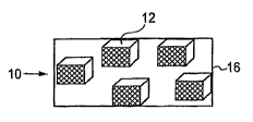
Figure 1 shows an enlarged perspective view of a plurality of cancellous cube
shaped
units of the present invention in a schematic container;
Figure 2 shows an enlarged perspective view of a plurality of cancellous disc
shaped
units of the present invention in a schematic container;
Figure 3 shows an enlarged perspective view of a plurality of cancellous
cubes, discs
and sphere shaped units of the present invention in a schematic container;
Figure 4 shows a perspective view of a filled porous mesh pouch with
demineralized
cancellous tissue units placed in the disc nucleus following the creation of a
lateral portal
through the disc annulus;
Figure 5 is a chart showing the angular motion in flexion and extension of
human
cadaveric motion segments (a) for an intact disc, (b) the disc following
nucleotomony, (c) the
disc directly following implantation of the present invention and (d) the
implanted disc after
allowing 30 minutes for additional hydration;
Figure 6 is a chart showing the deviation of the motion segment flexibility in
flexion-
extension from the intact disc;
Figure 7 is a chart showing the angular motion in left and right lateral
bending of human
cadaveric motion segments (a) for the intact disc, (b) the disc following
nucleotomy, (c) the disc
directly following implantation of the present invention, and (d) the
implanted disc after
allowing 30 minutes for additional hydration; and
Figure 8 is a chart showing the deviation of the motion segment flexibility in
lateral
bending from the intact disc.
CA 02658934 2009-01-26
WO 2008/013763 PCT/US2007/016528
8
DETAILED DESCRLP'.rION OF THE INVENTION
The preferred embodiment and best mode of the invention is seen in Figures l
to 4. The
present invention is directed toward an implant that formed of a plurality of
small, substantially
demineralized cancellous bone shapes 10 that can be loaded and packed into a
cannula or
hollow rod and then inserted by packing the small shaped bone units into a
disc nucleus in a
non-dehydrated state. This packed material is to be utilized to augment,
restore, or replace a
disc nucleus. In a preferred embodiment, the tissue forms described within are
to be delivered
into an expandable porous mesh pouch 16 that has been pre-placed in the disc
nucleus 20
through a small lateral opening 22 in the disc annulus such that the allograft
material will be
contained and not extrude out of the nucleus through an annular defect. The
deformable nature
of wet, demineralized cancellous bone will allow the tissue forms to pack
tightly together in a
confined space during delivery under sufficient pressure. A suitable amount of
tissue is inserted
so that the nuclear cavity is tightly filled and the resulting conglomerate
implant acts as a single
coherent mass under mechanical loading. This invention is implemented for
patients with
degenerative disc disease, particularly those in earlier stages of
degeneration who still possess a
competent annulus fibrosus.
Cancellous bone may be derived from proximal or distal femur, proximal or
distal tibia,
proxirnal humerus, talus, calceneus, patella, or iliium. Cancellous tissue is
first processed into
sheets or blocks, which preferably range in thickness of about 2m to 3 mm,
although sheets of
about 1.0 mm to about 4.0 mm can be used. Blood and lipids are flushed from
the tissue using
high pressure water. The cancellous tissue is then substantially demineralized
in dilute acid
until the bone contains less than 0.1% wtlwt residual calcium.
Demineralization of the
cancellous bone creates a material that is spongy and pliable in nature, yet
still possesses elastic
properties and shape memory following deformation.
CA 02658934 2009-01-26
WO 2008/013763 PCT/US2007/016528
9
Following decalcification, the cancellous tissue is cleaned and treated via
chemical or
thermal treatment or by high energy irradiation so that the cancellous tissue
is non-
osteoinductive.
In a preferred embodiment, the cancellous tissue is treated with hydrogen
peroxide for at
least 1 hour in order to further clean the tissue and to achieve a non-
osteoinductive material.
The tissue.is then soaked in ethanol as an additional cleaning step. After
these steps, the tissue
is soaked in phosphate buffered saline (PBS) in order to restore the pH of the
tissue within the
range of 6.6 to 7.4. After these treatment steps, small units of cancellous
tissue are fabricated
from the cancellous sheets or blocks. The cancellous tissue form units have a
defined shape
that may be cuboidal, spherical, or discoid in nature and are loaded into
filler tubes prior to
implantation. The cancellous shapes may have a single dimension ranging from
1.0mm to 4
mm and preferably are between 2mm to 3mm.
In the most preferred embodiment, the fully demineralized cancellous sheets
are then cut
into cube shaped tissue forms 12 with a side dimension of 2mm to 3 mm using a
chip press
cutting device. The cancellous cubes are then lyophilized to less than 6%
residual moisture.
Following the dehydration step, a specific amount of dry cancellous cubes is
weighted out
ranging between 0.4 to 1.2g. This amount of dry cancellous tissue is hydrated
in excess water
or saline and then loaded into a small diameter tube (2mm to 4mm in inner
diameter) that is to
be used to fill the disc nucleus during the surgical procedure.
In Figure 2, disc shaped tissue fonns 13 are formed using a mechanical press
that acts as
a multiple hole-punch. A preferred disc size is 2-3 mm in diameter and 2-3 mm
in height. In
another unit form spheres 14 are formed using a cutting device. A preferred
sphere size is
about 2-3mm in diameter. Figure 3 shows multiple unit configurations of
cuboidal, spherical or
discoid in shape used together. All of the shaped units are lyophilized to
less than 6% residual
moisture weighed in a dehydrated state, and then hydrated in excess water or
saline before
loading into an insertion tube or container.
CA 02658934 2009-01-26
WO 2008/013763 PCT/US2007/016528
A sufficient amount of cancellous bone is added to the expandable mesh pouch
container 16 such that the volume of the nucleus is restored when the implant
is packed so that
it conforms to the shape of the nuclear cavity. Due to the design of the
implant, the amount of
filling material loaded into the bag may thus be customized for the specific
size of the target
nuclear cavity of the patient. In certain embodiments, the pouch may be filled
with cancellous
bone until it expands to a volume greater than that of the existing nuclear
cavity, thereby
providing a degree of decompression or "lift" between the two adjacent
vertebrae. After the
pouch is tightly packed with the shaped demineralized cancellous bone shaped
units, the
implant is designed to possess mechanical properties that withstand the
compressive loads in
the spine and facilitate load transfer from the nucleus to the annulus. Once
filling is complete,
pouches will be closed or sealed to prevent the escape of any cancellous
tissue.
As shown in Figures 5 and 6, disc-shaped fully demineralized allograft
cancellous bone
units (3mm diameter x 3mm height were loaded into an expandable polyester mesh
in situ at a
packing density ranging from between 1.50 to 1.60 g/cc based on the hydrated
mass of the
tissue and the measured cavity size of the denucleated disc. The figures
represent the acute
restoration of stability to the spinal motion segment following nucleotomy and
the implantation
of the inventive implant device. Testing was performed on each cadaveric
motion segment
(either L2-L3 or L4-L5) at four different stages: the intact disc, the disc
following nucleotomy,
the denucleated disc directly following the implantation of the inventive
device and the
implanted disc after allowing for 30 minutes of hydration in saline. Figure 5
depicts the angular
motion in flexion and extension of human cadaveric motion segments over a
constant range of
bending moments. Figure 6 represents the deviation of the motion segment
flexibility in
flexion-extension from the intact disc. Figure 7 represents the angular motion
in left and right
lateral bending of human cadaveric motion segments over a constant range of
bending
moments: (a) for the intact disc, (b) the disc following nucleotomy, (c) the
disc directly
following implantation of the present invention, and (d) the implanted disc
after allowing 30
CA 02658934 2009-01-26
WO 2008/013763 PCT/US2007/016528
11
minutes for additional hydratioin. The data shows the biomechanical
instability introduced to
the discs following the nucleotomy and demonstrates the recovery of normal
range of motion
following the implantation of the inventive implant device. Figure 8 is a
chart showing the
deviation of the motion segment flexibility in lateral bending from the intact
disc. Error bars on
all figures indicated the standard deviation from the mean.
Additional embodiments of this invention may include the supplementation of
the
cancellous bone with synthetic material that is of similar physical dimensions
as the implanted
cancellous tissue forms. Such synthetics may include polymeric hydrogels,
biodegradable
polymers, rubbers, or other materials that are elastic in nature and capable
of being packed
together in a similar fashion to the cancellous tissue.
Other additional embodiments of this invention may include the addition of
cells and/or
biological agents to the cancellous bone either prior to implantation or post-
implantation.
Transplanted cells may include those derived from bone marrow, other
pluripotent stem cells,
chondrocytes, and nucleus pulposus cells. Bioactive molecules may include
viral particles,
plasmids, hormones, extracellular matrix proteins, platelet rich plasma, or
growth factors -such
as those in the TGF-8, FGF, VEGF, IGF, and BMP families. Another embodiment of
the
invention may include the addition of a radio-opaque marker to the cancellous
tissue in order to
make the implant visible during surgery. The radio-opaque marker may be
derived from
beryllium copper, brass, bronze, carbon steel, clad metals, copper, kovar,
molybdenum, nickel,
niobium, stainless steel, tantalum, titanium, zirconium, or other radio-opaque
material. Other
suitable materials may include barium, platinum, platinum iridium, gold and
iodine-containing
compounds.
This invention also utilizes a method of treating a degenerative spinal disc
by replacing
or augmenting the disc nucleus with allograft tissue through a minimally
invasive approach. In
a preferred embodiment, the allograft tissue form comprises small uniformly
shaped fully
demineralized, non-osteoinductive cancellous bone units. The target disc will
be accessed and
CA 02658934 2009-01-26
WO 2008/013763 PCT/US2007/016528
12
nuclear material will be removed via microdiscectomy or minimally invasive
nucleotomy.
Following this step, the resulting nuclear cavity is sized and an expandable,
porous pouch is
inserted into the disc nucleus via an existing annular tear or a small
annulotomy. The pouches
are initially empty and in a collapsed state such that it can be passed
through a small diameter
portal in the disc annulus (-3mm-4mm). This mesh bag may be made from
synthetic materials
such as polyester or biological material such as allograft bone, dermis, or
fascia, hyaluronic
acid, collagen, or other structural protein. In a preferred embodiment, a
woven fabric mesh is
utilized as the implantable pouch, with a pore size that is sufficiently small
such that allograft
material units do not extrude through the mesh openings. This containment
device may also be
sewn such that it expands into a disc nucleus-like shape upon addition of
implant material and
may have a radiographic marker in order to track its location following
implantation. The
porous nature of the pouch may allow the transfer of fluid from the
surrounding disc tissue to
the implant material and vice-versa. The porosity and mesh size of the pouch
may also be
critical for obtaining an appropriate biological response to the allograft
material contained
within it. By allowing cellular infiltration and fluid exchange, it may be
possible for tissue
remodeling or fibrous tissue formation to occur inside the implanted mesh
pouch within the
disc.
After the porous pouch has been inserted and positioned inside the disc, a
plurality of
small, demineralized non-osteoinductive cancellous bone units are passed into
the bag through a
hollow rod until the bag is appropriately filled. In a preferred embodiment,
the hollow rod has
an internal diameter between 3mm to 4 mm, and is utilized in combination with
cancellous
units that are cube shaped with 2mm to 3mm sides or disc-shaped with a
diameter of 2mm to 3
mm and a height of 2mm to 3 mm or spherical with a diameter of 2mm to 3 mm.
The
cancellous tissue forms may have a defined shape that may be spherical,
discoid, or cuboidal in
nature and may be loaded into filler tubes prior to implantation. The
cancellous tissue forms
may also have a single dimension of no more than 5 mm and no less than 1.0 mm
and will be
CA 02658934 2009-01-26
WO 2008/013763 PCT/US2007/016528
13
designed to pack tightly under pressure. It is recognized the size of the
individual units will be
considerably smaller than the diameter of the filler bag once it has been
expanded.
In operation, a small nucleotomy is created in the disc annulus by first
making an
incision in the disc and then expanding the same using dilators of increasing
size. The nucleus
is then mechanically removed while avoiding damage to the inner annulus or the
cartilaginous
end plates. Following the nucleotomy, an inflatable balloon is inserted in the
disc nucleus and
the nucleus is filled with radio contrast fluid to a specific pressure between
30 to 60psi such that
the nuclear cavity is visible under fluoroscopy. This step allows
visualization of the cavity
created by the nucleotomy and also provides a measurement of the cavity
volume, which will
be used to select the mesh size and determine the amount of fill material
needed for the implant.
After sizing, the porous mesh is inserted through the portal in the disc
annulus. In order to
ensure that the mesh is not restricted from deploying properly, an inflatable
balloon is placed
into the empty mesh in situ and the balloon is again filled with radio
contrast material.
Subsequently, the balloon is removed from the mesh and demineralized non-
osteoinductive
cancellous tissue in the form of cubes, discs or spheres is added to the mesh
by extruding the
filler implant material that has been pre-loaded in small diameter tubes.
Based on the empty
cavity volume of the disc nucleus, the mesh will be filled to a packing
density of 0.3 to 0.9g/cc
where the weight of the tissue is based upon its dry weight. After the filling
step, the mesh is
released from its holder tube and its opening is tied off to prevent migration
of the cancellous
tissue from the disc space. In another embodiment of the invention, a
degenerated or diseased
intervertebral disc is treated with the above noted steps where after the step
of removing a
portion of or the entire disc nucleus via mechanical disruption, at least one
region of the
vertebral end plates is removed or disrupted.
The principles, preferred embodiments and modes of operation of the present
invention
have been described in the foregoing specification. However; the invention
should not be
construed as limited to the particular embodiments which have been described
above. Instead,
CA 02658934 2009-01-26
WO 2008/013763 PCT/US2007/016528
14
the embodiments described here should be regarded as illustrative rather than
restrictive.
Variations and changes may be made by others without departing from the scope
of the present
inventions defined by the following claims: