Note: Descriptions are shown in the official language in which they were submitted.
CA 02659022 2009-01-26
WO 2008/016881 PCT/US2007/074746
LEAD AND METIIODS FOR
BRAIN MONITORING AND MODULATION
BACKGROUND OF THE INVENTION
100011 1. Field of the Invention. This irvention i-elates generally to medical
appai-atus
and methods, and moi-e specifically to leads used to electrically and/or
chemically modulate
and monitoi- tissues of the brain.
100021 Implanting medical devices such as probes oi- leads within the cranium
is an
increasingly important approach for ti-eatment of diseases such as Parkinson's
Disease,
essential tremor and dystonia. Implants may be used to treat a wide ari-ay of
disorders, such
as depression, epilepsy, dystonia, obsessive compulsive disordei-, obesity and
chronic pain.
Most of these devices interact with the bi-ain by applying cun-ent through an
electi-ode. In
addition, infusion of drugs through a chronically implanted lead has been pi-
oposed in the
medical literature either as a primai-y treatment, or as an adjunctive
treatment to electrical
stimulation, in patients with Alzheimer's and Pai-kinson's Diseases, ainong
others.
100031 Current implantable probes are typically configured as small diametei-
cylinders or
tubes, with several circumferential metal rings near the distal tip, and an
electrically passive
central axial lumen. The metal rings are used to provide electrical
stimulation, while the
central axial lumen can be used to deliver the probe over a guidewire or
stylet during the
implantation procedure.
100041 In most treatment protocols, a sequence of electrical pulses is applied
to one or more
conducting rings on the probe. Typically monopolar or bipolar stimulation of
the conducting
rings is used. In monopolar stimulation, a single circumferential ring is
stimulated with a
charge balanced biphasic electrical pulse, with a return path for the current
at a remote site,
such as a battery pack or conti-ol module. In bipolar stimulation, a
combination of rings are
stimulated with charge balanced biphasic electrical pulses of opposite
polarity. Stimulation
of conducting rings produce a field of action which is more or less synimetric
about the
probe, with some asymmetries arising because of anisotropy in the electrical
properties of the
adjacent neural or brain tissue.
100051 A symmetrical electrical field about the probe axis is not always
desirable. For
example, when the probe is not implanted at the center of the modulation
target or when the
I
CA 02659022 2009-01-26
WO 2008/016881 PCT/US2007/074746
brain target is asymmctric oi- irregular in shape. Additionally, there are
otten neui-onal
domains ncai- the targeted zonc. which should not be modulated. Moclulating
non-target
zones can lead to undesirable side effects, including sornatic sensation.
involuntary
movement and impaii-ed vision, among others.
100061 It is desii-able to not only modulate brain activity, but also to
monitor it along with
physiological and pathophysiological states. Monitoi-ing obtains information
on neuronal
activity neai- the stimulation sites, including fielcl potentials and
extracellularly recol-ded
action potentials. Such potentials may be observed on an ongoing basis, in the
course of
electrical stimulation for treatment, and in the course of special stimulation
and response
experiments designed to assess an individual's brain and the brain to electi-
ode intei-face.
Information obtained from monitoring at intei-vals may be used to conti-ol and
adjust
treatment on an ongoing. day-to-day basis by a patient, or in follow up visits
to a health
professional. Information obtained fi-om monitoi-ing may also be used to
dynamically adjust
the treatment by an automated control system or control algoi-ithm. and by
updating the
parameters of a controller.
100071 Monitoring at intei-vals can be used to track changes in the brain
response to
stimulation as a function of stimulus magnitude. Clinical decisions can be
based upon
estimated parameters, such as the threshold stimulus level which barely
generates a response,
and the stimulus level which just saturates the observed response. The shape
of the stimulus
response function, for example whether it is concave up, concave down, or
linear, may also
inform adjustments to treatment. The dynamic range from threshold to
saturation measured
near the stimulation site may directly correspond to the dynamic range of
clinical effect, or it
may be correlated with it. In either case, the locally measured dynamic range
gives
inforination which can accelerate the initial fitting and guide ongoing
adjustments in
treatment protocol. Brain plasticity in response to treatment may be tracked
by changes in
the dynamic range.
100081 Consider the application of monitoring at intervals to the treatment of
Parkinson's
disease. It is well known that the beneficial effects of electrical
stimulation to Parkinson's
patients do not appear for several minutes or hours after the stimulation
protocol is initiated.
If the protocol is discontinued during sleep and resumed at waking, the
beneficial effects of
treatment may not appear again for many hours. Monitoring at intervals offers
the
opportunity to track changes in the response to stimulation, so that
stimulation can be applied
2
CA 02659022 2009-01-26
WO 2008/016881 PCT/US2007/074746
dLU-ing onc pi-otocol in order to br-ing about the beneficial effects, and
under another moi-c
conservative protocol in oi-der to just maintain the beneficial effects. Such
a strategy would
conserve battery power, and could also reduce side effects.
100091 By monitoring fi-om moment to moment, a modulatoi-y treatment can be
clynam-
ically synchronized with natural brain i-hythms upon an observed pathological
or normal
physiological state. or conti-olled by an automatic control system or control
algorithm.
100101 Most procedures curi-ently performed monitor patient motions,
behaviors, or brain
activity at a site remote from the site of an electi-ically stimulating pi-
obe, and this information
is used to adjust brain stimulation parameters. Parameters are adjusted on a
short time scale,
to generate a desired effect and minimize side effects, and on a longer- time
scale, to account
for brain plasticity. Brain plasticity is due to an adaptive response by the
brain to an
intervention and it is well known that ongoing responses by the brain to an
intervention such
as modulating therapy often differ from the initial response. Useful
information may also be
obtained by monitoring electrical potentials near the site of electrical
stimulation and
therefore it would be desirable to monitor brain activity at the locus of
electrical stimulation.
Monitoring allows the course of the disease and healing processes to be
evaluated along with
the prognosis foi- various treatinent options.
100111 For these reasons as well as others, it would be desirable to provide
iinproved
probes for modulating and monitoring tissues such as the brain. It would be
particularly
desirable to provide an efficient design for generating a directed electrical
field that may be
steered towards the intended target, and/or away from other brain areas. It is
also desirable to
provide a probe with an efficient number and size of electrodes as well as
connector leads,
that integrates both electrical recording and stimulating or modulating
capabilities, where the
infonnation from recordings is obtained close to the treatment site and can be
used to define
the stimulating protocol. The protocol can then be adapted either statically
or dynamically
and as the disease state changes, the therapy can also be adjusted. Recording
and monitoring
of brain electrical activity is also used to detennine when the stimulation
protocol is applied
or whether it should be reserved for times when it is more effective, thereby
helping to
conserve power.
[00121 2. Description of Background Art. Prior patents and publications
describing
brain modulating probes and methods include: U.S. Publication Nos.
2006/0047325;
3
CA 02659022 2009-01-26
WO 2008/016881 PCT/US2007/074746
2006/0004422; 2005%0015130: 2004-0039434 and U.S. Patent Nos. 7.051.419;
7,047,052;
7,006,872: 6,094.598; 6.038.480: 6,01 1,996; 5,343,144; and 5,716,377.
BRIEF SUMMARY OF THE INVENTION
100131 The invention generally pi-ovides an implantable probe or lead capable
of
modulating or stimulating tissue and measuring and i-ecording local tissue r-
esponses as a
r-esult of the modulation. The teims "modulating" and "stimulating" are used
interchangeably
in order to refer to providing a stimulus that incites or suppresses activity
in the tissue. The
terms "probe" and "lead" ai-e also used interchangeably in order to i-efer to
any device that
inay be used to modulate the tissue and/or measure and recoi-d local tissue
responses.
Modulation of the tissue may include electrical and/oi- chemical stimulation
of the tissue, as
well as suppression of tissue activity. Measuring and recording tissue i-
esponses often entails
measuring local tissue potentials in response to the stimulation but could
also include
measuring and recording endogenous tissue potentials as well as chemical
activity in the
tissue. Often, the probe is used in tissues of the brain, typically being
implanted into deep
brain structui-es, or into the cerebrum oi- cerebellum.
(0014] The invention also provides methods where thei-apeutic modulation may
be directed
within tissues such as neural structures with improved effectiveness and
minimal undesirable
side effects. The present invention also includes methods to electrically
and/or chemically
monitor tissue activity so that the therapeutic intervention inay be modified
to improve its
effectiveness, or to conserve limited resources such as reagents or electrical
charge.
100151 The probe possesses electrodes for stimulating tissue such as the
brain, and/or for
recording tissue activity by measuring local tissue potentials. The
stimulating electrodes are
affanged so that they can be activated individually, or in combination. They
may
alternatively be activated in simultaneous or sequential coordination in order
to shape the
volume of stimulated brain tissue and regulate the magnitude and timing of
activity in a
stimulated brain. The probe often has a plurality annular shaped stimulating
electrodes
disposed axially along the probe. For the most efficient use of the probe,
each annular shaped
electi-ode has three independent stimulation sites disposed thereon, although
a greater number
of stimulation sites per annular region may be employed. By "independent
stimulation sites,"
it is meant that the electrode is separable into three isolated regions,
typically disposed in
4
CA 02659022 2009-01-26
WO 2008/016881 PCT/US2007/074746
120 ares of the annulai- clectrode, whcre cach region may be independently
enei-gizcd from
an external or othcr cnergy sourcc.
100161 In a first aspect of the invention, an appai-atus for stimulating and
monitoring bi-ain
tissue compi-ises an elongate membei- having proxiinal and distal ends, and a
plurality of
annular stimulating electrodes axially arranged along the elongate inember,
disposed near the
distal end, but may also be disposeci at othei- axial positions. Portions of
the elongate member
may be flexible, often neai- the proximal end and portions may also be rigid
near the distal
end. The annular stimulating electrodes are adapted to pass curi-ent into
tissue and at least
one of the annular stimulating electi-odes has at least thi-ee independent
stimulation regions or
points. The apparatus will usually but not necessai-ily further- compi-ise a
plurality of
measuring or recording electrodes disposed ad,jacent to the stimulating
electrodes and some
of the recording electrodes inay be arranged betwcen annular stiniulating
electi-odes and the
--ecording electrodes are adapted to measure local tissue potentials. The
recording electrodes
may be circumferentially disposed about the elongate membei- and sometimes
have a circular
shaped surface. There may also be a sui-face foi- recording and/or stimulating
at or near the
tip of the apparatus. The apparatus will usually include a plurality of
conductors which are
coupled with at least some of the annular stimulating and annular i-ecording
electrodes, and
an optional multiple contact connecting terminal inay be disposed near the
proximal end of
the elongate ineinber and that is coupled with the conductors. The apparatus
may have one
conductor per stimulating and/or one conductor per recording region. Often the
apparatus
also has a lumen that is axially disposed between the proximal and distal ends
and sometimes
the lumen is adapted to receive a guidewire or stylet.
100171 Often the tissue being treated is brain tissue, although other tissues
may also be
treated by the method and system of the present invention. Additionally, the
apparatus often
includes a lumen axially disposed along or within the elongate member. In some
cases the
lumen is adapted to receive a guidewire or stylet, which passes through the
lumen from a port
near the distal end of the elongate meinber. In other cases, one or more ports
in
coininunication with the lumen are disposed near the distal end of the
elongate member and
are adapted to deliver a therapeutic agent or other substance to the tissue
and/or to receive a
chemical substance from the tissue. In some cases, the ports are disposed
between the
annular stimulating electrodes and in other cases, at least one of the ports
is disposed at the
distal end of the elongate member. In some embodiments, the ports may comprise
a gating
member adapted to permit selective enablement of the ports. The gating member
may be a
CA 02659022 2009-01-26
WO 2008/016881 PCT/US2007/074746
semi-pei-meable membi-ane and may be chcmically controlled such as \N"hen the
gating
member is a chemically reactive hydrogel.
100181 In some einbodiments, an additional stimulating electi-ocie may be
disposeci in the
lumen, and often this additional electrode is a wire. Thei-apeutic agents may
also be delivered
thi-ough the lumen. In other embodiments, an additional stimulating electrode
may be placed
at the distal end of the clongate member and this electrode is also capable of
passing current
into the tissue. A therapeutic agent may also be used with this oi- other
embodiments
desci-ibed herein. Often, the conductors are helically wound along the
elongate member. A
fii-st group of conductors may be coupled with the stimulating electrodes, and
a second group
of conductors may be coupled with the recording electrodes. The first gi-oup
of conductors
may be wound in a helix having a first pitch, and the second gi-oup of
conductors may be
wound in a helix having a second pitch. In some cases, the first pitch is
different than the
second pitch. Conductors ai-e often comprised of stainless steel, MP35N or
tungsten because
of their biocompatibility and compatibility with MRI imaging systems, although
other
materials such as platinum-iridium alloy ar-e possible. Typically, the
plurality of annular-
stimulating electi-odes, as well as the i-ecording electrodes may also be
compatible with
magnetic resonance imaging (MRI). An object is compatible with MRI if it does
not
significantly distort image quality, cause tissue damage with heating and does
not move in
the presence of a magnetic field.
10019] In a second aspect of the present invention, ainethod of treating
tissue comprises
implanting a probe in the tissue. The probe may be coinpatible with magnetic
resonance
imaging and usually has a plurality of annular stimulating electrodes as well
as a plurality of
recording electrodes. At least one of the annular stimulating electrodes has
at least three
independent stimulation points or regions on it. The tissue can then be
stimulated with a
therapeutic electrical current from the annular stimulating electrodes, and
local tissue
potentials inay be measured, typically in response to the stimulation with the
recording
electrodes. Chemical substances from the tissue may also be collected in order
to provide
feedback on the effectivenss of the stimulation and this may include
controlling a gating
meinber so as to selectively open or close one or more ports disposed on the
probe. The ports
may also be adapted to control delivery of a therapeutic agent to and/or
received a chemical
substance from the tissue. The measured local tissue potentials may be
analyzed to provide
feedback on the effectiveness of the stimulation, and then stimulation may be
adjusted in
response to the feedback. Often the tissue being treated is brain tissue, and
the method may
6
CA 02659022 2009-01-26
WO 2008/016881 PCT/US2007/074746
furthei- compi-ise stinlulating the tissue with a therapeutic agent. The
method may also
compi-isc releasably coupling the probe to the tissue with an anchor.
100201 In a thii-d aspect of the present invention, a system for tl-eating
tissue comprises a
tissue probe compatible with magnetic resonance imaging and usually having a
plurality of
annular stimulating clecti-odes as well as a plurality of recoi-ding
electrodes adapted to
mcasure local tissue potentials. At least one of the annular stimulating
electi-odes has at least
three independent stimulation points oi- i-egions on the electrode, and the i-
egions ai-e adapted
to pass cun-ent into tissue. The system may also include a multiple contact
connector coupled
with the i-ecording and stimulating electrodes and an implantable and
controllable pulse
generator that is adapted to provide an electi-ical stimulus to the tissue
probe via the nlultiple
contact connector. Typically the tissue being treated is brain tissue, and the
system often may
coniprise an anchoring device. The anchoi-ing device is adapted to reinovably
fix the tissue
probe to a patient's head. The system also typically includes a patient pi-
ograminer that is
adapted to conti-ol the pulse generator.
100211 These and other embodiments are desci-ibed in fiarther detail in the
following
description related to the appended drawing figures.
BRIEF DESCRIPTION OF THE DRAWINGS
100221 Fig. I illustrates one embodiment of a tissue monitoi-ing and
modulation lead.
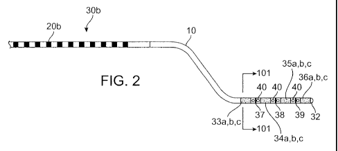
100231 Fig. 2 illustrates another embodilnent of a tissue monitoring and
modulating lead.
100241 Fig. 3 illustrates yet another embodiment of a tissue monitoring and
modulating
lead.
100251 Fig. 4 illustrates still another einbodiment of a tissue monitoring and
modulating
lead.
100261 Fig. 5 illustrates a cross-section of a tissue monitoring and
modulation lead.
[0027] Fig. 6 shows a cross-section of an alternative embodiment of a
monitoring and
modulation lead.
[0028] Fig. 7 shows a cross-section of yet another embodiment of a monitoring
and
modulation lead.
7
CA 02659022 2009-01-26
WO 2008/016881 PCT/US2007/074746
100291 Fig. 8 shows a cross-scction of still anothcr embodimcnt of a monitoi-
ing and
modulation lead.
100301 Figs. 9 shows anothei- cross-section of another embodiment of a monitoi-
ing and
modulation lead.
100311 Figs. 10 shows yet another cross-section of an embodiment of a
monitoring and
modulation lead.
100321 Figs. I I shows still anothei- cross-section of anothe-- embodiment of
a monitoring
and modulation lead.
100331 Figs. 12 shows another cross-section of another embodiment of a
monitoring and
modulation lead.
10034] Figs. 13A-13C highlight the recording and stimulating regions of an
exemplary
einbodiment of a monitoring and modulation lead.
100351 Fig. 14 illustrates a model of the magnitude of a dipole generated by
foui-
stimulation sites separated by 90".
100361 Fig. 15 illustrates a model of the magnitude of a dipole generated by
three
stimulation sites separated by 120 as compared with the model in Fig. 14.
100371 Fig. 16 shows a perspective view of an embodiment of a brain monitoring
and
modulation lead.
100381 Fig. 17 shows a brain monitoring and modulation lead implanted into a
patient's
head.
100391 Figs. 18A-18C show sample recordings of brain electrical potentials
from two
recording electrodes.
100401 Figs. 19A-19C show additional sample recordings of brain electrical
potentials from
two recording electrodes.
DETAILED DESCRIPTION OF THE INVENTION
[0041] In the drawings like numerals describe substantially similar
components. Probes
often have annular electrodes on their distal ends. An electrode divided into
two stimulation
sites is capable of orienting a dipole along one axis. When the annular
electrode is divided
8
CA 02659022 2009-01-26
WO 2008/016881 PCT/US2007/074746
into thi-ec stimulation sites, a dipole may be generated along any direction
in a plane. Thrcc
stimulation sites pei- annulai- elccti-ode is therefore advantageous as being
tlie minimum
number of stimulation sites per electrode i-equii-ed to ot-ient a dipole along
any direction in a
plane. Using the minimum number of stimulation sites is also advantageous
because it
minimizes the numbei- of conductoi-s which must pass through the probe and
pennits
maximum cui-rent density thi-ough any recording site to niodulate the brain
tissue.
100421 When cui-rent density is limited by b--ain tissue tolerance, a broken
ring of
stimulation sites can deliver a greater stimulus in some directions than
others. For example,
consider four stimulation sites arranged as a bi-oken t-ing ai-ound a
cylindrical pi-obe, with two
sites aligned with a transverse axis (X), and the other two sites aligned with
an or-thogonal
transverse axis (Y). This configuration mav generate an electi-ical dipole of
any orientation
within the plane of the stimulation sites by linear summation of two dipoles i-
esulting from
passing electrical cui-i-ent between opposite pairs of stimulating sites. To
generate a dipole of
magnitude (m) and orientation 0 relative to axis (X), a cui-rent of magnitude
(m/d) cos 0 is
passed through stimulating sites aligned with (X), and magnitude (m/d) sin 0
is passed
thi-ough the stimulating sites aligned with (Y), and where d is the distance
fi-om the origin.
As 0 changes, the locus of the dipole magnitude traces a circle. It may be
desired to linlit the
current density at any single electrode to be less than soine maximum value,
so that heat or
other undesired side effects of stimulation may be limited. With such a
constraint, the
maximum dipole that may be generated by a broken ring of four stimulation
sites as a
function of the angle 0 traces a square 243, as seen in Fig. 14. The largest
dipole magnitudes
are foi- orientations midway between the axes (X) and (Y), at the corners of
the square,
because both pairs of stimulation sites carry the inaximum pennitted current.
The smallest
dipole magnitudes are for orientations along the axes (X) and (Y), because
only one pair of
stimulation sites carries nonzero current.
100431 Compare the above scenario to an embodiment with three stimulation
sites arranged
in a broken ring or annulus about a cylindrical probe. If the axial extent of
the electrode ring
and maximum current density are the same as in the previous example, the
maximum
magnitude of the current through any electrode is 1/3 greater. When the
maximum current is
passed through one electrode, the return current is divided in various
proportions between the
other two electrodes. The maxiinum dipole that can be generated by a ring of
three
electrodes as a function of 0 traces a hexagon 246, similar to that
illustrated in Fig. 15. For
most orientations of the stimulating field, the magnitude of the maximum
dipole generated by
9
CA 02659022 2009-01-26
WO 2008/016881 PCT/US2007/074746
a broken i-ing of three stimulation sites is gi-eatei- than the dipole genei-
ated by a broken ring
of four stimulation sites as seen by the square 243) from Fig. 14 superimposed
in Fig. 15.
100441 Figs. 14 and 15 illusti-ate a simplifieci model which clai-ilies the
advantages of using
a prime number- of stimulation sites such as three. Thei-e ai-e three
stimulation sites on a
broken ring in the pi-eferi-ed embodiment of Fig. 1. Fig. 14 illustrates the
case of foui- electr-ic
monopoles 234a, 234b, 234c and 234d an-anged at points around a circle 230.
Monopoles
234a and 234c are equally and oppositely charged, and generate a dipole, as do
monopoles
234b and 234d. The radial position of points on the square 234 i-epresent the
maximum net
dipole that can be ci-eated by the sum of the two dipoles 234a, 234c and 234b,
234d, subject
to the constraint that the inaximum charge on a monopole is of magnitude one.
The sum of
the charge of the four monopoles is zero.
100451 Fig. 15 illustrates the case of three electric monopoles 235a, 235b and
235c
arranged at points ar-ound a cii-cle 230. The maximum net dipole square 243 of
Fig. 15 is
superimposed here for reference. Thi-ee electi-ic monopoles genei-ate an
oriented dipole more
efficiently, as diagramined by maximum net dipole hexagon. Two dipoles are
generated by
one monopole of one polarity, and two of the opposite or zei-o chai-ge. The
sum of the charge
of all three monopoles is zero. The radial position of points on the hexagon
246 represent the
maximum net dipole that can be created by the sum of the two dipoles, subject
to the
constraint that the maximum charge on any monopole cannot exceed the
inagnitude 1.2. The
larger maximum charge constraint is used here because the surface area of each
stimulation
site of a fixed axial length is greater if each portion occupies 1/3 of the
circumference, than if
each portion occupies 1/4 of the circumference. The sides of the hexagon
nearest the
electrodes 235a, 235b, 235c are generated in the situation where the
constraining electrodes
has positive polarity, and the sides of the hexagon opposite these are
generated in the
situation when the constraining electrode has negative polarity. It can be
seen that the radial
position of the hexagon 246 is farther fi-oin the origin than the square 243
at most directions
from the origin. For a fixed axial extent of the broken ring, three
stimulation sites can deliver
a larger effective stimulus compared to four stimulations sites.
Alternatively, for a fixed
effective stimulus, the axial length of a broken ring of 3 stimulation sites
can be shorter than
for a broken ring of 4 stimulation sites. The prefeiTed embodiment of the
invention has the
advantage over other probes of supporting better steerability of the electric
current for the
situation in which the maximum current density is constrained. This
description of the
CA 02659022 2009-01-26
WO 2008/016881 PCT/US2007/074746
invention does not pi-eclude using a stimulation protocol in which stimulation
sites on
diffei-ent broken rings ai-e stimulated simultancously oi- in coordination.
100461 It will be apparent to those skilled in the art that a stimulating
probe with a broken
ring of 6 stimulation sites (or any other inultiple of 3) can be used in
ainanner so as to obtain
the advantages of this invention. This may be accomplished by controlling the
ring of six
stimulation sites as three stimulation sites, each compi-ised of a paii- of
adjacent stimulation
sites.
100471 Therefore, at any axial position. the number of stimulation sites is a
priine number.
A prime number yields moi-e combinatoi-ial possibilities for simultaneously
using all
electrode sui-faces to achieve diffei-ent stimulation orientations. Using all
electrode surfaces
keeps current density as low as possible. In a preferred einbodiment, the
nwnber of
stimulation sites is 3. In anothei- embodiment, the number of stimulation
sites is 5.
Configurations with 2, 5 or 7 stimulation sites could achieve the cui-rent
density advantages
which this invention seeks to achieve also, although to a lesser degree.
100481 Referring now to Fig. 1, a tissue modulating and monitoring pi-obe is
illustrated.
Fig. I shows a preferred embodiment of the probe. It is a cylindrical probe,
with a flexible
probe body 10 and an optional multiple contact connecting tenninal 20a.
Additional details
on multiple contact connecting teiminals ai-e disclosed in U.S. Provisional
Application No.
60/820,914 the entire contents of which ai-e incorporated herein by reference.
Other
connectors may be used and ai-e well known in the art. At the distal end of
the probe 30a
there are one or more broken annular rings of stimulating sites. The
stimulating sites may be
aligned with matching angular position on all rings, or may be offset to
different angular
positions on different rings. There are also one or more circuinferential
electrode bands
suitable for recording local field potentials, and a recording electrode at or
near the most
distal point. In this preferred embodiment, the maximum diameter of the
multiple contact
terminal 20a is the same as the diameter of the flexible probe body 10.
[00491 In this embodiment, at four axial positions, three stimulation sites
33a, 33b, 33c,
34a, 34b, 34c, 35a, 35b, 35c, 36a, 36b, 36c are arranged as broken rings, for
a total of 12
stimulation sites. These are better seen in the cross-sectional views of Figs.
5-12. Also in
this embodiment are three recording bands 37, 38, 39 arranged in the gaps
between the
broken rings. The size of the recording sites is suitable for recording local
field potentials,
with an exposed area ranging from about 0.0005 inmZ to about 0.5mm2 but the
area could be
11
CA 02659022 2009-01-26
WO 2008/016881 PCT/US2007/074746
up to about 0.8mm'. Some embodiments havc smaller rccording sitcs that improve
extracellularly recordings of action potentials. Such recording sites range in
exposed area
from about 1 .9 x 10 5 nu1r to about 0.002 min', but they could be as large as
about 0. 1 mm' .
The form of the rccording sites could be the bare end of an insulated wire, a
thin film, ainetal
pad, oi- an insulated region with a poi-tion of the insulation removed to
expose an electrical
conductor within the wall of the device. Altei-native embodiments may have no
i-ecording
i-ings, oi- may have more recording rings. Additional recording rings or point
electrodes may
be located along the pi-obe body 10 oi- at the probe tip 32. The embodiment
docs not restrict
the aligninent of the recording electrodes (bands and/or points) with respect
to the stimulation
sites.
100501 There must be a nonconductive gap of at least l 00pm between
stimulating and
recording surfaces, and between recording surfaces, to reduce shunting and
improve the
isolation of the recor-ded signals. It is desirable that electr-ical signals
traversing through the
probe do not interfere with each other. It is especially desirable that the
high level electrical
stimulation signals not interfere with the low level recording signals.
Therefore, it is
preferable that the conductors cai-i-ying recoi-ding signals lay in an inner
helix, while
conductors cairying stimulation signals lay in an outer helix. The pitch of
the two helices
may be the same or inay be different, so that no pair of stiniulation and
recording conductors
traverse adjacent paths for an appreciable distance. This minimizes capacitive
coupling
between any stimulating conductors and any recording conductors. In other
embodiments, a
conductive coating inay be applied to the outside of the helix of recording
conductors. This
can be grounded to deci-ease electromagnetic interference between the two
types of
conductors. In yet another embodiment, a metal foil, which may be grounded, is
wrapped
between the inner and outer wire helices.
100511 In other embodiments, the conductors carrying recoi-ded signals lay
between
conductors carrying electrical stimulation signals. This embodiment has the
advantage that
the conductors lay in a single lamina and can be more compact and more
flexible, although in
some instances this embodiment may have the disadvantage that when stimulating
current
modulates a stimulating conductor, the stimulation signal may couple into
adjacent recording
conductors. Note that not all of the stimulus conductors are required to carry
a current at any
instant. In many uses of the probe, some of the recording conductors will
therefore be well
separated from active stimulating conductors at any instant. In another
embodiment, the
stimulating wires and recording wires course as adjacent groups of conductors
in a helix.
12
CA 02659022 2009-01-26
WO 2008/016881 PCT/US2007/074746
100521 The wires should be mechanically strong and electrically conductive.
Suitable ma-
terials include alloy MP35N (cobalt chi-ome alloy), stainless steel, and
tungsten or tungsten
alloy wire which has been gold plated to facilitate continuity with the
stimulation sites and to
the extra-cranial connector. It is important that the matei-ial be minimally
magnetic to
maximize MRi compatibility.
[0053] Stimulation sites are made of a rclatively inei-t material which
maximizes safe
charge ti-ansfei-, such as platinum, iridium oi- an alloy of platinum and ii-
idium. The body of
the probe is coated by a biocompatible polymer, such as silicone rubber or
pol_vurethane,
which supports bending with a short i-adius of curvature wher-e the pr-obe
exits the cranium.
100541 Fig. 2 illustrates an altei-native embodiment of the probe 30b. Probe
30b is similar to
the probe 30a of Fig. I except that it adds ports 40 which may pennit chemical
substances to
enter or leave the probe lumen. The ports 40 may be covered by a semi-
penneable
membrane. Alternatively a chemically controlled gating mechanism, such as a
cheinically
reactive hydrogel, may be placed near the ports. Such a hydrogel can swell oi-
contract
depending upon the chemical composition of the adjacent medium. The gating
mechanism
may operate based on bulk swelling and occlusion of the port, or the hydrogel
may be formed
with a mechanical accessory structure. An example of such as structui-e
includes a bimorph
beam as described by R. Bashir, J.Z. Hilt, O. Elibol, A. Gupta, and N. A.
Peppas in
"Micromechanical Cantilever as an Ultrasensitve pH Microsensor," published in
Applied
Physics Letters, 81(16):3091-3093, 2002. Another exainple includes a surface
covering
fenestrated with microports as disclosed by A. Baldi, M. Lei, Y. Gu, R.A.
Siegel and B. Ziaie
in an article entitled "A Microstructured Silicon Membrane with Entrapped
Hydrogels for
Environmentally Sensitive Fluid Gating," published in Sensor and Actuators B,
1 14(l):9-18,
2006, or another example includes a pad which displaces elements suited to
forming an
occlusive seal as described by A. Baldi, Y. Gu, P.E. Loftness, R.A. Siegel and
B. Ziaie in "A
Hydrogel-Actuated Environmentally Sensitive Microvalve for Active Flow
Control,"
published in the Journal of Microelectromechanical Systems, 12(5):613-621,
2003. The
entire contents of these references are incorporated herein by reference.
[00551 Since the hydrogels may be formulated such that their volume has
different
chemical dependencies, different hydrogels inay be associated with ports at
different pre-
determined positions on the lead, so that drugs may be delivered selectively
to pre-
determined positions on the probe. Likewise, samples of the extra-cellular
space or cerebral
spinal fluid (CSF) may be obtained from pre-detennined positions on the probe.
Examples of
13
CA 02659022 2009-01-26
WO 2008/016881 PCT/US2007/074746
chemical gating inechanisms that ai-e controlled directly by pH include those
described
previously in "Mici-omechanical Cantilevei- as an Ultrasensitve pH
Microsensor. Gating
mechanisnis controlled by the presence of carbon dioxide via a relationship to
pH include
those described by R. Stecge, H. Sebastiaan, W. Olthuis. P. Bergveld, A. Berg,
and J.
Kolkman in "Assessment of a New Prototype Hydrogel C02 Sensor; Comparison with
Air
Tonometry," as published in The Journal of Clinical Monitoring and Computing
2](2):83-90,
2007. Other examples of gating mechanisms conti-olled by the presence of
glucose are
disclosed by Theeuwes et al. in U.S. Patent No. 6,997,922. 1'he entire
contents of the above
listed references are incorporated herein by reference.
100561 Fig. 3 illustrates an alternative enlbodiment of pt-obe 30c in which
the probe tip 32a
is electi-ically conductive, sei-ving as an additional stimulation site. This
could sei-ve as a
conventional stimulation site, supporting inonopolar and bipolar stimulation.
In conjunction
with a distal ring of stimulation sites 36a-c it forms a group of stimulation
sites centered on
the vertices of a tetrahedron, suppor-ting steei-ing of the current near- the
tip in three
dimensions. The embodiment of Fig. 3 also has an additional recording
electrode 42 between
stimulating electrodes 36a - 36c and distal stimulating electrode 32a. Also,
multiple contact
connecting terminal 20c has a plurality of electrical contacts axially spaced
along two hemi-
cylidrical or D-shaped connectors, as further disclosed in U.S. Provisional
Patent Application
No. 60/820,914 the entire contents of which ai-e incorporated herein by
reference.
[0057] Fig. 4 illustrates an alternative embodiment of the probe, 30d,
demonstrating that
the multiple contact terminal 20d need not have the same diameter as the probe
body 10.
Here, contact terminal 20d is a larger diameter cylindrical shaped plug with
receptacles for
coupling the probe 30d with the rest of the monitoring and modulation system.
This
embodiment illustrates that the surface of recording electrodes need not be
circular, but may
be configured as recording points 43. Alternative embodiments may include
multiple
recording sites, some configured as rings, and other configured as points. In
other
embodiments the recording electrodes may take other shapes, including squares,
rectangles or
irregular shapes. In yet another alternative embodiment, the multiple contact
tenninal may
allow for a lumen or conduit for the passage fluid within the probe. Fluid may
pass in one or
more lumens, and may flow into or out of the brain, or both.
[0058] Fig. 5 illustrates an axial cross-sectional view of a preferred
embodiment, at section
line 101 in Fig. 1. In the preferred embodiment the central lumen 70 is
surrounded by a tube
14
CA 02659022 2009-01-26
WO 2008/016881 PCT/US2007/074746
72 made of a biocompatible polymer, such as polyurethane, silicone rubber or
polyamide. In
alternative embodiinents the lumen is a polymei- coating, and the insulated
recoi-ding
conductors 60 may reside in the inner lumen. Recording conductors 60 are wound
in a helix
fi-om the i-ecording sites to their tei-mination at the contact terminal 20.
Likewise, the
stimulating conductors 50 are wound in a helix from the stimulation sites to
their termination
at the contact tenninal 20. In a preferred embodinlent, the stimulating
conductors 50 have
larger size than the recoi-ding conductors 60 because resistive losses are a
greater concern foi-
the stimulating conductors 50, but all conductors may be of the same or
similar dimension in
alternative embodiments. In a preferred embodiment, the pitches of the
recording wire helix
and the stimulating wire helix are different, to decrease the average
capacitive coupling
between the wii-es. In alternative embodiments the helices could have the same
pitch. The
two helices may have the same or opposite orientation (one clockwise, the
other
countet-clockwise). Conductoi-s 50, 60 are embedded in a flexible polyiner,
and are insulated
in the prefen-ed embodiment, but could or could not rely on the sun-ounding
polymer for
insulation in an alternative embodiment. In the preferred embodiment, a layei-
of electrically
conductive inaterial 74 is interposed between the recording and stimulating
conductors,
which may be attached to a low impedance electrical reference. Alternative
embodiments
may use layer 74 or the central lining of the central lumen 72 as an internal
stimulating
electrode. Alternative embodiments may omit this layer 74 to simplify
manufacturing.
Stimulation sites 33a-c lay on the surface of the probe, with gaps of
nonconductive material
41 between them. The stimulation sites 33a-c may be of the form of sections of
a tube
adhered to the pi-obe, and welded or riveted to the conductors 50, or may be
fabricated with
thin film technology. Examples of thin film technology that could be used to
fabricate the
probe ai-e described, for example, in U.S. Patent Nos. 7,051,419 and 7,047,082
the entire
contents of which are incorporated herein by reference. The conductors 50, 60
in Fig. 5 are
shown as having a circular profile to suggest transversely cut round wires,
but alternative
fonns could use shaped wires such as those having a square, rectangular or
elliptical cross-
section, or thin film technologies may be used for the conductors. Fig. 5
shows 12
stimulating conductors 50 and 3 recording conductors 60 corresponding to the
preferred
embodiment, but alternative embodiments could have more or fewer conductors to
support
various numbers of electrodes.
100591 Fig. 6 illustrates an alternative embodiment, in which the stimulating
conductors 50
are arranged in groups rather than uniformly spaced around the circumference
of the probe.
CA 02659022 2009-01-26
WO 2008/016881 PCT/US2007/074746
Three groups of four ai-c illustrated, but alternatively the conductoi-s could
be arranged in 4
groups of thi-ee. Such embodiments could allow for ports communicating betwcen
the central
lumen 70 and the outside of the probe, or for improved flexibility of the
probe in conjunction
with reduced wall thickness bctwcen groups of conductors.
100601 Fig. 7 illustrates an axial cross-sectional view of an alternative
embodiment, at
section line 101 in Fig. 1. In this embodiment, the stimulating and i-ecording
conductors ai-e
in the same annular space of the probe, unlike prior embodiments where the
conductors are
separated. Because this embodiment places both conductoi-s in the same annulai-
space, the
centi-al lumen 70 inay be larger. In a preferred embodiment the stimulating
conductors 50
and recording conductors 60 alternate around the helix, but in alternative
embodiments the
stimulating conductors and recording conductoi-s could course as separate
groups. In
alternative embodiments, there inay be additional conductors betwecn the
stimulating 50 and
recording 60 conductors, which may be connected to the point of electrical
neutrality. In
alternative embodiments, the tube 72 may be coated with an electrically
conductive material,
which may be connected to the point of electrical neutrality.
100611 Fig. 8 illustrates an alternative embodiment wherein the recording
conductors 60
and stimulating conductors 50 are separated into groups. This embodiment has
the advantage
of reduced opportunities for undesirable capacitive coupling between
stimulating and
recording conductors compared to the embodiment illustrated in Fig. 7, but
increases the
opportunities for undesirable capacitive coupling between separate recording
conductors.
[0062] Fig. 9 illustrates an embodiment with dual lumens, central 70 and
annular 71, to
permit delivery or sampling of a fluid (gas or liquid) substance or drug, or
sampling of a
liquid or volatile substance. The lumens may communicate with ports, shown as
40 in Figs. 2
and 13A-13C, and such communication may be electrically or chemically gated.
The distal
ends of the lumens may be closed, permeable, selectively permeable, or open,
to release the
lumen contents or some fi-action or portion of the lumen contents. The distal
ends of the two
lumens may communicate with each other, so that one delivers a liquid
containing a drug
such a levodopa, or a gaseous medium with bioactive effects such as carbon
monoxide or
nitrous oxide, and another lumen retrieves the medium, after an opportunity to
exchange
substance or substances with the medium near ports 40 or other openings in the
probe. Other
therapeutic agents that may be delivered are well known in the art, such as
those disclosed in
U.S. Patent Nos. 6,094,598 and 6,227,203 both of which, the entire contents
are incorporated
16
CA 02659022 2009-01-26
WO 2008/016881 PCT/US2007/074746
hei-ein by i-eference ancl often. exti-acellulai- fluid such as cei-ebral
spinal fluid (CSF) is
sampled. In this embodimcnt. conductors for electrical stimulating and
recordino coui-se
togethei- within an additional annulus 79 ci-eated by an additional wall 78 in
the probe.
100631 Fig. 10 illustl-ates an ai-i-angement similar to that in Fig. 9, except
that the conductors
for stimulating and i-ecording coul-se through two separate annular rings 76
and 77, both
concentric to the inner two lumens 70 and 71. In other embodimeiits, there may
be more than
two lumens, and the lumens need not be concentric.
100641 Fig. 1 1 illustrates an arrangement similar to that in Fig. 9, except
that thei-e is a
single lumen 72. Additionally, conductors 50 and 60 are randomly oriented and
therefore
may allow the probe to be more easily fabricated as opposed to a probe with
conductors in a
defined pattern.
100651 Fig. 12 illustrates an ari-angement with no lumen for eithei- a guide
wire, oi- foi-
supporting mass transfer. The conductors course together through the center of
the probe.
100661 Figs. 13A-I3C illustrate an ai-rangement for the stimulating and r-
ecording conduc-
tors, similai- to the embodiments illustrated in Fig. 2. Fig. 13A shows a
probe having foui-
regions of stiinulating electrodes 36a-36c, 35a-35c, 34a-34c and 33a-33c, with
each region
having three independent stimulation sites. Additionally, the probe in Fig.
13A has recording
electrodes 37, 38 and 39 as well as ports 40. The probe of Fig. 13A is shown
in Figs. 13B-
13C with the circuinference of the probe unwi-apped, such that the upper edge
and the lower
edge of the conductors are actually continuous with each other. In the region
of the probe tip,
the conductors course in the axial direction, and turn to form helical
windings along the probe
body. Fig. 13B shows the recording electrode conductors 90a, 90b and 90c
coursing in the
axial direction near the probe tip and then turning to form helical windings
along the probe
body. Fig. 13C illustrates a similar pattern for stimulating electrode
conductors 92a, 92b,
92c, 94a, 94b, 94c, 96a, 96b, 96c and 98a, 98b, 98c.
100671 Fig. 16 shows a perspective view of a monitoring and modulation lead.
In Fig. 16,
four stimulation regions on the lead each contain three independent
stimulation electrodes.
All three stimulation electrodes 36a, 36b, 36c are only visible on the distal-
most region. Two
stimulating electrodes are visible in the other regions of the lead including
35a, 35b, 34a, 34b,
33a, 33b. Additionally, the lead has three recording electrodes 37, 38 and 39
as well as an
additional recording electrode 52 near the distal lead tip 32. An inner shaft
53 is contained
17
CA 02659022 2009-01-26
WO 2008/016881 PCT/US2007/074746
Nvithin ]cad body 10 and may be adapted to accommodate guidewires, stylets,
lumens, etc.
prcviously described herein.
100681 Fig. 17 shows a monitoring and modulating probe oi- lead I 2 secui-ed
to the skull of
a patient 1 I with a fixture 16 and iinplanted into brain tissue 14. An
extension lead 18
couples the probe 12 with a controllable pulse generator 19. The lead often
runs under the
patient's skin, although it may not and the conti-ollable pulse generator 19
may be implanted
or it may i-emain external to the body of the patient 11. Additional details
on a fixture for
securing the probe to the skull ai-e disclosed in U.S. Provisional Patent
Application No.
60/908,367 the entii-e contents of which are incorporated herein by reference.
100691 Table I below summarizes data collected that demonstrate that different
functional
stimulation effects can be achieved by stimulating different stimulation sites
around an
annular- ring. A lead similar to that illustt-ated in Fig. 16 was inserted
into the basal ganglia of
an anesthetized cat. The stimulating sites in the most distal annular ring
(36a, 36b and 36c)
were energized together and independently to electi-ically stimulate the
brain. The ground
was placed in the tempora]is muscle. Electrical stimulation of sufficient
magnitude evoked a
response in either the ipsilateral oi- contralateral or both facial muscles.
Stiinulation
rnagnitude was delivered in voltage steps, and the motor response was graded
on a rank-
ordered scale (NR - No Response; THR, Response Threshold; larger numbers
correspond to
larger magnitude of supra-threshold responses). When site 36a was stimulated
alone, the
response threshold for ipsilateral movement was lower than for contralateral
movement.
When site 36b was stimulated alone, the r-esponse threshold for ipsilateral
and contralateral
movement was the same. When site 36c was stimulated alone, the threshold for
contralateral
movement was lower than for ipsilateral movement. When all three sites were
stimulated
simultaneously, the threshold for ipsilateral movment was lower than for
contralateral
movement, but the threshold for both ipsilateral and contralateral movement
was lower than
with stimulation of any single site. Data froin this testing is suinmarized in
Table I below,
and this pattern of differential stimulation thresholds dernonstrates that
stimulating different
sites within an annular ring steers electrical current within the brain.
(00701 Figs. 18A-18C demonstrate that the lead can record field potentials,
and that
different recording sites record different potentials. The recording was
obtained from the
same lead illustrated in Fig. 16 as discussed above, and with the same
placement. The
response was evoked by sensory stimulation of the visual pathways by waving a
flashlight
18
CA 02659022 2009-01-26
WO 2008/016881 PCT/US2007/074746
befoi-e the eyes. In Figs. 18A. Ti-ace TI was i-ecorcled ti-om i-ecording site
38. and in Fig. 18B
trace T2 was recorded ti-om recoi-ding site 39. Specti-um analysis of these tt-
aces revealed
oscillations at I80 Hz. and 300 Hz. which are believed to i-esult froin
unintended coupling to
the power grid. A Christiano-Fitzgerald tiltei- was applied to i-emove signal
energy neai- these
fi-equencies, and the filtered traces are denoted T I a and T2a as shown in
Figs. 18A-18C. The
trace 0 in Fig. 18C is the arithmetic difference T 1 a - T2a. The traces look
similar, but they
ai-e not pi-opor-tional, as they would be if they i-esulted principally fi-om
electrical cross-talk.
At position A, Tl/Tla has a more sustained positivity compared to T2/T2a. At
position B.
the positivity in traces T I/T 1 a and T2/T2a are nearly identical. The
amplitude of the
triphasic wave between positions B and C differs considerably in traces T 1/T
I a and T2/T2a.
The amplitude of this recorded potential is somewhat less than the amplitudc
of an optimally
recorded field potential, reflecting the position of the lead near but not in
the optic tT-act.
100711 Figs. 19A-19C demonstrate that the lead can record spontaneous activity
field
potentials charactei-istic of placement in a gi-ey mattei- nucleus. The recoi-
ding was obtained
from a location 3mm dorsal to the location from which the recording in Figs.
18A-18C was
obtained. Because the amplitude of this recording was much greater than the
amplitude of
interference from the power grid, Christiano-Fitzgerald filtering was not
necessary. Trace T1
in Fig. 19A was recorded from recording site 38, and trace T2 in Fig. 19B was
recorded from
recording site 39. The trace n~ in Fig. 19C is the arithmetic difference TI -
T2. The traces
look similar, with a time course and amplitude chai-acteristic of field
potential recoT-dings.
The diffei-ence trace, A, has several transient waves with duration from 0.5
to 3.5 msec, and
amplitude of a few tens of millivolts, characteristic of action potential
wavefonns. Togethei-
with the recording shown in Figs. 18A-18C, these data demonstrate that a lead
such as that
illustrated in Fig. 16 can record field potentials from white matter and grey
matter, and with
suitable signal processing can also record action potential spikes.
19
CA 02659022 2009-01-26
WO 2008/016881 PCT/US2007/074746
Table I
Activated Stiniulation Ipsilateral Facial Muscle Cont--alateral Facial Muscle
Surfaces (V) Response Grade Response Grade
36a. 36b. 36c 1.0 NR NR
2.0 NR NR
2.2 THR NR
2.6 1 NR
2.7 1 THR
36a 1.0 NR NR
2.0 NR NR
3.0 NR NR
3.6 THR NR
4.0 1 NR
4.3 1 N R
4.5 2 THR
36b 1.0 NR NR
2.0 N R N R
2.4 THR THR
4.0 2 2
36c 1.0 NR NR
2.0 NR NR
3.0 NR NR
3.5 NR THR
4.0 THR 1
4.5 1 1
5.0 2 2
[0072] While the exemplary embodiments have been described in some detail for
clarity of
understanding and by way of example, a variety of additional modifications,
adaptations and
changes may be clear to those of skill in the art. Hence, the scope of the
present invention is
limited solely by the appended claims.