Note: Descriptions are shown in the official language in which they were submitted.
CA 02659392 2009-01-12
WO 2008/008986 PCT/US2007/073514
METHODS AND REAGENTS FOR TREATMENT AND DIAGNOSIS OF VASCULAR DISORDERS
AND AGE-RELATED MACULAR DEGENERATION
CROSS-REFERENCES TO RELATED APPLICATIONS
[00011 This application claims priority to U.S. Provisional Patent Application
No. 60/840,073,
filed August 23, 2006. This application also claims priority to U.S.
Provisional Patent Application No.
60/831,018, filed July 13, 2006. Both these applications are hereby
incorporated by reference.
STATEMENT AS TO RIGHTS TO INVENTIONS MADE UNDER
FEDERALLY SPONSORED RESEARCH AND DEVELOPMENT
[0002] This invention was made with government support under NIH RO1 EY11515
and R24
EY017404, awarded by the National Institutes of Health. The government has
certain rights in the
invention.
FIELD OF THE INVENTION
[0003] This invention relates to screening and therapeutic methods for
complement-mediated
diseases such as age-related macular degeneration and vascular diseases. The
invention finds application
in the fields of biology and medicine.
BACKGROUND OF THE INVENTION
[0004] Complement Factor H (CFH) is a multifunctional protein that acts as a
key regulator of
the complement system. See Zipfel, 2001, "Factor H and disease: a complement
regulator affects vital
body functions" Semin Thromb Hemost. 27:191-9. The Factor H protein activities
include: (1) binding to
C-reactive protein (CRP), (2) binding to C3b, (3) binding to heparin, (4)
binding to sialic acid; (5) binding
to endothelial cell surfaces, (6) binding to cellular integrin receptors (7)
binding to pathogens, including
microbes (see Figure 3 of U.S. patent publication No. 20070020647), and (8)
C3b co-factor activity. The
Factor H gene, known as HFI, CFH and HF, is located on human chromosome 1, at
position 1 q32. The
1q32 locus contains a number of complement pathway-associated genes. One group
of these genes,
referred to as the regulators of complement activation (RCA) gene cluster,
contains the genes that encode
Factor H, five Factor H-related proteins (FHR-1, FHR-2, FHR-3, FHR-4 and FHR-5
or CFHRI, CFHR2,
1
CA 02659392 2009-01-12
WO 2008/008986 PCT/US2007/073514
CFHR3, CFHR4 and CFHR5, respectively), and the gene encoding the beta subunit
of coagulation factor
XIII. The Factor H and Factor H related proteins are composed almost entirely
of short consensus repeats
(SCRs). Factor H and FHLI are composed of SCRs 1-20 and 1-7, respectively. FHR-
1, FHR-2, FHR-3,
FHR-4 and FHR-5 are composed of 5, 4, 5, 5 and 8 SCRs, respectively. The order
of genes, from
centromere to telomere is FH/FHL1, FHR-3, FHR-1, FHR-4, FHR-2 and FHR-5.
Factor H Gene
[0005] The Factor H cDNA encodes a polypeptide 1231 amino acids in length
having an apparent
molecular weight of 155 kDa (see Ripoche et al., 1988, Biochem J 249:593-602).
There is an
alternatively spliced form of Factor H known as FHL-1 (and also has been
referred to as HFLI or CFHT).
FHL-1 corresponds essentially to exons 1 through 9 of Factor H (see Ripoche et
al., 1988, Biochem J
249:593-602). The FHLI cDNA encodes a polypeptide 449 amino acids in length
having an apparent
molecular weight of 45-50 kDa. The first 445 amino acids of FHI and FHL1 are
identical, with FHL1
having four unique C-terminal amino acids (encoded by alternative exon 10A,
which is located in the
intron between exon 9 and exon 10. cDNA and amino acid sequence data for human
Factor H and FHL1
are found in the EMBL/GenBank Data Libraries under accession numbers Y00716
and X07523,
respectively. The 3926 base nucleotide sequence of the reference form of human
Factor H cDNA has
GenBank accession number Y00716 and the polypeptide has GenBank accession
number Y00716. The
1658 base nucleotide sequence of the reference form of HFL1, the truncated
form of the human Factor H,
has GenBank accession number X07523, and the polypeptide sequence has GenBank
accession number
X07523. The Factor H gene sequence (150626 bases in length) has GenBank
accession number
AL049744. The Factor H promoter is located 5' to the coding region of the
Factor H gene.
FHR-1 Gene
[0006] The FHR-1 gene is also known as CFHR1, CFHL1, CFHL, FHRI and HFLI. The
FHR-1
cDNA encodes a polypeptide 330 amino acids in length having an predicted
molecular weight of 39 kDa
(see Estaller et al., 1991, J. Immunol. 146:3190-3196). cDNA and amino acid
sequence data for human
FHR-1 are found in the EMBL/GenBank Data Libraries under accession number
M65292. The FHR-1
gene sequence is found under GenBank accession number AL049741.
FHR-2 Gene
[0007] The FHR-2 gene is also known as CFHR2, CFHL2, FHR2 and HFL3. The FHR-2
cDNA
encodes a polypeptide 270 amino acids in length having a predicted molecular
weight of 31 kDa (see
Strausberg et al., Proc. Natl. Acad. Sci USA 99:16899-16903). cDNA and amino
acid sequence data for
2
CA 02659392 2009-01-12
WO 2008/008986 PCT/US2007/073514
human FHR-2 are found in the EMBL/GenBank Data Libraries under accession
number BC022283. The
FHR-2 gene sequence is found under GenBank accession number AL139418.
FHR-3 Gene
[0008] The FHR-3 gene is also known as CFHR3, CFHL3, FHR3 and HLF4. The FHR-3
cDNA
encodes a polypeptide 330 amino acids in length having a predicted molecular
weight of 38 kDa (see
Strausberg et al., Proc. Natl. Acad. Sci USA 99:16899-16903). cDNA and amino
acid sequence data for
human FHR-3 are found in the EMBL/GenBank Data Libraries under accession
number BC058009. The
FHR-3 gene sequence is found under GenBank accession number AL049741.
FHR-4 Gene
[0009] The FHR-4 gene is also known as CFHR4, CFHL4 and FHR4. The FHR-4 cDNA
encodes a
polypeptide 331 amino acids in length having a predicted molecular weight of
38 kDa (see Skerka et al.,
1991, J. Biol. Chem. 272:5627-5634). cDNA and amino acid sequence data for
human FHR-4 are found
in the EMBL/GenBank Data Libraries under accession number X98337. The FHR-4
gene sequence is
found under GenBank accession numbers AF190816 (5' end), AL139418 (3' end) and
BX248415.
FHR-5 Gene
[0010] The FHR-5 gene is also known as CFHR5, CFHL5 and FHR5. The CFHR5 cDNA
encodes a
polypeptide 569 amino acids in length having an apparent molecular weight of
65 kDa (see McRae et al.,
2001, J. Biol.Chem. 276:6747-6754). cDNA and amino acid sequence data for
human CFHR5 are found
in the EMBL/GenBank Data Libraries under accession number AF295327. The 2821
base nucleotide
sequence of the reference form of human CFHR5 has GenBank accession number
AF295327, and the
polypeptide sequence has GenBank accession number AAK15619. The CFHR5 genomic
sequence is
found under GenBank accession numbers AL139418 (5' end) and AL353809 (3' end).
The FHR-5
promoter is located 5' to the coding region of the CFHR5 gene.
BRIEF SUMMARY OF THE INVENTION
[0011] In one aspect, the invention provides a screening method for
determining a human
subject's propensity to develop a vascular disorder and/or age-related macular
degeneration (AMD),
involving analysis of a biological sample from the subject to detect the
presence or absence of a deletion
in chromosome 1 between the 3' end of exon 22 of the complement factor H(CFH)
gene and the 5' end of
exon 1 of complement Factor H-related 4 (CFHR4) gene, wherein the presence of
a deletion is evidence
3
CA 02659392 2009-01-12
WO 2008/008986 PCT/US2007/073514
that the subject is at an increased risk of developing a vascular disorder and
a decreased risk of
developing AMD.
[0012] Examples of vascular disorders include aneurysms, such as abdominal
aortic aneurysm
(AAA) and brain intracranial aneurysm.
[0013] In one embodiment, the method comprises detecting the presence or
absence of at least a
portion of the complement Factor H-related 3 (CFHR3) gene. In a related
embodiment the entire protein
coding region of the CFHR3 gene is deleted. In a related embodiment the entire
CFHR3 gene is deleted.
In a related embodiment the entire CFHR3 gene and the region between the CFHR3
gene and
complement Factor H-related 1(CFHR1) gene are deleted.
[0014] In one embodiment, the method comprises detecting the presence or
absence of at least a
portion of the complement Factor H-related 1(CFHRI ) gene. In a related
embodiment the entire protein
coding region of the CFHRI gene is deleted. In a related embodiment the entire
CFHRl gene is deleted.
In a related embodiment the entire CFHRl gene and the region between the CFHRI
gene and
complement factor H-related 4 (CFHR4) gene are deleted. In a related
embodiment the entire CFHR1
gene and the region between the CFHRI gene and CFHR3 gene are deleted.
[0015] In one embodiment, the method comprises detecting the presence or
absence of at least a
portion of the CFHR3 gene and at least a portion of the CFHR1 gene. In a
related embodiment both the
entire protein coding regions of the CFHR3 and CFHR1 genes are deleted. In a
related embodiment the
entire CFHR3 and CFHR1 genes are deleted.
[0016] In one embodiment, a deletion or a partial deletion of an intergenic
sequence selected
from: a) a sequence between the CFH gene and the CFHR3 gene; b) a sequence
between the CFHR3
gene and the CFHR1 gene; c) a sequence between the CFHR1 gene and the CFHR4
gene. In yet another
embodiment, at least a portion of the CFH gene is deleted (e.g., at least a
portion of exon 22 is deleted).
[0017] In one embodiment, the presence or absence of the deletion is detected
by assaying for a
gene product encoded in chromosome 1 between the 3' end of exon 22 of the
complement factor H (CFH)
gene and the 5' end of exon 1 of complement Factor H-related 4 (CFHR4) gene,
where the absence of the
gene product, or a reduced level of expression of the gene product, indicates
the presence of deletion. In
another embodiment, the presence or absence of a CFHR1 gene product and/or a
CFHR3 gene product is
detected, where the absence of a gene product is indicative of a deletion. In
one instance, the gene
product is a protein. In another embodiment, detecting the presence or absence
of a deletion is performed
by analyzing a chromosome or nucleic acid (e.g., DNA or RNA) from the subject.
[0018] In one embodiment the presence or absence of the deletion is detected
by assaying for a
truncated CFHR1 or CRHR3 gene product, where detection of a truncated gene
product is indicative of a
4
CA 02659392 2009-01-12
WO 2008/008986 PCT/US2007/073514
deletion. In a preferred embodiment, the CFHR1 gene is partially deleted and
expresses a truncated
polypeptide gene product.
[0019] In one embodiment the subject has a genotype of T at position 1277 of
the coding region of
the CFH gene of the chromosome comprising the deletion.
[0020] The subject may be homozygous or heterozygous for deletions. Thus, in
one embodiment,
deletions are present in both chromosomes 1 of the subject.
[0021] The presence or absence of the deletion may be detected in a biological
sample from a
patient by, for example, analyzing a chromosome or nucleic acid (e.g., DNA or
RNA) sample from the
subject. The presence or absence of the deletion also may be detected by, for
example, determining the
presence or absence of protein encoded by the (deleted) DNA in a biological
sample from the subject,
e.g., a body fluid or tissue sample of the subject, by detecting a variant or
truncated form of the CFHR1 or
CFHR3 polypeptides in a body fluid or tissue sample of the subject, or by
measuring the level of CFHR1
or CFHR3 polypeptides in a body fluid or tissue sample of the subject.
[0022] The biological sample is any sample taken from a patient that is
suitable for use in the
invention. Examples of biological samples that include body fluids include
blood, serum, urine, cerebral
spinal fluid (CSF) and saliva. In one embodiment, the body fluid is blood,
serum or urine. Examples of
biological samples that comprise tissue samples include a skin biopsy and a
cheek scraping. In one
embodiment, the tissue sample is a skin biopsy.
[0023] Proteins (amount or presence) may be detected, for example, using an
immunoassay such
as a sandwich immunoassay, a competitive immunoassay, a radioimmunoassay,
fluorophore-labelled
immunoassay, an ELISA or a Western blot. Mass spectroscopy also may be used.
Variant proteins
(amount or presence) may be detected, for example, using variant-specific
antibodies. Truncated proteins
(amount or presence) may be detected, for example, by a difference in the size
of the protein by Western
blot analysis or mass spectroscopy.
[0024] In certain embodiments, the method comprises in the detecting step
determining the
presence of a deletion, for example, a deletion in a CFHR1 or CFHR3 gene, or
the absence or a reduction
of corresponding gene product (e.g., the amount or activity of the gene
product) indicating a higher risk of
the subject developing a vascular disorder.
[0025] In other embodiments, the method comprises in the detecting step
determining the
absence of a deletion, for example, the presence of a CFHR1 or CFHR3 gene, or
the presence or an
increase of the corresponding gene product (e.g., the amount or activity of
the gene product) indicating a
lower risk of the subject developing a vascular disorder.
[0026] In another embodiment, the method comprises in the detecting step
determining the
presence of a deletion, for example, a deletion in a CFHR1 or CFHR3 gene, or
the absence or a reduction
CA 02659392 2009-01-12
WO 2008/008986 PCT/US2007/073514
of the corresponding gene product (e.g., the amount or activity of the gene
product) indicating a lower
risk of the subject developing AMD.
100271 In yet another embodiment, the method comprises in the detecting step
determining the
absence of a deletion, for example, the presence of a CFHRI or CFHR3 gene, or
the presence or an
increase of the corresponding gene product (e.g., the amount or activity of
the gene product) indicating a
higher risk of the subject developing AMD. The increase in gene product, for
example, can be at least
10%, at least 20%, at least 50%, or more.
[0028] In certain embodiments, the method further comprises detecting at least
one other genetic
variant or biomarker indicative of AMD and/or vascular disease. Genetic
variants that may be detected in
the invention include genetic variants of complement factor H(CFH) gene, HTRA1
gene, complement
factor B (BF) gene and/or the complement component 2 (C2) gene. In an
embodiment, the genetic
variants include one or a plurality of polymorphic sites, such as those
described herein.
[0029] In another aspect, the invention provides a method for treating a
subject having (i.e.,
exhibiting symptoms of), or is at risk for developing, a vascular disorder, by
administering a CFHR1
polypeptide and/or a CFHR3 polypeptide to the subject. The polypeptide may be
a full-length CFHRI
polypeptide or a fragment or portion thereof. The polypeptide may be a full-
length CFHR3 polypeptide
or a fragment or portion thereof.
[0030] In another aspect the invention provides a pharmaceutical composition
comprising a
CFHR3 protein or fragment thereof and at least one pharmaceutically effective
excipient. In another
aspect the invention provides a pharmaceutical composition comprising a CFHRl
protein or fragment
thereof and at least one pharmaceutically effective excipient.
[0031] In another aspect the invention provides the use of a protein
comprising the gene product
of at least a portion of the CFHR3 and/or CFHR1 gene for the preparation of a
medicament for the
treatment of a vascular disorder.
[0032] In another aspect the invention provides gene therapy vectors
comprising nucleic acid
encoding a CFHR3 or CFHR1 protein, or fragment thereof. The vector may include
a promoter that
drives expression of the CFHR3 or CFHRI gene in multiple cell types.
Alternatively, the vector may
include a promoter that drives expression of the CFHR3 or CFHRI gene only in
specific cell types, for
example, in cells of the retina or in cells of the kidney. In a related aspect
pharmaceutical compositions
are provided containing a gene therapy vector encoding a CFHR3 or CFHR1
protein or fragment thereof
and a pharmaceutically acceptable excipient.
[0033] In another aspect the invention provides a method of treating a subject
having (i.e.,
exhibiting symptoms of), or susceptible to developing, age-related macular
degeneration (AMD), by
administering an agent that reduces the expression of the CFHRI and/or CFHR3
genes or reduces the
6
CA 02659392 2009-01-12
WO 2008/008986 PCT/US2007/073514
activity or amount of a gene product of the CFHRI and/or CFHR3 genes. Agents
include antisense RNA,
siRNA or ribozyme that reduces expression of the CFHRI and/or CFHR3 genes. In
a related aspect the
level of protein is reduced, for example by using plasmaphoresis or antibody-
based inhibition, for
example, using an anti-CFHR1 antibody and/or an anti-CFHR3 antibody.
[0034] In another aspect the invention provides a pharmaceutical composition
comprising an
anti-CFHR1 antibody and a pharmaceutically acceptable carrier. In one
embodiment, an anti-CFHR1
antibody specifically binds the amino-terminus of a CFHR1 polypeptide. In
another aspect the invention
provides a pharmaceutical composition comprising an anti-CFHR3 antibody and a
pharmaceutically
acceptable carrier. In one embodiment, an anti-CFHR3 antibody specifically
binds the carboxyl-terminus
of a CFHR3 antibody.
[0035] In another aspect the invention provides a diagnostic kit for
diagnosing susceptibility to a
vascular disorder and/or AMD in a subject, comprising nucleic acid primers or
probes that detect the
presence or absence of a deletion in the DNA sequence between the 3' end of
exon 22 of the complement
factor H (CFH) gene and the 5' end of exon 1 of complement Factor H-related 4
(CFHR4) gene on human
chromosome 1.
[0036] In another aspect the invention provides a diagnostic device comprising
nucleic acid
primers or probes that detect the presence or absence of a deletion in the DNA
sequence between the 3'
end of exon 22 of the complement factor H (CFH) gene and the 5' end of exon 1
of complement Factor H-
related 4 (CFHR4) gene on human chromosome I immobilized on a substrate, such
as a microarray.
[0037] In another aspect the invention provides a diagnostic kit for
diagnosing susceptibility to a
vascular disorder and/or AMD in a subject, comprising antibodies that detect
the presence or absence of
the complement Factor H-related 3(CFHR3) protein, or variant or truncated
forms thereof, and/or
complement Factor H related 1(CFHRl) protein, or variant or truncated forms
thereof, in a body fluid or
tissue sample of the subject.
[0038] In another aspect the invention provides a drug screening method for
screening for agents
for use in treating a vascular disorder. The method involves a) combining (i)
a cell that expresses CFHR3
and/or CFHR1 polypeptides; and (ii) a test agent; b) measuring the level of
CFHR3 and/or CFHR1
polypeptides secreted into the medium; and c) comparing the level of CFHR3
and/or CFHR1
polypeptides secreted into the medium in the presence of the test agent with a
reference value, said
reference value being the level of CFHR3 and/or CFHR1 polypeptides secreted
into the medium in the
absence of the test agent, where a higher level of CFHR3 and/or CFHR1
polypeptides secreted into the
medium in the presence of the test agent indicates the test agent may be
useful for treating the vascular
disorder.
7
CA 02659392 2009-01-12
WO 2008/008986 PCT/US2007/073514
[0039] In another aspect the invention provides a method for identifying a CFH
protein likely to
protect against AMD development, by identifying a subject with a deletion in
the DNA sequence between
the 3' end of exon 22 of the complement factor H (CFH) gene and the 5' end of
exon 1 of complement
Factor H-related 4 (CFHR4) gene on human chromosome 1; determining the
sequence of the CFH gene
encoded by the gene contained in the chromosome containing the deletion; and
determining the sequence
of the protein encoded by the CFH gene, wherein said protein is different from
wild-type CFH, said
protein being a CFH protein likely to protect against AMD development. The
invention also provides a
protective CFH protein obtained using the method.
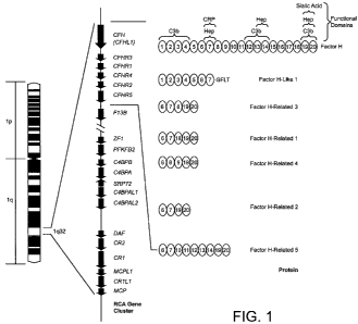
BRIEF DESCRIPTION OF THE FIGURES
[0040] Figure 1 is a diagram showing the organization of the regulators-of-
complement-
activation (RCA) gene cluster on chromosome 1 q32 and the arrangement of
approximately 60-amino acid
domains known as short consensus repeats (SCRs) in complement Factor H (CFH),
Factor H-Like 1
(CFHL1) and Factor H-Related 1, 2, 3, 4 and 5(CFHR1, CFHR2, CFHR3, CFHR4 and
CFHR5). CFH
has 20 SCRs. The interacting partners with some of these SCRs has been
determined and is shown on the
top right (CRP, C reactive protein; Hep, heparin). Complement factor H-like
1(CFHL1) is a splice
isoform of CFH, while complement factor H-related proteins 1-5 (CFHR1-5) are
each encoded by a
unique gene (CFHRI-5). The SCRs of CFHR1-5 are similar to some of the SCRs in
CFH, as denoted by
the numbers in the ovals. For example, CFHR5 has 9 SCRs, with the first two
being similar to SCRs 6
and 7 of Factor H and therefore having CRP and heparin binding properties.
SCRs 5-7 of CFHR5 have
the numbers 12-14 within the corresponding ovals because these SCRs are
similar to SCRs 12-14 of
Factor H and have C3b and heparin binding properties.
[0041] Figure 2 shows regions of homology (genomic duplications) in the genes
encoding CFH
and the Factor H-related proteins. Exons are indicated as vertical lines.
Regions labeled with the same
letter (e.g., A, A', and A") have substantially identical sequences.
[0042] Figure 3 shows a Western blot of serum proteins from seven patients
using an anti-human
CFH antibody. FHL-1, CFHR1 and CFHR2 indicate the positions of the truncated
form of CFH, CFHR1
and CFHR2, respectively. The anti-human CFH antibody employed also cross-
reacts with CFHR1 and
CFHR2. No CFHR1 is detected in the serum of two patients (197-02 and 325-02)
that have a
homozygous deletion of the CFHR3 and CFHRI genes, as determined by SSCP
analysis and direct DNA
sequencing.
[0043] Figure 4 shows a SSCP analysis of the CFH, CFHR3 and CFHR1 genes. 1, 2,
3, and 4
indicate four different SSCP patterns observed using primers from exon 22 of
the CFH gene to PCR
amplify DNA. SSCP patterns 1, 2 and 3 correspond to homozygous non-deletion or
heterozygous
8
CA 02659392 2009-01-12
WO 2008/008986 PCT/US2007/073514
deletion of CFHR3 and CFHR1, and pattern 4 corresponds to homozygous deletion
of CFHR3 and
CFHR1.
[0044] Figure 5 shows a PCR analysis of the CFH and CFH-related genes 1 to 5
in leukocytes
from 20 patients that are separated into four groups according to the SSCP
pattems using the CFHexon
22 primers (patterns 1-4 are as described in Figure 4). From left to right, in
each panel (gel), 5 leukocyte-
derived DNA samples each from patients displaying SSCP patterns 1, 2, 3 and 4
were subjected to PCR
using primers specific for CFH, CFHRI, CFHR2, CFHR3, CFHR4 and CFHR5, as
indicated. When
SSCP analysis and direct DNA sequencing show a homozygous deletion of the
CFHR3 and CFHRI
genes, no PCR amplifiable CFHR3 and CFHRI DNA are detected.
[0045] Figure 6 shows an amino acid alignment of the CFH (SEQ ID NO: 2), CFHR1
(SEQ ID
NO: 4), and CFHR3 (SEQ ID NO: 6) proteins.
[0046] Figure 7 shows a nucleotide alignment of the CFH (SEQ ID NO: 1), CFHRI
(SEQ ID
NO: 3), and CFHR3 genes (SEQ ID NO: 5).
DETAILED DESCRIPTION
1. Definitions
[0047] The following definitions are provided to aid in understanding the
invention. Unless otherwise
defmed, all terms of art, notations and other scientific or medical terms or
terminology used herein are
intended to have the meanings commonly understood by those of skill in the
arts of medicine and
molecular biology. In some cases, terms with commonly understood meanings are
defmed herein for
clarity and/or for ready reference, and the inclusion of such definitions
herein should not be assumed to
represent a substantial difference over what is generally understood in the
art.
[0048] A "vascular disorder" is a disease or condition of the vascular system.
One type of vascular
disorder is an aneurysm such as abdominal aortic aneurysm or brain
intracranial aneurysm. Other types
of vascular disorder include hypertension, cerebral vascular accidents, trans-
ischemic accidents (e.g.,
stroke). Still other types of vascular disorders include coronary artery
disease, peripheral artery disease,
varicose veins, and peripheral vascular disease.
[0049] A "nucleic acid", "polynucleotide" or "oligonucleotide" is a polymeric
form of nucleotides of
any length, may be DNA or RNA, and may be single- or double-stranded. Nucleic
acids may include
promoters or other regulatory sequences. Oligonucleotides are usually prepared
by synthetic means. A
reference to the sequence of one strand of a double-stranded nucleic acid
defmes the complementary
sequence and except where otherwise clear from context, a reference to one
strand of a nucleic acid also
refers to its complement. For certain applications, nucleic acid (e.g., RNA)
molecules may be modified to
increase intracellular stability and half-life. Possible modifications
include, but are not limited to, the use
9
CA 02659392 2009-01-12
WO 2008/008986 PCT/US2007/073514
of phosphorothioate or 2'-O-methyl rather than phosphodiesterase linkages
within the backbone of the
molecule. Modified nucleic acids include peptide nucleic acids (PNAs) and
nucleic acids with
nontraditional bases such as inosine, queosine and wybutosine and acetyl-,
methyl-, thio-, and similarly
modified forms of adenine, cytidine, guanine, thymine, and uridine which are
not as easily recognized by
endogenous endonucleases.
[0050] "Hybridization probes" are nucleic acids capable of binding in a base-
specific manner to a
complementary strand of nucleic acid. Such probes include nucleic acids and
peptide nucleic acids
(Nielsen et al., 1991). Hybridization may be performed under stringent
conditions which are known in
the art. For example, see, e.g., Berger and Kimmel (1987) METHODS IN
ENZYMOLOGY, VOL. 152: GUIDE
TO MOLECULAR CLONING TECHNIQUES, San Diego: Academic Press, Inc.; Sambrook et
al. (1989)
MOLECULAR CLONING: A LABORATORY MANUAL, 2nd Ed., Vols. 1-3, Cold Spring Harbor
Laboratory;
Sambook (2001) 3rd Edition; Rychlik, W. and Rhoads, R.E., 1989, Nucl. Acids
Res. 17, 8543; Mueller,
P.R. et al. (1993) In: CURRENT PROTOCOLS IN MOLECULAR BIOLOGY 15.5, Greene
Publishing
Associates, Inc. and John Wiley and Sons, New York; and Anderson and Young,
QUANTITATIVE FILTER
HYBRIDIZATION IN NUCLEIC ACID HYBRIDIZATION (1985)). As used herein, the term
"probe" includes
primers. Probes and primers are sometimes referred to as "oligonucleotides."
[0051] The term "primer" refers to a single-stranded oligonucleotide capable
of acting as a point of
initiation of template-directed DNA synthesis under appropriate conditions, in
an appropriate buffer and
at a suitable temperature. The appropriate length of a primer depends on the
intended use of the primer
but typically ranges from 15 to 30 nucleotides. A primer sequence need not be
exactly complementary to
a template but must be sufficiently complementary to hybridize with a
template. The term "primer site"
refers to the area of the target DNA to which a primer hybridizes. The term
"primer pair" means a set of
primers including a 5' upstream primer, which hybridizes to the 5' end of the
DNA sequence to be
amplified and a 3' downstream primer, which hybridizes to the complement of
the 3' end of the sequence
to be amplified.
[0052] Exemplary hybridization conditions for short probes and primers is
about 5 to 12 degrees C
below the calculated Tm. Formulas for calculating Tm are known and include: Tm
= 4 C x (number of
G's and C's in the primer) + 2 C x (number of A's and T's in the primer) for
oligos <14 bases and
assumes a reaction is carried out in the presence of 50mM monovalent cations.
For longer oligos, the
following formula can be used: Tm = 64.9 C + 41 C x (number of G's and C's in
the primer - 16.4)/N,
where N is the length of the primer. Another commonly used formula takes into
account the salt
concentration of the reaction (Rychlik, supra, Sambrook, supra, Mueller,
supra.): Tm = 81.5 C + 16.6 C
x(log10[Na+] + [K+]) + 0.41 C x (%GC) - 675/N, where N is the number of
nucleotides in the
oligo. The aforementioned formulae provide a starting point for certain
applications; however, the
CA 02659392 2009-01-12
WO 2008/008986 PCT/US2007/073514
design of particular probes and primers may take into account additional or
different factors. Methods for
design of probes and primers for use in the methods of the invention are well
known in the art.
[0053] The term "polymorphism" refers to the occurrence of two or more
genetically determined
alternative sequences or alleles in a population. A "polymorphic site" is the
locus at which sequence
divergence occurs. Polymorphic sites have at least two alleles. A diallelic
polymorphism has two alleles.
A triallelic polymorphism has three alleles. Diploid organisms may be
homozygous or heterozygous for
allelic forms. A polymorphic site may be as small as one base pair. Examples
of polymorphic sites
include: restriction fragment length polymorphisms (RFLPs); variable number of
tandem repeats
(VNTRs); hypervariable regions; minisatellites; dinucleotide repeats;
trinucleotide repeats; tetranucleotide
repeats; and simple sequence repeats. As used herein, reference to a
"polymorphism" can encompass a
set of polymorphisms (i.e., a haplotype).
[0054] A "single nucleotide polymorphism (SNP)" occurs at a polymorphic site
occupied by a single
nucleotide, which is the site of variation between allelic sequences. The site
is usually preceded by and
followed by highly conserved sequences of the allele. A SNP usually arises due
to substitution of one
nucleotide for another at the polymorphic site. Replacement of one purine by
another purine or one
pyrimidine by another pyrimidine is called a transition. Replacement of a
purine by a pyrimidine or vice
versa is called a transversion. A synonymous SNP refers to a substitution of
one nucleotide for another in
the coding region that does not change the amino acid sequence of the encoded
polypeptide. A non-
synonymous SNP refers to a substitution of one nucleotide for another in the
coding region that changes
the amino acid sequence of the encoded polypeptide. A SNP may also arise from
a deletion or an
insertion of a nucleotide or nucleotides relative to a reference allele.
[0055] The term "deletion," when referring to a nucleic acid sequence, has the
usual meaning in
genetics of an allele in which one or more bases are missing compared to a
reference or wild-type
sequence. Deletions may be as short as one base-pair. Deletions detected in
the present invention may be
longer, such as a deletion of at least 100 bp, at least 200 bp, at least 300
bp, at least 400 bp, at least 500
bp, at least 600 bp, at least 700 bp, at least 800 bp, at least 900 bp, at
least 1000 bp, at least 1100 bp, at
least 1200 bp, at least 1300 bp, at least 1400 bp, at least 1500 bp, at least
1600 bp, at least 1700 bp, at
least 1800 bp, at least 1900 bp, at least 2000 bp, at least 2500 bp, at least
3000 bp, at least 3500 bp, at
least 4000 bp, at least 4500 bp, at least 5000 bp, at least 6000 bp, at least
7000 bp, at least 8000 bp, at
least 9000 bp, at least 10,000 bp, at least 15,000 bp, at least 20,000 bp, at
least 30,000 bp, at least 40,000
bp, at least 50,000 bp, at least 75,000 bp, at least 100,000 bp, at least
125,000 bp, at least 150,000 bp, at
least 200,000 bp or at least 250,000 bp.
[0056] The term "haplotype" refers to the designation of a set of
polymorphisms or alleles of
polymorphic sites within a gene of an individual. For example, a"112" Factor H
haplotype refers to the
11
CA 02659392 2009-01-12
WO 2008/008986 PCT/US2007/073514
Factor H gene comprising allele 1 at each of the first two polymorphic sites
and allele 2 at the third
polymorphic site. A "diplotype" is a haplotype pair.
[0057] An "isolated" nucleic acid means a nucleic acid species that is the
predominant species
present in a composition. Isolated means the nucleic acid is separated from at
least one compound with
which it is associated in nature. A purified nucleic acid comprises (on a
molar basis) at least about 50, 80
or 90 percent of all macromolecular species present.
[0058] Two amino acid sequences are considered to have "substantial identity"
when they are at
least about 80% identical, preferably at least about 90% identical, more
preferably at least about 95%, at
least about 98% identical or at least about 99% identical. Percentage sequence
identity is typically
calculated by determining the optimal alignment between two sequences and
comparing the two
sequences. Optimal alignment of sequences may be conducted by inspection, or
using the local
homology algorithm of Smith and Waterman, 1981, Adv. Appl. Math. 2: 482, using
the homology
alignment algorithm of Needleman and Wunsch, 1970, J. Mol. Biol. 48: 443,
using the search for
similarity method of Pearson and Lipman, 1988, Proc. Natl. Acad. Sci. U.S.A.
85: 2444, by computerized
implementations of these algorithms (e.g., in the Wisconsin Genetics Software
Package, Genetics
Computer Group, 575 Science Dr., Madison, Wis.) using default parameters for
amino acid comparisons
(e.g., for gap-scoring, etc.). It is sometimes desirable to describe sequence
identity between two
sequences in reference to a particular length or region (e.g., two sequences
may be described as having at
least 95% identity over a length of at least 500 basepairs). Usually the
length will be at least about 50,
100, 200, 300, 400, 500, 600, 700, 800, 900 or 1000 amino acids, or the full
length of the reference
protein. Two amino acid sequences can also be considered to have substantial
identity if they differ by 1,
2, or 3 residues, or by from 2-20 residues, 2-10 residues, 3-20 residues, or 3-
10 residues.
[0059] "Linkage" describes the tendency of genes, alleles, loci or genetic
markers to be inherited
together as a result of their location on the same chromosome. Linkage can be
measured by percent
recombination between the two genes, alleles, loci or genetic markers.
Typically, loci occurring within a
50 centimorgan (cM) distance of each other are linked. Linked markers may
occur within the same gene
or gene cluster. "Linkage disequilibrium" or "allelic association" means the
preferential association of a
particular allele or genetic marker with a specific allele or genetic marker
at a nearby chromosomal
location more frequently than expected by chance for any particular allele
frequency in the population. A
marker in linkage disequilibrium can be particularly useful in detecting
susceptibility to disease, even if
the marker itself does not cause the disease.
[0060] The terms "susceptibility," "propensity," and "risk" refer to either an
increased or
decreased likelihood of an individual developing a disorder (e.g., a
condition, illness, disorder or disease)
relative to a control population. In one example, the control population may
be individuals in the
12
CA 02659392 2009-01-12
WO 2008/008986 PCT/US2007/073514
population (e.g., matched by age, gender, race and/or ethnicity) without the
disorder, or without the
genotype or phenotype assayed for. In some contexts, the terms diagnosing and
screening are used
interchangeably (e.g., a person skilled in the art can diagnose a propensity
to develop the disease).
[0061] The term "diagnose" and "diagnosis" refer to the ability to determine
or identify whether
an individual has a particular disorder (e.g., a condition, illness, disorder
or disease).
[0062] The term "screen" or "screening" as used herein has a broad meaning. It
includes
processes intended for the diagnosis or for determining the susceptibility,
propensity, risk, or risk
assessment of an asymptomatic subject for developing a disorder later in life.
Screening also includes the
prognosis of a subject, i.e., when a subject has been diagnosed with a
disorder, determinating in advance
the progress of the disorder as well as the assessment of efficacy of therapy
options to treat a disorder.
[0063] The terms "portion," "fragment" and/or "truncated form" when used in
reference to a
Factor H-related gene product (e.g., CFHR3 or CFHR1 gene product), refers to a
nucleic acid or
polypeptide sequence that is less than the full-length sequence (i.e., a
portion of the full-length gene or
polypeptide). A portion or fragment or truncated form of CFHR3 or CFHR1 gene
or polypeptide can be
at least 25, at least 50, at least 75, at least 100, at least 150, at least
200, at least 250, or at least 300
nucleotides or amino acids in length. Typically the portion includes at least
1, often at least two, and
sometimes at least 3 or 4 complete SCRs.
[0064] As used herein, the term "gene product" means an RNA (e.g., mRNA) or
protein that is
encoded by the gene. A "protein coding region" is a region of DNA/RNA sequence
within a gene that
encodes a polypeptide or protein.
[0065] An "assay" is a procedure wherein the presence or amount or a property
of a test
substance, e.g., a nucleic acid or gene product, is detected or measured.
[0066] The terms "inhibit" and "reduce" refer to any inhibition, reduction, or
decrease in
expression or activity including partial or complete inhibition of gene
expression or gene product activity.
2. Association of Polymorphisms in the CFHR1 And CFHR3 Genes and Risk Of
Developing AMD and
Vascular Disorders
[0067] A correlation between polymorphic sites and haplotypes in the CFH gene
and the
likelihood of developing AMD has been discovered. See Hageman et al., 2005,
Proc. Natl. Acad. Sci.
U.S.A. 102:7227-32; Haines et al., 2005, Science 308:419-21; Klein et al.,
2005, Science 308:385-9;
Edwards et al., 2005, Science 308:421-4 and U.S. patent publication No.
20070020647, each incorporated
by reference in its entirety for all purposes. Both CFH risk haplotypes and
CFH protective haplotypes are
known. Polymorphisms particularly associated with increased risk include a
variant allele at: rs 1061170
(402H; exon 9); rs203674 (intron 10) and the polymorphism at residue 1210
(1210C; exon 22).
13
CA 02659392 2009-01-12
WO 2008/008986 PCT/US2007/073514
Polymorphisms particularly associated with decreased risk include the
protective H2 haplotype, which
includes a variant allele in IVS6 (intron 6, rs3766404) and the H4 haplotype,
which includes a variant
allele in IVS1 (intron 1, rs529825) and a variant allele (162) (exon 2,
rs800292).
[0068] It has now been discovered that an AMD protective haplotype is
genetically linked to
deletions in the DNA sequence between the 3' end of exon 22 of the complement
factor H (CFH) gene
and the 5' end of exon 1 of complement Factor H-related 4 (CFHR4) gene on
human chromosome 1(i.e.,
the DNA sequence encoding the CFHR1 and CFHR3 proteins). See Example 1, infra.
The discovery
that deletions at the CFHRI and CFHR31oci are associated with decreased risk
of developing AMD has a
number of specific applications, including screening individuals to ascertain
risk of developing AMD and
identification of new and optimal therapeutic approaches for individuals
afflicted with, or at increased
risk of developing, AMD. As discussued in Example 1, below, the deletion
genotype is predominantly
associated with the CFHH4 haplotype. See Hageman et al., 2005, Proc. Natl.
Acad. Sci. U.S.A.
102:7227-32. Thus, this deletion acts as a marker for decreased risk of
conditions for which the H4
haplotype is protective.
[0069] Moreover, it has now been discovered that deletions in the DNA sequence
between the 3'
end of exon 22 of the complement factor H(CFH) gene and the 5' end of exon I
of complement Factor H-
related 4 (CFHR4) gene on human chromosome 1(i.e., the DNA sequence encoding
the CFHR1 and
CFHR3 proteins) are associated with increased risk of developing a vascular
disease such as aortic
aneurysm. See Example 1, infra. The discovery that deletions at the CFHR1 and
CFHR3 loci are
associated with increased risk of developing a vascular disorder has a number
of specific applications,
including screening individuals to ascertain risk of developing a vascular
disorder and identification of
new and optimal therapeutic approaches for individuals afflicted with, or at
increased risk of developing,
vascular disorders.
3. Screening Methods
[0070] Based on the discoveries described herein, a subject's risk for AMD or
vascular disease
can be assessed by determining whether or not a the subject has a deletion
within the region of
chromosome 1 lying between the 3' end of exon 22 of the complement factor
H(CFH) gene and the 5'
end of exon 1 of complement Factor H-related 4 (CFHR4). The extent of the
deletion may vary in
different individuals or populations. For example, in one embodiment the all
of most of the region
between CFH exon 22 and CFHR4 exon 1 is deleted. Alternatively, a portion of
the region may be
deleted, such as, for example, a deletion of less than the entire region
between CFH exon 22 and CFHR4
exon 1 but including the CFHR1 encoding sequence, or including the CFHR3
encoding sequence,
14
CA 02659392 2009-01-12
WO 2008/008986 PCT/US2007/073514
including both, or including a non-coding (e.g., intragenic) sequence. An
individual may be homozygous
for deletion (both chromosomes I have a deletion in the region) or may be
heterozygous for deletion.
[0071] For example and not limitation, the homozygous deletion of CFHRI and/or
CFHR3 can
be detected from the absence of CFHR1 and/or CFHL3 protein in a body fluid or
tissue sample (see
Figure 3), by the absence of RNA encoded in the region between the 3' end of
CFH exon 22 and the 5'
end of CFHR4 exon 1(e.g., absense of absence of CFHRI and/or CFHL3 mRNAs), or
by absense of
genomic DNA in the region in the region between the 3' end of CFH exon 22 and
the 5' end of CFHR4
exon 1. The present or absense of DNA or RNA sequences can be determined using
art known methods,
such as PCR. The absense of a nucleic acid sequence is deduced from the
absense of an amplified PCR
product in an assay of a tissue sample (see Figure 5). It will be understood
that, although PCR is
frequently cited herein as a method for genetic analysis, many other
analytical methods are known and are
suitable for detection of a deletion. For example and not limitation several
are described below in the
section captioned "Analysis of Nucleic Acid Samples."
[0072] The heterozygous deletion of CFHR1 and/or CFHR3 can be determined, for
illustration
and not limitation, (1) from a reduction in the amount of protein in a body
fluid or tissue sample as
compared to the amount from a control having both alleles of CFHRl and/or
CFHR3 genes, (2) from a
reduction in the amount of RNA, DNA, or amplified PCR product in a tissue
sample as compared to the
amount from similar sample of a homozygote without the deletion, or (3) by an
assay using direct DNA
sequencing, quantitative PCR or other methods known in the art. For example,
the amount of a gene
product may be reduced in a heterozygote by at least 10%, at least 20%, at
least 30%, about 50% or or
more compared to a homozygote without the deletion. Quantitative PCR and
methods are available that
would be able to detect a two-fold difference in mRNA or DNA in a sample.
[0073] As noted, a deletion lies in the region between CFH exon 22 and CFHR4
exon 1 but need
not span the entire region. Deletions of a portion of the CFHR1 and/or CFHR3
genes ("partial
deletions") may result in truncated forms of CFHR1 and/or CFHR3 RNAs and
polypeptides. Such partial
deletions can be identified by a difference in size of a protein in a body
fluid or tissue sample compared to
the full-length protein, by detecting a size difference in the RNA, and by
various methods well known in
the art, including PCR amplification of DNA or RNA in a biological sample
using primers selected to
distinguish between a nucleic acid comprising a deletion and a nucleic acid
not containg a deletion.
Methods known in the art can be used to distinguish homozygotes from
heterozygotes (see, e.g., Example
1).
[0074] The selection, design and manufacture of suitable primers or probes for
analysis of
nucleic acid is well known in the art. A person of ordinary skill in the art
can use suitable combinations
of primers to detect deletions. In an embodiment, the primers or probes are
designed to hybridize at any
CA 02659392 2009-01-12
WO 2008/008986 PCT/US2007/073514
position in the DNA sequence between the 3' end of exon 22 of the complement
factor H(CFH) gene and
the 5' end of exon 1 of complement Factor H-related 4 (CFHR4) gene. For
instance, both primers may be
located in the CHFR3 gene to detect its presence or absence. In another
example. In other examples, one
or more primers are located within intergenic (non-coding) sequence, e.g.,
intergenic sequence between
between CFHR3 and CFHRI or between CFHRI and CFHR4.
[0075] In another embodiment, the invention includes a method of detecting a
nonreciprocal transfer of
genetic information, such as gene conversion. In one instance, the gene
conversion results in replacement
of a 3' portion of the CFH gene with a portion of the 3' CFHR1 gene, such that
a chimeric protein with
sequence derived from both the CFH gene and the CFHR1 gene is produced.
3.1 Analysis of Nucleic Acid Samples
[0076] Methods for detection of polymorphisms and deletions in genetic
sequences are well
known in the art and can be adapted for use in the present invention.
[0077] In one embodiment, genomic DNA is analyzed. For assay of genomic DNA,
virtually
any biological sample containing genomic DNA or RNA, e.g., nucleated cells, is
suitable. For example,
genomic DNA can be obtained from peripheral blood leukocytes collected from
case and control subjects
(QlAamp DNA Blood Maxi kit, Qiagen, Valencia, CA). Other suitable samples
include saliva, cheek
scrapings, biopsies of retina, kidney, skin, or liver or other organs or
tissues; amniotic fluid, cerebral
spinal fluid (CSF) samples; and the like. Alternatively RNA or cDNA can be
assayed. Methods for
purification or partial purification of nucleic acids from patient samples for
use in diagnostic or other
assays are well known
[0078] Methods for detecting polymorphisms and deletions in nucleic acids
include, without
limitation, Southern blot analysis (see Kees et al., "Homozygous Deletion of
the p16/MTS1 Gene in
Pediatric Acute Lymphoblastic Leukemia Is Associated With Unfavorable Clinical
Outcome," Blood
89:4161-4166, Fizzotti et al., "Detection of homozygous deletions of the
cyclin-dependent kinase 4
inhibitor (p16) gene in acute lymphoblastic leukemia and association with
adverse prognostic features,"
Blood 85(10):2685-2690, Kitada et al., "Mutations in the parkin gene cause
autosomal recessive juvenile
parkinsonism," Nature 392 (9):605-608); Northern Blot Analysis (see Fieschi et
al., "A novel form of
complete IL-12/IL-23 receptor bl deficiency with cell surface-expressed
nonfunctional receptors,"
Immunobiology 104(7):2095-2101) and amplification based method such as PCR-
based methods are used
to detect deletions in samples. PCR primers may be designed to target DNA
sequences flanking a known
mutation, in which a change in PCR product size in comparison to amplification
reactions using WT
DNA identifies a mutant template. Primers may also be targeted to deleted
sequences, wherein an
absence of a PCR product identifies a mutant template (Kitada et al.,
"Mutations in the parkin gene cause
autosomal recessive juvenile parkinsonism," Nature 392:605-608) including
multiplex PCR (Chong et al.,
16
CA 02659392 2009-01-12
WO 2008/008986 PCT/US2007/073514
"Single-tube multiplex-PCR screen for common deletional determinants of a-
thalassemia," Blood 95
(1):360-362).
[0079] Polymorphisms (e.g., deletions) can also be detected using allele-
specific probes; use of
allele-specific primers; direct sequence analysis; denaturing gradient gel
electropohoresis (DGGE)
analysis; single-strand conformation polymorphism (SSCP) analysis; and
denaturing high performance
liquid chromatography (DHPLC) analysis. Other well known methods to detect
polymorphisms in DNA
include use of: Molecular Beacons technology (see, e.g., Piatek et al., 1998;
Nat. Biotechnol. 16:359-63;
Tyagi, and Kramer, 1996, Nat. Biotechnology 14:303-308; and Tyagi, et al.,
1998, Nat. Biotechnol.
16:49-53), Invader technology (see, e.g., Neri et al., 2000, Advances in
Naicleic Acid and Protein Analysis
3826:117-125 and U.S. Patent No. 6,706,471), nucleic acid sequence based
amplification (Nasba)
(Compton, 1991), Scorpion technology (Thelwell et al., 2000, Nuc. Acids Res,
28:3752-3761 and Solinas
et al., 2001, "Duplex Scorpion primers in SNP analysis and FRET applications"
Nuc. Acids Res, 29:20),
restriction fragment length polymorphism (RFLP) analysis, and the like.
[0080] The design and use of allele-specific probes for analyzing
polymorphisms are described
by e.g., Saiki et al., 1986; Dattagupta, EP 235,726; and Saiki, WO 89/11548.
Briefly, allele-specific
probes are designed to hybridize to a segment of target DNA from one
individual but not to the
corresponding segment from another individual, if the two segments represent
different polymorphic
forms. Hybridization conditions are chosen that are sufficiently stringent so
that a given probe essentially
hybridizes to only one of two alleles. Typically, allele-specific probes are
designed to hybridize to a
segment of target DNA such that the polymorphic site aligns with a central
position of the probe.
[0081] Exemplary probes for analyzing deletions and polymorphisms are shown in
Table 1 of
Example 1, but many others may be designed by one of skill.
[0082] Allele-specific probes are often used in pairs, one member of a pair
designed to hybridize
to the reference allele of a target sequence and the other member designed to
hybridize to the variant
allele. Several pairs of probes can be immobilized on the same support for
simultaneous analysis of
multiple polymorphisms within the same target gene sequence.
[00831 The design and use of allele-specific primers for analyzing
polymorphisms are described
by, e.g., WO 93/22456 and Gibbs, 1989. Briefly, allele-specific primers are
designed to hybridize to a
site on target DNA overlapping a polymorphism and to prime DNA amplification
according to standard
PCR protocols oinly when the primer exhibits perfect complementarity to the
particular allelic form. A
single-base mismatch prevents DNA amplification and no detectable PCR product
is formed. The
method works best when the polymorphic site is at the extreme 3'-end of the
primer, because this position
is most destabilizing to elongation from the primer.
17
CA 02659392 2009-01-12
WO 2008/008986 PCT/US2007/073514
[0084] Amplification products generated using PCR can be analyzed by the use
of denaturing
gradient gel electrophoresis (DGGE). Different alleles can be identified based
on sequence-dependent
melting properties and electrophoretic migration in solution. See Erlich, ed.,
PCR Technology, Principles
and Applications for DNA Amplification, Chapter 7 (W.H. Freeman and Co, New
York, 1992).
[0085] Alleles of target sequences can be differentiated using single-strand
conformation
polymorphism (SSCP) analysis. Different alleles can be identified based on
sequence- and structure-
dependent electrophoretic migration of single stranded PCR products (Orita et
al., 1989). Amplified PCR
products can be generated according to standard protocols, and heated or
otherwise denatured to form
single stranded products, which may refold or form secondary structures that
are partially dependent on
base sequence.
[0086] Alleles of target sequences can be differentiated using denaturing high
performance
liquid chromatography (DHPLC) analysis. Different alleles can be identified
based on base differences
by alteration in chromatographic migration of single stranded PCR products
(Frueh and Noyer-Weidner,
2003, Clin Chem Lab Med. 41(4):452-61). Amplified PCR products can be
generated according to
standard protocols, and heated or otherwise denatured to form single stranded
products, which may refold
or form secondary structures that are partially dependent on the base
sequence.
[0087] Direct sequence analysis of polymorphisms can be accomplished using DNA
sequencing
procedures that are well-known in the art. See Sambrook et al., MOLECULAR
CLONING, A LABORATORY
MANUAL (2nd Ed., CSHP, New York 1989) and Zyskind et al., RECOMBINANT DNA
LABORATORY
MANUAL (Acad. Press, 1988).
[0088] Homozygote deletions can be identified by a variety of methods known in
the art. For
example, in one approach DNA samples are amplified for further analysis. In an
embodiment, two
CFHR1-specific primer pairs are used, for instance, ("CFHL1ex6.F" [5'-
AGTCGGTTTGGACAGTG -3'
(SEQ ID NO: 7)] &"CFHL1ex6R" [5'- GCACAAGTTGGATACTCC -3' (SEQ ID NO: 8)];
and/or
"CHFL1ex6.F2" [5'- CATAGTCGGTTTGGACAGTG -3' (SEQ ID NO: 9)] & "CFHL1ex6.R" [5'-
GCACAAGTTGGATACTCC -3' (SEQ ID NO: 8)]). In another embodiment, CFHR3-specific
primer
pairs are used. for instance, ("CFHL3ex3.F" [5'- TCATTGCTATGTCCTTAGG -4' (SEQ
ID NO: 10)] &
"CFHL3ex3.R" [5'- TCTGAGACTGTCGTCCG -3' (SEQ ID NO: 11)]; and/or
"CFHL3ex3seq.F" [5'-
TT"fTGGATGTTTATGCG -3' (SEQ ID NO: 12)] & "CFHL3ex3seq.R" [5'-
AAATAGGTCCGTTGGC
-3' (SEQ ID NO: 13)]). Absence of the correct-sized PCR product indicates that
the CFHL1 and/or
CFHL3 gene(s) are deleted.
[0089] Similarly, heterozygote deletions can be identified by a variety of
methods known in the
art. For example, in one approach DNA samples are amplified for further
analysis, for example with the
same primers listed above, followed by direct sequencing. Heterozygotes are
characterized, for instance,
18
CA 02659392 2009-01-12
WO 2008/008986 PCT/US2007/073514
by chromatograms in which one peak is approximately half the height of the
second peak (in contrast to
equal sized peaks) at the SNP positions (rs460897, rs16840561, rs4230,
rs414628 for CFHR1; rs1061170
for CFHR3). In another embodiment, a protocol employing ParAllele genotyping
data, a copy number
analysis is performed, in which samples that fail to genotype key markers
(MRD_3855, MRD_3856,
MRD_3857, rs385390, rs389897) in the region of these two genes are identified.
All samples assigned a
copy number of 0 (designated CNO) allow the haplotypes that contain the
deletion to be defined. Having
defmed a deletion haplotype, linkage disequilibrium is used to infer whether
samples could not carry a
deletion. Specifically, if a sample is homozygous for a different allele than
one that defines the
haplotype, then it does not carry a deletion.
[0090] A wide variety of other methods are known in the art for detecting
polymorphisms in a
biological sample. For example and not limitation, see, e.g., Ullman et al.
"Methods for single nucleotide
polymorphism detection" U.S. Pat. No. 6,632,606; Shi, 2002, "Technologies for
individual genotyping:
detection of genetic polymorphisms in drug targets and disease genes" Am
JPharmacogenomics 2:197-
205; and Kwok et al., 2003, "Detection of single nucleotide polymorphisms"
Curr Issues Biol. 5:43-60).
3.2 Analysis of Protein Samples
[0091] Methods for protein analysis that can be adapted for detection of
proteins such as the
CFHR1 and CFHR3 gene products and variants or fragments thereof are well
known. These methods
include analytical biochemical methods such as electrophoresis (including
capillary electrophoresis and
one- and two-dimensional electrophoresis), chromatographic methods such as
high performance liquid
chromatography (HPLC), thin layer chromatography (TLC), hyperdiffusion
chromatography, mass
spectrometry, and various immunological methods such as fluid or gel
precipitin reactions,
immunodiffusion (single or double), immunoelectrophoresis, radioimmnunoassay
(RIA), enzyme-linked
immunosorbent assays (ELISAs), immunofluorescent assays, Western blotting and
others.
[0092] For example, a number of well established immunological binding assay
formats suitable
for the practice of the invention are known (see, e.g., Harlow, E.; Lane, D.
ANTIBODIES: A LABORATORY
MANUAL. Cold Spring Harbor, N.Y: Cold Spring Harbor Laboratory; 1988; and
Ausubel et al., (2004)
CURRENT PROTOCOLS IN MOLECULAR BIOLOGY, John Wiley & Sons, New York NY. The
assay may be,
for example, competitive or non-competitive. Typically, immunological binding
assays (or
immunoassays) utilize a "capture agent" to specifically bind to and, often,
immobilize the analyte. In one
embodiment, the capture agent is a moiety that specifically binds to a variant
or wild-type CFHR1 or
CFHR3 polypeptide or subsequence (e.g., a fragment or truncated form of CFHR1
or CFHR3). The
19
CA 02659392 2009-01-12
WO 2008/008986 PCT/US2007/073514
bound protein may be detected using, for example, a detectably labeled anti-
CFHR1 or anti-CFHR3
antibody.
3.3 Screening Using Multiple Polymorphisms and Markers
[0093] In diagnostic methods, analysis of CFHR1 and/or CFHR3 polymorphisms can
be
combined with analysis of polymorphisms in other genes associated with AMD or
vascular disease (e.g.,
AAA), detection of protein markers of AMD (see, e.g., Hageman et al., patent
publications US
20030017501; US 20020102581; WO0184149; and WO0106262; and US patent
applications 11/706,154
(entitled "Protective Complement Proteins and Age-Related Macular
Degeneration") and 11/706,074
(entitled "Variants in Complement Regulatory Genes Predict Age-Related Macular
Degeneration"); Gorin
et al., US20060281120; and Hoh, WO2007/044897, each of which are incorporated
herein by reference in
their entirety for all purposes), assessment of other risk factors of AMD or
vascular disease (such as
family history).
[0094] For example, analysis of CFHR1 and/or CFHR3 polymorphisms (e.g.,
deletions) can be
combined with the analysis of polymorphisms in the Complement Factor H gene
(CFH). Genetic variants
of the CFH gene that may be detected include, but are not limited to, a
genotype of a T at position 1277 of
the coding region of human CFH, any one or more of rs529825; rs800292;
rs3766404; rs1061147;
rs1061170; and rs203674; any one of more of intron 2 (IVS2 or insTT);
rs2274700; exon 10A; and
rs375046; one or both of rs529825 and rs800292; one or more of rs1061147,
rs1061170 and rs203674; at
least one of rs529825 and rs800292; and rs3766404; and at least one of
rs1061147, rs1061170 and
rs203674; at least rs529825, rs800292, rs3766404, rs1061170, and rs203674;
and/or exon 22 (R1210C).
See. e.g., Hartman et al., 2006, "HTRAl promoter polymorphism in wet age-
related macular
degeneration" Science 314:989-92, incorporated herein by reference.
[0095] In certain embodiments, the analysis of CFHR1 and/or CFHR3
polymorphisms can be
combined with analysis of polymorphisms in the HTRAl gene (also known as the
PRSS 11 gene), the
complement factor B (BF) gene, and/or the complement component 2 (C2) gene.
Genetic variants of the
HTRAI gene that may be detected include, but are not limited to, at least one
of rs10490924, rs11200638,
rs760336, and rs763720. Each of the single nucleotide polymorphisms (SNPs)
within the HTRAl gene
are associated with increased risk of developing AMD. The genetic variants of
the BF gene that may be
detected include the presence of an A or G at rs641153 of the BF gene, or an R
or Q at position 32 of the
BF protein; and/or an A or T at rs4151667 of the BF gene, or L or H at
position 9 of the BF protein. The
genetic variants of the C2 protein that may be detected include a G or T at
rs547154 of the C2 gene;
and/or a C or G at rs9332379 of the C2 gene, or E of D at position 318 of the
C2 protein. See, e.g., Gold
CA 02659392 2009-01-12
WO 2008/008986 PCT/US2007/073514
et al., 2006 "Variation in factor B (BF) and complement component 2 (C2) genes
is associated with age-
related macular degeneration " Nat Genet. 38:458-62.
[0096] In addition, the analysis of CFHR1 and/or CFHR3 polymorphisms can be
combined with
an analysis of protein markers associated with AMD. The protein markers may
include, but are not
limited to, fibulin-3, vitronectin, 0-crystallin A2, P-crystallin A3, (3-
crystallin A4, 0-crystallin S, glucose-
regulated protein 78 kD (GRP-78), calreticulin, 14-3-3 protein epsilon,
serotransferrin, albumin, keratin,
pyruvate carboxylase, villin 2, complement 1 q binding protein/hyaluronic acid
binding protein
("complement 1 q component"), amyloid A (al amyloid A), amyloid P component,
C5 and CSb-9 terminal
complexes, HLA-DR, fibrinogen, Factor X, prothrombin, complements 3,5 and 9,
complement reactive
protein (CRP), HLA-DR, apolipoprotein A, apolipoprotein E, antichymotrypsin,
p2 microglobulin,
thrombospondin, elastin, collagen, ICAM-1, LFA1, LFA3, B7, IL-1, IL-6, IL-12,
TNF-alpha, GM-CSF,
heat shock proteins, colony stimulating factors (GM-CSF, M-CSFs), and IL-10.
4. Therapeutic Methods
[0097] In an embodiment, the invention provides methods of treatment and/or
prophylaxis of
diseases associated with a deletion within a CFHR1 and/or CFHR3 gene, or with
reduced amount or
activity of a CFHR1 and/or CFHR3 gene product, though the administration of a
CFHR1 or CFHR3
polypeptide, or at least one portion of a CFHR1 and/or a CFHR3 polypeptide, or
mixtures thereof, to a
subject. In one instance, the disease is vascular disease.
[0098] In an embodiment, the invention provides methods of treatment and/or
prophylaxis of
diseases associated with an absence of a deletion within a CFHR1 and/or CFHR3
gene, or with
unchanged or increased amount or activity of a CFHR1 and/or CFHR3 gene
product, though the
administration of at least one agent that reduces or inhibits CFHR1 or CFHR3
polypeptide to a subject.
In one instance, the disease is AMD.
4.1 Prevention and Treatment of Vascular Disorders
[0099] A subject identified as having an elevated likelihood of developing a
vascular disorder
(e.g., aneurysm) can be treated by administering CFHR1 and/or CFHR3
polypeptides or biologically
active fragments or variants thereof. The therapeutic polypeptide can be
administered systemically (e.g.,
by intravenous infusion) or locally (e.g., directly to an organ or tissue,
such as the eye or the liver). The
polypeptides may have the sequence of wild-type (naturally occurring)
polypeptides or may have an
amino acid sequence substantially identical to the naturally occurring form.
21
CA 02659392 2009-01-12
WO 2008/008986 PCT/US2007/073514
[0100] CFHR1 and CFHR3 polypeptides or biologically active fragments or
variants thereof
may be isolated from blood (serum or plasma) or produced using conventional
recombinant technology
(see Ausubel et al., 2004, CURRENT PROTOCOLS IN MOLECULAR BIOLOGY, Greene
Publishing and
Wiley-Interscience, New York). Recombinant expression generally involved
introducing the CFHR1 or
CFHR3 gene into an expression vector that include a promoter to drive
transcription of the DNA which is
adapted for expression in prokaryotic (e.g., E. coli) and eukaryotic (e.g.,
yeast, insect or mammalian cells)
hosts. Suitable host cells include bacteria such as E. coli, yeast,
filamentous fungi, insect cells, and
mammalian cells, which are typically immortalized, including mouse, hamster,
human, and monkey cell
line. Usually, the promoter is a eukaryotic promoter for expression in a
mammalian cell. Usually,
transcription regulatory sequences comprise a heterologous promoter and
optionally an enhancer, which
is recognized by the host cell. Conunercially available expression vectors can
be used. Expression
vectors can include host-recognized replication systems, amplifiable genes,
selectable markers, host
sequences useful for insertion into the host genome, and the like.
[0101] In another embodiment the recombinant CFHR1 or CFHR3 is a full-length
polypeptide, a
variant thereof, or fragment thereof. In one embodiment the fragment is a
biologically active fragment.
In this context, a biologically active CFHR1 or CFHR3 polypeptide has an
activity associated with wild-
type CFHR1 or CFHR3. For example, in some embodiments the fragment has heparin
and/or CRP and/or
C3b-protein binding activity. Preferably the fragment has substantial sequence
identity to at least a
portion of the wild-type proteins. Biologically active fragments may comprise
varying lengths of
sequence substantially identical to wild-type proteins, such as, for example,
at least 100, 200, 500, 700,
900 or 1100 residues. Alternatively, biologically active fragments may
comprise at least one SCR
substantially identical to CFHR1 or CFHR3, preferably at least 2, 3, 4, or 5
SCRs.
[0102] In specific embodiments, the biologically active fragment includes at
least SCR 6-7. In
another embodiment, the biologically active fragment includes at least SCR 19-
20. In another
embodiment, the biologically active CFH includes at least SCRl.
[0103] In certain embodiments the therapeutic Factor H polypeptides are
chimeric or fusion
proteins, and comprise sequence from other proteins. For example, a
therapeutic Factor H polypeptide
may contain portions of human CFHR1 or CFHR3 as well as portions comprised, at
least in part, of SCR
(or CCP) consensus domains from other proteins (e.g., CR1, MCP, DAF, C4BP,
CR2, CFH) and/or
artificial SCR (CCP) consensus sequences. See U.S. Pat. No. 5,545,619,
incorporated herein by
reference.
22
CA 02659392 2009-01-12
WO 2008/008986 PCT/US2007/073514
4.1.1 Therapeutic Compositions Containing CFHRI or CFHR3 Polypeptides
[0104] The invention provides therapeutic preparations of CFHR1 or CFHR3
polypeptides,
which may be wild-type or variants (e.g., neutral or protective variants), and
may be full length forms,
truncated forms, or biologically active fragments, including splice variants
and recombinant fusion
proteins. Therapeutic CFHRI or CFHR3 polypeptides can be made recombinantly.
Therapeutic proteins
can be recombinantly produced (e.g., in cultured bacterial or eukaryotic
cells) and purified using methods
well known in the art and described herein. Alternatively, CFHRI or CFHR3
polypeptides can be
isolated from cultured RPE cells (e.g., primary cultures) or other cells that
express CFHR1 or CFHR3
endogenously. Recombinant or purified polypeptides subject to FDA approval
must be tested for potency
and identity, be sterile, be free of extraneous material, and all ingredients
in a product (i.e., preservatives,
diluents, adjuvants, and the like) must meet standards of purity, quality, and
not be deleterious to the
patient.
[0105] The invention provides a composition comprising a CFHR1 polypeptide or
CFHR3
polypeptide, and a pharmaceutically acceptable excipient or carrier. The term
"pharmaceutically
acceptable excipient or carrier" refers to a medium that is used to prepare a
desired dosage form of a
compound. A pharmaceutically acceptable excipient or carrier can include one
or more solvents, diluents,
or other liquid vehicles; dispersion or suspension aids; surface active
agents; isotonic agents; thickening
or emulsifying agents; preservatives; solid binders; lubricants; and the like.
Remington's Pharmaceutical
Sciences, Fifteenth Edition, E.W. Martin (Mack Publishing Co., Easton, PA,
1975) and Handbook of
Pharmaceutical Excipients, Third Edition, A.H. Kibbe ed. (American
Pharmaceutical Assoc. 2000),
disclose various carriers used in formulating pharmaceutical compositions and
known techniques for the
preparation thereof. The pharmaceutical compositions may be formulated using
slow release agents or
biodegradeable agents following techniques known in the art. In one
embodiment, the pharmaceutically
acceptable excipient is not deleterious to a mammal (e.g., human patient) if
administered to the eye (e.g.,
by intraocular injection). For intraocular administration, for example and not
limitation, the therapeutic
agent can be administered in a Balanced Salt Solution (BSS) or Balanced Salt
Solution Plus (BSS Plus)
(Alcon Laboratories, Fort Worth, Texas, USA). In a related aspect, the
invention provides a sterile
container, e.g. vial, containing a therapeutically acceptable CFHR1 or CFHR3
polypeptides, optionally as
a lyophilized preparation.
[0106] The amount of CFHRl or CFHR3 polypeptide, or biologically active
fragment thereof, to
be administered to an individual can be determined. In one embodiment,
exogenous CFHR1 or CFHR3
can be administered to an individual in an amount sufficient to achieve a
level similar to the plasma
concentration of CFHRI or CFHR3 in a healthy individual, i.e., an amount
sufficient to achieve a plasma
level of from about 50 to 600 micrograms/ml, such as from about 100 to 560
micrograms/ml. The
23
CA 02659392 2009-01-12
WO 2008/008986 PCT/US2007/073514
amount of CFHR1 or CFHR3 to be administered to an individual (e.g., a 160
pound subject) can be, for
example and not for limitation, from about 10 milligrams to about 5000
milligrams per dose, from about
50 milligrams to about 2000 milligrams per dose, from about 100 milligrams to
about 1500 milligrams
per dose, from about 200 milligrams to about 1000 milligrams per dose, or from
about 250 milligrams to
about 750 milligrams per dose. The frequency with which CFHR1 or CFHR3 can be
administered to an
individual can be, for example and not for limitation, twice per day, once per
day, twice per week, once
per week, once every two weeks, once per month, once every two months, once
every six months, or once
per year. The amount and frequency of administration of CFHRI or CFHR3 to an
individual can be
readily determined by a physician by monitoring the course of treatment.
[0107] Alternatively, the CFHR1 or CFHR3 polypeptide, or biologically active
fragment thereof,
can be administered to an individual using gene therapy or cell therapy
methods as described further
below.
4.1.2 Gene Therapy Methods
[0108] In another approach, CFHR1 or CFHR3 polypeptide is administered by in
vivo expression
of protein encoded by exogenous polynucleotide (i.e., via gene therapy). In
one example, gene therapy
involves introducing into a cell a vector that expresses CFHR1 or CFHR3
polypeptides or biologically
active fragments of CFHR1 or CFHR3. The cell may be an endogenous cell (i.e.,
a cell from the patient)
or engineered exogenous cell.
[0109] Vectors can be viral or nonviral. A number of vectors derived from
animal viruses are
available, including those derived from adenovirus, adeno-associated virus,
retroviruses, pox viruses,
alpha viruses, rhadboviruses, and papillomaviruses. Usually the viruses have
been attenuated to no longer
replicate (see, e.g., Kay et al. 2001, Nature Medicine 7:33-40).
[0110] The nucleic acid encoding the polypeptide is typically linked to
regulatory elements, such
as a promoters and an enhancers, which drive transcription of the DNA in the
target cells of an individual.
The promoter may drive expression of the gene in all cell types.
Alternatively, the promoter may drive
expression of the CFHRI or CFHR3 gene only in specific cell types, for
example, in cells of the retina,
the liver or the kidney. The regulatory elements, operably linked to the
nucleic acid encoding the
polypeptide, are often cloned into a vector.
[0111] As will be understood by those of skill in the art, gene therapy
vectors contain the
necessary elements for the transcription and translation of the inserted
coding sequence (and may include,
for example, a promoter, an enhancer, other regulatory elements). Promoters
can be constitutive or
inducible. Promoters can be selected to target preferential gene expression in
a target tissue, such as the
RPE (for recent reviews see Sutanto et al., 2005, "Development and evaluation
of the specificity of a
24
CA 02659392 2009-01-12
WO 2008/008986 PCT/US2007/073514
cathepsin D proximal promoter in the eye" Curr Eye Res. 30:53-61; Zhang et
al., 2004, "Concurrent
enhancement of transcriptional activity and specificity of a retinal pigment
epithelial cell-preferential
promoter" Mol Vis. 10:208-14; Esumi et al., 2004, "Analysis of the VMD2
promoter and implication of
E-box binding factors in its regulation" JBiol Chem 279:19064-73; Camacho-
Hubner et al., 2000, "The
Fugu rubripes tyrosinase gene promoter targets transgene expression to pigment
cells in the mouse"
Genesis. 28:99-105; and references therein).
101121 Suitable viral vectors include DNA virus vectors (such as adenoviral
vectors, adeno-
associated virus vectors, lentivirus vectors, and vaccinia virus vectors), and
RNA virus vectors (such as
retroviral vectors). In one embodiment, an adeno-associated viral (AAV) vector
is used. For recent
reviews see Auricchio et al., 2005, "Adeno-associated viral vectors for
retinal gene transfer and treatment
of retinal diseases" Curr Gene Ther. 5:339-48; Martin et al., 2004, Gene
therapy for optic nerve disease,
Eye 18:1049-55; Ali, 2004, "Prospects for gene therapy" Novartis Found Symp.
255:165-72; Hennig et
al., 2004, "AAV-mediated intravitreal gene therapy reduces lysosomal storage
in the retinal pigmented
epithelium and improves retinal function in adult MPS VII mice" Mol Ther.
10:106-16; Smith et al.,
2003, "AAV-Mediated gene transfer slows photoreceptor loss in the RCS rat
model of retinitis
pigmentosa" Mol Ther. 8:188-95; Broderick et al., 2005, "Local administration
of an adeno-associated
viral vector expressing IL-10 reduces monocyte infiltration and subsequent
photoreceptor damage during
experimental autoimmune uveitis" Mol Ther. 12:369-73; Cheng et al., 2005,
"Efficient gene transfer to
retinal pigment epithelium cells with long-term expression. Retina 25:193-201;
Rex et al., "Adenovirus-
mediated delivery of catalase to retinal pigment epithelial cells protects
neighboring photoreceptors from
photo-oxidative stress. Hum Gene Ther. 15:960-7; and references cited
therein).
[0113] Gene therapy vectors must be produced in compliance with the Good
Manufacturing
Practice. (GMP) requirements rendering the product suitable for administration
to patients. The present
invention provides gene therapy vectors suitable for administration to
patients including gene therapy
vectors that are produced and tested in compliance with the GMP requirements.
Gene therapy vectors
subject to FDA approval must be tested for potency and identity, be sterile,
be free of extraneous material,
and all ingredients in a product (i.e., preservatives, diluents, adjuvants,
and the like) must meet standards
of purity, quality, and not be deleterious to the patient. For example, the
nucleic acid preparation is
demonstrated to be mycoplasma-free. See, e.g, Islam et al., 1997, An academic
centre for gene therapy
research=and clinical grade manufacturing capability, Ann Med 29, 579-583.
[0114] Methods for administering gene therapy vectors are known. In one
embodiment, CFHR1
or CFHR3 expression vectors are introduced systemically (e.g., intravenously
or by infusion). In one
embodiment, expression vectors are introduced locally (i.e., directly to a
particular tissue or organ, e.g.,
liver). In one embodiment, expression vectors are introduced directly into the
eye (e.g., by intraocular
CA 02659392 2009-01-12
WO 2008/008986 PCT/US2007/073514
injection). As will be understood by those of skill in the art, the promoter
chosen for the expression
vectors will be dependent upon the target cells expressing the CFHR1 or CFHR3
polypeptides. In some
embodiments, a cell type-specific promoter is used and in other embodiments, a
constitutive or general
promoter is used. For recent reviews see, e.g., Dinculescu et al., 2005,
"Adeno-associated virus-vectored
gene therapy for retinal disease" Hum Gene Ther. 16:649-63; Rex et al., 2004,
"Adenovirus-mediated
delivery of catalase to retinal pigment epithelial cells protects neighboring
photoreceptors from photo-
oxidative stress" Hum Gene Ther. 15:960-7; Bennett, 2004, "Gene therapy for
Leber congenital
amaurosis" Novartis Found Symp. 255:195-202; Hauswirth et al., "Range of
retinal diseases potentially
treatable by AAV-vectored gene therapy" Novartis Found Symp. 255:179-188, and
references cited
therein).
[0115] Thus in one aspect, the invention provides a preparation comprising a
gene therapy vector
encoding a CFHR1 or CFHR3 polypeptide, optionally a viral vector, where the
gene therapy vector is
suitable for administration to a human subject and in an excipient suitable
for administration to a human
subject (e.g., produced using GLP techniques). Optionally the gene therapy
vector comprises a promoter
that is expressed preferentially or specifically in retinal pigmented
epithelium cells.
[0116] Nonviral methods for introduction of CFHRl or CFHR3 gene sequences,
such as
encapsulation in biodegradable polymers (e.g., polylactic acid (PLA);
polyglycolic acid (PGA); and co-
polymers (PLGA) can also be used (for recent reviews see, e.g., Bejjani et
al., 2005, "Nanoparticles for
gene delivery to retinal pigment epithelial cells" Mol Vis. 11:124-32;
Mannermaa et al., 2005, "Long-
lasting secretion of transgene product from differentiated and filter-grown
retinal pigment epithelial cells
after nonviral gene transfer" Curr Eye Res. 2005 30:345-53; and references
cited therein). Alternatively,
the nucleic acid encoding a CFHR1 or CFHR3 polypeptide may be packaged into
liposomes, or the
nucleic acid can be delivered to an individual without packaging without using
a vector.
4.1.3. Cell Therapy Methods
[0117] In another approach, CFHR1 or CFHR3 polypeptide is administered by in
vivo
expression of protein encoded by endogenous or exogenous CFHR1 or CFHR3
polynucleotide (i.e., via
cell therapy). For example, hepatocyte transplantation has been used as an
alternative to whole-organ
transplantation to support many forms of hepatic insufficiency (see, e.g.,
Ohashi et al., Hepatocyte
transplantation: clinical and experimental application, JMoI Med. 2001 79:617-
30). According to this
method, hepatocytes or other CFHR1- or CFHR3 -expressing cells are
administered (e.g., infused) to a
patient in need of treatment. These cells migrate to the liver or other organ,
and produce the therapeutic
protein. Also see, e.g., Alexandrova et al., 2005, "Large-scale isolation of
human hepatocytes for
therapeutic application" Cell Transplant. 14(10):845-53; Cheong et al.,
2004,"Attempted treatment of
26
CA 02659392 2009-01-12
WO 2008/008986 PCT/US2007/073514
factor H deficiency by liver transplantation" Pediatr Nephrol. 19:454-8;
Ohashi et al., 2001, "Hepatocyte
transplantation: clinical and experimental application" JMoI Med. 79:617-30;
Serralta et al., 2005,
"Influence of preservation solution on the isolation and culture of human
hepatocytes from liver grafts"
Cell Transplant. 14(10):837-43; Yokoyama et al., 2006, "In vivo engineering of
metabolically active
hepatic tissues in a neovascularized subcutaneous cavity" Am. J. Transplant.
6(1):50-9; Dhawan et al.,
2005, "Hepatocyte transplantation for metabolic disorders, experience at
King's College hospital and
review of literature." Acta Grastroenterol. Belg. 68(4):457-60; Bruns et al.,
2005, "Injectable liver: a
novel approach using fibrin gel as a matrix for culture and intrahepatic
transplantation of hepatocytes"
Tissue Eng. 11(11-12):1718-26. Other cell types that may be used include, for
illustration and not
limitation, kidney and pancreatic cells. In one embodiment, the administered
cells are engineered to
express a recombinant form of the CFHRI or CFHR3 protein.
[0118] In another, related approach, therapeutic organ transplantation is
used. Most of the
body's systemic CFHRI and CFHR3 is produced by the liver, making
transplantation of liver tissue the
preferred method. See, Gerber et al., 2003, "Successful (?) therapy of
hemolytic-uremic syndrome with
factor H abnormality" Pediatr Nephrol. 18:952-5.
[0119] In another approach, a CFHRI or CFHR3 protein is delivered to the back
of the eye by
injection into the eye (e.g., intravitreal) or via encapsulated cells.
Neurotech's Encapsulated Cell
Technology (ECT), as an example, is a unique technology that allows for the
sustained, longterm delivery
of therapeutic factors to the back of the eye. See (http://www.neurotech.fr).
ECT implants consist of
cells that have been genetically modified to produce a specific therapeutic
protein that are encapsulated in
a semi-permeable hollow fiber membrane. The cells continuously produce the
therapeutic protein that
diffuses out of the implant and into the eye (Bush et al., 2004, "Encapsulated
cell-based intraocular
delivery of ciliary neurotrophic factor in normal rabbit: dose-dependent
effects on ERG and retinal
histology" Invest Ophthalmol Vis Sci. 45:2420-30.). CNTF delivered to the
human eye by ECT devices
was recently shown to be completely successful and associated with minimal
complications in 10 patients.
enrolled in a Phase I clinical trial (Sieving et al., 2006, "Ciliary
neurotrophic factor (CNTF) for human
retinal degeneration: phase I trial of CNTF delivered by encapsulated cell
intraocular implants" Proc Natl
Acad Sci USA 103(10):3896-901). Also see Song et al., 2003, "Photoreceptor
protection by
cardiotrophin-1 in transgenic rats with the rhodopsin mutation s334ter" IOVS,
44(9):4069-75; Tao et al.,
2002, "Encapsulated Cell-Based Delivery of CNTF Reduces Photoreceptor
Degeneration in Animal
Models of Retinitis Pigmentosa" IOVS, 43 10:3292-3298; and Hammang et al.,
U.S. Pat. No. 6,649,184.
In one embodiment of the present invention, a form of CFHRI or CFHR3 is
expressed in cells and
administered in an encapsulated form. In one embodiment, the cells used are
the NTC-201 human RPE
27
CA 02659392 2009-01-12
WO 2008/008986 PCT/US2007/073514
line (ATCC # CRL-2302) available from the American Type Culture Collection
P.O. Box 1549,
Manassas, VA 20108.
4.2 Prevention and Treatment of AMD
[0120] A subject identified as having an elevated likelihood of developing
AMD, exhibiting
symptoms of AMD, or susceptible to AMD, can be treated by reducing the
expression, activity or amount
of a gene product of the CFHRI and/or CFHR3 genes. Any method of reducing
levels of CFHR1 or
CFHR3 in the eye or systemically may be used for treatment including, for
example, inhibiting
transcription of a CFHRI or CFHR3 gene, inhibiting translation of CFHR1 or
CFHR3 RNA, decreasing
the amount or activity of CFHR1 or CFHR3 proteins (e.g., by plasmaphoresis,
antibody-directed
plasmaphoresis, or complexing with a CFHR1 or CFHR3 binding moiety (e.g.,
heparin or antibody), or
by administration of inhibitory nucleic acids. In some embodiments levels of
CFHR1 or CFHR3 are
preferentially reduced in the eye (e.g., RPE) relative to other tissues. For
illustration and not limitation,
several methods are briefly described below.
4.2.1 Inhibitory Nucleic Acids
[0121] Inhibitory nucleic acids are known and include antisense nucleic acids,
interfering RNAs,
ribozymes and others (see, e.g., Gomes et al., 2005, "Intraocular delivery of
oligonucleotides" Curr
Pharm Biotechnol. 6:7-15; and Henry et al., 2004, "Setting sights on the
treatment of ocular angiogenesis
using antisense oligonucleotides" Trends Pharmacol Sci 25:523-7; PCT
Publications WO 98/53083; WO
99/32619; WO 99/53050; WO 00/44914; WO 01/36646; WO 01/75164; WO 02/44321; and
U.S. Patent
No. 6,107,094; Sui et al., 2002, "A DNA vector-based RNAi technology to
suppress gene expression in
mammalian cells" Proc Natl Acad Sci USA 99:5515-20; and Kasahara and Aoki,
2005, "Gene silencing
using adenoviral RNA vector in vascular smooth muscle cells and
cardiomyocytes" Methods Mol
Med.112:155-72; US Patent Nos. US6180399; US5869254; US6025167; US5854038;
US5591610;
US5667969; US5354855;US5093246; US5180818; US5116742; US5037746; and
US4987071; Dawson
et al., 2000, "Hammerhead ribozymes selectively suppress mutant type I
collagen mRNA in osteogenesis
imperfecta fibroblasts" Nucleic Acids Res. 28:4013-20; Blalock et al., 2004
"Hammerhead ribozyme
targeting connective tissue growth factor mRNA blocks transforming growth
factor-beta mediated cell
proliferation" Exp Eye Res. 78:1127-36; Kuan et al., 2004, "Targeted gene
modification using triplex-
forming oligonucleotides" Methods Mol Biol. 262:173-94.
[0122] It will be understood that inhibitory nucleic acids can be administered
as a
pharmaceutical composition or using gene therapy or cell therapy methods.
28
CA 02659392 2009-01-12
WO 2008/008986 PCT/US2007/073514
4.2.2 Antibodies and Antibody Therapy
101231 In one aspect, an anti-CFHR1 or anti-CFHR3 binding agents (e.g.,
antibodies) that reduce
the activity or amount of the proteins is administered to an individual with
or at risk for AMD. The
antibody can be administered systemically or locally (see, e.g., Gaudreault et
al., 2005, "Preclinical
pharmacokinetics of Ranibizumab (rhuFabV2) after a single intravitreal
administration" Invest
Ophthalmol Vis Sci. 46:726-33).
[0124] In one embodiment, an anti-CFHR1 antibody specifically binds an epitope
of CFHR1, in
particular human CFHR 1. In certain embodiments, an anti-CFHRI antibody
specifically binds an epitope
located within the amino-terminus of a CFHRI polypeptide. In particular, an
anti-CFHR1 antibody
specifically binds an epitope located between amino acids 1-143 of SEQ ID NO:
4 as shown in Figure 6.
In other embodiments, an anti-CFHRI antibody specifically binds an epitope
within the CFHR1 short
consensus repeats (SCRs) 6 and/or 7 as shown in Figure 1. The amino acid
sequence of CFHR1 SCR6 is
35% homologous to the corresponding CFH SCR, and the amino acid sequence of
CFHR SCR7 is 45%
homologous to the corresponding CFH SCR. Anti-CFHR1 antibodies of the
invention specifically bind
CFHR1 and do not cross-react with CFH or other factor H related proteins
including CFHT, CFHR2,
CFHR3, CFHR4, or CFHR5. A variety of immunoassay formats may be used to select
antibodies that are
specifically immunoreactive with a particular protein. For example, solid-
phase ELISA immunoassays are
routinely used to select monoclonal antibodies specifically immunoreactive
with an antigen. See Harlow
and Lane (1988) Antibodies, A Laboratory Manual, Cold Spring Harbor
Publications, New York.
Epitope mapping of the CFHR1 protein is within the skill of the art to
determine epitopes that are most
immunogenic for the generation of anti-CFHRl antibodies.
[0125] In another embodiment, an anti-CFHR3 antibody specifically binds an
epitope of
CFHR3, in particular human CFHR3. In certain embodiments, an anti-CFHR3
antibody specifically
binds an epitope located within the carboxyl-terminus of a CFHR3 polypeptide.
For example, an anti-
CFHR3 antibody may specifically bind to an epitope between amino acids 144-330
of SEQ ID NO: 6 as
shown in Figure 6. In other embodiments, an anti-CFHR3 antibody specifically
binds an epitope within
the CHFR3 SCRs 8, 19 and/or 20 as shown in Figure 1. The amino acid sequence
of CFHR3 SCR8 is
63% homologous to the corresponding CRH SCR, the amino acid sequence of CFHR3
SCR19 is 62%
homologous to the corresponding CFH SCR, and the amino acid sequence of CFHR3
SCR20 is 36%
homologous to the corresponding CFH SCR. Anti-CFHR3 antibodies of the
invention specifically bind
CFHR3 and do not cross-react with CFH or other factor H related proteins
including CFHT, CFHR1,
CFHR2, CFHR4, or CFHR5. Epitope mapping of the CFHR3 protein is within the
skill of the art to
determine epitopes that may be immunogenic for the generation of anti-CFHR3
antibodies.
29
CA 02659392 2009-01-12
WO 2008/008986 PCT/US2007/073514
[0126] It is understood that each of the antibodies discussed above can be an
intact antibody, for
example, a monoclonal antibody. Alternatively, the binding protein can be an
antigen binding fragment
of an antibody, or can be a biosynthetic antibody binding site. Antibody
fragments include Fab, Fab',
(Fab')2 or Fv fragments. Techniques for making such antibody fragments are
known to those skilled in
the art. A number of biosynthetic antibody binding sites are known in the art
and include, for example,
single Fv or sFv molecules, described, for example, in U.S. Patent Nos.
5,476,786. Other biosynthetic
antibody binding sites include bispecific or bifunctional binding proteins,
for example, bispecific or
bifunctional antibodies, which are antibodies or antibody fragments that bind
at least two different
antigens. For example, bispecific binding proteins can bind CFHR1, CFHR3,
and/or another antigen.
Methods for making bispecific antibodies are known in art and, include, for
example, by fusing
hybridomas or by linking Fab' fragments. See, e.g., Songsivilai et al. (1990)
CLIN. Exp. IMMUNOL. 79:
315-325; Kostelny et al. (1992) J. IMMUNOL. 148: 1547-1553.
[0127] Anti-CFHRl and anti-CFHR3 antibodies can be produced using techniques
well known
in the art. Monoclonal antibodies can be produced using standard fusion
techniques for forming
hybridoma cells. See G. Kohler, et al., Natatre, 256:456 (1975).
Alternatively, monoclonal antibodies can
be produced from cells by the method of Huse, et al., Science, 256:1275
(1989).
[0128] It is understood that the CDRs of the antibodies described herein
confer the binding
specificity to CFHR1 or CFHR3. The antibodies described herein can be used as
diagnostic and/or
therapeutic agents. It is understood that the antibodies of the invention can
be modified to optimize
performance depending upon the intended use of the antibodies. For example,
when the antibody is being
used as a therapeutic agent, the antibody can be modified to reduce its
immunogenicity in the intended
recipient. Alternatively or in addition, the antibody can be fused or coupled
to another protein or peptide,
for example, a growth factor, cytokine, or cytotoxin. Such modifications can
be achieved by using
routine gene manipulation techniques known in the art.
[0129] Various techniques for reducing the antigenicity of antibodies and
antibody fragments are
known in the art. These techniques can be used to reduce or eliminate the
antigenicity of the antibodies of
the invention. For example, when the antibodies are to be administered to a
human, the antibodies
preferably are engineered to reduce their antigenicity in humans. This process
often is referred to as
humanization. Preferably, the humanized binding proteins have the same or
substantially the same
affmity for the antigen as the original non-humanized binding protein it was
derived from.
[0130] In one well known humanization approach, chimeric proteins are created
in which
immunoglobulin constant regions of antibodies from one species, e.g., mouse,
are replaced with
immunoglobulin constant regions from a second, different species, e.g., a
human. In this example, the
CA 02659392 2009-01-12
WO 2008/008986 PCT/US2007/073514
resulting antibody is a mouse-human chimera, where the human constant region
sequences, in principle,
are less immunogenic than the counterpart murine sequences. This type of
antibody engineering is
described, for example, Morrison, et al. (1984) Proc. Nat. Acad. Sci. 81: 6851-
6855, Neuberger et al.,
1984, Nature 312: 604-608; U.S. Patent Nos. 6,893,625 (Robinson); 5,500,362
(Robinson); and
4,816,567 (Cabilly).
[0131] In another approach, known as CDR grafting, the CDRs of the light and
heavy chain
variable regions of an antibody of interest are grafted into frameworks (FRs)
from another species. For
example, murine CDRs can be grafted into human FR sequences. In some
embodiments, the CDRs of the
light and heavy chain variable regions of an anti-CFHRI antibody or an anti-
CFHR3 antibody are grafted
into human FRs or consensus human FRs. In order to create consensus human FRs,
FRs from several
human heavy chain or light chain amino acid sequences are aligned to identify
a consensus amino acid
sequence. CDR grafting is described, for example, in U.S. Patent Nos.
7,022,500 (Queen); 6,982,321
(Winter); 6,180,370 (Queen); 6,054,297 (Carter); 5,693,762 (Queen); 5,859,205
(Adair); 5,693,761
(Queen); 5,565,332 (Hoogenboom); 5,585,089 (Queen); 5,530,101 (Queen); Jones
et al. (1986) NATURE
321: 522-525; Riechmann et al. (1988) NATURE 332: 323-327; Verhoeyen et al.
(1988) SctENCE 239:
1534-1536; and Winter (1998) FEBS LET'r430: 92-94.
[0132] In addition, it is possible to create fully human antibodies in mice.
In this approach,
human antibodies are prepared using a transgenic mouse in which the mouse's
antibody-producing genes
have been replaced by a substantial portion of the human antibody producing
genes. Such mice produce
human immunoglobulin instead of murine immunoglobulin molecules. See, e.g., WO
98/24893
(Jacobovitz et al.) and Mendez et al., 1997, Nature Genetics 15: 146-156.
Fully human anti-CFHR1
and/or anti-CFHR3 monoclonal antibodies can be produced using the following
approach. Transgenic
mice containing human immunoglobulin genes are immunized with the antigen of
interest, e.g., CFHRl
or CFHR3. Lymphatic cells from the mice then are obtained from the mice, which
are then fused with a
myeloid-type cell line to prepare immortal hybridoma cell lines. The hybridoma
cell lines are screened
and selected to identify hybridoma cell lines that produce antibodies specific
to CFHR1 or CFHR3.
5. Drug Screening/Antagonists of Risk Variant Factor H or Variant CFHRS
[0133] The invention provides a drug screening method for screening for agents
for use in
treating vascular disorders. The method involves combining (i) a cell that
expresses CFHR3 and/or
CFHRl polypeptides; and (ii) a test agent; b) measuring the level of CFHR3
and/or CFHR1 gene
expression in the cell and c) comparing the level of CFHR3 and/or CFHR1 gene
expression in the cell
with a reference value, where the reference value is the level of CFHR3 and/or
CFHR1 gene expression in
31
CA 02659392 2009-01-12
WO 2008/008986 PCT/US2007/073514
the absence of the test agent, where a higher level of CFHR3 and/or CFHR1 gene
expression in the
presence of the test agent indicates the test agent may be useful for treating
the vascular disorders.
Compounds from natural product libraries or synthetic combinatorial libraries
may be screened. The
level of CFHR3 and/or CFHR1 gene expression using a variety of approaches
including measuring
protein levels, measuring mRNA levels or other methods.
[0134] In one embodiment the method involves combining (i) a cell that
expresses CFHR3
and/or CFHR1 polypeptides; and (ii) a test agent; b) measuring the level of
CFHR3 and/or CFHR1
polypeptides produced by the cell (e.g., secreted into the medium); and c)
comparing the level of CFHR3
and/or CFHR1 polypeptides secreted into the medium in the presence of the test
agent with a reference
value, said reference value being the level of CFHR3 and/or CFHR1 polypeptides
produced (or secreted
into the medium) in the absence of the test agent, where a higher level of
CFHR3 and/or CFHR1
polypeptides secreted into the medium in the presence of the test agent
indicates the test agent may be
useful for treating the vascular disorders. Compounds from natural product
libraries or synthetic
combinatorial libraries may be screened.
6. Identifying Protective Forms of Complement Factor H Proteins
[0135] As described above, deletions at the CFHRl and CFHR31oci are linked to
the presence
of a protective haplotype. Protective haplotypes and protective forms of CFH
proteins are described in
Hageman et al., 2005, Proc. Natl. Acad. Sci. U.S.A. 102:7227-32 and U.S.
patent publication No.
20070020647. In one aspect, the invention provides a method for identifying a
CFH protein likely to
protect against development of AMD when administered to a subject having, or
at risk of developing,
AMD. The method involves identifying a subject with a deletion in the DNA
sequence between the 3'
end of exon 22 of the complement factor H (CFH) gene and the 5' end of exon 1
of complement Factor H-
related 4 (CFHR4) gene on human chromosome 1; determining the sequence of the
CFH gene encoded by
the gene contained in the chromosome containing the deletion; and determining
the sequence of the
protein encoded by the CFH gene, wherein said protein is different from wild-
type CFH, said protein
being a CFH protein likely to protect against AMD development. The invention
also provides a
protective CFH protein obtained using the method. U.S. patent publication No.
20070020647 discloses
the use of protective forms of CFH protein to protect against AMD development
and to treat AMD.
7. Kits and Diagnostic Devices
[0136] The invention provides reagents, devices and kits for detecting CFHRI
or CFHR3
deletions. A number of assay systems are known in the art, and it is within
the skill of the art to arrive at
means to determine the presence of variations associated with vascular
disorders or AMD. The kit
32
CA 02659392 2009-01-12
WO 2008/008986 PCT/US2007/073514
reagents, such as multiple primers, multiple probes, combinations of primers,
or combinations of probes,
may be contained in separate containers prior to their use for diagnosis or
screening. In an embodiment,
the kit contains a first container containing a probe, primer, or primer pair
for a first CFHR1 or CFHR3
allele described herein, and a second container containing a probe, primer, or
primer pair for a second
CFHRI or CFHR3 allele described herein.
[0137] The kits may contain one or more pairs of CFHRI and/or CFHR3 allele-
specific
oligonucleotides hybridizing to different forms of a polymorphism. The allele-
specific oligonucleotides
may be provided immobilized on a substrate.
[0138] The invention also provides devices and reagents useful for diagnostic,
prognostic, drug
screening, and other methods are provided. In one aspect, a device comprising
immobilized primer(s) or
probe(s) specific for detecting deletions in the CFHR1 and/or CFHR3 genes and
optionally also including
immobilized primer(s) or probe(s) specific for detecting polymorphic sites in
CFH that are associated
with AMD. Exemplary probes and polymorphic sites are described in U.S. patent
publication No.
20070020647.
[0139] In one aspect, a device comprising immobilized primer(s) or probe(s)
specific for one or
more Factor H and/or CFHR5 and/or CFHR1 and/or CFHR3 gene products
(polynucleotides or proteins)
is provided. The primers or probes can bind polynucleotides (e.g., based on
hybridization to specific
polymorphic sites) or polypeptides (e.g., based on specific binding to a
variant polypeptide).
[0140] In one embodiment, an array format is used in which a plurality (at
least 2, usually at
least 3 or more) of different primers or probes are immobilized. The term
"array" is used in its usual
sense and means that each of a plurality of primers or probes, usually
immobilized on a substrate, has a
defmed location (address) e.g., on the substrate. The number of primers or
probes on the array can vary
depending on the nature and use of the device. For example, a dipstick format
array can have as few as 2
distinct primers or probes, although usually more than 2 primers or probes,
and often many more, will be
present. One method for attaching the nucleic acids to a surface is by making
high-density
oligonucleotide arrays (see, Fodor et al., 1991, Science 251:767-73; Lockhart
et al., 1996, Nature Biotech
14:1675; and U.S. Pat. Nos. 5,578,832; 5,556,752; and 5,510,270). It is also
contemplated that, in some
embodiments, a device comprising a single immobilized probe can be used.
[0141] In one embodiment, an array format is used in which a plurality (at
least 2, usually at
least 3 or more) of different primers or probes are immobilized. The term
"array" is used in its usual sense
and means that each of a plurality of primers or probes, usually immobilized
on a substrate, has a defined
location (address) e.g., on the substrate. The number of primers or probes on
the array can vary depending
on the nature and use of the device.
33
CA 02659392 2009-01-12
WO 2008/008986 PCT/US2007/073514
[0142] In one embodiment, the immobilized probe is an antibody or other CFHR1
or CFHR3
binding moiety.
[0143] It will be apparent to the skilled practitioner guided by this
disclosure than various
polymorphisms and haplotypes can be detected, and used in combination with a
deletion in the DNA
sequence between the 3' end of exon 22 of the complement factor H (CFH) gene
and the 5' end of exon 1
of complement Factor H-related 4 (CFHR4) gene on human chromosome 1, to assess
the propensity of an
individual to develop a Factor H related condition. Examples of CFH
polymorphisms that may be
assayed for include the following SNPs and combinations of SNPs: rs529825;
rs800292; rs3766404;
rs1061147; rs1061170; rs203674; and optionally including exon 22 (R1210C). In
one embodiment the
array includes primers or probes to determine the allele at at least one of
the following polymorphic sites:
rs529825; rs800292; intron 2 (IVS2 or insTT); rs3766404; rs1061147; rs1061170;
exon 10A; rs203674;
rs375046; and optionally including exon 22 (R1210C). In an embodiment the
array includes primers or
probes to determine the allele at at least one of the following polymorphic
sites: (a) rs3753394; (b)
rs529825; (c) rs800292; (d) intron 2 (IVS2 or insTT); (e) rs3766404; (f)
rs1061147; (g) rs1061170; (h)
rs2274700; (i) rs203674; (j) rs3753396; (j) rs1065489; and optionally
including exon 22 (R1210C). In
one embodiment, the array includes primers or probes to determine the allele
at at least one of the
following polymorphic sites: rs800292 (162V); IVS 2(-18insTT); rs1061170
(Y402H); and rs2274700
(A473A). In one embodiment, the array includes primers or probes to determine
the allele at at least one
of the following polymorphic sites: rs9427661 (-249T>C); rs9427662 (-20T>C);
and rs 12097550 (P46S).
[0144] The array can include primers or probes to determine the allele at two
of the above sites,
at least three, at least four, at least five or at least six. In one
embodiment the primers or probes
distinguish alleles at rs529825. In one embodiment the primers or probes
distinguish alleles at rs800292.
In one embodiment the primers or probes distinguish alleles at rs3766404. In
one embodiment the
primers or probes distinguish alleles at rs1061147. In one embodiment the
primers or probes distinguish
alleles at rs1061170. In one embodiment the primers or probes distinguish
alleles at rs203674. In one
embodiment the primers or probes distinguish alleles at exon 22 (R1210C). In
one embodiment the
primers or probes distinguish alleles at rs529825 and rs800292. In one
embodiment the primers or probes
distinguish alleles at two or three of rs1061147, rs1061170 and rs203674. In
one embodiment the primers
or probes distinguish alleles at rs529825 and rs800292, at rs3766404, two or
three of rs1061147,
rs 1061170 and rs203674. In one embodiment the primers or probes distinguish
alleles at rs529825,
rs800292, rs3766404, rsl061170 and rs203674. In one embodiment, the primers or
probes distinguish
alleles at exon 22 (R1210C) and at rs529825; at rs800292; at rs3766404; at
rsl061147; at rsl061170; at
rs203674; at rs529825 and rs800292; at two or three of rs1061147, rs1061170
and rs203674; at rs529825
and rs800292, rs3766404, and two or three of rs1061147, rs1061170 and
rs203674; or at rs529825,
34
CA 02659392 2009-01-12
WO 2008/008986 PCT/US2007/073514
rs800292, rs3766404, rs1061170 and rs203674. In one embodiment, the primers or
probes distinguish
alleles at (a) any one or more of rs529825; rs800292; rs3766404; rs1061147;
rs1061170; and rs203674;
(b)any one of more of intron 2 (IVS2 or insTT); rs2274700; exon 10A; and
rs375046; (c) one or both of
rs529825 and rs800292; (d) one or more of rs1061147, rs1061170 and rs203674;
(e) at least one of
rs529825 and rs800292; and rs3766404; and at least one of rs1061147, rs1061170
and rs203674; (f) at
least rs529825, rs800292, rs3766404, rs1061170, and rs203674; (g) exon 22
(R1210C); (h) exon 22
(R 1210C) and any of (a)-(g); or (i) any one or more of rs529825; rs800292;
rs3766404; rs 1061147;
rs 1061170; rs203674; intron 2(IV S2 or insTT); rs2274700; exon l 0A;
rs375046; and exon 22 (R 1210C)
and any one or more of rs9427661, rs9427662 and rs 12097550.
[0145] The array can include primers or probes to determine the allele at two
of the above sites,
at least three, at least four, at least five or at least six. In one
embodiment the primers or probes
distinguish alleles at rs529825. In one embodiment the primers or probes
distinguish alleles at rs800292.
In one embodiment the primers or probes distinguish alleles at intron 2 (IVS2
or insTT). In one
embodiment the primers or probes distinguish alleles at rs3766404. In one
embodiment the primers or
probes distinguish alleles at rs1061147. In one embodiment the primers or
probes distinguish alleles at
rs1061170. In one embodiment the primers or probes distinguish alleles at exon
10A. In one
embodiment the primers or probes distinguish alleles at rs2274700. In one
embodiment the primers or
probes distinguish alleles at rs203674. In one embodiment the primers or
probes distinguish alleles at
rs375046. In one embodiment the primers or probes distinguish alleles at exon
22 (R1210C). In one
embodiment the primers or probes distinguish alleles at rs529825 and rs800292.
In one embodiment the
primers or probes distinguish alleles at two or three of rs1061147, rs1061170
and rs203674. In one
embodiment the primers or probes distinguish alleles at of rs529825 and
rs800292, at intron 2, at
rs3766404, at two or three of rs1061147, rs1061170 and rs203674, at exon 10A,
at rs2274700, and at
rs375046. In one embodiment the primers or probes distinguish alleles at
rs529825, rs800292, intron 2
(IVS2 or insTT), rs3766404, rs1061170, exon 10A, rs2274700, rs203674, and
rs375046. In one
embodiment, the primers or probes distinguish alleles at exon 22 (R1210C) and
at either at rs529825; at
rs800292; at intron 2(IVS2 or insTT); at rs3766404; at rs1061147; at
rs1061170; at rs2274700, at exon
10A; at rs203674; at rs375046; at rs529825 and rs800292; at two or three of
rs1061147, rs1061170 and
rs203674; at rs529825 and rs800292, intron 2(NS2 or insTT), rs3766404, two or
three of rs1061147,
rs1061170 and rs203674, rs2274700, exon 10A, and rs375046; or at rs529825,
rs800292, intron 2 (IVS2
or insTT), rs3766404, rs 1061170, rs2274700, exon l 0A, rs203674, and
rs375046. In one embodiment,
the device distinguishes any combination of allelles at the sites listed above
in the context of kits.
[0146] In one embodiment, the substrate comprises fewer than about 1000
distinct primers or
probes, often fewer than about 100 distinct primers or probes, fewer than
about 50 distinct primers or
CA 02659392 2009-01-12
WO 2008/008986 PCT/US2007/073514
probes, or fewer than about 10 distinct primers or probes. As used in this
context, a primer is "distinct"
from a second primer if the two primers do not specifically bind the same
polynucleotide (i.e., such as
cDNA primers for different genes). As used in this context, a probe is
"distinct" from a second probe if
the two probes do not specifically bind the same polypeptide or polynucleotide
(i.e., such as cDNA
probes for different genes). Primers or probes may also be described as
distinct if they recognize different
alleles of the same gene (i.e., CFHor CFHR5). Thus, in one embodiment
diagnostic devices of the
invention detect alleles of CFH only, CFHR5 only, CFH and CFHR5 only, or CFH,
CFHR5 and up to 20,
preferably up to 10, or preferably up to 5 genes other than CFH and/or CFHR5.
That is, the device is
particularly suited to screening for AMD and related complement-associated
diseases. In one
embodiment, the device comprises primers or probes that recognize CFH and/or
one or more of CFHRI -5
only. In a related embodiment, the device contains primers and probes for up
to 20, preferably up to 10,
or preferably up to 5 other genes than CFH or CFHRI-5.
[0147] In one embodiment, the immobilized primer(s) is/are an allele-specific
primer(s) that can
distinguish between alleles at a polymorphic site in the Factor H or CHRF5
gene. Exemplary allele-
specific primers to identify alleles at polymorphic sites in the Factor H gene
are shown in TABLE 16A of
U.S. patent publication No. 20070020647, incorporated by reference in its
entirety for all purposes. The
immobilized allele-specific primers hybridize preferentially to nucleic acids,
either RNA or DNA, that
have sequences complementary to the primers. The hybridization may be detected
by various methods,
including single-base extension with fluorescence detection, the
oligonucleotide ligation assay, and the
like (see Shi, M.M., 2001, Enabling large-scale pharmacogenetic studies by
high-throughput mutation
detection and genotyping technologies" Clin. Chem. 47(2):164-172). Microarray-
based devices to detect
polymorphic sites are conunercially available, including Affymetrix (Santa
Clara, CA), Protogene (Menlo
Park, CA), Genometrix (The Woodland, TX), Motorola BioChip Systems
(Northbrook, IL), and Perlegen
Sciences (Mountain View, CA).
[0148] The invention provides reagents and kits for detecting CFHR1 and/or
CFHR3 proteins.
A number of assay systems are known in the art, and it is within the skill of
the art to arrive at means to
determine the presence or absence of CFHRI and/or CFHR3, or variant or
truncated forms thereof,
associated with vascular disorders or AMD. The kit reagents, such as anti-
CFHR3 or CFHR1 antibodies
or other CFHR3 or CFHR1 binding moieties, may be contained in separate
containers prior to their use
for diagnosis or screening. In an embodiment, the kit contains a first
container containing an antibody or
binding moiety that specifically binds to CFHR1 protein, or a variant or
truncated form thereof, and a
second container containing an antibody or binding moiety that specifically
binds to CFHR3 protein, or a
variant or truncated form thereof. In some embodiments the binding moieties is
an aptamer, such as a
nucleic acid aptamer. Aptamers are RNA or DNA molecules selected in vitro from
vast populations of
36
CA 02659392 2009-01-12
WO 2008/008986 PCT/US2007/073514
random sequence that recognize specific ligands by forming binding pockets.
Aptamers are nucleic acids
that are capable of three dimensional recognition that bind specific proteins
or other molecules. See, e.g.,
US20050176940 "Aptamers and Antiaptamers".
[0149] Thus, the invention provides reagents for conducting the screening
methods of the
invention, comprising a binding moiety capable of specifically binding CFHR1
and/or CFHR3 protein or
a portion thereof (e.g., a labeled binder that reacts preferentially with
CFHR1 and/or CFHR3 protein or a
portion thereof or a labeled binder that reacts preferentially with CFHR1
mR.NA and/or CFHR3 mRNA
or a portion thereof, or a labeled binder that reacts preferentially with
CFHR1 DNA and/or CFHR3
DNA). The binding moiety may comprise, for example, a member of a ligand-
receptor pair, i.e., a pair of
molecules capable of having a specific binding interaction (such as antibody-
antigen, protein-protein,
nucleic acid-nucleic acid, protein-nucleic acid, or other specific binding
pair known in the art).
Optionally the binding moiety is labled (e.g., directly labeled) or is
accompanied by a labeled molecule
that reacts with the binding moiety (indirectly labeled). Detectable labels
can be directly attached to or
incorporated into the detection reagent by chemical or recombinant methods.
Examples of detectable
labels include, but are not limited to, radioisotopes, fluorophores,
chromophores (e.g., colored particles),
mass labels, electron dense particles, magnetic particles, spin labels, and
molecules that emit
chemiluminescence. Methods for labeling are well known in the art.
[0150] The kits may contain an instruction manual with instructions how to use
the anti-CFHR3
or CFHR1 antibodies or other CFHR3 or CFHR1 binding moieties to detect CFHR3
or CFHR1 proteins
in body fluids or in tissue samples.
[0151] The kits may contain a control antibody or binding moiety. An example
of a control
antibody or binding moiety is an antibody that specifically binds to CFH
protein.
[0152] The kits may contain one or more pairs of antibodies or binding
moieties that specifically
bind to different (i.e., not wild-type or full-length) forms (e.g., variant or
truncated) of CFHR1 or CFHR3
proteins.
[0153] In one embodiment, the antibodies or binding moieties are immobilized
to a solid support
such as an ordered array.
[0154] In one embodiment, the antibodies or binding moieties are used in
Western blots.
37
CA 02659392 2009-01-12
WO 2008/008986 PCT/US2007/073514
EXAMPLES
Example 1:
[0155] Polymerase chain reaction (PCR) amplification, single-strand
conformation
polymorphism (SSCP) analysis and direct DNA sequencing were used to
characterize a deletion in the
CFHR3 and CFHRI genes located between the CFH and CFHR4 genes on chromosome 1.
Examples of
primers that can be used for PCR amplification of the CFH gene and CFH-related
genes 1 to 5 are shown
in Table 1 A. Examples of primers that can be used for SSCP analysis of the
CFH and CFHR3 genes are
shown in Table l B. Examples of primers that can be used for direct DNA
sequencing of the CFH,
CFHRI and CFHR3 genes are shown in Table 1 C and 1 D.
38
CA 02659392 2009-01-12
WO 2008/008986 PCT/US2007/073514
.-. ,-,
~----O m 00 M M M
h O~ V) o0 ~O V) N N Z Z z Z
V~ M~ N M-+ ++ N N a+ try ~ M O
a a 2
,-,
^t- N
- CP
0
.-r
00 N 71
zzzzzz z00 z zoz ~-.Q U N y~ ~ 1
~a~~oo QQ Z oo H <
00
CYw Oaa~ w a O w Z z vQ Q z z
w~ww..v~ ww w~ 22FQQ
~ ~~~¾" ~~ ~~ u a~ Q aa
¾UU w¾ u (D rACA
.. ., .~ ..
~ ~~u 0 " ~0 U
(D
~CV Q
F-'
j EdCF7~¾ iFQdU > ~U > U CU7~
QC7U > V~ U U
VF" U 0 E''UU C7F
kHFU `~ u U~
C7U U U U U
~"' V C7 ~ F U~ F 0 0 ~ l-' Q Q U. (7 C7 U N N pp U
~~c7c7QF F d c7F~ QO~OUO~O0O
¾u¾Hu ¾ ¾H ¾~" u u 2u
(D U(-') d ~OQ H
y F U~ UU) V) U V) Q U
to d d C7..F.~.~¾ F
00 pl~O~po p~ ~~ zN z N
zOOZzcy z0 z..p
~zz 2z zz ooz z
0 2 z 2
2z
aA a~ zza a
22 2 a Q 2 Nz
O O w~ O w oa 2 2 vo v 0
rA vW a~ ~a W ~aw aO~F w w z Q
~w ~v a
~ M d~" U d`. M Q' C.J M d"~ a M cn CIO U r~ CJ a V]
L0~d~~ y~d b
p uC7
F¾ co C7C7(j~ F Q ~CF7
'hIm
~ H
C~7UE-+C7FV
C7 ~ U
~~CF7QU U Q ~7C~7UU U H CQ7CU7
F- C7U C7 d ~~C7~ C7 C7
~QFHQ
C7 U ~
~ C7 C~7 U C7 EU-
~ v
QO w
r.
~ ~ y U N N Mr, r,M
p p~ R' R' R~ ~ L~ M L~ s. N N U U V DC
kxxxx~x wx 0
U UUUUUU U~' Ux~
U C) U a ~) ~) V-)
U x x v) v) V)
Q 0.1 U UU> > U UU
39
CA 02659392 2009-01-12
WO 2008/008986 PCT/US2007/073514
0 O O I A W cnln W WN
z z z >:
O ON
_ ----^ r-
z z
z Q Q
za
~ aa~ ¾ 2 2
~,
U C7 dQ C7
U ~ ~ U ~
u VUCU7U ¾ CU7E~- Q C~7U
HEQ-U~ U H~ ~¾
Q ~Q ~U
C7
C7
& u
z ¾ 2z ¾ qzz
U qHH2
UUC7 C7 C7U QQ
Nwciuo¾a~aH~w~w~U
C7C-nC7C7QC7~Q~Q ~¾v U
,-= ,~
M M O M O
~z2 z 2 o 00
'z2o a z
o~v H o QN
z a CD z U ao
u a~H Z
E-4 ¾a
C~7 ~ U U U Q Q U U
E~ 0
E~ C¾7~F"U Q Q~ CQ7 dU
"'
0
H ~H"~ c-z~~
U
E ¾UEU+U ¾~E - E-4 UQ
N N ~
It tn
"q www w ww w ww
U U U U U U U U U U U
CA 02659392 2009-01-12
WO 2008/008986 PCT/US2007/073514
[0156] In a study directed toward further characterization of CFH and its
associated haplotypes
on chromosome 1 q, a complete deletion of the entire CFHLI and CFHL3 genes was
identified. In
examining SSCP gels generated using CFH exon 22 primers (Table 1), several
additional patterns of
variation were observed due to the amplification of CFHR1 in addition to CFH.
By designing another set
of CFH-specific primers, it was determined that there were no variations in
exon 22 of CFH. CFHRI -
specific primers were generated and used to identify a deletion of CFHRI.
Further analysis of the
CFHRl, CFHR2, CFHR3, CFHR4 and CFHR5 genes and intervening sequence 5' to
CFHR3 (Table 1 D)
using specific primers revealed a deletion that extends across the entire
length of the CFHRI and CFHR3
genes. The precise boundaries of the complete deletion have not be determined,
but the mapping of the
boundaries is within the skill of the art.
[0157] SSCP analysis and direct DNA sequencing was used to determine the
frequency of the
homozygous deletion of the CFHR3 and CFHR1 genes in a set of 1074 patients
with and without a
clinical history of AMD. The cohort included patients who had other systemic
diseases, including
vascular diseases, irrespective of their AMD status. As shown in Table 2,
homozygous deletion of the
CFHRI and CFHR3 genes was found in -2.7% of the persons tested.
Table 2. Frequency of homozygous deletion of CFHR1 and CFHR3 genes
Genotype* Count Percent
+/+, +/0 1046 97.3%
0/0 28 2.7%
+/0, A/A 1074 100%
*Genotype refers to the deletion (A) or non-deletion (+) of the CFHRI and
CFHR3 genes by SSCP
analysis and direct sequencing.
[0158] Initial analysis suggested that the deletion homozygotes were more
common in control
individuals than in AMD cases. To determine whether there was an association
of the homozygous
deletion of the CFHR3 and CFHR1 genes with AMD, a subset of the above patient
population was
analyzed by SSCP analysis and direct DNA sequencing. As shown in Table 3, in a
study of 576 AMD
patients and 352 age-matched non-AMD control patients, deletion homozygotes
make up 5.1 % of
controls and 1.2% of cases. The homozygous deletion of CFHRI and CFHR3 is
strongly associated with
controls, with x2 = 10.2 and P value = 0.0014, demonstrating a highly
significant protective effect of the
homozygous CFHR1/CFHR3 deletion for AMD.
41
CA 02659392 2009-01-12
WO 2008/008986 PCT/US2007/073514
Table 3. Association of homozygous deletion of CFHR1 and CFHR3 genes with non-
AMD
Genotype Non-AMD patients AMD patients
Count +/+, +/0 352 576
Count 0/0 18 7
Frequency +/+, +/0 0.951 0.988
Frequency 0/0 0.049 0.012
*Genotype refers to the deletion (0) or non-deletion (+) of the CFHR1 and
CFHR3 genes by SSCP
analysis and direct sequencing.
[0159] To deterrnine whether there was an association of the homozygous
deletion of the
CFHR3 and CFHR1 genes with vascular disorders, two subsets of the above
patient population were
analyzed by SSCP analysis and direct DNA sequencing. As shown in Table 4A, a
study of 26 patients
with abdominal aortic aneurysm (AAA) and 133 non-AAA patients revealed that
the homozygous
deletion of CFHR1 and CFHR3 was strongly associated with AAA, with x2 =
6.982329 and P = 0.0082.
As shown in Table 4B, a second study of 86 patients with abdominal aortic
aneurysm (AAA) and 221
non-AAA patients revealed that the homozygous deletion of CFHRI and CFHR3 was
associated with
AAA, with x2 = 4.05 and P = 0.0442.
Table 4. Association of homozygous deletion of CFHRI and CFHR3 genes with AAA
A. Study 1
Genotype Controls AAA
Count +/+, +/A 126 19
Count A/A 7 7
Total +/+, +/A, A/A 133 26
*Genotype refers to the deletion (A) or non-deletion (+) of the CFHRI. and
CFHR3 genes by SSCP
analysis and direct sequencing.
B. Study 2
Genotype Controls AAA
Count +/+, +/A 221 86
Count 0/0 12 11
Total +/+, +/A, 0/0 233 97
*Genotype refers to the deletion (0) or non-deletion (+) of the CFHRI and
CFHR3 genes by SSCP
analysis and direct sequencing.
[0160] To determine whether previously identified protective haplotypes in the
CFH gene were
associated with the del (0) CFHR1 allele, haplotype analysis was performed. As
shown in Tables 5A-5E,
the relationship between the del (0) CFHRI allele and SNPs in the CFH gene
revealed strong linkage
42
CA 02659392 2009-01-12
WO 2008/008986 PCT/US2007/073514
disequilibrium. The SNPs used in this haplotype analysis are described in U.S.
patent publication No.
20070020647. In the table, letters refer to genotypes and numbers refer to
SSCP shift patterns.
Table 5. CFHgene haplotype analysis in subjects with the deUdel (0/0) CFHRI
allele
A. Promoter 1 to Exon 3
Promoter I Promoter 4 Exon 2 Exon 3a Exon 3
rs3753394 rs800292 same SNP as 3a
162V
C-257T G184A IVS2-18insTT
I AA TT GG SS SS
2 AA CC GG SS SS
3 AA CT GG SS SS
4 AA CC GG SS+G100Rhet SS+GlO0Rhet
AA CT GG SS SS
6 AA CT GG SS SS
7 AA CC GG SS SS
8 AA TT GG SS SS
9 AA CT GG SS SS
AA CC GG SS SS
11 AA CC GG SS SS
12 AA CC GG SS SS
13 AA CT GG SS SS
14 GG SS
GG SS
16 GG SS
17 GG SS
18 GG S S
19 GG S S
GG SS
21 GA SS
22 GG SS
23 SS
24 SS
ss
26 SS
B. IVS 6 to Exon 7b
IVS 6 IVS 6 IVS6 IVS6 Exon 7b
shift N or Del rs16840419 rs3766404 rs1061147
A307A
A921C
1 3 NN GA CT CC
2 5 NDe1 5 (GG) 5 (CC?) CC
3 2 NN GG CC CC
43
CA 02659392 2009-01-12
WO 2008/008986 PCT/US2007/073514
IVS 6 IVS 6 IVS6 IVS6 Exon 7b
shift N or Del rs16840419 rs3766404 rs1061147
4 2 NN GG CC CC
3 NN GA CT CC
6 1 NN AA TT AC
7 5 NDeI 5 (GG) 5 CC? CC
8 1 NN AA TT CC
9 3 NN GA CT CC
2 NN GG CC CC
11 No DNA (3) NN No DNA (GA) No DNA (CT) AC
12 2 NN GG CC CC
13 1 NN AA TT CC
14
16
17
18 AA
19
21
22
23
24
26
C. Exon 9 to Exon 16b
Exon 9 Exon l0A Exon l0a Exon 13b Exon 16b
rs1061170 rs2274700 rs3753396 rs375046
Y402H A473A Q672Q IVS15
C1204T CFHtrunc G2016A A2089G
1 TT 1 AA AA
2 TT AA AA
3 TT 1 AA AA AA
4 TT I AA AA AA
5 TT 1 AA AA 4
6 CT 1 GA AA
7 TT AA
8 TT 1 AA AA AA
9 TT 1 AA AA AA
10 TT 1 AA AA AC?
11 CT 1 GA AA
12 TT AA
13 TT
14 TT 1 AA
15 TT 1 AA
16 CT 1 GA
44
CA 02659392 2009-01-12
WO 2008/008986 PCT/US2007/073514
Exon 9 Exon l0A Exon 10a Exon 13b Exon 16b
rs1061170 rs2274700 rs3753396 rs375046
Y402H A473A Q672Q IVS15
C1204T CFHtrunc G2016A A2089G
17 TT I AA AA CC
18 CC 1 GG
19 TT 1 AA
20 TT 1 AA
21 TT 1 AA
22 TT 1 AA
23 TT
24 TZ
25 TT
26 Tr
D. Exon 17a to Exon 19a
Exon 17a Exon 17b Exon 18a Exon 18b Exon 19a
rs1065489 rs1065489 rs534399
A892V E936D E936D V1007L
C2748T G2881T G2881T G3092T
1 1 CC GG GG GG
2 1 CC
3 1 CC GG GG GG
4 1 CC GG GG GG
1 CC GG GG GG
6 3 CC GG GG TT
7 1
8 1 CC GG GG GG
9 1 CC GG GG GG
1 CC GG GG GG
11 1 CC GG GG GG
12 1 CC
13 1 CC
14 GG
GG
16 GG
17 GG
18 1 CC GG GG GG
19 GG
GG
21 GG
22 GG
23 GG
24 GG
GG
CA 02659392 2009-01-12
WO 2008/008986 PCT/US2007/073514
Exonl7a Exonl7b Exon 18a Exon 18b Exon 19a
rs1065489 rs1065489 rs534399
A892V E936D E936D V1007L
C2748T G2881T G2881T G3092T
26 GG
E. Exon 20b to Exon22 split (detects both CFH and CFHRI)
Exon 20b Exon 22b Exon 22s lit
1191 / 1197/ 1210 1197
1 4 4 4
2 4
3 4 4 4
4 2 4 4
4 4 4
6 4 4 4
7 4
8 4 4 4
9 4 4 4
6 4 4
11 4 4 4
12 4
13 4
14 4
4 4
16 4 4
17 4
18 4 4
19 4 4
6 4
21 4 4
22 4 4
23 4 4
24 4 4
4 4
26 4 4
[0161] As shown in Table 6, in two studies it was found that the deletion of
the CFHR1 and
CFHR3 genes was associated with 402T-containing haplotypes. This deletion is
almost never found on
the same 402C-containing haplotype as the major CFHrisk allele, Y402H. The del
(0) CFHRI mutation
is predominantly associated with the CFHH4 haplotype, a haplotype with T at
position 1277 of the
coding region of CFH (codon 402) shown previously shown to be protective for
AMD. However, not
every del (0) CFHRI chromosome is on H4, and the protection of del/del (A/A)
CFHRI homozygotes for
46
CA 02659392 2009-01-12
WO 2008/008986 PCT/US2007/073514
AMD is even stronger than H4 homozygotes. Heterozygous deletion of the CFHR3
and CFHRI genes
was detected by direct DNA sequencing of the CFH, CFHR1 and CFHR3 genes using
a CFHexon 22
primer.
Table 6. Association of homozygous deletion of CFHR1 and CFHR3 genes with the
TT genotype at
position 1277 of the coding region of CFH (codon 402)
A. Study 1
CFH402 Genotype
Genotype TT TC CC
Count +/+, +/A 102 209 150
Count 0/0 11 2 0
Count +/A, A/A 113 211 150
*Genotype refers to the deletion (0) or non-deletion (+) of the CFHR1 and
CFHR3 genes by SSCP
analysis and direct sequencing.
**CFH402 Genotype refers to the nucleotide on both alleles at position 1277 of
the coding region of
human CFH. A T results in a tyrosine at codon 402, whereas a C results in a
histidine at codon 402.
***Of the 474 patients, approximately 22+/-4% are heterozygous (+/0) for the
deletion of the CFHR1
and CFHR3 genes, as determined by direct DNA sequencing.
B. Study 2
CFH402 Genotype
Genotype TT TC CC
Count +/+, +/0 192 393 283
Count A/A 23 3 0
Count +/+, +/0, 0/0 215 396 283
*Genotype refers to the deletion (0) or non-deletion (+) of the CFHR1 and
CFHR3 genes by SSCP
analysis and direct sequencing.
**CFH402 Genotype refers to the nucleotide on both alleles at position 1277 of
the coding region of
human CFH. A T results in a tyrosine at codon 402, whereas a C results in a
histidine at codon 402.
[0162] By Western blotting, it was determined that CFHRI protein, normally an
abundant serum
protein, is absent in sera derived from individuals homozygous for the
CFHR1/CFHR3 deletion. Figure 3
shows a representative Western blot of serum proteins from seven (out of a
sample set of 52) patients
using an anti-human CFH antibody. Serum proteins were separated by one-
dimensional SDS-
polyacrylamide gel electrophoresis and transferred to a nitrocellulose
membrane. After transfer, the
membrane was blocked with 5% non-fat dry milk, washed, and then incubated with
a goat anti-human
CFH (Calbiochem, 1:1000 dilution). After incubation, the membrane was washed,
and then incubated
47
CA 02659392 2009-01-12
WO 2008/008986 PCT/US2007/073514
with horse radish peroxidase-conjugated rabbit anti-goat Ig antibody (Abcam,
1:4000 dilution). After
incubation, the membrane was washed, and then incubated with extravidin
(1:1500 dilution). Samples
197-02 and 325-02 were from patients with a TT 402 genotype (protective CFH H4
haplotype) and have
homozygous deletion of CFHRI and CFHR3 genes, as determined by SSCP analysis
and direct
sequencing. Figure 3 shows that no CFHR1 is detected in the serum from
patients having a homozygous
deletion of the CFHRI and CFHR3 genes.
[0163] Western blotting using the same anti-human CFH antibody was used to
detect CFH and
CFHR1 in serum from an additional 40 patients, separated according to SSCP
patteins using the CFH
exon 22 primers. Patterns 1-3 correspond to homozygous, or heterozygous for,
non-deletion of CFHRI
and CFHR3 (+/+, +/A), and pattern 4 corresponds to homozygous deletion of
CFHR1 and CFHR3 (0/0)
(see Figure 4). All 10 of the serum samples from patients displaying SSCP
pattern 4 show no CFHR1,
whereas all 30 of the serum samples from patients displaying SSCP patterns 1-3
show at least some
CFHRI (data not shown). Thus, analysis of serum from individuals with a CFHRl
del/del (A/A)
genotype shows that they lack any detectable CFHRI protein. This protein
analysis confirms that these
individuals lack both the CFHRI gene and encoded protein. Individuals who are
heterozygous for
deletion of CFHR1 and CFHR3 can be recognized by protein analysis of serum
samples by virtue of the
intensity of the band corresponding to CFHR1 being roughly half the intensity
in heterozygous (+/A)
patients as compared to homozygous non-deletion (+/+) patients.
[0164] PCR experiments using leukocyte-derived DNA were performed to confirm
that patients
having a homozygous deletion of CFHRI and CFHR3 do not have CFHR1 and CFHR3
DNA. Figure 5
shows a PCR analysis of CFH and CFHRl -5 from DNA samples from 20 patients,
separated into four
groups according to SSCP patterns using the CFH exon 22 primers mentioned
above. Patterns 1-3
correspond to homozygous non-deletion or heterozygous deletion of CFHRl and
CFHR3 (+/+, +/A), and
pattern 4 corresponds to homozygous deletion of CFHRI and CFHR3 (0/0). From
left to right, 5
samples each from patients displaying SSCP patterns 1, 2, 3 and 4 were
subjected to PCR using primers
specific for CFH, CFHRI, CFHR2, CFHR3, CFHR4 and CFHR5, as indicated. This
figure shows that
CFH, CFHR4 and CFHR5 DNA are amplified in all of the samples, whereas CFHRI
and CFHR3 DNA
are amplified in samples from patients displaying SSCP patterns 1-3, but not
from patients displaying
SSCP pattern 4. The CFHR2 DNA was amplified in some, but not all, of the
samples. Thus, when SSCP
and direct sequencing show a homozygous deletion of the CFHRI and CFHR3 genes,
no PCR
amplifiable CFHRI and CFHR3 DNA are detected in samples.
48
CA 02659392 2009-01-12
WO 2008/008986 PCT/US2007/073514
Example 2: Production of Anti-CFHR1 and Anti-CFHR3 Monoclonal Antibodies
[0165] Mice will be immunized with recombinant human CFHR1 or CFHR3. Two mice
with
sera displaying the highest anti-CFHR1 and anti-CFHR3 activity by Enzyme
Linked Immunosorbent
Assay (ELISA) will be chosen for subsequent fusion and spleens and lymph nodes
from the appropriate
mice will be harvested. B-cells will be harvested and fused with an myeloma
line. Fusion products will
be serially diluted on one or more plates to near clonality. Supernatants from
the resulting fusions will be
screened for their binding to hCFHRI or hCFHR3 by ELISA. Supernatants
identified as containing
antibodies to CFHRI or CFHR3 will be further characterized by in vitro
functional testing as discussed
below. A panel of hybridomas will be selected and the hybridomas will be
subcloned and expanded. The
monoclonal antibodies will then be purified by affinity chromatography on
Protein A/G resin under
standard conditions.
[0166] Anti-CFHR1 and anti-CFHR3 antibodies may be further characterized by in
vitro
functional testing using complement activation assays well known in the art.
For example, complement
activation assays may be conducted in solution (e.g., fluid phase in blood) or
on immobilized surfaces.
Exemplary assays may measure the ability of the anti-CFHRI and/or anti-CFHR3
antibodies to block or
reduce CFH, C3b, heparin and/or C-reactive protein (CRP) binding to a
substrate.
***
[0167] Although the present invention has been described in detail with
reference to specific
embodiments, those of skill in the art will recognize that modifications and
improvements are within the
scope and spirit of the invention, as set forth in the claims which follow.
All publications and patent
documents cited herein are incorporated herein by reference as if each such
publication or document was
specifically and individually indicated to be incorporated herein by
reference. Citation of publications
and patent documents (patents, published patent applications, and unpublished
patent applications) is not
intended as an admission that any such document is pertinent prior art, nor
does it constitute any
admission as to the contents or date of the same. The invention having now
been described by way of
written description, those of skill in the art will recognize that the
invention can be practiced in a variety
of embodiments and that the foregoing description is for purposes of
illustration and not limitation of the
following claims.
49