Note: Descriptions are shown in the official language in which they were submitted.
CA 02659591 2011-02-08
WO 20081008384 PCT/US2007/015803
PAPILLA SPREADER
INVENTOR:
Brian K. Rucker
PRIORITY CLAIM
[0001) This invention claims the benefit of priority of U.S..Provisional
Application Serial
No. 60/830,835, entitled "Papilla Spreader," filed July 14, 2006.
TECHNICAL FIELD
[0002] The present invention relates to apparatus and methods for facilitating
entry into a
bodily opening, and more specifically, to a device that can be used to grasp
tissue around the
bodily opening and spread the tissue apart to facilitate entry into the
opening.
BACKGROUND INFORMATION
[0003) There are many instances in which it is surgically desirable or
necessary to obtain
access through a constrained bodily opening. For example, it may become
desirable to access
a patient's common bile duct to remove a gallstone or treat a biliary
stricture. In order to
access the common bile duct, an endoscopic retrograde cholangiopancreatography
(ERCP)
procedure may be performed, in which a physician inserts an endoscope into a
patient's
mouth, through the esophagus, stomach, and into the duodenum. The endoscope
may
comprise a working lumen through which a wire guide, catheter and/or other
device may be
loaded. = Such devices may be guided, via the working lumen of the endoscope,
into the
duodenum, through the papilla of Vater, and then into the common bile duct.
100041 There are several problems that may be encountered when advancing a
wire guide or
other device through the papilla of Vater and into the common bile duct.
First, the insertion
of the wire guide may be rendered difficult due to folds of soft tissue in the
vicinity of the
papillar opening, i.e., the folds of tissue may partially or fully block or
impede access through
1
CA 02659591 2009-01-06
WO 2008/008384 PCT/US2007/015803
the opening. Further, it may be difficult to achieve the proper angle
necessary to gain entry
from the duodenum into the common bile duct. If the proper angle is not
achieved, an errant
wire guide entry may cause damage to the relatively sensitive pancreatic duct.
[0005] One known technique for facilitating access into the common bile duct
during an
ERCP procedure is performing a sphincterectomy at the sphincter of Oddi.
Several
drawbacks are associated with sphincterectomies. For example, cutting the
sphincter may
lead to bleeding and acute pancreatitis. Further, an endoscopic
sphincterectomy permanently
destroys the sphincter of Oddi, thus exposing the biliary tree to the risk of
future infection.
[0006] In view of the drawbacks of previously-known techniques, there is a
need for a device
that facilitates access into an anatomical opening while reducing the
likelihood of damaging a
patient's anatomy.
SUMMARY
[0007] The present invention provides apparatus and methods for facilitating
entry through a
bodily opening, such as the papilla of Vater. The apparatus comprises a
spreader having a
plurality of arms, each of the arms having proximal and distal ends. The
proximal end of
each arm is joined at a proximal region of the spreader and extends distally
therefrom. Each
of the arms are formed of a resilient material and shaped so that the distal
ends tend to be
spaced apart from each other when the spreader is in an open position and
adjacent to each
other when the spreader is in a closed position.
[0008] At least one engaging member is disposed near the distal end of each
arm. Each
engaging member has a shape and configuration adapted to grasp tissue. When
the spreader
is transformed from the closed position to the open position, the engaging
member is adapted
to grasp tissue in the vicinity of the bodily opening and spread the tissue in
a direction away
from the bodily opening. When the tissue is moved away from the opening,
improved
visualization and/or access into the bodily opening may be achieved.
[0009] In one embodiment, the spreader is delivered to a target site using a
catheter having
proximal and distal ends and a first lumen disposed therebetween. The first
lumen is adapted
to receive the spreader and constrain the arms of the spreader in the closed
position. The
catheter may be proximally retracted with respect to the arms of the spreader
to permit the
arms to assume a predetermined expanded configuration in the open position. If
desired,
2
CA 02659591 2012-05-30
while the spreader engages and spreads the tissue in a direction away from the
bodily opening, the
catheter may be advanced between the at least two of the arms of the spreader
and through the
bodily opening.
[0010] In order to remove the spreader, the catheter may be distally advanced
over the proximal
region of the spreader and over each of the arms. The spreader is transformed
from the open
position to the closed position in which the arms are radially constrained by
the catheter, causing a
weakened engagement between the spreader and the tissue. Alternatively, the
spreader may be left
inside the patient after the procedure.
[0011] In order to ensure controlled deployment of the spreader, the apparatus
may comprise first
and second retainers. The second retainer is adapted to be coupled to the
first retainer prior to
deployment of the spreader, and further configured to be disengaged from the
first retainer after the
spreader is deployed. In some embodiments, the first and second retainers are
adapted to be re-
engaged with each other to capture and retrieve the spreader.
[OOl la] In summary, an apparatus for facilitating entry through a bodily
opening is provided, the
apparatus comprising: a spreader having a proximal region and a plurality of
arms, each of the arms
having a proximal end and a distal end, wherein the proximal end of each of
the arms is joined to
the proximal region of the spreader and extends distally therefrom, each of
the arms being formed
of a resilient material and shaped so that the distal ends tend to be spaced
apart from each other
when the spreader is in an open position and adjacent to each other when the
spreader is in a closed
position; at least one engaging member disposed near the distal end of each
arm, each engaging
member disposed near the distal end of each arm, each engaging member adapted
to grasp tissue;
and a catheter having proximal and distal ends and a first lumen disposed
therebetween, wherein
the first lumen is adapted to receive the spreader and constrain the arms of
the spreader in the
closed position; wherein a transformation of the spreader from the closed
position to the open
position is adapted to spread tissue in the vicinity of the bodily opening;
and wherein the catheter
comprises an outer diameter configured to allow the catheter to be advanced
between at least two of
the arms of the spreader when the spreader is in the open position.
[001 lb] Also provided is an apparatus for facilitating entry through a bodily
opening, the apparatus
comprising: a spreader having a proximal region and a plurality of arms, each
of the arms having a
3
CA 02659591 2012-05-30
proximal end and a distal end, wherein the proximal end of each of the arms is
joined to a first
retainer and extends distally therefrom, each of the arms being formed of a
resilient material and
shaped so that the distal ends tend to be spaced apart from each other when
the spreader is in an
open position and adjacent to each other when the spreader is in a closed
position; at least one
engaging member disposed near the distal end of each arm, each engaging member
adapted to
grasp tissue situated in the vicinity of the bodily opening; a second retainer
adapted to be coupled
to the first retainer prior to deployment of the spreader, and further
configured to be disengaged
from the first retainer after the spreader is deployed; and a catheter having
proximal and distal ends
and a first lumen disposed therebetween, wherein the first lumen is adapted to
receive the spreader
and constrain the arms of the spreader in the closed position, wherein the
catheter comprises an
outer diameter configured to allow the catheter to be advanced between at
least two of the arms of
the spreader when the spreader is in the open position.
[0012] Other systems, methods, features and advantages of the invention will
be, or will become,
apparent to one with skills in the art upon examination of the following
figures and detailed
description. It is intended that all such additional systems, methods,
features and advantages be
within the scope of the invention, and be encompassed by the following claims.
BRIEF DESCRIPTION OF THE DRAWINGS
[0013] The invention can be better understood with reference to the following
drawings and
description. The components in the figures are not necessarily to scale,
emphasis instead being
placed upon illustrating the principles of the invention. Moreover, in the
figures, like referenced
numerals designate corresponding parts throughout the different views.
[0014] FIGS. lA-113 are, respectively, side and front views illustrating one
embodiment of a
spreader according to the present invention.
[0015] FIGS. 2A-2C are side views illustrating alternative designs of an
engaging member of the
spreader of FIGS. I A-1 B.
[0016] FIG. 3 Illustrates a catheter and retainer system that may be used to
deploy the spreader of
FIGS. IA-1B.
3a
CA 02659591 2009-01-06
WO 2008/008384 PCT/US2007/015803
[00171 FIG. 4 illustrates a catheter and alternative retainer system that may
be used to deploy
the spreader of FIGS. 1 A-1 B.
[0018] FIG. 5 illustrates a further alternative retainer system that may be
used to deploy the
spreader of FIGS. 1 A- I B.
[00191 FIG. 6 is a schematic of a patient's anatomy in the vicinity of the
papilla of Vater.
100201 FIGS. 7A-7D describe method steps for using the spreader of FIGS. 1 A-1
B.
[0021] FIG. 8 is an alternative embodiment of the device of FIGS. lA-1B.
[0022] FIG. 9 is a top view illustrating a further alternative embodiment of
the device of
FIGS. IA-113.
[00231 FIG. 10 is a top view of the device of FIG. 9 in an assembled state.
100241 FIG. 1-1 is an elevated side view of the device ofFFG. 9 in an
assembled state.
[00251 FIG. 12 is a side view of apparatus suitable for delivering the device
of FIGS. 9-11.
100261 FIG. 13 is a side view of an alternative apparatus suitable for
delivering the device of
FIGS. 9-11..
[00271 FIG. 14 is a perspective view of a further alternative embodiment of
the device of
FIGS. 1A-1B.
DETAILED DESCRIPTION OF THE PREFERRED EMBODIMENTS
100281 In the present application, the term "proximal" refers to a direction
that is generally
towards a physician during a medical procedure, while the term "distal" refers
to a direction
that is generally towards a target site within a patent's anatomy during a
medical procedure.
[00291 The present invention provides apparatus suitable for spreading tissue,
such as soft
tissue in the vicinity of a bodily opening, passageway or cavity, to
facilitate access into the
opening. In a preferred embodiment, the apparatus comprises a spreader that is
configured to
engage tissue. The spreader has a constrained delivery state, and an
unconstrained state in
which a plurality of arms deploy in a radially outward direction to engage
tissue and urge the
tissue in a direction away from the opening.
10030] Referring to FIGS. IA-1B, a first embodiment of a spreader according to
the present
invention is shown. Spreader 20 comprises proximal region 40 and plurality of
arms 22, 24
and 26 extending from proximal region 40. Plurality of arms 22, 24 and 26 may
be
independently -manufactured and then joined together at proximal region 40, or
may be
4
CA 02659591 2009-01-06
WO 2008/008384 PCT/US2007/015803
integrally formed during manufacture. While three arms are preferred, it is
contemplated that
greater or fewer than three arms may be used.
[0031] Plurality of arms 22, 24 and 26 comprise engaging members 32, 34 and
36,
respectively, which preferably are outwardly bent to facilitate grasping of
tissue, as explained
in greater detail below. Engaging members 32, 34 and 36 may be integrally
formed with
arms 22, 24 and 26, or may comprise sharpened members that are attached to
distal regions of
one or more arms 22, 24 and 26.
[0032] Spreader 20 may be made from any suitable resilient material such as
stainless steel,
nitinol, plastic, and the like. In addition, arms 22, 24 and 26 may have a
cross-sectional
shape that is round, square, triangular, pie-shaped, truncated cone, and the
like.
[0033] Spreader 20 may comprise a retainer system to ensure controlled
deployment. A first
retainer, such as looped region 42 of FIG. 1 A, may be integrally formed with
proximal region
40 or separately attached thereto. The first retainer preferably is provided
with a shape that
will complement a shape provided on a second retainer of a delivery/deployment
system so
that the first and second retainers will matingly join with each other. As
will be explained in
greater detail below, by coupling the first and second retainers together,
controlled
deployment of spreader 20 may be achieved.
{0034] Referring now to FIGS. 2A-2C, various engaging members suitable for
spreader 20
are described. In FIG. 2A, engaging member 32 comprises a curved region 46,
which is
curved in an outward direction as shown. In FIG. 2B, engaging member 32'
comprises one
or more barbs 47, which may be disposed on curved region 46 and adapted to
grasp tissue. In
FIG. 2C, engaging member 32" comprises roughened surface 48, which may be
formed
integral to curved region 46 and adapted to grasp tissue. While various
embodiments of
engaging member 32 have been depicted, engaging members 34 and/or 36 of
spreader 20 also
may comprise any of the features shown in FIGS. 2A-2C.
[0035] Referring now to FIG. 3, a catheter and first retainer system that may
be used to
deploy spreader 20 are described. Catheter 50 comprises proximal and distal
ends and at
least one lumen extending therebetween. In the embodiment of FIG_ 3, catheter
50 has three
lumens 52, 54 and 56. Lumen 56 is configured to receive, deliver and
facilitate deployment
of spreader 20, as explained in greater detail below. The other lumen(s) may
be adapted to
perform other auxiliary functions, such as receiving a wire guide, an
extraction basket
adapted to remove a stone, a lithotripsy basket adapted to disintegrate a
stone, a balloon
CA 02659591 2009-01-06
WO 2008/008384 PCT/US2007/015803
catheter adapted to treat a biliary stricture, and/or other devices. The other
lumen(s) may also
be adapted for the passage of fluids, such as the delivery of contrast. The
distal region of
catheter 50 also may comprise an external balloon (not shown), which is
adapted to be
selectively inflated, for example, to treat a biliary stricture.
[0036] Catheter 50 preferably comprises a flexible, tubular member that may be
formed from
one or more semi-rigid polymers. For example, catheter 50 may be manufactured
from
polyurethane, polyethylene, tetrafluoroethylene, polytetrafluoroethylene,
perfluoalkoxl,
fluorinated ethylene propylene, or the like. The catheter may have a length
and an outer
diameter sufficient to extend through a working channel of conventional
endoscope 110 (see
FIGS. 7A-7C). For example, catheter 50 may comprise an outer diameter of about
6 to 7
French- in order to, fit within the- working channel. Catheter 50 also may
comprise a
hydrophilic coating overlying its outer surface. The hydrophilic coating, when
applied to the
outer surface of catheter 50, imparts suppleness and kink resistance to the
catheter. The
hydrophilic coating also may provide a lubricated surface to facilitate
movement through the
working channel of endoscope 110.
[0037] In the embodiment of FIG. 3, the first retainer of the retainer system
is in the form of
a looped region 42 at proximal region 40 of spreader 20. The second retainer
adapted for use
with spreader 20 is in the form of wire 70 having proximal and distal ends and
hook member
72 disposed at the distal end. Hook member 72 may be integrally formed at the
distal end of
wire 70 or attached thereto. Hook member 72 is adapted to be disposed through.
looped
region 42 of spreader 20. When spreader 20 and wire 70 are disposed within
lumen 56, hook
member 72 is disposed through looped region 42 and arms 22, 24 and 26 are in a
closed
position at a location distal to wire 70. In the closed position, arms 22, 24
and 26 are adjacent
to one another and held in a constrained state within the confines of lumen
56.
[0038] As will be explained in greater detail below, catheter 50 may be
retracted proximally
with respect to wire 70 and spreader 20, thereby exposing arms 22, 24 and 26
and causing the
arms to assume a predetermined, radially expanded open position, as shown in
FIG. 3.
Further, when catheter 50 is proximally retracted past hook member 72, the
coupling junction
where looped region 42 is coupled to hook member 72 is exposed, allowing
spreader 20 to be
released from wire 70.
[0039] Referring now to FIG. 4, an alternative retainer system, which may be
used to deploy
spreader 20, is described. In FIG. 4, the second retainer comprises rod 80
having proximal
6
CA 02659591 2009-01-06
WO 2008/008384 PCT/US2007/015803
and distal ends and bore 85 disposed at least partially through rod 80 near
the distal end. Rod
80 is configured to be disposed within lumen 56 and is adapted for
longitudinal movement
therein. Rod 80 may comprise any suitable material, such as stainless steel.
100401 Bore 85 is adapted to receive looped region 42 of spreader 20. More
specifically,
looped region 42 is sufficiently flexible to allow lateral compression so as
to fit in bore 85.
Bore 85 may extend partially or completely through rod 80, and preferably is
angled to
accommodate looped region 42, as shown in FIG. 4. In a preferred embodiment,
rod 80
comprises reduced diameter region 86, which may be formed distal to bore 85
and transition
into bore 85, as shown in FIG. 4. When looped region 42 is disposed within
bore 85,
proximal region 40 of spreader 20 is aligned beneath (adjacent to) reduced
diameter region
86, so that the over-all- r-adia+ profile of the apparatus is- not-increased.
Further, when arms 22,
24 and -26 are in the closed position at a location distal to rod 80, the
overall radial profile of
the constrained arms is preferably about the same as the outer diameter of rod
80. Therefore,
the constrained arms do not substantially increase the radial delivery profile
of the apparatus.
100411 As will be explained in greater detail below, catheter 50 may be
retracted proximally
with respect to wire 80 and spreader 20, thereby exposing arms 22, 24 and 26
and causing the
arms to assume their predetermined, radially expanded open position, as shown
in FIG. 4.
Further, when catheter 50 is proximally retracted past bore 85, the coupling
junction where
looped region 42 is coupled to rod 80 is exposed, allowing spreader 20 to be
released from
rod 80.
,100421 Referring now to FIG. 5, an alternative retainer system, which may be
used to deploy
spreader 20, is described. First retainer 95 is operably attached to arms 22,
24 and 26 of
spreader 20. A proximal end of second retainer 90 is attached to operating
wire 91, as shown
in FIG. 5. First retainer 95 and second retainer 90 preferably are cylindrical
in cross-
sectional shape and have substantially identical outer diameters when mating,
as described
below.
[00431 First retainer 95 comprises partially rounded notch 97 formed therein,
and has
rounded knob 96 formed proximal to notch 97. Similarly, second retainer 90
comprises
partially rounded notch 93 formed therein, and has rounded knob 92 disposed
distal to notch
93. During delivery of the device, rounded knob 92 is aligned with notch 97,
while rounded
knob 96 is aligned with notch 93, as shown in FIG. 5, thereby securing first
retainer 95 to
second retainer 90. In this embodiment, the first and second retainers are
matingly held
7
CA 02659591 2011-02-08
WO 2008/008384 PCT/US2007/015803
together when disposed within the confines of catheter lumen 56, thereby
inhibiting
movement of the retainers with respect to each other. When a distal end of
catheter 50 is
retracted proximally past first retainer 95 and second retainer 90, the
coupling region between
the retainers is exposed, and since the retainers are no longer radially
constrained, they will
releasably detach from one another.
[0044] While FIGS. 3-5 illustrate two exemplary retaining systems that may be
used to
deliver spreader 20, many other retaining systems may be provided. For
example, the first
and second retainers may be provided in accordance with any of the retainer
mechanisms
described in U.S. Patent Application Publication No.: 20070282355, filed May
30, 2007.
[0045]= As mentioned above, spreader 2U may be used to spread tissue in the
vicinity of a
bodily opening to facilitate access into the opening. In FIGS. 6-7D, one
exemplary use of
spreader 20 is described, whereby spreader 20 facilitates access into the
common bile duct
via the papilla of Vater during an ERCP procedure.
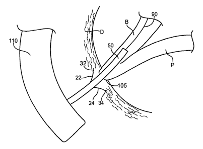
[0046] As shown in FIG. 6, the pertinent anatomy depicts common bile duct B
leading from
liver L into duodenum D. Bile duct B joins with pancreatic duct P just before
papilla of
Vater 105, as shown in FIG. 6. Papilla of Vater 105 is a small opening in
duodenum D that
drains the secretions from bile duct B and pancreatic duct P. Stomach S also
empties into
duodenum D, as shown in FIG. 6.
[0047] During a first step of an FRCP procedure, endoscope 110 is inserted
into a patient's
mouth, through the esophagus, through stomach S, and into duodenum D, as
schematically
shown in FIG. 6. In a preferred embodiment, endoscope 110 is a side-viewing
endoscope.
The distal end of endoscope 110 is positioned in the vicinity of papilla of
Vater 105. Papilla
of Vater 105 may be located by visualizing the pancreas (not shown), and then
tracing bile
duct B and/or pancreatic duct P to the wall of duodenum D and papilla of Vater
105.
[00481 It may be difficult to gain and/or maintain access to bile duct B
during an ERCP
procedure for various reasons. First, accessing the small opening in papilla
of Vater 105 may
be troublesome. For example, the papillar opening may be fully or partially
covered by soft
tissue T, as shown in FIG. 7A. Further, even when cannulation through the
papilla is
achieved, it is easier to access pancreatic duct P rather than bile duct B,
which is slightly
angled to the side, as depicted in FIG. 6. Therefore, in accordance with one
aspect, spreader
20 may be provided to facilitate access into bile duct B during an ERCP
procedure.
8
CA 02659591 2009-01-06
WO 2008/008384 PCT/US2007/015803
100491 In FIG. 7A, endoscope 110 has been maneuvered into a patient's duodenum
D and
positioned in the vicinity of papilla of Vater 105, as explained above.
Catheter 50 is
advanced through a working lumen (not shown) of endoscope 110 and is
positioned adjacent
to papilla of Vater 105, as shown in FIG. 7A.
100501 As explained above with respect to FIGS. 3-5, spreader 20 preferably is
pre-loaded
into lumen 56 of catheter 50. Lumen 56 constrains arms 22, 24 and 26 in a
closed position
such that the arms are adjacent to one another. In a preferred embodiment,
spreader 20 is
adapted to be deployed in a controlled manner using a retainer system
comprising first and
second retainers. The first retainer may comprise looped region 42 at the
proximal region of
spreader 20, while the second retainer may comprise wire 70 having hook member
72 (FIG.
3); or may comprise rod 80 having bore 85 (FIG. 4), or another retaining
means. During
delivery of catheter 50, the first and second retainers are coupled together
to secure spreader
20 within lumen 56.
[00511 In a next step, shown in FIG. 7B, catheter 50 is retracted proximally
with respect to
spreader 20 so that engaging members 32, 34 and 36 of the arms of the spreader
are exposed
and no longer radially constrained. At this time, engaging members 32, 34 and
36 grasp
tissue T in the vicinity of papilla of Vater 105 and urge tissue T in a radial
direction away
from the papillar opening, as depicted in FIG. 7B. Further retraction of
catheter 50 causes the
distal ends of arms 22, 24 and 26 to spread apart from one another.
Preferably, prior to
deployment of spreader 20, lumen 56 is aligned with papilla of Vater 105 so
that when arms
22, 24 and 26 expand radially outward, they expand away from the papillar
opening.
100521 After deployment of arms 22, 24 and 26, catheter 50 may be further
retracted with
respect to spreader 20 to expose the coupling junction between the first and
second retainers.
In the embodiment of FIG. 3, catheter 50 is retracted proximal to hook member
72, allowing
looped region 42 of spreader 20 to be released from wire 70. In the embodiment
of FIG. 4,
catheter 50 is retracted proximal to bore 85, allowing looped region 42 of
spreader 20 to be
released from rod 80. Therefore, spreader 20 is left securely in place in
front of papilla of
Vater 105.
100531 Referring now to FIG. 7C, in a next step wire guide 90 may be advanced
distally
between one or more arms 22, 24 and 26 of spreader 20 and guided into bile
duct B. Wire
guide 90 may be advanced through a dedicated wire guide lumen of catheter 50,
such as
lumen 52 of FIG. 3. Subsequently, catheter 50 may be advanced distally over
wire guide 90,
9
CA 02659591 2009-01-06
WO 2008/008384 PCT/US2007/015803
between one or more arms 22, 24 and 26 of spreader 20, and into bile duct B,
as shown in
FIG. 7C (in this illustration, it should be noted that arm 26 is obscured by
catheter 50).
10054] Advantageously, since the arms of spreader 20 hold tissue T in a spread
position away
from papilla of Vater 105, it may be easier for endoscope 110 to view the
papillar opening.
Therefore, it may be easier to cannulate papilla of Vater 105 and achieve
access into bile duct
B. Importantly, the likelihood of entering and damaging pancreatic duct P may
be reduced.
10055] If desired, one or more procedures may be performed while catheter 50
is disposed
within bile duct B. For example, an extraction basket (not shown) may be
advanced through
catheter lumen 54 and used to remove a gallstone lodged within bile duct B.
Alternatively, as
mentioned above, catheter 50 may be used to treat a biliary stricture, for
example, by
disposing an- inflation balloon on an exterior surface of catheter 50.
Further, a lithotripsy
probe or other device may be inserted into bile duct B via the papillar
opening once access is
achieved and maintained.
10056] Once the desired surgical procedure(s) are completed, spreader 20 may
be left inside
the patient or removed. In order to remove spreader 20, a reverse sequence of
one or more
steps from 7A-7B may be used. For example, the hook member 72 on the distal
end of wire
70 may be advanced and engaged with the looped region 42 of spreader 20.
Catheter 50 is
then advanced distally such that lumen 56 is advanced over proximal region 40
of spreader
20 to collapse the arms 22, 24 and 26 together, thereby allowing spreader 20
to be removed.
Alternatively, a forceps or other grasping device can be deployed through a
separate sheath,
the forceps being used to grasp and pull the spreader 20 into the sheath. The
catheter or
sheath is advanced over proximal region 40 and then over arms 22, 24 and 26 to
cause the
arms to move radially inward and disengage tissue T. The advancement of the
catheter or
sheath will cause arms 22, 24 and 26 to be radially constrained therein. If
desired, spreader
20 may be retracted proximally within the catheter or sheath by engaging
looped region 42.
10057] Alternatively, spreader 20 may be left inside the patient, as shown in
FIG. 7D.
Spreader 20 may be configured to detach from tissue T over a period of time
and may pass
naturally through the patient. Spreader 20 may also comprise a biodegradable
material that
will eventually dissolve and pass harmlessly out of the body. If desired,
spreader 20 may be
designed to permanently engage tissue T, in which case the spreader will be
left inside the
patient.
CA 02659591 2009-01-06
WO 2008/008384 PCT/US2007/015803
100581 In a further alternative embodiment, spreader 20 may be removable when
made from
a shape memory material, whereby the spreader can assume a relaxed
configuration in which
it readily disengages from tissue upon application of a certain cold or hot
medium. More
specifically, a shape memory material may undergo a substantially reversible
phase
transformation that allows it to "remember" and return to a previous shape or
configuration.
For example, in the case of nickel-titanium alloys, a transformation between
an austenitic
phase and a martensitic phase may occur by cooling and/or heating (shape
memory effect) or
by isothermally applying and/or removing stress (superelastic effect).
Austenite is
characteristically the stronger phase and martensite is the more easily
deformable phase.
100591 In an example of the shape memory effect, a nickel-titanium alloy
having an initial
configuration in the austenitie-phase may be-cooled-below a transformation
temperature (Mf)
to the martensitic phase and then deformed to a second configuration. Upon
heating to
another transformation temperature (Af), the material may spontaneously return
to its initial
configuration. Generally, the memory effect is one-way,.which means that the
spontaneous
change from one configuration to another occurs only upon heating. However, it
is possible
to obtain a two-way shape memory effect, in which a shape memory material
spontaneously
changes shape upon cooling as well as upon heating.
10060] Applying these shape-memory properties to spreader 20, it will be
possible to retract
catheter 50 to expose spreader 20 to body temperature and cause arms 22, 24
and 26 to
expand radially outward and grasp tissue. When it is desired to remove
spreader 20, a
second, predetermined temperature may be applied to spreader 20, e.g., by
injecting. fluid
through catheter 50 or by direct contact of a temperature-inducing element, to
cause arms 22,
24 and 26 to transform to a relaxed state in which they more easily let go of
the tissue. Upon
application of this temperature, spreader 20 may assume a relatively
atraumatic posture so
that it can be safely passed through the body.
100611 Referring now to FIG. 8, an alternative spreader is described. Spreader
120 is similar
to spreader 20 of FIGS. 1-7, with a main exception that biasing member 140 is
added. As
shown in FIG. 8, biasing member 140 may be coupled directly to engaging
members 132 and
136 of arms 122 and 126, respectively. Biasing member 140 also may be coupled
directly to
arms 122, 124 and/or 126 at a location proximal of the engaging members. As
depicted,
biasing member 140 may comprise a cylindrical, zig-zag shaped member having a
plurality
of substantially straight sections separated by a plurality of bends* although
other
11
CA 02659591 2009-01-06
WO 2008/008384 PCT/US2007/015803
configurations are possible. Biasing member 140 may be manufactured using a
nickel-
titanium alloy and may comprise a reduced profile delivery configuration and a
radially
expanded spreading configuration. In the expanded state, biasing member 140
may provide a
larger tissue engaging area compared to the use of arms 22, 24 and 26 by
themselves, and
may facilitate entry into bodily openings that are otherwise difficult to
access.
[0062] While reference has been made to facilitating access to the common bile
duct via the
papilla of Vater, spreader 20 may be used to help spread tissue to gain access
to many other
constrained bodily openings, passageways, ducts or cavities. Alternatively,
spreader 20 may
be used to open an annular passageway itself, as opposed to tissue in the
vicinity of the
passageway. In the latter embodiment, arms 22, 24 and 26 may be designed to
have a
stronger radial- force, for example, to-spread-open a sphincter, such as the-
sphincter of Oddi or
another passageway.
[0063] Referring now to FIGS. 9-14, further alternative embodiments of a
spreader are
described. In FIG. 9, spreader 200 is formed from first and second portions
202 and 204.
First portion 202 comprises first and second arms 212 and 213, which are
separated by
central region 211. Similarly, second portion 204 comprises third and fourth
arms 216 and
217, which are separated by central region 215, as shown in FIG. 9. The four
arms 212, 213,
216 and 217 may be provided as generally explained above with respect to the
arms 22, 24
and 26 of spreader 20. In particular, each of the four arms 212, 213, 216 and
217 may
comprise a curvature and/or an engaging member bent outwardly and adapted to
grasp tissue.
[0064] In a preferred embodiment, central regions 211 and 215 may comprise a
wire or other
suitable material bent into a substantially circumferential shape spanning
between 180-360
degrees. Alternatively, central regions 211 and 215 may comprise a wire
forming a 360
degree loop, U-shape, or the like, for purposes described below.
[0065] Referring to FIGS. 10-11, in a next step, first and second portions 202
and 204 are
coupled together. In FIG: 10, central regions 211 and 215 may be aligned and
then coupled
together using an adhesive 220. Once coupled, the four arms 212, 213, 216 and
217 of
spreader 200 preferably extend in opposing circumferential directions to
facilitate spreading
tissue, as explained above. Alternatively, as shown in FIG. 11, central
regions 211 and 215
may be braided or twisted together. In either of the embodiments shown in
FIGS. 10-11, the
overlapping portions may form a loop, U-shape, or the like. In a further
alternative
12
CA 02659591 2009-01-06
WO 2008/008384 PCT/US2007/015803
embodiment, a sleeve may be disposed over central regions 211 and 215 to help
secure first
and second portions 202 and 204 together.
100661 Referring now to FIG. 12, a catheter suitable for delivering spreader
200 is described.
Catheter 240 preferably comprises proximal and distal ends and. at least one
lumen 244
extending therebetween. Catheter 240 further comprises an' exterior surface
having at least
one ridge 242 formed therein, as shown in FIG. 12. In this embodiment,
spreader 200 is
delivered substantially from the outside of catheter 240, as opposed to
through an interior
lumen. Specifically, overlapping central regions 211 and 215 may be disposed
around a
perimeter of catheter 240 using a frictional fit, or by being held into place
within ridge 242,
or using an additional securing mechanism.
[00671- In FIG. 1-2, at least one filament 250 is employed- to hold the four
arms 212, 21-3, 216
and 217 of spreader 200 together in the delivery .state. Filament 250
preferably extends
through lumen 244 and spans the entire length of catheter 240. Once spreader
200 is
positioned at a desired location, a proximal end of filament 250 is retracted
proximally to
thereby release arms 212, 213, 216 and 217 and allow their radial expansion,
as shown in
FIGS. 10-11 above.
[00681 In an alternative embodiment, shown in FIG. 13, a splittable sheath is
employed to
hold the four arms 212, 213, 216 and 217 of spreader 200 together in the
delivery state.
Splittable sheath 260 has a proximal end coupled to wire 264, which extends
longitudinally
through lumen 244. Splittable sheath 260 further comprises at least one tear
line 262. In use,
when it becomes desirable to deploy spreader 200, wire 264 is retracted
proximally within
lumen 244, thereby causing the at least one tear line 262 to tear apart and
release spreader
200. At this time, splittable sheath 260 may be withdrawn into lumen 244,
while arms 212,
213, 216 and 217 are released and radially expand to engage tissue, as
depicted in FIGS. 10-
11 above.
100691 Referring to FIG. 14, in a further alternative embodiment, spreader 300
is similar to
spreader 200 of FIGS. 9-11, with a main exception that it is manufactured from
a single
component instead of two separate components that are coupled together.
Spreader 300
comprises a base portion 302 having a circular configuration, and a plurality
of integral arms
303-306 extending therefrom, as shown in FIG. 14. Base portion 302 may be
sized to be fit
over an outer surface of catheter 240, for example, using a frictional fit, or
by being placed in
13
CA 02659591 2009-01-06
WO 2008/008384 PCT/US2007/015803
ridge 242. Alternatively, spreader 300 may be configured for delivery
internally though a
lumen of a catheter, as generally described above with respect to FIGS. 3-4.
[00701 While various embodiments of the invention have been described, it will
be apparent
to those of ordinary skill in the art that many more embodiments and
implementations are
possible within the scope of the invention. Accordingly, the invention is not
to be restricted
except in light of the attached claims and their equivalents.
14