Note: Descriptions are shown in the official language in which they were submitted.
CA 02659785 2012-10-11
52281-12
METHODS AND DEVICES FOR DELIVERING AGENTS
ACROSS BIOLOGICAL BARRIERS
[0001]
Background
[0002] Numerous drugs and therapeutic agents have been developed in the battle
against disease and illness. However, a frequent therapeutic limitation of
these drugs
is their delivery: how to transport drugs across biological barriers in the
body (e.g.,
the skin, the oral mucosa, the blood-brain barrier), which normally do not
transport
drugs at rates that are therapeutically useful.
[0003] Drugs are commonly administered orally as pills or capsules. However,
many drugs cannot be effectively delivered in this manner due to degradation
in the
gastrointestinal tract and/or elimination by the liver. Moreover, some drugs
cannot
effectively diffuse across the intestinal mucosa. Patient compliance may also
be a
problem, for example, in therapies requiring that pills be taken at particular
intervals
over a prolonged period.
[0004] Another common technique for delivering drugs across a biological
barrier is
the use of a needle, such as those used with standard syringes or catheters,
to
transport drugs across (through) the skin. While effective for this purpose,
needles
generally cause pain; local damage to the skin at the site of insertion;
bleeding,
which increases the risk of disease transmission; and a wound sufficiently
large to be
a site of infection.
[0005] An alternative delivery technique is the transdermal patch, which
usually
relies on diffusion of the drug across the skin. However, this method is not
useful for
many drugs, due to the poor permeability (i.e., effective barrier properties)
of the
skin. The rate of diffusion depends in part on the size and hydrophilicity of
the drug
molecules and the concentration gradient across the stratum comeum. Few drugs
-1-
=
CA 02659785 2012-07-04
, 52281-12
have the necessary physiochemical properties to be effectively delivered
through the
skin by passive diffusion. Iontophoresis, electroporation, ultrasound, and
heat (so-
called active systems) have been used in an attempt to improve the rate of
delivery.
While providing varying degrees of enhancement, these techniques are not
suitable
for all types of drugs, failing to provide the desired level of delivery. In
some cases,
they are also painful and inconvenient or impractical for continuous
controlled drug
delivery over a period of hours or days. Attempts have been made to design
alternative devices for active transfer of drugs through the skin.
[0006] Thus, there remains a need for better drug delivery devices, which make
smaller incisions, deliver drug with greater efficiency (greater drug delivery
per
quantity applied) and less variability of drug administration, and/or are
easier to use.
Summary
[0007] In one aspect, the invention relates to a
delivery device which includes a microneedle with an integrated agent
reservoir.
The integrated reservoir may include, for example, an opening extending
through the
entirety of the width or depth of the needle or a depression in one side of
the needle.
In such a configuration, when an agent is placed within the integrated
reservoir and
the microneedle is applied to the biological barrier (e.g., the skin, the oral
mucosa
barrier, the blood-brain barrier, etc.) of a patient, the agent, being located
predominantly within the interior volume of the microneedle, is largely
protected
from contacting the barrier as the microneedle passes through the barrier.
This
greatly reduces the loss of the agent cause by contact with the barrier. Such
loss can
be significant given the small quantity of agent delivered by microneedle
technologies and can affect the therapeutic effectiveness of the agent.
[0008] In one embodiment, the integrated reservoir encompasses between 20%--
50% of the volume of the microneedle. In other embodiments, integrated
reservoir
encompasses as little as 10% or and as much as 70% of the volume of the first
-2-
CA 02659785 2009-02-02
WO 2007/019539
PCT/US2006/030981
microneedle. The integrated reservoir is filled, in one embodiment with a
biologically active agent, such as a drug or a vaccine.
[0009] In various embodiments, the microneedle is made of, for example and
without limitation, stainless steel, titanium, or a biodegradable polymer. The
microneedle can be between 150 and 3000 microns long, and between 10 and 2000
microns wide.
[0010] Additional features of the invention include microneedles with depth
guards
and the use of base elements, which in some embodiments are wider than the
microneedles, themselves. The base elements provide for greater structural
stability
for longer microneedles. The depth guard prevents the wider base elements from
entering the biological barrier, which would enlarge the disruption in the
barrier
caused by the microneedle.
[0011] In another embodiment, microneedles are combined into arrays. The
arrays
of microneedles allow for administration of larger volumes of agent and for
concurrent administration of multiple agents. The microneedles in the array
may be
attached to a substrate.
[0012] In another aspect, the invention relates to manufacturing the delivery
devices
described above. The method of manufacture may include dipping the microneedle
into a solution containing the agent. In an alternative embodiment, a
predetermined
volume of the agent is dispensed into the integrated reservoir.
[0013] In another aspect, the invention relates to methods of administering an
agent
across a biological barrier. The administration method includes applying one
of the
microneedle devices described above against a biological barrier, thereby
puncturing
the barrier and positioning the integrated reservoir beyond the barrier. In
one
embodiment, the method includes providing a plurality of microneedles coupled
to a
substrate. At least one of the microneedles includes an opening which defines
an
integrated reservoir. The reservoir is filled with an agent. The plurality of
microneedles are applied against the skin of a patient, puncturing the skin
and
-3-
CA 02659785 2012-07-04
52281-12
positioning the integrated reservoir beneath the surface of the skin. The
puncture
= depth is limited by a depth guard coupled to at least one of the
microneedles.
= In one aspect, the invention provides a device for delivering an agent
across a
biological barrier, the device comprising: a first microneedle comprising an
integrated
first reservoir that is an opening through the entirety of the width of the
microneedle; a
first agent contained predominantly within an interior volume of the first
microneedle,
the first agent being in a dried or semi-solid state; a first base element
from which the
first microneedle extends; and a substrate to which the first base element is
coupled;
wherein the first agent is selectively deposited in a predetermined volume
into the
first integrated reservoir by a dispensing device.
In another aspect, the invention provides a method of manufacturing a device
for
delivering an agent across a biological barrier, comprising: providing a first
microneedle having a first integrated reservoir that is an opening through the
entirety
of the width of the first microneedle; and filling the first integrated
reservoir with a first
agent by selectively depositing a predetermined volume of the first agent into
the first
integrated reservoir utilizing a dispensing device, such that the first agent
is contained
predominantly within the interior volume of the first microneedle.
In another aspect, the invention provides use of a device for delivering an
agent
transdermally, wherein the device comprises: a first microneedle comprising an
integrated first reservoir that is an opening through the entirety of the
width of the
microneedle; an agent contained predominantly within an interior volume of the
first
microneedle, the first agent being in a dried or semi-solid state; a first
base element
from which the first microneedle extends; and a substrate to which the first
base
element is coupled; wherein the agent is selectively deposited in a
predetermined
volume into the first integrated reservoir by a dispensing device.
-4-
CA 02659785 2012-07-04
, 52281-12
In another aspect, the invention provides use of a device for delivering an
agent
transdermally, wherein the device comprises: a plurality of microneedles, each
of
which extends from a base element; a substrate that is coupled to the base
element
of each of the plurality of microneedles; an integrated reservoir in at least
one
microneedle of the plurality of microneedles, the integrated reservoir being
an
opening through the entirety of the width of the at least one microneedle; an
agent
contained predominantly within an interior volume of the at least one
microneedle, the
agent being in a dried or semi-solid state; wherein the agent is selectively
deposited
in a predetermined volume into the integrated reservoir by a dispensing
device.
In another aspect, the invention provides use of a device for delivering an
agent
transdermally, wherein the device comprises: a plurality of microneedles
arranged in
a plane, each microneedle of the plurality of microneedles comprising an
integrated
reservoir which is an opening through the entirety of a width of that
microneedle
suitable for containing an agent predominantly within an interior volume of
that
microneedle; a planar support structure; and a plurality of base elements,
each of the
plurality of base elements coupling one of the plurality of microneedles to
the planar
support structure.
-4a-
CA 02659785 2012-07-04
, 52281-12
Brief Description of the Figures
[0014] The invention may be better understood from the following illustrative
description with reference to the following drawings.
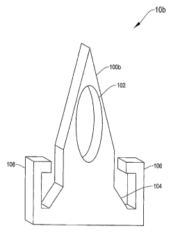
[0015] Figures lA through 1D depict microneedles with integrated drug
reservoirs
according to several illustrative embodiments of the invention.
[0016] Figures 2A through 2C depict arrays of microneedles with integrated
drug
reservoirs according to illustrative embodiments of the invention.
[0017] Figure 3 depicts a microneedle with an integrated drug reservoir which
is
filled with an agent according to an illustrative embodiment of the invention.
[0018] Figures 4A through 4C depict a method of forming a microneedle with an
integrated drug reservoir using injection molding according to an illustrative
embodiment of the invention.
[0019] Figures 5A through 5C depict a method of forming a microneedle with an
integrated drug reservoir using a stamping process according to an
illustrative
embodiment of the invention.
[0020] Figures 6A through 6C depict a method of forming a microneedle with an
integrated drug reservoir using a chemical etching technique according to an
illustrative embodiment of the invention.
[0021] Figures 7A through 7D depict four methods of filling microneedle
integrated
agent reservoirs according to illustrative embodiments of the invention.
[0022] Figures 8A through 8C and 9A through 9C illustrate methods of
administering an agent transdermally to a patient according to two embodiments
of the invention.
-4h-
CA 02659785 2009-02-02
WO 2007/019539
PCT/US2006/030981
[0023] Figures 10A-10E depict a medical device incorporating a microneedle
with
an integrated agent reservoir and an external reservoir, and a method of using
the
same, according to an illustrative embodiment of the invention.
Description of Illustrative Embodiments
[0024] Throughout the description below reference to ranges of values are
intended
to refer to the specified range, and any smaller range, or single value within
that
range. Thus, a range of 1 to 10 refers, for example, to the ranges 1 to 10, 3
to 7, or
5. In addition, like reference numerals refer to like elements.
[0025] The devices disclosed herein are useful in transport of material into
or across
biological barriers including the skin (or parts thereof); the blood-brain
barrier;
mucosal tissue (e.g., oral, nasal, ocular, vaginal, urethral,
gastrointestinal,
respiratory); blood vessels; lymphatic vessels; or cell membranes (e.g., for
the
introduction of material into the interior of a cell or cells). The biological
barriers
could be in humans or other types of animals, as well as in plants, insects,
or other
organisms, and embryos.
[0026] For internal tissues, application of the microneedle devices can be
achieved
with the aid of a catheter or laparoscope. For certain applications, such as
for drug
delivery to an internal tissue, the devices can be surgically implanted.
[0027] Skin is a biological barrier of particular use with the microneedle
device
disclosed herein. However, skin is only one example of a biological barrier.
It will
be understood that any biological barrier can be substituted for "skin"
throughout.
[0028] Specifically with respect to skin, the stratum corneum is the outer
layer,
generally between 10 and 50 cells, or between 10 and 20 gm thick. Unlike other
tissue in the body, the stratum corneum contains "cells" (called
keratinocytes) filled
with bundles of cross-linked keratin and keratohyalin surrounded by an
extracellular
matrix of lipids. It is this structure that is believed to give skin its
barrier properties,
which prevents therapeutic transdermal administration of many drugs.
-5-
CA 02659785 2009-02-02
WO 2007/019539
PCT/US2006/030981
[0029] Below the stratum come= is the viable epidermis, which is between 50
and
100 inn thick. The viable epidermis contains no blood vessels, and it
exchanges
metabolites by diffusion to and from the dermis. Beneath the viable epidermis
is the
dermis, which is between 1 and 3 mm thick and contains blood vessels,
lymphatics,
and nerves.
[0030] Figures 1A¨C depict three versions of agent delivery devices (generally
referred to as agent delivery devices 10) for delivering agents across
biological
barriers. Each agent delivery device 10 includes a microneedle (generally
referred
to as microneedle 100) with integrated agent reservoirs 102 according to
illustrative
embodiments of the invention. Microneedles 100 include microprotrusions,
microabraders, microblades, and other elements on the submicron to millimeter
scale used to pierce, cut, or otherwise disrupt the surface of a biological
barrier. The
microneedle 100 can be constructed from a variety of materials, including
metals,
ceramics, semiconductors, organics, polymers (e.g., biodegradable polymers),
and
composites. Preferred materials of construction include medical grade
stainless steel,
gold, titanium, nickel, iron, gold, tin, chromium, copper, alloys of these or
other
metals, silicon, silicon dioxide, and polymers. Representative biodegradable
polymers include polymers of hydroxy acids such as lactic acid and glycolic
acid
polylactide, polyglycolide, polylactide-co-glycolide, and copolymers with PEG,
polyanhydrides, poly(ortho)esters, polyurethanes, poly(butyric acid),
poly(valeric
acid), and poly(lactide-co-caprolactone). Representative non-biodegradable
polymers include polycarbonate, polymethacrylic acid, ethylenevinyl acetate,
polytetrafluorethylene (TEFLONTm), and polyesters.
[0031] Generally, a microneedle 100 should have the mechanical strength to
remain
intact for delivery of an agent, while being inserted into the barrier, while
remaining
in place for up to a number of days, and while being removed. In embodiments
where the microneedle 100 is formed of biodegradable polymers, however, this
mechanical requirement is less stringent, since the microneedle 100 or the tip
thereof
can break off, for example in the skin, and will biodegrade. Therefore,
biodegradable microneedles 100 can provide an increased level of safety, as
compared to nonbiodegradable ones. Nonetheless, even a biodegradable
-6-
CA 02659785 2009-02-02
WO 2007/019539
PCT/US2006/030981
microneedle 100 still needs to remain intact at least long enough for the
microneedle
100 to serve its intended purpose (e.g, its delivery function). The
microneedle 100
should preferably be sterilizable using standard methods.
[0032] In general, one benefit of delivering an agent via a microneedle 100 is
that
while the microneedle 100 disrupts a patient's skin, thereby providing access
to the
blood flow of a patient, it does not disrupt the skin deep enough to generate
a
response from the patient's nerves. Thus agent delivery via a microneedle 100
typically is less painful than standard injection delivery devices. To this
end, the
height (or length) of the microneedle 100 generally is between about 100 m
and
about 3 mm. In transderrnal applications, the "insertion depth" of the
microneedle
100 is preferably between about 100 gm and about 1 mm, so that insertion of
the
microneedle 100 into the skin does not penetrate through the lower dermis. In
such
applications, the actual length of the microneedle 100 may be longer, since
some
portion of the microneedle 100 distal the tip may not be inserted into the
skin; the
uninserted length depends on the particular device design and configuration.
[0033] In order to reduce injury and the risk of infection to the patient, the
microneedle 100 is formed to be between 10 gm and about 2mm wide, preferably
between 100 and 300 pm wide. A microneedle 100 will be generally planar,
cylindrical, conical, or rectangular in shape, though other polygonal and
irregular
shapes are also suitable. The distal end of the microneedle 100 preferably
tapers to a
point.
[0034] The agent delivery device 10a illustrated in Figure 1A, includes
microneedle
100a. Microneedle 100a includes an integrated reservoir 102 for holding agents
to
be delivered across a biological barrier, such as the skin. The integrated
reservoir
102 consists of an opening that passes through a side of the microneedle 100a.
The
integrated reservoir 102 encompasses a substantial portion of the volume of
the
microneedle 100a. For example, the reservoir 102 encompasses between 10% and
70% of the volume of the microneedle 100a. In other configurations, the
integrated
reservoir 102 encompasses between 20 and 50% of the volume of the microneedle
100a. Thus, the exposed surface area of any agent stored in the integrated
reservoir
102 is relatively low in relation to the total volume of the stored agent. The
-7-
CA 02659785 2009-02-02
WO 2007/019539
PCT/US2006/030981
integrated reservoir 102 is contained wholly within the physical bounds of the
microneedle 100a. The integrated reservoir 102 can take on virtually any
shape,
whether it be polygonal, irregular, circular, or elliptical.
[0035] Figure 1B depicts a second agent delivery device 10b for delivering
agents
across biological barriers. The delivery device 10b includes a microneedle
100b
with an integrated reservoir 102 according to a second embodiment of the
invention.
The microneedle 100b is coupled to a base element 104. The base element 104
can
be wider than the microneedle 100b to provide additional strength and
stability.
[0036] To prevent the base element 104 from widening the wound in a patient's
skin
during insertion, the delivery device 10b includes a depth guard 106. The
depth
guard 106 includes a rigid member that extends from the base element 104
toward
the distal end of the microneedle 100b to a point beyond the base element 104.
In an
alternative embodiment, the depth guard 106 extends out directly from the
microneedle 100b, substantially perpendicular to the length of the microneedle
100b.
In both embodiments, upon application of the microneedle 100b to the skin of a
patient, the depth guard 106 acts as a barrier and prevents the microneedle
100b
from being inserted so deep within the skin that the wider base element 104
further
disrupts the skin surface. In embodiments in which the base element 104 is not
substantially wider than the microneedle 100b, the depth guard 106 prevents
the
microneedle 100b from penetrating too deeply.
[0037] Figure 1C depicts a third illustrative embodiment of a delivery device
10c
according to an illustrative embodiment of the invention. Delivery device 10c
includes microneedle 100c with an integrated reservoir 102. In addition to the
features of the delivery devices 10a and 10b depicted in Figures 1A-1B, the
delivery
device 10c includes a substrate 108 to which the base element 104 is coupled.
In the
illustrative embodiment, the substrate 108 is formed integrally with the base
element
104, microneedle 100c, and depth guard 106. The substrate can be, for example,
between 300 ptin ¨500 pm wide and between about 400 Am and about lmm long.
As shown, the substrate 108 is generally parallel to the base element 104 and
microneedle 100c, though in other embodiments, the substrate 108 is generally
perpendicular to, or at an angle to the base element 104 and substrate 108.
The
-8-
CA 02659785 2009-02-02
WO 2007/019539
PCT/US2006/030981
substrate 108 includes two alignment holes 110 for aligning a plurality of
microneedles 100c into an array. The alignment holes 110 can be, for example,
spaced between about 100 pm to about 300 pm apart, and be between about 50 Am
to about 200 pm in diameter.
[0038] In another embodiment of the delivery device 10d, depicted in Figure
1D, the
microneedle 100d includes an integrated reservoir 102d, which does not pass
through the entirety of the side of the microneedle 100d. Instead, the
integrated
reservoir 102d is formed by creating a depression into one or more sides of
the
microneedle 100d into which an agent can be placed. As with the version of the
integrated reservoir 102 in which the reservoir passes through the entirety of
a side
of a microneedle 100a, described above in relation to Figure 1A, the
depression
integrated reservoir 102d preferably takes up a substantial portion of the
volume of
the microneedle 100d. The integrated reservoir 102d can take on virtually any
shape, whether it be polygonal, irregular, circular, or elliptical.
[0039] Figures 2A ¨ 2C illustrate arrays of microneedles according to three
illustrative embodiments of the invention. Microneedle arrays (generally
microneedle arrays 200) are useful, for example and without limitation, in at
least
the three following circumstances: 1) if the reservoir 102 of a single
microneedle
100 may not be able to hold a sufficient volume of an agent to be effective;
2) if it
desired to deliver the agent or agents to a greater surface area of a
biological barrier;
and 3) if multiple agents are to be administered concurrently and the multiple
agents
are not sufficiently compatible to store or administer in a single microneedle
100
integrated reservoir 102.
[0040] Figure 2A depicts a delivery device 10e including a two-dimensional
microneedle array 200a according to an illustrative embodiment of the
invention.
The two-dimensional microneedle array 200a includes four microneedles 100a, as
described in relation to Figure 1A. The inclusion of only four microneedles
100a in
the two-dimensional microneedle array 200a is for illustrative purposes only.
The
two-dimensional microneedle array 200a may include a smaller or larger number
of
microneedles 100a. For example, the two-dimensional microneedle array 200a may
include as few as three microneedles 100a. The dimensionality of the
microneedle
-9-
CA 02659785 2009-02-02
WO 2007/019539
PCT/US2006/030981
array 200a refers to the geometric relationship among the microneedles 100a in
the
array, and thus, two microneedles by definition could only form a one
dimensional
array. The two-dimensional microneedle array can include as many as sixteen
microneedles 100a, or more. Other microneedle 100 implementations, for example
and without limitation, microneedles 100b-100d, may be incorporated into the
two-
dimensional microneedle array 200a.
[0041] The microneedles 100a in the two-dimensional microneedle array 200a are
attached to a substrate 108. The microneedles 100a may be integrally formed
with
the substrate 108 or they may be physically attached, for example with an
adhesive,
to the substrate 108. In the two-dimensional array 200a, the substrate 108
serves as
a depth guard 106. In other implementations, one or more of the microneedles
100a
on the two-dimensional array 200a include independent depth guards 106.
[0042] In the two-dimensional microneedle array 200a depicted in Figure 2A,
two
of the microneedles 100a include a first agent 202a stored in their
corresponding
integrated reservoirs 102 and two of the microneedles 100a include a different
agent
202b (agents will be referred to hereinafter generally as agents 202).
[0043] Two-dimensional microneedle array 200a may also include a feature in
which the substrate 108 is coated with an adhesive for adhering to the
patient's skin.
The adhesive keeps the integrated reservoirs 102 of the microneedles 100
beneath
the skin for extended periods of time, for example, to allow for gradual
absorption of
agents stored in the reservoir 102.
[0044] Figure 2B depicts a second illustrative embodiment of a delivery device
10f
having a two-dimensional microneedle array 200b. Two-dimensional microneedle
array 200b includes four microneedles 100d. Microneedles 100d resemble
microneedles 100a with the addition of alignment holes 110, as previously
depicted
in microneedle 100c. In this two-dimensional array 200b, alignment elements
204
pass through the alignment holes 110 of the microneedles 100d and into base
structure 206. Spacers 208 can be placed on the alignment elements between the
microneedles 100c to keep them apart and firmly in place.
-10-
CA 02659785 2009-02-02
WO 2007/019539
PCT/US2006/030981
[0045] Figure 2C depicts a delivery device lOg including a one-dimensional
microneedle array 200c according to an illustrative embodiment of the
invention.
The one-dimensional microneedle array 200c includes ten microneedles 100c. The
one-dimensional microneedle array 200c may have fewer than ten microneedles
100c (as few as two) or it can include additional microneedles 100c. The one-
dimensional microneedle array 200c may be formed by manufacturing a single
integrated set of microneedles, or each microneedle 100c may be formed
independently and then joined together. The microneedles 100c can be joined
using,
for example, adhesives, bonding, or alignment elements 204.
[0046] Figure 3 depicts delivery device 10b depicted in Figure 1B having an
agent
202 place in the integrated reservoir 102. The term agent refers to a single
agent 202
or a combination of several agents 202. The agents 202 may be biologically
active
or biologically inactive. Sample agents 202 include, without limitation,
drugs,
vaccines, allergens, antigens, excipients, anti-coagulants, surfactants,
radiological
dyes or markers, toxins, or any other agent, compound or substance suitable
for
introduction into a biological barrier. As stored, the agents 202 may be, for
example, dry (e.g., a film), or in a semi-solid gel.
[0047] One class of agents 202 includes therapeutic agents in all the major
therapeutic areas including, but not limited to, anti-infectives, such as
antibiotics and
antiviral agents; analgesics, including fentanyl, sufentanil, remifentanil,
buprenorphine and analgesic combinations; anesthetics; anorexics;
antiarthritics;
antiasthmatic agents such as terbutaline; anticonvulsants; antidepressants;
antidiabetic agents; antidiarrheals; antihistamines; anti-inflammatory agents;
antimigraine preparations; antimotion sickness preparations such as
scopolamine
and ondansetron; antinauseants; antineoplastics; antiparkinsonism drugs;
antipruritics; antipsychotics; antipyretics; antispasmodics, including
gastrointestinal
and urinary; anticholinergics; sympathomimetrics; xanthine derivatives;
cardiovascular preparations, including calcium channel blockers such as
nifedipine;
beta blockers; beta-agonists such as dobutamine and ritodrine; antianythmics;
antihypertensives such as atenolol; ACE inhibitors such as ranitidine;
diuretics;
vasodilators, including general, coronary, peripheral, and cerebral; central
nervous
-11-
CA 02659785 2009-02-02
WO 2007/019539
PCT/US2006/030981
system stimulants; cough and cold preparations; decongestants; diagnostics;
hormones such as parathyroid hormone; hypnotics; immunosuppressants; muscle
relaxants; parasympatholytics; parasympathomimetrics; prostaglandins;
proteins;
peptides; psychostimulants; sedatives; and tranquilizers. These agents may
take the
form of peptides, proteins, carbohydrates (including monosaccharides,
oligosaccharides, and polysaccharides), nucleoproteins, mucoproteins,
lipoproteins,
glycoproteins, nucleic acid molecules (including any form of DNA such as cDNA,
RNA, or a fragment thereof, oligonucleotides, and genes), nucleotides,
nucleosides,
lipids, biologically active organic or inorganic molecules, or combinations
thereof.
[0048] Further specific examples of agents 202 include, without limitation,
growth
hormone release hormone (GHRH), growth hormone release factor (GHRF), insulin,
insultropin, calcitonin, octreotide, endorphin, TRN, NT-36 (chemical name: N-
[[(s)-
4-oxo-2-azetidinyl]carbony1]-L-histidyl-L-p- rolinamide), liprecin, pituitary
hormones (e.g., HGH, HMG, desmopressin acetate, etc), follicle luteoids, aANF,
growth factors such as growth factor releasing factor (GFRF), bMSH, GH,
somatostatin, bradykinin, somatotropin, platelet-derived growth factor
releasing
factor, asparaginase, bleomycin sulfate, chymopapain, cholecystokinin,
chorionic
gonadotropin, erythropoietin, epoprostenol (platelet aggregation inhibitor),
gluagon,
HCG, hirulog, hyaluronidase, interferon alpha, interferon beta, interferon
gamma,
interleukins, interleukin-10 (IL-10), erythropoietin (EPO), granulocyte
macrophage
colony stimulating factor (GM-CSF), granulocyte colony stimulating factor (G-
CSF), glucagon, leutinizing hormone releasing hormone (LHRH), LHRH analogs
(such as goserelin, leuprolide, buserelin, triptorelin, gonadorelin, and
napfarelin,
menotropins (urofollitropin (FSH) and LH)), oxytocin, streptokinase, tissue
plasminogen activator, urokinase, vasopressin, deamino [Va14, D-Arg8] arginine
vasopressin, desmopressin, corticotropin (ACTH), ACTH analogs such as ACTH (1-
24), ANP, ANP clearance inhibitors, angiotensin II antagonists, antidiuretic
hormone agonists, bradykinn antagonists, ceredase, CSI's, calcitonin gene
related
peptide (CGRP), enkephalins, FAB fragments, IgE peptide suppressors, IGF-1,
neurotrophic factors, colony stimulating factors, parathyroid hormone and
agonists,
parathyroid hormone antagonists, parathyroid hormone (PTH), PTH analogs such
as
PTH (1-34), prostaglandin antagonists, pentigetide, protein C, protein S,
renin
-12-
CA 02659785 2009-02-02
WO 2007/019539
PCT/US2006/030981
inhibitors, thymosin alpha-1, thrombolytics, TNF, vasopressin antagonists
analogs,
alpha-1 antitrypsin (recombinant), and TGF-beta.
[0049] The biologically active agents 202 can also be in various forms, such
as free
bases, acids, charged or uncharged molecules, components of molecular
complexes
or nonirritating, pharmacologically acceptable salts. Further, simple
derivatives of
the active agents 202 (such as ethers, esters, amides, etc.), which are easily
hydrolyzed at body pH, enzymes, etc., can be employed.
[0050] Additional agents 202 may be stored in the same integrated reservoir
102 as
a therapeutic agent 202, or they may be stored in integrated reservoirs 102
integrated
into separate microneedles 100. For example, the integrated reservoir 102 may
contain a viscosity enhancing agent 202 such as maleic acid, malic acid,
malonic
acid, tartaric acid, adipic acid, citraconic acid, fumaric acid, glutaric
acid, itaconic
acid, meglutol, mesaconic acid, succinic acid, citramalic acid, tartronic
acid, citric
acid, tricarballylic acid, ethylenediaminetetraacetic acid, aspartic acid,
glutamic acid,
carbonic acid, sulfuric acid, phosphoric acid, hydrochloric acid, hydrobromic
acid,
nitric acid, sulfuric acid, benzene sulfonic acid, methane sulfonic acid,
glycolic acid,
gluconic acid, glucuronic acid, lactic acid, pyruvic acid, tartronic acid,
propionic
acid, pentanoic acid, carbonic acid, adipic acid, citraconic acid, and
levulinic acid.
[0051] Additional potential agents 202 include surfactants, such as
zwitterionic,
amphoteric, cationic, anionic, or nonionic, including, without limitation,
sodium
lauroamphoacetate, sodium dodecyl sulfate (SDS), cetylpyridinium chloride
(CPC),
dodecyltrimethyl ammonium chloride (TMAC), benzalkonium, chloride,
polysorbates such as Tween 20 and Tween 80, other sorbitan derivatives, such
as
sorbitan laurate, and alkoxylated alcohols, such as laureth-4.
[0052] Still other useful agents 202 include include polymeric materials or
polymers
that have amphiphilic properties, for example and without, cellulose
derivatives,
such as hydroxyethylcellulose (HEC), hydroxypropylmethylcell- ulose (HPMC),
hydroxypropycellulose (HPC), methylcellulose (MC), hydroxyethylmethylcellulose
(HEMC), or ethylhydroxy-ethylcellulose (EHEC), as well as pluronics.
-13-
CA 02659785 2009-02-02
WO 2007/019539
PCT/US2006/030981
[0053] Further agents 202 compatible for use in the integrated reservoir 102
include
biocompatible carriers, which include, without limitation, human albumin,
bioengineered human albumin, polyglutamic acid, polyaspartic acid,
polyhistidine,
pentosan polysulfate, polyamino acids, sucrose, trehalose, melezitose,
raffinose and
stachyose.
[0054] Stabilizing agents 202, which can comprise, without limitation, a non-
reducing sugar, a polysaccharide or a reducing sugar, may be stored in the
integrated
reservoir 102. Suitable non-reducing sugars for use in the methods and
compositions
of the invention include, for example, sucrose, trehalose, stachyose, or
raffinose.
Suitable polysaccharides for use in the methods and compositions of the
invention
include, for example, dextran, soluble starch, dextrin, and insulin. Suitable
reducing
sugars for use in the methods and compositions of the invention include, for
example, monosaccharides such as, for example, apiose, arabinose, lyxose,
ribose,
xylose, digitoxose, fucose, quercitol, quinovose, rhamnose, allose, altrose,
fructose,
galactose, glucose, gulose, hamamelose, idose, mannose, tagatose, and the
like; and
disaccharides such as, for example, primeverose, vicianose, rutinose,
scillabiose,
cellobiose, gentiobiose, lactose, lactulose, maltose, melibiose, sophorose,
and
turanose, and the like.
[0055] Other agents 202 include "pathway patency modulators", which can
comprise, without limitation, osmotic agents 202 (e.g., sodium chloride),
zwitterionic compounds (e.g., amino acids), and anti-inflammatory agents, such
as
betamethasone 21-phosphate disodium salt, triamcinolone acetonide 21-disodium
phosphate, hydrocortamate hydrochloride, hydrocortisone 21-phosphate disodium
salt, methylprodnisolone 21-phosphate disodium salt, methylprednisolone 21-
succinaate sodium salt, paramethasone disodium phosphate and prednisolone 21-
succinate sodium salt, and anticoagulants, such as citric acid, citrate salts
(e.g.,
sodium citrate), dextrin sulfate sodium, aspirin and EDTA.
[0056] In yet another embodiment of the invention, the integrated reservoir
102
includes a solubilising/complexing agent 202, for example, alpha-cyclodextrin,
beta-
cyclodextrin, gamma-cyclodextrin, glucosyl-alpha-cyclodextrin, maltosyl-alpha-
cyclodextrin, glucosyl-beta-cyclodextrin, maltosyl-beta-cyclodextrin,
hydroxypropyl
-14-
CA 02659785 2009-02-02
WO 2007/019539
PCT/US2006/030981
beta-cyclodextrin, 2-hydroxypropyl-beta-cyclodextrin, 2-hydroxypropyl-gamma-
cyclodextrin, hydroxyethyl-beta-cyclodextrin, methyl-beta-cyclodextrin,
sulfobutylether-alpha-cyclodextrin, sulfobutylether-beta-cyclodextrin,
sulfobutylether7 beta-cyclodextrin, and sulfobutylether-gamma-cyclodextrin.
[0057] Additional useful agents 202 include non-aqueous solvents, such as
ethanol,
isopropanol, methanol, propanol, butanol, propylene glycol, dimethysulfoxide,
glycerin, N,N-dimethylformamide and polyethylene glycol 400.
[0058] In order to facilitate filling of the integrated reservoir 102,
hydrophilic
compounds can be applied to the surfaces of the microneedle 100 defining the
integrated reservoir 102. The hydrophilic compound can be selected from the
following group: hydroxyethyl starch, dextran, poly(vinyl alcohol),
poly(ethylene
oxide), poly(2-hydroxyethylmethacrylate), poly(n-vinyl pyrolidone),
polyethylene
glycol and mixtures thereof, and like polymers. A hydrophobic compound, such
as
TEFLONTm, silicon or other low energy material, can be applied to the
remainder of
the microneedle 100. Alternatively, either a hydrophobic or hydrophilic
compound
can be applied to the entirety of the microneedle 102, including the surfaces
defining
the reservoir 102.
[0059] Microneedles 100, as depicted in Figures 1A-1C, can be formed using a
variety of microfabrication techniques known in the art. For example, the
microneedles 100 can be fabricated using lithography; etching techniques, such
as
wet chemical, dry, and photoresist removal; thermal oxidation of silicon;
electroplating and electroless plating; diffusion processes, such as boron,
phosphorus, arsenic, and antimony diffusion; ion implantation; film
deposition, such
as evaporation (filament, electron beam, flash, and shadowing and step
coverage),
sputtering, chemical vapor deposition (CVD), epitaxy (vapor phase, liquid
phase,
and molecular beam), electroplating, screen printing, and lamination. See
generally
Jaeger, Introduction to Microelectronic Fabrication (Addison-Wesley Publishing
Co., Reading Mass. 1988); Runyan, et al., Semiconductor Integrated Circuit
Processing Technology (Addison-Wesley Publishing Co., Reading Mass. 1990);
Proceedings of the IEEE Micro Electro Mechanical Systems Conference 1987-1998;
-15-
CA 02659785 2009-02-02
WO 2007/019539
PCT/US2006/030981
Rai-Choudhury, ed., Handbook of Microlithography. Micromachining &
Microfabrication (SPIE Optical Engineering Press, Bellingham, Wash. 1997).
[0060] More particularly, Figures 4A-6C depict specific methods of forming
microneedles 100 with integrated agent reservoirs 102 as described in relation
to
Figures 1A¨C.
[0061] Figure 4A depicts a method of forming a microneedle 100 using an
injection
molding technique according to an illustrative embodiment of the invention.
The
first step, depicted in Figure 4A, includes providing a microneedle injection
mold
402. The microneedle injection mold 402 can be formed using one or more of the
microfabrication processes mentioned above. The interior of the microneedle
injection mold 402 includes the relevant features of the microneedle 100. In
the
second step, depicted in Figure 4B, a molten material, for example, a molten
metal
or plastic, is injected into the microneedle injection mold 402. After the
molten
material solidifies, the microneedle injection mold 402 is opened yielding the
microneedle 100a depicted in Figure 4C.
[0062] In similar methods, the microneedle injection mold 402 is formed from a
transparent material. Light sensitive material is injected into the
microneedle
injection mold 402 is then set by the application of, for example, ultraviolet
light.
After the material is set, the microneedle injection mold 402 is opened to
yield the
microneedle 100a.
[0063] Figures 5A-5C depict a method of forming a microneedle 100 with an
integrated reservoir 102 using a stamping process according to one
illustrative
embodiment of the invention. The first step, depicted in Figure 5A includes
providing a microneedle stamping mold 502. As with the microneedle injection
mold 502 described with respect to Figure 4A, the microneedle stamping mold
502
can be fabricated using one or more of the microfabrication techniques
described
above. As depicted in Figure 5B, the microneedle stamping mold 502 is then
stamped into the material 504 being used to form the microneedle 100. The
material
may be heated to a semi-solid or liquid state prior to stamping. If the
material is
heated prior to stamping, the material is allowed to cool before the
microneedle
-16-
CA 02659785 2009-02-02
WO 2007/019539
PCT/US2006/030981
stamping mold 502 is removed. After the microneedle stamping mold 502 is
removed, excess material, if any, is removed, resulting in the microneedle 100
with
an integrated reservoir 102 depicted in Figure 5C. The stamping process can be
used to form a strip or a sheet of microneedles. In addition, a substrate can
be
processed in a reel-to-reel fashion resulting in a continuous chain of
microneedles.
[0064] In additional implementations of the methods described in relation to
Figures
4A-5C, microneedles 100 can be formed using a multi-step process that may
include
both injection molding and stamping steps. For example the exterior shape of
the
microneedle 100, i.e., the microneedle 100 without a reservoir 102, is formed
using
injection molding or a first stamping step. Subsequently, a stamp may puncture
the
microneedle 100 to form the integrated reservoir 102.
[0065] Figures 6A-6C depict a method of forming a microneedle 100 with an
integrated agent reservoir 102 using an etching process according to an
illustrative
embodiment of the invention. A substrate 602 is provided from which the
microneedle 100 is to be formed, as depicted in Figure 6A. The substrate 602
may
formed from a semiconductor material, such as silicon oxide, or any other
semiconductor material suitable for insertion into a patient. Figure 6B
illustrates the
application to the substrate 602 of a mask 604 defining the features of the
microneedle 100. For example, the mask includes a reservoir portion 606. The
chemical composition of the mask 604 depends upon the chemistry being used in
the
etch. Such mask/etch chemistry combinations are well known in the art of
semiconductor substrate processing. See, e.g., Jansen, et al., "The Black
Silicon
Method IV: The Fabrication of Three-Dimensional Structures in Silicon with
High
Aspect Ratios for Scanning Probe Microscopy and Other Applications," IEEE
Proceedings of Micro Electro Mechanical Systems Conference, pp. 88-93 (1995).
In
the sample illustrated in Figures 6A-6C, reactive ions etch away portions of
the
substrate 602 not protected by the mask 604, thereby yielding the microneedle
100
depicted in Figure 6C. Etching can be used to multiple microneedles 100 at the
same time. For example, masks corresponding to multiple microneedles 100 can
be
deposited linearly or in two dimensions across a substrate.
-17-
CA 02659785 2009-02-02
WO 2007/019539
PCT/US2006/030981
[0066] In other embodiments, the etching process includes a wet chemical etch
or a
combination of wet and dry etching. For example, in a first step, the process
includes applying a first mask 604 corresponding to the exterior outline of
the
microneedle 100. A dry etch removes the unmasked material of the substrate
604.
Subsequently, the process includes applying a second mask 604 leaving an area
of
the microneedle 100 exposed for forming the integrated agent reservoir 102.
Various etching methods and etching times are then employed to form the
reservoir
102.
[0067] The processes described above with respect to Figures 4A-6C can also be
used to form microneedle arrays 200. In particular, the one-dimensional
microneedle array 200c can readily be formed using a dry etching technique by
applying a mask corresponding to the entire array shape.
[0068] Figures 7A through 7D depict methods of filling integrated reservoirs
102
according to illustrative embodiments of the invention. The integrated
reservoirs
102 can be either wholly or partially filled. In Figure 7A, the integrated
reservoirs
102 are filled using a dip process. The dip process includes physically
dipping a
microneedle 100 into a solution 702a of water or other solvent, which includes
the
agent 202. The solution can be either in a liquid or semi-solid gel-like
state. The
dipping process is well suited for filling one- and two-dimensional
microneedle
arrays 200. As described with respect to Figure 3, the interior surface of the
integrated reservoir 102 can be coated with a hydrophilic compound 701 while
the
remaining surface area of the microneedle 100 can be coated with a hydrophobic
compound 703. As a result of the coatings and surface tension forces, when the
microneedle 100 is removed from the solution, a volume of the aqueous solution
702a remains within the agent reservoir 102 but the remaining surface area of
the
microneedle 100 is substantially free of the aqueous solution 702a. In
alternative
embodiments, no coatings are applied, and residual aqueous solution 702a falls
from
the microneedle 100 due to gravity, while the integrated reservoir 102 remains
filled
due to surface tension forces.
[0069] Figure 7B depicts a deposition reservoir filling process according to
an
illustrative embodiment of the invention. The process includes providing a
-18-
CA 02659785 2009-02-02
WO 2007/019539
PCT/US2006/030981
microneedle 100 with an integrated reservoir 102. A dispensing device 704
(e.g., a
micropipette or a syringe) deposits a predetermined volume of an aqueous
solution
702b including the desired agent 202 into the integrated reservoir 102. The
aqueous
solution 702b dries or forms a gel within the integrated reservoir 102. As
described
above, the exterior surfaces of the microneedle 100 and the surfaces of the
integrated
reservoir 102 may be coated with hydrophobic and hydrophilic compounds to aid
in
the deposition process.
[0070] Figure 7C depicts a third reservoir filling process according to an
illustrative
embodiment of the invention. The process includes providing multiple
microneedles
100 with integrated reservoirs 102 arranged in a plane 705. A dispensing
device 706
(e.g., a micropipette or a syringe) deposits a predetermined volume of an
aqueous
solution 702c including the desired agent 202 into the integrated reservoirs
102 of
the microneedles 100 (step 710). The aqueous solution 702c dries or forms a
gel
within the integrated reservoirs 102 (step 712). Subsequently, using a forming
tool,
the microneedles are bent out of plane to be substantially perpendicular to
the plane
705 (step 714).
[0071] Figure 7D depicts yet another alternative implementation of a process
of
filling the microneedle 100 reservoirs 102. The process includes providing a
several
microneedles 100 arranged in a plane (step 720). The microneedles 100 are
attached
to a planar support structure 722 at about where the microneedles 100 meet
corresponding base elements 104. The reservoirs 102 of the microneedles 100
are
then filled with an agent 200 (step 724). After the agent 200 dries or forms a
gel,
force is applied to the base elements 104 of the microneedles 100 to rotate
the
microneedles 100 out of the plane of the planar structure (step 726).
[0072] When depositing agents 202 into one-dimensional microneedle arrays
200c,
the process may include multiple dispensing devices 704 corresponding to each
microneedle 100 or to subsets of microneedles in the one-dimensional array.
The
multiple fluid dispensing devices 704 may all hold the same agent, or they may
hold
different agents. Microneedles 100 can be filled prior to attachment to a
substrate or
to other microneedles, or they may be filled subsequent to such attachment.
-19-
CA 02659785 2009-02-02
WO 2007/019539
PCT/US2006/030981
[0073] Figures 8A-9C depict methods of administering agents 202 using a
microneedle 100 having an integrated agent reservoir 102 according to
illustrative
embodiments of the invention. For illustrative purposes, the biological
barrier
illustrated in the figures is the skin of a patient. The illustrated methods
also apply
to administering agents across other biological barriers. Figures 8A-8C depict
three
steps of administering an agent 202 using a microneedle 100a that does not
have a
depth guard 106, while the microneedle 100b in Figures 9A-9C has a depth guard
106. While the Figures 8A-9C depict transdermal delivery using single
rnicroneedles 100a and 100b, the methods illustrated therein also apply to
transderrnal delivery using microneedle arrays 200.
[0074] As depicted in Figure 8A, an exemplary administration process includes
providing a microneedle 100a having an integrated reservoir 102 filled with an
agent
202. A microneedle applier (e.g., a patient, doctor, nurse, certified nurse's
assistant,
etc.) then applies the microneedle 100a to the skin 802 of the patient such
that the
microneedle 100a pierces the skin 802. The microneedle 100a may be applied
manually or by using an impacting device which forces the microneedle 100a
against the skin. Upon application, the microneedle 100a extends to a depth
great
enough such that the integrated reservoir 102 is located beneath the surface
of the
skin 802, but not deep enough to trigger a pain response in the patient, as
depicted in
Figure 8B. The bloodstream of the patient absorbs the agent 202 in the agent
reservoir 102, as depicted in Figure 8C.
[0075] Figures 9A-9C are similar to Figures 8A-8C, though the microneedle 100b
in Figures 9A-9C includes a depth guard 106. Thus, when the microneedle
applier
applies the microneedle 100b to the skin 902 of the patient, the microneedle
100b
pierces the skin 902 to the depth at which the depth guard 106 rests upon the
surface
of the skin 902. As with the method illustrated in Figures 8A-8C, this depth
is great
enough that the integrated reservoir 102 sits beneath the surface of the skin
902 and
shallow enough such that the application of the microneedle 100b does not
trigger a
pain response in the patient. As depicted in Figure 9B, the depth guard 106
also
prevents the wider base element 104 from expanding the puncture wound 904
caused by the application of the microneedle 100b.
-20-
CA 02659785 2009-02-02
WO 2007/019539
PCT/US2006/030981
[0076] Figures 10A-10E depict a medical device 1000 incorporating a
microneedle
1002 with an integrated agent reservoir 1004 and an external reservoir 1006,
and a
method of using the same, according to an illustrative embodiment of the
invention.
The medical device 1000 includes a external reservoir 1006 storing at least
one
agent 1008. The microneedle 1002 is retractably mounted to the interior of the
external reservoir 1006 such that the integrated agent reservoir 1004 of the
microneedle 1002 can be withdrawn into the interior of the exterior reservoir
1006
and such that it can be forced out of the exterior reservoir 1006. The
exterior
reservoir 1006 is sealed such that the microneedle 1002 can move back and
forth
through the seal 1010 without the agent 1008 leaking from the external
reservoir
1006.
[0077] In operation, the microneedle 1002 begins in a retracted position, as
depicted
in Figure 10A, such the integrated reservoir 1004 is positioned within the
external
reservoir 1006 and is exposed to the agent 1008. The medical device 1000 then
forces the microneedle 1002 out of the external reservoir 1006 and through a
biological barrier 1012, as depicted in Figure 10B. A volume of agent 1008
remains
within the integrated reservoir 1004 of the microneedle 1002 as a result of
capillary
forces, thereby transporting the agent 1008 across the biological barrier
1012. After
a predetermined time, during which the agent 1008 in the integrated reservoir
1004
is absorbed into the target biological tissue, the microneedle 1002 is
withdrawn to
the initial position (see Figure 10C) such that the integrated reservoir 1004
fills with
an additional volume of the agent 1008. The process then repeats (see Figures
10D
and 10E).
[0078] This retractable microneedle medical device 1000 can be used in
situations in
which an agent is administered over a prolonged period of time. For example,
the
device 1008 can be implanted within a patient, allowing continuous internal
administration of accurately dosed agents without the need for external
intervention.
[0079] The invention may be embodied in other specific forms without departing
from the spirit or essential characteristics thereof. The foregoing
embodiments are
therefore to be considered in all respects illustrative, rather than limiting
of the
invention.
-21-