Note: Descriptions are shown in the official language in which they were submitted.
CA 02660240 2009-02-05
WO 2008/051926 PCT/US2007/082126
CARDIAC HARNESS ASSEMBLY FOR TREATING CONGESTIVE
HEART FAILURE AND FOR PACING/SENSING
CROSS-REFERENCES TO RELATED APPLICATIONS
This application is a continuation-in-part application of U.S. Serial No.
11/515,226 filed September 1, 2006, which is a continuation-in-part
application of
U.S. Serial No. 10/704,376 filed November 7, 2003, the entire contents of each
are
incorporated herein by reference. This application is related to U.S. Serial
Nos.
10/793,549; 10/777,451; 11/097,405; 10/931,449; 11/158,913; 10/795,574;
11/051,823; 10/858,995; 10/964,420; 11/002,609; 11/304,077; and 11/193,043,
all of
which are incorporated by reference.
BACKGROUND OF THE INVENTION
The present invention relates to a device for treating heart failure. More
specifically, the invention relates to a cardiac harness configured to be fit
around at
least a portion of a patient's heart. The cardiac harness includes electrodes
attached to
a power source for use in defibrillation or pacing/sensing.
Congestive heart failure ("CHF") is characterized by the failure of the heart
to pump
blood at sufficient flow rates to meet the metabolic demand of tissues,
especially the demand
for oxygen. One characteristic of CHF is remodeling of at least portions of a
patient's heart.
Remodeling involves physical change to the size, shape and thickness of the
heart wall. For
example, a damaged left ventricle may have some localized thinning and
stretching of a
portion of the myocardium. The thinned portion of the myocardium often is
functionally
impaired, and other portions of the myocardium attempt to compensate. As a
result, the other
portions of the myocardium may expand so that the stroke volume of the
ventricle is
maintained notwithstanding the impaired zone of the myocardium. Such expansion
may
cause the left ventricle to assume a somewhat spherical shape.
Cardiac remodeling often subjects the heart wall to increased wall tension or
stress,
which further impairs the heart's functional performance. Often, the heart
wall will dilate
further in order to compensate for the impairment caused by such increased
stress. Thus, a
cycle can result, in which dilation leads to further dilation and greater
functional impairment.
CA 02660240 2009-02-05
WO 2008/051926 PCT/US2007/082126
-2-
Historically, congestive heart failure has been managed with a variety of
drugs.
Devices have also been used to improve cardiac output. For example, left
ventricular assist
pumps help the heart to pump blood. Multi-chamber pacing has also been
employed to
optimally synchronize the beating of the heart chambers to improve cardiac
output. Various
skeletal muscles, such as the latissimus dorsi, have been used to assist
ventricular pumping.
Researchers and cardiac surgeons have also experimented with prosthetic
"girdles" disposed
around the heart. One such design is a prosthetic "sock" or "jacket" that is
wrapped around
the heart.
Patients suffering from congestive heart failure often are at risk to
additional cardiac
failures, including cardiac arrhythmias. When such arrhythmias occur, the
heart must be
shocked to return it to a normal cycle, typically by using a defibrillator.
Implantable
cardioverter/defibrillators (ICD's) are well known in the art and typically
have a lead from the
ICD connected to an electrode implanted in the right ventricle. Such
electrodes are capable
of delivering a defibrillating electrical shock from the ICD to the heart.
Other prior art devices have placed the electrodes on the epicardium at
various
locations, including on or near the epicardial surface of the right and left
heart. These devices
also are capable of distributing an electrical current from an implantable
cardioverter/defibrillator for purposes of treating ventricular defibrillation
or
hemodynamically stable or unstable ventricular tachyarrhythmias.
Patients suffering from congestive heart failure may also suffer from cardiac
failures,
including bradycardia and tachycardia. Such disorders typically are treated by
both
pacemakers and implantable cardioverter/defibrillators. The pacemaker is a
device that paces
the heart with timed pacing pulses for use in the treatment of bradycardia,
where the
ventricular rate is too slow, or to treat cardiac rhythms that are too fast,
i.e., anti-tachycardia
pacing. As used herein, the term "pacemaker" is any cardiac rhythm management
device
with a pacing functionality, regardless of any other functions it may perform
such as the
delivery cardioversion or defibrillation shocks to terminate atrial or
ventricular fibrillation.
Particular forms and uses for pacing/sensing can be found in U.S. Patent Nos.
6,574,506
(Kramer et al.) and 6,223,079 (Bakels et al.); and U.S. Publication No.
2003/0130702
(Kramer et al.) and U.S. Publication No. 2003/0195575 (Kramer et al.), the
entire contents of
which are incorporated herein by reference thereto.
The present invention solves the problems associated with prior art devices
relating to
a harness for treating congestive heart failure and placement of electrodes
for use in
defibrillation, or for use in pacing/sensing.
CA 02660240 2009-02-05
WO 2008/051926 PCT/US2007/082126
-3-
SUMMARY OF THE INVENTION
The present invention includes a passive restraint device consisting of a
wireform cardiac harness delivered through a mini-thoracotomy using a delivery
system. In one embodiment, defibrillation electrodes/leads are attached
directly onto
the cardiac harness. There is a need to provide the cardiac harness in
combination
with epicardial pace/sense electrodes to provide optimal Cardiac
Resynchronization
Therapy (CRT) in patients with inter- and intra-ventricular contraction
dyssynchrony.
The pace/sense electrodes could be integrated into fixed positions on the
harness,
however, there is benefit to being able to adjust the position of the
pace/sense
electrodes relative to the harness once on the heart. While the harness
configured with
integrated pace/sense electrodes could be moved to some degree in an attempt
to
optimize the electrode position, it is assumed that the harness is deployed
into an
optimal position for passive restraint and that it would be undesirable to
alter that
position. The benefit of adjusting the pace/sense electrode position is
largely related
to where the electrodes are positioned once the harness is deployed. The
pace/sense
electrodes may be located over a tissue region where there is insufficient
sensing or
pacing ability (e.g., over fat, ischemic, fibrotic, or necrotic tissue), or
where there is a
sub-optimal resynchronization effect. Besides sensing and pacing for CRT
applications, there may be benefit to altering the placement of one or more
pace/sense
electrodes relative to the harness for bradycardic pacing (e.g., for backup
VVI pacing,
or for chronic pacing in locations other than the RV apex, which is thought to
exacerbate heart failure symptoms). There is a further benefit of moving one
or more
defibrillation electrodes (either in combination with or independent of one or
more
pace/sense electrodes) relative to the harness to alter the defibrillation
vector, local
voltage gradients, and/or impedance to improve the ability to defibrillate the
heart.
The embodiments disclosed herein relate to various means to provide pace/sense
and/or defibrillation electrodes which are associated with the cardiac
harness, yet are
movable relative to the harness. Typically the terms "electrode" and "lead"
are used to
note a specific part of the device as a whole ("electrode" meaning the
pace/sense
electrode or the defibrillation electrode, and "lead" being the body of the
device that
contains everything else (conductors, insulation, connectors, etc.)).
Sometimes,
CA 02660240 2009-02-05
WO 2008/051926 PCT/US2007/082126
-4-
however, either term is used generically to refer to the lead/electrode device
as a
whole. This lead/electrode device may have a pace/sense electrode or
defibrillation
electrode or both.
One of the advantages of having a movable pace/sense electrode used in
conjunction
with a passive restraint device such as the disclosed cardiac harness, is that
it allows not only
pacing and defibrillation therapies, but also treats congestive heart failure
two different ways
at the same time. Congestive heart failure is treated by the cardiac harness
by wall stress
reduction and congestive heart failure also can be treated by biventricular
pacing to increase
heart pump efficiency. In fact, it is contemplated that these effects are not
only additive, but
may be synergistic in that the end results could be better than the individual
contributions of
the therapies separately. A further advantage of providing movable pace/sense
leads
independently of the cardiac harness allows for optimization of the pace/sense
function, and
in the case of the cardiac harness having defibrillation electrodes attached
to it, one can
decouple the pace/sense function from the defibrillation function, thus
allowing one to
optimally place both devices for optimal defibrillation therapy and pace/sense
therapy.
Integrated intravenous lead systems do not allow decoupling of the pace/sense
function from
the defibrillation function since they are integrated and the positions of the
electrodes are
fixed relative to each other. Thus, an important advantage of the present
invention provides
for the decoupling of the pace/sense function from the defibrillation function
since the
pace/sense electrodes can be moved independently to an optimal position on the
heart during
delivery.
In one embodiment, a single pace/sense electrode (with optional defibrillation
electrode) is attached to a delivery member that allows it to be slipped under
a previously
delivered cardiac harness. In this embodiment, the tension of the harness
provides the
compression required for the pace/sense electrodes to firmly contact the heart
tissue. It may
be necessary to provide a surface area on the pace/sense electrode at least as
wide as a cell on
the cardiac harness to ensure a more even distribution of the compression.
Preferably, a
delivery member would be a flattened "paddle-like" member that offers a low
profile and
resists side-to-side movement during advancement. The delivery member may be
similar to
the current push arm used to deploy the cardiac harness, though it may benefit
from being
wider, and having less of a "nub" at the end, and being either stiffer or more
flexible. Holes
in the delivery member offer the ability to secure the pace/sense electrode to
the member with
a thread-like material (release lines) and release it once it is in the
desired position under the
CA 02660240 2009-02-05
WO 2008/051926 PCT/US2007/082126
-5-
cardiac harness. As with other embodiments to be described, it is beneficial
to connect the
proximal end of the pace/sense electrode to a pace/sense analyzer prior to
releasing the
pace/sense electrode from the delivery member. This allows the user to make
positional
adjustments as necessary to optimize the desired electrical performance and/or
effect on
resynchronization.
While the pace/sense electrode and delivery member could be manufactured and
packaged together, it may be desirable to allow the user the ability to load a
separate sterile
pace/sense electrode into a sterile delivery member (in the sterile field) at
the time of surgery.
In one embodiment, the pace/sense electrode could be inserted under a loose
release line
mechanism on the delivery member that is then cinched down on the pace/sense
electrode by
the user prior to delivery.
In the embodiments just described, the pace/sense electrode is placed under
the
harness after the harness has been delivered. There is a benefit to having the
separate
pace/sense electrode be deployed onto the heart at the same time as the
cardiac harness. The
pace/sense electrodes could be laced to any of the same push arms as the
cardiac harness, and
released onto the heart at the same time as the cardiac harness. In another
embodiment, the
pace/sense electrodes could be laced directly to the cardiac harness (with or
without the
support of an independent set of push arms). In this case, the release lines
attached to the
pace/sense electrode and/or delivery member could be removed independently of
the release
lines that attach the push arms to the harness. This allows the user to adjust
the harness and
electrodes together after the harness is deployed and the primary harness
delivery system
removed. In another embodiment, the pace/sense electrodes could be laced to
delivery
members that are positioned under the cardiac harness, but are not attached to
the harness.
There is an added benefit of this configuration in that the delivery members
provide support
to the harness to help prevent row flipping and cell interlocks as the harness
is advanced onto
the heart. In another embodiment, the delivery members are attached to the
same slider as the
push arms laced to the cardiac harness and all release lines are connected to
the same pull
ring. In another embodiment, the delivery members are attached to a separate
sliding
mechanism, preferably in front of the slider to which the push arms are
connected.
Alternatively, there could be one sliding mechanism, but the delivery members
could be
detached from it after deployment onto the heart. At this point, usage of the
delivery
members would be similar to the case of having a separate sliding mechanism.
Either way,
the release lines from the pace/sense electrodes and the cardiac harness are
connected to
separate removal mechanisms. The pace/sense electrodes may be able to be
released
CA 02660240 2009-02-05
WO 2008/051926 PCT/US2007/082126
-6-
independently of the other electrodes. The delivery members may also be
removed from the
slider independently of one another. This allows the pace/sense electrodes to
be advanced
either ahead of or with the cardiac harness. It also allows the removal of the
primary cardiac
harness delivery system, leaving behind the delivery members attached to the
pace/sense
electrodes. Each pace/sense electrode may then be manipulated under the
harness as
necessary before being released from the delivery member in order to find the
optimal
position on the heart for the pace/sense therapy.
It should be noted that the same or similar pace/sense electrode delivery
techniques
described above could be used to deploy a pace/sense electrode onto any
position on the
surface of the heart, including the right or left atrium. There are particular
advantages of
being able to place a pace/sense electrode on the left atrial epicardial
surface. As is typically
recommended for CRT procedures, atrial sensing and optional pacing allows for
improved
timing between atrial and ventricular contractions (assuming a ventricular
pace/sense
electrode is present). Placement of a pace/sense electrode onto the atrial
epicardial surface
prevents the need for venous access to the right atrium, thus allowing the
cardiothoracic
surgeon to perform the whole procedure. It also allows the possibility of left
atrial electrode
placement, which is not feasible from a venous approach. Left atrial sensing
and optional
pacing particularly optimizes left atrial and left ventricular contraction
timing.
In the described embodiments, consideration is made for the interaction of the
cardiac
harness and the pace/sense electrode, which relies on the tension of the
harness to hold the
electrode in place. It may be that once the harness and pace/sense electrode
are fibrosed in
place, little relative motion exists. However, this may not be the case
thereby requiring
features in the pace/sense electrode and/or the cardiac harness to minimize
relative movement
between the devices, or if relative motion exists, minimize the friction or
propensity for
material abrasion in the chronic setting. Because silicone rubber in its
unaltered cured state
can abrade against itself and against other materials, it may be important to
utilize
implantable materials in the cardiac harness, and/or the pace/sense electrodes
that are
positioned against it, that have more abrasion resistant surfaces. Examples of
abrasion
resistant materials include, but are not limited to, application of a
lubricious silicone oil or
hydrophilic coating to the lead body surface; silicone extruded tubing (e.g.,
platinum-cured
Nusil 4755) which has the surface modified with plasma; oxidative reduction of
the silicone
surface to a silicon suboxide; plasma enhanced chemical vapor deposition of a
silicon
suboxide (these processes should reduce the tackiness of the surface and
increase toughness);
silicone extruded tubing that has a Teflon or Parylene deposited upon the
surface; a sleeve of
CA 02660240 2009-02-05
WO 2008/051926 PCT/US2007/082126
-7-
TEFLON or ePTFE over the surface of the pace/sense electrode (the material
could also be
used in place of silicone); matrix of braided or wound fibers (e.g., TEFLON,
polypropylene,
or polyester) or a matrix of an otherwise porous material (e.g., ePTFE),
impregnated with
silicone or another implantable elastic material; silicone extruded tubing
with a layer of
polyurethane (e.g., 55D polyurethane, a more lubricious and abrasion resist
implantable
material) over the surface (either as a sleeve slipped over the surface, a
sleeve melted down
onto the surface, or coextruded onto the surface); polyurethane used in place
of silicone; and
a chemical blend of silicone and polyurethane, such as Elast-Eon 2A, produced
by Aortech
Biomaterials plc. A pace/sense electrode covered by an ePTFE sheet may not
only reduce
contact force (and frictional force), but the wireform harness may sink down
to be flush with
the top surface of the ePTFE, and the contact force (and frictional force)
could be reduced to
zero, thus eliminating the frictional and wear abrasion concerns between two
devices in
contact on a beating heart.
Mechanical features on the pace/sense electrode may help minimize migration of
the
pace/sense electrode placed adjacent the cardiac harness, and/or minimize
relative movement
between the materials that could cause material abrasion. One embodiment of a
mechanical
feature includes protrusions on the pace/sense electrode that are designed to
hook within the
harness wireforms and stabilize the pace/sense electrode relative to the
harness. The
protrusions are rounded, but could have any specific shape that would lend
itself to securing
each to the wireforms. During delivery, it may be possible to shield or cover
the protrusions
until the final position is determined. This could be done by covering the
protrusions with
material releasing the material with a release line. A retractable sleeve over
the protrusions
also could be used. Another embodiment would be to have the protrusions facing
the side
opposite the harness during delivery, and then torquing the pace/sense
electrode to flip the
protrusion up against the harness when the final or near-final pace/sense
electrode position is
attained.
Another embodiment of the pace/sense electrode is that it has a geometry in
the
region of the electrodes that is wider than the rest of the lead, preferably
at least as wide as a
hinge on the harness wireform, to help distribute the contact force of the
harness against the
pace/sense electrode. A reduction in contact force should help reduce the
propensity of the
material to abrade. Also, the material on the harness wireform side of the
lead is preferably
an abrasion resistant material, similar to those described above, but in this
case preferably
constructed from an ePTFE sheet. Besides being flexible and lubricious
implantable
material, the ePTFE has the advantage of allowing silicone, molded around the
lead
CA 02660240 2009-02-05
WO 2008/051926 PCT/US2007/082126
-8-
components, to impregnate its matrix and form a secure bond. An alternative to
the ePTFE
sheet would be a "fabric" or "mesh" of fibers, such as polyester. In one
aspect of the
invention, there are various materials that can be chosen for use on both the
pace/sense
electrode and cardiac harness to resist abrasion between the two. In addition,
composite
designs may also resist abrasion. Coils, braids, and/or weaves of metal (e.g.,
stainless steel,
nitinol, platinum, MP35N), or abrasion-resistant polymers (e.g., polyester,
polyimide,
TEFLON, KEVLAR), may allow protection of the conductor and conductor
insulation. The
above materials may be incorporated within a matrix of polymer (e.g.,
silicone) within the
pace/sense electrode. The outer layer of polymer may even be allowed to abrade
as a
sacrificial layer before the more abrasion-resistant material stops or
significantly impedes
further material loss. The key to avoiding abrasion is to limit the contact
force and relative
motion between the materials. A layer of material may be applied to the
pace/sense electrode
and/or harness that is expected to abrade and allow the mating materials to
"sink into" one
another. Thus the contact area between the materials will be increased from an
initial point
contact between curved surfaces to a more widespread contact surface. The
benefit is that the
local contact force between the materials will drop, and frictional (abrasive)
forces will be
reduced. The relative motion between the materials may also be reduced,
further reducing
potential for abrasion. A further aspect includes use of soft materials on the
pace/sense
electrodes and cardiac harness. The soft materials "sink into" one another,
decreasing contact
force and relative movement that can cause abrasion. Similar to constructions
mentioned
previously, material examples include a low durometer polymer, porous polymer,
or
brush/carpet-like material. In another aspect, the pace/sense electrodes are
fixed relative to
the cardiac harness, thereby preventing relative motion and substantially
eliminating friction.
As mentioned previously, any feature that helps secure the harness and
pace/sense electrode
together and prevent relative motion will help avoid abrasion.
If a porous material (e.g., fiber mesh, ePTFE, or other open cell polymer
matrix) is
used on the pace/sense electrode, the final open pore size may be optimized to
achieve certain
features of the pace/sense electrode, depending on where and how the
pace/sense electrode is
used. It may be desirable to limit the pore size to minimize tissue in-growth
and facilitate
later removal of the pace/sense electrode, or a portion of the pace/sense
electrode, if it ever
became necessary. However, in the region adjacent the cardiac harness
wireforms, there may
be an advantage of encouraging tissue in-growth that could serve to stabilize
the pace/sense
electrode and/or harness and minimize relative movement between the two.
CA 02660240 2009-02-05
WO 2008/051926 PCT/US2007/082126
-9-
There also may be an advantage to having the outer layer of the pace/sense
electrode
in contact with the harness and/or the material on the harness itself, consist
of a soft material
that compresses or dimples when the harness wireforms are pressed against it.
This may help
reduce the contact pressure between the pace/sense electrode and the harness,
as well as to
help the materials lock into one another, especially when fibrosed in place.
In another aspect
of the invention, the material at the interface of the cardiac harness and the
pace/sense
electrode could be made of a soft material that helps the harness settle into
the lead material.
This could be a porous or foam-like material, or a matrix of thin protrusions
on the surface, to
create a brush-like or carpet-like surface, into which the harness settles. In
another aspect,
the material at the interface could be made of a tacky material, such as a gel
or low-durometer
silicone, that helps the materials to stick to one another. In another aspect,
the material on the
pace/sense electrode and/or the cardiac harness could be designed to ensure
that the tissue
grows in and around the pace/sense electrode and harness, linking them
together. Examples
of such materials include ePTFE, DACRON, and porous silicone. Pore size could
be 10-100
microns, preferably 20-30 micron. The above mentioned "brush" or "carpet-like"
features
also could enhance tissue in-growth. The material also could be selectively
coated or
impregnated with a drug that promotes fibrin deposition for an enhanced acute
effect. In
another aspect, an adhesive is applied between the pace/sense electrode and
cardiac harness.
The adhesive could be externally applied to the surface of the harness and
pace/sense
electrode just before or after deployment onto the heart.
In one aspect of the invention, a malleable retractor (or similar blunt, flat
tool) is used
to lift an already deployed harness (by placing the tool under the cardiac
harness and lifting it
away from the heart or turning it on its edge) and the pace/sense electrode is
inserted under
the tool. The tool serves to provide a clear path for inserting the electrode
without hang-ups
on the harness. Once the pace/sense electrode is under the harness the tool
may be removed.
While the focus is on pacing, sensing, and defibrillation electrodes, the
concepts may
also be applied to any other sort of sensor placed on the heart (e.g.,
magnetic, ultrasound, pH,
impedance, etc.).
One advantage of a pace/sense electrode not attached to the cardiac harness,
is that it
allows the physician to scout a position for the pace/sense electrode. This
could be done
before deploying the harness, after deploying the harness but before deploying
the
implantable electrode, or after deployment of both the harness and the
implantable pace/sense
electrode with the intent to move the implantable electrode to provide a
better target. A
combination of the above techniques also could be done. For example, the scout
electrode
CA 02660240 2009-02-05
WO 2008/051926 PCT/US2007/082126
- 10-
could be used first to target a position, and then used again after deployment
of the
implantable pace/sense electrode to help confirm or adjust the proper position
of the
pace/sense electrode. Scouting involves moving an electrode around the surface
of the heart
to find a target location to position the implantable pace/sense electrode.
This location is
determined by a combination of the desired anatomic location of the electrode,
the quality of
the electro-gram, and the ability to pace the site. Importantly, one could use
the same
pace/sense electrode as that intended for permanent implantation, however, if
the electrode
contains a steroid eluting plug or collar, it may be important to provide a
resorbable coating
over the electrode to prevent early loss of the steroid before it is in the
final implant position.
Such a coating could be mannitol or polyethylene glycol (PEG). In another
embodiment, one
could use a non-implantable electrode probe to find the desired position. By
not being
permanently implanted, this probe may more easily incorporate the following
features:
cheaper to make and use; potentially reusable; easier to use; could be made
with a specific
feature to improve tissue contact (pre-shape curve, use of a steerable handle,
or other
stiffening/maneuvering mechanism); multi-electrode capability with a multi-pin
connector to
allow the ability to easily switch between electrodes at the proximal end
(this would also
allow the ability to connect to a multi-electrode mapping system, e.g., Bard
EP, Pruka,
Biosense, etc. for quick assessment of the ideal location); anatomic
positioning could be
enhanced with the incorporation of sensors to identify the position of the
electrodes relative
to the heart and relative to adequately conductive tissue. Examples of such
sensors include
magnetic hall sensors (such as used in the J&J/Biosense catheters), or
ultrasound sensors
(such as used in the Boston Scientific/Cardiac Pathways catheters).
In one aspect of the method of delivery, with some of the embodiments
disclosed
herein, the order of the deployment of the cardiac harness and the pace/sense
electrodes may
vary: deploy the pace/sense electrode then the cardiac harness; deploy the
harness and the
pace/sense electrodes at the same time; or deploy the harness then deploy the
pace/sense
electrodes.
In the disclosed embodiments, it is preferred that the implantable pace/sense
electrode
be deployed under the pericardium from an opening at the apex. However, it is
possible that
the electrode could be deployed from outside the pericardium. To accomplish
this, an
incision is made in the pericardium, somewhere other than at the apex, and the
distal end of
the pace/sense electrode is advanced onto the epicardium through the incision.
The potential
advantage of this approach would be to allow the pericardium to act as a means
to prevent
CA 02660240 2009-02-05
WO 2008/051926 PCT/US2007/082126
-11-
direct contact (that could cause material wear) between the pace/sense
electrode body and
harness.
The emphasis for the delivery systems listed below are on the implantable
pace/sense
electrode, but could apply to a non-implantable scouting probe as well. In one
aspect of the
invention, the pace/sense electrode has a lumen for receiving a guidewire. The
pace/sense
electrode is advanced over the guidewire which is atraumatic and has precise
steering. The
guidewire could be advanced atraumatically beyond the AV groove. In another
aspect, the
pace/sense electrode is attached to a delivery member with a release line.
There is an
additional benefit of using a release line to hold features of the pace/sense
electrode (such as
soft "wings" or "flaps" that extend beyond the location of the electrodes)
tightly against the
main body of the lead (e.g., wrapped around the lead body) with the release
line. Securing
the features allows easier deployment by preventing the features from
interfering with the
harness. Then, with either the same release line that is attached to the
delivery member, or a
separate line, the features may be released (or "unfurled" as the case may be)
and allowed to
take the intended shape against the heart and/or harness. In another aspect, a
stylet is placed
in a lumen in the pace/sense electrode for push force and torquability. The
removable stylet
could be straight or shaped round or flat. A stylet provides the ability to
advance the
pace/sense electrode, move it laterally, or to flip the pace/sense electrode
over. A stylet can
be inserted and removed from inside the pace/sense electrode to provide
sufficient columnar
support during advancement of the pace/sense electrode under the cardiac
harness. In another
aspect, use of a tool to create a space under the harness that allows the
pace/sense electrode to
be advanced without catching on the cardiac harness. Such a tool could be a
malleable
retractor, or other customized flat, stiff, low-profile tool to create the
desired space.
Secure contact between the pace/sense electrode and myocardium is important
for
optimal sensing and pacing. The following features allow the ability to fix
the pace/sense
electrodes securely to the epicardial surface of the heart: use of the
pericardium to hold the
pace/sense electrode against the epicardial surface; use of the cardiac
harness to compress the
pace/sense electrode and/or pace/sense electrode body against the heart; or
provide an
expandable member on the pericardial side of pace/sense electrode (pace/sense
electrode
placed in space between epicardium and pericardium). If the pace/sense
electrode is
positioned on the outside of the harness, the expandable member expands
against pericardium
and forces electrodes into the epicardium. If the pace/sense electrode is
positioned under the
harness, the expandable member expands against the harness and possibly also
the
pericardium to force the electrodes onto the epicardium. Examples of an
expandable member
CA 02660240 2009-02-05
WO 2008/051926 PCT/US2007/082126
-12-
include an inflatable bladder (using air or fluid), or an expandable cage
(e.g., nitinol
wireforms). The member could be self-expanding or expanded by the user. Other
features
used to fix the pace/sense electrode include: tissue adhesive (a lumen in the
pace/sense
electrode with a distal port at one or more locations on the pace/sense
electrode, including
positions near the electrode, could be used to transport a tissue adhesive,
e.g., cyanoacrylate,
that would fix lead to the epicardial and/or pericardial tissue); pre-filled
bladder of adhesive
could also be punctured to allow the adhesive to dispense; elastic band
(elasticity achieved
through strain of a metal wireform such as the nitinol in the harness or with
an elastic rubber-
like polymer wherein the band would be attached to the lead and then made to
elongate
around the heart or relative to points/devices fixed relative to the heart);
friction pads (the
friction of features on the pace/sense electrode help hold the pace/sense
electrode and/or
electrodes against the heart surface).
In keeping with the invention, a cardiac harness and assembly is configured to
fit at
least a portion of a patient's heart and is associated with one or more
electrodes capable of
providing defibrillation and electrodes used for pacing and/or sensing
functions. In one
embodiment, an adapter having a cavity is used to retain one or more
pacing/sensing
electrodes. The adapter is configured to retain the pacing/sensing electrodes
so that
electrodes are placed in direct contact with the epicardial surface of the
heart, or proximate
the epicardial surface of the heart. The adapter has a cavity for receiving
one or more
pacing/sensing electrodes and in one embodiment, the cavity is sized and
shaped for
receiving the pacing/sensing electrodes in an interference fit. In other
words, the
pacing/sensing electrodes are pressed into the cavity of the adapter in a snap-
fit relationship
so that there is an interference fit without any further fastening means. In
another
embodiment, a fastener is used to securely retain the pacing/sensing
electrodes in the cavity.
In another embodiment, after the pace/sense electrodes are pressed into the
cavity, silicone
rubber or other dielectric material is molded over the pace/sense electrodes
in order to further
secure the electrodes in the cavity.
In one embodiment, the adapter resembles a clam shell configuration that has
an
opened and closed configuration. In the open configuration, the pace/sense
electrodes are
pressed into a cavity and the electrodes are retained in the adapter when the
two halves of the
clam shell configuration are moved to the closed position and fastened
together. In another
embodiment, the adapter is formed in two parts with the cavity formed in a
first portion and
in a second portion. The pace/sense electrodes are pressed into the cavity of
either the first
CA 02660240 2009-02-05
WO 2008/051926 PCT/US2007/082126
- 13-
portion or second portion and then the first portion is mated to the second
portion so that the
cavity surrounds the pace/sense electrodes.
In the clam shell and two portion embodiments of the adapter, it is preferred
that the
cavity have apertures for receiving electrodes on the pace/sense electrodes.
The electrodes
typically are in the form of a small metal protrusion or button, such that the
button or
protrusion extends through the aperture in the cavity so that the metallic
surface of the
protrusion or button can come into direct contact with the surface of the
heart, or come into
nearly direct contact with the surface of the heart.
In one embodiment, the adapter includes a cavity for receiving a pace/sense
electrode.
After the pace/sense electrode is pressed into the cavity, a dielectric
material is molded over
the pace/sense electrode to retain the pace/sense electrode in the adapter.
Preferably, the
adapter is formed from a silicone rubber material as is the molded layer
retaining the
pace/sense electrode in the cavity. The electrode portion of the pace/sense
electrode is not
covered by dielectric material so that it can contact the heart directly.
The present invention also includes a method of delivery and a method of use
of the
adapter and the associated pace/sense electrodes in conjunction with a cardiac
harness. In
one embodiment, after the pace/sense electrodes have been attached to the
adapter, the
adapter assembly is positioned under an already implanted cardiac harness.
Preferably, the
adapter assembly is delivered minimally invasively to a desired position under
the cardiac
harness. In one embodiment, the adapter assembly is releasably attached to the
distal end of a
push arm which has an atraumatic distal end so that the push arm, with the
adapter assembly
attached thereto, can be advanced under the implanted cardiac harness without
catching on or
moving the cardiac harness. After the push arm has been used to position the
adapter
assembly (with the pacing/sensing electrodes attached thereto), the adapter
assembly is
released from the push arm and the push arm is removed from the body.
Optionally, a
malleable retractor is used to lift portions of the harness to create free or
open space under the
harness as the push arm and adapter assembly are advanced onto the heart.
Since the cardiac
harness has a number of rows of undulating hinges that surround the heart
which impart a
slight compressive pressure on the heart, the adapter assembly is held in
position on the heart
by the same compressive pressure without any further fastening means.
Alternatively, a
suture or other fastener can be used to more securely fasten the adapter
assembly to the
epicardial surface of the heart. The adapter assembly is positioned under the
cardiac harness
so that the electrodes on the pacing/sensing electrodes are facing the
epicardial surface of the
heart and preferably in direct contact with the heart. It is preferred that
the adapter be formed
CA 02660240 2009-02-05
WO 2008/051926 PCT/US2007/082126
-14-
of a dielectric material that is compatible with the material of the cardiac
harness. In one
embodiment, the cardiac harness is formed of a nitinol alloy wire that is
coated with a
silicone rubber. In this embodiment, the adapter is formed of a silicone
rubber as well in
order to reduce the frictional engagement between the adapter and the cardiac
harness.
Further, portions of the pacing/sensing electrodes also can be coated with a
dielectric material
compatible with the silicone rubber coating on the cardiac harness.
Preferably, the
pacing/sensing electrodes are also coated with silicone rubber or a similar
material in order to
reduce frictional engagement and reduce the likelihood of the development of
abrasions
thereby exposing the bare metal of the cardiac harness or any metal associated
with the
pacing/sensing lead. In another embodiment, the cardiac harness and the
adapter have
coatings of dissimilar materials to reduce frictional engagement.
The adapter and the associated pacing/sensing electrodes can be used with any
of the
embodiments disclosed herein. For example, in one embodiment, defibrillating
electrodes are
attached to the cardiac harness for providing a defibrillating shock to the
heart. In this
embodiment, after the cardiac harness with electrodes is mounted on the heart,
the adapter
assembly is positioned on the heart under the cardiac harness for the purpose
of providing
pacing/sensing functions. In another embodiment, the cardiac harness, without
defibrillating
electrodes, is mounted on the heart and the adapter assembly with
pacing/sensing electrodes
is placed under the cardiac harness for providing pacing/sensing therapy.
One embodiment of the method of use for the adapter and the associated
pace/sense
electrodes in conjunction with a cardiac harness includes fitting an existing
pace/sense
electrode into the adapter. An example of an existing pace/sense electrode is
a Capsure Epi
Lead manufactured by Medtronic, Inc., Minneapolis, MN.
Other types of existing electrodes, such as a coronary sinus lead, can be
retained by an
adapter for placement against the surface of the heart and underneath a
cardiac harness. An
example of an existing coronary sinus lead is the Quicksite manufactured by
St. Jude
Medical, Inc.
In another embodiment, a moveable or modular pace/sense electrode spine or
structure includes a spine body having a "paddle-like" shape that retains one
bipolar pair of
button type electrodes exposed on a front surface of the spine body. This
moveable electrode
spine may also be used in conjunction with a cardiac harness. It has also been
contemplated
that the moveable electrode spine retains only one electrode, and in use,
multiple moveable
electrodes may be positioned under the cardiac harness.
CA 02660240 2009-02-05
WO 2008/051926 PCT/US2007/082126
- 15-
Another example of a moveable or modular pace/sense electrode spine or
structure
may include a spine body with a low profile having a general shape of a circle
or other
geometry that retains one bipolar pair of button electrodes exposed on a front
surface of the
spine body. The pair of electrodes may be placed side-by-side horizontally,
linearly in a
column, or diagonally.
Yet another example of a moveable or modular pace/sense electrode spine or
structure
includes a low profile, circular shaped spine body having an Omni directional
bipolar
electrode pair. All examples of the moveable electrode spine may include grip
pads
positioned on the front surface of the spine body, and the grip pads can take
any shape, such
as rectangular, square, or circular.
A moveable or modular defibrillation electrode may also be used in conjunction
with
a cardiac harness that is placed on a beating heart. In this embodiment a
defibrillation lead
having a lead body retains a defibrillation electrode coil for providing a
defibrillating shock
through the heart. The moveable defibrillation electrode may be useful in
adding another
electrode for defibrillation where an additional current vector would be
useful to lower the
defibrillation threshold.
In accordance with the present invention, a cardiac harness is configured to
fit at least
a portion of a patient's heart and is associated with one or more electrodes
capable of
providing defibrillation or pacing functions. In one embodiment, rows or
strands of
undulations are interconnected and associated with coils or defibrillation
and/or
pacing/sensing electrodes. In another embodiment, the cardiac harness includes
a number of
panels separated by coils or electrodes, wherein the panels have rows or
strands of
undulations interconnected together so that the panels can flex and can expand
and retract
circumferentially. The panels of the cardiac harness are coated with a
dielectric coating to
electrically insulate the panels from an electrical shock delivered through
the electrodes.
Further, the electrodes are at least partially coated with a dielectric
material to insulate the
electrodes from the cardiac harness. In one embodiment, the strands or rows of
undulations
are formed from Nitinol and are coated with a dielectric material such as
silicone rubber. In
this embodiment, the electrodes are at least partially coated with the same
dielectric material
of silicone rubber. The electrode portion of the leads are not covered by the
dielectric
material so that as the electrical shock is delivered by the electrodes to the
epicardial surface
of the heart, the coated panels and the portion of the electrodes that are
coated are insulated
by the silicone rubber. In other words, the heart received an electrical shock
only where the
CA 02660240 2009-02-05
WO 2008/051926 PCT/US2007/082126
- 16-
bare metal of the electrodes are in contact with or are adjacent to the
epicardial surface of the
heart. The dielectric coating also serves to attach the panels to the
electrodes.
In another embodiment, the electrodes have a first surface and a second
surface, the
first surface being in contact with the outer surface of the heart, such as
the epicardium, and
the second surface faces away from the heart. Both the first surface and the
second surface
do not have a dielectric coating so that an electrical charge can be delivered
to the outer
surface of the heart for defibrillating or for pacing. In this embodiment, at
least a portion of
the electrodes are coated with a dielectric coating, such as silicone rubber,
ParyleneTM or
polyurethane. The dielectric coating serves to insulate the bare metal
portions of the
electrode from the cardiac harness, and also to provide attachment means for
attaching the
electrodes to the panels of the cardiac harness.
The number of electrodes and the number of panels forming the cardiac harness
is a
matter of choice. For example, in one embodiment the cardiac harness can
include two
panels separated by two electrodes. The electrodes would be positioned 180
apart, or in
some other orientation so that the electrodes could be positioned to provide a
optimum
electrical shock to the epicardial surface of the heart, preferably adjacent
the right ventricle or
the left ventricle. In another embodiment, the electrodes can be positioned
180 apart so that
the electrical shock carries through the myocardium adjacent the right
ventricle thereby
providing an optimal electrical shock for defibrillation or periodic shocks
for pacing. In
another embodiment, three leads are associated with the cardiac harness so
that there are
three panels separated by the three electrodes.
In yet another embodiment, four panels on the cardiac harness are separated by
four
electrodes. In this embodiment, two electrodes are positioned adjacent the
left ventricle on or
near the epicardial surface of the heart while the other two electrodes are
positioned adjacent
the right ventricle on or near the epicardial surface of the heart. As an
electrical shock is
delivered, it passes through the myocardium between the two sets of electrodes
to shock the
entire ventricles.
In another embodiment, there are more than four panels and more than four
electrodes
forming the cardiac harness. Placement of the electrodes and the panels is a
matter of choice.
Further, one or more electrodes may be deactivated.
In another embodiment, the cardiac harness includes multiple electrodes
separating
multiple panels. The embodiment also includes one or more pacing/sensing
electrodes
(multi-site) for use in sensing heart functions, and delivering pacing stimuli
for
CA 02660240 2009-02-05
WO 2008/051926 PCT/US2007/082126
- 17-
resynchronization, including biventricular pacing and left ventricle pacing or
right ventricular
pacing.
In each of the embodiments, an electrical shock for defibrillation, or an
electrical
pacing stimuli for synchronization or pacing is delivered by a pulse
generator, which can
include an implantable cardioverter/defibrillator (ICD), a cardiac
resynchronization therapy
defibrillator (CRT-D), and/or a pacemaker. Further, in each of the foregoing
embodiments,
the cardiac harness can be associated with multiple pacing/sensing electrodes
to provide
multi-site pacing to control cardiac function. By associating multi-site
pacing with the
cardiac harness, the system can be used to treat contractile dysfunction while
concurrently
treating bradycardia and tachycardia. This will improve pumping function by
altering heart
chamber contraction sequences while maintaining pumping rate and rhythm. In
one
embodiment, pacing/sensing electrodes are positioned under the cardiac harness
and on the
epicardial surface of the heart adjacent to the left and right ventricle for
pacing both the left
and right ventricles.
In another embodiment, the cardiac harness includes multiple electrodes
separating
multiple panels. In this embodiment, at least some of the electrodes are
positioned on or near
(proximate) the epicardial surface of the heart for providing an electrical
shock for
defibrillation, and other of the electrodes are positioned on the epicardial
surface of the heart
to provide pacing stimuli useful in synchronizing the left and right
ventricles, cardiac
resynchronization therapy, and biventricular pacing or left ventricular pacing
or right
ventricular pacing.
In another embodiment, the cardiac harness includes multiple electrodes
separating
multiple panels. At least some of the electrodes provide an electrical shock
for defibrillation,
and one of the electrodes, a single site electrode, is used for pacing and
sensing a single
ventricle. For example, the single site electrode is used for left ventricular
pacing or right
ventricular pacing. The single site electrode also can be positioned near the
septum in order
to provide bi-ventricular pacing.
In yet another embodiment, the cardiac harness includes one or more electrodes
associated with the cardiac harness for providing a pacing/sensing function.
In this
embodiment, a single site electrode is positioned on the epicardial surface of
the heart
adjacent the left ventricle for left ventricular pacing. Alternatively, a
single site electrode is
positioned on the surface of the heart adjacent the right ventricle to provide
right ventricular
pacing. Alternatively, more than one pacing/sensing electrode is positioned on
the epicardial
surface of the heart to treat synchrony of both ventricles, including bi-
ventricular pacing.
CA 02660240 2009-02-05
WO 2008/051926 PCT/US2007/082126
-18-
In another embodiment, the cardiac harness includes coils that separate
multiple
panels. The coils have a high degree of flexibility, yet are capable of
providing column
strength so that the cardiac harness can be delivered by minimally invasive
access.
All embodiments of the cardiac harness, including those with electrodes, are
configured for delivery and implantation on the heart using minimally invasive
approaches
involving cardiac access through, for example, subxiphoid, subcostal, or
intercostal incisions,
and through the skin by percutaneous delivery using a catheter.
BRIEF DESCRIPTION OF THE DRAWINGS
FIGURE 1 depicts a schematic view of a heart with a prior art cardiac harness
placed thereon.
FIGS. 2A-2B depict a spring hinge of a prior art cardiac harness in a relaxed
position
and under tension.
FIG. 3 depicts a prior art cardiac harness that has been cut out of a flat
sheet of
material.
FIG. 4 depicts the prior art cardiac harness of FIG. 3 formed into a shape
configured
to fit about a heart.
FIG. 5A depicts a flattened view of one embodiment of the cardiac harness of
the
invention showing two panels connected to two electrodes.
FIG. 5B depicts a cross-sectional view of an electrode.
FIG. 5C depicts a cross-sectional view of an electrode.
FIG. 6A depicts a cross-sectional view of an undulating strand or ring.
FIG. 6B depicts a cross-sectional view of an undulating strand or ring.
FIG. 6C depicts a cross-sectional view of an undulating strand or ring.
FIG. 7A depicts an enlarged plan view of a cardiac harness showing three
electrodes
separating three panels, with the far side panel not shown for clarity.
FIG. 7B depicts an enlarged partial plan view of the cardiac harness of FIG.
7A
showing an electrode partially covered with a dielectric material which also
serves to attach
the panels to the electrode.
FIG 8A depicts a transverse cross-sectional view of the heart showing the
position of
electrodes for defibrillation and/or pacing/sensing functions.
FIG. 8B depicts a transverse cross-sectional view of the heart showing the
position of
electrodes for defibrillation and/or pacing/sensing functions.
CA 02660240 2009-02-05
WO 2008/051926 PCT/US2007/082126
- 19-
FIG. 8C depicts a transverse cross-sectional view of the heart showing the
position of
electrodes for defibrillation and/or pacing/sensing functions.
FIG. 8D depicts a transverse cross-sectional view of the heart showing the
position of
electrodes for defibrillation and/or pacing/sensing functions.
FIG. 9 depicts a plan view of one embodiment of a cardiac harness having
panels
separated by and attached to flexible coils.
FIG. 10 depicts a flattened plan view of a cardiac harness similar to that of
FIG. 9 but
with fewer panels and coils.
FIG. 11 depicts a plan view of one embodiment of a cardiac harness having
panels
separated by and attached to flexible coils.
FIG. 12 depicts a plan view of a cardiac harness similar to that shown in FIG.
11
mounted on the epicardial surface of the heart.
FIG. 13 depicts a perspective view of a cardiac harness similar to that of
FIG. 9 where
the harness has been folded to reduce its profile for minimally invasive
delivery.
FIG. 14 depicts the cardiac harness of FIG. 13 in a partially bent and folded
condition
to reduce its profile for minimally invasive delivery.
FIG. 15A depicts an enlarged plan view of a cardiac harness showing continuous
undulating strands with electrodes overlaying the strands.
FIG. 15B depicts an enlarged partial plan view of the cardiac harness of FIG.
15A
showing continuous undulating strands with an electrode overlying the strands.
FIG. 15C depicts a partial cross-sectional view taken along lines 15C-15C
showing
the electrode and undulating strands.
FIG. 15D depicts a partial cross-sectional view taken along lines 15D-15D
showing
the undulating strands in notches in the electrode.
FIG. 16 depicts a top view of a fixture for winding wire to construct the
cardiac
harness.
FIG. 17 depicts a plan view of a portion of a cardiac harness showing panels
separated
by electrodes.
FIGS. 18A, 18B and 18C depict various views of a mold used for injecting a
dielectric material around the cardiac harness and the electrodes.
FIGS. 19A, 19B and 19C depict various views of molds used in injecting a
dielectric
material around the cardiac harness and the electrodes.
FIG. 20 depicts a top view of a portion of an electrode having a metallic coil
winding.
FIG. 21 depicts a side view of the electrode portion shown in FIG. 20.
CA 02660240 2009-02-05
WO 2008/051926 PCT/US2007/082126
-20-
FIG. 22 depicts a cross-sectional view taken along lines 22-22 showing lumens
extending through the electrode.
FIG. 23 depicts a cross-sectional view taken along lines 23-23 depicting
another
embodiment of lumens extending through the electrode.
FIG. 24 depicts a top view of a portion of an electrode having multiple coil
windings.
FIG. 25A depicts a side view of a portion of a defibrillator electrode
combined with a
pacing/sensing electrode.
FIG. 25B depicts a top view of the electrode portion of FIG. 25A.
FIGS. 26A-26C depict various views of a defibrillator electrode combined with
a
pacing/sensing electrode.
FIG. 27 depicts a side view of an introducer for delivering the cardiac
harness through
minimally invasive procedures.
FIG. 28 depicts a perspective end view of a dilator with the cardiac harness
releasably
positioned therein.
FIG. 29 depicts an end view of the introducer with the cardiac harness
releasably
positioned therein.
FIG. 30 depicts a schematic cross-sectional view of a human thorax with the
cardiac
harness system being delivered by a delivery device inserted through an
intercostal space and
contacting the heart.
FIG. 31 depicts a plan view of the heart with a suction device releasably
attached to
the apex of the heart.
FIG. 32 depicts a plan view of the heart with the suction device attached to
the apex
and the introducer positioned to deliver the cardiac harness over the heart.
FIG. 33 depicts a plan view of the cardiac harness being deployed from the
introducer
onto the epicardial surface of the heart.
FIG. 34 depicts a plan view of the heart with the cardiac harness being
deployed from
the introducer onto the epicardial surface of the heart.
FIG. 35 depicts a plan view of the heart with the cardiac harness having
electrodes
attached thereto, surrounding a portion of the heart.
FIG. 36 depicts a schematic view of the cardiac harness assembly mounted on
the
human heart together with leads and an ICD for use in defibrillation or
pacing.
FIG. 37 depicts an exploded a side view of a delivery system with the
introducer tube,
dilator tube, and ejection tube shown prior to assembly.
FIG. 38 depicts a cross-sectional view of the introducer tube taken along
lines 38-38.
CA 02660240 2009-02-05
WO 2008/051926 PCT/US2007/082126
-21-
FIG. 39 depicts a cross-sectional view taken along lines 39-39 showing the
cross-
section of the dilator tube.
FIG. 40 depicts a cross-sectional view taken along lines 40-40 extending
through the
plate of the ejection tube and showing the various lumens in the plate.
FIG. 41 depicts a cross-sectional view taken along lines 41-41 of the proximal
end of
the ejection tube.
FIG. 42 depicts a longitudinal cross-sectional view and schematic of the
ejection tube
with the leads from the electrodes extending through the lumens in the plate
and the tubing
from the suction cup extending through a lumen in the plate.
FIG. 43A is a plan view depicting the adapter with a cavity for receiving
pacing/sensing electrodes.
FIGS 43B and 43C are cross-sectional views taken along the lines 43B-43B and
43C-
43C respectively depicting the adapter of FIG. 43A.
FIG. 44 is a plan view of the adapter depicting the outer surface of the
adapter that
faces away from the epicardial surface of the heart.
FIG. 45 is a plan view of the adapter depicting the cavity for receiving the
pacing/sensing electrodes.
FIG. 46 is a plan view of the adapter depicting the surface facing away from
the
epicardial surface of the heart.
FIG. 47A is a plan view of an adapter depicting a clam shell configuration
having a
cavity for receiving pacing/sensing electrodes.
FIG. 47B is a plan view of an adapter depicting first and second mating
portions.
FIG. 48 is a plan view an adapter depicting a cavity and a pair of
pacing/sensing
electrodes for insertion into the cavity.
FIG. 49 is a plan view depicting an adapter assembly in which a pair of
pacing/sensing electrodes have been inserted into the cavities of the adapter.
FIG. 50A is a plan view depicting an adapter releasably attached to a pusher
rod.
FIG. 50B is a front plan view depicting a malleable retractor for use with the
push
arm and adapter of FIG. 50A.
FIG. 50C is a side plan view depicting the malleable retractor of FIG. 50B.
FIG. 51 is an enlarged partial plan view depicting the distal end of the
adapter
assembly and push arm of FIG. 50.
FIG. 52 is a partial plan view depicting a cardiac harness assembly mounted on
a
heart with a push arm delivering an adapter assembly under the cardiac
harness.
CA 02660240 2009-02-05
WO 2008/051926 PCT/US2007/082126
-22-
FIG. 53 is an enlarged partial plan view depicting an adapter assembly mounted
on
the epicardial surface of a heart and under the cardiac harness assembly.
FIG. 54 is an enlarged partial plan view depicting a cardiac harness and an
adapter
assembly mounted under the cardiac harness assembly.
FIG. 55 is a cross-sectional view depicting a human thorax with the adapter
assembly
being delivered by insertion through an intercostal space.
FIG. 56 is a plan view depicting a cardiac harness assembly mounted on a human
heart with the adapter assembly being mounted on the epicardial surface of the
heart under
the cardiac harness assembly.
FIG. 57 is a plan view depicting the cardiac harness assembly mounted on a
human
heart with the adapter assembly mounted under the cardiac harness and the
leads connected to
an ICD for use in pacing/sensing.
FIG. 58A is a partial plan view depicting a pace/sense electrode with a
stylet.
FIG. 58B is a side view depicting the pace/sense electrode of FIG. 58A showing
protrusions on one side of the pace/sense electrode.
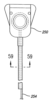
FIG. 59 is a cross-sectional view taken along lines 59-59 depicting the stylet
lumen.
FIG. 60A is a partial plan view depicting a pace/sense electrode mounted on a
delivery member and attached with release lines.
FIG. 60B is a side view depicting the pace/sense electrode and delivery member
of
FIG. 60A.
FIG. 61A is an exploded plan view depicting a pace/sense electrode for
insertion into
loops on a delivery member.
FIG. 61B is a plan view depicting the pace/sense electrode and delivery member
of
FIG. 61A with the loops being tightened around the pace/sense electrode.
FIG. 62 is a partial plan view depicting a pace/sense electrode having a lumen
for
receiving a guidewire.
FIG. 63 is a plan view depicting another embodiment of an adapter with a
cavity for
receiving a pacing/sensing electrode.
FIG. 63A is a perspective view depicting a sinus lead adapter retaining a
bipolar
coronary sinus lead.
FIG. 64 is a partial plan view of a modular pacing/sensing electrode spine.
FIG. 65 is a partial plan view of another embodiment of a moveable or modular
pacing/sensing electrode spine.
CA 02660240 2009-02-05
WO 2008/051926 PCT/US2007/082126
-23-
FIG. 66 is a partial plan view of another embodiment of a moveable or modular
pacing/sensing electrode spine.
FIG. 67 is a partial plan view of yet another embodiment of a modular
pacing/sensing
electrode spine.
FIG. 68 is a partial plan view of a modular defibrillation electrode spine.
FIG. 69 depicts a flattened plan view of a cardiac harness having panels
separated by
a spine that retains a defibrillation coil and two bipolar pairs of
electrodes.
FIG. 70 depicts a flattened plan view of a another embodiment of a cardiac
harness
having panels separated by a spine that retains a defibrillation coil and one
bipolar pair of
electrodes.
FIG. 71 depicts a cross-sectional view of an electrode tip within a rubber cup
during a
silicone rubber molding process.
DETAILED DESCRIPTION OF THE PREFERRED EMBODIMENTS
This invention relates to a method and apparatus for treating heart failure.
It is
anticipated that remodeling of a diseased heart can be resisted or even
reversed by
alleviating the wall stresses in such a heart. The present invention discloses
embodiments and methods for supporting the cardiac wall and for providing
defibrillation and/or pacing functions using the same system. Additional
embodiments and aspects are also discussed in Applicants' co-pending
application
entitled "Multi-Panel Cardiac Harness" U.S. Serial No. 60/458,991 filed March
28,
2003, the entirety of which is hereby expressly incorporated by reference.
Prior Art Devices
FIG. 1 illustrates a mammalian heart 10 having a prior art cardiac wall stress
reduction device in the form of a harness applied to it. The harness surrounds
a portion of the
heart and covers the right ventricle 11, the left ventricle 12, and the apex
13. For
convenience of reference, longitudinal axis 15 goes through the apex and the
AV groove 14.
The cardiac harness has a series of hinges or spring elements that
circumscribe the heart and,
collectively, apply a mild compressive force on the heart to alleviate wall
stresses.
The term "cardiac harness" as used herein is a broad term that refers to a
device fit
onto a patient's heart to apply a compressive force on the heart during at
least a portion of the
cardiac cycle.
CA 02660240 2009-02-05
WO 2008/051926 PCT/US2007/082126
-24-
The cardiac harness illustrated in FIG. 1 has at least one undulating strand
having a
series of spring elements referred to as hinges or spring hinges that are
configured to deform
as the heart expands during filling. Each hinge provides substantially
unidirectional
elasticity, in that it acts in one direction and does not provide as much
elasticity in the
direction perpendicular to that direction. For example, FIG. 2A shows a prior
art hinge
member at rest. The hinge member has a central portion and a pair of arms. As
the arms are
pulled, as shown in FIG. 2B, a bending moment is imposed on the central
portion. The
bending moment urges the hinge member back to its relaxed condition. Note that
a typical
strand comprises a series of such hinges, and that the hinges are adapted to
elastically expand
and retract in the direction of the strand.
In the harness illustrated in FIG. 1, the strands of spring elements are
constructed of
extruded wire that is deformed to form the spring elements.
FIGS. 3 and 4 illustrate another prior art cardiac harness, shown at two
points during
manufacture of such a harness. The harness is first formed from a relatively
thin, flat sheet of
material. Any method can be used to form the harness from the flat sheet. For
example, in
one embodiment, the harness is photochemically etched from the material; in
another
embodiment, the harness is laser-cut from the thin sheet of material. The
harness shown in
FIGS. 3 and 4 has been etched from a thin sheet of Nitinol, which is
superelastic material that
also exhibits shape memory properties. The flat sheet of material is draped
over a form, die
or the like, and is formed to generally take on the shape of at least a
portion of a heart.
With further reference to FIGS. 1 and 4, the cardiac harnesses have a base
portion
which is sized and configured to generally engage and fit onto a base region
of a patient's
heart, an apex portion which is sized and shaped so as to generally engage and
fit on an apex
region of a patient's heart, and a medial portion between the base and apex
portions.
In the harness shown in FIGS. 3 and 4, the harness has strands or rows of
undulating
wire. As discussed above, the undulations have hinge/spring elements which are
elastically
bendable in a desired direction. Some of the strands are connected to each
other by
interconnecting elements. The interconnecting elements help maintain the
position of the
strands relative to one another. Preferably the interconnecting elements allow
some relative
movement between adjacent strands.
The undulating spring elements exert a force in resistance to expansion of the
heart.
Collectively, the force exerted by the spring elements tends toward
compressing the heart,
thus alleviating wall stresses in the heart as the heart expands. Accordingly,
the harness helps
to decrease the workload of the heart, enabling the heart to more effectively
pump blood
CA 02660240 2009-02-05
WO 2008/051926 PCT/US2007/082126
-25-
through the patient's body and enabling the heart an opportunity to heal
itself. It should be
understood that several arrangements and configurations of spring members can
be used to
create a mildly compressive force on the heart to reduce wall stresses. For
example, spring
members can be disposed over only a portion of the circumference of the heart
or the spring
members can cover a substantial portion of the heart.
As the heart expands and contracts during diastole and systole, the
contractile cells of
the myocardium expand and contract. In a diseased heart, the myocardium may
expand such
that the cells are distressed and lose at least some contractility. Distressed
cells are less able
to deal with the stresses of expansion and contraction. As such, the
effectiveness of heart
pumping decreases. Each series of spring hinges of the above cardiac harness
embodiments
is configured so that as the heart expands during diastole the spring hinges
correspondingly
will expand, thus storing expansion forces as bending energy in the spring. As
such, the
stress load on the myocardium is partially relieved by the harness. This
reduction in stress
helps the myocardium cells to remain healthy and/or regain health. As the
heart contracts
during systole, the disclosed prior art cardiac harnesses apply a moderate
compressive force
as the hinge or spring elements release the bending energy developed during
expansion
allowing the cardiac harness to follow the heart as it contracts and to apply
contractile force
as well.
Other structural configurations for cardiac harnesses exist, however, but all
have
drawbacks and do not function optimally to treat CHF and other related
diseases or failures.
The present invention cardiac harness provides a novel approach to treat CHF
and provides
electrodes associated with the harness to deliver an electrical shock for
defibrillation or a
pacing stimulus for resynchronization, or for biventricular pacing/sensing.
The Present Invention Embodiments
The present invention is directed to a cardiac harness system for treating the
heart.
The cardiac harness system of the present invention couples a cardiac harness
for treating the
heart coupled with a cardiac rhythm management device. More particularly, the
cardiac
harness includes rows or undulating strands of spring elements that provide a
compressive
force on the heart during diastole and systole in order to relieve wall stress
pressure on the
heart. Associated with the cardiac harness is a cardiac rhythm management
device for
treating any number of irregularities in heart beat due to, among other
reasons, congestive
heart failure. Thus, the cardiac rhythm management device associated with the
cardiac
harness can include one or more of the following: an implantable
cardioverter/defibrillator
with associated leads and electrodes; a cardiac pacemaker including leads and
electrodes used
CA 02660240 2009-02-05
WO 2008/051926 PCT/US2007/082126
-26-
for sensing cardiac function and providing pacing stimuli to treat synchrony
of both vessels;
and a combined implantable cardioverter/defibrillator and pacemaker, with
associated leads
and electrodes to provide a defibrillation shock and/or pacing/sensing
functions.
The cardiac harness system includes various configurations of panels connected
together to at least partially surround the heart and assist the heart during
diastole and systole.
The cardiac harness system also includes one or more leads having electrodes
associated with
the cardiac harness and a source of electrical energy supplied to the
electrodes for delivering
a defibrillating shock or pacing stimuli.
In one embodiment of the invention, as shown in a flattened configuration in
FIG. 5, a
cardiac harness 20 includes two panels 21 of generally continuous undulating
strands 22. A
panel includes rows or undulating strands of hinges or spring elements that
are connected
together and that are positioned between a pair of electrodes, the rows or
undulations being
highly elastic in the circumferential direction and, to a lesser extent, in
the longitudinal
direction. In this embodiment, the undulating strands have U-shaped hinges or
spring
elements 23 capable of expanding and contracting circumferentially along
directional line 24.
The cardiac harness has a base or upper end 25 and an apex or lower end 26.
The undulating
strands are highly elastic in the circumferential direction when placed around
the heart 10,
and to a lesser degree in a direction parallel to the longitudinal axis 15 of
the heart. Similar
hinges or spring elements are disclosed in co-pending and co-assigned U.S.
Serial No.
60/458,991 filed March 28, 2003, the entire contents of which are incorporated
herein by
reference. While the FIG. 5 embodiment appears flat for ease of reference, in
use it is
substantially cylindrical (or tapered) to conform to the heart and the right
and left side panels
would actually be one panel and there would be no discontinuity in the
undulating strands.
The undulating strands 22 provide a compressive force on the epicardial
surface of the
heart thereby relieving wall stress. In particular, the spring elements 23
expand and contract
circumferentially as the heart expands and contracts during the diastolic and
systolic
functions. As the heart expands, the spring elements expand and resist
expansion as they
continue to open and store expansion forces. During systole, as the heart 10
contracts, the
spring elements will contract circumferentially by releasing the stored
bending forces thereby
assisting in both the diastolic and systolic function.
As just discussed, bending stresses are absorbed by the spring elements 23
during
diastole and are stored in the elements as bending energy. During systole,
when the heart
pumps, the heart muscles contract and the heart becomes smaller.
Simultaneously, bending
energy stored within the spring elements 23 is at least partially released,
thereby providing an
CA 02660240 2009-02-05
WO 2008/051926 PCT/US2007/082126
-27-
assist to the heart during systole. In a preferred embodiment, the compressive
force exerted
on the heart by the spring elements of the harness comprises about 10% to 15%
of the
mechanical work done as the heart contracts during systole. Although the
harness is not
intended to replace ventricular pumping, the harness does substantially assist
the heart during
systole.
The undulating strands 22 can have varying numbers of spring element 23
depending
upon the amplitude and pitch of the spring elements. For example, by varying
the amplitude
of the pitch of the spring elements, the number of undulations per panel will
vary as well. It
may be desired to increase the amount of compressive force the cardiac harness
20 imparts on
the epicardial surface of the heart, therefore the present invention provides
for panels that
have spring elements with lower amplitudes and a shorter pitch, thereby
increasing the
expansion force imparted by the spring element. In other words, all other
factors being
constant, a spring element having a relatively lower amplitude will be more
rigid and resist
opening, thereby storing more bending forces during diastole. Further, if the
pitch is smaller,
there will be more spring elements per unit of length along the undulating
strand, thereby
increasing the overall bending force stored during diastole, and released
during systole.
Other factors that will affect the compressive force imparted by the cardiac
harness onto the
epicardial surface of the heart include the shape of the spring elements, the
diameter and
shape of the wire forming the undulating strands, and the material comprising
the strands.
As shown in FIG. 5, the undulating strands 22 are connected to each other by
grip
pads 27. In the embodiments shown in FIG. 5, adjacent undulating strands are
connected by
one or more grip pads attached at the apex 28 of the spring elements 23. The
number of grip
pads between adjacent undulating strands is a matter of choice and can range
from one grip
pad between adjacent undulating strands, to one grip pad for every apex on the
undulating
strand. Importantly, the grip pads should be positioned in order to maintain
flexibility of the
cardiac harness 20 without sacrificing the objectives of maintaining the
spacing between
adjacent undulating strands to prevent overlap and to enhance the frictional
engagement
between the grip pads and the epicardial surface of the heart. Further, while
it is desirable to
have the grip pads attached at the apex of the spring elements, the invention
is not so limited.
The grip pads 27 can be attached anywhere along the length of the spring
elements, including
the sides 29. Further, the shape of the grip pads 27, as shown in FIG. 5, can
vary to suit a
particular purpose. For example, grip pad 27 can be attached to the apex 28 of
one
undulating strand 22, and be attached to two apices on an adjacent undulating
strand (see
FIG. 7). As shown in FIG. 5, all of the apices point toward each other, and
are said to be
CA 02660240 2009-02-05
WO 2008/051926 PCT/US2007/082126
-28-
"out-of-phase." If the apices of the undulations were aligned, they would be
"in-phase." The
apices are all out-of-phase since the number of spring elements in each
undulating strand is
the same, however, the invention contemplates that the number of spring
elements in each
undulating strand may vary since the heart is tapered from its base near the
top to its apex 13
at the bottom. Thus, there would be more spring elements and a longer
undulating strand per
panel at the top or base of the cardiac harness than at the bottom of the
cardiac harness near
the apex of the heart. Accordingly, the cardiac harness would be tapered from
the relatively
wide base to a relatively narrow bottom toward the apex of the heart, and this
would affect
the alignment of the apices of the spring elements, and hence the ability of
the grip pads 27 to
align perfectly and attach to adjacent apices of the spring elements. A
further disclosure and
embodiments relating to the undulating strands and the attachment means in the
form of grip
pads is found in co-pending and co-assigned U.S. Serial No. 60/486,062 filed
July 10, 2003,
the entire contents of which are incorporated herein by reference. While the
connections
between adjacent undulating strands 22 is preferably grip pads 27, in an
alternative
embodiment (not shown) the undulating strands are connected by interconnecting
elements
made of the same material as the strands. The interconnecting elements can be
straight or
curved as shown in FIGS. 8A-8B of commonly owned U.S. Patent No. 6,612,979 B2,
the
entire contents of which is incorporated by reference herein.
It is preferred that the undulating strands 22 be continuous as shown in FIG.
5. For
example, every pair of adjacent undulating strands are connected by bar arm
30. It is
preferred that the bar arms form part of a continuous wire that is bent to
form the undulating
strands, and then welded at its ends along the bar arm. The weld is not shown
in FIG. 5, but
can be by any conventional method such as laser welding, fusion bonding, or
conventional
welding. The type of wire used to form the undulating strands may have a
bearing on the
method of attaching the ends of the wire used to form the undulating strand.
For example, it
is preferred that the undulating strands be made out of a nickel-titanium
alloy, such as
Nitinol, which may lose some of its superelastic or shape memory properties if
exposed to
high heat during conventional welding.
Associated with the cardiac harness of the present invention is a cardiac
rhythm
management device as previously disclosed. Thus, associated with the cardiac
harness as
shown in FIG. 5, are one or more electrodes for use in providing
defibrillating shock. As can
be seen immediately below, any number of factors associated with congestive
heart failure
can lead to fibrillation, acquiring immediate action to save the patient's
life.
CA 02660240 2009-02-05
WO 2008/051926 PCT/US2007/082126
-29-
Diseased hearts often have several maladies. One malady that is not uncommon
is
irregularity in heartbeat caused by irregularities in the electrical
stimulation system of the
heart. For example, damage from a cardiac infarction can interrupt the
electrical signal of the
heart. In some instances, implantable devices, such as pacemakers, help to
regulate cardiac
rhythm and stimulate heart pumping. A problem with the heart's electrical
system can
sometimes cause the heart to fibrillate. During fibrillation, the heart does
not beat normally,
and sometimes does not pump adequately. A cardiac defibrillator can be used to
restore the
heart to normal beating. An external defibrillator typically includes a pair
of electrode
paddles applied to the patient's chest. The defibrillator generates an
electric field between
electrodes. An electric current passes through the patient's heart and
stimulates the heart's
electrical system to help restore the heart to regular pumping.
Sometimes a patient's heart begins fibrillating during heart surgery or other
open-
chest surgeries. In such instances, a special type of defibrillating device is
used. An open-
chest defibrillator includes special electrode paddles that are configured to
be applied to the
heart on opposite sides of the heart. A strong electric field is created
between the paddles,
and an electric current passes through the heart to defibrillate the heart and
restore the heart to
regular pumping.
In some patients that are especially vulnerable to fibrillation, an
implantable heart
defibrillation device may be used. Typically, an implantable heart
defibrillation device
includes an implantable cardioverter defibrillator (ICD) or a cardiac
resynchronization
therapy device (CRT-D) which usually has only one electrode positioned in the
right
ventricle, and the return electrode is the defibrillator housing itself,
typically implanted in the
pectoral region. Alternatively, an implantable device includes two or more
electrodes
mounted directly on, in or adjacent the heart wall. If the patient's heart
begins fibrillating,
these electrodes will generate an electric field therebetween in a manner
similar to the other
defibrillators discussed above.
Testing has indicated that when defibrillating electrodes are applied external
to a heart
that is surrounded by a device made of electrically conductive material, at
least some of the
electrical current disbursed by the electrodes is conducted around the heart
by the conductive
material, rather than through the heart. Thus, the efficacy of defibrillation
is reduced.
Accordingly, the present invention includes several cardiac harness
embodiments that enable
defibrillation of the heart and other embodiments disclose means for
defibrillating,
resynchronization, left ventricular pacing, right ventricular pacing, and
biventricular
pacing/sensing.
CA 02660240 2009-02-05
WO 2008/051926 PCT/US2007/082126
-30-
In further keeping with the invention, the cardiac harness 20 includes a pair
of leads
31 having conductive electrode portions 32 that are spaced apart and which
separate panels
21. As shown in FIG. 5, the electrodes are formed of a conductive coil wire 33
that is
wrapped around a non-conductive member 34, preferably in a helical manner. A
conductive
wire 35 is attached to the coil wire and to a power source 36. As used herein,
the power
source 36 can include any of the following, depending upon the particular
application of the
electrode: a pulse generator; an implantable cardioverter/defibrillator; a
pacemaker; and an
implantable cardioverter/defibrillator coupled with a pacemaker. In the
embodiment shown
in FIG. 5, the electrodes are configured to deliver an electrical shock, via
the conductive wire
and the power source, to the epicardial surface of the heart so that the
electrical shock passes
through the myocardium. Even though the electrodes are spaced so that they
would be about
180 apart around the circumference of the heart in the embodiment shown, they
are not so
limited. In other words, the electrodes can be spaced so that they are about
45 apart, 60
apart, 90 apart, 120 apart, or any arbitrary arc length spacing, or, for
that matter, essentially
any arc length apart around the circumference of the heart in order to deliver
an appropriate
electrical shock. As previously described, it may become necessary to
defibrillate the heart
and the electrodes 32 are configured to deliver an appropriate electrical
shock to defibrillate
the heart.
Still referring to FIG. 5, the electrodes 32 are attached to the cardiac
harness 20, and
more particularly to the undulating strands 22, by a dielectric material 37.
The dielectric
material insulates the electrodes from the cardiac harness so that electrical
current does not
pass from the electrode to the harness thereby undesirably shunting current
away from the
heart for defibrillation. Preferably, the dielectric material covers the
undulating strands 22
and covers at least a portion of the electrodes 32. In the FIG. 5 embodiment,
the middle
panel undulating strands are covered with the dielectric material while the
right and left side
panels are bare metal. While it is preferred that all of the undulating
strands of the panels be
coated with the dielectric material, thereby insulating the harness from the
electric shock
delivered by the electrodes, some or all of the undulating strands can be bare
metal used to
deliver the electrical shock to the epicardial surface of the heart for
defibrillation or for
pacing.
As will be described in more detail, the electrodes 32 have a conductive
discharge
first surface 38 that is intended to be proximate to or in direct contact with
the epicardial
surface of the heart, and a conductive discharge second surface 39 that is
opposite to the first
surface and faces away from the heart surface. As used herein, the term
"proximate" is
CA 02660240 2009-02-05
WO 2008/051926 PCT/US2007/082126
-31 -
intended to mean that the electrode is positioned near or in direct contact
with the outer
surface of the heart, such as the epicardial surface of the heart. The first
surface and second
surface typically will not be covered with the dielectric material 37 so that
the bare metal
conductive coil can transmit the electrical current from the power source
(pulse generator),
such as an implantable cardioverter/defibrillator (ICD or CRT-D) 36, to the
epicardial surface
of the heart. In an alternative embodiment, either the first or the second
surface may be
covered with dielectric material in order to preferentially direct the current
through only one
surface. Further details of the construction and use of the leads 31 and
electrodes 33 of the
present invention, in conjunction with the cardiac harness, will be described
more fully
herein.
Importantly, the dielectric material 37 used to attach the electrodes 32 to
the
undulating strands 22 insulates the undulating strands from any electrical
current discharged
through the conductive metal coils 33 of the electrodes. Further, the
dielectric material in this
embodiment is flexible so that the electrodes can serve as a seam or hinge to
fold the cardiac
harness 20 into a lower profile for minimally invasive delivery. Thus, as will
be described in
more detail (see FIGS. 13 and 14), the cardiac harness can be folded along its
length, along
the length of the electrodes, in order to reduce the profile for intercostal
delivery, for example
through the rib cage or other area typically used for minimally invasive
surgery for accessing
the heart. Minimally invasive approaches involving the heart typically are
made through
subxiphoid, subcostal or intercostal incisions. When the cardiac harness is
folded, it can be
reduced into a circular or a more or less oval shape, both of which promote
minimally
invasive procedures.
In further keeping with the invention, cross sectional views of the leads 31
and the
electrode portion 32 are shown in FIGS. 5B, 5C, and 5D. As shown in FIG. 5B,
the electrode
32 has the coil wire 33 wrapped around the non-conducting member 34 in a
helical pattern.
The dielectric material 37 provides a spaced connection between the electrode
and the bar
arms 30 at the ends of the undulating strands 22. The electrodes do not touch
or overlap with
the bar arms or any portion of the undulating strands. Instead, the dielectric
material provides
the attachment means between the electrodes and the bar arms of the undulating
strands.
Thus, the dielectric material 37 not only acts as an insulating non-conductive
material, but
also provides attachment means between the undulating strands and the
electrodes. Because
the dielectric material 37 is relatively thin at the attachment points, it is
highly flexible and
permits the electrodes to be flexible along with the cardiac harness panels
21, which will
expand and contract as the heart beats as previously described.
CA 02660240 2009-02-05
WO 2008/051926 PCT/US2007/082126
-32-
Referring to FIG. 5C, the non-conductive member 34 extends beyond the coil
wire 33
for a distance. The non-conductive member preferably is made from the same
material as the
dielectric material 37, typically a silicone rubber or similar material. While
it is preferred
that the dielectric material be made from silicone rubber, or a similar
material, it also can be
made from ParyleneTM (Union Carbide), polyurethanes, PTFE, TFE, and ePTFE. As
can be
seen, the non-conductive member provides support for the dielectric material
to attach the bar
arms 30 of the undulating strands 22 in order to connect the strands to the
electrode 32. A
conductive wire 35 extends through the non-conducting member and attaches to
the proximal
end of the coil wire 33 so that when an electrical current is delivered from
the power source
36 through conductive wire 35, the electrode coil 33 will be energized. The
conductive wire
35 is also covered by non-conducting material 34. Referring to FIG. 5D, it can
be seen that
the non-conductive member 34 continues to extend beyond the bottom (apex) of
the cardiac
harness and that conductive wire 35 continues to extend out of the non-
conductive member
and into the power source 36. In the embodiment shown in FIGS. 5B-5D, the
cardiac harness
is insulated from the electrodes by the dielectric material 37 so that there
is no shunting of
electrical currents by the cardiac harness 20 from the electrical shock
delivered by the
electrodes during defibrillation or pacing functions.
While it is preferred that the cardiac harness 20 be comprised of undulating
strands 22
made from a solid wire member, such as a superelastic or shape memory material
such as
Nitinol, and be insulated from the electrodes 32, it is possible to use some
or all of the
undulating strands to deliver the electrical shock to the epicardial surface
of the heart. For
example, as shown in FIG. 6A, a composite wire 45 can be used to form the
undulating
strands 22 and, importantly, to effectively transmit current to deliver an
electrical shock to the
epicardial surface of the heart. The composite wire 45 includes a current
conducting wire 47
made from, for example silver (Ag), and which is covered by a Nitinol tube 46.
In order to
improve the surface conductivity of the outer Nitinol tube 46, a highly
conductive coating is
placed on the Nitinol tube. For example, the Nitinol tube can be covered with
a deposition
layer of platinum (Pt) or platinum-iridium (Pt-Ir), or an equivalent material
including iridium
oxide (IROX). The composite wire, so constructed, will have superior
mechanical
performance to expand and contract due to the Nitinol tubing, and also will
have improved
electrical properties resulting from the current conducting wire 47 and
improved
electrolytic/electrochemical properties via the surface layer of platinum-
iridium. Thus, if
some portion or all of the undulating strands 22 are made from a composite
wire 45, the
cardiac harness 20 will be capable of delivering a defibrillating shock on
selected portions of
CA 02660240 2009-02-05
WO 2008/051926 PCT/US2007/082126
-33-
the heart via the undulating strands and will also function to impart
compressive forces as
previously described.
In contrast to the current conducting undulating strands of FIG. 6A, are the
non-
conducting insulated undulating strands 22 as shown by cross sectional view
FIG. 6B. As
previously described, some or all of the undulating strands 22 can be covered
with dielectric
material 37 in order to insulate the strands from the electrical current
delivered through the
electrodes while delivering shock on the epicardial surface of the heart.
Thus, as shown in
FIG. 6B, the undulating strands 22 are covered by dielectric material 37 to
provide insulation
from the electrical shock delivered by the electrodes 32, yet maintain the
flexibility and the
expansive properties of the undulating strands.
An important aspect of the invention is to provide a cardiac harness 20 that
can be
implanted minimally invasively and be attached to the epicardial surface of
the heart, without
requiring sutures, clips, screws, glue or other attachment means. Importantly,
the undulating
strands 22 may provide relatively high frictional engagement with the
epicardial surface,
depending on the cross-sectional shape of the strands. For example, in the
embodiment
disclosed in FIG. 6C, the cross-sectional shape of the undulating strands 22
can be circular,
rectangular, triangular or for that matter, any shape that increases the
frictional engagement
between the undulating strands and the epicardial surface of the heart. As
shown in FIG. 6C,
the middle cross-section view having a flat rectangular surface (wider than
tall) not only has a
low profile for enhancing minimally invasive delivery of the cardiac harness,
but it also has
rectangular edges that may have a tendency to engage and dig into the
epicardium to increase
the frictional engagement with the epicardial surface of the heart. With the
proper cross-
sectional shape for the undulating strands, coupled with the grip pads 27
having a high
frictional engagement feature, the necessity for suturing, clipping, or
further attachment
means to attach the cardiac harness to the epicardial surface of the heart
becomes
unnecessary.
In another embodiment as shown in FIGS. 7A and 7B, a different configuration
for
cardiac harness 20 and the electrodes 32 are shown, as compared to the FIG. 5
embodiments.
In FIGS. 7A and 7B, three electrodes are shown separating the three panels 21
with
undulating strands 22 extending between the electrodes. As with previous
embodiments,
springs 23 are formed by the undulating strands so that the undulating strands
can expand and
contract during the diastolic and systolic functions, and apply a compressive
force during
both functions. The far side panel of FIG. 7A is not shown for clarity
purposes. The position
of the electrodes around the circumference of the heart is a matter of choice,
and in the
CA 02660240 2009-02-05
WO 2008/051926 PCT/US2007/082126
-34-
embodiment of FIG. 7A, the electrodes can be spaced an equal distance apart at
about 120 .
Alternatively, it may be important to deliver the electrical shock more
through the right
ventricle requiring the positioning of the electrodes closer to the right
ventricle than to the left
ventricle. Similarly, it may be more important to deliver an electrical shock
to the left
ventricle as opposed to the right ventricle. Thus, positioning of electrodes,
as with other
embodiments, is a matter of choice.
Still referring to FIGS. 7A and 7B, in this embodiment electrodes 32 extend
beyond
the bottom or apex portion of the cardiac harness 20 in order to insure that
the electrical
shock delivered by the electrodes is delivered to the epicardial surface of
the heart and
including the lower portion of the heart closer to the apex 13. Thus, the
electrodes 22 have a
distal end 50 and a proximal end 51 where the proximal end is positioned
closer to the apex
13 of the heart and the distal end is positioned closer to the base or upper
portion of the heart.
As used herein, distal is intended to mean further into the body and away from
the attending
physician, and proximal is meant to be closer to the outside of the body and
closer to the
attending physician. The proximal ends of the electrodes are positioned closer
to the apex of
the heart and provide several functions, including the ability to deliver an
electrical shock
closer to the apex of the heart. The electrode proximal ends also function to
provide support
for the cardiac harness 20 and the panels 21, and lend support not only during
delivery (as
will be further described herein) but in separating the panels and in gripping
the epicardial
surface of the heart to retain the harness on the heart without slipping.
While the FIGS. 7A and 7B embodiments show electrodes 32 separating three
panels
21 of the cardiac panel 20, more or fewer electrodes and panels can be
provided to suit a
particular application. For example, in one preferred embodiment, four
electrodes 32
separate four panels 21, so that two of the electrodes can be positioned on
opposite sides of
the left ventricle and two of the electrodes can be positioned on opposite
sides of the right
ventricle. In this embodiment, preferably all four electrodes would be used,
with a first set of
two electrodes on opposite sides of the right ventricle acting as one (common)
electrode and a
second set of two electrodes on opposite sides of the left ventricle acting as
the opposite
(common) electrode. Alternatively, two of the electrodes can be activated
while the other
two electrodes act as dummy electrodes in that they would not be activated
unless necessary.
At present, commercially available implantable cardioverter/defibrillators
(ICD's) are
capable of delivering approximately thirty to forty joules in order to
defibrillate the heart.
With respect to the present invention, it is preferred that the electrodes 22
of the cardiac
harness 20 of the present invention deliver defibrillating shocks having less
than thirty to
CA 02660240 2009-02-05
WO 2008/051926 PCT/US2007/082126
-35-
forty joules. The commercially available ICD's can be modified to provide
lower power
levels to suit the present invention cardiac harness system with electrodes
delivering less than
thirty to forty joules of power. As a general rule, one objective of the
electrode configuration
is to create a uniform current density distribution throughout the myocardium.
Therefore, in
addition to the number of electrodes used, their size, shape, and relative
positions will also all
have an impact on the induced current density distribution. Thus, while one to
four
electrodes are preferred embodiments of the invention, five to eight
electrodes also are
envisioned.
In keeping with the present invention, the cardiac harness and the associated
cardiac
rhythm management device can be used not only for providing a defibrillating
shock, but also
can be used as a pacing/sensing device for treating the synchrony of both
ventricles, for
resynchronization, for biventricular pacing and for left ventricular pacing or
right ventricular
pacing. As shown in FIGS. 8A-8D, the heart 10 is shown in cross-section
exposing the right
ventricle 11 and the left ventricle 12. The cardiac harness 20 is mounted
around the outer
surface of the heart, preferably on the epicardial surface of the heart, and
multiple electrodes
are associated with the cardiac harness. More specifically, electrodes 32 are
attached to the
cardiac harness and positioned around the circumference of the heart on
opposite sides of the
right and left ventricles. In the event that fibrillation should occur, the
electrodes will provide
an electrical shock through the myocardium and the left and right ventricles
in order to
defibrillate the heart. Also mounted on the cardiac harness, is a
pacing/sensing lead 40 that
functions to monitor the heart and provide data to a pacemaker. If required,
the pacemaker
will provide pacing stimuli to synchronize the ventricles, and/or provide left
ventricular
pacing, right ventricular pacing or biventricular pacing. Thus, for example,
in FIG. 8C, pairs
of pacing/sensing electrodes 40 are positioned adjacent the left and right
ventricle free walls
and can be used to provide pacing stimuli to synchronize the ventricles, or
provide left
ventricular pacing, right ventricular pacing or biventriculator pacing. The
use of proximal Y
connectors can simplify the transition to a post-generator such as Oscor's,
iLink-B15-10. The
iLink-B15-10 can be used to link the right and left ventricular free-wall
pace/sense electrodes
40, as shown in 8D.
In another embodiment of the invention, as shown in FIGS. 9-14, cardiac
harness 60
is similar to previously described cardiac harness 20. With respect to cardiac
harness 60, it
also includes panels 61 consisting of undulating strands 62. In the disclosed
embodiments,
the undulating strands are continuous and extend through coils as will be
described. The
undulating strands act as spring elements 63 as with prior embodiments that
will expand and
CA 02660240 2009-02-05
WO 2008/051926 PCT/US2007/082126
-36-
contract along directional line 64. The cardiac harness 60 includes a base or
upper end 65
and an apex or lower end 66. In order to add stability to the cardiac harness
60, and to assist
in maintaining the spacing between the undulating strands 62, grip pads 67 are
connected to
adjacent strands, preferably at the apex 68 of the springs. Alternatively, the
grip pads 67
could be attached from the apex of one spring element to the side 69 of a
spring element, or
the grip pad could be attached from the side of one spring to the side of an
adjacent spring on
an adjacent undulating strand. In further keeping with the invention as shown
in the FIGS. 9-
14, in order to add stability and some mechanical stiffness to the cardiac
harness 60, coils 62
are interwoven with the undulating strands, which together define the panels
61. The coils
typically are formed of a coil of wire such as Nitinol or similar material
(stainless steel,
MP35N), and are highly flexible along their longitudinal length. The coils 72
have a coil
apex 73 and a coil base 74 to coincide with the harness base 65 and the
harness apex 66. The
coils can be injected with a non-conducting material so that the undulating
strands extend
through gaps in the coils and through the non-conducting material. The non-
conducting
material also fills in the gaps which will prevent the undulating strands from
touching the
coils so there is no metal-to-metal touching between the undulating strands
and the coils.
Preferably, the non-conducting material is a dielectric material 77 that is
formed of silicone
rubber or equivalent material as previously described. Further, a dielectric
material 78 also
covers the undulating strands in the event a defibrillating shock or pacing
stimuli is delivered
to the heart via an external defibrillator (e.g., transthoracic) or other
means.
Importantly, coils 72 not only perform the function of being highly flexible
and
provide the attachment means between the coils and the undulating strands, but
they also
provide structural columns or spines that assist in deploying the harness 60
over the
epicardial surface of the heart. Thus, as shown for example in FIG. 12, the
cardiac harness
60 has been positioned over the heart and delivered by minimally invasive
means, as will be
described more fully herein. The coils 72, although highly flexible along
their longitudinal
length, have sufficient column strength in order to push on the apex 73 of the
coils so that the
base portion 74 of the coils and of the harness 65 slide over the apex of the
heart and along
the epicardial surface of the heart until the cardiac harness 60 is positioned
over the heart,
substantially as shown in FIG. 12.
Referring to the embodiments shown in FIGS. 9 and 11, the cardiac harness 60
has
multiple panels 61 and multiple coils 72. More or fewer panels and coils can
be used in order
to achieve a desired result. For example, eight coils are shown in FIGS. 9 and
11, while
fewer coils may provide a harness with greater flexibility since the
undulating strands 62
CA 02660240 2009-02-05
WO 2008/051926 PCT/US2007/082126
-37-
would be longer in the space between each coil. Further, the diameter of the
coils can be
varied in order to increase or decrease flexibility and/or column strength in
order to assist in
the delivery of the harness over the heart. The coils preferably have a round
cross-sectional
wire in the form of a tightly wound spiral or helix so that the cross-section
of the coil is
circular. However, the cross-sectional shape of the coil need not be circular,
but may be
more advantageous if it were oval, rectangular, or another shape. Thus, if
coils 72 had an
oval shape, where the longer axis of the oval was parallel to the
circumference of the heart,
the coil would flex along its longitudinal axis and still provide substantial
column strength to
assist in delivery of the cardiac harness 60. Further, an oval-shaped coil
would provide a
lower profile for minimally invasive delivery. The wire cross-section also
need not be
round/circular, but can consist of a flat ribbon having a rectangular shape
for low profile
delivery. The coils also can have different shapes, for example they can be
closed coils, open
coils, laser-cut coils, wire-wound coils, multi-filar coils, or the coil
strands themselves can be
coiled (i.e., coiled coils). The electrode need not have a coil of wire,
rather the electrode
could be formed by a zig-zag-shaped wire (not shown) extending along the
electrode. Such a
design would be highly flexible and fatigue resistant yet still be capable of
providing a
defibrillating shock.
The cardiac harness embodiments 60 shown in FIGS. 9-12, can be folded as shown
in
FIGS. 13 and 14 and yet remain highly flexible for minimally invasive
delivery. The coils 72
act as hinges or spines so that the cardiac harness can be folded along the
longitudinal axis of
the coils. The grip pads typically connecting adjacent undulating strands 62
have been
omitted for clarity in these embodiments, however, they would be used as
previously
described.
In an alternative embodiment, similar to the embodiment shown in FIGS. 9-12,
the
cardiac harness 60 includes both coils 72 and electrodes 32. In this
embodiment, as with the
previously described embodiments, a series of undulating strands 22 extend
between the coils
and the electrodes to form panels 21. In this embodiment, for example, the
coils and
electrodes form hinge regions so that the panels can be folded along the
longitudinal axis of
the coils and electrodes for minimally invasive delivery. Further, in this
embodiment, there
are two coils and four electrodes, with two of the electrodes positioned
adjacent the right
ventricle, with the remaining two electrodes being positioned adjacent the
left ventricle. The
coils not only act as a hinge, but provide column strength as previously
described so that the
cardiac harness can be delivered minimally invasively by delivery through, for
example, the
intercostal space between the ribs and then pushing the harness over the
heart. Likewise, the
CA 02660240 2009-02-05
WO 2008/051926 PCT/US2007/082126
-38-
electrodes provide column strength as well, yet remain flexible along their
longitudinal axis,
as do the coils.
Referring to FIGS. 15A-15D, the electrodes 32 or the coils 72 can be mounted
on the
inner surface (touching the heart) or outer surface (away from the heart) of
the cardiac
harness. Thus, the cardiac harness 20 includes continuous undulating strands
22 that extend
circumferentially around the heart without any interruptions. The undulating
strands are
interconnected by any interconnecting means, including grip pads 27, as
previously
described. In this embodiment, electrodes 32 or coils 72, or both, are mounted
on an inner
surface 80 or an outer surface 81 of the cardiac harness 20. A dielectric
material 82 is
molded around the electrodes or coils and around the undulating strands in
order to connect
the electrodes and coils to the cardiac harness. Alternatively, as shown in
FIG. 15D, the
electrodes 32 or coils 72 can be formed into a fastening means by forming
notches 83 into the
electrode (or coil) and which are configured to receive portions of the
undulating strand 22.
The undulating strands 22 are spaced from the coils or electrodes so that
there is no
overlapping/touching of metal. The notches 83 are filled with a dielectric
material,
preferably silicone rubber, or similar material that insulates the undulating
strands of the
cardiac harness from the electrodes yet provides a secure attachment means so
that the
electrodes or coils remain firmly attached to the undulating strands of the
cardiac harness.
Importantly, the electrodes 32 do not have to be in contact with the
epicardial surface of the
heart in order to deliver a defibrillating shock. Thus, the electrodes 32 can
be mounted on the
outer surface 81 of the cardiac harness, and not be in physical contact with
the epicardial
surface of the heart, yet still deliver a defibrillating shock as previously
described.
It is to be understood that several embodiments of cardiac harnesses can be
constructed and that such embodiments may have varying configurations, sizes,
flexibilities,
etc. Such cardiac harnesses can be constructed from many suitable materials
including
various metals, fabrics, plastics and braided filaments. Suitable materials
also include
superelastic materials and materials that exhibit shape memory properties. For
example, a
preferred embodiment cardiac harness is constructed of Nitinol. Shape memory
dielectric
materials can also be employed. Such shape memory dielectric materials can
include shape
memory polyurethanes or other dielectric materials such as those containing
oligo(e-
caprolactone) dimethacrylate and/or poly(e-caprolactone), which are available
from
mnemoScience.
In keeping with the invention, as shown in FIG. 16, the undulating strands 22
and 62
can be formed in many ways, including by a fixture 90. The fixture 90 has a
number of stems
CA 02660240 2009-02-05
WO 2008/051926 PCT/US2007/082126
-39-
91 that are arranged in a pre-selected pattern that will define the shape of
the undulating
strands 22 and 62. The position of the stems will define the shape of the
undulating strands,
and determine whether the previously disclosed apex of the springs is either
in-phase or out-
of-phase. The shape of stems 91 will define the shape of the springs in terms
of radius of
curvature, or other shape, such as a keyhole shape, a U-shape, and the like.
The spacing
between the stems will determine the pitch and the amplitude of the undulating
strands which
is a matter of choice. Preferably, in one exemplary embodiment, a Nitinol wire
92 or other
superelastic or shape memory wire having a 0.012 inch diameter, is woven
between stems 91
in order to form the undulating strands. Other wire diameters can be used to
suit a particular
need and can range from about 0.007 inch to about 0.020 inch diameter. Other
wire cross-
section shapes are envisioned to be used with fixture 90, particularly a flat
rectangular-shaped
wire and an oval-shaped wire. The Nitinol wire is then heat set to impart the
shape memory
feature. Any free ends can be connected together by laser bonding, laser
welding, or other
type of similar connection consistent with the use of Nitinol, or the ends may
remain free and
be encapsulated in a dielectric material to keep them atraumatic, depending
upon the design.
Again referring to FIG. 16, after the Nitinol wire is heat set to impart the
shape
memory feature, the wire is jacketed with NuSil silicone tubing (Helix
Medical) having 0.029
inch outside diameter by 0.012 inch inside diameter. Thereafter, the jacketed
Nitinol wire is
placed in molds for transfer of liquid silicone rubber which will insulate the
Nitinol wire from
any electrical shock from any electrodes associated with the cardiac harness,
or any other
device providing a defibrillating shock to the heart. The dimensions of the
silicone tubing
will of course vary for different wire dimensions.
In another embodiment of the invention, shown in FIG. 17, cardiac harness 100
includes multiple panels 101 similar to those previously described. Further,
undulating
strands 102 form the panels and have multiple spring elements 103 that expand
and contract
along directional line 104, also as previously described for other
embodiments. In the cardiac
harness 100 shown in FIG. 17, the amplitude of the spring elements is
relatively smaller than
in other embodiments, and the pitch is higher, meaning there are more spring
elements per
unit of length relative to other embodiments. Thus, the cardiac harness 100
should generate
higher bending forces as the heart expands and contracts during the diastolic
and systolic
cycles. In other words, the spring elements 103 of cardiac harness 100 will
resist expansion,
thereby imparting higher compressive forces on the wall of the heart during
the diastolic
function and will release these higher bending forces during the systolic
function as the heart
contracts. It may be important to provide undulating strands 102 that
alternate in amplitude
CA 02660240 2009-02-05
WO 2008/051926 PCT/US2007/082126
-40-
and pitch within a panel, starting at the base of the harness and extending
toward the apex.
For example, the pitch and amplitude of an undulating strand closer to the
base or the harness
may be configured to impart higher compressive forces on the epicardial
surface of the heart
than the undulating strands closer to the apex or the lower part of the
harness. It also may be
desirable to alternate the amplitude and pitch of the spring elements from one
undulating
strand to the next. Further, where multiple panels are provided, it may be
advantageous to
provide one amplitude and pitch of the spring elements of the undulating
strands of one
panel, and a different amplitude and pitch of the spring elements of the
undulating strands of
an adjacent panel. The FIG. 17 embodiment can be configured with electrodes as
previously
described in other embodiments, or with coils, both of which assist with the
delivery of the
cardiac harness by providing column support to the harness.
The cardiac harness of the present invention, having either electrodes or
coils, can be
formed using injection molding techniques as shown in FIGS. 18A-18C and 19A-
19C. The
molds in FIGS. 18A-18C are substantially the same as the molds shown in FIGS.
19A-19C,
with the exception of the undulating pattern grooves that receive the
undulating strands
previously described. In referring to FIG. 18A, bottom mold 110 includes a
pattern for
receiving the cardiac harness and a coil or an electrode. For illustration
purposes, FIG. 18B
shows top mold 111 and FIG. 18C shows end view mold 112. The top mold mates
with the
bottom mold. As can be seen, the cardiac harness undulating strands will fit
in undulating
strand groove 113, which extend into coil groove 114. The previously described
electrodes
or coils fit into coil grooves 114. Injection port 115 is positioned midway
along the mold
fixtures, however, more than one injection port can be used to insure that the
flow of polymer
is uniform and consistent. Preferably, silicone rubber is injected into the
molds so that the
silicone rubber flows over the undulating strands and the electrodes or the
coils. When the
cardiac harness assembly is taken out of the mold, the undulating strands will
be attached to
the electrodes or the coils by the silicone rubber according to the pattern
shown. Other
patterns may be desired and the molds are easily altered to provide any
pattern that ensures a
secure attachment between the undulating strands and the electrodes or the
coils.
Importantly, the molds of FIGS. 18 and 19 can be used to inject the dielectric
material or
silicone rubber inside the coils and, if necessary, between the gaps in the
coils in order to
insure that the coils and the undulating strands are insulated from each
other. The silicone
rubber fills the inside of the coils, extrudes through the gaps in the coils,
and forms a skin on
the inner and outer surface of the coil. This skin is selectively removed (as
will be described)
to expose portions of the electrode coils so that they can conduct current as
described.
CA 02660240 2009-02-05
WO 2008/051926 PCT/US2007/082126
-41-
Further, it is desired that the coils and the undulating strands do not
overlap or touch in order
to reduce any frictional engagement between the metallic coils and the
metallic undulating
strands. In order to increase the frictional engagement between the cardiac
harness and the
epicardial surface of the heart, small projections (not shown) can be molded
along the surface
of the coils that will contact the epicardial surface. As previously described
with respect to
the grip pads, these small projections, preferably formed of silicone rubber,
will engage the
epicardial surface of the heart and increase the frictional engagement between
the coils and
the surface of the heart in order to secure the harness to the heart without
the use of sutures,
clips, or other mechanical attachment means.
In further keeping with the invention, as shown in FIGS. 20-23, a portion of a
lead
having an electrode 120 is shown in the form of a conductive coil 121. The
coil can be
formed of any suitable wire that is conductive so that an electrical shock can
be transmitted
through the electrode and through the myocardium of the heart. In this
embodiment, the coil
wire is wrapped around a dielectric material 122 in a helical configuration,
however, a spiral
wrap or other configuration is possible as long as the coil has superior
fatigue resistance and
longitudinal flexibility. Importantly, conductive coils 121 have high fatigue
resistance which
is necessary since the coil is on or near the surface of the beating heart so
that the coil is
constantly flexing along its longitudinal length in response to heart
expansion and
contraction. The cross-section of the wire preferably is round or circular,
however, it also can
be oval shaped or flat (rectangular) in order to reduce the profile of the
electrode for
minimally invasive delivery. A circular, oval or flat wire will have a
relatively high fatigue
resistance as well as a relatively low profile for delivery purposes. Also, a
flat wire coil is
highly flexible along the longitudinal axis and it has a relatively high
surface area for
delivering an electrical shock. The electrode 120 has a first surface 123 and
a second
surface 124. The first surface 123 will be proximate the epicardial surface of
the heart, or
other portions of the heart, while the second surface will be opposite the
first surface and
away from the epicardial surface of the heart. A conductive wire (not shown)
extends
through the dielectric material 122 and attaches to the coil wire 121 at one
or more locations
along the coil or coils, and the conductive wire is connected to a power
source (e.g., an ICD)
at its other end. As shown in FIG. 22, the cross-section of the electrode 120
can be circular,
or as shown in FIG. 23, can be oval for reduced profile for minimally invasive
delivery.
Other cross-sectional shapes for electrode 120 are available depending upon
the particular
need. All of these cross-sectional shapes will have relatively high fatigue
resistance. As
shown in FIGS. 22 and 23, multiple lumens 125 can be provided to carry one or
more
CA 02660240 2009-02-05
WO 2008/051926 PCT/US2007/082126
-42-
conductive wires from the electrode to the power source (pulse generator, ICD,
CRT-D,
pacemaker, etc.). The lumens also can carry sensing wires that transmit data
from a sensor
on or in the heart to a pacemaker so that the heart can be monitored. Further,
the lumens 125
can be used for other purposes such as drug delivery (therapeutic drugs,
steroids, etc.), dye
injection for visibility under fluoroscopy, carrying a guide wire (not shown)
or a stylet to
facilitate delivery of the electrodes and the harness, or for other purposes.
The lumens 125
can be used to carry a guide wire (not shown) or a stylet in such a way that
the column
stiffness of the coil is increased by the guide wire or stylet, or in a manner
that will vary the
column stiffness as required. By varying the column stiffness of the coils
with a guide wire
or a stylet in lumens 125, the ability to push the cardiac harness over the
heart (as will be
described) will be enhanced. The guide wires or stylets also can be used, to
some extent, to
steer the coils and hence the cardiac harness during delivery and implantation
over the heart.
The guide wire or stylet in lumens 125 can be removed after the cardiac
harness is implanted
so that the coils (electrodes) become more flexible and atraumatic.
In keeping with the invention, as shown in FIGS. 20-23, the electrode 120 not
only
provides an electrical conduit for use in defibrillation, but also has
sufficient column strength
when attached to the cardiac harness to assist in the delivery of the harness
by minimally
invasive means. As will be further described, the coils 121 provide a highly
flexible
electrode along its longitudinal length, and also provide a substantial amount
of column
strength when coupled with a cardiac harness to assist in the delivery of the
harness.
In further keeping with the invention of FIGS. 20-23, a dielectric material
such as
silicone rubber 126 can be used to coat electrodes 120. During the molding
process
(previously described), when the electrode 120 is attached to the cardiac
harness, silicone
rubber 126 will coat the entire electrode 120. Soda blasting (or other known
material
removal process) can be used to remove portions of the silicone rubber skin
from the coils
121 in order to expose first surface 123 and second surface 124 (or portions
of those surfaces)
so that the bare metal coil is exposed to the epicardial surface of the heart.
Preferably, the
silicone rubber is removed from both the first surface and the second surface,
however, it also
may be advantageous to remove the silicone rubber from only the first surface,
which is
proximate to or in contact with the epicardial surface of the heart. The
electrode 120 has a
surface area 128 which essentially includes all of the bare metal surface area
that is exposed
and that will deliver a shock. The amount of surface area per electrode can
vary greatly
depending upon a particular application, however, surface areas in the range
from about 50
mrri to about 600 mrri are typical. While it is possible to remove the
silicone rubber from
CA 02660240 2009-02-05
WO 2008/051926 PCT/US2007/082126
-43-
only the second surface (facing away from the heart), and leaving the first
surface coated with
silicone rubber, an electrical shock can still be delivered from the bare
metal second surface,
however, the electrical shock delivered may not be as efficient as with other
embodiments.
While the dimensions of the electrodes can vary widely due to the variations
in the size of the
heart to be treated in conjunction with the size of the cardiac harness,
generally the length of
the electrode ranges from about 2 cm to about 16 cm. The coil 121 has a length
in the range
of about 1 cm to about 12 cm. Commercially available leads having one or more
electrodes
are available from several sources and may be used with the cardiac harness of
the present
invention. Commercially available leads with one or more electrodes is
available from
Guidant Corporation (St. Paul, Minnesota), St. Jude Medical (Minneapolis,
Minnesota) and
Medtronic Corporation (Minneapolis, Minnesota). Further examples of
commercially
available cardiac rhythm management devices, including defibrillation and
pacing systems
available for use in combination with the cardiac harness of the present
invention (possibly
with some modification) include, the CONTAK CD , the INSIGNIA Plus pacemaker
and
FLEXTREND leads, and the VITALITYTM AVT ICD and ENDOTAK RELIANCE
defibrillation leads, all available from Guidant Corporation (St. Paul, MN),
and the InSync
System available from Medtronic Corporation (Minneapolis, MN).
In an alternative embodiment, as shown in FIG. 24, the conductive coils 121
need not
be continuous along the length of the electrode 120, but can be spatially
isolated or staggered
along the electrode. For example, multiple coil sections 127, similar to the
coil 121 shown in
FIG. 20, can be spaced along the electrode with each coil section being
attached to the
conductive wire so it receives electrical current from the power source. The
coil sections can
be from about 0.5 cm to about 2.0 cm long and be spaced from about 0.5 cm to
about 4 cm
apart along the electrode. The dimensions used herein are by way of example
only and can
vary to suit a particular application
When removing portions of the silicone rubber from the electrode 120 using
soda
blasting or a similar technique, it may be desirable to leave portions of the
electrode masked
or insulated so that the masked portion is non-conductive. By masking portions
of two
electrodes positioned, for example, on opposite sides of the left ventricle,
it is possible to
vector a shock at a desirable angle through the myocardium and ventricle. The
shock will
travel from the bare metal (unmasked) portion of one electrode through the
myocardium and
the ventricle to the bare metal (unmasked) portion of the opposing electrode
at a vector angle
determined by the position of the masking on the electrodes.
CA 02660240 2009-02-05
WO 2008/051926 PCT/US2007/082126
-44-
The cardiac rhythm management devices associated with the present invention
are
implantable devices that provide electrical stimulation to selected chambers
of the heart in
order to treat disorders of cardiac rhythm and can include pacemakers and
implantable
cardioverter/defibrillators and/or cardiac resynchronization therapy devices
(CRT-D). A
pacemaker is a cardiac rhythm management device which paces the heart with
timed pacing
pulses. As previously described, common conditions for which pacemakers are
used is in the
treatment of bradycardia (ventricular rate is too slow) and tachycardia
(cardiac rhythms are
too fast). As used herein, a pacemaker is any cardiac rhythm management device
with a
pacing functionality, regardless of any other functions it may perform such as
the delivery of
cardioversion or defibrillation shocks to terminate atrial or ventricular
fibrillation. An
important feature of the present invention is to provide a cardiac harness
having the capability
of providing a pacing function in order to treat the synchrony of both
ventricles. To
accomplish the objective, a pacemaker with associated leads and electrodes are
associated
with and incorporated into the cardiac harness of the present invention. The
pacing/sensing
electrodes, alone or in combination with defibrillating electrodes, provide
treatment to
synchronize the ventricles and improve cardiac function.
In keeping with the invention, a pacemaker and a pacing/sensing electrode are
incorporated into the design of the cardiac harness. As shown in FIGS. 25A and
25B, a lead
(not shown) having a defibrillator electrode 130 at its distal end, shown in
partial section, not
only incorporates wire coils 131 used to deliver a defibrillating electrical
shock to the
epicardial surface of the heart, but also incorporates a pacing/sensing
electrode 132. The
defibrillator electrode 130 can be attached to any cardiac harness embodiment
previously
described herein. In this embodiment, a non-penetrating pacing/sensing
electrode 132 is
combined with the defibrillating electrode 130 in order to provide data
relating to heart
function. More specifically, the pacing/sensing electrode 132 does not
penetrate the
myocardium in this embodiment, however, it may be beneficial in other
embodiments for the
pacing or sensing electrode to penetrate the myocardium. One advantage of a
non-
penetrating pacing/sensing electrode is that there is no danger of puncturing
a coronary artery
or causing further trauma to the epicardium or myocardium. It is also easier
to design since
there is no requirement of a penetration mechanism (barb or screw) on the
pacing/sensing
electrode. The pacing/sensing electrode 132 is in direct contact with the
epicardial surface of
the heart and will provide data via lead wire 133 to the pulse generator
(pacemaker), which
will interpret the data and provide any pacing function necessary to achieve,
for example,
ventricular resynchronization therapy, left ventricular pacing, right
ventricular pacing,
CA 02660240 2009-02-05
WO 2008/051926 PCT/US2007/082126
-45-
synchrony of both ventricles, and/or biventricular pacing. As shown in FIG.
25B, the
pacing/sensing electrode 132 is incorporated into a portion of a cardiac
harness 134, and
more particularly the undulating strands 135 are attached by dielectric
material 136 to the
pacing/sensing electrode. As can be seen in FIGS. 25A and 25B, the wire coils
131 of the
defibrillating electrode 130 are wrapped around the dielectric material 136,
and the dielectric
material insulates the pacing/sensing electrode 132 from both the wire coils
131 and from the
undulating strands 135 of the cardiac harness. Multiple pacing/sensing
electrodes 132 can be
incorporated along defibrillating electrode 130, and multiple pacing and
sensing electrodes
can be incorporated on other electrodes associated with the cardiac harness.
In one of the preferred embodiments, multi-site pacing (as previously shown in
FIGS.
8A-8D) using pacing/sensing electrodes 132 enables resynchronization therapy
in order to
treat the synchrony of both ventricles. Multi-site pacing allows the
positioning of the
pacing/sensing electrodes to provide bi-ventricular pacing or right
ventricular pacing, left
ventricular pacing, depending upon the patient's needs.
In another embodiment, shown in FIGS. 26A-26C, a defibrillating electrode is
combined with pacing/sensing electrodes, for attachment to any of the cardiac
harness
embodiments disclosed herein. In this embodiment, the defibrillating electrode
130 is formed
of wire coils 131 wrapped in a helical manner. The helical wire can be a wound
wire having
a single strand or a quadrafilar wire having four wires bundled together to
form the coil. The
wire coils 131 are wrapped around dielectric material 136 in a manner similar
to that
described for the embodiments in FIGS. 25A and 25B. In this embodiment, the
pacing/sensing electrode 132 is in the form of a single ring for unipolar
operation, and two
rings for bi-polar operation. The pacing/sensing electrode rings 132 are
mounted coaxially
with the defibrillating electrode wire coils 131, and the conducting wires
from the wire coils
and the pacing/sensing ring electrode are shown extending through the
dielectric material 136
and being insulated from each other. The conducting wires from the
defibrillating electrode
130 and from the pacing/sensing ring electrodes 132 can be bundled into a
common lead wire
133 which extends to the pulse generator (an ICD, CRT-D, and/or a pacemaker).
As can be
seen in FIGS. 26A-26C, the pacing/sensing electrode rings 132 have a diameter
that is
somewhat larger than the defibrillator electrode coils 131 in order to insure
preferential
contact by the electrode rings against the epicardial surface of the heart.
Preferably, several
pairs of pacing/sensing electrode rings (bipolar) would be positioned on the
cardiac harness
and be positioned to come into contact with, for example, the left ventricle
free wall. Multi-
site pacing allows the pacing/sensing electrode rings 132 to be used for both
pacing and
CA 02660240 2009-02-05
WO 2008/051926 PCT/US2007/082126
-46-
resynchronization concurrently. Further, the pacing/sensing electrode rings
132 also can be
used in the absence of defibrillating electrodes 130. The prior disclosure
relating to molding
of the cardiac harness to the defibrillator electrode applies equally as well
to the
pacing/sensing electrode rings. The wire coil 131 and the pacing/sensing
electrode rings 32
can be fabricated in several ways including by laser cutting stainless steel
tubing or using
highly conductive materials in wire form, such as biocompatible platinum wire.
As
previously disclosed, the wire coils 131 can be quadrafilar wire (platinum)
for improved
flexibility and conformability to the epicardial surface of the heart and be
biocompatible.
The surface of the pacing/sensing electrodes can vary greatly depending upon
the application.
As an example, in one embodiment, the surface area of the pacing/sensing
electrodes are in
the range from about 2 mrri to about 12 mm~, however, this range can vary
substantially.
While the disclosed embodiments show the pacing/sensing electrodes combined
with the
defibrillating electrodes, the pacing/sensing electrodes can be formed
separately and mounted
on the cardiac harness with or without defibrillating electrodes.
The defibrillating electrode 130 as disclosed herein, can be used with
commercially
available pacing/sensing electrodes and leads. For example, Oscor (Model HT
52PB)
endocardial/passive fixation leads can be integrated with the defibrillator
electrode 130 by
molding the leads into the fibrillator electrode using the same molds
previously disclosed
herein.
The foregoing disclosed invention incorporating cardiac rhythm management
devices
into the cardiac harness combines several treatment modalities that are
particularly beneficial
to patients suffering from congestive heart failure. The cardiac harness
provides a
compressive force on the heart thereby relieving wall stress, and improving
cardiac function.
The defibrillating and pacing/sensing electrodes associated with the cardiac
harness, along
with ICD's and pacemakers, provide numerous treatment options to correct for
any number of
maladies associated with congestive heart failure. In addition to the
defibrillation function
previously described, the cardiac rhythm devices can provide electrical pacing
stimulation to
one or more of the heart chambers to improve the coordination of atrial and/or
ventricular
contractions, which is referred to as resynchronization therapy. Cardiac
resynchronization
therapy is pacing stimulation applied to one or more heart chambers, typically
the ventricles,
in a manner that restores or maintains synchronized bilateral contractions of
the atria and/or
ventricles thereby improving pumping efficiency. Resynchronization pacing may
involve
pacing both ventricles in accordance with a synchronized pacing mode. For
example, pacing
at more than one site (multi-site pacing) at various sites on the epicardial
surface of the heart
CA 02660240 2009-02-05
WO 2008/051926 PCT/US2007/082126
-47-
to desynchronize the contraction sequence of a ventricle (or ventricles) may
be therapeutic in
patients with hypertrophic obstructive cardiomyopathy, where creating
asynchronous
contractions with multi-site pacing reduces the abnormal hyper-contractile
function of the
ventricle. Further, resynchronization therapy may be implemented by adding
synchronized
pacing to the bradycardia pacing mode where paces are delivered to one or more
synchronized pacing sites in a defined time relation to one or more sensing
and pacing events.
An example of synchronized chamber-only pacing is left ventricle only
synchronized pacing
where the rate in synchronized chambers are the right and left ventricles
respectively. Left-
ventricle-only pacing may be advantageous where the conduction velocities
within the
ventricles are such that pacing only the left ventricle results in a more
coordinated contraction
by the ventricles than by conventional right ventricle pacing or by
ventricular pacing.
Further, synchronized pacing may be applied to multiple sites of a single
chamber, such as
the left ventricle, the right ventricle, or both ventricles. The pacemakers
associated with the
present invention are typically implanted subcutaneously on a patient's chest
and have leads
threaded to the pacing/electrodes as previously described in order to connect
the pacemaker
to the electrodes for sensing and pacing. The pacemakers sense intrinsic
cardiac electrical
activity through the electrodes disposed on the surface of the heart.
Pacemakers are well
known in the art and any commercially available pacemaker or combination
defibrillator/pacemaker can be used in accordance with the present invention.
The cardiac harness and the associated cardiac rhythm management device system
of
the present invention can be designed to provide left ventricular pacing. In
left heart pacing,
there is an initial detection of a spontaneous signal, and upon sensing the
mechanical
contraction of the right and left ventricles. In a heart with normal right
heart function, the
right mechanical atrio-ventricular delay is monitored to provide the timing
between the initial
sensing of right atrial activation (known as the P-wave) and right ventricular
mechanical
contraction. The left heart is controlled to provide pacing which results in
left ventricular
mechanical contraction in a desired time relation to the right mechanical
contraction, e.g.,
either simultaneous or just preceding the right mechanical contraction.
Cardiac output is
monitored by impedance measurements and left ventricular pacing is timed to
maximize
cardiac output. The proper positioning of the pacing/sensing electrodes
disclosed herein
provides the necessary sensing functions and the resulting pacing therapy
associated with left
ventricular pacing.
CA 02660240 2009-02-05
WO 2008/051926 PCT/US2007/082126
-48-
An important feature of the present invention is the minimally invasive
delivery of the
cardiac harness and the cardiac rhythm management device system which will be
described
immediately below.
Delivery of the cardiac harness 20,60, and 100 and associated electrodes and
leads
can be accomplished through conventional cardio-thoracic surgical techniques
such as
through a median sternotomy. In such a procedure, an incision is made in the
pericardial sac
and the cardiac harness can be advanced over the apex of the heart and along
the epicardial
surface of the heart simply by pushing it on by hand. The intact pericardium
is over the
harness and helps to hold it in place. The previously described grip pads and
the compressive
force of the cardiac harness on the heart provide sufficient attachment means
of the cardiac
harness to the epicardial surface so that sutures, clips or staples are
unnecessary. Other
procedures to gain access to the epicardial surface of the heart include
making a slit in the
pericardium and leaving it open, making a slit and later closing it, or making
a small incision
in the pericardium.
Preferably, however, the cardiac harness and associated electrodes and leads
may be
delivered through minimally invasive surgical access to the thoracic cavity,
as illustrated in
FIGS. 27-36, and more specifically as shown in FIG. 30. A delivery device 140
may be
delivered into the thoracic cavity 141 between the patient's ribs to gain
direct access to the
heart 10. Preferably, such a minimally invasive procedure is accomplished on a
beating
heart, without the use of cardio-pulmonary bypass. Access to the heart can be
created with
conventional surgical approaches. For example, the pericardium may be opened
completely
or a small incision can be made in the pericardium (pericardiotomy) to allow
the delivery
system 140 access to the heart. The delivery system of the disclosed
embodiments comprises
several components as shown in FIGS. 27-36. As shown in FIG. 27, an introducer
tube 142
is configured for low profile access through a patient's ribs. A number of
fingers 143 are
flexible and have a delivery diameter 144 as shown in FIG. 27, and an expanded
diameter
145 as shown in FIG. 29. The delivery diameter is smaller than the expanded
diameter. An
elastic band 146 expands around the distal end 147 of the fingers and prevents
the fingers
from overexpanding during delivery of the cardiac harness. The distal end of
the fingers is
the part of the delivery device 140 that is inserted through the patient's
ribs to gain direct
access to the heart.
The delivery device 140 also includes a dilator tube 150 that has a distal end
151 and
a proximal end 152. The cardiac harness 20,60,100 is collapsed to a low
profile
configuration and inserted into the distal end of the dilator tube, as shown
in FIG. 28. The
CA 02660240 2009-02-05
WO 2008/051926 PCT/US2007/082126
-49-
dilator tube has an outside diameter that is slightly smaller than the inside
diameter of the
introducer tube 142. As will be discussed more fully herein, the distal end
151 of the dilator
tube is inserted into the proximal end 147 of the introducer tube in close
sliding engagement
and in a slight frictional engagement. The slidable engagement between the
dilator tube and
the introducer tube should be with some mild resistance, however, there should
be
unrestricted slidable movement between the two tubes. The distal end 151 of
the dilator tube
will expand the fingers 143 of the introducer tube 142 as the dilator tube is
pushed distally
into the introducer tube as shown in FIG. 29. In the embodiments shown in
FIGS. 27-36, the
cardiac harness 20,60,100 is equipped with leads (previously described) having
electrodes for
use in defibrillation or pacing functions.
As shown in FIG. 31, the delivery system 140 also includes a releasable
suction
device, such as suction cup 156 at the distal end of the delivery device. The
negative
pressure suction cup 156 is used to hold the apex of the heart 10. Negative
pressure can be
applied to the suction cup using a syringe or other vacuum device commonly
known in the
art. A negative pressure lock can be achieved by a one-way valve stop-cock or
a tubing
clamp, also known in the art. The suction cup 156 is formed of a biocompatible
material and
is preferably stiff enough to prevent any negative pressure loss through the
heart while
manipulating the heart and sliding the cardiac harness 20,60,100 onto the
heart. Further, the
suction cup 156 can be used to lift and maneuver the heart 10 to facilitate
advancement of the
harness or to allow visualization and surgical manipulation of the posterior
side of the heart.
The suction cup has enough negative pressure to allow a slight pulling in the
proximal
direction away from the apex of the heart to somewhat elongate the heart
(e.g., into a bullet
shape) during delivery to facilitate advancing the cardiac harness over the
apex and onto the
base portion of the heart. After the suction cup 156 is attached to the apex
of the heart and a
negative pressure is drawn, the cardiac harness, which has been releasably
mounted in the
distal end 151 of the dilator tube 150, can be advanced distally over the
heart, as will be
described more fully herein.
As shown in FIG. 30, the delivery device 140, and more specifically introducer
tube
142, has been advanced through the intercostal space between the patient's
ribs during
insertion of the introducer tube, the fingers 143 are in their delivery
diameter 144, which is a
low profile for ease of access through the small port made through the
patient's ribs.
Thereafter, the dilator tube 150, with the cardiac harness 20,60,100 mounted
therein, is
advanced distally through the introducer tube so that the fingers 143 are
expanded until they
achieve their expanded diameter 145. The suction cup 156 can be attached to
the apex 13 of
CA 02660240 2009-02-05
WO 2008/051926 PCT/US2007/082126
-50-
the heart 10 either before or after the dilator tube is advanced to spread the
fingers 143 of the
introducer tube 142. Preferably, the dilator tube has already expanded the
fingers on the
introducer tube so that there is a larger opening for the suction cup as it is
advanced through
the inside of a dilator tube, out of the distal end of the introducer tube,
and placed in contact
with the apex of the heart. Thereafter, a negative pressure is drawn allowing
the suction cup
to securely attach to the apex of the heart. Visualizing equipment that is
commonly known in
the art may be used to assist in positioning the suction cup to the apex. For
example,
fluoroscopy, magnetic resonance imaging (MRI), dye injection to enhance
fluoroscopy, and
echocardiography, and intracardiac, transesophageal, or transthoracic echo,
all can be used to
enhance positioning and in attaching the suction cup to the apex of the heart.
After negative
pressure is drawn and the suction cup is securely attached (releasably) to the
apex of the
heart, the heart can then be maneuvered somewhat by pulling on the tubing 157
attached to
the suction cup, or by manipulating the introducer tube 142, the dilator tube
150, both in
conjunction with the suction cup. As previously described, it may be
advantageous to pull on
the tubing 157 to allow the suction cup to pull on the apex of the heart and
elongate the heart
somewhat in order to facilitate sliding the harness over the epicardium.
As more clearly shown in FIGS. 32-36, the cardiac harness 20,60,100 is
advanced
distally out of the dilator tube and over the suction cup 156. The suction cup
is tapered so
that the distal end of the harness slides over the narrow portion of the taper
(the proximal end
of the suction cup 158). The suction cup becomes wider at its distal end where
it is attached
to the apex of the heart, and the cardiac harness continues to slide and
expand over the
suction cup as it is advanced distally. As the cardiac harness continues to be
advanced
distally, it slides over the apex of the heart and continues to expand as it
is pushed out of the
dilator tube and along the epicardial surface of the heart. Since the harness
and the electrodes
32,120,130 are coated with the previously described dielectric material,
preferably silicone
rubber, the cardiac harness should slide easily over the epicardial surface of
the heart. The
silicone rubber offers little resistance and the epicardial surface of the
heart has sufficient
fluid to allow the harness to easily slide over the wet surface of the heart.
The pericardium
previously has been cut so that the cardiac harness is sliding over the
epicardial surface of the
heart with the pericardium over the cardiac harness to help hold it onto the
surface of the
heart. As shown in FIGS. 35 and 36, the cardiac harness 20,60,100 has been
completely
advanced out of the dilator tube so that the harness covers at least a portion
of the heart 10.
The suction cup 156 has been withdrawn, and the introducer tube 142 and
dilator tube 150
also have been withdrawn proximally from the patient. Prior to removing the
introducer tube,
CA 02660240 2009-02-05
WO 2008/051926 PCT/US2007/082126
-51 -
a power source 170 (such as an ICD, CRT-D, and/or pacemaker) can be implanted
by
conventional means. The electrodes will be attached to the pulse generator to
provide a
defibrillating shock or pacing functions as previously described.
In the embodiments shown in FIGS. 27-36, the cardiac harness 20,60,100 was
advanced through the dilator tube by pushing on the proximal end of the
electrodes
32,120,130, on the lead wires 31,133, and on the proximal end (apex 26) of the
cardiac
harness. Even though the electrodes are designed to be atraumatic and
longitudinally
flexible, the electrodes have sufficient column strength so that pushing on
the proximal ends
of the electrodes assists in pushing the cardiac harness out of the dilator
tube and over the
epicardial surface of the heart. In one embodiment, advancement of the cardiac
harness is
accomplished by hand, by the physician simply pushing on the electrodes and
the leads to
advance the cardiac harness out of the dilator tube to slide onto the
epicardial surface of the
heart.
As shown in the embodiments of FIGS. 27-36, the delivery device 140, and more
specifically introducer tube 142 and dilator tube 150, have a circular cross-
section. It may be
preferable, however, to chose other cross-sectional shapes, such as an oval
cross-sectional
shape for the delivery device. An oval delivery device may be more easily
inserted through
the intercostal space between the patient's ribs for a low profile delivery.
Further, as the
cardiac harness 20,60,100 is advanced out of a delivery device 140 having an
oval cross-
section, the harness distal end will quickly form into a more circular shape
in order to assume
the configuration of the epicardial surface of the heart as it is advanced
distally over the heart.
In the embodiments shown in FIGS. 35 and 36, the cardiac harness 20,60,100
remains
firmly attached to the epicardial surface of the heart without the need for
any further
attachment means, such as sutures, clips, adhesives, or staples. Further, the
pericardial sac
helps to enclose the harness to prevent it from shifting or sliding on the
epicardial surface of
the heart.
Importantly, during delivery of the cardiac harness 20,60,100, the harness
itself, the
electrodes 32,120,130, as well as leads 31 and 132 have sufficient column
strength in order
for the physician to push from the proximal end of the harness to advance it
distally through
the dilator tube 150. While the entire cardiac harness assembly is flexible,
there is sufficient
column strength, especially in the electrodes, to easily slide the cardiac
harness over the
epicardial surface of the heart in the manner described.
In an alternative embodiment, if the cardiac harness 20,60,100 includes coils
72, as
opposed to the electrodes and leads, the harness can be delivered in the same
manner as
CA 02660240 2009-02-05
WO 2008/051926 PCT/US2007/082126
-52-
previously described with respect to FIGS. 27-36. The coils have sufficient
column strength
to permit the physician to push on the proximal end of the coils to advance
the cardiac
harness distally to slide over the apex of the heart and onto the epicardial
surface.
In another embodiment, delivery of the cardiac harness 20,60,100 can be by
mechanical means as opposed to the hand delivery previously described. As
shown in FIGS.
37-42, delivery system 180 includes an introducer tube 181 that functions the
same as
introducer tube 142. Also, a dilator tube 182, which is sized for slidable
movement within
the introducer tube, also functions the same as the previously described
dilator tube 150. An
ejection tube 183 is sized for slidable movement within the dilator tube, that
is, the outer
diameter of the ejection tube is slightly smaller than the inner diameter of
the dilator tube. As
shown in FIGS. 40 and 41, the ejection tube has a distal end 184 and a
proximal end 185,
wherein the distal end of the ejection tube has a plate that fills the entire
inner diameter of the
ejection tube. The plate has a number of lumens 187 for receiving leads 31,132
and for
receiving the suction cup 156 and associated tubing 157. Thus, lumens 188 are
sized for
receiving leads 31,132 therethrough, while lumen 189 is sized for receiving
suction cup 156
and the associated tubing 157. The number of lumens 188 in plate 186 will be
defined by the
number of leads 31,132 associated with the cardiac harness 20,60,100. Thus, as
shown in
FIG. 40, there are four lumens 188 for receiving four leads therethrough, and
one lumen 189
for receiving the suction cup 156 and tubing 157 therethrough. The leads and
the tubing 157
extend proximally out the proximal end 185 of the ejection tube. As shown in
FIG. 42, the
suction cup and cardiac harness are on the left side of the schematic, and the
ejection tube
183 is on the right hand side of the schematic. For clarity, the dilator tube
and the introducer
tube have been omitted, however, in practice the cardiac harness would be
mounted in the
dilator tube, and the dilator tube would extend into the introducer tube,
while the ejection
tube would extend into the dilator tube. As can be seen in FIG. 42, the leads
31,132 extend
through lumens 188, while the tubing 157 associated with the suction cup
extends through
lumen 189. The tubing and the leads extend proximally out of the proximal end
of the
ejection tube, and extend out of the patient during delivery of the harness.
As previously
described, after the introducer is positioned through the rib cage, and the
apex of the heart is
acquired by the suction cup, the harness can be advanced out of the dilator by
advancing the
ejection tube 183 in a distal direction toward the apex of the heart. The
leads, the cardiac
harness and electrodes all provide sufficient column strength to allow the
plate 186 to impart
a pushing force against the cardiac harness to advance it distally over the
heart as previously
described. After the cardiac harness is pushed over the epicardial surface of
the heart, the
CA 02660240 2009-02-05
WO 2008/051926 PCT/US2007/082126
-53-
ejection tube can be withdrawn proximally so that the tubing 157 and the leads
31,132 slide
through lumens 189,188 respectively. The ejection tube 183 continues to be
withdrawn
proximally so that the proximal end of the leads and the proximal end of
tubing 157 are
pulled through the distal end 184 of the ejection tube so that the ejection
tube is clear of the
leads and the tubing.
As with the previous embodiment, suitable materials for the delivery system
140,180
can include the class of polymers typically used and approved for
biocompatible use within
the body. Preferably, the tubing associated with delivery systems 140 and 180
are rigid,
however, they can be formed of a more flexible material. Further, the delivery
systems
140,180 can be curved rather than straight, or can have a flexible joint in
order to more
appropriately maneuver the cardiac harness 20,60,100 over the epicardial
surface of the heart
during delivery. Further, the tubing associated with delivery systems 140,180
can be coated
with a lubricious material to facilitate relative movement between the tubes.
Lubricious
materials commonly known in the art such as TeflonTM can be used to enhance
slidable
movement between the tubes.
The present invention includes a passive restraint device consisting of a
wireform
cardiac harness delivered through a mini-thoracotomy using a delivery system.
As
previously disclosed, defibrillation electrodes/leads are attached directly
onto the cardiac
harness. There is a need to provide the cardiac harness in combination with
epicardial
pace/sense electrodes to provide optimal Cardiac Resynchronization Therapy
(CRT) in
patients with inter- and intra-ventricular contraction dyssynchrony. While the
pace/sense
electrodes could be integrated into fixed positions on the harness, there is a
benefit to being
able to adjust the position of the pace/sense electrodes relative to the
harness once on the
heart. While the harness configured with integrated pace/sense electrodes
could be moved to
some degree in an attempt to optimize the electrode position, it is assumed
that the harness is
deployed into an optimal position for passive restraint and that it would be
undesirable to
alter that position. The benefit of adjusting the pace/sense electrode
position is largely
related to where the electrodes are positioned once the harness is deployed.
The pace/sense
electrodes may be located over a tissue region where there is insufficient
sensing or pacing
ability (e.g., over fat, ischemic, fibrotic, or necrotic tissue), or where
there is a sub-optimal
resynchronization effect. Besides sensing and pacing for CRT applications,
there may be
benefit to altering the placement of one or more pace/sense electrodes
relative to the harness
for bradycardic pacing (e.g., for backup VVI pacing, or for chronic pacing in
locations other
than the RV apex, which is thought to exacerbate heart failure symptoms).
There is a further
CA 02660240 2009-02-05
WO 2008/051926 PCT/US2007/082126
-54-
benefit of moving one or more defibrillation electrodes (either in combination
with or
independent of one or more pace/sense electrodes) relative to the harness to
alter the
defibrillation vector, local voltage gradients, and/or impedance to improve
the ability to
defibrillate the heart. The embodiments disclosed herein relate to various
means to provide
pace/sense and/or defibrillation electrodes which are coupled to the cardiac
harness, yet are
movable relative to the harness. Typically the terms "electrode" and "lead"
are used to note a
specific part of the device as a whole ("electrode" meaning the pace/sense
electrode or the
defibrillation electrode, and "lead" being the body of the device that
contains everything else
(conductors, insulation, connectors, etc.)). Sometimes, however, either term
is used
generically to refer to the lead/electrode device as a whole. This
lead/electrode device may
have a pace/sense electrode or defibrillation electrode or both.
In keeping with the invention, a cardiac harness and assembly is configured to
fit at
least a portion of a patient's heart and is associated with one or more
electrodes capable of
providing defibrillation and electrodes used for pacing and/or sensing
functions. In one
embodiment, shown in FIGS. 43A-49, an adapter 200 having a housing 202 is used
to retain
one or more pacing/sensing electrodes 204. The adapter is configured to retain
the
pacing/sensing electrodes so that electrodes are placed in direct contact with
the epicardial
surface of the heart, or proximate the epicardial surface of the heart. The
adapter has a cavity
206 for receiving one or more pacing/sensing electrode and in one embodiment,
the cavity is
sized and shaped for receiving the pacing/sensing electrodes in an
interference fit. In other
words, the pacing/sensing electrodes are pressed into the cavity of the
adapter in a snap-fit
relationship so that there is an interference fit requiring no other fastening
means. In another
embodiment, a fastener 208 is used to securely retain the pacing/sensing
electrodes in the
cavity. Fasteners can include, but are not limited to sutures, staples, clips,
adhesives, or
polymer coatings over the electrodes. Fasteners 208 can be inserted through
first apertures
216 and into the adapter 200 in order to more firmly attach the pace/sense
electrodes 204 to
the cavity 206. In another embodiment, after the pace/sense electrodes are
pressed into the
cavity, silicone rubber or other dielectric material is molded over the
pace/sense electrodes in
order to further secure the electrodes in the cavity.
In all embodiments of the adapter 200 disclosed thus far, it is preferred that
the cavity
206 be configured to receive the pace/sense electrode 204 so that the
electrode 218 on the
pace/sense electrode faces away from the cavity. The electrodes 218 typically
are in the form
of a small metal protrusion or button, such that the button or protrusion
extends outwardly
from the pace/sense electrode so that the metallic surface of the protrusion
or button can
CA 02660240 2009-02-05
WO 2008/051926 PCT/US2007/082126
-55-
come into direct contact with the surface of the heart, or come into nearly
direct contact with
the surface of the heart. The electrodes 218 are electrically connected to a
power source (see
FIG. 54). The adapter 200 also has second apertures 217 for receiving release
lines as will be
further described infra.
In one embodiment, the adapter 200 includes a cavity 206 for receiving a
pace/sense
electrode 204. After the pace/sense electrode is pressed into the cavity,
dielectric material is
molded over the pace/sense electrode to retain the pace/sense electrode in the
adapter. When
molding dielectric material over the pace/sense electrode 204, care must be
taken to make
sure electrode 218 remains exposed (i.e., not covered). Preferably, the
adapter is formed
from a silicone rubber material as is the molded layer retaining the
pace/sense electrode in the
cavity.
In one embodiment, shown in FIG. 47A, the adapter 200 resembles a clam shell
configuration 210 that has an open and closed configuration. In the open
configuration
(shown in FIG. 47A), the pace/sense electrodes 204 are pressed into cavity 206
and the
electrodes are retained in the adapter when the two halves of the clam shell
configuration are
moved to the closed position (not shown). In another embodiment, shown in FIG.
47B, the
adapter 200 is formed in two parts with the cavity 206 formed in a first
portion 212 and in a
second portion 214. The pace/sense electrodes 204 are pressed into the cavity
206 of either
the first portion 212 or second portion 214 and then the first portion is
mated to the second
portion (not shown) so that the cavity surrounds the pace/sense electrodes. In
these
embodiments (FIGS. 47A and 47B) an aperture in the cavity corresponds with
electrodes 218
so that the electrodes extend through the aperture to directly contact the
surface of the heart.
The present invention also includes a method of delivery and a method of use
of the
adapter and the associated pace/sense electrodes in conjunction with a cardiac
harness.
Preferably, the mounting of the cardiac harness and placement of the
pace/sense electrode
under the harness is performed on a beating heart. In one embodiment, shown in
FIGS. 50-
57, after the pace/sense electrodes 204 have been attached to the adapter 200,
an adapter
assembly 220 (which includes the adapter with the pace/sense electrodes
attached) is
positioned under an already implanted cardiac harness 222. Preferably, the
adapter assembly
is delivered minimally invasively to a desired position under the cardiac
harness. In one
embodiment, the adapter assembly 220 is releasably attached to the distal end
224 of a push
arm 226 which has an atraumatic distal end 228 so that the push arm, with the
adapter
assembly attached thereto, can be advanced through an introducer tube 229 and
under the
implanted cardiac harness without catching on or moving the cardiac harness.
In this
CA 02660240 2009-02-05
WO 2008/051926 PCT/US2007/082126
-56-
embodiment, the adapter assembly 220 is releasably attached to the push arm
226 by release
lines 230. The release lines 230 are wound through third apertures 232 in the
push arm 226
and threaded through the second apertures 217 in the adapter in order to
releasably attach the
adapter assembly to the push arm. The release lines 230 are threaded and tied
in a manner
similar to that disclosed in U.S. Serial No. 10/715,150 filed November 17,
2003, the entire
contents of which are incorporated herein by reference. After the push arm 226
has been
used to position the adapter assembly under the cardiac harness 222, the
adapter assembly is
released from the push arm by pulling on the release line 230 and the push arm
is then
withdrawn from the body. As seen for example in FIGS. 52 and 53, the cardiac
harness 222
has rows of undulating hinges 234. It is preferred that the adapter assembly
220 be sized to
span one or more of the hinges 234 so that the adapter assembly does not
protrude through
any of the hinges. While the size of the adapter 200 is a matter of choice and
can be varied to
fit a particular need, the adapter approximate dimensions are about one inch
long, one inch
wide, and one-eighth inch thick (25.4 mm x 25.4 mm x 3.2 mm). These dimensions
are
exemplary, and as stated these dimensions can vary to suit a particular
purpose. Since the
cardiac harness 222 has a number of rows of undulating hinges 234 that
surround the heart
and form a slight compressive pressure on the heart, the adapter assembly 220
is held in
position on the heart without any further fastening means. Further, if the
pericardium is
intact, it may provide a slight compressive pressure on the harness and on the
adapter
assembly as well. Alternatively, a suture or other fastener (not shown) can be
used to more
securely fasten the adapter assembly 220 to the epicardial surface of the
heart. The adapter
assembly is positioned under the cardiac harness so that the electrodes 218 on
the
pacing/sensing electrodes 204 are facing the epicardial surface of the heart
and preferably in
direct contact with the heart.
While it is believed that the compressive pressure of the cardiac harness 222
on the
adapter assembly 220 is sufficient to hold the adapter assembly and pace/sense
electrodes 204
firmly onto the epicardial surface of the heart, for added security
protrusions 235 can be
formed onto the surface of the adapter assembly that faces the cardiac harness
222. The
protrusions 235 can, for example, be knobs or raised nubs on the surface of
the adapter
assembly which will engage the wireform of the cardiac harness, thereby
preventing relative
movement between the adapter assembly (and pace/sense electrodes) and the
cardiac harness.
During delivery of the adapter assembly, a sheet of material such as ePTFE or
similar
material can cover the adapter assembly 220 so that the protrusions 235 do not
catch on the
cardiac harness 222 as the push arm 226 advances the adapter assembly onto the
epicardial
CA 02660240 2009-02-05
WO 2008/051926 PCT/US2007/082126
-57-
surface of the heart. The cover can then be removed after the adapter assembly
and
pace/sense electrodes are positioned thereby allowing the protrusions 235 to
engage with the
wireforms of the cardiac harness.
In another embodiment, shown in FIGS. 50B and 50C, a malleable retractor 237
can
be used in conjunction with push arm 226 (FIG. 50A) to assist in advancing the
push arm and
the adapter assembly 220 under the cardiac harness. In this embodiment, the
malleable
retractor has curved portion 239 that is atraumatic and will not catch on the
cardiac harness as
the retractor 237 is advanced under the cardiac harness. The malleable
retractor is used to
create space under the harness for the advancement of the push arm 226 and
adapter
assembly 220 so that they do not catch on the cardiac harness during delivery.
A portion of
the retractor 237 can be more flexible than other portions in order to
manipulate the retractor
under the cardiac harness. The retractor 237 is used to lift portions of the
cardiac harness to
create free space for the advancement of the push arm and the adapter
assembly. Retractor
237 can be used independently or separately from the push arm 226 and adapter
assembly
220, or the retractor can be releasably attached to the push arm 226 in order
to assist in lifting
the harness and creating free space as the push arm and adapter assembly are
advanced under
the cardiac harness.
It is preferred that the adapter 200 be formed of a dielectric material that
is compatible
with the material of the cardiac harness 222. In one embodiment, shown in FIG.
54, the
cardiac harness 222 is formed of a nitinol alloy wire 236 that is coated with
a silicone rubber
238. In this embodiment, the adapter is formed of a silicone rubber 240 as
well in order to
reduce the frictional engagement between the adapter and the cardiac harness.
Further,
portions of the pacing/sensing electrodes also can be coated with a dielectric
material
compatible with the silicone rubber coating on the cardiac harness.
Preferably, the
pacing/sensing electrodes are also coated with silicone rubber or a similar
material in order to
reduce frictional engagement and reduce the likelihood of the development of
abrasions
thereby exposing the bare metal of the cardiac harness or any metal associated
with the
pacing/sensing electrodes. As will be more fully described, other abrasion
resistant materials
are contemplated as are materials intentionally designed to abrade that may be
useful as
coatings on the pace/sense electrodes, adapter, and cardiac harness.
The adapter 200 and the associated pacing/sensing electrodes 204 can be used
with
any of the embodiments disclosed herein. For example, in one embodiment, shown
in FIGS.
54-57, defibrillating electrodes 242 are attached to the cardiac harness 222
for providing a
defibrillating shock to the heart. In this embodiment, after the cardiac
harness 222 with
CA 02660240 2009-02-05
WO 2008/051926 PCT/US2007/082126
-58-
electrodes 242 is mounted on the heart, the adapter assembly 220 is positioned
on the heart
under the cardiac harness for the purpose of providing pacing/sensing
functions. The leads
244 from the pacing/sensing electrodes 204 and the defibrillating electrodes
242 are
connected to a power source 246 (an ICD as previously described). In another
embodiment,
the cardiac harness, without defibrillating electrodes, is mounted on the
heart and the adapter
assembly with pacing/sensing electrodes is placed under the cardiac harness
for providing
pacing/sensing therapy.
In one embodiment, shown in FIGS. 58A-59, a single pace/sense electrode 250
(with
optional defibrillation electrode) is attached to a delivery member that
allows it to be slipped
under a previously delivered cardiac harness (similar to the embodiment shown
in FIGS. 52-
53). In this embodiment, the compressive force of the cardiac harness provides
the
compression required for the pace/sense electrode to firmly contact the heart
tissue and to
firmly hold the pace/sense electrode onto the epicardial surface of the heart.
It may be
necessary to provide a surface area on the pace/sense electrode at least as
wide as a cell
(several hinges) on the cardiac harness to ensure a more even distribution of
the compression.
A stylet 254 can be inserted and removed from a lumen 256 inside the
pace/sense electrode to
provide sufficient columnar support during advancement of the pace/sense
electrode under
the cardiac harness. The stylet 254 is placed in the pace/sense electrode for
push force and
torquability. The stylet could be straight or shaped round or flat. The stylet
provides the
ability to advance the pace/sense electrode, move it laterally, or to flip the
pace/sense
electrode over.
Mechanical features on the pace/sense electrode may help minimize migration of
the
pace/sense electrode placed under the cardiac harness, and/or minimize
relative movement
between the materials that could cause material abrasion. As shown in FIG.
58B, one
embodiment of a mechanical feature includes protrusions 270 on the pace/sense
electrode 250
that are designed to hook within the cardiac harness wireforms and stabilize
the pace/sense
electrode relative to the harness. The protrusions are rounded, but could have
any specific
shape that would lend itself to securing each to the wireforms. During
delivery, it may be
possible to shield or cover the protrusions until the final position is
determined. This could
be done by covering the protrusions with material and then releasing the
material with a
release line. A retractable sleeve over the protrusions also could be used.
Another
embodiment would be to have the protrusions facing the side opposite the
harness during
delivery, and then torquing the pace/sense electrode to flip the protrusion up
against the
harness when the final or near-final pace/sense electrode position is
attained.
CA 02660240 2009-02-05
WO 2008/051926 PCT/US2007/082126
-59-
In another embodiment, as shown in FIGS. 60A-60B, a delivery member 260,
similar
to push arm 226, is used to advance the pace/sense electrode 250 under the
cardiac harness.
Preferably, the delivery member would be a flattened "paddle-like" member that
offers a low
profile and resists side-to-side movement during advancement (delivery member
260 is
similar to malleable retractor 237). The delivery member may be similar to the
current push
arm used to deploy the cardiac harness, though it may benefit from being wider
and having
less of a "nub" at the end, and being either stiffer or more flexible.
Delivery member 260
also can be similarly shaped to malleable retractor 237 (FIGS. 50B and 50C)
and operate in a
similar manner to create space under the cardiac harness as the pace/sense
electrode is
advanced onto the heart and under the harness. Apertures 262 in the delivery
member offer
the ability to secure the pace/sense electrode to the member with release
lines 264 and release
it once it is in the desired position under the cardiac harness. The release
lines are tied in the
manner previously described and shown in U.S. Serial No. 10/715,150. As with
other
embodiments, it is beneficial to connect the proximal end of the pace/sense
electrode to a
pace/sense analyzer (not shown) prior to releasing the pace/sense electrode
from the delivery
member to allow the user to make positional adjustments as necessary to
optimize the desired
electrical performance and/or effect on resynchronization.
While the pace/sense electrode 250 and delivery member 260 could be
manufactured
and packaged together, it may be desirable to allow the user the ability to
load a separate
sterile pace/sense electrode into a sterile delivery member (in the sterile
field) at the time of
surgery. In one embodiment, as shown in FIGS. 61A-61B, the pace/sense
electrode 250
could be inserted under a loose release line mechanism 264 on the delivery
member 260 that
is then cinched down by the physician prior to delivery. A loop 266 is
provided to add
tension in order to tighten the release line after the pace/sense electrode is
inserted under the
loose release line 264. The proximal end 268 of the release line 264 can be
pulled to release
the loops holding the pace/sense electrode on the delivery member after the
pace/sense
electrode and the delivery member are advanced under the cardiac harness.
In the embodiments just described, the pace/sense electrode is placed under
the
harness after the harness has been delivered. There may also be a benefit to
having the
separate pace/sense electrode be deployed onto the heart at the same time as
the cardiac
harness. The pace/sense electrodes could be laced to any of the same push arms
as the
cardiac harness (as seen for example in FIG. 7A), and released onto the heart
at the same time
as the cardiac harness. The pace/sense electrode 250 could be laced directly
to the cardiac
harness 222 shown in FIG. 52 for example (with or without the support of an
independent set
CA 02660240 2009-02-05
WO 2008/051926 PCT/US2007/082126
-60-
of push arms). In this case, the release lines 264 attached to the pace/sense
electrode 250 and
delivery member 252 could be removed independently of the release lines that
attach the push
arms to the harness. This allows the user to adjust the cardiac harness and
pace/sense
electrode together after the harness is deployed and the primary delivery
system removed.
In another embodiment, the pace/sense electrodes 250 could be laced to
delivery
members 252 that are positioned under the cardiac harness (before delivery to
the heart), but
are not attached to the harness. There is an added benefit of this
configuration in that the
delivery members provide support to the harness to help prevent row flipping
and cell
interlocks as the harness is advanced onto the heart.
In another embodiment, the delivery members 252 are attached to the same
slider as
the push arms laced to the cardiac harness (similar to FIGS. 27-35) and all
release lines are
connected to the same pull ring. In another embodiment (not shown), the
delivery members
are attached to a separate sliding mechanism, preferably in front of the
slider to which the
push arms carrying the cardiac harness are connected. Alternatively, there
could be one
sliding mechanism, but the delivery members could be detached from it after
deployment
onto the heart. At this point, usage of the delivery members would be similar
to the case of
having a separate sliding mechanism. Either way, the release lines from the
pace/sense
electrodes and the cardiac harness are connected to separate removal
mechanisms. The
pace/sense electrodes may be able to be released independently of the
defibrillating
electrodes. The delivery members may also be removed from the slider
independently of one
another. This allows the pace/sense electrodes to be advanced either ahead of
or with the
cardiac harness. It also allows the removal of the primary cardiac harness
delivery system,
leaving behind the delivery members attached to the pace/sense electrodes.
Each pace/sense
electrode may then be manipulated under the harness as necessary before being
released from
the delivery member.
It should be noted that the same or similar pace/sense electrode delivery
techniques
described above could be used to deploy a pace/sense electrode 250 onto any
position on the
surface of the heart, including the right or left atrium. There are particular
advantages of
being able to place a pace/sense electrode on the left atrial epicardial
surface. As is typically
recommended for CRT procedures, atrial sensing and optional pacing allows for
improved
timing between atrial and ventricular contractions (assuming a ventricular
pace/sense
electrode is present). Placement of a pace/sense electrode onto the atrial
epicardial surface
prevents the need for venous access to the right atrium, thus allowing the
cardiothoracic
surgeon to perform the whole procedure. It also allows the possibility of left
atrial electrode
CA 02660240 2009-02-05
WO 2008/051926 PCT/US2007/082126
-61-
placement, which is not feasible from a venous approach. Left atrial sensing
and optional
pacing particularly optimizes left atrial and left ventricular contraction
timing.
In another embodiment, shown in FIG. 63, a single adapter 200a having a
housing
202a is used to retain one pacing/sensing electrode 204. The adapter is
configured to retain
the pacing/sensing electrode so that the electrode is placed in direct contact
with the
epicardial surface of the heart, or proximate the epicardial surface of the
heart. The adapter
has a cavity 206a for receiving one pacing/sensing electrode and in one
embodiment, the
cavity is sized and shaped for receiving the pacing/sensing electrodes in an
interference fit.
In other words, the pacing/sensing electrode is pressed into the cavity of the
adapter in a
snap-fit relationship so that there is an interference fit requiring no other
fastening means.
The single adapter 200a may have the same characteristics as the adapter 200
shown in FIG.
43a, including the same material and it may also have a similar size so the
cardiac harness has
enough surface area to contact and hold the single adapter 200a without
slipping through the
wireforms of the cardiac harness.
As discussed above in relation to the adapter 200, a fastener may also be used
to
securely retain the pacing/sensing electrode 204 in the cavity 206a of the
single adapter 200a.
Fasteners can include, but are not limited to sutures, staples, clips,
adhesives, or polymer
coatings over the electrodes. Fasteners can be inserted through first
apertures 216a and into
the adapter 200a in order to more firmly attach the pace/sense electrodes 204
to the cavity
206. In another embodiment, after the pace/sense electrodes are pressed into
the cavity,
silicone rubber or other dielectric material is molded over the pace/sense
electrode in order to
further secure the electrodes in the cavity.
In all embodiments of the single adapter 200a disclosed thus far, it is
preferred that
the cavity 206a be configured to receive the pace/sense electrode 204 so that
the electrode
218 on the pace/sense electrode faces away from the cavity. In use, existing
pace/sense
electrodes may be fitted into the cavity 206a of the single adapter 200a to
form a single
adapter assembly 220a. Existing pace/sense electrodes may also be used with
the adapter 200
discussed above. An example of an existing pace/sense electrode is a Capsure
Epi Lead
manufactured by Medtronic Inc., Minneapolis, MN. The single adapter 200a also
has second
apertures 217a for receiving release lines as described above in with regard
to the adapter
200.
While it is believed that the compressive force of the cardiac harness 222 on
the
single adapter assembly 220a is sufficient to hold the single adapter assembly
and pace/sense
electrode 204 firmly onto the epicardial surface of the heart, for added
security, protrusions
CA 02660240 2009-02-05
WO 2008/051926 PCT/US2007/082126
-62-
(not shown) can be formed onto the surface of the adapter assembly that face
the cardiac
harness 222. These protrusions are similar to the protrusions 235 on the
adapter 200 shown
in FIG. 51. As described above, during delivery of the single adapter assembly
220a, a sheet
of material such as ePTFE or similar material can cover the single adapter
assembly so that
the protrusions do not catch on the cardiac harness 222 as a push arm advances
the adapter
assembly onto the epicardial surface of the heart. The cover can then be
removed after the
single adapter assembly or multiple single adapter assemblies are positioned
thereby allowing
the protrusions to engage with the wireforms of the cardiac harness.
In use, one or more of the single adapter assemblies 220a can be delivered to
the heart
and positioned under the cardiac harness 222 using the same methods as
described above in
regard to the adapter 200 and adapter assembly 220. In another embodiment, the
single
adapter assemblies 220a can be delivered to the heart using the same methods
as described
above in regard to the pace/sense electrodes 250. Placing two single adapter
assemblies 220a
under the cardiac harness has the same advantages as placing two pace/sense
electrodes 250
as described above.
In another embodiment, shown in FIG. 63A, a sinus lead adapter 360 having a
housing 362 is used to retain a coronary sinus lead 364. The adapter is
configured to retain
the coronary sinus lead so that electrodes 366 of the lead are placed in
direct contact with the
epicardial surface of the heart, or proximate the epicardial surface of the
heart. The
electrodes on the coronary sinus lead are ring electrodes and the lead may
include 1, 2, 3, or
more electrodes at its distal tip. The adapter has a cavity 368 for the
coronary sinus lead, and
in one embodiment the cavity is sized and shaped for receiving the
pacing/sensing electrodes
in an interference fit. In other words, the lead is pressed into the cavity of
the adapter in a
snap-fit relationship so that there is an interference fit requiring no other
fastening means.
The sinus lead adapter may be formed of a dielectric material, such as
silicone rubber. In use,
the adapter may favorably insulate the portion of the electrode ring not in
contact with the
surface of the heart. The insulation provided by the adapter limits current
loss and prevents
phrenic nerve stimulation. Further, due to the positioning of the lead in the
adapter, any
steroid emitted from a steroid collar may possibly be more concentrated on the
epicardial
surface. The size of the sinus lead adapter is such that the compressive
forces of the cardiac
harness hold the adapter on the surface of the heart without the adapter
slipping through the
wireforms of the cardiac harness.
A fastener may also be used to securely retain the coronary sinus lead 364 in
the
cavity 368 of the sinus lead adapter 360. Fasteners can include, but are not
limited to sutures,
CA 02660240 2009-02-05
WO 2008/051926 PCT/US2007/082126
-63-
staples, clips, adhesives, or polymer coatings over the electrodes. Fasteners
can be inserted
through apertures 370 and into the adapter in order to more firmly attach the
coronary sinus
lead to the cavity. In another embodiment, after the coronary sinus lead is
pressed into the
cavity, silicone rubber or other dielectric material is molded over the lead
in order to further
secure the electrodes in the cavity. In this embodiment, care must be taken
not to cover the
surfaces of the ring electrodes 366 with the dielectric material.
Existing coronary sinus leads may be used with the sinus lead adapter 360. An
example of an existing coronary sinus lead is the Quicksite manufactured by
St. Jude
Medical, Inc. The coronary sinus lead shown in FIG. 63A has bipolar ring
electrodes, and it
has been contemplated that a unipolar coronary sinus lead may also be fitted
in the adapter.
The adapter may also include apertures (not shown) for receiving release lines
as described
above in with regard to the adapter 200.
In use, one or more of the sinus lead adapters 360 retaining coronary sinus
leads 364
can be delivered to the heart and positioned under the cardiac harness using
the same
methods as described above in regard to the adapter 200 and adapter assembly
220. It has
also been contemplated that coronary sinus leads may be delivered and
positioned between a
cardiac harness and the surface of the heart without the use of the adapter
360. In one
embodiment, the coronary sinus lead, with or without the adapter, may be
delivered to the
heart and positioned under a cardiac harness by itself without the aid of
delivery member,
such as a push arm.
While the compressive force of the cardiac harness is sufficient to hold the
sinus lead
adapter 360 firmly onto the epicardial surface of the heart, for added
security, protrusions
(not shown) can be formed onto the back surface of the adapter that face the
cardiac harness.
These protrusions are similar to the protrusions 235 on the adapter 200 shown
in FIG. 51. As
described above, during delivery of the adapter, a sheet of material such as
ePTFE or similar
material can cover the adapter so that the protrusions do not catch on the
cardiac harness as a
push arm advances the adapter onto the epicardial surface of the heart. The
cover can then be
removed after one or more sinus lead adapters are positioned thereby allowing
the protrusions
to engage with the wireforms of the cardiac harness.
In another embodiment, shown in FIG. 64, a moveable or modular pace/sense
electrode spine 280 includes a spine body 282 having a "paddle-like" shape
that retains one
bipolar pair of button type electrodes 284 exposed on a front surface 286 of
the spine body.
The "paddle-like" shape of the modular electrode spine has a low profile. It
has also been
contemplated that this could be a unipolar electrode with a single electrode
disposed on the
CA 02660240 2009-02-05
WO 2008/051926 PCT/US2007/082126
-64-
spine body. The electrodes can be configured with a steroid eluting component
to reduce scar
tissue development and prevent exit block from forming. In this embodiment,
the modular
pace/sense electrode spine is configured with a standard IS-1 type connector.
The electrodes
284 are in communication with a power source, and in the embodiment shown, the
associate
leads 288 are connected to a power source. There is also an aperture 290
disposed through
the modular spine for receiving release lines in the same manner as described
above with
regard to the adapter 200.
The spine body 282 can be formed with a dielectric material that is molded
over the
pair of pace/sense electrodes 284, and care must be taken to make sure the
surface of the
electrodes remain exposed so that they can be positioned on the epicardial
surface of the
heart. It is preferred that the spine body is formed of a silicone rubber
material. In order to
reduce frictional engagement between the modular spine 280 and the cardiac
harness and
reduce the likelihood of development of abrasions, the modular spine can be
backed with
ePTFE or can be plasma treated as discussed in detail below. The modular spine
may also
include grip pads 292 attached to the front surface 286 of the spine body 282
to add a self-
anchoring feature to the modular spine. The spine body provides a large
surface area that
comes in contact with the cardiac harness to remain positioned on the heart.
It is
contemplated that the spine body in this embodiment may have a length between
about 3 cm
and about 10 cm and a width between about 0.5 cm and about 4 cm.
FIG. 65 shows another embodiment of a moveable or modular pace/sense electrode
spine 294. The embodiment includes a spine body 296 with a low profile having
a general
shape of a circle that retains one bipolar pair of button electrodes 298
exposed on a front
surface 300 of the spine body. In this embodiment, the pair of electrodes are
placed side-by-
side horizontally, however, they may also be placed linearly in a column, or
diagonally. It
has been contemplated that a single electrode be disposed on the spine body
and that the
spine body may be any geometric shape such as square, rectangle, or star. The
electrodes are
in communication with a power source, and in the embodiment shown, the
associated leads
302 connect to a power source (not shown).
There also are release apertures 304 disposed through the spine body 296 for
receiving release lines in the same manner as described above with regard to
the adapter 200
for delivery of the modular spine. The spine body can be formed with a
dielectric material
that is molded over the pair of pace/sense electrodes 298. It is preferred
that the spine body is
formed of a silicone rubber material. In order to reduce frictional engagement
between the
modular spine 294 and the cardiac harness and reduce the likelihood of
development of
CA 02660240 2009-02-05
WO 2008/051926 PCT/US2007/082126
-65-
abrasions, the modular spine can be backed with ePTFE or it can be plasma
treated. The
modular spine may also include grip pads (not shown) attached to the front
surface of the
spine body to help prevent sliding or other movement once the modular spine is
positioned on
the heart and under the cardiac harness. The spine body provides a large
surface area to come
in contact with the cardiac harness and remain positioned on the heart. It is
contemplated that
the spine body in this embodiment have a diameter between about 2 cm and about
8 cm.
Another embodiment is shown in FIG. 66 of a moveable or modular pace/sense
electrode spine 306 that includes a low profile spine body 308 shaped as a
stylet that retains
one pair of button electrodes 310 exposed on a front surface 312 of the spine
body. It has
also been contemplated that that a single electrode be disposed on the spine
body. The
electrodes are in communication with a power source, and in the embodiment
shown, the
associate leads 314 are connected to a power source. An aperture may be
disposed through
the spine body for receiving release lines in the same manner as described
above with regard
to the adapter 200. When delivering this embodiment of the modular spine, the
release line
associated with a push arm or stylet can be tied around spine body 308.
The spine body 308 can be formed with a dielectric material that is molded
over the
pair of pace/sense electrodes 310, and care must be taken to make sure the
surface of the
electrodes remain exposed so that they can be positioned on the epicardial
surface of the
heart. It is preferred that the spine body is formed of a silicone rubber
material. In order to
reduce frictional engagement between the modular spine and the cardiac harness
and reduce
the likelihood of development of abrasions, the modular spine may be backed
with ePTFE or
may be plasma treated. The modular spine may also include grip pads or
protrusions (not
shown) attached to the spine body to help prevent sliding or other movement
once the
modular spine 306 is positioned on the heart and under the cardiac harness. It
is
contemplated that the spine body in this embodiment have a width of about 0.5
cm and the
length may vary between about 2 cm and about 10 cm.
FIG. 67 shows another embodiment of a moveable or modular pace/sense electrode
spine 316 with a different electrode configuration. This embodiment includes a
low profile,
circular shaped spine body 318 having an Omni directional bipolar electrode
pair 320.
Another difference with this embodiment is that it includes disc shaped grip
pads 322, which
could be any geometry and placed in any configuration in order to hold and
increase the
functional engagement between the electrodes and the epicardial surface of the
heart. The
spine body can be formed with a dielectric material that is molded over the
Omni directional
CA 02660240 2009-02-05
WO 2008/051926 PCT/US2007/082126
-66-
bipolar electrode pair. It is preferred that the spine body is formed of a
silicone rubber
material.
In keeping with the invention, a moveable or modular defibrillation electrode
may
also be mounted under the cardiac harness that is placed on a beating heart.
FIG. 68 shows a
defibrillation spine 324 with a spine body 326 retaining a defibrillation
electrode coil 328.
The defibrillation electrode is in communication with a power source, and in
the embodiment
shown, the associate lead 329 is connected to a power source. Although not
shown, there
may also be apertures disposed through the spine body for receiving release
lines associated
with a push arm in the same manner as described above with regard to the
adapter 200. The
spine body can be formed with a dielectric material that is molded over the
defibrillation
electrode, and care must be taken to make sure a portion of the coil remains
exposed so it can
be positioned on the epicardial surface of the heart. It is preferred that the
spine body is
formed of a silicone rubber material. In order to reduce frictional engagement
between the
defibrillation lead and the cardiac harness and reduce the likelihood of
development of
abrasions, the defibrillation lead can be backed with ePTFE or can be plasma
treated. The
defibrillation lead may also include grip pads 330 attached to the spine body
to help self-
anchor the defibrillation lead. The spine body provides a large surface area
to come in
contact with the cardiac harness and remain positioned on the surface of the
heart.
Using the defibrillation spine 324 may be useful in adding another electrode
for
defibrillation where an additional current vector would be useful to lower the
defibrillation
threshold. The defibrillation spine can be used in place of sub muscularly
placed patch
electrodes and may be more effective since it is placed on the epicardial
surface for minimal
energy loss. In addition, epicardial placement would allow for easier
manipulation to get the
exact vector needed to optimize therapy. It has been contemplated that the
defibrillation
spine shown in FIG. 68 could also be a patch, array, or other type of
defibrillation electrode
configuration instead of a defibrillation electrode coil.
In the embodiments described herein, consideration is made for the interaction
of the
cardiac harness and the pace/sense electrode, which relies on the tension of
the harness to
hold the electrode in place. It may be that once the harness and pace/sense
electrode are
fibrosed in place, little relative motion exists. However, this may not be the
case requiring
features in the pace/sense electrode and/or the cardiac harness to minimize
relative movement
between the devices, or if relative motion exists, minimize the friction or
propensity for
material abrasion in the chronic setting. Because silicone rubber in its
unaltered cured state
can abrade against itself and against other materials, it may be important to
utilize
CA 02660240 2009-02-05
WO 2008/051926 PCT/US2007/082126
-67-
implantable materials in the cardiac harness 222, adapter 200, the pace/sense
electrodes 204,
250, 280, 294, 306, 316, and/or the defibrillation spine 328 that are
positioned against it, that
have more abrasion resistant surfaces. Examples of abrasion resistant
materials include, but
are not limited to: application of a lubricious silicone oil or hydrophilic
coating to the
pace/sense electrode body surface; silicone extruded tubing (e.g., platinum-
cured Nusi14755)
which has the surface modified with plasma; oxidative reduction of the
silicone surface to a
silicon suboxide; plasma enhanced chemical vapor deposition of a silicon
suboxide (these
processes should reduce the tackiness of the surface and increase toughness);
silicone
extruded tubing that has a TEFLON or Parylene deposited upon the surface; a
sleeve of
TEFLON or ePTFE over the surface of the adapter 200 or the pace/sense
electrode 204, 250,
280, 294, 306, 316 or the defibrillation spine 328 (the material could also be
used in place of
silicone); a matrix of braided or wound fibers (e.g., TEFLON, polypropylene,
or polyester) or
a matrix of an otherwise porous material (e.g., ePTFE), impregnated with
silicone or another
implantable elastic material; silicone extruded tubing with a layer of
polyurethane (e.g., 55D
polyurethane, a more lubricious and abrasion resist implantable material) over
the surface
(either as a sleeve slipped over the surface, a sleeve melted down onto the
surface, or
coextruded onto the surface); polyurethane used in place of silicone; and a
chemical blend of
silicone and polyurethane, such as Elast-Eon 2A, produced by Aortech
Biomaterials plc. In
one embodiment, the cardiac harness 222 has a coating of silicone rubber over
the nitinol
wireform. The adapter 200, the pace/sense electrodes 204, 250, 280, 294, 306,
316, and/or
the defibrillation spine 328 are constructed with a sheet of ePTFE over the
devices which will
not only reduce contact force (and frictional force) but the wireform of the
cardiac harness
will sink down to be flush with the top surface of the ePTFE thereby reducing
the contact
force (and frictional force) to zero. Once the contact and frictional forces
are substantially
reduced, the frictional and wear abrasion between the two devices are
effectively eliminated.
Another embodiment of the pace/sense electrode 250 is that it has a geometry
in the
region of the electrodes that is wider than the rest of the lead, preferably
at least as wide as
one or more hinges on the cardiac harness wireform, to help distribute the
contact force of the
harness against the pace/sense electrode. A reduction in contact force should
help reduce the
propensity of the material to abrade. Also, the material on the harness
wireform side of the
electrode is preferably an abrasion resistant material, similar to those
described above, but in
this case preferably constructed from an ePTFE sheet. Besides being flexible
and lubricious
implantable material, the ePTFE has the advantage of allowing silicone, molded
around the
CA 02660240 2009-02-05
WO 2008/051926 PCT/US2007/082126
-68-
lead components, to impregnate its matrix and form a secure bond. An
alternative to the
ePTFE sheet would be a "fabric" or "mesh" of fibers, such as polyester.
There also is a benefit to a method of using a malleable retractor (or similar
blunt, flat
tool) to lift an already deployed harness (by placing the tool under the
harness and lifting it
away from the heart or turning on its edge) and inserting the pace/sense
electrode or
defibrillation lead under the tool. Such a tool could be a malleable
retractor, or other
customized flat, stiff, low-profile tool to create the desired space. The tool
serves to provide
a clear path for inserting the lead without hang-ups on the harness. Once the
pace/sense
electrode is under the harness the tool may be removed.
While the focus is on pacing, sensing, and defibrillation electrodes, the
concepts also
may be applied to any other sort of sensor placed on the heart (e.g.,
magnetic, ultrasound, pH,
impedance, etc.).
One advantage of a pace/sense electrode not attached to the cardiac harness,
is that it
allows the physician to scout a position for the pace/sense electrode. This
could be done
before deploying the harness, after deploying the harness but before deploying
the pace/sense
electrode, or after deployment of both the harness and the implantable
pace/sense electrode
with the intent to move the implantable electrode to provide a better target.
A combination of
the above techniques could also be accomplished. For example, the scout
electrode could be
used first to target a position, and then used again after deployment of the
implantable
pace/sense electrode to help confirm or adjust the proper position of the
pace/sense electrode.
Scouting involves moving an electrode around the surface of the heart to find
a target
location to position the implantable pace/sense electrode. This location is
determined by a
combination of the desired anatomic location of the electrode, the quality of
the electrogram,
and the ability to pace the site. Importantly, one could use the same
pace/sense electrode for
scouting as that intended for permanent implantation. If such an electrode is
used for
scouting and it contains a steroid eluting plug or collar, it may be important
to provide a
resorbable coating over the electrode to prevent early loss of the steroid
before it is in the
final implant position. Such a coating could be mannitol or polyethylene
glycol (PEG). In
another embodiment, one could use a non-implantable electrode probe to scout
the desired
position. By not being permanently implanted, this probe may more easily
incorporate the
following features: cheaper to make and use; potentially reusable; easier to
use; it could be
made with a specific feature to improve tissue contact (pre-shape curve, use
of a steerable
handle, or other stiffening/maneuvering mechanism); have multi-electrode
capability with a
multi-pin connector to allow the ability to easily switch between electrodes
at the proximal
CA 02660240 2009-02-05
WO 2008/051926 PCT/US2007/082126
-69-
end (this also would allow the ability to connect to a multi-electrode mapping
system, e.g.,
Bard EP, Pruka, Biosense, etc. for quick assessment of the ideal location);
and anatomic
positioning could be enhanced with the incorporation of sensors to identify
the position of the
electrodes relative to the heart and relative to adequately conductive tissue.
Examples of
such sensors include magnetic hall sensors (such as used in the J&J/Biosense
catheters), or
ultrasound sensors (such as used in the Boston Scientific/Cardiac Pathways
catheters).
With some of the embodiments disclosed herein, the order of the deployment of
the
cardiac harness and the pace/sense electrodes or defibrillation lead may vary:
deploy the
pace/sense electrode and/or defibrillation lead then the harness; deploy the
harness and the
pace/sense electrode and/or defibrillation lead at the same time; and/or
deploy the harness
then deploy the pace/sense electrode and/or defibrillation lead.
In the disclosed embodiments, it is preferred that the implantable pace/sense
electrode
250 be deployed under the pericardium from an opening at the apex. However, it
is possible
that the electrode could be deployed from outside the pericardium. To
accomplish this, a slit
in the pericardium, somewhere other than at the apex would be made, and the
pace/sense
electrode advanced onto the epicardium through the slit. The potential
advantage of this
approach would be to allow the pericardium to act as a means to prevent direct
contact (that
could cause material wear) between the pace/sense electrode body and cardiac
harness. The
slit could be a small incision in the range of about .25 inch to about 1.00
inch (1.016 mm to
25.4 mm) and the incision could be closed with a suture (or other fastener
like a staple)
around the lead.
The emphasis for the delivery mechanism listed below are on the implantable
pace/sense electrode, but could apply to a non-implantable scouting probe as
well. In one
aspect of the invention, shown in FIG. 62, the pace/sense electrode 250 is
advanced over a
guidewire 274, that is atraumatic and has precise steering. The guidewire
extends through a
lumen 276 in the pace/sense electrode so that after the guidewire is
positioned under the
pericardium, the pace/sense electrode is advanced over the guidewire and into
contact with
the epicardial surface of the heart. After the pace/sense electrode is in
position, the guidewire
is withdrawn from the patient. Lumen 276 can be positioned anywhere on or
through the
pace/sense electrode 250. For example, the lumen could extend through the lead
wire or
coaxially next to the lead wire and through the pace/sense electrode so that
the lumen extends
all the way through the entire pace/sense electrode and associated leads. The
guidewire
preferably is inserted through a small incision in the pericardium as
previously described.
The guidewire could be advanced atraumatically beyond the AV groove.
CA 02660240 2009-02-05
WO 2008/051926 PCT/US2007/082126
-70-
Secure contact between the pace/sense electrode and myocardium is important
for
optimal sensing and pacing. The following features allow the ability to fix
the pace/sense
electrodes securely to the epicardial surface of the heart. One can use the
pericardium to hold
the pace/sense electrode against the epicardial surface. One can use the
cardiac harness to
compress the pace/sense electrode and/or pace/sense electrode body against the
heart. An
expandable member (such as an expandable balloon, not shown) is positioned on
the
pericardial side of pace/sense electrode (pace/sense electrode placed in space
between
epicardium and pericardium). If the pace/sense electrode is on the outside of
the harness, the
expandable member expands against pericardium and forces electrodes into the
epicardium.
If the pace/sense electrode is under the harness, the expandable member
expands against the
harness and also the pericardium to force the electrodes into contact with the
epicardium.
Examples of an expandable member include an inflatable bladder (using air or
fluid), or an
expandable cage (e.g., nitinol wireforms). The member could be self-expanding
or expanded
by the user. Other features used to fix the pace/sense electrode to the
epicardial surface of the
heart include: tissue adhesive (a lumen in the pace/sense electrode with a
distal port at one or
more locations on the pace/sense electrode, including positions near the
electrode, could be
used to transport a tissue adhesive, e.g., cyanoacrylate, that would fix lead
to the epicardial
and/or pericardial tissue); a pre-filled bladder of adhesive could also be
punctured to allow
the adhesive to dispense; an elastic band (elasticity achieved through strain
of a metal
wireform such as the nitinol in the harness or with an elastic rubber-like
polymer wherein the
band would be attached to the electrode and then made to elongate around the
heart or
relative to points/devices fixed relative to the heart); or friction pads (the
friction of features
on the pace/sense electrode help hold the pace/sense electrode and/or
electrodes against the
heart surface).
In another aspect, the material at the cardiac harness-pace/sense electrode
interface
could be made of a soft material that helps the harness settle into the lead
material. This
could be a porous or foam-like material, or a matrix of thin protrusions on
the surface, to
create a brush-like or carpet-like surface, into which the harness settles.
There also may be
an advantage to having the outer layer of the pace/sense electrode in contact
with the cardiac
harness and/or the material on the harness itself, consist of a soft material
that compresses or
dimples when the harness wireforms are pressed against it. This may help
reduce the contact
pressure between the pace/sense electrode and the harness, as well as to help
the materials
lock into one another, especially when fibrosed in place. In another aspect,
the material at the
cardiac harness-pace/sense electrode interface could be made of a tacky
material, such as a
CA 02660240 2009-02-05
WO 2008/051926 PCT/US2007/082126
-71-
gel or low-durometer silicone, that helps the materials to stick to one
another. In another
aspect, the material on the pace/sense electrode and/or the cardiac harness
could be designed
to ensure that the tissue grows in and around the pace/sense electrode and
harness, linking
them together. Examples of such materials include ePTFE, DACRON, and porous
silicone.
Pore size could be 10-100 microns, preferably 20-30 micron. If a porous
material (e.g., fiber
mesh, ePTFE, or other open cell polymer matrix) is used on the pace/sense
electrode, the
final open pore size may be optimized to achieve certain features of the
pace/sense electrode,
depending on where and how the pace/sense electrode is used. It may be
desirable to limit
the pore size to minimize tissue in-growth and facilitate later removal of the
pace/sense
electrode, or a portion of the pace/sense electrode, if it ever became
necessary. However, in
the region adjacent the cardiac harness wireforms, there may be an advantage
of encouraging
tissue in-growth that could serve to stabilize the pace/sense electrode and/or
cardiac harness
and minimize relative movement between the two. The above mentioned brush-like
or
carpet-like features could also enhance tissue in-growth. The material could
also be
selectively coated or impregnated with a drug that promotes fibrin deposition
for an enhanced
acute effect.
In one aspect of the invention, there are various materials that can be chosen
for use
on both the pace/sense electrode and cardiac harness to resist abrasion
between the two. In
addition, composite designs may also resist abrasion. Coils, braids, and/or
weaves of metal
(e.g., stainless steel, nitinol, platinum, MP35N), or abrasion-resistant
polymers (e.g.,
polyester, polyimide, TEFLON, KEVLAR), may allow protection of the conductor
and
conductor insulation. The above materials may be incorporated within a matrix
of polymer
(e.g., silicone rubber) within the pace/sense electrode. The outer layer of
polymer may even
be allowed to abrade as a sacrificial layer before the more abrasion-resistant
material stops or
significantly impedes further material loss. The key to avoiding abrasion is
to limit the
contact force and relative motion between the materials. A layer of material
may be applied
to the pace/sense electrode and/or harness that is expected to abrade and
allow the mating
materials to "sink into" one another. Thus the contact area between the
materials will be
increased from an initial point contact between curved surfaces to a more
widespread contact
surface. The benefit is that the local contact force between the materials
will drop, and
frictional (abrasive) forces will be reduced. The relative motion between the
materials may
also be reduced, further reducing potential for abrasion. A further aspect
includes use of soft
materials on the pace/sense electrodes and cardiac harness. The soft materials
"sink into" one
another, decreasing contact force and relative movement that can cause
abrasion. Similar to
CA 02660240 2009-02-05
WO 2008/051926 PCT/US2007/082126
-72-
constructions mentioned previously, material examples include a low durometer
polymer,
porous polymer, or brush/carpet-like material. As mentioned previously, any
feature that
helps secure the harness and pace/sense electrode together and prevent
relative motion will
help avoid abrasion.
Delivery and implantation of an ICD, CRT-D, pacemaker, leads, and any other
device
associated with the cardiac rhythm management devices can be performed by
means well
known in the art. Preferably, the ICD/CRT-D/pacemaker, are delivered through
the same
minimally invasive access site as the cardiac harness, electrodes, and leads.
The leads are
then connected to the ICD/CRT-D/pacemaker in a known manner. In one embodiment
of the
invention, the ICD or CRT-D or pacemaker (or combination device) is implanted
in a known
manner in the abdominal area and then the leads are connected. Since the leads
extend from
the apical ends of the electrodes (on the cardiac harness) the leads are well
positioned to
attach to the power source in the abdominal area.
It may be desired to reduce the likelihood of the development of fibrotic
tissue over
the cardiac harness so that the elastic properties of the harness are not
compromised. Also, as
fibrotic tissue forms over the cardiac harness and electrodes over time, it
may become
necessary to increase the power of the pacing stimuli. As fibrotic tissue
increases, the right
and left ventricular thresholds may increase, commonly referred to as "exit
block." When
exit block is detected, the pacing therapy may have to be adjusted. Certain
drugs such as
steroids, have been found to inhibit cell growth leading to scar tissue or
fibrotic tissue
growth. Examples of therapeutic drugs or pharmacologic compounds that may be
loaded
onto the cardiac harness or into a polymeric coating on the harness, on a
polymeric sleeve, on
individual undulating strands on the harness, or infused through the lumens in
the electrodes
and delivered to the epicardial surface of the heart include steroids, taxol,
aspirin,
prostaglandins, and the like. Various therapeutic agents such as
antithrombogenic or
antiproliferative drugs are used to further control scar tissue formation.
Examples of
therapeutic agents or drugs that are suitable for use in accordance with the
present invention
include 17-beta estradiol, sirolimus, everolimus, actinomycin D (ActD), taxol,
paclitaxel, or
derivatives and analogs thereof. Examples of agents include other
antiproliferative substances
as well as antineoplastic, antiinflammatory, antiplatelet, anticoagulant,
antifibrin,
antithrombin, antimitotic, antibiotic, and antioxidant substances. Examples of
antineoplastics
include taxol (paclitaxel and docetaxel). Further examples of therapeutic
drugs or agents
include antiplatelets, anticoagulants, antifibrins, antiinflammatories,
antithrombins, and
antiproliferatives. Examples of antiplatelets, anticoagulants, antifibrins,
and antithrombins
CA 02660240 2009-02-05
WO 2008/051926 PCT/US2007/082126
-73-
include, but are not limited to, sodium heparin, low molecular weight heparin,
hirudin,
argatroban, forskolin, vapiprost, prostacyclin and prostacyclin analogs,
dextran,
D-phe-pro-arg-chloromethylketone (synthetic antithrombin), dipyridamole,
glycoprotein
IIb/IIIa platelet membrane receptor antagonist, recombinant hirudin, thrombin
inhibitor
(available from Biogen located in Cambridge, MA), and 7E-3B (an antiplatelet
drug from
Centocor located in Malvern, PA). Examples of antimitotic agents include
methotrexate,
azathioprine, vincristine, vinblastine, fluorouracil, adriamycin, and
mutamycin. Examples of
cytostatic or antiproliferative agents include angiopeptin (a somatostatin
analog from Ibsen
located in the United Kingdom), angiotensin converting enzyme inhibitors such
as
Captopril (available from Squibb located in New York, NY), Cilazapril
(available from
Hoffman-LaRoche located in Basel, Switzerland), or Lisinopril (available from
Merck
located in Whitehouse Station, NJ); calcium channel blockers (such as
Nifedipine),
colchicine, fibroblast growth factor (FGF) antagonists, fish oil (omega 3-
fatty acid),
histamine antagonists, Lovastatin (an inhibitor of HMG-CoA reductase, a
cholesterol
lowering drug from Merck), methotrexate, monoclonal antibodies (such as PDGF
receptors),
nitroprusside, phosphodiesterase inhibitors, prostaglandin inhibitor
(available from
G1axoSmithKline located in United Kingdom), Seramin (a PDGF antagonist),
serotonin
blockers, steroids, thioprotease inhibitors, triazolopyrimidine (a PDGF
antagonist), and nitric
oxide. Other therapeutic drugs or agents which may be appropriate include
alpha-interferon,
genetically engineered epithelial cells, and dexamethasone.
As previously discussed, electrodes may also be positioned on the cardiac
harness
such as in FIGS. 25A through 26B. FIG. 69 shows another embodiment of a
cardiac harness
340 having a defibrillation electrode 342 and pacing/sensing electrodes 344
integrated into a
spine 346 of the cardiac harness. This embodiment includes two pairs of
pacing/sensing
electrodes being retained by the spine. Any number of pacing/sensing
electrodes can be
retained by the spine, including one electrode, one pair of electrodes as
shown in FIG. 70, or
three pairs of electrodes. A greater the number of electrode pairs on the
cardiac harness
provides more positions on the heart where pacing/sensing features may be
optimal.
When molding the pacing/sensing electrodes 344 into the spine, a rubber cup
348
having a top cup portion 350 and a bottom cup portion 352 is used. As shown in
FIG. 71, the
pacing/sensing electrode 344 is captured between the top cup portion and the
bottom cup
portion and placed into a silicone rubber mold 354. During the molding
process, the halves
of the rubber cup are pressed together by the mold sealing the porous tip of
the electrodes.
The rubber cup allows the silicone rubber mold to freely flow around the
electrode tip. After
CA 02660240 2009-02-05
WO 2008/051926 PCT/US2007/082126
-74-
the molding is done, the molding surrounding the electrode tip is cut away and
the top cup
portion of the rubber cup is removed revealing the pacing/sensing electrode.
The bottom cup
portion may be integrated into the silicone rubber mold. Since the
pacing/sensing electrode is
not bonded, it is possible for the electrode to emit a steroid, such as
dexamethasone sodium
phosphate, through a silicone matrix as known in the art.
In February of 2006, a study involving a canine was conducted to evaluate
certain
embodiments of the cardiac harness being used in connection with
pacing/sensing electrodes.
There were four basic cardiac harness and pace/sense electrode spine
configurations tested
during the study. Deployment #1 consisted of a cardiac harness and four
modular
pacing/sensing spines 306 as shown in FIG. 66, each having one bipolar
electrode pair. The
spines 306 were co-laced to the delivery system and delivered simultaneously
with the
cardiac harness. Spine "MA(l)" was positioned basal on the anterolateral wall
of the right
ventricle, and spine "MA(2)" was located mid-wall on the left posterolateral.
Spine "MB"
was positioned on the posterolateral wall of the right ventricle. The third
spine "MC(l)" was
positioned basal on the posterolateral wall of the left ventricle, spine
"MC(2)" was located
mid-wall on the left posterolateral. Spine "MD" was positioned on the
anterolateral wall of
the left ventricle. The pacing and sensing performances of the electrode
bipole on each spine
was evaluated utilizing the diagnostic capabilities of a CRT-D pulse
generator. A summary
of these results is tabulated in the below table.
Deployment #1 - Performance Results
E\aluL1tion Spine Sense Pace PLice Siu'lial
~ ID Amplitudc Impedance Thi-csliold Quality
[mV] [_Q ] [u]
1 MA > 1355 4.0 Good
25.0
2 MC 20.4 1118 2.0 Good
3 MB 24.9 1229 2.4 Good
4 MD 16.1 1118 2.8 Good
MA(2) >25.0 1149 >7.0 Good
6 MC(2) 9.9 1168 1.8 Good
In general, the pace/sense performances of the above four bipoles tested were
excellent. All sense amplitudes and pace impedances were well above minimum
CA 02660240 2009-02-05
WO 2008/051926 PCT/US2007/082126
-75-
acceptable levels, and the signal quality of the sensed electrograms were
noise-free
and robust. Acute pace capture thresholds were slightly higher than desirable
(i.e.
>2V), however, optimal performance from these prototype pacing/sensing
electrodes
and associated construction (e.g. stainless steel electrodes with mini-clip
adapters) was
not expected.
Deployment #2 consisted of a cardiac harness and four modular pacing/sensing
spines
280 as shown in FIG. 64, each having one bipolar electrode pair. The spines
280 were
independently deployed between the epicardium and the cardiac harness already
positioned
on the beating canine heart by lacing them to a push-arm. Spine designated
"PA" was
positioned basal on the left posterolateral, and spine "PB" was positioned
basal on the right
posterolateral. The third spine "PD" was located basal on the left
posterolateral. Spine
"PE(l)" was located mid-wall on the right ventricular free wall, and spine
"PE(2)" was
located basal on the right ventricular free wall. The pacing and sensing
performances of the
electrode bipole on each spine was evaluated utilizing the diagnostic
capabilities of a CRT-D
pulse generator. A summary of these results is tabulated in the below table.
Deployment #2 - Performance Results
Evaluation Spine Sense Pace Pace Si"nal
9 ID Amplitude Impedanee Tht-eshold Quality
[õIV] [~?] [\11
7 PD 22.6 1349 2.8 Intermittent
Good
8 PE(1) > 1355 >7.5 Good
25.0
9 PE(2) 24.1 1321 2.4 Good
PA 7.1 988 1.2 Good
11 PB 16.6 975 2.6 Good
In general, the pace/sense performances of the above four bipoles tested were
very good. All sense amplitudes and pace impedances were well above minimum
acceptable levels, and the signal quality of the sensed electrograms were
noise-free
and robust. However, evaluation #7 encountered variable and intermittent
signal
amplitude and quality, and the reason for this was not positively determined.
Acute
pace capture thresholds were slightly higher than desirable (i.e., >2V), and
one
CA 02660240 2009-02-05
WO 2008/051926 PCT/US2007/082126
-76-
location (evaluation #8) could not be captured. The probable explanations for
these
pace capture results mirror those stated above in deployment #1.
Deployment #3 consisted of a cardiac harness with integrated defibrillation
and
pacing/sensing electrodes, such as the cardiac harness 340 shown in FIG. 69.
However, the
cardiac harness used in this study had three bipolar pairs of electrodes per
spine, with each
electrode being designated a number from 1 to 6 starting from the bottom or
basal end of the
cardiac harness. In the below table, "RA" stands from right anterolateral,
"RP" stands from
right posterolateral, "LA" stands for left anterolateral, and "LP" stands for
left posterolateral.
The pacing and sensing performances of the electrode bipoles along each
cardiac harness
spine was evaluated utilizing the diagnostic capabilities of a CRT-D pulse
generator. A
summary of these results is tabulated in the below table.
Deployment #3 - Performance Results
Evaluation Bipole Bipole Sense Pace Pace
9 ID Epicardial Amplitude Impedance Tlireshold
Location [mV] [s2] [V]
12 RA RA 12.9 905 1.8
1,2 basal
13 LA LP 3.0 797 >7.0
1,2 basal
14 RA RA 15.0 1034 1.4
3,4 mid-wall
15 LA LP 18.9 1168 1.8
3,4 mid-wall
16 RA RA >25 1055 1.4
5,6 apical
17 LA LP 16.0 >2000 >7.5
5,6 apical
18 RB RP 6.5 939 3.0-
1,2 basal 3.5
19 LB LA 3.8 645 4.0
1,2 basal
CA 02660240 2009-02-05
WO 2008/051926 PCT/US2007/082126
-77-
20 RB RP >25.0 1258 2.4
3,4 mid-wall
21 LB LA 12.0 1072 1.2
3,4 mid-wall
22 RB RP 13.3 1643 >7.5
5,6 apical
23 LB LA 11.0 1028 1.2-
5,6 apical 1.4
All unacceptable and marginal values were derived from basal and apical
bipole locations only. In contrast, all four of the mid-wall bipole locations
provided
excellent pace/sense performance with consistently robust signal quality. The
reasons
for the relatively poorer performance of some of the basal and apical bipoles
were not
determined, but one hypothesis is that the affected bipoles were not making
good
contact with the myocardium because of the intervening fat/vessel.
Deployment #4 was similar to deployment #3 except that the cardiac harness
included
20% barium-loaded posterior row connectors. Further after the cardiac harness
was
positioned on the heart, a modular pacing/sensing spine 280, such as the one
shown in FIG.
64, was deployed between the cardiac harness, epicardium, and the two left
ventricular
harness electrode spines, and was advanced until it was positively positioned
on the
epicardium of the left atrium. The modular spine is designated "PC" in the
table below.
Further, "RA" stands from right anterolateral, "RP" stands from right
posterolateral, "LA"
stands for left anterolateral, and "LP" stands for left posterolateral. The
pacing and sensing
performances of the electrode bipoles along each cardiac harness spine was
evaluated
utilizing the diagnostic capabilities of a CRT-D pulse generator. A summary of
these results
is tabulated in the below table.
CA 02660240 2009-02-05
WO 2008/051926 PCT/US2007/082126
-78-
Deployment #4 - Performance Results
Evaluation Bipole Bipole Sense Pace Pace
1~ ID Epicardial Amplitude Impeclance Tln-eshold Qu,
L.ocitloll [111~ ] 10-1 1\11
24 RA RA 8.6 1174 1.8
3,4 mid-wall
25 LA LP 16.1 1072 1.4
3,4 mid-wall
26 RB RP 9.8 1201 1.0
3,4 mid-wall
27 LB LA 20.6 1168 2.4
3,4 mid-wall
28 PC Left 7.6 693 2.8
atrium
29 RB RP 12.3 994 Not
3,4 mid-wall applicable app
30 LB LA 10.6 1072 Not
3,4 mid-wall applicable app
In general, the pacing/sensing performances of the bipoles tested were very
good. Sense amplitudes and pace impedances were well above minimum acceptable
levels, and the signal quality of the sensed electrograms were generally noise-
free and
robust. All pace capture thresholds were also acceptable.
Although the present invention has been described in terms of certain
preferred
embodiments, other embodiments that are apparent to those of ordinary skill in
the art are
also within the scope of the invention. Accordingly, the scope of the
invention is intended to
be defined only by reference to the appended claims. While the dimensions,
types of
materials and coatings described herein are intended to define the parameters
of the
invention, they are by no means limiting and are exemplary embodiments.