Note: Descriptions are shown in the official language in which they were submitted.
CA 02660287 2009-02-05
WO 2008/021770 PCT/US2007/075236
MEDICAL DEVICE FOR REPAIR OF TISSUE AND METHOD FOR IMPLANTATION
AND FIXATION
BACKGROUND OF THE INVENTION
1. Field of the Invention.
[0001] The present invention relates to medical devices for repairing tissue
and more
specifically to devices which facilitate tissue regeneration and to surgical
methods for the
implantation and fixation thereof.
2. Description of the Related Art.
[0002] Various parts of the human body are comprised of fibrocartilage.
Fibrocartilage forms the disc, meniscus, and labrums, located in the spine and
temporo-
mandibular joint, knee, and shoulder and hip, respectively. Additionally,
fibrocartilage is
present in other parts of the human body, such as fingers, wrists, and ankles.
Fibrocartilage is
a resilient, compressive tissue capable of accepting and withstanding high
loads imparted
during bodily movement. Generally, fibrocartilage is found between two
adjacent bones,
such as the locations set forth hereinabove.
[0003] The fibrocartilage of the knee forms menisci 10, 11, shown in FIG. 1.
Menisci
10, 11 are semi-lunar, wedge-shaped portions of tissue that sit atop the tibia
and articulate
with the tibia and femur during movement of the tibia and/or femur relative to
one another.
Menisci 10, 11 have top articulating surfaces 12 which interface with the
femoral condyle
and bottom articulating surfaces (not shown) which interface with tibia
plateau 14. Menisci
10, 11 function as shock absorbers between the femur and the tibia to
distribute compressive
and shear loads from the curved condyles of the femur to the relatively flat
plateau of the
tibia. While much of menisci 10, 11 can be classified as avascular and
aneural, each menisci
10, 11 has three distinct zones of vascularity, shown in FIG. 2, red zone 16,
red/white zone
18, and white zone 20. Red zone 16, comprised of approximately the outer
peripheral third
of each meniscus, is rich in blood supply and is highly vascular. White zone
20, comprised
of approximately the inner peripheral third of each meniscus, is completely
void of blood
supply and is avascular. Red/white zone 18, comprised of the area between the
red zone and
white zone, has some limited vascularity with limited blood supply. As a
patient ages, the
size of the white zone 20 will increase and the size of red zone 16 and
red/white zone 18 will
correspondingly decrease.
CA 02660287 2012-05-18
WO 2008/021770 PCT/US2007/075236
[0004] Due to the high stress imparted on fibrocartilage, injuries and
pathologies can
occur in the fibrocartilage which are manifested in the form of tears, such as
tear 22 shown in
FIG. 3, defects, and/or degeneration. Tears may occur due to the existence of
prior defects in the
fibrocartilage, shear loading of the fibrocartilage, and/or compounded loading
resulting from
repetitive compressive loading occurring over a period of time. Additionally,
fibrocartilage
can deteriorate as a result of aging, resulting in hard and/or soft areas
which further facilitate
the creation of tears therein.
[0005] One common procedure for treating fibrocartilage tears is to surgically
remove
part or all of the fibrocartilage surrounding the tear, such as removing a
portion of the
meniscus. These procedures, known as meniscectomies or partial meniscectomies
when
performed on the meniscus, are commonly utilized in the case of "unrepairable"
or complex
tears such as radial tears, horizontal tears, and vertical longitudinal tears
occurring outside the
vascular zone. Additionally, these procedures may be performed when there is
fibrillation
and/or degeneration caused by defects in an avascular or limited vascular
area, since these
injuries are unlikely to heal. As shown in FIG. 4, a partial meniscectomy may
be performed
in which the meniscus is removed along lines extending inwardly toward the
inner meniscus
from the peripheral ends of tear 22. In some cases, implants may be inserted
to replace the
portion of the meniscus removed during the procedure. Meniscectomies, and
similar
fibrocartilage procedures, typically provide immediate pain relief and
restoration of knee
function to a patient. However, cartilage wear on the condylar or tibial
plateau surfaces and
the eventual development of osteoarthritis may occur as a result of the
meniscectomy.
Additionally, the onset of osteoarthritis may lead to more chronic conditions
resulting in the
need for a total knee replacement procedure.
[0006] Another method for treating fibrocartilage tears, including tears of
the
meniscus, is to attempt to surgically repair the torn tissue. This technique
is most commonly
performed when the tear is a longitudinal vertical tear located in the
vascular area of the
fibrocartilage, such as red zone 16 of meniscus 10, shown in FIG. 2. To
facilitate tissue
regeneration, the tear walls may be rasped or trephined to induce bleeding.
Additionally, the
tear walls may be stabilized with sutures or other retention devices.
[0007] A further method for treating fibrocartilage tears is the subject of
U.S. Patent
No. 7,988,716 to Schwartz ("Schwartz `716"). The stent of Schwartz `716 is
designed with an
interior, longitudinally-extending bore and external threads or ribs. Stent
24, shown in FIG. 6,
is inserted through fibrocartilage tissue and positioned to extend across
2
CA 02660287 2012-05-18
WO 2008/021770 PCT/US2007/075236
walls 26, 28 of fibrocartilage tear 22, shown in FIG. 5, to secure the sides
of the tear together. The
threads or ribbing of stent 24, denoted by slanted, dashed lines in FIG. 6,
effectively retain the stent,
and corresponding tear walls 26, 28, in position. Additionally, the outer wall
of stent 24 includes a
plurality of apertures, not shown, extending from the interior of the
longitudinal bore to the exterior
surface of stent 24. These apertures allow for the dissemination of blood,
biological factors, and
cells from stent 24, as blood, biological factors, and cells flow through
stent 24 from a vascular
region of the fibrocartilage to a semi-vascular or avascular tear region of
the fibrocartilage. The
dissemination of blood, biological factors, and cells via stent 24 stimulates
tissue regeneration.
While the device disclosed in Schwartz `716 is effective, the walls of the
fibrocartilage tear may
actually be pushed apart during implantation of the stent and prevent
effective healing of the tear.
Additionally, even when the sides of the tear are properly aligned, the tear
walls may loosen or
migrate over time. Further, the blood dissemination apertures in the stent may
not be as effective in
providing maximum blood flow to the area of interest as desired to effect
healing.
[0008] What is needed is a device that is an improvement over the prior art.
SUMMARY OF THE INVENTION
[00091 The present invention relates to medical devices for repairing tissue
and more
specifically to devices which facilitate tissue regeneration and to surgical
methods for the
implantation and fixation of such devices. In one embodiment, the medical
device is an elongate
conduit that includes a longitudinal bore extending therethrough to facilitate
the transfer of blood,
biological factors, and cells from a vascular region of tissue to a tear or
damaged area located in an
avascular and/or semi-vascular region of tissue. A filament and/or filaments
are attached to the
conduit and are positioned to fixate the adjacent tear walls in mutual
engagement. In another
embodiment, a series of conduits are connected via a filament and/or filaments
to facilitate the
implantation of multiple conduits while fixating the adjacent tear walls.
[00101 Advantageously, the present medical device allows for the provision of
blood,
biological factors, and cells from a vascular region of tissue to a torn or
damaged area located in an
avascular and/or semi-vascular region of tissue and provides for fixation of
the tear walls or
damaged area and the securement of a conduit in a desired position.
Additionally, because the
conduit itself anchors one side of the primary tear fixation, the conduit can
be located with one end
adjacent the plane of a tear, damaged area, or implant, allowing the
3
CA 02660287 2009-02-05
WO 2008/021770 PCT/US2007/075236
conduit to efficiently deliver blood, biological factors, and cells thereto
and increase the
rapidity of the healing process. Moreover, in addition to facilitating the
transfer of blood,
biological factors, and cells from a vascular region to an avascular and/or
semi-vascular
region, the conduit can also provide for delivery of biological treatments,
drugs, and other
substances, such as blood, platelet rich plasma, growth factors, or cells, to
the tear or defect
area through the bore of the conduit. The desired substance can be delivered
before, during,
or after the conduit is inserted and positioned.
[0011] In one form thereof, the present invention provides a medical device
including
an elongate conduit formed of biocompatible material, the device body having
an exterior, a
first end, a second end, and a longitudinal bore; and a filament attached to
the device body,
whereby the filament can be positioned to fixate tissue in a desired position.
[0012] In another form thereof, the present invention provides a method for
implanting a medical device in tissue, the tissue having a first area of
vascularity and a
second area of vascularity, the vascularity of the second area being less than
the vascularity
of the first area, the method including the steps of: inserting a device into
tissue, the device
including a conduit and a filament attached to the conduit, the conduit having
a first end, a
second end, and a bore therethrough; positioning the first end of the conduit
adjacent the
outside wall of a torn or damaged area of tissue; positioning the filament
through the tissue to
secure the conduit and fixate the tissue in a desired position; and securing
the filament.
[0013] In another form thereof, the present invention provides a method for
implanting a medical device in tissue, the method including the steps of:
inserting a device
into tissue, the device including a plurality of conduits and a filament
attached to the
conduits, the conduits having a first end, a second end, and a bore
therethrough; positioning
the first end of each of the conduits adjacent the outside wall of a torn or
damaged area of
tissue; positioning the filament through the tissue to secure the conduit and
fixate the tissue in
a desired position; and securing the filament.
BRIEF DESCRIPTION OF THE DRAWINGS
[0014] The above-mentioned and other features and advantages of this
invention, and
the manner of attaining them, will become more apparent and the invention
itself will be
better understood by reference to the following descriptions of embodiments of
the invention
taken in conjunction with the accompanying drawings, wherein:
[0015] FIG. 1 is a perspective view of the menisci and other knee anatomy;
4
CA 02660287 2009-02-05
WO 2008/021770 PCT/US2007/075236
[0016] FIG. 2 is a partial cross-sectional view along line 2-2 of FIG. 1;
[0017] FIG. 3 is a perspective view of the menisci, including a tear in the
lateral
meniscus and other knee anatomy;
[0018] FIG. 4 is a perspective view of the menisci and other knee anatomy
following
a partial meniscectomy of the lateral meniscus;
[0019] FIG. 5 is a partial cross-sectional view along line 5-5 of FIG. 3;
[0020] FIG. 6 is a partial cross-sectional view of the lateral meniscus of
FIG. 3
including a prior art stent;
[0021] FIG. 7 is a plan view of an exemplary embodiment of the conduit of the
present invention;
[0022] FIG. 7A is a plan view of a conduit according to another exemplary
embodiment;
[0023] FIG. 7B is a cross-sectional view along line 7B-7B of FIG. 7A;
[0024] FIG. 7C is a plan view of a conduit according to another exemplary
embodiment;
[0025] FIG. 8 is a plan view of a conduit according to another exemplary
embodiment;
[0026] FIG. 9 is a plan view of a conduit according to another exemplary
embodiment;
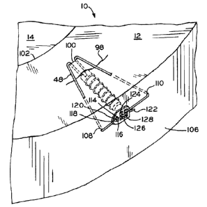
[0027] FIG. 10 is a perspective view of an exemplary embodiment of a device
incorporating a conduit according to another exemplary embodiment;
[0028] FIG. 11 is a perspective view of the device of FIG. 10 implanted in a
meniscus;
[0029] FIG. 12 is a cross-sectional view along line 12-12 of FIG. 11;
[0030] FIG. 13 is a perspective view of a device according to another
exemplary
embodiment;
[0031] FIG. 14 is a perspective view of the device of FIG. 13 implanted in a
meniscus;
[0032] FIG. 15 is a perspective view of a device according to another
exemplary
embodiment;
[0033] FIG. 16 is a elevational view along line 16-16 of the device of FIG.
15;
[0034] FIG 17 is a perspective view of the device of FIG. 10 implanted in a
meniscus
and secured according to another exemplary embodiment;
CA 02660287 2009-02-05
WO 2008/021770 PCT/US2007/075236
[0035] FIG. 18 is a perspective view of the device of FIG. 10 implanted in a
meniscus
according to another exemplary embodiment;
[0036] FIG. 19 is a perspective view of the device of FIG. 10 implanted in a
meniscus
and secured according to another exemplary embodiment;
[0037] FIG. 19A is a perspective view of the device of FIG. 10 implanted in a
meniscus and secured according to another exemplary embodiment;
[0038] FIG. 20 is a perspective view of the device of FIG. 10 implanted in a
meniscus
and secured according to another exemplary embodiment;
[0039] FIG. 21 is a perspective view of the device of FIG. 10 implanted in a
meniscus
and including a scaffold replacement;
[0040] FIG. 22 is a perspective view of a device according to another
exemplary
embodiment;
[0041] FIG. 23 is a perspective view of a device according to another
exemplary
embodiment implanted in a meniscus;
[0042] FIG. 24 is a perspective view of a filament and stops used in the
device of
FIG. 23;
[0043] FIG. 25 is a perspective view of an exemplary stop of the device of
FIG. 23;
and
[0044] FIG. 26 is a perspective view of a stop according to another exemplary
embodiment.
[0045] Corresponding reference characters indicate corresponding parts
throughout
the several views. The exemplifications set out herein illustrate preferred
embodiments of the
invention and such exemplifications are not to be construed as limiting the
scope of the
invention in any manner.
DETAILED DESCRIPTION
[0046] FIG. 7 shows conduit 30 according to one embodiment of the present
invention. The term "conduit", as used herein, means only an elongate body and
does not
define any other structural features. Conduit 30 includes a first end 32, a
second end 34, and
through bore 36 extending from first end 32 to second end 34. In one
embodiment, bore 36
has a non-circular cross-section. Conduit 30 can be manufactured from any
biocompatible
material. Conduit 30 has a length from first end 32 to second end 34 as small
as 2 mm, 3
mm, or 4 mm and as large as 10 mm, 12 mm, or 15 mm. Additionally, conduit 30
may be
6
CA 02660287 2009-02-05
WO 2008/021770 PCT/US2007/075236
coated with biocompatible substances to facilitate tissue regeneration,
improve circulation, or
achieve any other biologically desirable responses. For example, interior 38
of through bore
36 may be coated with an anti-coagulant to prevent coagulation of blood within
through bore
36, thereby promoting the delivery of blood to a torn or damaged tissue area.
Alternatively,
bore 36 may contain a scaffold material to promote tissue regeneration or to
improve healing
outcomes.
[0047] Conduit 30 may also be made of any porous material which would allow
for
the transfer of blood from a vascular to an avascular area as a result of
physiological
processes in the patient's body. Moreover, such a porous construct may be two-
piece, shown
in FIG. 7A and 7B, wherein first end 32' of conduit 30' is closed and
constructed of a porous
material, while the remainder of conduit 30' is made of a substantially solid,
biocompatible
material. This allows for blood or other fluid to enter second end 34 via bore
36 and exit
through the porous material at end 32'. Alternatively, conduit 30 may be
constructed entirely
of porous material and lack bore 36, as shown in FIG. 7C. Fluid would enter
conduit 30 from
end 34 and travel, due to the interconnected porosity of the porous material,
through conduit
30, exiting at first end 32. The flow of fluid may be directed by altering the
material
properties of the porous material along the length of conduit 30.
[0048] As shown in FIG. 7, conduit 30 further includes a plurality of
apertures 42, 44
at end 32 of conduit 30. Apertures 42, 44 extend from interior 38 of through
bore 36 to
exterior surface 46. Apertures 42, 44 may receive filament 48, as shown in
FIG. 10 and
described in detail hereinbelow, for securing conduit 30 within tissue and
fixating a tear,
damaged tissue, or an implant in a desired position. As used herein, the term
"filament" is
inclusive of single or multiple strands, threads, fibers, strings, wires or
sutures. In another
exemplary embodiment, apertures 42, 44 are positioned adjacent one another on
the same
side of conduit 30, i.e., along the axial length of conduit 30, and receive
filament 48 in the
same manner described in detail herein below. Location of apertures 42, 44 on
the same side
of conduit 30 provides for eccentric loading of conduit 30 when filament 48 is
fully secured,
which impedes pull out of conduit 30. Additionally, conduit 30 may include
slot 47, shown
in FIG. 7, in end 32 of conduit 30 which allow for the exit of blood or other
substances
therethrough. Slot 47 aids the surgeon in positioning conduit 30 within tissue
by eliminating
the need for the surgeon to precisely align end 32 of conduit 30 with the
plane of a tear,
damaged area, or implant to provide blood thereto. As long as the surgeon
positions a portion
of slot 47 in or adjacent the plane of the tear, damaged area, or implant,
blood or other
7
CA 02660287 2012-05-18
WO 2008/021770 PCT/US2007/075236
substances will be delivered to the tear, damaged area, or implant. In effect,
slot 47 provides
an increased length, only a portion of which the surgeon must locate adjacent
the tear,
damaged tissue, or implant, thereby increasing the likelihood of a successful
implantation.
[0049] In an exemplary embodiment, end 32 is perforated with a plurality of
apertures
of sufficient size and spacing to provide a substantially similar benefit as
slot 47, described
above. In another exemplary embodiment, shown in FIGS.7A-7B, conduit 30'
includes
closed end 32' perforated by a plurality of apertures 49 of sufficient size to
allow for the
dissemination of blood therethrough. In another embodiment, the entire length
of conduit 30
is perforated by a plurality of apertures 49 of sufficient size to allow for
the dissemination of
blood therethrough. Additionally, in another exemplary embodiment, the entire
length of
conduit 30 is porous, allow the release of fluid along the entire length of
conduit 30.
[0050] FIGS. 8-10 show conduits 50, 60, 70, respectively, according to
additional
embodiments of the present invention. Conduits 50, 60, 70 include several
features which are
identical to the embodiment of FIG. 7 discussed above and identical reference
numerals have
been used to indicate identical or substantially identical features
therebetween. Conduits 50,
60, shown in FIGS. 8 and 9, respectively, include surface features, such as
outwardly
extending ribs 52 and outwardly extending thread 62, respectively, on external
surface 46 of
conduits 50, 60. Ribs 52 and threads 62 provide an additional mechanism for
fixation of
conduits 50, 60 within tissue. As shown in FIG. 10, conduit 70 further
includes nose 72.
Nose 72 is separated from main body portion 74 via tapering section 76. During
implantation, nose 72 facilitates insertion of conduit 70 into the tissue and
can be positioned
such that nose 72 is in a vascular tissue, such as the synovium, while ribs 52
and/or threads 62
provide fixation. Additionally, nose 72 may itself be tapered to further ease
insertion.
[0051] As shown in FIG. 10, conduit 70 includes filament 48 attached thereto,
forming
completed medical device 78. The devices of the present invention are an
improvement over
the stent disclosed in U.S. Patent No. 7,988,716 to Schwartz, which is
assigned to the assignee
of the present invention. Filament 48 may be manufactured from any flexible,
biocompatible
material, such as polyglactin, polydioaxanone, surgical gut, nylon,
polypropeylyene,
polyglycolic acid, polylactic acid, co-polymers, Vicryl , and Ethibond Excel .
Vicryl
and Ethibond Excel are registered trademarks of Johnson & Johnson
Corporation, One
Johnson & Johnson Plaza, New Brunswick, New Jersey 08933. Filament 48 and
conduit 70
may be preassembled or may be assembled by the surgeon before or during
8
CA 02660287 2009-02-05
WO 2008/021770 PCT/US2007/075236
surgery. Filament 48 and conduit 70 may be connected together by inserting a
first end (not
shown) of filament 48 into interior 38 of through bore 36. The first end of
filament 48 is then
threaded through aperture 42 and wrapped half-way around exterior surface 46
until the first
end reaches aperture 44. In another embodiment, filament 48 is wrapped
substantially
entirely around exterior surface 46. The first end of filament 48 is then
inserted through
aperture 44 into interior 38 of through bore 36. First end of filament 48 is
then pulled out of
through bore 36 through end 32. In another embodiment, exterior surface 46
includes a
groove (not shown) on at least a portion of exterior surface 46 transverse to
the longitudinal
axis of conduit 70. As filament 48 is pulled from end 32 of conduit 70,
filament 48 tightens,
seating filament 48 within the groove. Once device 78 is assembled, device 78
may be
inserted into the meniscus as described in detail hereinbelow.
[0052] In another embodiment, the first end of filament 48 is inserted through
aperture 42 into interior 38 of through bore 36 and pulled out of through bore
36 through
aperture 44. In this embodiment, a portion of filament 48 extends through
interior 38 of
through bore 36 in a direction transverse to the longitudinal axis of conduit
70. In another
embodiment, device 80, as shown in FIGS. 15-16, includes conduit 82 having
nose 72,
through bore 36, and overmolded end 84. Device 80 include several features
which are
identical to the embodiment of FIG. 10 discussed above and identical reference
numerals
have been used to indicate identical or substantially identical features
therebetween. As best
seen in FIG. 16, overmolded end 84 includes apertures 86, 88 extending from
rim 90 of first
end 32 toward second end 34 along a portion of conduit 82. Apertures 86, 88
may be formed
to be slightly larger than filaments 92, 94 and, during manufacturing, shrink
around the ends
of filaments 92, 94 to retain the ends therein. Utilizing overmolded end 84
prevents filaments
92, 94 from extending into through bore 36 and provides an uninterrupted path
for the flow of
blood and other substances therethrough. In another embodiment, a
biocompatible adhesive
is used to secure the ends of filaments 92, 94 within apertures 86, 88. Once
device 80 is
assembled, device 80 may be inserted into the meniscus as described in detail
hereinbelow.
[0053] The method for inserting the devices will now be described in detail
with
reference to medical device 78, shown in FIG. 10. Device 78 may be inserted
into meniscus
as shown in FIGS. 11 and 12. In one embodiment, the entire procedure is
performed
arthroscopically using standard techniques, procedures, and devices. Device 78
is inserted
from the interior side of tear 98 at insertion point 100, located between
inner rim 102 of
meniscus 10 and the interior side of tear 98. In another exemplary embodiment,
the insertion
9
CA 02660287 2012-05-18
WO 2008/021770 PCT/US2007/075236
point is the face of tear 98. Device 78 is inserted along a plane
substantially parallel to the
bottom articulation surface of meniscus 10. While device 78 may be inserted at
any angle
relative to the bottom articulation surface of meniscus 10, insertion along a
plane
substantially parallel to the bottom articulation surface provides the optimal
purchase for
conduit 70. In one embodiment, insertion of device 78 is performed using a
compatible
insertion tool, such as those disclosed in U.S. Patent No. 7,988,716 to
Schwartz. The insertion
tool (not shown) may be inserted into the interior of through bore 36 to
retain device 78
thereon and advance device 78 through meniscus 10. In one embodiment, the
insertion device
is cannulated. The use of a cannulated insertion tool allows for the delivery
of biological
substances through the insertion device and conduit 70 directly to the torn or
damaged area
of meniscus 10. In another exemplary embodiment, device 78 is inserted
utilizing any
technique known technique, including an all-inside technique, inside-out
technique, and/or an
outside-in technique.
[0054] Device 78 is advanced via the insertion tool until end 32 of conduit 70
is
substantially aligned with the plane of tear 98, damaged area, or regenerative
or replacement
meniscus implant 134 (Fig. 21). Additionally, when inserted to align with a
damaged area of
tissue, the deterioration of the damaged tissue may provide tactile feedback
to the surgeon
that the outer plane of the damaged area has been encountered. As shown in
FIG. 18, conduit
70 may be positioned adjacent a tear, damaged area, or regenerative or
replacement meniscus
implant 134 with nose 72 extending from outer wall 106 of meniscus 10. In this
position,
nose 72 extends into the synovium and/or other tissue surrounding the knee
joint, which is a
highly vascular membrane surrounding the knee. In the same manner as set forth
above with
reference to red zone 16 of meniscus 10, blood, biological factors, cells, and
fluid from the
synovium and/or other tissue surrounding the knee joint can be delivered to a
torn or
damaged area of meniscus 10 via conduit 70.
[0055] Referring to FIG. 11, once conduit 70 is positioned, the insertion tool
is
removed, leaving conduit 70 in position and filament 48 extending from
insertion point 100.
Ends (not shown) of filament 48 are then looped over tear 98 and inserted in
meniscus 10 at
second insertion points 103, 104, located between outer wall 106 of meniscus
10 and tear 98
or between inner rim 102 of meniscus 10 and tear 98, using, for example, a
needle. The ends
of filament 48 are advanced through meniscus 10 at diverging angles until the
ends exit outer
wall 106 at points 108, 110. The ends of filament 48 are then tightened by
pulling the ends
away from outer wall 106. In addition to the stitching method set out above,
filament 48 can
CA 02660287 2009-02-05
WO 2008/021770 PCT/US2007/075236
be positioned via any method known to one of ordinary skill in the art,
including any
horizontal or vertical mattress suture technique.
[0056] With filament 48 taut, fixating inner and outer walls of tear 98 in
mutual
engagement, the ends of filament 48 are secured to one another. Once secured,
device 78 is
secured and the walls of tear 98 are fixed in their relative positions. In one
exemplary
embodiment, the ends of filament 48 are secured by tying the ends together to
form knot 112,
shown in FIG. 11. Excess portions of filament 48 may then be trimmed and
discarded.
[0057] As shown in FIG. 17, in another exemplary embodiment, first end 114 of
filament 48 is secured to a retention device, such as buckle 116, by inserting
first end 114
through an aperture in end 118 of buckle 116 and tying end 114 to form knot
120. Second
end 122 of filament 48 may then be secured to buckle 116 by inserting second
end 122
through opening 128 in buckle 116, looping end 122 around bar 126, through
opening 124,
and threading end 122 back through opening 128. In this manner, filament 48 is
looped back
onto itself and retained by friction within buckle 116. For large tears or
damaged areas,
multiple devices may be implanted in accordance with the method described
hereinabove.
[0058] As shown in FIG. 19, in another exemplary embodiment, first end 114 and
second end 122 of filament 48 are secured, via knots for example, to hooks
130. Hooks 130
are curved and terminate at sharpened tips 132. At any time during the
procedure, tips 132
are inserted through the upper articulation surface 12 of meniscus 10. Once
conduit 70 is
properly positioned and hooks 130 attached to meniscus 10 via tips 132,
filament 48 acts to
fixate tear 98 and secure conduit 70 in position, as described hereinabove.
[0059] In another exemplary embodiment, shown in FIG. 19A, first end 114 and
second end 122 of filament 48 are pulled tight through top articulating
surface 12 of meniscus
10. Knot 115 is tied using first end 114 and knot 117 is tied using second end
122 to secure
the walls of tear 98 in mutual engagement. Due to the physical properties of
meniscus 10,
knots 115, 117 will sink into top articulating surface 12, preventing any
damage to or pain in
the patient's knee. Similarly, any other securement method or device disclosed
herein may
potentially be used atop top articulating surface 12 to secure ends 114, 122
of filament 48
together and fixate tissue in the desired position.
[0060] Additionally, in another exemplary embodiment shown in FIGS. 20-21,
conduit 70 is positioned within meniscus 10 in a similar manner as described
hereinabove.
To secure conduit 70 in position within meniscus 10 and fixate tear 22 or
regenerative or
replacement meniscus implant 134, shown in FIG. 21, slide 136 is used. Slide
136 has a
11
CA 02660287 2009-02-05
WO 2008/021770 PCT/US2007/075236
body with a bore extending therethrough and flange 138 projecting from an end
of the body
of slide 136. First end 114 of filament 48 is threaded through the bore of
slide 136 toward
flange 138. Second end 122 of filament 48 is then secured to first end 114 of
filament 48 via
slipknot 140. By pulling first end 114 of filament 48 away from outer wall 106
of meniscus
10, slipknot 140 moves toward outer wall 106 and pushes slide 136 into
meniscus 10. Once
filament 48 is taught, flange 138 will contact outer wall 106 of meniscus 10,
preventing
slipknot 140 from sliding further. Slipknot 140 can then be tightened to
secure ends 114, 122
of filament 48 together. Once secured, ends 114, 122 of filament 48 may be
trimmed and the
removed portion discarded.
[0061] While the devices of the present invention may be implanted as an
alternative
to a meniscectomy, the devices may also be implanted in native meniscus tissue
or a
regenerative or replacement meniscus implant following a meniscectomy to
encourage and/or
promote tissue regeneration and, when a regenerative or replacement meniscus
implant is
used, the device may further fixate the implant to the natural meniscus
tissue, as shown in
FIG. 21. As shown in FIG. 21, regenerative or replacement meniscus implant 134
is fixated
via filament 48 in position against natural meniscus 10. Implant 134 further
receives blood,
biological factors, cells, and other fluids from the red zone 16 of meniscus
or, in another
embodiment shown in FIG. 18, from the synovium via conduit 70.
[0062] As shown in FIG. 13-14, two conduits 70, 70' are connected together via
filament 142. In connecting the conduits, a first end of filament 142 is
inserted through
interior 38 of through bore 36 of conduit 70, pulled from aperture 42, and
wrapped half way
around conduit 70. The end is then inserted through aperture 44, shown in
hidden lines in
FIG. 10, and pulled from interior 38 of through bore 36, as discussed in
detail hereinabove.
Filament 142 is then inserted through interior 38' of through bore 36' of
conduit 70', pulled
from aperture 42', and wrapped half way around conduit 70'. The end is then
inserted
through aperture 44' and pulled from interior 38' of through bore 36', as
discussed in detail
hereinabove. The ends of filament 142 are then connected together via slipknot
144, forming
device 146. While two conduits are depicted in FIG. 13, any number of conduits
needed to
facilitate tissue regeneration and healing may be connected together.
Generally, as the size of
the tear or damaged area increases, the number of conduits needed to
facilitate tissue
regeneration and healing will correspondingly increase.
[0063] By using multiple conduits, blood and/or other substances can be
delivered to
multiple points along the plane of a tear or damaged area of tissue and
fixated by the
12
CA 02660287 2009-02-05
WO 2008/021770 PCT/US2007/075236
tightening of only a single filament. The insertion of device 146 will now be
described in
detail. Conduits 70, 70' are inserted individually relative to tear 148 using
the same
procedure discussed hereinabove with respect to conduit 70 and tear 98. Once
each conduit
70, 70' is properly inserted, as shown in FIG. 14, filament 142 remains
partially exposed
along top articulating surface 12 of meniscus 10. Filament 142 is then
tightened, by pulling
end 150 of filament 142 away from top articulating surface 12 until the inner
and outer walls
of tear 148 are in mutual engagement. The interference of top articulating
surface 12 of
meniscus 10 with the tightening of filament 142 secures conduits 70, 70' in
their desired
positions.
[0064] In one exemplary embodiment, a knot (not shown) is used to fix filament
142,
and correspondingly secure device 146, in position. In one exemplary
embodiment, slipknot
144 is used to retain filament 142 in the tightened position. To tighten
filament 142, end 150
is pulled away from top articulating surface 12 of meniscus 10 and, at the
same time, slipknot
144 slides downwardly toward top articulating surface 12. Once slipknot 144 is
tightened,
excess filament 142 can be trimmed and discarded. Due to the resilient nature
of
fibrocartilage tissue, filament 142 and slipknot 144 will become integrated
with meniscus
preventing any adverse effects, such pain or discomfort during articulation of
the condyles of
the femur against top articulation surface 12 and filament 142. In one
embodiment, a series
of devices 78, shown in FIG. 10, may be utilized with a single tear. Each
device 78 can then
be fixated in the manner discussed hereinabove providing additional tension on
tear 98,
shown in FIG. 11, and placing knot 112 outside of the contact area of meniscus
10 and
against outer wall 106.
[0065] Referring to FIGS. 23-25, another exemplary embodiment utilizing
conduits
70, 70' is shown. In this embodiment, conduits 70, 70' are inserted through
tear 170
individually and prior to attachment of filament 172. Specifically, conduits
70, 70' are
inserted through the one of the walls forming tear 170 closest to outer wall
106 of meniscus
10. In one exemplary embodiment, noses 72, 72' of conduits 70, 70' may extend
from outer
wall 106 of meniscus 10, as described in detail above. Once conduits 70, 70'
are inserted into
meniscus 10, filament 172 and stops 174 may be secured to conduits 70, 70'.
While
described and depicted herein as utilizing two conduits 70, 70' and two stops
174, any
number of conduits may be used in conjunction with any number of stops to
provide the
desired fixation of tear 170.
13
CA 02660287 2009-02-05
WO 2008/021770 PCT/US2007/075236
[0066] Referring to FIGS. 24-25, stop 174 include apertures 176, 178 extending
therethrough. As shown in FIG. 25, stop 174 is formed as an elongate rod. By
forming stops
174 as elongate rods, stops 174 allow for fluid to pass through bores 36, 36'
(FIG. 13) of
conduits 70, 70'. However, stops 174 may be formed in any other geometric
shape, such as
square rods or circular discs. For example, referring to FIG. 26, stop 174' is
shown in the
form of a circular disc have apertures 176', 178' extending therethrough. Stop
174' may
replace stop 174 and may be utilized in the same manner as described herein
with reference
to stop 174. In connecting filament 172 to stops 174, first end 180 of
filament 172 is
threaded through aperture 176 of stop 174. First end 180 is then threaded
through aperture
178 of stop 174. A second stop 174 is then provided and connected to filament
172 in a
similar manner. First end 180 of filament 172 is then used to tie slipknot 182
on filament 172
and the remainder of first end 180 is then trimmed.
[0067] Once stops 174 are attached to filament 172, as shown in FIG. 24,
filament
172 and stops 174 are inserted into meniscus 10. Specifically, longitudinal
axes 184 (FIG.
25) of stops 174 are aligned with bores 36, 36' of conduits 70, 70' and
inserted at insertion
points 190. Since stops 174 have an outer diameter that is less then the inner
diameter of
bores 36, 36', once aligned, stops 174 may be inserted through bores 36, 36',
respectively, and
then through outer wall 106 of meniscus 10. Stops 174 may be inserted in
unison or,
alternatively, may be inserted individually. For example, one of stops 174 may
be inserted
through bore 36 of conduit 70 and outer wall 106. Then, the other of stops 174
may be
inserted through bore 36' of conduit 70' and outer wall 106. Once inserted as
described in
detail above, end 186 and a portion of filament 172 extends from articulating
surface 12 of
meniscus 10. End 186 of filament 172 may then be pulled away from articulating
surface 12.
As end 186 is pulled away from articulating surface 12 of meniscus 10,
slipknot 182 of
filament 172 tightens as filament 172 slides through apertures 176, 178 of
stops 174 and
bores 36, 36' of conduits 70, 70'. In this manner, pulling on end 186 of
filament 172 allows
for the tightening of the entire construct to fixate the opposing sides of
tear 170 adjacent one
another and fixate conduits 70, 70' in their desired positions. Once
tightened, end 186 of
filament 172 may be trimmed to remove any excess material.
[0068] In another exemplary embodiment, conduit 160, shown in FIG. 22,
includes
filament 162 secured through apertures in projection 164. Projection 164 may
be
overmolded, as described in detail above, or may allow for sliding movement of
filament 162
within projection 164. If sliding movement of filament 162 is allowed, end 166
of filament
14
CA 02660287 2009-02-05
WO 2008/021770 PCT/US2007/075236
162 could be pulled away from projection 164 drawing end 168 toward projection
164. In
another embodiment, projection 164 is replaced by apertures located adjacent
one another on
the same side of conduit 160, i.e., along the axial length of conduit 160.
These apertures
accept filament 162 in the same manner as apertures 42, 44, described in
detail above with
reference to FIGS. 7-10. The use of either projection 164 or the apertures
located on the
same side of conduit 160 provides for eccentric loading of conduit 160 when
filament 162
finally secured, which impedes pull out of conduit 160.
[0069] While this invention has been described as having a preferred design,
the
present invention can be further modified within the spirit and scope of this
disclosure. This
application is therefore intended to cover any variations, uses, or
adaptations of the invention
using its general principles. Further, this application is intended to cover
such departures
from the present disclosure as come within known or customary practice in the
art to which
this invention pertains and which fall within the limits of the appended
claims.