Note: Descriptions are shown in the official language in which they were submitted.
CA 02660292 2009-01-28
WO 2008/015670 PCT/IL2007/000952
ARTHROSCOPIC BONE TRANSPLANTING PROCEDURE, AND MEDICAL
INSTRUMENTS USEFUL THEREIN
FIELD AND BACKGROUND OF THE INVENTION
The present invention relates to an arthroscopic bone transplanting procedure
and to medical instruments useful in such a procedure as may be supplied in
the form
of a kit. The invention is particularly useful in the treatment of an anterior
shoulder
instability, where a section of the coracoid is transplanted to the glenoid,
and is
therefore described below with respect to said transplant.
The range of movements the human shoulder can make far exceeds any other
joint in the body. The shoulder joint is a ball and socket joint, similar to
the hip;
however, the socket of the shoulder joint is extremely shallow, and thus
inlierently
unstable. Muscles and tendons serve to keep the bones in approximation. In
addition,
in order to compensate for the shallow socket, the shoulder joint has a cuff
of fibrous
cartilage called a labrum that forms a cup for the head of the arm bone
(humerus) to -
move within. This cuff of cartilage makes the shoulder joint much more stable,
yet
allows for a very wide range of movement. When the labrum of the shoulder
joint is
damaged, the stability of the shoulder joint is compromised, leading to
subluxation
and dislocation of the joint. Recurrent dislocations may cause damage to the
bones of
the joint -the humeral head and the glenoid. In particular, damage to the
anterior-
inferior part of the glenoid will cause a decrease in the area of contact with
the
humeral head.
When bone deficiencies associated with anterior shoulder instability are
present, the prognostic factors for the success of soft tissue repair are
poor. Current
standards of success are predicated on the restoration of motion and strength
and the
return to full functional activities, including competitive athletics.
Reestablishment of
anterior shoulder stability requires the recognition and the treatment of
osseous
defects.
Several surgical procedures have been described for the management of
osseous deficiencies in association with anterior shoulder instability,
involving the
transplantation of a portion of the coracoid process to the anterior-inferior
section of
the glenoid. The procedure described by Latarjet in 1954 involves the
transplantation
of a large section of the coracoid together with the conjoined tendon attached
to it to
1
CA 02660292 2009-01-28
WO 2008/015670 PCT/IL2007/000952
reinforce the glenoid fossa and create an antero-inferior musculotendinous
sling. The
procedure has been performed since its disclosure with positive results as an
open
surgical intervention.
However, up to the present, no minimally invasive technique for performing it
has been developed.
OBJECTS AND BRIEF SUMMARY OF THE PRESENT INVENTION
An object of the present invention is to provide an arthroscopic bone
transplanting procedure which is particularly useful in the treatment of
anterior
shoulder instability, but may be used in other procedures involving implanting
of a
section of a first bone to a second bone. A further object of the invention is
to provide
instruments, wliich may be supplied in kit form, particularly useful in such
an
arthroscopic procedure.
According to one aspect of the present invention, there is provided an
arthroscopic procedure for transplanting a section of a first bone to a second
bone,
coinprising the following steps: (a) making small incisions to open portals
for the
introduction of medical instruments; (b) drilling a tlireaded bore in said
section of said
first bone; (c) attaching a first cannula to said section of said first bone;
(d) separating
said section from said first bone; (e) positioning said separated section of
said first
bone on said second bone; (f) replacing said first cannula by a second cannula
attached to said separated bone section by a cannulated device; (g)
introducing a
guide wire through the cannulated device; (h) removing the cannulated device;
(i) drilling a bore into the second bone by a cannulated drill guided by said
guide
wire; (j) removing the guide wire; (k) and applying a bone screw through said
bore in
said separated section of the first bone and said bore in said second bone.
The preferred embodiment of the invention described below is particularly
useful for the treatment of anterior shoulder instability, or other disorders
where it is
desired to use at least two bone screws for attaching a section of a first
bone to a
second bone. When such a procedure is used, in step (b), two threaded bores at
a
fixed distance from each other are drilled in said section of the first bone;
in step (c),
the first cannula is a T-handle cannula and is attached in said first bore by
sutures or
flexible wires; in step (f), the second cannula is a double cannula and is
attaclied to
said section of the first bone by two cannulated devices; in step (g), two
guide wires
are introduced through the two cannulated devices, which cannulated devices
are then
2
CA 02660292 2009-01-28
WO 2008/015670 PCT/IL2007/000952
removed in step (h); in step (i), two bores are drilled into the second bone
by a
cannulated drill guided by said guide wires; in step (j), the two guide wires
are
removed; and in step (k), two bone screws are applied through the two bones in
the
separated section of the first bone, and the two bores in the second bone.
Other aspects of the invention involve the construction of medical
instruments,
which may be supplied in a kit, particularly useful for the above-described
bone
transplanting procedures.
Further features of the invention will be apparent from the description below.
BRIEF DESCRIPTION OF THE DRAWINGS
The present invention is herein described below, the reference to the
accompanying drawings, wherein:
Fig. l a is a schematic drawing of the gleno-humeral joint in the shoulder;
Fig. lb is a schematic lateral view illustrating damage to the glenoid fossa;
Fig. 2a is a schematic anterior view of the bone reconstruction;
Fig. 2b is a transverse section through the reconstructed joint. and
Figs. 3-20 illustrate various medical instruments, which may be supplied in
kit
form, particularly useful in an arthroscopic bone transplanting procedure for
reconstructing the shoulder joint in accordance with the present invention, in
which:
Fig. 3 shows a standard Kirschner wire;
Fig. 4 is a cannulated bone drill;
Fig. 5 shows a drill guide for drilling a second bore at a pre-
determined distance from a first bore;
Fig. 6 is a thread tapping tool;
Fig. 7a is a suture loader;
Fig. 7b is a suture retriever;
Fig. 8 shows a flexible wire;
Fig. 9 is a cannula with a T-handle;
Fig. 10 shows osteotomes, straight and cuived;
Fig. 11 is a cannulator for a double cannula;
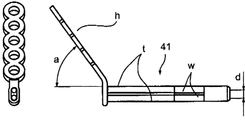
Fig. 12 is a double cannula;
Fig. 13 shows a suture hook;
Fig. 14 shows a cannulated device
Fig. 15 is a cannulated devicedriver;
3
CA 02660292 2009-01-28
WO 2008/015670 PCT/IL2007/000952
Fig. 16 is a cannulated spike;
Figs. 17a and 17b are side and top views, respectively, of a clamping device
for holding a transplanted bone section to the receiving site;
Fig. 18 shows ca.nnulated bone drills;
Fig. 19 is a cannulated bone screw; and
Fig. 20 is a screwdriver with a long cannulated shaft for the bone screws.
THE CONSTRUCTION OF THE SHOULDER JOINT
Fig. 1 a illustrates the bones of the shoulder joint. The liead 1 of the upper
arm
bone, the humerus 2, forms a ball-and.-socket joint with the shallow glenoid
cavity 3.
The glenoid is the lateral part of the shoulder blade scapula 4. Two hook-like
projections of the scapula seen overhanging the glenoid are the acromion 5 and
the
coracoid process 6. A group of muscles collectively know as the Rotator Cuff
originate on the scapula and insert on the humeius. These serve to stabilize
the joint
by keeping the huineral head in contact witll the glenoid cavity. The clavicle
7
connects the acromion to the breastbone sternum. The glenoid labrum 8, which
is a
flexible fibrous ligament, surrounds the glenoid rim enlarging its area of
contact with
the humerus. When dislocations in the direction shown by the arrow occur, the
anterior-inferior part of the labrum is torn away from the glenoid, causing
instability
of the joint. Recurring dislocations may lead to osseous lesions.
Fig. lb illustrates the type of dainage to the glenoid socket caused by such
dislocations. The pear-shape of the intact glenoid is shown at "A"; while bone
loss at
the inferior, wider section "A", caused by a dislocation, is shown at "B" and
results in
an inverted pear shape narrower lower section as shown at "C". This causes a
partial
loss of contact with the humeral head.
Figs. 2a and 2b illustrate a bone reconstruction in accordance with the
present
invention.
DESCRIPTION OF A PREFERRED EMBODIMENT
The description below describes a kit of instruments, and the method of their
use, for performing coracoid transfer (Latarjet procedure) arthroscopically.
The kit
consists of various instruments, including drills, drill guides, osteotomes,
cannulae,
suture manipulators, screws, screwdrivers and others, specific for the purpose
of the
method disclosed by the invention.
4
CA 02660292 2009-01-28
WO 2008/015670 PCT/IL2007/000952
The procedure consists of the following main steps:
= Opening portals (small incisions); introducing the arthroscope and
instruments
= Preparation of the coracoid and glenoid surfaces
= Drilling and threading two holes in the coracoid at a fixed distance
= Passing sutures or flexible wires through the holes
= Attaching the coracoid by sutures or flexible wires to a camiula
= Separating the section of the coracoid to be transferred
= Positioning the graft on the glenoid
= Attaching a double cannula to the coracoid with a cannulated device
= Introducing K-wires through the cannulated device
= Removing the cannulated device
= Drilling into the glenoid with a cannulated drill over the K-wires
= Attaching the transplanted coracoid onto the glenoid with bone screws
= Removing the K-wires
= Final fixing of the transplant (tightening the screws)
= Removing the camlula.
In the reconstruction of the shoulder joint according to the present invention
illustrated in Figs. 2a and 2b, 20 indicates the glenoid, 21 illustrates the
coracoid graft
implanted thereto by a pair of cannulated devices 22 and 23, 24 indicates the
humeral
head, and 25 indicates the conjoined tendon.
A Bone Transplantation Procedure and the Medical Instruments Used Therein
Figs. 3-20 illustrate the various medical instruments, preferably supplied in
kit
form, for performing an arthroscopic bone transplanting procedure in
accordance with
the present invention.
Portals (small incisions) are first made for introducing the arthroscope and
instruments and for preparing the coracoid and glenoid surfaces, leaving the
conjoined
tendon (shown in Fig. 2b) attached to the coracoid. Two threaded holes are
drilled in
the coracoid process, using the bone drill shown at 32 in Fig. 4 with a
diameter of
about 3 mm. A Kirschner wire 31 (Fig. 3) is inserted at a safe distance from
the
lateral tip of the process for guiding the bone drill, and the first hole is
drilled. For
placing the second hole, the drill is inserted through the drill guide shown
at 33 in
Fig. 5. A guide pin 33a fixed at distance "d" from the center of the drill nut
33b
ensures a predetermined distance of about 9 mm from the first hole. Both
lloles are
5
CA 02660292 2009-01-28
WO 2008/015670 PCT/IL2007/000952
threaded now with the elongated tap shown at 34 in Fig. 6. For safeguarding
the
integrity of the transplant, inserts may be implanted in the holes.
Suture strands or flexible wires are now attached to the coracoid process for
securing during separation by threading them through the holes. A suture
loader 35,
Fig. 7a, and a suture retriever 36, in Fig. 7b are provided in the kit for
manipulating
the sutures. An alternative flexible wire 37 is shown in Fig. 8. The
sutures/wires are
drawn out through the shaft of a T-handle cannula shown at 38 in Fig. 9 and
are fixed
at the proximal, handle section of the cannula for holding the coracoid graft
during
separation and transfer to the receiving site. Osteotomes, such as those shown
at 39a,
3 9b in Fig. 10, serve to separate the lateral section of the coracoid. At
least one
osteotome is provided in the kit.
Preparing for the transfer of the separated section of the coracoid, the
subscapularis muscle is dissected and split to allow for transferring the T-
handle
cannula 38 with the coracoid transplant to the anterior-inferior, damaged
section of
the glerioid. The cannulator shown at 40 in Fig. 11 is used to dissect tissue
and to free
a passage to the receiving site. A double cannula shown at 41 in Fig. 12 is
inserted
through the passage freed by the cannulator.
The two tubes "t" of the double cannula 41 are fixed, so that the distance of
their centerlines "d" is identical to that of the drill guide 33 in Fig. 5.
Handle "h"
attached to the tube is offset at an angle "a" relative to the axis of the
tubes and is
formed to provide a firm grip. Angle "a" should be of an order of 40 to 65
degrees to
allow maneuvering without obstructing the field of vision, and the length of
the tubes
measured from the handle should be about 150nv.n. A window "w" is cut in each
of
the tubes near the distal end to enable observation of the interior of the two
tubes, and
the position of an instrument introduced into the tubes.
When the double cannula has been inserted to face the coracoid transplant, the
T-handle cannula 38 is released from the sutures/wires attached to the graft
and is
withdrawn. Using a suture hook shown at 42 in Fig. 13, the sutures/wires are
drawn
through the tubes of the double cannula and an elongated cannulated holding
device
such as screws 43 shown in Fig. 14 are inserted over them into the tubes of
the
cannula. The screws are driven into the coracoid using a suitable instrument,
such as
the screw driver shown at 44 in Fig. 15 until the coracoid is firmly attached
to the
cannula. An alternative device for holding the separated coracoid bone
transplatit to
6
CA 02660292 2009-01-28
WO 2008/015670 PCT/IL2007/000952
the double cannula is shown at 45 in Fig. 16. The distal section of the spike
in Fig. 16
is expandable to hold the device to the walls of the bores of the graft.
The sutures/wires holding the coracoid can now be removed. The exact
positioning on the glenoid may be assisted by using a suitable instrument,
such as the
clamping device shown at 46 in Figs. 17a and 17b. Once the transplant is in
the
correct position on the glenoid, Kirschner wires (31, Fig. 3) are driven into
the
glenoid through the cannulated devices holding the coracoid. The devices are
now
removed using the screwdriver 44, Fig. 15, or by releasing the spike 45.
The double cannula serves as a drill guide. With a catmulated drill 47a,
Fig. 18, inserted over one of the Kirschner wires, a first hole is drilled
into the
glenoid. Leaving the first drill in position, the other drill 47b in Fig. 18,
with the
longer shaft, is used to drill a second hole over the second Kirschner wire.
After removing the drills, cannulated bone screws 48, Fig. 19, are inserted
over the K-wires into the coracoid graft and are screwed part-way into the
glenoid
using the cannulated device driver with a long shaft 49, Fig. 20, for use with
the
cannulated bone screws.
The K-wires can now be pulled out and the optional bone clamping device is
removed. The bone screws 48 are drawn tight and the double carmula is
withdrawn to
conclude the procedure.
While the invention has been described with respect to a preferred
embodiment, it will be appreciated that this is set fortli merely for purposes
of
example, and that many other variations, modifications and applications of the
invention may be made.
7
