Note: Descriptions are shown in the official language in which they were submitted.
CA 02661709 2009-04-07
LOOP SUTURE
BACKGROUND
Technical Field
The present disclosure relates to sutures and more particularly to sutures
which include
a proximal end having a first outer diameter and a distal end having an inner
hollow region with
an inner diameter which is configured to accommodate at least a portion of the
first outer
diameter of the proximal end of the suture to form a stitch.
Background of Related Art
Sutures are well known for tying approximated tissues of a wound together at
intervals
along the length of the wound. Although sutures have been proven to
sufficiently close a
wound, the process can be somewhat time consuming depending upon the handling
characteristics of the suture and the wound location. For example, some suture
materials are
not easily tied into knots, or can not withstanding the constant movement
through tissue when
closing a larger wound and as a result break in the middle of the suturing
process.
There are great advantages in providing a suture capable of closing a wound in
tissue
following surgery, i.e., plastic, general, or laparoscopic, which eliminates
the need for tying a
knot and also forms individual stitches thereby exposing the suture to less
stress and
decreasing the likelihood of the suture breaking before the wound is closed.
1
CA 02661709 2009-04-07
SUMMARY
Accordingly, the present disclosure describes sutures which include an
elongate body
that has a proximal end and a distal end. The proximal end has a first outer
diameter and the
distal end has an inner hollow region with an inner diameter which is
configured to receive at
least a portion of the first outer diameter of the proximal end of the suture.
In embodiments, the sutures have an elongate body that has a distal end and a
proximal
end. The proximal end has a first outer diameter, and the distal end has a
second outer
diameter which is greater than the first outer diameter of the proximal end.
The distal end
further includes an inner hollow region defining an inner diameter within the
distal end which is
configured to receive at least a portion of the first outer diameter of the
proximal end. Methods
of suturing tissue are also described.
BRIEF DESCRIPTION OF THE DRAWINGS
Various embodiments are illustrated and described herein, wherein:
FIG. 1A is a side view of a suture containing a distal end having an inner
hollow region
defined therein in accordance with one embodiment of this disclosure;
FIG. 1 B is a side view of a suture containing a tip in accordance with one
embodiment of
this disclosure;
FIG. 2A is a side view of a suture containing at least one slot in accordance
with one
embodiment of this disclosure;
FtG. 2B is a side view of a suture forming a closed loop stitch in accordance
with one
embodiment of this disclosure;
FIG. 2C is a side view of a suture containing a distal end having an inner
hollow region
defined therein including internal friction-enhancing members.
FIG. 3 is a side view of a suture containing a plug in accordance with one
embodiment of
this disclosure; and
2
CA 02661709 2009-04-07
FIGS. 4A-4C are side views of a suture containing a distal end having an inner
hollow
region defined therein including friction-enhancing members.
DETAILED DESCRIPTION OF THE EMBODIMENTS
Turning now to FIG. 1 A, suture 10 is shown as a hollow monofilament having an
elongate body which includes a proximal end 20 defining a first outer diameter
25 and a distal
end 30 defining a second outer diameter 35 which is greater than or equal to
first outer diameter
25. Distal end 30 also includes hollow region 40 defining an inner diameter 45
of distal end 30
which is configured to receive at least a portion of first outer diameter 25
of proximal end 20 of
suture 10 to form a closed loop stitch. It is envisioned that suture 10 may be
a monofilament or
a multifilament suture (see FIGS. 4A-C) or a combination thereof and proximal
end 20 may be
hollow or solid or any combination thereof.
Suture 10 may be made from any material suitable for manufacturing surgical
sutures or
ligatures. Suture materials include for example any bioabsorbable, non-
bioabsorbable,
synthetic or natural materials and combinations thereof. Some suitable
examples of absorbable
materials include trimethylene carbonate, caprolactone, dioxanone, glycolic
acid, lactic acid,
glycolide, lactide, homopolymers thereof, copolymers thereof, and combinations
thereof. Some
specific examples of suitable non-absorbable materials which may be utilized
to form the suture
include polyolefins, such as polyethylene, polypropylene, copolymers of
polyethylene and
polypropylene, and blends of polyethylene and polypropylene. Some other useful
materials
include nylons, and cat-gut.
It is envisioned that the suture described herein may also be made from
biomaterials not
commonly associated as suture materials. Since the sutures are capable of
forming a closed
stitch loop which does not require the formation of a knot, these sutures can
be formed of
biomaterials which do not possess a minimal ability to form knots. One example
of such a
3
CA 02661709 2009-04-07
material would include biomaterials which having a low coefficient of
friction. Another example
would include biomaterials displaying a very high modulus.
As shown in FIG. 1A, in some embodiments, hollow region 40 is sealed within
distal end
30 of suture 10 and is capable of storing at least one bioactive agent. In
these embodiments,
hollow region 40 can also serve as a vehicle for delivery of the bioactive
agent. It is envisioned
that at least a portion of proximal end 20 of suture 10 will penetrate into
distal end 30 and enter
into inner hollow region 40 to interact with and possibly release a bioactive
agent stored therein.
In embodiments where the bioactive agent is an adhesive, proximal end 20 may
be positioned
more permanently within inner hollow region 40 of distal end 30 after reacting
with the adhesive.
The term "bioactive agent", as used herein, is used in its broadest sense and
includes
any substance or mixture of substances that have clinical use. Consequently,
bioactive agents
may or may not have pharmacological activity per se, e.g., a dye, or
fragrance. Alternatively a
bioactive agent could be any agent which provides a therapeutic or
prophylactic effect, a
compound that affects or participates in tissue growth, cell growth, cell
differentiation, an anti-
adhesive compound, a compound that may be able to invoke a biological action
such as an
immune response, or could play any other role in one or more biological
processes. It is
envisioned that the bioactive agent may be positioned on any part of the
sutures described
herein. For example, the bioactive agent may be simply coated on an outer or
inner surface of
the suture or combined with the material used to form the suture or may
impregnate the suture
surface. In addition, the inner hollow region may act as a reservoir in
storing the bioactive agent.
The bioactive agent may be positioned on the suture in any amount,
configuration, and suitable
form of matter, i.e., films, powders, liquids, gels and the like.
Examples of classes of bioactive agents which may be utilized in accordance
with the
present disclosure include antimicrobials, analgesics, antipyretics,
anesthetics, antiepileptics,
antihistamines, anti-inflammatories, cardiovascular drugs, diagnostic agents,
sympathomimetics, cholinomimetics, antimuscarinics, antispasmodics, anti-
adhesives,
4
CA 02661709 2009-04-07
hormones, growth factors, muscle relaxants, adrenergic neuron blockers,
antineoplastics,
immunogenic agents, immunosuppressants, gastrointestinal drugs, diuretics,
steroids, lipids,
lipopolysaccharides, polysaccharides, and enzymes. It is also intended that
combinations of
bioactive agents may be used.
Suitable antimicrobial agents which may be included as a bioactive agent
stored within
the suture described herein includes triclosan, also known as 2,4,4'-trichloro-
2'-hydroxydiphenyl
ether, chlorhexidine and its salts, including chlorhexidine acetate,
chlorhexidine gluconate,
chlorhexidine hydrochloride, and chlorhexidine sulfate, silver and its salts,
including silver
acetate, silver benzoate, silver carbonate, silver citrate, silver iodate,
silver iodide, silver lactate,
silver laurate, silver nitrate, silver oxide, silver palmitate, silver
protein, and silver sulfadiazine,
polymyxin, tetracycline, aminoglycosides, such as tobramycin and gentamicin,
rifampicin,
bacitracin, neomycin, chloramphenicol, miconazole, quinolones such as oxolinic
acid,
norfloxacin, nalidixic acid, pefloxacin, enoxacin and ciprofloxacin,
penicillins such as oxacillin
and pipracil, nonoxynol 9, fusidic acid, cephalosporins, and combinations
thereof. In addition,
antimicrobial proteins and peptides such as bovine lactoferrin and
lactoferricin B may be
included as a bioactive agent in the present disclosure.
Other bioactive agents which may be included as a bioactive agent within the
sutures
described herein include: lubricants; local anesthetics; non-steroidal
antifertility agents;
parasympathomimetic agents; psychotherapeutic agents; tranquilizers;
decongestants; sedative
hypnotics; steroids; sulfonamides; sympathomimetic agents; vaccines; vitamins;
antimalarials;
anti-migraine agents; anti-parkinson agents such as L-dopa; anti-spasmodics;
anticholinergic
agents (e.g. oxybutynin); antitussives; bronchodilators; cardiovascular agents
such as coronary
vasodilators and nitroglycerin; alkaloids; analgesics; narcotics such as
codeine,
dihydrocodeinone, meperidine, morphine and the like; non-narcotics such as
salicylates, aspirin,
acetaminophen, d-propoxyphene and the like; opioid receptor antagonists, such
as naltrexone
and naloxone; anti-cancer agents; anti-convulsants; anti-emetics;
antihistamines; anti-
CA 02661709 2009-04-07
inflammatory agents such as hormonal agents, hydrocortisone, prednisolone,
prednisone, non-
hormonal agents, allopurinol, indomethacin, phenylbutazone and the like;
prostagiandins and
cytotoxic drugs; estrogens; antibacterials; antibiotics; anti-fungals; anti-
virals; anticoagulants;
anticonvulsants; antidepressants; antihistamines; and immunological agents.
Other examples of suitable bioactive agents which may be included within the
suture
described herein include viruses and cells, peptides, polypeptides and
proteins, analogs,
muteins, and active fragments thereof, such as immunoglobulins, antibodies,
cytokines (e.g.
lymphokines, monokines, chemokines), blood clotting factors, hemopoietic
factors, interleukins
(IL-2, IL-3, IL-4, IL-6), interferons (R-IFN, (a-IFN and y-IFN),
erythropoietin, nucleases, tumor
necrosis factor, colony stimulating factors (e.g., GCSF, GM-CSF, MCSF),
insulin, anti-tumor
agents and tumor suppressors, blood proteins, gonadotropins (e.g., FSH, LH,
CG, etc.),
hormones and hormone analogs (e.g., growth hormone), vaccines (e.g., tumoral,
bacterial and
viral antigens); somatostatin; antigens; blood coagulation factors; growth
factors (e.g., nerve
growth factor, insulin-like growth factor); protein inhibitors, protein
antagonists, and protein
agonists; nucleic acids, such as antisense molecules, DNA and RNA;
oligonucleotides;
polynucleotides; and ribozymes.
Turning now to FIG. 1 B, suture 10 is shown to include to a tip 50 capable of
penetrating
tissue. In addition, where distal end 30 is closed, tip 50 can serve to
penetrate distal end 30 to
obtain access to hollow region 40 and form a closed loop stitch, as shown in
FIG. 1C. In
embodiments, tip 50 may be formed from the suture material. In embodiments,
tip 50 may be a
surgical needle connected to the suture material. It is envisioned that suture
10 can be
connected to any suitable surgical needle capable of penetrating tissue,
closing a wound and
entering into hollow region 40 of distal end 30 of suture 10. Some non-
limiting examples include
curved needles, straight needles, tapered needles, triangular-body needles,
round body
needles, blunt and trocar needles. It is further envisioned that the sutures
described herein may
be attached to a suture needle using any conventional method known to those
skilled in the art
6
CA 02661709 2009-04-07
including swaging, crimping, heat-shrinking, adhesives, male/female mating
engagement and so
forth.
In some embodiments, suture 10 may include a tip 50 which is designed and
configured
to penetrate through tissue, approximate tissue and/or close a wound. Tip 50
may be blunt,
sharp or any combination thereof and can be made from any suitable material
capable of
penetrating tissue to close a wound. Some useful examples of suitable
materials include, but
are not limited too, metals, such as stainless steels, metal alloys, shape
memory alloys, such as
Nitinol, and any shapeable polymeric materials, such as lactide, glycolide,
caprolactone, and the
like.
As shown in FIG. 1 B, tip 50 may be preformed and positioned within proximal
end 20 of
suture 10. In some embodiments, an adhesive, such as a cyanoacrylate, may be
used to
position tip 50 within proximal end 20 of suture 10. In still other
embodiments, tip 50 may be
heat-shrunk into position within suture 10.
In embodiments where tip 50 is made from a shape-memory alloy, it is
envisioned that
tip 50 may penetrate hollow region 40 of distal end 30 to form a close-loop
stitch and then be
exposed to an electrical, magnetic or temperature force which makes the shape-
memory alloy
return to its original shape and dimension which further tightens the closed-
loop stitch creating a
tighter and more secure wound closure.
Turning now to the embodiment of FIG. 2A, flared distal end 130 of suture 110
is shown
to further include slot 160. Slot 160 is positioned along flared distal end
130 to allow the
passage of proximal end 120 of suture 110 into inner hollow region 140 to form
a closed stitch
loop. It is envisioned that slot 160 may be positioned on any portion of
flared distal end 130 to
allow entry of proximal end 120 into inner hollow region 140.
As shown in the embodiments of FIG. 2B, flared distal end 130 of suture 110
may
include more than one slot 160a and 160b to allow proximal end 120 of suture
110 to penetrate
at least a portion of flared distal end 130 and pass through inner hollow
region 140 before
7
CA 02661709 2009-04-07
exiting distal flared end 130 through additional slot 160b. Suture 110 is
shown forming a 3602
closed loop stitch. Such a stitch is designed to be formed quickly and does
not necessarily
require the use of a knot to keep the wound closed. Proximal end 120 is shown
to include at
least one external friction-enhancing member 125 which is designed to assist
in holding suture
110 in the closed loop position. As shown, external friction-enhancing members
125 are barbs
or barb-like structures positioned along the outer surface of proximal end 20
of suture 10. It is
envisioned that the external friction-enhancing members may take any shape or
geometric
contour suitable for maintaining the proximal end of the suture within the
inner hollow region.
Suitable non-limiting examples include treads, bumps, grooves, spikes, ridges,
and the like.
In FIG. 2C, inner hollow region 140 of flared distal end 130 of suture 110 is
shown
further including internal friction-enhancing members 145. Internal friction-
enhancing members
145 may act as a ratchet type mechanism to secure the proximal end of the
suture in the inner
hollow region 130 to form a closed loop stitch. As shown, internal friction-
enhancing members
145 are barbs or barb-like structures positioned along the inner surface of
inner hollow region
130. It is envisioned that the internal friction-enhancing members may take
any shape or
geometric contour suitable for maintaining the proximal end of the suture
within the inner hollow
region. Suitable non-limiting examples include treads, bumps, grooves, spikes,
ridges, and the
like.
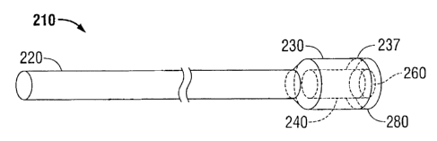
Turning now to FIG. 3, suture 210 is shown to include a plug 280 which is
dimensioned
to fit within slot 260. Since slot 260 creates an opening which interconnects
inner hollow region
240 with an outer surface 237 of distal flared end 230, inner hollow region
240 may be unable to
store a bioactive agent. Thus, plug 280 is designed to store at least one of
the bioactive agents
previously described herein and release the bioactive agent when penetrated by
proximal end
220 of suture 210, which in some embodiments may further include a sharpened
tip, upon
entering or exiting inner hollow region 240 of distal flared end 230 of suture
210. Plug 280 may
be made from any bioabsorbable or non-bioabsorbable material.
8
CA 02661709 2009-04-07
In the embodiments shown in FIGS. 4A-4C, the sutures are shown including at
least one
external friction-enhancing member and/or at least one internal friction-
enhancing member. As
shown, the external members may be added to the outer surface of the proximal
end of the
suture, and the internal members mat be positioned on the inner surface of the
inner hollow
region of the distal end of the suture, the inner surface of a slot, or any
combination thereof. In
FIG. 4A, a multifilament suture 410 is shown having an inner hollow region 440
which includes a
plurality of internal friction-enhancing members 445 to assist in holding
suture 410 in a closed
loop position after passing through at least a portion of distal end 430. As
further shown in Figs.
4B and 4C, external friction-enhancing members 425 and internal friction-
enhancing members
445 may take any shape, dimension or contour capable of assisting with holding
the suture in a
closed loop stitch. Suitable friction-enhancing members may also be formed
from any material
capable of assisting in positioning at least a portion of the proximal end of
the suture within the
inner hollow region of the distal end of the suture.
It is envisioned that the sutures described herein may be made of any suitable
size,
shape and dimension to close a wound in living tissue. It is further
envisioned that the sutures
described herein may be used to close a wound in any type of tissue. In
particularly useful
embodiments, the sutures may be used to close sub-dermal tissue, such as
wounds common to
plastic, laparoscopic and general surgeries.
In embodiments, the sutures described herein may be used to suture wounded
tissues
and form knotless wound closures. Methods of suturing tissue include the steps
of: providing a
suture having a proximal end with a first outer diameter and a distal end
having an inner hollow
region with an inner diameter which is configured to receive at least a
portion of the first outer
diameter of the proximal end of the suture, passing the proximal end of the
suture through the
tissue; and engaging the distal end of the suture with at least a portion of
the proximal end of
the suture and forming a closed loop. In embodiments, the closed loop can vary
in size and is
adjustable to apply the appropriate amount of tension and force to keep the
wound closed.
9
CA 02661709 2009-04-07
The sutures described herein may be made using any known method for forming a
suture. Some non-limiting examples include, molding, extruding, coextruding,
and the like. In
particular embodiments, the sutures described herein may begin as a hollow
monofilament
made of a suitable suture material. The distal end the hollow suture is then
expanded to allow
the proximal end of the suture to be received within the inner hollow region
of the expanded
distal end of the suture. The distal end can be expanded using any suitable
method, including
but not limited to, the use of heat, pressure, physical force and any
combination thereof. In
embodiments, a preformed tip may be positioned in the proximal end of the
hollow suture using
any suitable means known to those skilled in the art.
It will be understood that various modifications may be made to the
embodiments
disclosed herein. Therefore, the above description should not be construed as
limiting, but
merely as an exemplification of preferred embodiments. Those skilled in the
art will envision
other modifications within the scope and spirit of the present disclosure.
Various modifications
and variations of the suture and uses thereof will be apparent to those
skilled in the art from the
foregoing detailed description. Such modifications and variations are intended
to come within
the scope of the following claims.