Note: Descriptions are shown in the official language in which they were submitted.
CA 02661899 2008-11-06
WO 2008/060328 PCT/US2007/011506
TELEMEDICINE PLATFORM FOR STANDARDIZED INTERPRETATION
OF VASCULAR DATA USING VASCULAR ANALYSIS
FIELD OF THE INVENTION
The invention relates, in general, to the use of Dynamic Vascular Analysis
(DVAT"') (formerly described as DCA or Dynamic Cerebrovascular Analysis) and
Hemodymanic Vascular Analysis (HVATM) methodologies for distinguishing among
various vascular states. In particular, the invention relates to a
telemedicine system,
which includes both hardware and software, to automate, standardize and
distribute the
analysis of vascular data such as Transcranial Doppler ("TCD") data, deploy
DVA/HVA to extract more information from such data and extend neurovascular
expertise world-wide for clinical and research applications. The invention
further
includes using such telemedicine system for assessing vascular health and the
effects of
treatments, risk factors and substances, including therapeutic substances, on
blood
vessels, especially cerebral blood vessels, but not linuted thereto.
BACKGROUND OF THE INVENTION
DVA and HVA provide methodologies of distinguishing among various
vascular states. The ability to differentiate such vascular states (that may
otherwise be
indistinguishable until after a vascular event) is particularly applicable in
many fields,
one example being subarachnoid bleed from a ruptured aneurysm.
Vascular system disease processes and injury can affect the tone of a vessel
or
create points of blockage along the vessel (e.g., from inflammation from
surrounding
blood or atherosclerosis). Various methodologies exist today for assessing
vascular
function (more commonly referred to as endothelial function). These tests
generally
measure the response to a physiological stimulus such as breath holding or
hyperventilation. Arterial blockages, however, are often detected by
measurements of
mean blood flow velocity by either Transcranial Doppler ("TCD") ultrasound or
an
-1-
CA 02661899 2008-11-06
WO 2008/060328 PCT/US2007/011506
angiographical evaluation of the arterial segment (showing only a cross
section
silhouette of a vascular narrowing).
Stenosis is defined as a narrowing caused by inflammation, external
compression, or arteriosclerosis within an arterial segment. Stenosis includes
relative
hyperemic conditions as well as vasospasm. For example, vasospasm represents a
supra-physiologic stenosis given the acute development and lack of time for
the
vasculature to compensate. It should also be kept in mind that when there is
atherosclerotic stenosis secondary to inflammatory changes at any particular
point,
other stenotic regions usually exist elsewhere in the vascular system (i.e.,
both
proximate and distal to that point). The most common form of stenosis is
atherosclerotic narrowing. In the coronaries and elsewhere, stenosis is
assessed by a
variety of methods. In the coronaries, for example, stenosis is measured
primarily by
angiography. As discussed above, however, angiography provides only a cross
section
silhouette of a vascular narrowing. As such, angiographic analysis is highly
susceptible
to being inaccurate (at time) due to the asymmetry of the narrowing within the
artery
(i.e., when the projection of view is changed, it may appear that the
narrowing is either
nonexistent or much smaller than would be measured physiologically).
Stenotic events and conditions resulting in significant flow alteration,
including
those needing therapeutic intervention, are composed of three discreet micro-
physiological states depending on the three regions defined by the stenosis.
The
regions defined by the stenosis will be the pre-stenotic region, the stenotic
region and
the post-stenotic region. The three physiologic states in these regions will
be a distal
Perfusion-Impedance Mismatch ("PIMM") in the pre-stenotic region, a hyperemic
breakthrough at the site of stenosis in order to conserve volume and pressure
of flow,
and a proximal PIMM in the post stenotic region.
PIMM is defined as the imbalance of force vectors such that the impedance or
resistance vector contributes more to the balance than the forward force
vector. The net
result of this condition is a reduction in forward flow. There may be two
reasons for
PIMM to occur. The first possible reason is "proximal" PIlVIM incurred by a
drop in
-2-
CA 02661899 2008-11-06
WO 2008/060328 PCT/US2007/011506
proximal perfusion pressure as a result of a significant stenosis. The second
possible
cause is a "distal" PIN1M resulting from the increase in the resistance (or
impedance)
vector that induces the imbalance. Distal PIMM also occurs when significant
small
vessel disease is present. A combination of both types of PINIM can
significantly
inhibit forward movement of blood and when it is present in a post stenotic
region it
likely indicates a state of compensatory flow from other vessels.
Traditionally, neurological critical care defines two distinct types of
cerebral
vascular events. The first event is an ischemic flow or low flow. The second
event is a
vessel rupture (most commonly an aneurysm resulting from an over-dilated
vessel).
When a patient suffers or bleeds from an aneurysm, it typically occurs in the
subarachnoid space (i.e., a subarachnoid hemorrhage). The initial response to
a
subarachnoid hemorrhage is a neurologic injury accompanied by loss of
consciousness.
Patients surviving the initial event, however, frequently also have a
secondary
response to the hemorrhage. In particular, it is well documents that in the
early phases
of recovery, patients go into a state of hyperemia. Hyperemia is defined as a
pathological increase in blood flow volume that exceeds the metabolic needs of
the
tissue being served by that vessel.
Another secondary response, often occurring five to ten days after the initial
event, is the development of vasospasm. Vasospasm is defined as the pathologic
constriction of the muscles of the vessel, causing a significant narrowing,
which leads
to a secondary ischemic or low flow stroke. Prevention and treatment of
vasospasm
(and more importantly prevention of the clinical or morbid state associated
with
vasospasm) primarily include hypertension and hypervolemic therapy. These
therapies
endeavor to increase vascular volume with fluid infusion and by raising the
patient's
blood pressure artificially with pharmacological agents. In the course of
raising the
patient's blood pressure and/or increasing the blood volume, however, it is
possible to
induce the state of cerebral hyperemia. Thus, treatment of one condition
(vasospasm)
may unintentionally induce the other (hyperemia).
-3-
CA 02661899 2008-11-06
WO 2008/060328 PCT/US2007/011506
As can be seen from the foregoing discussion, it is important to be able to
distinguish between naturally occurring hyperemia, therapy-induced hyperemia
and
whether that hyperemia is actually becoming a vasospasm. The practicality of
making
such distinctions, however, is difficult to accomplish by traditional
methodologies. For
example, the current treatment modalities for vasospasm include transporting a
patient
to an angiography suite and performing angioplasty on the spastic lesion.
Similarly,
premature treatment of an apparent vasospastic condition (i.e., by
hypertension and
hypervolemic therapy) may actually increase a patient's risk of hyperemic
swelling
from the initial vascular event or cerebral edema. As such, it is critical to
determine if
and when a patient is transitioning from a hyperemic state to the early stages
of
vasospasm. Conversely, instituting hypertensive and/or hypervolemic therapy
too late
after the onset of vasospasm is of little or no value, as it provides no
difference to the
clinical outcome. In this regard, unnecessarily beginning hypertensive and/or
hypervolemic therapy too far after the onset of vasospasm may be detrimental
to the
patient's health in view of the well known incidence of induced congestive
heart failure
among certain older (i.e., middle age and older) patients undergoing
aggressive
hypertensive and/or hypervolemic therapy.
Thus, the timing and use of hypertensive and/or hypervolemic therapy
following a subarachnoid hemorrhage depends largely on being able to better
define
when a patient is transitioning from a hyperemic state to vasospasm.
Currently, making
such determinations may involve the comparison of peak systolic velocity
ratios
(derived from TCD ultrasound or other methodologies) of an intracranial vessel
versus
the extra cranial carotid artery. This comparison is referred to as the
Lindegaard ratio.
This type of analysis, however, is not highly accurate. Some studies have
shown that
the Lindegaard ratio is no better than 50% predictive for identifying the
transition from
hyperemia to vasospasm.
Other methodologies have been explored but have not come into widespread
use for evaluating and differentiating among vascular states. One such
methodology
involves measuring blood pressure waves with a catheter being pulled through a
point
of narrowing within the corner artery. Similarly, some efforts have been
directed to
-4-
CA 02661899 2008-11-06
WO 2008/060328 PCT/US2007/011506
conducting vascular assessments using intravascular ultrasound ("IVUS"). These
studies, however, have focused almost entirely on the use of the resultant
ultrasound
images to evaluate the physiological responses to the injection of
vasodilators (e.g.,
adenosine) in order to calculate an anomaly defined ratio called the coronary
flow
volume reserve or the arterial flow volume reserve.
DVA/HVA may be used to quantitatively distinguish the transition from a
hyperemic state to vasospasm (which may vary dynamically and dramatically on a
day-
to-day, or even moment-to-moment, basis in a neurocritical care unit). It
should be
further understood, however, that the physiological principals described
herein may be
extended and/or applied to differentiate other forms of vascular problems and
vascular
stenosis.
Hydrocephalus is a condition characterized by increased intracranial pressure
resulting in decreased intracranial blood flow. Raised intracranial pressure
puts
additional external force on vessels, compressing small vessels such as
terminal
capillaries and/or venules. Specifically, this flow limitation affects the
deeper brain
structures fed by deep penetrating arteries such as those in the
periventricular space.
This decrease in flow characteristically results in edema formation at the
ventricular
horns, which is believed to be a watershed ischemic event.
Very little is known in most cases about the cause of hydrocephalus. It has
been
observed to affect patients with a variety of conditions including, for
example,
meningitis or intracranial hemorrhage (e.g., subarachnoid hemorrhage).
Further, it has
been speculated that it may be precipitated by certain metabolic disorders or
general
inflammatory states. It may also affect people, particularly the elderly, who
exhibit no
preexisting condition. The hydrocephalus condition often seen in the elderly
is known
as Normal Pressure Hydrocephalus (NPH).
Accurate diagnosis of NPH is complicated by the fact that it is characterized
by
the "classical symptom triad" of incontinence, dementia and unsteadiness of
gait,
though other symptoms are often present or more prevalent. These symptoms can
often
-5-
CA 02661899 2008-11-06
WO 2008/060328 PCT/US2007/011506
be mistakenly attributed to other causes. As a result, NPH is frequently
misdiagnosed
because it historically requires a high index of suspicion on the part of the
treating
physician. Once suspected, NPH is difficult to definitively assess and
diagnose
accurately. Conventionally, confirming a diagnosis of NPH may entail
performing an
invasive procedure, known as cisternogram, comprising injection of a
radioactive tracer
substance into the subdural space (i.e., the cerebrospinal fluid space) and
monitoring
the uptake of the tracer at particular points in the cranium using a nuclear
detector at
24, 48 and 72 hour intervals after the initial injection in an effort to semi-
quantitate the
clearance of that radionuclide tracer.
Other methods of diagnosing hydrocephalus and NPH may include repeated
lumbar puncture testing, which is the withdrawal of anywhere from 20 to 40
cc's of
spinal fluid to see if a patient gains clinical improvement. The most marked
improvements being in gait and mentation. Continuous pressure monitoring of
the
spinal fluid pressure may also be performed via an indwelling catheter.
However, this
methodology is performed only at those institutions having specialized
critical care
units dedicated to this task. Furthermore, this method entails a high risk of
infection
(i.e., meningitis).
While a cisternogram or other clinical study may be indicative of NPH
condition, these studies alone typically do not definitively diagnose a
patient with NPH
because they do not sufficiently exclude other causes of the observed
symptoms. The
only definitive diagnostic procedure currently available entails a major
invasive
neurosurgical procedure. The presence of the symptoms alone, however, usually
does
not warrant performing such a procedure. Accordingly, it has been notoriously
difficult
to both accurately and quickly assess and diagnose NPH.
Finally, by the time the classic triad of symptoms appears in a patient
sufficient
to arouse the suspicions of the treating physician, considerable injury to the
central
nervous system may have already occurred. Given that the central nervous
system has
very little capacity for damage repair, especially in the elderly, it is
highly desirable to
have a system capable of being used to both preventively monitor patients
before
-6-
CA 02661899 2008-11-06
WO 2008/060328 PCT/US2007/011506
symptoms become evident and to quickly and accurately diagnose a patient once
the
symptoms have been expressed.
The use of the DVA/HVA methodologies described above has been uniquely
applied for the diagnosis and evaluation of hydrocephalus, including NPH, both
before
and after surgical correction. It has been used to track the natural history
and
progression of the onset of NPH. It has also been.used to generate a reference
database
useful for future diagnoses that includes a variety of intracranial pressure
data such as
natural history NPH data, supine data, and Trendelenberg (head down tilt of
approximately 15 degrees) data.
One common shortcoming of most diagnostic systems relates to the lack of
sensitivity and specificity associated with the differential diagnosis of
various
conditions (i.e., increased intracranial pressure and/or flow variations) that
may be
explain any number of physiological phenomenon. DVA/HVA enables observation of
the abnormal flow characteristics in patients suffering from hydrocephalus
which are
especially apparent during a tilt table (Trendelenberg) test. The fundamental
feature of
the test is the ability to detect and observe a homogenous global increase in
both the
pulsatility index and flow acceleration, thus enabling discrimination between
homogenous and heterogeneous affects from global intracranial events. For
example, a
global event could be global inflammation which would typically cause a patchy
distribution when the TCD data was correlated (i.e., a heterogeneous event) or
it could
be a metabolic disorder affecting all vessels homogeneously without
necessarily
excluding any particular region. These metabolic disorders may include, for
example,
Fabry Disease, Diabetes or Alzheimer's Disease.
Additionally, DVA/HVA provides a means to identify critical variables that
affect intracranial blood flow that in turn cause dementia. Dementia in as
much as a
function of deterioration of blood flow dynamics as it is due to the loss of
brain tissue
and deposition of pathologic substances. Accordingly, the invention provides a
reliable
and efficient means for diagnosing and assessing patients suffering from
dementia as
-7-
CA 02661899 2008-11-06
WO 2008/060328 PCT/US2007/011506
well as monitoring and optimizing treatments and regimens designed to combat
the
onset and progression of the condition.
Thus, there is a need for better diagnosis as well a decision tool to allow
physicians to analyze vascular test data, such as TCD-derived data, using
vascular
methodologies such as DVA/HVA. Further, there is a need for a tool that
provides
comparisons between a patient's readings and normative data sets, as well as a
system
to do so.
Furthermore, the expertise to make such uses of the test data is not wide
spread.
As such, every location, capable of performing vascular tests on a patient,
does not also
have the capability to analyze, process, diagnose or otherwise use this data.
Therefore,
this invention, among other benefits, provides a distributed system and method
that
permit wide-spread or remote use of these methodologies which may achieve the
benefits recited above and in the foregoing descriptions.
SUMMARY OF THE INVENTION
The invention relates, in general, to the use of Dynamic Vascular Analysis
(DVATM) and Hemodymanic Vascular Analysis (HVATM) methodologies for
distinguishing among various vascular states. In particular, the invention
relates to a
telemedicine system, which includes both hardware and software, to automate,
standardize and distribute the analysis of vascular data such as Transcranial
Doppler
("TCD") data, deploy DVA/HVA to extract more information from such data and
extend neurovascular expertise world-wide for clinical and research
applications. The
invention further includes using such telemedicine system for assessing
vascular health
and the effects of treatments, risk factors and substances, including
therapeutic
substances, on blood vessels, especially cerebral blood vessels, but not
limited thereto.
The present invention includes a system and method for analyzing vascular
data, such as, but no limited to, Doppler data or TCD, with algorithms such as
DVA or
HVA. The data may be measured by a vascular property measuring device, such as
a
-8-
CA 02661899 2008-11-06
WO 2008/060328 PCT/US2007/011506
TCD but not limited thereto. The invention allows rapid and efficient analysis
of the
data, and provides mechanisms for comparing patient data to known or measured
normative data sets. Further, the present invention provides more accurate and
less
invasive diagnoses based on vascular conditions. Additionally, the present
invention
also provides a methodology for differentiating among various vascular states
and
conditions.
In one embodiment of the present invention, such differentiation is made by a
telemedicine system on TCD data deploying DVA or HVA algorithms to extract
information from such TCD data. Further, the present invention may extend
neurovascular expertise world-wide for clinical and research applications. The
system
and method of the present invention include software and hardware that
distinguish
between vascular states which may be used for assessing vascular health, the
effects of
treatments, risk factors and substances, including therapeutic substances, on
blood
vessels, especially cerebral blood vessels, but not limited thereto. In the
present
invention, a telemedicine platform enable objective, reproducible,
computational
processing to provide a variety of information including, but not limited to,
data
measures (e.g. TCD data), vascular analysis indices (e.g. DVA indices), and
other
parameters and hymodynamic information in a telemedicine service model across
multiple instrument systems which can be supervised by a global group of
experts.
This extends neurovascular expertise, making it possible for facilities not
associated
with neurovascular centers of excellence to develop a neurovascular diagnostic
capability. Further, it may help expand the use of cerebrovascular hemodynamic
information in other clinical disciplines. Furthermore, other benefits of the
device and
method of the present invention, as well as variations on the data, data
source, analysis
algorithms and dissemination of the data and results, within the level of
ordinary skill
are contemplated here, even if not expressly stated.
BRIEF DESCRIPTION OF THE FIGURES
Figures 1A, 1B and 1C illustrate the high-level components of the embodiment
of the
invention described below..
-9-
CA 02661899 2008-11-06
WO 2008/060328 PCT/US2007/011506
Figure 2 illustrates a method according to one embodiment of the present
invention;
Figures 3A and 3B illustrate the components of a telemedicine server according
to an
embodiment of the present invention;
Figures 4A and 4B illustrate a data review tool according to one embodiment of
the
present invention;
Figure 5 illustrates a process for importing data according to one embodiment
of the
present invention;
Figure 6 illustrates a process for analyzing velocimetry data according to one
embodiment of the present invention;
Figures 7A and 7B illustrate noise removal from a velocimetry waveform
according to
one embodiment of the present invention;
Figure 8 illustrates 19 vessel segments available for evaluation by a
methodology such
as DVA or HVA;
Figure 9 illustrates 19 vessel segments available for evaluation;
Figure 10 illustrates a method for reviewing velocimetry data for a patient
according to
one embodiment of the present invention;
Figure 11 illustrates a method for adjusting cursor placement on a velocimetry
waveform according to one embodiment of the present invention;
Figure 12 illustrates a method for generating a report on velocimetry data for
a patient
according to one embodiment of the present invention; and
-10-
CA 02661899 2008-11-06
WO 2008/060328 PCT/US2007/011506
Figure 13 illustrates a comparison graph comparing velocimetry data to a
reference data
set.
DETAILED DESCRIPTION OF THE INVENTION
While the claims at the conclusion of the specification set forth the present
invention, the following detailed description and accompanying drawings are
intended
to set forth a preferred embodiment for carrying out the invention. It is
understood,
however, that the subject matter of the present invention may be embodied in
many
different forms and variations known to those skilled in the art.
While the description below discusses the utilization of TCD as the data
source
of the invention, it should be realized that the present invention may use any
data
source and should not be construed as being limited to the TCD. Furthermore,
the
present invention may use the multivariate analysis of data from diverse
sources and
need not be limited to a single data source such as a TCD (e.g. use of blood
pressure
information).
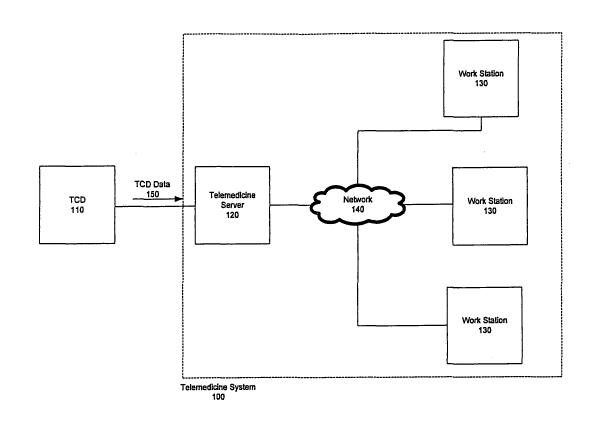
Figure 1A illustrates a telemedicine system 100 for analyzing data, such as
Transcranial Doppler (TCD) data, using, for example, decision tools for
dynamic
vascular analysis such as DVA or HVA, in accordance with this embodiment of
the
invention. In Fig. 1A, the system includes a telemedicine server 120 and a
plurality of
workstations 130, which may include, but are not limited to, personal
computers or
terminals. The workstations 130 may be located at any location in which they
are
capable of accessing the network 140 including, but not limited to, on-site,
remote or in
one or more regional centers. The server 120 receives data 150 from a device
110. The
server 120 may be connected to TCD device 110 using any conventional means for
connection known in the art including, but not limited to, direct connections,
through an
interface port, such as a parallel port or USB port, through a computer
network, such as
a local area network (LAN), through the Internet or wirelessly using various
wireless
technologies. In other embodiments, as illustrated in Fig. 1B, the device 110
may
include a computer that also acts as the telemedicine server 120. In yet
another
-11-
CA 02661899 2008-11-06
WO 2008/060328 PCT/US2007/011506
embodiment, as illustrated in Fig. 1C, the device 110 may write data to a file
server
140. In such an embodiment, the telemedicine server 120 is capable of reading
data
150 from the file server 140.
The data 150 from device 110 may be processed on the telemedicine server 120,
and users may interact with that data through the plurality of workstations
130. The
workstations 130 may be connected to the server 120 through any type of
conventional
network 140 known to one of skill in the art. This may include, but is not
limited to, a
LAN or the Internet.
In operation, data 150 may flow from the device to the telemedicine server
120,
where the data 150 may be processed in accordance with the methods of the
present
invention, as described in detail below. A user may access a review tool on a
workstation 130 to review the results of the processing and may make any
necessary
adjustments thereto. Again, users may be located at any location including,
but not
limited to, on-site, remote locations or in one or more regional centers. As
such, where
desired, remote access is provided to the user. The adjusted data may be
updated on the
telemedicine server 120. After the update, the telemedicine server 120 may
generate a
report that may be reviewed by a user through a workstation 130. Again, where
desired, the processing and storage of the data by the server and access and
review by
the user, as well as report generation, may be remotely performed. After
generation of
the report, the data and/or report may alternatively be reviewed by another
user such as
a physician. This again may be done remotely where desired. The physician may
review the report and enter comments, interpretations or provide a diagnosis,
thereby
eliminating the need for the physician to dictate the comments,
interpretations or
diagnosis and then have that information entered on the report by a
transcription
service. This improves report accuracy and reduces the time required to
produce a
report. The physician may also electronically sign the report, after which the
system
will "lock" the report to prevent further modification. At this time, the
physician may
then send the locked report to the requesting physician. Further, the reports
may be
queried or viewed on-line. Any and all portions of the present invention may
have
remote access and any of the server, the work stations, users, physicians,
data storage,
-12-
CA 02661899 2008-11-06
WO 2008/060328 PCT/US2007/011506
report generator and any other portion of the present invention, may be
located
remotely to the other portions, in some cases separated by many, many miles.
Access
to all data in the telemedicine platform is controlled and restricted by a
role-based
security system. The security system prevents users from accessing any
information
they are not authorized to access.
Figure 2 illustrates a process for analyzing a patient's data according to
this
embodiment of the present invention. The process may begin in step S210 where
the
patient may be scanned by a TCD device. The data collected in step S210 may
then be
imported by a telemedicine server in step S215. In step S220, the telemedicine
server
may then process the imported data. The processing, described in detail below,
may
include identification of relevant features of the data for each vessel
scanned. As
mentioned above, in this embodiment of the present invention, this data may be
Doppler data, but is not limited thereto. In step S230, a review of the result
or results
may be provided to enable a user to adjust the identified features. In step
S240, a report
may be generated that compares the patient's readings to a normative data set,
for
example, a reference data set. The system may optionally suggest the
likelihood of
certain outcomes or various diagnoses. In step S250, notification may be
generated that
the report is ready for review. Lastly, in step S260, the report may be
displayed or
printed.
Figure 3A illustrates one embodiment of the telemedicine server 120 that
includes five modules, but is not limited thereto. The five modules shown in
Fig. 3A
are a data conversion module 310, a data processing module 320, a data storage
module
330, a notification module 340, and a report generation module 350. While Fig.
3A
illustrates a telemedicine server 120 having five specific modules, it should
be realized
that the server may be configured to have any number of modules, distributing
the
currently described functions or adding other data storage, display or
processing
functions.
[paragraph number] The data conversion module 310 may converts the reviewed
data, for example data 150 from a de.vice 110, into a unified data format. In
this regard,
the device 110 could optionally output data directly into the unified data
format, in
-13-
CA 02661899 2008-11-06
WO 2008/060328 PCT/US2007/011506
which case the data conversion module 310 would leave the data unmodified.
Alternatively, the data conversion module could be altogether omitted. As
explained
below, the data from the device 110, in this embodiment, forms a Doppler graph
for
each vessel scanned. The data processing module 320 takes the data formatted
by the
data conversion module 310 and executes algorithms to identify features of
interest in
the data, for example, on those Doppler graphs, and stores the results using
the data
storage module 330. The data storage module 330 optionally allows read and
write
access to this data, to processed data and/or to generated reports. The data
storage
module 330 may use data storage on storage space of the telemedicine server
itself,
storage space on another server or storage space on another device attached to
or
remote from the telemedicine server, storage space attached to or remote from
the work
stations or storage space attached to or remote from the device. Once the data
produced
by the data processing module has been approved, the notification module 340
may
notify users that the report or reports are ready for review. The report
generation
module 350 produces reports/results, may allow users to review reports/results
and data
and may send the reports/results to the patient's physician or to a storage
location.
Figure. 3B illustrates another embodiment of the telemedicine server 120 that
further includes a web server module 360. The web server module 360 may
provide
web services that allow the device 110 to upload data, may allow users to
review data,
raw or processed, through a web page, and may allow users to view
reports/results
through a web page.
Figures 4A and 4B illustrate a workstation 130 which may include a data review
tool 410 or a report review tool 420. These tools can take on many forms , for
example, standalone applications or web-based applications, applications
executing in a
browser or a combination thereof. It would be apparent to one of ordinary
skill in the
art that any operating system, for example, Windows XPO, SunOSO, Linux or
UnixO,
but not limited thereto, may support the tools. One example of an
implementation is in
a platform-agnostic language like JavaO. As illustrated in Figs. 4A and 4B and
as
discussed above, workstation 130 may be connected to the telemedicine server
120
through a network.
-14-
CA 02661899 2008-11-06
WO 2008/060328 PCT/US2007/011506
Some examples of the data 150 provided by device 110 are listed below. This
list includes, but is not limited, to the following:
=Patient information - This may include information to identify the patient,
for
example, name, address, social security number, or a patient identification
number;
physical data about the patient, for example, gender, height, weight or
handedness;
and, medical information about the patient, like referring physician and
insurance
information, and other patient information
=Session information - This may include the time and data of the session, for
example
the TCD session, a unique patient identifier, information about the person
performing
the procedure, and the referring physician or other session information.
=Exam information - This may include a unique identification code for the
exam, an
accession code, the start and end times of the TCD exam, and comments of the
technician or physician or other exam information.
=Device information - This may include information about the TC device,
including
manufacturer, model and software version or other device information.
=Vascular test readings, for example vessel velocity readings other
information. In the
case of a Doppler reading, this may include velocimetry data, taken for each
blood
vessel. For each blood vessel, the data can include the fast Fourier transform
data
describing the velocimetry waveform. One embodiment, uses 512 time slices and
256
different sample frequencies. The data may also include an image of the
waveform in a
standard graphics format, such as JPEG or other graphic formats.
The format of this data can dependent upon the manufacturer of the device.
Some
possible formats, for example but not limited thereto, can include an X1VII.
file, a
DICOM-format file, an HL 7-format file, Microsoft Access database, a SQL-
-15-
CA 02661899 2008-11-06
WO 2008/060328 PCT/US2007/011506
compatible database, a flat file. If necessary, the conversion of this data to
the format
used by the invention can be accomplished through known data mapping
techniques
from the format of the device into the invention's data format.
The telemedicine system 100 may have a data conversion module 310, as
illustrated in Figs. 3A and 3B. The data conversion module 310 performs the
optional
step S215, illustrated in Fig. 2, where data from the device 100 may be
converted into
the format used by the invention. Fig. 5 illustrates an example process for
importing
data, for example TCD data, in step S215. In step S510, the invention
determines the
data format. The data format in this case is determined by the manufacturer of
the TCD
device, and may be in any of a number of formats, including XML, a Microsoft
Access , or a relational database. The data conversion module 310 maybe
configured
to scan a data source for new data, or alternatively, may receive notification
when data
is available for conversion. Once the format is determined, data for an exam,
for
example from the TCD data source, may be read into memory in step S520. In
step
S530, the module 310 may map fields from the data read in step S520 into
fields in the
telemedicine data format. The mapping used in step S530 may be determined by
the
data format used by the device. One of ordinary skill in the art would realize
how to
make mapping decisions and affect this data mapping from one set of data
fields to
another set of data fields. In step S540, the telemedicine data may then be
written to
data storage. For example, the data may be written to a fixed storage, to an
X1VII, file,
to storage module 330, or any suitable place without limitation.
The telemedicine system 100 may include a data processing module 320, as
illustrated in Figs. 3A and 3B. The data processing module 320 performs step
S220,
where the data is processed to identify specific features on the data. Fig. 6
illustrates
one example of a processing method for step S220 from a TCD device. The data
that is
processed in this example includes the velocimetry waveforms from a TCD device
for
each vessel. In step S610, the processing module 3201oads the data to be
processed
into memory from the telemedicine server. This data may have been stored by
the data
conversion module 310 in step S540 of the data import process S215 illustrated
in Fig.
5. In step S620, if there is another vessel to process, the next vessel may be
processed
-16-
CA 02661899 2008-11-06
WO 2008/060328 PCT/US2007/011506
according to the following steps: 1) wave form processing step S630, 2)
feature
extraction/feature identification step S640, 3) selection of parameters of
interest S650.
Given that ultrasound waves are echoed by objects in the body in addition to
blood cells, velocimetry waveforms will often have noise from the echoes of
those
other objects. Figures 7A and 7B, illustrate data before and after the noise
removal of
step S60. Step S640 algorithmically identifies the relevant parameters for
many
useable wave forms within the Doppler data provided. Step S650 identifies the
"best"
wave or waves for which all identified parameters are closest to the mean
parameter
values for the waves within the Doppler data for that vessel. If, in step S620
there are
no more vessels to process, step S670 may be performed, where the processed
data, i.e.,
the original data plus the identified "best" waves, can be written to the
telemedicine
server. Other data can optionally be written to the server as well.
DVA/HVA involves the analysis of the vascular test data, for example, TCD
data. As applied to evaluating and differentiating among vascular states and
conditions,
DVA/HVA may include TCD and/or Intravascular Ultrasound ("IVUS") data
(collectively "data") that is collected and evaluated (via software) as a
function of time
and velocity. Some factors that can be measured or considered when evaluating
and
differentiating among vascular states are (a) a simultaneous consideration of
the
ultrasound data values (peak systolic velocity (PSV or Sys), end diastolic
velocity
(EDV or Dia), peak systolic time (PST), end diastolic time (EDT), mean flow
velocity
(MFV), systolic acceleration (SA), pulsatility index (PI), the natural
logarithm of the
SA (In SA) for each of the established 19 vessel segments within the cerebral
vasculature; (b) a comparison of the data values against a reference database
and/or
quantifying the degree of variance from mean values; or (c) a series of
indices (e.g.
blood flow velocity ratios or other vascular data) that are representative of
the vascular
status/performance/health of each of.the 19 vessel segments. Of course, the
analysis
need not be limited to these 19 vessel segments. Further, the list of factors
above is
exemplary and not exhaustive.
-17-
CA 02661899 2008-11-06
WO 2008/060328 PCT/US2007/011506
The examples of figures 8 and 9 depict 19 intracranial vessel segments. The
vessel segments depicted in Figures 8 and 9 represent the left and right
vertebral artery
(VA), basilar artery (BA), posterior cerebral artery/PCA t(towards)(P1),
posterior
cerebral artery/PCA a (away)(P2), internal carotid artery/ICA t(towards)(C1),
middle
cerebral artery (Ml), anterior cerebral artery (Al), anterior communicating
artery
(ACOM), carotid siphon (towards)(C4), carotid siphon (away)(C2), and the
ophthalmic
artery (OA).
Peak systolic velocity (PSV) is the velocity at the identified maximum. End
diastolic velocity (EDV) is the velocity at the identified minimum. The mean
flow
velocity (MFV) is
PSV - EDV
MFV = 3 + EDV in approximation and more completely
MFV = 1 r fv(t)dt.
ti - to ro
The pulsatility index (PI) is
PI - PSV - EDV
MFV
The systolic acceleration (SA) is identified as the point of maximum
acceleration on the
velocity envelope between the end diastolic and peak systolic velocities. This
value
may be automatically calculated by the algorithm via known methods of
calculating
maxima of a data set or may be calculated via the following formula:
SA = PSV - EDV
PST - EDT
-18-
CA 02661899 2008-11-06
WO 2008/060328 PCT/US2007/011506
The derived indices can include the dynamic work or compliance index the
dynamic flow index, and the dynamic pressure index.
1. The Dynamic compliance Index (DCI or Acceleration/Mean Flow
Velocity Index (VAI)) relates to the force of flow to the mean flow
velocity and describes kinetic efficiency of a segment in moving blood
forward. It is given by the formula
DCI = InSA
MFV
That is, the DCI is the natural logarithm of the systolic acceleration
divided by the mean flow velocity.
II. The Dynamic Flow Index (DFI or Velocity/Impedance Index (VPI))
relates the mean flow velocity to the impedance (pulsatility
index) and describes how capacitance volume affects flow through
the conductance vessel. It is given by the formula
DFI - MFV
PI
III. The Dynamic Pressure Index (DPI or Acceleration/Impedance Index
(API)) relates the force of flow to impedance and describes the
effect of capacitance vessel volume on the force of flow. It is
given by the formula
DPI = InSA
PI
That is, the DPI is the natural logarithm of the Systolic Acceleration
value divided by the Pulsatility Index Value.
-19-
CA 02661899 2008-11-06
WO 2008/060328 PCT/US2007/011506
The basic values and derived indices may be computed based on the relevant
identified features or selected parameters, in this embodiment, the maxima and
minima.
Thus, if cursor placements, i.e. feature identified or selected parameters are
changed,
the factors may be recomputed based on the new placements. As explained below,
the
review tool has the capability to recompute the factors dynamically as cursor
placements are adjusted.
The telemedicine system 100 has a data review too1410, as illustrated in Fig.
4A. Once the data processing step S220 has been completed, a user may perform
the
data review step S230, illustrated in Fig. 2, where the processed data is
reviewed using
the data review tool 410. One benefit of this review is to ensure that
features are
properly identified or the parameters appropriately selected, i.e. the
features are
identified/parameters are selected so that the factors computed from them are
correct.
Fig. 10 illustrates one method of data review S230 using the data review tool
410. In
step S1010, the data review tool 410 loads vessel data from the telemedicine
server.
This may be performed by reading a data file from a remote server.
Alternatively,
other methods can be used such as requesting data from a web service. One of
ordinary
skill in the art would understand that other known techniques of receiving
data from
other devices may be used.
As explained above, one form of velocimetry data consists of a series of
waveforms, one waveform for each vessel scanned, where features may be
identified or
parameters selected therefore in steps S620 to S650, as illustrated in Fig. 6.
In this
example, such identification or selection is done by placement of cursors to
identify the
features or select the parameters. The data, in this example, waveforms, may
be
displayed along with the cursors that identify the features. The user may then
see
which vessel waveforms have been reviewed or approved. The system may use
various
indications to distinguish reviewed or approved vessels. For example, vessel
names
that are reviewed or approved can be shown in color, e.g. in green. In step
S1020, if
vessels are remaining to be reviewed or approved, the user may select one of
the
unreviewed vessels and review the cursor placement in step S1030. Such a
review is
explained below and illustrated in Fig. 11. In fig. 10, once the feature
identification
-20-
CA 02661899 2008-11-06
WO 2008/060328 PCT/US2007/011506
and parameter selection has been completed or approved, in this example, the
placement of the cursors on a wave has been reviewed or approved, , step
S1030, the
user reviews or approves that vessel, corresponding to the wave, in step
S1040. After
step S1020, step S1050 may be performed, where the updated velocimetry data
may be
written to the telemedicine server. In this embodiment, step S1020 concludes
and step
S1050 may be performed if all vessels have been reviewed or approved.
Step S230, as illustrated in Fig. 10, may include a cursor adjustment or
alteration process in step S1030. Cursor adjustment here refers feature
identification or
parameter selection. In the embodiment described here, such identification and
selection is affected by changing placement of a cursor. Nevertheless, any
known
method of feature identification and parameter adjustment known to those
skilled in the
art may be used. Figure 11 illustrates an example of such cursor adjustment
S1030 for
a single vessel. In step S1110, if after looking at the waveform, the user
determines
that no cursor adjustment is necessary, the user can simply conclude review or
approve
the vessel, as in steps S1180 and S1190. Otherwise, if the user determines, in
step
S1120, that adjustment or alteration may be necessary, the user may perform
step
S1130, where the user selects and identifies the appropriate features or
parameters. In
this example, this selection is affected by placing the cursor on the "best"
maxima and
minima for each wave. In step S1140, if the peak cursor (i.e., the cursor at
the
maximum of the wave) is not the "best," the reviewer performs step S1150,
where he
adjusts the placement of the peak cursor. In step S1160, if the valley cursor
(i.e., the
cursor at the minimum of the wave) is not the "best," the reviewer performs
step
S1170, where he adjusts the placement of the valley cursor. It is within the
level of
ordinary skill in the art to repeat, vary or omit these steps or the order of
performing
these steps. Steps S1163 through S1166 show adjustments of cursors which are
lines as
well as points in this particular embodiment, related to other features of the
data or
waves. In step S1170, if no more adjustment is necessary, step S1180 may be
performed. In step S1180 the factors for the vessel may be recalculated to
reflect the
new cursor placements. In step S1190 the vessel may be identified as reviewed
or
approved.
-21-
CA 02661899 2008-11-06
WO 2008/060328 PCT/US2007/011506
The telemedicine system 100 may have a report generation module 350, as
illustrated in Figs. 3A and 3B. The report generation module 350 may perform
step
S240, illustrated in Fig. 2, where the report showing the comparison between a
patient's
data and known reference data is generated. Fig. 12 illustrates an example
process for
generating a report in step S240. In step S1210, the patient data that was
updated in
step S230, illustrated in Figs. 2 and 10, may be loaded into memory. In step
S1220,
reference patient data, e.g.., data for a healthy patient of comparable
physiological
characteristics, is loaded into memory. In step S1230, the two data sets are
compared
to create a graph that may show variation of the patient's data from the
reference data.
An example graph is shown in Fig. 13. In step S1240, the data for the report
may be
written to the data storage module.
The telemedicine system 100 may have a notification module 340, as illustrated
in Figs. 3A and 3B. When step S240 is completed by the report generation
module
350, the optional notification module 340 notifies readers, in step S250, that
the report
is ready for display or printing, as illustrated in Fig. 2. One embodiment
generates an
email that is sent to an email address. Another embodiment displays a visual
alert on a
workstation. Other known forms of notification are also possible, including
but not
limited to text messages, communicating with cell phone or notification
through a web
page. Optionally, a webpage of reports that are ready for review may be
displayed.
The telemedicine system 100 has optional report review tool 420, as
illustrated
in Fig. 4. After step S250 is completed by the notification module 340,
readers may
use the report review tool 420 to perform step S260, illustrated in Fig. 2.
The report
may include data relating to the patient, test device, test procedure or
comparison
graphs generated in step S1230, Fig. 12. The information contained in the
report may
be any desired information as in apparent to those skilled in the art. An
example
comparison graph is shown in Fig. 13. The reader may use the comparison graphs
to
diagnose likely conditions or to determine whether or not certain medical
procedures
are likely to be successful. The reader may also use the review tool to
document the
diagnosis or document comments by entering information into the report. After
the
reader has concluded entering information into the report, the reader may
indicate so by
-22-
CA 02661899 2008-11-06
WO 2008/060328 PCT/US2007/011506
any suitable method. For example, the reader may electronically sign the
report. After
the conclusion of the entry or "signing" of the report, the report may be
"locked" to
prevent further modification. The locked reports may be sent back to the
server or to
one of the storage devices or to other users. Further, readers can optionally
query or
review reports on-line. Further still, access to the reports, data, or the
entire system
altogether can be optionally role-based and restricted to certain users or
optionally have
various levels of security or require various levels of authentication of the
user. The
readers can be located any where including on-site, remote or in one or more
regional
centers.
While the foregoing explanations are made to better illustrate and describe
the
invention, they are not intended to limit the scope of the claims. The scope
of the
invention is to be defined by the claims appended hereto, and by their
equivalents, and
all equivalent structures, acts and configurations known to those skilled in
the art are
contemplated herein.
- 23 -