Note: Descriptions are shown in the official language in which they were submitted.
CA 02662292 2015-04-07
=
Title: Isolation of Chondrocytes
The invention relates to a method for isolating cells from a tissue
sample.
Cartilage disorders are highly debilitating disorders including, for
instance, articular cartilage trauma, meniscus injury, chondrogenesis
disorders and arthritis. There are at present no optimal therapies available
for
treating these disorders. Cartilage tissue is neither innervated nor
penetrated
by the vascular or lymphatic systems and it is generally believed that due to
this lack of a vasculature, damaged cartilage tissue does not receive
sufficient
or proper stimuli to elicit a repair response. Repair of arthritic joints thus
requires orthopaedic surgery to replace the worn-out joints by a prosthesis or
by a biological graft. Particularly arthritis is an enormous medical and
economic problem.
Current approaches for cartilage repair rely on removal of tissue
debris, access to the wound healing system of bone by penetrating the
subchondral bone plate, and tissue transplantation and cell based therapies.
Current clinical therapies typically involve autologous cells. Examples of
such
therapies are autologous chondrocytes implantation (ACT) and mosaicplasty
(also known as autologous osteochondral grafts). Due to severe drawbacks,
both therapies can currently only address a limited share of the cartilage
repair market.
For mosaicplasty, a major disadvantage is the limitation to small
defects due to limited availability of donor tissue for transplantation. For
ACT,
drawbacks include the necessity to perform two surgical operations, one for
harvesting cartilage tissue, and another for implantation of in vitro expanded
chondrocytes obtained from the harvested cartilage tissue. Apart from the fact
that high costs are involved, the ACT process is long since in vitro cell
expansion is necessary, during which cartilage cells de-differentiate, and
lose
their phenotype. Hence, a long rehabilitation of several months is needed
CA 02662292 2009-02-27
WO 2008/026929 PCT/NL2007/050431
2
following the surgical implantation procedure for the cells to regain their
original phenotype. Only then true cartilage repair can commence.
Recently, a second generation ACT has been developed involving
autologous chondrocytes in a biomaterial matrix. This technique solves some of
the problems of ACT, particularly the long and open surgical procedure that
was required in ACT. However, important drawbacks remain: two surgical
procedures have to be carried out, involving high costs and long
rehabilitation.
One of the reasons why two surgical procedures have to be carried out is that
the current processes for isolating chondrocytes from a tissue sample
extracted
from the patient takes a long time.
Hyaline cartilage, the most abundant form of cartilage, is glass
smooth, glistening and bluish white in appearance and of this form of
cartilage
articular cartilage is the most common. Articular cartilage covers the ends of
long bones of synovial joints. It is characterized by a particular structural
organization, consisting of chondrocytes embedded in an extracellular
material, typically referred to as "cartilage matrix", which is an
extracellular
matrix rich in proteoglycans, collagen fibrils, other proteins, and water.
Chondrocytes are the only cell type found in normal articular cartilage but
contribute less then 2% of the wet weight in human healthy adult
cartilaginous tissue.
The extracellular matrix of cartilage tissue consists predominantly
of cartilage specific proteoglycan molecules with highly negatively charged
sulphated glycosaminoglycan (GAG) side chains, as well as type II collagen
fibrils. The GAG side chains are able to bind water molecules, thereby
sequestering water and generating an internal swelling pressure within the
cartilage matrix. These hydrogel-like properties are essential for the
interstitial fluid flow patterns observed inside the matrix during functional
loading of cartilage, at which point water is forced out of the tissue to an
amount that allows the negatively charged GAG chains to repel each other.
Upon release of the compressive load, water is imbibed back into the tissue
CA 02662292 2009-02-27
WO 2008/026929 PCT/NL2007/050431
3
matrix. The collagenous network, together with water bound GAG, enables
articular cartilage to withstand large compressive loads which gives the
tissue
its unique function in synovial joints: smooth and pain-free articulation,
spreading of the applied load onto the subchondral bone and absorbing
mechanical shocks.
In normal cartilaginous tissue, proteoglycans are slowly but
continuously turned over, the degraded molecules are released from the
cartilage and are replaced by newly synthesized components. It is the
coordinate control of synthesis and degradation of the matrix components by
the chondrocytes that maintain normal cartilage.
After a tissue sample of the cartilage of the patient has been
extracted, the chondrocytes present in that sample have to be isolated from
the
extracellular matrix before they can be expanded and implanted with the aim
of repairing a cartilage tissue defect. Enzymatic liberation of cells located
within an extracellular matrix, requires diffusion of the enzyme to the
substrate (e.g. collagen), digestion of collagen, and liberation of the cells.
Known procedures for chondrocyte isolation are carried out by
incubating a cartilage tissue sample with a solution of collagenase for a
period
of from 16 to 22 hours. The current belief is that shorter incubation times do
not produce sufficient cell yields for expansion and tissue repair purposes,
whereas longer exposure to collagenase is believed to compromise cell
viability.
The shortest incubation time described in the art appears to be 2 hours (Jakob
et al., Connective Tissue Research, 44 (2003), 173-180). Digestion was never
terminated before 2 hours.
The present invention provides a method for isolating cells,
preferably chondrocytes, from a tissue sample which is considerably shorter
than prior art methods. Thus, unlike the known isolation procedures, a method
for isolating cells according to the invention can be completed within the
usual
duration of a surgical procedure for repairing a cartilage defect. Surgical
procedures for cartilage defect treatments usually last between 30 and 90
CA 02662292 2009-02-27
WO 2008/026929 PCT/NL2007/050431
4
minutes. It has now been shown, surprisingly, that a substantial and
sufficient
number of cells can be isolated within 30 minutes, or even 10 minutes.
The current view in the art is that a high number of chondrocytes is
required for use in cartilage defect repair: typically at least 1 million
chondrocytes for repair of a defect having a volume of 1 milliliter.
Surprisingly,
it has now been shown that a cell number of less than 1 million chondrocytes
may be sufficient to apply to a cartilage tissue defect of a volume of 1
milliliter
to achieve cartilage formation.
The present invention thus relates to a method for isolating cells
from a tissue sample comprising subjecting the tissue sample to a digestion
enzyme for a period of less than 2 hours, and harvesting the isolated cells.
Although it is preferred that chondrocytes are isolated from a
cartilage tissue sample, particularly intended for use in cartilage tissue
repair,
the invention also contemplates isolation of other types of cells from other
types of tissue samples. Examples of primary cells that may be isolated in
accordance with the invention include chondrocytes, nerve cells, osteoblasts,
osteoclasts, hepatocytes, cardiomyocytes, myocytes, Schwann cells or
urothelial cells. For use in tissue repair, the type and source of the primary
cells may be chosen dependent on the type of tissue that is intended to be
repaired. The following overview gives examples of how cell types of primary
cells may be selected with a view to repair of a specific tissue type.
CA 02662292 2009-02-27
WO 2008/026929 PCT/NL2007/050431
Repair tissue Primary cell source
Bone osteoblasts from trabecular bone in long bone,
pelvic bone, clavicula, compact subchonclral bone
Cartilage Chondrocytes derived from nose, knee or hip
joint elbow, ear, ankle or trachea cartilage,
isolated chondron
Liver Hepatocytes from liver
Heart Heart valves Cardiomyocytes from hart muscle, vascular
myofibroblasts form vascular tissue in the hart
Muscle Myocytes from smooth muscle
Nerve Schwann cells, neural cells from epineurial tubes
Bladder Urothelial cells from urological tract
Intestine Cells from jejunum, duodenum
Ligaments and Tendons Cells from cruciate ligaments or tendon
Hair Cells from hair follicle, such as dermal
papilla
cells, outer root sheath or matrix epithelial cells
Preferably, the primary cells are of a cell type that naturally occurs
5 in the tissue that will be repaired. In a preferred embodiment,
chondrocytes
are isolated from a sample of articular cartilage, e.g. for repair of
cartilage
defects.
Prior to subjecting the tissue sample to the digestion enzyme, a
method according to the invention may comprise mincing of the tissue sample
to obtain smaller fragments of the tissue, preferably approximately 0.5 to 2
mm in diameter, more preferably about 1 mm. Mincing may be performed by
any suitable method, for instance using scissors, one or more razor blades (a
set of parallel razorbla.des can be used to make slices, or two such sets can
be
used to make cubes), a scalpel, straining through a steel or nylon mesh screen
or sieve, or disaggregating it through a needle.
In a preferred embodiment, the tissue sample is subjected to a
treatment to increase extracellular matrix permeability prior to subjecting it
to
the digestion enzyme. It is contemplated that one of the factors determining
the efficiency of the isolation of cells from the tissue sample, is the access
of
the digestion enzyme to the cells and extracellular matrix in the sample. The
permeability of cartilage is determined by chemical and mechanical factors,
CA 02662292 2014-05-23
WO 2008/026929 PCT/NL2007/050431
6
water and proteoglycan interactions. It is preferred that the treatment to
increase extracellular matrix permeability, particularly for cartilage tissue,
comprises increasing repulsive forces between glycosaminoglycans present in
the extracellular matrix. In a preferred embodiment, this treatment comprises
contacting the tissue sample to an acid, a base, diraethyl sulfoxide (DMSO),
cathepsin, glycerol, or cations, or any other agent which may increase the
Donan osmotic pressure of the extracellular matrix or cause the extracellular
matrix to swell, prior to subjecting it to the digestion enzyme.
Suitable examples of cations include Na, K+, NH4+, Pb2+, Mg2+, Znz+,
Fe2+, Cd2+, and Cu2+. These may for instance be introduced in the form of
their
chloride salts, preferably in a concentration between 10 mIVI and 2 M. A
suitable acid is for instance hydrochloric acid, preferably in a concentration
of
10-100mM, resulting in a decrease of the pH of the extracellular matrix.
Dimethylsulfoxide (DMSO) and glycerol may be used in a concentration
between 5 and 30 %v /v. Other suitable agents for this step include disodium
ethylenediaminetetraacetate (EDTA) or ethyleneglycolbis (8-aminoethyl ether)
N,N'-tetraacetic acid (EGTA), both preferably used in a concentration of 0.01-
011 M) or citrate in Tris buffer, pH 5.8 and 7.4, at 4 and 37 . After the
permeability of the tissue is increased, the tissue sample may be washed with
for instance phosphate buffered saline before subjecting it to a digestion
enzyme.
The step of increasing permeability preferably lasts from 1 minute
up to no more than 1 hour, preferably maintaining the total isolation time of
the cells to be within the 2 hour range. It is preferably performed at a
temperature between 17 C and 37 C.
The tissue sample, possibly in the form of small fragments, is then
incubated in a digestion solution. The digestion solution comprises one or
more
enzymes chosen from the group consisting of collagenases, pronases, dispasesTm
,
trypsins, hyaluronidases, chondroitinases, elastases, and heparitinases. The
type of enzyme will depend on the type of tissue used. It is also contemplated
CA 02662292 2014-05-23
WO 2008/026929
PCT/NL2007/050431
7
to use different enzymes sequentially or simultaneously. For cartilage, it is
preferred that collagenase type II is used. A suitable amount of enzyme is for
instance 0.05-20 wt.%, preferably below 10 wt.%, more preferably 0.15-2 wt.%,
based on the weight of the digestion solution.
The conditions (e.g. pH and temperature) under which a method
according to the invention is carried out will be chosen such that they are
optimal for the cells that are being isolated and for the digestion enzyme and
possible other agents used. To this end, the digestion solution may further
comprise buffering agents which help to maintain the pH in the range which
approximates physiological conditions. They are preferably present at
concentration ranging from about 1 mM to about 100 mM. Suitable buffering
agents for use in the present invention include both organic and inorganic
acids and salts thereof such as citrate, succinate, tartrate, fumarate,
gluconate, oxalate, lactate, acetate phosphate and borate buffers.
Additionally,
there may be mentioned, histidine, glycine and urea buffers and buffers such
as Tris, MOPS and HEPES.
The digestion solution may further comprise such compounds as:
chelating agents, e.g. diethylenetriaminepentacetic acid (DTPA),
ethylenediaminetetraacetic acid (EDTA, e.g. as VerseneTm), ethylene
bis(oxyethylenenitrolo)tetraacetic acid (EGTA); reducing agents, such as
dithiotreitol, dithioerythrito1,13-mercaptoethanol, glutathione, thioredoxine,
cysteine, etc.; ions necessary for activation of the enzyme such as CaC12,
MgC12, NaC1 and/or KC1; and/or organic solvents or lipid/membrane modifying
agents such as dimethyl sulfoxide (DMS0); nonionic detergents such as triton
X100TM and/or osmoprotectants such as sucrose.
In accordance with the invention, the tissue sample is subjected to
the digestion enzyme for a period of less than 2 hours. In a preferred
embodiment, the tissue sample is subjected to the digestion enzyme for a
period of less than 1 hour, more preferably for a period of at least 1 minute,
more preferably from 5 minutes to 1 hour, and even more preferably for a
CA 02662292 2009-02-27
WO 2008/026929 PCT/NL2007/050431
8
period of from 10 to 30 minutes. It is further preferred that, if the method
encompasses a pre-treatment to increase extracellular matrix permeability,
the periods of time specified in this paragraph cover both the subjecting of
the
tissue sample to the digestion enzyme and the pre-treatment. As mentioned
above, it is an important advantage of the invention that a method for
isolating cells is provided that can be completed within the usual duration of
a
surgical procedure for repairing a tissue defect, such as a cartilage tissue
defect.
The isolated cells may then be harvested in the usual manner, e.g.
by filtration, washing, centrifuging, and/or magnetic bead extraction. During
this step, the cells are separated from the digestion enzyme and possible
other
agents used, thereby effectively ending the digestion process. If desired, the
digestion enzyme may be inactivated prior to this separation step, e.g. by
adjustment of the pH.
Filtration may be carried out by pouring the cell suspension still
comprising the digestion solution on a tissue culture grade filter.
Subsequently, the cells may be washed, for instance by pouring phosphate
buffered saline (PBS) onto the cells while they are still on the filter.
Finally,
the cells may be resuspended in a suitable medium.
Washing may be carried out by centrifuging the cell suspension still
comprising the digestion solution and aspirating the supernatant, followed by
resuspending the cells in a relatively large volume of PBS. The cells may then
centrifuged again. This procedure can be repeated several times to achieve the
desired degree of washing.
,Isolation using magnetic beads may be carried out by adding
magnetic beads coated with a general receptor (e.g. a5131 integrin) or a
suitable
antibody, to the cell suspension still comprising the digestion solution. The
cells will bind to the magnetic beads via their membrane expressed epitope
(e.g. fibronectin). With the aid of a magnet, the beads with the cells
attached
thereto are concentrated to the bottom of a tube and the supernatant may be
CA 02662292 2009-02-27
WO 2008/026929 PCT/NL2007/050431
9
aspirated. The magnetic beads with the cells may be washed with e.g. PBS.
This process can be repeated several times. Finally, the cells may be
separated
from the magnetic beads by trypsin treatment, competition by washing with
epitope containing buffer (e.g. fibronectin) or washing with 1mM to 10 mM
aqueous HC1 solution.
The invention will now be elucidated by the following, non-
restrictive examples.
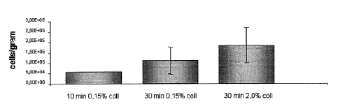
Example I
Cartilage from human subjects was dissected to approximate lx1
mm cubes, incubated in a 0.15% or 2% collagenase solution (type II
collagenase, dissolved in DMEM, filter sterilized through 0.22 pm filter
and supplemented with 10% V/V FBS; approximately 10 ml of collagenase
suspension per g of cartilage) on an orbital- (xyz-) shaker at 37 C and 5%
CO2
for 10 or 15 min. Next, undigested cartilage was separated through a cell
strainer, the cell suspension was centrifuged at 4 C, 300 g for 10-20 minutes,
the supernatant was aspirated off, and the centrifugation was repeated 2
times. Next, cells were resuspended in chondrocyte medium and viable cells
were counted using trypan blue staining. The results are shown in Figure 1.
Example II
Cartilage was retrieved from adult human cartilage biopsies (n=4)
from the tibia. The cartilage was minced into < 1 mm3 pieces. Approximately 1
gram of cartilage per group was transferred to 50 ml tubes and the exact
weight of cartilage was determined (table1). Chondrocytes were isolated by
means of different pretreatment as described in Table 1, immediately followed
by collagenase type II (Worthington) digestion for 30 minutes. The digested
suspension was filtered through a 100 gm mesh nylon filter and the resulting
cell suspension was centrifuged at 300 g for 10 minutes at 4 C. The cell
pellet
was washed twice with phosphate buffered saline (PBS) and finally
CA 02662292 2009-02-27
WO 2008/026929 PCT/NL2007/050431
resuspended in 1 ml PBS. The number of cells and the number of dead cells
were determined with a Burker-Turk counting chamber and Trypan blue
staining. The results are summarized in Table 1 and Figure 2.
5
treatment total n cells weight total n sd normalized
to
cartilage cells/g #11
(g)
1. 0.25% T 15' 1.42E+06 0.83 1.71E+06
8.49E+04 2.63
2. 2 mg/ml HA 15' 8.40E+05 0.84 1.00E+06
1.48E+05 1.54
3. 1% DMSO 15' 7.40E+05 0.84 8.81E+05
1.56E+05 1.35
4. 5% DMSO 15' 1.08E+06 0.99 1.09E+06
1.70E+05 1.67
5. 10% DMSO 15' 8.98E+05 0.9 9.97E+05
6.01E+04 1.53
6. 140mM NaC1 15' 7.10E+05 0.98 7.24E+05
7.07E+04 1.11
7. 500mM NaC1 15' 7.45E+05 0.98 7.60E+05
1.77E+04 1.17
8. 1M NaC1 15' 7.13E+05 0.73 9.76E+05
1.13E+05 1.50
9. 1% 1.02E+06 1
1.02E+06 3.54E+03 1.56
DMS0+140mM
NaC1 15'
10. 140 mM NaC1 1.30E+06 0.84 1.55E+06
6.72E+04 2.38
1570.25% T 15'
11. 2% coil 30' 6.25E+05 0.96 6.51E+05 1.41E+04
1.00
12. 2% coil 120' 1.85E+06 0.79 2.34E+06 1.52E+05
3.59
13. 0.15% coil 23 hrs 9.53E+05 0.87 1.09E+06
1.24E+05 1.68
Table 1 Cartilage was pretreated for 15 minutes (15') with trypsin-EDTA (T)
0.25%,
Hyaluronidase (HA) 2mg / ml in PBS, DMSO, NaCl or a combination of DMSO + NaCl
(#9) or
NaCl followed by trypsin-EDTA (#10). All experimental samples (1-10) were
subsequently
digested with 2% collagenase type II for 30 minutes. Controls were digested
with 2 %
10 collagenase for 30 minutes or 120 minutes or overnight for 23 hours in
0.15% collagenase type
II. Cell viability was between 80 and 95% for all groups.
The cell number results show that pretreatment of cartilage in all
experimental groups results in a significantly higher cell yield after 30
minutes collagenase digestion compared to 30 minutes collagenase digestion
alone. The results also show that the cell yield in group 1-5 and 8-10 is not
significantly different or is even higher than overnight digestion (23 hrs)
with
0.15% collagenase type II (table 1 #13). However, it is also shown that upon
CA 02662292 2009-02-27
WO 2008/026929 PCT/NL2007/050431
11
collagenase digestion with 2% collagenase type II for 2hrs, the cell yield
increased even more than with pretreatment (see also Example III).
Thus, it is concluded that with pretreatment of cartilage prior to
collagenase digestion it is possible to increase the cell yield compared to
collagenase digestion alone. Moreover, within 45 minutes similar or higher
cell
yield can be established by a combination of pretreatment and collagenase
digestion at a higher concentration compared to a conventional 23 hrs
digestion with 0.15% collagenase.
Example III
Cartilage was retrieved from adult human cartilage biopsies (n=3)
from the tibia. Cartilage was minced into < 1 mm3 pieces. Approximately 1
gram of cartilage per group was transferred to 50 ml tubes and the exact
weight of cartilage was determined (table1). Chondrocytes were isolated by
means of different pretreatment as described in Table 2, immediately followed
by collagenase type II (Worthington) digestion for 120 minutes. The digested
suspension was filtered through a 1001.tm mesh nylon filter and the resulting
cell suspension was centrifuged at 300g for 10 minutes at 4 C. The cell pellet
was washed twice with phosphate buffered saline (PBS) and finally
resuspended in 1 ml PBS. The number of cells and the number of dead cells
were determined with a Burker-Turk counting chamber. The results are
summarized in Table 2 and Figure 3.
CA 02662292 2009-02-27
WO 2008/026929
PCT/NL2007/050431
12
weight normalized
cartilage total n
against
treatment total n cells (g) cells/g sd #12
1. 0.25% T 15' 4.81E+06 1.17 4.11E+06
1.84E+05 1.56
2. 2 mg/ml HA 15' 3.17E+06 1.08 2.94E+06
1.27E+05 1.11
3.1% DMSO 15' 3.88E+06 1.069 3.63E+06 2.26E+05
1.38
4. 5% DMSO 15' 2.79E+06 0.95 2.94E+06
2,97E+05 1.11
5. 10% DMSO 15' 3.38E+06 0.92 3.67E+06
4.53E+05 1.39
6. 140mM NaC1
15' 2.14E+06
0.8 2.68E+06 2.83E+04 1.01
7. 500mM NaC1
15' 1.49E+06
1 1.49E+06 1.13E+05 0.56
8. 1M NaC1 15' 1.43E+06 1 1.43E+06 1.06E+05
0.54
9. 1%
DMS0+140mM
NaC1 15' 2.03E+06 0.99 2.05E+06 2.69E+05
0.78
10. 140 mM NaC1
1570.25% T 15' 2.62E+06 1.06 _ 2.47E+06
7.00E+05 0.94
11. 2% coil 30' 1.15E+06 1.11 1.03E+06
1.31E+05 0.39
12. 2% coil 120' 2.69E+06 1.02 2.64E+06
3.82E+05 1.00
13. 0.15% coil 23
hrs 3.58E+06
1.27 2.82E+06 3.68E+05 1.07
Table 2: Cartilage was pretreated with trypsin-EDTA (T) 0.25%, Hyaluronidctse
(HA) 2mg /m1
in PBS, DMSO, NaC1 or a combination of DMSO + NaC1 (#9) or NaC1 followed by
trypsin-
EDTA (#10). All experimental samples (1-10) were subsequently digested with 2%
collagenase
type II for 120 minutes. Controls were digested with 2 % collagenase for 30
minutes or 120
minutes or overnight for 23 hours in 0.15% collagenase type II. Cell viability
was between 80
and 88% for all groups.
Cell number results show that pretreatment of cartilage in all
experimental groups, except #7 and #8, results in a significantly equal or
higher cell yield after 120 minutes collagenase digestion compared to 120
minutes collagenase digestion alone. Pretreatment with 500mM (#7) or 1M
(#8) NaCl results in lower cell yield upon collagenase type II digestion. The
results also show that the cell yield in group 1- is not significantly
different or
is even higher than overnight digestion (23 hrs) with 0.15% collagenase type
II
(table 1 #13).
Thus, it is concluded that pretreatment of cartilage with trypsine or
DMSO prior to collagenase digestion makes it possible to increase the cell
yield
CA 02662292 2009-02-27
WO 2008/026929 PCT/NL2007/050431
13
compared to collagenase digestion alone. Moreover, within 135 minutes similar
of higher cell yield can be established by a combination of pretreatment of
cartilage as described, followed by collagenase digestion at a higher
concentration compared to a conventional 23 hrs digestion with 0.15%
collagenase.