Note: Descriptions are shown in the official language in which they were submitted.
CA 02662965 2009-03-10
WO 2008/036239 PCT/US2007/020150
1
USE OF LXR AGONISTS FOR THE TREATMENT
OF OSTEOARTHRITIS
FIELD OF THE INVENTION
The present invention relates to methods of treating or preventing
osteoarthritis with LXR agonists.
BACKGROUND OF THE INVENTION
Osteoarthritis, also known as degenerative joint disease, is characterized
by degeneration of articular cartilage as well as proliferation and remodeling
of
subchondral bone. The usual symptoms are stiffness, limitation of motion, and
pain. Osteoarthritis is the most common form of arthritis, and prevalence
rates
increase markedly with age.
Existing osteoarthritis treatment approaches include exercise, medicines,
rest and joint care, surgery, pain relief techniques, alternative therapies,
and
weight control. The commonly used medicines in treating osteoarthritis include
nonsteroidal anti-inflammatory drugs (NSAIDs), for example, aspirin,
ibuprofen,
naproxen sodium, ketoprofen; topical pain-relieving creams, rubs, and sprays
(for
example, capsaicin cream) applied directly to the skin; corticosteroids,
typically
injected into affected joints to relieve pain temporarily; and hyaluronic
acid.
Surgery may be performed to resurface (smooth out) bones, reposition bones,
and replace joints. Although various medications have been used for treating
the
disease, they are not effective for long term control and prevention.
Liver X receptors (LXRs), originally identified from liver as orphan
receptors, are members of the nuclear hormone receptor super family and have
been found to be negative regulators of macrophage inflammatory gene
expression (see Published U.S. Patent Application No. 2004/0259948; Joseph
SB et al., Nat. Med. 9:213-19 (2003)). LXRs are ligand-activated transcription
factors and bind to DNA as obligate heterodimers with retinoid X receptors.
While LXRa is restricted to certain tissues such as liver, kidney, adipose,
intestine, and macrophages, LXR(3 displays a ubiquitous tissue distribution
pattern. Activation of LXRs by oxysterols (endogenous ligands) in macrophages
CA 02662965 2009-03-10
WO 2008/036239 PCT/US2007/020150
2
results in the expression of several genes involved in lipid metabolism and
reverse cholesterol transport, including ABCA1, ABCG1, and apolipoprotein E.
SUMMARY OF THE INVENTION
One aspect is for a method for the treatment of a mammal suffering from
osteoarthritis comprising administering to the mammal in need thereof an LXR-
responsive gene expression-inducing amount of an LXR agonist.
Another aspect is for a method of inducing expression of apolipoprotein D
in a mammal having osteoarthritic cartilage comprising administering to the
mammal in need thereof an effective amount of an LXR agonist.
A further aspect relates to a method of preventing osteoarthritis-
comprising: (a) determining a baseline apolipoprotein D expression level in
normal cartilage of a subject; and (b) maintaining baseline apolipoprotein D
expression level in cartilage of the subject via treatment with LXR agonist.
An additional aspect is for a method for the treatment of a mammal
suffering from osteoarthritis comprising administering to the mammal in need
thereof an aggrecanase activity-inhibiting amount of an LXR agonist.
A further aspect is for a method of inhibiting activity of aggrecanase in a
mammal having osteoarthritic cartilage comprising administering to the mammal
in need thereof an effective amount of an LXR agonist.
Another aspect relates to a method for the treatment of a mammal
suffering from osteoarthritis comprising administering to the mammal in need
thereof an effective amount of an LXR agonist to inhibit elaboration of pro-
inflammatory cytokines in osteoarthritic lesions.
An additional aspect relates to a method of detecting an osteoarthritic
phenotype in a subject comprising: (a) determining a baseline apolipoprotein D
expression level in normal cartilage; (b) obtaining a cartilage sample from a
subject suspected of having osteoarthritis; and (c) detecting the level of
expression of apolipoprotein D in the sample; wherein a lower amount of
apolipoprotein D expression in the sample compared to baseline apolipoprotein
D
expression is indicative of osteoarthritis.
A further aspect is for a method of identifying an LXR ligand capable of
reducing an osteoarthritic effect in cartilage comprising: (a) providing a
sample
containing LXR; (b) contacting the sample with a test compound; and (c)
CA 02662965 2009-03-10
WO 2008/036239 PCT/US2007/020150
3
determining whether the test compound induces apolipoprotein D expression,
inhibits aggrecanase activity, inhibits elaboration of pro-inflammatory
cytokines,
or a combination thereof.
Other aspects and advantages of the present invention will become
apparent to those skilled in the art upon reference to the detailed
description that
hereinafter follows.
BRIEF DESCRIPTION OF THE FIGURES
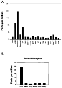
Figure IA is a bar graph showing relative expression levels of nuclear
receptor
(NR) expression in cartilage with severe osteoarthritis (OA). Figure 1B is a
bar
graph showing relative expression levels of retinoid receptor expression in
cartilage with severe OA.
Figure 2A is a bar graph showing ApoD expression in normal cartilage, and
cartilage with mild OA and severe OA. Disease severity was assessed
macroscopically by examining the sizes and depth of the lesions in the
cartilage
specimens. Figure 2B is a bar graph showing TNFa expression in normal
cartilage, and cartilage with mild OA and severe OA.
Figure 3 is a bar graph showing that cytokine-induced proteoglycan
degradation/release from human OA cartilage explants is inhibited by LXR
agonists, and that cytokine-induced reduction of total proteogycan content in
these explants is prevented by LXR agonists.
Figure 4A is a Western blot showing aggrecanase-generated aggrecan
neoepitopes using BC-3 antibody, which recognizes the N-terminus on
aggrecanase-generated aggrecan catabolites. Cartilage explants from two
human donors with end stage OA (after joint replacement surgery) were used.
Donor #259 is a 57 year-old male patient, and donor #261 is a 55 year-old
female
patient. Lanes 1, 5: vehicle. Lanes 2, 6: T0901317 (2 pM). Lanes 3, 7: 1L-10
+ oncostatin M (OSM) (10 ng/ml each). Lanes 4, 8: IL-10+ OSM + TO901317.
Figure 4B is a Western blot showing aggrecanase-generated aggrecan
neoepitopes using AGEG antibody, which recognizes a different epitope on
aggrecanase-generated aggrecan catabolites. Lanes 1, 5: vehicle. Lanes 2, 6:
CA 02662965 2009-03-10
WO 2008/036239 PCT/US2007/020150
4
T0901317 (2 NM). Lanes 3, 7: IL-1p + OSM (10 ng/ml each). Lanes 4, 8: IL-1P
+ OSM + T0901 317.
Figure 5A is a bar graph showing inhibition of total prostagiandin E2 (PGE2)
production from cytokine-treated human cartilage explants by LXR agonists.
Figure 5B compares the quantities of arachidonic acid in the forms of membrane
phospholipids PC and PE in the explants treated with vehicle control or LXR
agonist GW3965 (2 M) for 21 days. Cartilage samples from 2 human OA
donors were used in this study.
DETAILED DESCRIPTION OF THE INVENTION
Applicants specifically incorporate the entire contents of all cited
references in this disclosure. Further, when an amount, concentration, or
other
value or parameter is given as either a range, preferred range, or a list of
upper
preferable values and lower preferable values, this is to be understood as
specifically disclosing all ranges formed from any pair of any upper range
limit or
preferred value and any lower range limit or preferred value, regardless of
whether ranges are separately disclosed. Where a range of numerical values is
recited herein, unless otherwise stated, the range is intended to include the
endpoints thereof, and all integers and fractions within the range. It is not
intended that the scope of the invention be limited to the specific values
recited
when defining a range.
The practice of the present invention will employ, unless otherwise
indicated, conventional techniques of cell biology, cell culture, molecular
biology,
transgenic biology, microbiology, recombinant DNA, and immunology, which are
within the skill of the art. Such techniques are explained fully in the
literature.
See, for example, Molecular Cloning: A Laboratory Manual; 2nd Ed., ed. by
Sambrook, Fritsch and Maniatis (Cold Spring Harbor Laboratory Press: 1989);
DNA Cloning, Volumes I and II (D. N. Glover ed., 1985); Oligonucleotide
Synthesis (M. J. Gait ed., 1984); U.S. Patent No. 4,683,195; Nucleic Acid
Hybridization (B. D. Hames & S. J. Higgins eds. 1984); Transcription and
Translation (B. D. Hames & S. J. Higgins eds. 1984); Culture of Animal Cells
(R.
1. Freshney, Alan R. Liss, Inc., 1987); Immobilized Cells and Enzymes (IRL
CA 02662965 2009-03-10
WO 2008/036239 PCT/US2007/020150
Press, 1986); B. Perbal, A Practical Guide to Molecular Cloning (1984);
Methods
in Enzymology (Academic Press, Inc., N.Y.); Gene Transfer Vectors for
Mammalian Cells (J. H. Miller and M. P. Calos eds., 1987, Cold Spring Harbor
Laboratory); Methods in Enzymology, Vols. 154 and 155 (Wu et al. eds.),
5 Immunochemical Methods in Cell and Molecular Biology (Mayer and Walker,
eds., Academic Press, London, 1987); Handbook of Experimental Immunology,
Volumes I-IV (D. M. Weir and C. C. Blackwell, eds., 1986); Manipulating the
Mouse Embryo, (Cold Spring Harbor Laboratory Press, Cold Spring Harbor, N.Y.,
1986).
Here, Applicants show that LXRa and LXRO (liver X receptor a and (3) are
expressed in normal, medium osteoarthritic, and severe osteoarthritic
cartilages.
Applicants also demonstrate for the first time a plausible lipid defect in
osteoarthritis because the expression of Apolipoprotein D (ApoD), which is
expressed at a very high level in normal cartilage, is dramatically down
regulated
in medium and severe osteoarthritic cartilage. LXR ligands induce the
expression of ApoD via an LXR responsive element present in the ApoD
promoter region. In accordance with the expression data, protein levels of
proapolipoprotein D are also reduced in osteoarthritic cartilage samples when
compared to normal cartilage. Because ApoD is a lipid (arachidonic acid and
cholesterol) binding protein, its reduction in osteoarthritic cartilage may
account
for increased lipid levels that are observed in the osteoarthritic cartilage.
Increased arachidonic acid in the cartilage is expected to result in increased
levels of lipid mediators of inflammation (PGE2, leukotrienes, and the like)
in the
diseased tissue. Osteoarthritic cartilage also shows increased activity of
cartilage-degrading enzymes (aggrecanases and metalloproteases).
Applicants also show for the first time that LXR ligand inhibits the activity
of aggrecanases in human osteoarthritis articular cartilage tissue explants.
LXR
ligands also inhibit the expression of TNFa, and a number of other pro-
inflammatory cytokines. Therefore, an LXR ligand is expected to be
therapeutically efficacious in osteoarthritis, and more efficacious than the
current
as well as upcoming osteoarthritic therapies, by normalizing the lipid defect,
inhibiting the expression and/or activity of aggrecanases/metalloproteases,
and
inhibiting the elaboration of pro-inflammatory cytokines in osteoarthritic
lesions.
Further, LXR ligands induce the c-jun/c-fos family of proteins and, as a
result,
CA 02662965 2009-03-10
WO 2008/036239 PCT/US2007/020150
6
enhance AP1 activity, which is required for cartilage formation. Therefore,
with
LXR ligands, for the first time, an osteoarthritis treatment may not only
inhibit
cartilage degradation but also may induce cartilage regeneration.
I. Definitions
In the context of this disclosure, a number of terms shall be utilized.
As used herein, the term "about" or "approximately" means within 20%,
preferably within 10%, and more preferably within 5% of a given value or
range.
The term "aggrecanase activity" refers to at least one cellular process
interrupted or initiated by an aggrecanase enzyme binding to aggrecan.
Generally, activity refers to proteolytic cleavage of aggrecan by aggrecanase.
Other aggrecanase activities include, but are not limited to, binding of
aggrecanase to aggrecan and a biological response resulting from the binding
to
or cleavage of aggrecan by aggrecanases.
The term "cytokine elaboration" refers to production of cytokines by
cartilaginous tissue or chondrocytes.
The terms "effective amount", "therapeutically effective amount", "an LXR-
responsive gene expression-inducing amount", "aggrecanase activity-inhibiting
amount", and "effective dosage" as used herein, refer to the amount of an
effector
molecule that, when administered to a mammal in need, is effective to at least
partially ameliorate or to at least partially prevent conditions related to
osteoarthritis.
As used herein, the term "expression" includes the process by which DNA
is transcribed into mRNA and translated into polypeptides or proteins.
The term "induce" or "induction" of apolipoprotein D (ApoD) expression
refers to an increase, induction, or otherwise augmentation of apolipoprotein
D
mRNA and/or protein expression. The increase, induction, or augmentation can
be measured by one of the assays provided herein. Induction of apolipoprotein
D
expression does not necessarily indicate maximal expression of apolipoprotein
D.
An increase in ApoD expression can be, for example, at least about 10%, 20%,
30%, 40%, 50%, 60%, 70%, 80%, 90% or more. In one embodiment, induction is
measured by comparing ApoD mRNA expression levels from normal cartilage to
that of ApoD mRNA expression levels from osteoarthritic cartilage.
CA 02662965 2009-03-10
WO 2008/036239 PCT/US2007/020150
7
The term "inhibit" or "inhibition" of aggrecanase or aggrecanase activity
refers to a reduction, inhibition, or otherwise diminution of at least one
activity of
aggrecanase. The reduction, inhibition, or diminution of binding can be
measured by one of the assays provided herein. Inhibition of aggrecanase
activity does not necessarily indicate a complete negation of aggrecanase
activity. A reduction in activity can be, for example, at least about 10%,
20%,
30%, 40%, 50%, 60%, 70%, 80%, 90% or more. In one embodiment, inhibition is
measured by a reduction in the detection of cleavage products of aggrecan.
The term "inhibit" or "inhibition" of elaboration of pro-inflammatory
cytokines refers to a reduction, inhibition, or otherwise diminution of the
activity of
a cytokine such as, for example, iNOS, MCP-3, COX-2, MIP1(3, MMP-9, IP-10,
IL-1(3, IL-1a, G-CSF, TNFa, MCP-1, IL-6. The reduction, inhibition, or
diminution
of cytokine elaboration can be measured by one of the assays provided herein.
Inhibition of pro-inflammatory cytokine elaboration does not necessarily
indicate a
complete negation of pro-inflammatory cytokine elaboration. A reduction in
elaboration can be, for example, at least about 10%, 20%, 30%, 40%, 50%, 60%,
70%, 80%, 90% or more. In one embodiment, inhibition is measured by
comparing TNFa mRNA expression levels from normal cartilage to that of TNFa
mRNA expression levels from osteoarthritic cartilage.
"Liver X receptor" or "LXR" refers to both LXRa and LXRP, and variants,
isoforms, and active fragments thereof. LXR(3 is ubiquitously expressed, while
LXRa expression is limited to liver, kidney, intestine, spleen, adipose
tissue,
macrophages, skeletal muscle, and, as demonstrated herein, cartilage.
Representative GenBank accession numbers for LXRa sequences include the
following: human (Homo sapiens, Q13133), mouse (Mus musculus, Q9ZOY9),
rat (Rattus norvegicus, Q62685), cow (Bos taurus, Q5E9B6), pig (Sus scrofa,
AAY43056), chicken (Gallus gallus, AAM90897). Representative GenBank
accession numbers for LXRR include the following: human (Homo sapiens,
P55055), mouse (Mus musculus, Q60644), rat (Rattus norvegicus, Q62755), cow
(Bos taurus, Q5BIS6).
The term "mammal" refers to a human, a non-human primate, canine,
feline, bovine, ovine, porcine, murine, or other veterinary or laboratory
mammal.
Those skilled in the art recognize that a therapy which reduces the severity
of a
CA 02662965 2009-03-10
WO 2008/036239 PCT/US2007/020150
8
pathology in one species of mammal is predictive of the effect of the therapy
on
another species of mammal.
The term "modulate" encompasses either a decrease or an increase in
activity or expression depending on the target molecule. For example, an ApoD
modulator is considered to modulate the expression of ApoD if the presence of
such ApoD modulator results in an increase or decrease in ApoD expression.
II. LXR Agonists
LXR agonists useful in the present invention include natural oxysterols,
synthetic oxysterols, synthetic nonoxysterols, and natural nonoxysterols.
Exemplary natural oxysterols include 20(S) hydroxycholesterol, 22(R)
hydroxycholesterol, 24(S) hydroxycholesterol, 25-hydroxycholesterol, 24(S),25
epoxycholesterol, and 27-hydroxycholesterol. Exemplary synthetic oxysterols
include N,N-dimethyl-3R-hydroxycholenamide (DMHCA). Exemplary synthetic
nonoxysterols include N-(2,2,2-trifluoroethyl)-N-{4-[2,2,2-trifluoro-l-hydroxy-
l-
(trifluoromethyl)ethyl]phenyl}benzene sulfonamide (T0901317; Tularik 0901317),
[3-(3-(2-chloro-trifluoromethylbenzyl-2,2-
diphenylethylamino)propoxy)phenylacetic acid] (GW3965), N-methyl-N-[4-(2,2,2-
trifluoro-l-hydroxy-l-trifluoromethyl-l-ethyl)-phenyl]-benzenesulfonamide
(T0314407), 4,5-dihydro-l-(3-(3-trifluoromethyl-7-propyl-benzisoxazol-6-
yloxy)propyl)-2,6-pyrimid inedione, 3-chloro-4-(3-(7-propyl-3-trifluoromethyl-
6-
(4,5)-isoxazolyl)propylthio)-phenyl acetic acid (F3MethylAA), and acetyl-
podocarpic dimer. Exemplary natural nonoxysterols include paxilline,
desmosterol, and stigmasterol.
Other useful LXR agonists are disclosed, for example, in Published U.S.
Patent Application Nos. 2006/0030612, 2005/0131014, 2005/0036992,
2005/0080111, 2003/0181420, 2003/0086923, 2003/0207898, 2004/0110947,
2004/0087632, 2005/0009837, 2004/0048920, and 2005/0123580; U.S. Patent
Nos. 6,316,503, 6,828,446, 6,822,120, and 6,900,244; WO01/41704; Menke JG
et al., Endocrinology 143:2548-58 (2002); Joseph SB et al., Proc. Natl. Acad.
Sci.
USA 99:7604-09 (2002); Fu X et al., J. Biol. Chem. 276:38378-87 (2001);
Schultz
JR et ai., Genes Dev. 14:2831-38 (2000); Sparrow CP et al., J. Biol. Chem.
277:10021-27 (2002); Yang C et al., J. Biol. Chem., Manuscript M603781200
CA 02662965 2009-03-10
WO 2008/036239 PCT/US2007/020150
9
(July 20, 2006); Bramlett KS et al., J. Pharmacol. Exp. Ther. 307:291-96
(2003);
Ondeyka JG et al., J. Antibiot (Tokyo) 58:559-65 (2005).
Ill. Methods of Treatment/Prevention
According to one modulatory method, LXR activity is stimulated in a cell by
contacting the cell with an LXR agonist. Examples of such LXR agonists are
described above in Section II. Other LXR agonists that can be used to
stimulate
the LXR activity can be identified using screening assays that select for such
compounds, as described in detail herein (Section V).
Modulatory methods can be performed in vitro (e.g., by culturing the cell
with an LXR agonist or by introducing an LXR agonist into cells in culture)
or,
alternatively, in vivo (e.g., by administering an LXR agonist to a subject or
by
introducing an LXR agonist into cells of a subject). For practicing a
modulatory
method in vitro, cells can be obtained from a subject by standard methods and
incubated (i.e., cultured) in vitro with an LXR agonist to modulate LXR
activity in
the cells.
1. Prophylactic Methods
In one aspect, the invention provides a method for preventing in a subject
osteoarthritis by administering to the subject an LXR agonist that induces
ApoD
expression and/or inhibits aggrecanase activity and/or inhibits the
elaboration of
pro-inflammatory cytokines in osteoarthritic lesions. Administration of a
prophylactic LXR agonist can occur prior to the manifestation of
osteoarthritis
symptoms, such that osteoarthritis is prevented or, altematively, delayed in
its
progression.
2. Therapeutic Methods
Another aspect of the invention pertains to methods of modulating LXR
activity for osteoarthritis therapeutic purposes. Accordingly, in an exemplary
embodiment, a modulatory method of the invention involves contacting a cell
with
an LXR agonist that modulates ApoD expression and/or aggrecanase activity
and/or inhibits the elaboration of pro-inflammatory cytokines in
osteoarthritic
lesions. These modulatory methods can be performed in vitro (e.g., by
culturing
the cell with an LXR agonist) or, alternatively, in vivo (e.g., by
administering an
CA 02662965 2009-03-10
WO 2008/036239 PCT/US2007/020150
LXR agonist to a subject). As such, the present invention provides methods of
treating an individual afflicted with osteoarthritis that would benefit from
modulation of ApoD expression and/or aggrecanase activity and/or pro-
inflammatory cytokine elaboration in osteoarthritic lesions.
5
IV. Administration of LXR Agonists
LXR agonists are administered to subjects in a biologically compatible
form suitable for pharmaceutical administration in vivo to enhance ApoD
expression and/or suppress aggrecanase activity and/or suppress elaboration of
10 pro-inflammatory cytokines. By "biologically compatible form suitable for
administration in vivo" is meant a form of the LXR agonist to be administered
in
which any toxic effects are outweighed by the therapeutic effects of the
agonist.
The term "subject" is intended to include living organisms in which an immune
response can be elicited, for example, mammals. Administration of LXR agonists
as described herein can be in any pharmacological form including a
therapeutically effective amount of an LXR agonist alone or in combination
with a
pharmaceutically acceptable carrier.
A therapeutically effective amount of an LXR agonist may vary according
to factors such as the disease state, age, sex, and weight of the individual,
and
the ability of the LXR agonist to elicit a desired response in the individual.
Dosage regime may be adjusted to provide the optimum therapeutic response.
For example, several divided doses may be administered daily, or the dose may
be proportionally reduced as indicated by the exigencies of the therapeutic
situation.
The therapeutic or pharmaceutical compositions of the present invention
can be administered by any suitable route known in the art including, for
example, oral, intravenous, subcutaneous, intramuscular, transdermal,
intrathecal, or intracerebral or administration to cells in ex vivo treatment
protocols. Administration can be either rapid as by injection orover a period
of
time as by slow infusion or administration of slow release formulation. For
treating or preventing osteoarthritis, administration of the therapeutic or
pharmaceutical compositions of the present invention can be performed, for
example, by oral administration or by intra-articular injection.
CA 02662965 2009-03-10
WO 2008/036239 PCT/US2007/020150
11
Furthermore, LXR agonists can be stably linked to a polymer such as
polyethylene glycol to obtain desirable properties of solubility, stability,
half-life,
and other pharmaceutically advantageous properties (see, e.g., Davis et al.,
Enzyme Eng. 4:169-73 (1978); Burnham NL, Am. J. Hosp. Pharm. 51:210-18
(1994)).
LXR agonists can be in a composition that aids in delivery into the cytosol
of a cell. For example, an LXR agonist may be conjugated with a carrier moiety
such as a liposome that is capable of delivering the agonist into the cytosol
of a
cell. Such methods are well known in the art (see, e.g., Amselem S et al.,
Chem.
Phys. Lipids 64:219-37 (1993)). In addition, an LXR agonist can be delivered
directly into a cell by microinjection.
LXR agonists can be employed in the form of pharmaceutical
preparations. Such preparations are made in a manner well known in the
pharmaceutical art. One preferred preparation utilizes a vehicle of
physiological
saline solution, but it is contemplated that other pharmaceutically acceptable
carriers such as physiological concentrations of other non-toxic salts, five
percent
aqueous glucose solution, sterile water or the like may also be used. As used
herein "pharmaceutically acceptable carrier" includes any and all solvents,
dispersion media, coatings, antibacterial and antifungal agents, isotonic and
absorption delaying agents, and the like. The use of such media and agents for
pharmaceutically active substances is well known in the art. Except insofar as
any conventional media or agent is incompatible with the LXR agonist, use
thereof in the therapeutic compositions is contemplated. Supplementary active
compounds can also be incorporated into the compositions. It may also be
desirable that a suitable buffer be present in the composition. Such solutions
can, if desired, be lyophilized and stored in a sterile ampoule ready for
reconstitution by the addition of sterile water for ready injection. The
primary
solvent can be aqueous or alternatively non-aqueous. LXR agonists can also be
incorporated into a solid or semi-solid biologically compatible matrix which
can be
implanted into tissues requiring treatment.
The carrier can also contain other pharmaceutically-acceptable excipients
for modifying or maintaining the pH, osmolarity, viscosity, clarity, color,
sterility,
stability, rate of dissolution, or odor of the formulation.
CA 02662965 2009-03-10
WO 2008/036239 PCT/US2007/020150
12
Dose administration can be repeated depending upon the pharmacokinetic
parameters of the dosage formulation and the route of administration used.
It is also provided that certain formulations containing LXR agonists are to
be administered orally. Such formulations are preferably encapsulated and
formulated with suitable carriers in solid dosage forms. Some examples of
suitable carriers, excipients, and diluents include lactose, dextrose,
sucrose,
sorbitol, mannitol, starches, gum acacia, calcium phosphate, alginates,
calcium
silicate, microcrystalline cellulose, polyvinylpyrrolidone, cellulose,
gelatin, syrup,
methyl cellulose, methyl- and propylhydroxybenzoates, talc, magnesium,
stearate, water, mineral oil, and the like. The formulations can additionally
include lubricating agents, wetting agents, emulsifying and suspending agents,
preserving agents, sweetening agents, or flavoring agents. The compositions
may be formulated so as to provide rapid, sustained, or delayed release of the
active ingredients after administration to the patient by employing procedures
well
known in the art. The formulations can also contain substances that diminish
proteolytic degradation and/or substances which promote absorption such as,
for
example, surface active agents.
It is especially advantageous to formulate compositions in dosage unit
form for ease of administration and uniformity of dosage. Dosage unit form as
used herein refers to physically discrete units suited as unitary dosages for
the
mammalian subjects to be treated; each unit containing a predetermined
quantity
of active compound calculated to produce the desired therapeutic effect in
association with the required pharmaceutical carrier. The specification for
the
dosage unit forms of the invention are dictated by and directly dependent on
(a)
the unique characteristics of the LXR agonist and the particular therapeutic
effect
to be achieved and (b) the limitations inherent in the art of compounding such
an
active compound for the treatment of sensitivity in individuals. The specific
dose
can be readily calculated by one of ordinary skill in the art, e.g., according
to the
approximate body weight or body surface area of the patient or the volume of
body space to be occupied. The dose will also be calculated dependent upon the
particular route of administration selected. Further refinement of the
calculations
necessary to determine the appropriate dosage for treatment is routinely made
by
those of ordinary skill in the art. Such calculations can be made without
undue
experimentation by one skilled in the art in light of the LXR agonist
activities
CA 02662965 2009-03-10
WO 2008/036239 PCT/US2007/020150
13
disclosed herein in assay preparations of target cells. Exact dosages are
determined in conjunction with standard dose-response studies. It will be
understood that the amount of the composition actually administered will be
determined by a practitioner, in the light of the relevant circumstances
including
the condition or conditions to be treated, the choice of composition to be
administered, the age, weight, and response of the individual patient, the
severity
of the patient's symptoms, and the chosen route of administration.
Toxicity and therapeutic efficacy of such LXR agonists can be determined
by standard pharmaceutical procedures in cell cultures or experimental
animals,
for example, for determining the LD50 (the dose lethal to 50% of the
population)
and the ED5o (the dose therapeutically effective in 50% of the population).
The
dose ratio between toxic and therapeutic effects is the therapeutic index and
it
can be expressed as the ratio LD5o/ED50. LXR agonists that exhibit large
therapeutic indices are preferred. While LXR agonists that exhibit toxic side
effects may be used, care should be taken to design a delivery system that
targets such agonists to the site of affected tissue in order to minimize
potential
damage to uninfected cells and, thereby, reduce side effects.
The data obtained from the cell culture assays and animal studies can be
used in formulating a range of dosage for use in humans. The dosage of such
LXR agonists lies preferably within a range of circulating concentrations that
include the ED50 with little or no toxicity. The dosage may vary within this
range
depending upon the dosage form employed and the route of administration
utilized. For any LXR agonist used in a method of the invention,-the
therapeutically effective dose can be estimated initially from cell culture
assays.
A dose may be formulated in animal models to achieve a circulating plasma
concentration range that includes the IC50 (i.e., the concentration of LXR
agonist
that achieves a half-maximal inhibition of symptoms) as determined in cell
culture. Such information can be used to more accurately determine useful
doses in humans. Levels in plasma may be measured, for example, by high
performance liquid chromatography.
Monitoring the influence of LXR agonists on the expression of ApoD
and/or activity of aggrecanase and/or the elaboration of pro-inflammatory
cytokines can be applied not only in basic drug screening, but also in
clinical
trials. For example, the effectiveness of an LXR agonist can be monitored in
CA 02662965 2009-03-10
WO 2008/036239 PCT/US2007/020150
14
clinical trials of subjects exhibiting decreased ApoD gene expression in
chondrocytes and/or increased aggrecanase activity and/or increased
elaboration
of pro-inflammatory cytokines in osteoarthritic lesions. In such clinical
trials, the
expression of ApoD and/or the activity of aggrecanase and/or the elaboration
of
pro-inflammatory cytokines can be used as a "read out" or markers of the
phenotype of different osteoarthritis stages.
Thus, to study the effect of LXR agonists on osteoarthritis, for example, in
a clinical trial, cells can be isolated and RNA prepared and analyzed for the
levels
of expression of ApoD and other genes implicated in osteoarthritis (for
example,
TNFa). The levels of gene expression (i.e., a gene expression pattem) can be
quantified by Northern blot analysis or RT-PCR, by measuring the amount of
protein produced, or by measuring the levels of activity of ApoD or other
genes,
all by methods well known to those of ordinary skill in the art. In this way,
the
gene expression pattern can serve as a marker, indicative of the physiological
response of the cells to the LXR agonist. Accordingly, this response state may
be determined before, and at various points during, treatment of the
individual
with the LXR agonist.
The present invention also provides a method for monitoring the
effectiveness of treatment of a subject with an LXR agonist comprising the
steps
of (i) obtaining a pre-administration sample from a subject prior to
administration
of the LXR agonist; (ii) detecting the level of expression of ApoD and/or the
level
of aggrecanase activity and/or the level of elaboration of pro-inflammatory
cytokines in the pre-administration sample; (iii) obtaining one or more post-
administration samples from the subject; (iv) detecting the level of
expression or
activity of ApoD and/or the level of aggrecanase activity and/or the level of
elaboration of pro-inflammatory cytokines in the post-administration samples;
(v)
comparing the level of expression of ApoD and/or the level of aggrecanase
activity and/or the level of elaboration of pro-inflammatory cytokines in the
pre-
administration sample with the ApoD expression and/or aggrecanase activity
and/or the level of elaboration of pro-inflammatory cytokines in the post
administration sample or samples; and (vi) altering the administration of the
LXR
agonist to the subject accordingly. For example, increased administration of
the
LXR agonist may be desirable to increase ApoD expression to higher levels than
detected and/or reduce aggrecanase activity to lower levels than detected
and/or
CA 02662965 2009-03-10
WO 2008/036239 PCT/US2007/020150
reduce elaboration of pro-inflammatory cytokines to lower levels than
detected,
that is, to increase the effectiveness of the LXR agonist. Alternatively,
decreased
administration of the LXR agonist may be desirable to decrease ApoD expression
to lower levels than detected or activity and/or to increase aggrecanase
activity to
5 higher levels than detected and/or to increase elaboration of pro-
inflammatory
cytokines to higher levels than detected, that is, to decrease the
effectiveness of
the LXR agonist. According to such an embodiment, ApoD expression and/or
aggrecanase activity and/or pro-inflammatory cytokine elaboration may be used
as an indicator of the effectiveness of an LXR agonist, even in the absence of
an
10 observable phenotypic response.
Furthermore, in the treatment of osteoarthritis, compositions containing
LXR agonists can be administered exogenously, and it would likely be desirable
to achieve certain target levels of LXR agonist in sera, in any desired tissue
compartment, and/or in the affected tissue. It would, therefore, be
advantageous
15 to be able to monitor the levels of LXR agonist in a patient or in a
biological
sample including a tissue biopsy sample obtained from a patient and, in some
cases, also monitoring the levels of ApoD expression and/or aggrecanase
activity
and/or pro-inflammatory cytokine elaboration. Accordingly, the present
invention
also provides methods for detecting the presence of LXR agonist in a sample
from a patient.
V. Screening Assays
In one embodiment, expression levels of LXR-responsive genes or activity
levels of proteins therefrom can be used to facilitate design and/or
identification
of compounds that treat osteoarthritis through an LXR-based mechanism.
Accordingly, the invention provides methods (also referred to herein as
"screening assays") for identifying modulators, i.e., LXR agonists, that have
a
stimulatory or inhibitory effect on, for example, ApoD expression and/or
aggrecanase activity and/or cytokine elaboration. Compounds thus identified
can
be used in the treatment of osteoarthritis as described elsewhere herein.
Test compounds can be obtained, for example, using any of the numerous
approaches in combinatorial library methods known in the art, including
spatially
addressable parallel solid phase or solution phase libraries; synthetic
library
CA 02662965 2009-03-10
WO 2008/036239 PCT/US2007/020150
16
methods requiring deconvolution; the `one-bead one-compound' library method;
and synthetic library methods using affinity chromatography selection.
Examples of methods for the synthesis of molecular libraries can be found
in, for example: DeWitt SH et al., Proc. Natl. Acad. Sci. U.S.A. 90:6909-13
(1993); Erb E et al., Proc. Natl. Acad. Sci. USA 91:11422-26 (1994);
Zuckermann
RN et al., J. Med. Chem. 37:2678-85 (1994); Cho CY et al., Science 261:1303-05
(1993); Carrell et al., Angew. Chem. Int. Ed. Engl. 33:2059 (1994); Carrell et
al.,
Angew. Chem. Int. Ed. Engt. 33:2061 (1994); Gallop MA et al., J. Med. Chem.
37:1233-51 (1994).
Libraries of compounds may be presented in solution (e.g., Houghten RA
et al., Biotechniques 13:412-21 (1992)), or on beads (Houghten RA et al.,
Nature
354:82-84 (1991)), chips (Fodor SA et al., Nature 364:555-56 (1993)), bacteria
(U.S. Patent No. 5,223,409), spores (U.S. Patent No. 5,223,409), plasmids
(Cull
MG et al., Proc. Natl. Acad. Sci. USA 89:1865-69 (1992)) or on phage (Scott JK
& Smith GP, Science 249:386-90 (1990); Devlin JJ et al., Science 249:404-06
(1990); Cwirla SE et al., Proc. Natl. Acad. Sci. 87:6378-82 (1990); Felici F
et al.,
J. Mol. Biol. 222:301-10 (1991); U.S. Patent No. 5,223,409.).
An exemplary screening assay is a cell-based assay in which a cell that
expresses LXR is contacted with a test compound, and the ability of the test
compound to modulate ApoD expression and/or aggrecanase activity and/or
cytokine elaboration through an LXR-based mechanism. Determining the ability
of the test compound to modulate ApoD expression and/or aggrecanase activity
and/or cytokine elaboration can be accomplished by monitoring, for example,
DNA, mRNA, or protein levels, or by measuring the levels of activity of ApoD,
aggrecanase, and/or TNFa, all by methods well known to those of ordinary skill
in
the art. The cell, for example, can be of mammalian origin, e.g., human.
Novel modulators identified by the above-described screening assays can
be used for treatments as described herein.
EXAMPLES
. The present invention is further defined in the following Examples. It
should be understood that these Examples, while indicating preferred
embodiments of the invention, are given by way of illustration only. From the
above discussion and these Examples, one skilled in the art can ascertain the
CA 02662965 2009-03-10
WO 2008/036239 PCT/US2007/020150
17
preferred features of this invention, and without departing from the spirit
and
scope thereof, can make various changes and modification of the invention to
adapt it to various uses and conditions.
Example 1
To identify transcripts expressed in either arthritic or normal articular
cartilage, tissue samples were obtained from arthritis patients with end-stage
knee replacement and nonarthritic amputee individuals. The presence or
absence of arthritis was confirmed by histology.
The Human Genome U95Av2 (HG-U95Av2) GeneChip Array (Affymetrix,
Santa Clara, CA) was used for expression profiling. The HG-U95Av2 chip
contains 25-mer oligonucleotide probes representing -12,000 primarily full-
length
sequences (-16 probe pairs/sequence) derived from the human genome. For
each probe designed to be perfectly complimentary to a target sequence, a
partner probe is generated that is identical except for a single base mismatch
in
its center. These probe pairs allow for signal quantitation and subtraction of
nonspecific noise.
RNA was extracted from individual articular cartilage tissue, converted to
biotinylated cRNA, and fragmented according to the Affymetrix protocol. The
fragmented cRNAs were diluted in 1x MES buffer containing 100 Ng/mI herring
sperm DNA and 500 Ng/mI acetylated BSA and denatured for 5 min at 99 C
followed immediately by 5 min at 45 C. Insoluble material was removed from
the
hybridization mixtures by a brief centrifugation, and the hybridization mix
was
added to each array and incubated at 45 C for 16 hr with continuous rotation
at
60 rpm. After incubation, the hybridization mix was removed and the chips were
extensively washed with 6x SSPET and stained with SAPE solution as described
in the Affymetrix protocol.
The raw florescent intensity value of each transcript was measured at a
resolution of 6 mm with a Hewlett-Packard Gene Array Scanner. GeneChip
software 3.2 (Affymetrix), which uses an algorithm to determine whether a gene
is "present" or "absent", as well as the specific hybridization intensity
values or
average differences" of each gene on the array, was used to evaluate the
fluorescent data. The average difference for each gene was normalized to
CA 02662965 2009-03-10
WO 2008/036239 PCT/US2007/020150
18
frequency values by referral to the average differences of 11 control
transcripts of
known abundance that were spiked into each hybridization mix according to the
procedure of Hill AA et al., Science 290:809-12 (2000). The frequency of each
gene was calculated and represents a value equal to the total number of
individual gene transcripts per 106 total transcripts.
Figure IA depicts the mRNA levels in severe osteoarthritic cartilage
(expressed as parts per million (ppm)) for 19 different members of the nuclear
hormone receptor superfamily (LXRa, LXR(3, Rev-erba, Rev-erbp, GR, EAR2,
COUP TF-I, COUP TF-II, CAR, PXR, MR, SF-1, TR-2, TR-4, NOR-1, Nurr1,
Nur77, SHP, FXR). The lower quantitative limit of detection for these gene
chips
studies was determined to be approximately 5 ppm. The data shown in Figure 1
provides evidence that LXR(3, Rev-erba, and GR appear to be expressed by
articular cartilage at the level of sensitivity of the gene chips. In Figure 1
B, the
expression levels of the six retinoid receptor family members (Retinoic Acid
Receptors (RARs) and Retinoid X Receptors (RXRs)) are shown. These data
show that RXRa is expressed in the articular cartilage tissue at levels that
are
easily detectable. RXRa is a heterodimeric partner of LXR and the biologically
active unit of LXR ligand action is LXR-RXR Heterodimer. These data provided
an impetus to look at the functional effects of LXR expression in articular
cartilage.
Example 2
Figure 2A shows the comparison of ApoD mRNA levels in normal
cartilage and cartilage obtained from medium and severe osteoarthritic
patients (expressed as parts per million (ppm)). The lower quantitative limit
of
detection for these gene chips studies was determined to be approximately 5
ppm. The data shown in Figure 2A provides evidence that the expression of
ApoD message is dramatically reduced in mild and severe osteoarthritic
cartilage
when compared to the normal cartilage. Figure 2B shows the comparison of
TNFa mRNA levels in normal cartilage and cartilage obtained from medium
and severe osteoarthritic patients (expressed as parts per million (ppm)). The
lower quantitative limit of detection for these gene chips studies was
determined
to be approximately 5 ppm. The data shown in Figure 2B provides evidence that
CA 02662965 2009-03-10
WO 2008/036239 PCT/US2007/020150
19
the expression of TNFa is significantly induced in mild and severe
osteoarthritic
cartilage when compared to the normal cartilage.
Example 3
Fresh cartilage explants (-20 pieces, a total of -200 mg/well) from a
human OA donor (#154, from National Disease Research Interchange) were
cultured for 10 days in 1 ml of DMEM/F12 containing 1% Nutridoma (Roche
Applied Science, Indianapolis, IN). During the 10 days, the explants were
exposed to cytokines (1 ng/mi IL1P plus 5 ng/ml Oncostatin M) with or without
LXR agonists (2 M GW3965, a reported LXR agonist, or 2 M of Formula I
below, an LXR agonist).
CO2H
O
I I
N/
CF3
(I)
Every 2 days the culture medium was replaced with fresh cytokines and LXR
agonists. Accumulative release of proteoglycans was measured in these cultures
after using DMMB (dimethylmethylene blue) assay. The explants at the end of
the 10-day treatment were then digested with proteinase K and assayed for
total
proteoglycan content. LXR agonists significantly reduced cytokine-induced
release of proteoglycan into the culture medium; consequently, a 10-day
treatment of OA cartilage explants with LXR agonist significantly increased
total
proteoglycan content in the explants (Fig 3). Since both ILl R and Oncostatin
M
CA 02662965 2009-03-10
WO 2008/036239 PCT/US2007/020150
are present in joints with OA and are believed to play role in OA disease
progression, our data suggest that LXR agonist may have a structure-modifying
effect in OA cartilage.
5
Example 4
Fresh cartilage from human OA donors was cut into pieces (-10 mg/piece,
-2x2x2 mm). The cartilage explants were randomized into 24 well plates (-250
mg wet weight/well). Three wells of explants were included for each treatment
10 group. The explants were cultured in 1 ml DMEM/F-12 with 10% FBS for 3
days,
then the complete medium was replaced with serum-free medium. Twelve hours
later, the medium was removed and fresh serum-free medium (1 ml) was added,
followed by LXR agonist T0901317 treatment (2 pM). IL1 p/Oncostatin M(10
ng/mI each) were added 8 hours later. The explants were then cultured in the
15 presence or absence of LXR agonist T0901317 and IL1(3/Oncostatin M for
additional 20 hours. 180 NI of pooled culture medium from each treatment group
was deglycosylated with chondroitinase ABC, keratanase, keratanase II in the
presence of 50 mM EDTA at 37 C for 3 hrs. The samples were then
concentrated and separated in a 4-12% SDS-PAGE gel. Western analysis was
20 performed using either mouse BC3 neoepitope antibody (1:1500), or rabbit
anti-
AGEG antibody (1:1000) as the primary antibody, and anti-mouse or anti-rabbit
IgG antibody conjugated with alkaline peroxidase (1:5000) as the secondary
antibody. Figure 4A shows the result using BC3 antibody, and Figure 4B shows.
the result using AGEG antibody. In the experiment usirig cartilage from donor
#259, cytokine treatment induced release of both BC3 and AGEG containing
aggrecan fragments into the culture medium. Treatment with T0901317 blocked
the induction of BC3 and AEEG release by cytokines. In the experiment using
donor #261, BC3- and AEGE-containing aggrecan fragments were released into
the culture medium from untreated cartilage explants. T0901317 treatment
reduced the amount of these fragments in the culture medium. Release of
AGEG-containing fragment from the explants was also induced by cytokine
treatment, and it was blocked by T0901317 treatment.
CA 02662965 2009-03-10
WO 2008/036239 PCT/US2007/020150
21
Example 5
Fresh cartilage explants (-20 pieces, a total of -200 mg/well) from a
human OA donor (provided by National Disease Research Interchange) were
cultured for 21 days in 1 ml of DMEM/F12 containing 1% Nutridoma (Roche
Applied Science, Indianapolis, IN). During the 21 days, the explants were
exposed to cytokines (10 ng/mI IL1 P plus 10 ng/ml Oncostatin M) with or
without
LXR agonists (2 M GW3965 or Formula I). Every 2-3 days the culture medium
was replaced with fresh cytokines and LXR agonists. Total amounts of
prostaglandin E2 (PGE2) in the culture medium samples collected on day 7, 14,
21 were measured using an EIA assay (Cayman).
Fig. 5 shows that both LXR agonists strongly inhibit cytokine
(ILl R/Oncostatin M)-induced PGE2 synthesis at all 3 time points. Lipid
profiling
analysis (Lipomics Inc.) results show that the amounts of two forms of
membrane
phospholipids where most arachidonic acid (AA) is from are reduced by LXR
activation, suggesting that the decrease of total PGE2 is mediated at least
partly
by reduced total AA content in OA cartilage. Expression of enzymes involved in
PGE2 synthesis may also be inhibited by LXR activity.
PGE2 is the principal proinflammatory prostanoid found in joints with
rheumatoid arthritis (RA) or OA. Increased PGE2 in cartilage may also play a
role in inflammation-mediated structural damages that characterize arthritic
diseases. More importantly, PGE2 contributes to one of the key features of
inflammation, pain hypersensitivity. Therefore, LXR agonists have great
potential
to be OA therapeutics that will relieve pain by blocking PGE2 production in OA
joints, as well as prevent disease-progression by blocking cartilage matrix
degradation.