Note: Descriptions are shown in the official language in which they were submitted.
CA 02663026 2010-11-10
WO 2008/039980 PCT/US2007/079876
1
APOPTOTIC CELL-MEDIATED TRANSFECTION OF MAMMALIAN CELLS
WITH INTERFERING RNA
BACKGROUND
[0002] RNA interference (RNAi) is a mechanism in molecular biology where the
presence of certain fragments of double-stranded RNA (dsRNA) interferes with
the
expression of a particular gene, which shares a homologous sequence with the
dsRNA.
RNAi is a gene silencing process that requires active participation of
cellular machinery.
Although the specific mechanism is poorly understood, it is known that the
ribonuclease
enzyme Dicer binds to and cleaves short double-stranded RNA molecules (dsRNA)
to
produce double-stranded fragments of 21-23 base pairs with two-base single-
stranded
overhangs on each end. The short double-stranded fragments produced by Dicer,
called
small interfering RNAs (siRNAs), are then separated, presumably by an enzyme
with
helicase activity, and integrated into a multiprotein complex called the RNA-
induced
silencing complex (RISC).
[0003] Synthetic siRNAs and short hairpin RNAs (shRNAs) can be designed to
have identical function. Whereas, siRNA are 2 strands of complementary RNA
that can be
synthesized, a shRNA is encoded by DNA as a single RNA molecule that hybridze
to itself
with a loop at one end. The loop is then cleaved intracellularly yielding a
molecule similar
to a siRNA. There are thousands of RNAi sequences available that are capable
of
downregulating gene expression. (See, e.g. Behlke, 2006, Mol Ther vol. 13
p644). This
method has become a universally accepted means of downregulating expression of
any gene
in mammalian cells.
[0004] Presently, RNAi molecules are delivered via electroporation, cationic-
and
liposome-mediated transfection, viral delivery, and direct injection (Behlke,
2006, Mol Ther
vol. 13 p644). One group has shown that bacteria can be used to deliver RNAi
molecules to
CA 02663026 2009-03-09
WO 2008/039980 PCT/US2007/079876
2
mammalian cells to screen for targeting siRNA molecules (Zhao et al., 2005,
Nat Methods
vol 2 p967).
[0005] Antigen-presenting cells (APCs) like dendritic cells (DCs) are a major
target
for manipulation of immune responses and they have been modified using RNAi
(Li et al.,
2004, Immu Res vol 30 p215). However, there is no available method that
permits
guaranteed co-delivery of multiple antigens and RNAi molecules to the same
APC.
SUMMARY
[0006] The invention utilizes apoptotic cells (ACs) for the delivery to living
cells of
short RNAs capable of downregulating gene expression via RNA interference
(RNAi). The
invention addresses the problem of delivering RNAi molecules to mammalian
cells in vivo,
and the ability to link presence of an already synthesized antigen(s) with an
RNAi molecule
as part of the same package to be delivered.
[0007] In one embodiment the invention provides a method of generating ACs
containing an RNAi molecule, which includes the steps of (1) providing an RNAi
molecule,
such as short interfering RNA (siRNA) or a vector capable of expressing a
short hairpin
RNA (shRNA), directed to a target gene of interest; (2) introducing the RNAi
molecule into
a pre-apoptotic cells (pre-ACs), preferably by transfection; and (3) inducing
apoptosis, e.g.,
by UV exposure or expression of a pro-apoptotic protein like BAX, to create an
AC
containing the RNAi molecule.
[0008] In one embodiment the RNAi molecule contains a polynucleotide sequence
substantially complementary to a messenger RNA (mRNA) encoding the target
gene. In a
preferred embodiment the RNAi molecule comprises a double-stranded RNA
(dsRNA),
which contains a sense sequence corresponding a partial sequence of the target
gene mRNA
and an antisense sequence that is substantially complementary and capable of
specifically
hybridizing to a target gene mRNA
[0009] In one embodiment the RNAi molecule comprises a short double-stranded
RNA molecule (dsRNA) of about 19-27 base pairs. In a preferred embodiment, the
RNAi
molecule is a siRNA, comprising a short double-stranded RNA molecule (dsRNA)
of about
19-23 base pairs, each strand having a single-stranded overhang of about two
bases on one
end.
CA 02663026 2009-03-09
WO 2008/039980 PCT/US2007/079876
3
[0010] In another embodiment, the RNAi molecule is provided by a vector
capable
of expressing a short hairpin RNA (shRNA) or a short interfering RNA (siRNA).
In a
preferred embodiment, the vector contains one or more than one RNA polymerase
III
promoter controlling transcription of the RNAi molecule.
[0011] In one embodiment, the RNAi molecule is introduced into the mammalian
cell by transfection, electroporation or microinjection. In another
embodiment, the RNAi
molecule is introduced into the mammalian cell by delivering a DNA plasmid or
viral
vector encoding a short hairpin RNA (shRNA).
[0012] In one embodiment, the method includes the further step of introducing
a
plasmid DNA or viral expression vector containing a polynucleotide sequence
encoding a
pro-apoptotic protein, such as BAX protein, into the pre-apoptotic mammalian
cells.
[0013] In one embodiment the RNAi molecule and the expression vector
containing
a polynucleotide sequence encoding a pro-apoptotic protein are both introduced
into the
mammalian cell, e.g. by co-transfection in vitro or by introducing the RNAi
molecule and
expression vector into an organ or tissue by electroporation, gene-gun, or
injection.
[0014] In one embodiment, the present invention provides a method of
transfecting
a mammalian cell, which includes the steps of: (a) providing a mammalian cell
expressing a
target gene, wherein the mammalian cell is capable of phagocytosis; and (b)
exposing the
mammalian cell to an apoptotic cell, containing an RNAi molecule capable of
downregulating the target gene, under conditions whereby the apoptotic cell is
taken up by
the mammalian cell. The RNAi molecule then downregulates expression of the
target gene
in the mammalian cell. In alternative embodiments, the mammalian cells are
exposed to the
apoptotic cells in vivo or in vitro. In a preferred embodiment, the mammalian
cell is an
antigen presenting cell.
[0015] In another embodiment, the present invention provides a mammalian host
cell, comprising: (a) One or several RNAi molecules capable of downregulating
a target
gene; and (b) an expression vector capable of expressing a pro-apoptotic
protein. In a
preferred embodiment the mammalian host cell expresses one or several
antigens, like
autoantigens or donor antigens. Mammalian host cells in accordance with this
aspect of the
present invention can be converted to ACs for use in cell-mediated
transfection procedures.
[0016] Many cells can process ACs, in particular, antigen presenting cells
(APCs)
CA 02663026 2009-03-09
WO 2008/039980 PCT/US2007/079876
4
like dendritic cells (DCs) that direct immune responses. The ability to
deliver antigen and a
RNAi molecule capable of modifying the function of an APC, like DC, as part of
the same
package will permit increased control over induced immune responses (i.e.,
tolerogenic vs
immunogenic) for antigens present in ACs. This approach can be adapted for use
in
prevention of transplant rejection (with donor antigens) and treatment of
autoimmune
diseases (with autoantigens).
BRIEF DESCRIPTION OF THE DRAWINGS
[0017] These features, aspects and advantages of the present invention will
become
better understood with regard to the following description, appended claims
and
accompanying drawings where:
[0018] Figure 1 shows schematic depictions of the plasmids used to generate
mammalian cells containing an RNAi molecule (shRUC and shII) and/or to
generate ACs
(BAX), as well as plasmids containing reporter genes (RUC and LUC) used to
monitor the
downregulation of a target gene (RUC) in accordance with a method of the
present
invention;
[0019] Figure 2 shows Renilla luciferase (RUC) activity from COS-7 cells
expressing the RUC cDNA and co-cultured with differently treated COS-7 ACs;
[0020] Figure 3 shows the effects of duration of expression of shRUC prior to
induction of apoptosis on Renilla luciferase activity in live cells; and
[0021] Figure 4 shows the effects of UV- and BAX-induced ACs containing shRUC
on RUC mRNA levels expressed by live cells.
DETAILED DESCRIPTION
[0022] According to one embodiment of the present invention, there is provided
a
method for generating an apoptotic cell (AC) that contains an interfering RNA
(RNAi)
molecule capable of down regulating a chosen target gene. According to another
embodiment of the present invention, there is provided method for delivering
the RNAi
molecule to a mammalian cell expressing the target gene using the AC.
According to
another embodiment of the present invention, there is provided a mammalian
host cell
containing an RNAi molecule and a vector capable of expressing a pro-apoptotic
protein.
[0023] As used in this disclosure, except where the context requires
otherwise, the
CA 02663026 2009-03-09
WO 2008/039980 PCT/US2007/079876
term "comprise" and variations of the term, such as "comprising," "comprises"
and
"comprised" are not intended to exclude other additives, components, integers
or steps.
[0024] As used in this disclosure, the term "substantially complementary" and
variations of the term, such as "substantial complement," means that at least
90% of all of
the consecutive residues in a first strand are complementary to a series of
consecutive
residues of the same length of a second strand. As will be understood by those
with skill in
the art with reference to this disclosure, one strand can be shorter than the
other strand and
still be substantially complementary. With respect to the invention disclosed
in this
disclosure, for example, the RNAi, siRNA or shRNA can be shorter or longer
than the
complementary messenger RNA (mRNA) for the target gene interest; however, it
is
preferable that the RNAi molecule is shorter than and substantially
complementary to its
corresponding mRNA.
[0025] One step of the method is providing an RNAi molecule directed to a
target
gene of interest.
[0026] "RNAi molecule" refers to a nucleic acid that forms a double stranded
RNA,
which double stranded RNA has the ability to reduce or inhibit expression of a
gene or
target gene when the RNAi molecule present in the same cell as the gene or
target gene. In
general, RNAi molecules are fragments of double-stranded RNA (dsRNA), which
share a
homologous sequence with a target gene. The dsRNA of an RNAi molecule
typically
contains a "sense" sequence corresponding a partial sequence of the target
gene messenger
RNA (mRNA) and an "antisense" sequence that is substantially complementary and
capable
of specifically hybridizing to a target gene mRNA.
[0027] RNAi molecules include small interfering RNAs (siRNAs), which are
comprised of short dsRNA molecules. In one embodiment, a siRNA comprises a
dsRNA
containing an antisense sequence substantially or completely complementary to
a target
gene mRNA. The portions of the siRNA that hybridize to form the dsRNA are
typically
substantially or completely complementary to each other. The sequences of the
siRNA can
correspond to the full length target gene, or a subsequence thereof.
Typically, the siRNA is
at least about 15-50 nucleotides in length (e.g., each complementary sequence
of the double
stranded siRNA is 15-50 nucleotides in length, and the double stranded siRNA
is about 15-
50 base pairs in length), preferably about 19-27 base pairs in length, e.g.,
19, 20, 21, 22, 23,
CA 02663026 2009-03-09
WO 2008/039980 PCT/US2007/079876
6
24, 25, 26 or 27 nucleotides in length.
[0028] In a preferred embodiment, the double stranded portion of the siRNA is
about 19-23 base pairs and contains two-base single-stranded overhangs on each
end,
mimicking the product naturally produced by the endoribonuclease Dicer in
vivo. Suitable
siRNAs are integrated into a multiprotein complex called the RNA-induced
silencing
complex (RISC), which initiates the degradation of homologous mRNA.
[0029] Synthesis of the siRNA can readily be accomplished by phosphoramidite
chemistry and can be obtained from a number of commercial sources well known
in the art,
as will be understood by those with skill in the art with reference to this
disclosure.
[0030] An alternative to individual chemical synthesis of siRNA is to
construct a
sequence for insertion in an expression vector. Several RNAi vectors for the
transcription of
inserts are commercially available (e.g., Ambion, Austin, TX; Invitrogen,
Carlsbad, CA).
Some use an RNA polymerase III (Pol III) promoter to drive expression of both
the sense
and antisense strands separately, which then hybridize in vivo to make the
siRNA. Other
vectors are based on the use of Pol III to drive expression of short "hairpin"
RNAs
(shRNA), individual transcripts that adopt stem-loop structures, which are
processed into
siRNAs by the RNAi machinery. An example of an RNAi vector is the pTZU6 vector
shown in Figure 1.
[0031] Accordingly, RNAi molecules also include short "hairpin" RNA (shRNA),
which functions in a similar manner as siRNA. Whereas siRNA is comprised of
two
strands of complementary RNA that can be synthesized, a shRNA is encoded by
DNA as a
single RNA molecule that hybridizes to itself with a loop at one end. The
"hairpin" loop of
the shRNA is cleaved intracellularly yielding a molecule similar to a siRNA.
[0032] A typical shRNA vector design incorporates two inverted repeats,
containing
the sense and antisense target sequences, separated by a loop sequence.
Commonly used
loop sequences contain 8-9 bases. A terminator sequence consisting of 5-6 poly
dTs may be
present at the 3' end and cloning sequences can be added to the 5' ends of the
complementary oligonucleotides. Referring to Figure 1, two specific inserts
encoding are
shown, shRUC and shIl, which encode shRNAs. The polynucleotide sequences for
these
inserts are SEQ ID NO:1 and SEQ ID NO:2.
[0033] Any gene expressed within living cells, which are capable of
phagocytosis
CA 02663026 2009-03-09
WO 2008/039980 PCT/US2007/079876
7
and uptake of apoptotic cells, can be selected as the target gene. For
example, one could
deliver plasmid DNA that expresses a RNAi molecule that regulates immunity,
e.g., by
downregulation of CD40 expression to induce tolerance. One or several RNAi
molecules
can be designed to downregulate the expression of one or several chosen target
genes in
living cells following a routinely used method, such as computer software or
random
selection of target sequence within the messenger RNA of the target gene
followed by
experimental determination of target RNA degradation.
[0034] Downregulation is the process by which a cell decreases the number of a
cellular component, such as RNA or protein in response to external variable.
RNAi down
regulates a gene function by mRNA degradation. Thus, the degree of RNA
interference
achieved is directly proportional to the level of mature mRNA and the
translated proteins.
The terms "downregulate," "downregulation," "downregulating" or
"downregulated"
interchangeably refer to a protein or nucleic acid (RNA) that is transcribed
or translated at a
detectably lower level, in comparison to a normal or untreated cell.
Downregulation can be
detected using conventional techniques for detecting and/or measuring target
mRNA (i.e.,
RT-PCR, PCR, hybridization) or target proteins (i.e., ELISA,
immunohistochemical
techniques, enzyme activity). Downregulation can be 10%, 20%, 30%, 40%, 50%,
60%,
70%, 80%, 90% etc. in comparison to a normal or untreated cell. In certain
instances,
downregulation is 1-fold, 2-fold, 3-fold, 4-fold or more lower levels of
transcription or
translation in comparison to a normal or untreated cell.
[0035] Another step of the method is introducing an RNAi molecule into a cell,
which has not undergone apoptosis, i.e., a pre-apoptotic cell (pre-AC). Any
mammalian
cell can be used because they can be all induced to undergo apoptosis and are
capable of
carrying out RNAi reactions. The RNAi molecules are delivered into living
cells that will
be made apoptotic either in vitro or directly in vivo, depending on the
desired application.
[0036] In one embodiment the pre-ACs express known or unknown antigens
capable of eliciting an immune response. For example, the specific antigen may
be
autoantigen that is recognized by the immune system of patients suffering from
a specific
autoimmune disease.
[0037] The RNAi molecules can be delivered directly as RNA by transfecting
cells
with short interfering RNAs (siRNAs) using electroporation or other accepted
methods
CA 02663026 2009-03-09
WO 2008/039980 PCT/US2007/079876
8
described in the literature. For example, delivery of siRNA directly in cells
can be achieved
by using microinjection or the use of transfection reagent specialized for
siRNA-delivery.
[0038] Alternatively, the preferred method is to deliver a DNA expression
vector
encoding a short hairpin RNA (shRNA) that functions as a RNAi molecule,
delivered via
electroporation, cationic- or liposome-mediated transfection, viral delivery,
or direct
injection. This approach permits higher concentrations of RNAi molecules in
ACs.
[0039] After introducing the RNAi molecule into the pre-apoptotic cell, the
next
step of the method is inducing apoptosis, thereby creating an AC containing
the RNAi
molecule
[0040] As will be appreciated by one of skill in the art, apoptosis is a form
of cell
death in which a programmed sequence of events leads to the elimination of
cells without
releasing harmful substances into the surrounding area. Apoptosis plays a
crucial role in
developing and maintaining health by eliminating old cells, unnecessary cells,
and
unhealthy cells. The human body replaces perhaps a million cells a second.
Apoptosis is
also called programmed cell death or cell suicide. Strictly speaking, the term
apoptosis
refers only to the structural changes cells go through, and programmed cell
death refers to
the complete underlying process, but the terms are often used interchangeably.
[0041] Morphological features associated with cells undergoing apoptosis
include,
membrane blebbing, aggregation of chromatin at the nuclear membrane, shrinking
of the
cytoplasm and condensation of the nucleus, fragmentation of the cell into
smaller bodies,
formation of apoptotic bodies, and pore formation in the mitochondrial
membrane,
involving proteins of the bcl-2 family. Biochemical features associated with
the energy
(ATP)-dependent process of programmed cell death include non-random mono- and
oligonucleosomal length fragmentation of DNA (ladder pattern after agarose gel
electrophoresis), release of cytochrome c, apoptosis-inducing factor (AIF) and
other factors
into the cytoplasm by mitochondria, activation of the caspase cascade, and
alterations in
membrane biochemistry (i.e. translocation of phosphatidylserine from the
cytoplasmic to
the extracellular side of the membrane).
[0042] Apoptosis can be induced experimentally by exposing cells to various
stimuli, including chemicals or radiation. Topoisomerase inhibitors such as
etoposide (also
known as VP-16) are potent inducers of apoptosis, and are widely used in the
study of
CA 02663026 2009-03-09
WO 2008/039980 PCT/US2007/079876
9
programmed cell death. Alternatively, cells transfected in vitro can be made
apoptotic using
exposure to ultra violet light or co-delivery of a gene or cDNA coding for a
pro-apoptotic
protein, for example, the BAX protein. For UV induced apoptosis, cells are
simply exposed
to UV-B light for 10 min at a distance of 10 cm. For BAX-induced apoptosis,
delivery and
expression of the cDNA into cells is sufficient to trigger apoptosis.
[0043] In one embodiment, the method includes the further step of introducing
a
plasmid DNA or viral expression vector containing a polynucleotide sequence
encoding a
pro-apoptotic protein into the mammalian cells. With reference to Figure 1,
there is shown
a map for such vector, pND2-BAX, wherein expression of the BAX cDNA is under
the
control of the hCMV IE1 enhancer/promoter. The polynucleotide sequence
encoding the
BAX protein is set forth in SEQ ID NO:3.
[0044] Cells can be transfected in vitro, made apoptotic and then injected
into a
patient, preferably intravenously. A similar approach can be used to generate
ACs
containing RNAi molecules in vivo. In this case the preferred approach is to
deliver
plasmid DNA coding for shRNA of choice as well as a pro-apoptotic protein. The
DNA
can be delivered into a chosen organ or tissue, using electroporation, gene-
gun, or injection.
[0045] In one embodiment the invention further provides a method of
transfecting
mammalian cells by exposing a live cell containing a target gene to an AC
containing an
RNAi molecule directed to the target gene so that the RNAi molecule
downregulates
expression of the target gene.
[0046] The live mammalian cells can be cell lines grown in vitro, or cells of
any
given tissue in a living body in vivo. Living cells expressing one or several
genes targeted
by the RNAi molecule gene are exposed to the ACs containing the RNAi molecule.
Any
endogenous or exogenous gene expressed within living cells can be the target
of the RNAi
molecule. Expression of an exogenous gene can be accomplished by introduction
of an
expression vector containing a polynucleotide encoding a target gene of
interest. Again,
these cells can be cells grown in vitro or can be cells of any tissue in vivo.
[0047] The in vitro experiments disclosed herein demonstrate that RNAi
molecules
present in ACs can transfect living cells with the RNAi molecules. The ACs are
phagocytosed and processed by the living cells, and the RNAi molecules that
were present
in the ACs downregulate the expression of the target gene(s) in living cells.
CA 02663026 2009-03-09
WO 2008/039980 PCT/US2007/079876
[0048] Most cells have some phagocytic ability, however, the two most
important
cell types whose major function is phagocytosis are polymorphonuclear
leukocytes and the
monocyte-macrophage lineage cells (monocytes, macrophages, Kupffer cells,
Langerhans
cells, dendritic cells, and glial cells). As will be appreciated by one of
skill in the art,
phagocytosis of ACs occurs constantly in vivo to remove dead cells.
Accordingly, it is
expected that phagocytosis and uptake of ACs containing RNAi molecules will
also occur
in vivo, as has been shown for ACs carrying genomic DNA. (Holmgren et al,
1999, Blood
vol 11 p3956)
[0049] Many cells can process ACs, in particular, antigen-presenting APCs,
like
DCs, that direct immune responses. An antigen-presenting cell (APC) is a cell
that displays
foreign antigen complexed with MHC on its surface. T-cells may recognize this
complex
using their T-cell receptor (TCR). Although almost every cell in the body is
technically an
APC, since it can present antigen to CD8+ T cells via MHC class I molecules,
the term is
often limited to those specialized cells that can prime T cells (i.e.,
activate a T cell that has
not been exposed to antigen. These cells generally express MHC class II as
well as MHC
class I molecules, and can stimulate CD4+ ("helper") cells as well as CD8+
("cytotoxic") T
cells. Traditional antigen-presenting cells include macrophages; dendritic
cells;
Langherhans cells; and B-lymphocytes. Other cells, like fibroblasts (skin),
thymic epithelial
cells, thyroid epithelial cells, glial cells (brain), pancreatic beta cells
and vascular
endothelial cells, can be stimulated by certain cytokines such as IFN-y, to
express the major
histocompatibility complex proteins required for interaction with naive T
cells.
[0050] A significant advantage of AC-mediated transfection of APCs with RNAi
molecules is that it will permit the co-delivery of any and all antigens
present in ACs
together with one or possibly several selected RNAi molecules to the same
APCs. In
addition, AC-mediated transfection is a physiological means of delivering RNAi
that could
result in a high number of transfected cells, because ACs are rapidly
phagocytosed and
recruit APCs in vivo.
[0051] The ability to deliver antigen and a RNAi molecule capable of modifying
the
function of APCs, like DCs, as part of the same package permits increased
control over
induced immune responses (i.e., tolerogenic vs immunogenic) for antigens
present in ACs.
Important applications for this approach include the prevention of transplant
rejection (with
CA 02663026 2009-03-09
WO 2008/039980 PCT/US2007/079876
11
donor antigens) and treatment of autoimmune diseases (with autoantigens).
[0052] The clinical potential applications of this approach are multiple, and
include
any situation where a gene must be downregulated for therapeutic purposes. The
approach
is particularly well-suited for manipulation of immune responses because
antigen-presenting
cells are very efficient at taking in and processing ACs. The ability to
deliver antigen(s)
and RNAi molecules as a single package means that a specific dendritic cell
will mount an
immune response directed by the RNAi molecules to the antigen(s) of the ACs.
For
example, if one wishes to induce tolerance or immunity to a specific antigen,
one could
deliver plasmid DNA coding the antigen, a RNAi molecule that regulates
immunity, for
example downregulation of CD40 expression to induce tolerance, and a pro-
apoptotic
protein. Such ACs would be processed by APCs which would be more likely to
trigger
tolerance for the antigen(s) carried by ACs.
[0053] The invention provides for the generation of mammalian ACs containing a
chosen RNAi molecule that downregulates the expression of a chosen target
gene. The
ACs can be generated using UV or a pro-apoptotic cDNA like that coding for the
BAX
protein. The invention may be appreciated in certain aspects with reference to
the following
examples, offered by way of illustration, not by way of limitation. Materials,
reagents and
the like to which reference is made in the following examples are obtainable
from
commercial sources, unless otherwise noted.
[0054] Figure 1 shows schematic depictions of the plasmids used to generate
mammalian cells containing an RNAi molecule (shRUC and shII) and/or to
generate ACs
(BAX), as well as plasmids containing reporter genes (RUC and LUC) used to
monitor the
downregulation of a target gene (RUC) in accordance with a method of the
present
invention. The plasmid maps were prepared using Plasmid Processor W software
(T.
Kivirauma, P. Oikari and J.Saarela, Dept. of Biochemistry & Biotechnology, U.
of Kuopio,
plasmid@uku.fi, Home: http://www.uku.fi/-kiviraum/plasmid/plasmid.html,
Archive:
iubio/ibmpc/plasmid-processor*, ebi/dos/plasmid).
[0055] Referring to Figure 1, the sequence of shRUC for Renilla luciferase
site C
introduced into the pTZU6-shRUC plasmid is SEQ ID NO:1. The sequence of shII
for
HIV-1 rev (site II) introduced into the pTZU6-shII plasmid is SEQ ID NO:2. The
sequence
of BAX for human BAX inserted into the pND2-BAX plasmid is SEQ ID NO:3. The
CA 02663026 2010-11-10
WO 2008/039980 PCT/US2007/079876
12
sequence of LUC for Firefly luciferase inserted into the pND2-LUC plasmid is
SEQ ID
NO:4. The sequence of RUC for Renilla luciferase introduced into the pND2-RUC
plasmid
is SEQ ID NO:5.
[0056] As an example, Figure 2 shows the effect of ACs containing a short
hairpin
RNA (shRUC) that causes degradation of the Renilla luciferase mRNA. Simian COS-
7
cells expressing Renilla luciferase cDNA were incubated with UV- or BAX-
induced
apoptotic COS-7 cells containing shRUC, and Renilla luciferase activity was
measured.
[0057] COS-7 cells were transfected with 5 g RUC plasmid DNA coding for
Renilla luciferase to measure effects of ACs and 2 g LUC plasmid DNA coding
for firefly
luciferase for normalization. Differently treated COS-7 cells were made
apoptotic and
added to the live COS-7 cells 3 hours after the live cells had been
transfected with
luciferase. UV- and BAX-induced apoptosis yielded -80% and -30% ACs,
respectively.
The ratio of cells induced to be apoptotic added to living cells expressing
luciferase cDNA
was 2:1. Cells were then harvested after 20 hours culture to measure
luciferase activities.
Staining of live and apoptotic COS-7 cells showed uptake of ACs by live cells
(data not
shown).
[0058] All transfections were performed using Superfect (Qiagen, Valencia,
CA).
Measurements were performed in triplicate from 2 separate experiments.
[0059] Figure 2 shows Renilla luciferase (RUC) activity from COS-7 cells
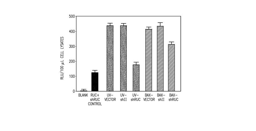
expressing the RUC cDNA and co-cultured with differently treated COS-7 ACs.
Referring
now to Figure 2: Blank shows background luminescence activity from
untransfected cells;
RUC+shRUC control: shows RUC activity when cells were co-transfected with
luciferase
plasmids (5 tg RUC, 2 gg LUC) and plasmid encoding shRUC (10 g) to confirm
downregulating activity of shRUC (no ACs added); UV-Vector shows RUC activity
when
added ACs were generated by transfecting COS-7 cells with 10 g plasmid vector
alone and
UV exposure 48 hrs post transfection; UV-shII shows RUC activity when the pre-
ACs were
transfected with 10 gg plasmid DNA encoding a shRNA targeting the HIV virus II
gene as
negative control and made apoptotic as described for UV-AC vector; UV-AC shRUC
shows
RUC activity when the pre-ACs were transfected with 10 g plasmid DNA encoding
a
shRNA targeting the RUC cDNA and made apoptotic as described for UV-AC vector;
BAX-vector shows RUC activitiy when the pre-ACs were co-transfected with
plasmid DNA
*Trademark
CA 02663026 2009-03-09
WO 2008/039980 PCT/US2007/079876
13
coding for BAX (10 g) and plasmid vector alone (10 g) and ACs were harvested
30 hrs
post transfection (no UV-treatment); BAX-shII shows RUC activity when the pre-
ACs were
transfected with plasmid DNA coding for BAX and control shRNA and processed as
described for BAX-vector; and BAX-shRUC shows RUC activity when the pre-ACs
were
transfected with plasmid DNA coding for BAX and shRUC and processed as
described for
BAX-vector.
[0060] These results show ACs containing shRUC decreased luciferase activity
in
live cells expressing an RUC target gene. In contrast, co-cultivation with ACs
containing a
control shRNA (shII) targeting the HIV-1 rev gene did not. Addition of ACs
containing
vector alone did not affect Renilla luciferase activity (data not shown).
[0061] Figure 3 shows the effects of duration of expression of shRUC prior to
induction of apoptosis of shRUC-containing cells on Renilla luciferase
activity in live cells.
The data indicate that ACs containing shRUC that had been expressed for 12 and
24 hrs did
not downregulate activity of luciferase after incubating the apopotic and live
cells.
Expression of shRUC for 48 hrs was necessary to observe loss of luciferase
actvity. These
data indicate that the loss of luciferase activity after adding ACs containing
shRUC was not
due to shRUC plasmid contamination into cells expressing RUC luciferase cDNA,
but to
expression of shRUC contained by ACs.
[0062] Figure 4 shows the effects of UV- and BAX-induced ACs containing shII
or
shRUC on levels of Renilla luciferase mRNA in live cells exposed to the AC.
Live COS-7
cells transfected with luciferase cDNA were co-cultured with COS-7 ACs
containing
control shRNA (shII) or shRNA targeting RUC mRNA (shRNA), and induced with UV
or
BAX, as described for Figure 2. Total RNA was isolated and semi-quantitative
RT-PCR
was performed with 100, 200 and 400 ng total RNA template using primers for
RUC and
the housekeeping gene GAPD-H. Products were separated using agarose gel
electrophoresis and cDNA band densities were determined. RUC cDNA amount was
normalized for GAPD-H cDNA amount when comparing shII and shRUC treatments for
a
given method of apoptosis induction. Data is shown as percentage of RUC cDNA
found in
shRUC-treated cells compared to shII-treated cells.
[0063] These results show that shRUC contained by ACs decreased RUC mRNA
levels in live cells exposed to the ACs.
CA 02663026 2010-11-10
WO 2008/039980 PCT/US2007/079876
14
REFERENCES
[0065] Behlke, M.A. (2006) Progress Towards In Vivo Use of siRNAs. Molecular
Therapy 13/4:644-670
[0066] Holmgren, L, Szeles, A., Rajnavolgyi, E., Foldman, J., Klein, G.,
Ernberg, I.
and Falk, K.I. (1999) Horizontal Transfer of DNA by the Uptake of Apoptotic
Bodies,
Blood 93/11:3956-3963.
[0067] Li, M., Qian, H., Ichim, T.M., Ge, W-W., Popov, I.A., Rycerz, K., Neu,
J.,
White, D., Zhong, R., and Min, W.-P. (2004) Induction of RNA Interference in
Dendritic
Cells. Immunologic Research 30/2:215-230.
[0068] Zhao H.-F., L'Abbe D., Jolicoeur, N., Wu, M,. Li, Z., Zhenbao, Y., and
Shen
S-H. (2005) High-Throughput Screening of Effective siRNAs from RNAi Libraries
Delivered via Bacterial Invasion, Nature Methods 2/12:967-973.