Note: Descriptions are shown in the official language in which they were submitted.
CA 02663250 2009-03-11
WO 2008/034031 PCT/US2007/078450
BIOERODIBLE ENDOPROSTHESES AND METHODS OF MAKING THE SAME
CROSS-REFERENCE TO RELATED APPLICATIONS
This application claims priority under 35 USC 119(e) to U.S. Provisional
Patent
Application Serial No. 60/844,898, filed on September 15, 2006, the entire
contents of which
are hereby incorporated by reference.
TECHNICAL FIELD
The invention relates to bioerodible endoprostheses, and to methods of making
the
same.
BACKGROUND
The body includes various passageways such as arteries, other blood vessels,
and
other body lumens. These passageways sometimes become occluded or weakened.
For
example, the passageways can be occluded by a tumor, restricted by plaque, or
weakened by
an aneurysm. When this occurs, the passageway can be reopened or reinforced
with a
medical endoprosthesis. An endoprosthesis is typically a tubular member that
is placed in a
lumen in the body. Examples of endoprostheses include stents, covered stents,
and stent-
grafts.
Endoprostheses can be delivered inside the body by a catheter that supports
the
endoprosthesis in a compacted or reduced-size form as the endoprosthesis is
transported to a
desired site. Upon reaching the site, the endoprosthesis is expanded, e.g., so
that it can
contact the walls of the lumen.
The expansion mechanism may include forcing the endoprosthesis to expand
radially.
For example, the expansion mechanism can include the catheter carrying a
balloon, which
carries a balloon-expandable endoprosthesis. The balloon can be inflated to
deform and to
fix the expanded endoprosthesis at a predetermined position in contact with
the lumen wall.
The balloon can then be deflated, and the catheter withdrawn from the lumen.
-1-
CA 02663250 2009-03-11
WO 2008/034031 PCT/US2007/078450
It is sometimes desirable for an implanted endoprosthesis to erode over time
within
the passageway. For example, a fully erodible endoprosthesis does not remain
as a
permanent object in the body, which may help the passageway recover to its
natural
condition. Erodible endoprostheses can be formed from, e.g., a polymeric
material, such as
polylactic acid, or from a metallic material, such as magnesium, iron or an
alloy thereof.
SUMMARY
The invention relates to bioerodible endoprostheses and methods of making the
endoprostheses.
In one aspect, the invention features an endoprosthesis including a member.
The
member includes a bioerodible material and an antioxidant carried by the
member.
In another aspect, the invention features a method of making an
endoprosthesis. The
method includes incorporating a bioerodible material with an antioxidant to
form at least a
portion of the endoprosthesis.
Embodiments can include one or more of the following features.
The endoprosthesis can include a carrier layer carrying the antioxidant. The
antioxidant can be on a surface of the member. The antioxidant can be within a
matrix or a
carrier material. The carrier can include pores. The carrier can be
bioerodible or non-
bioerodible. The carrier can be a metal and/or a polymer.
In some embodiments, the antioxidant is encapsulated by the bioerodible
material.
The bioerodible material can be iron or magnesium. The antioxidant can be in a
layer having
a thickness of from about 0.5 micrometer to about 10 micrometers. The
antioxidant can
include a phenol. The antioxidant can include an eugenol, an isoeugenol,
and/or an acetyl-
eugenol.
The endoprosthesis can further include a drug carried by the member. In some
embodiments, the member includes a tubular member constructed to maintain
patency of a
body vessel. The endoprosthesis can be in the form of a stent.
In some embodiments, the method includes adsorbing the antioxidant on the
surface.
In some embodiments, the bioerodible material is in the form of a tubular
member, and the
antioxidant is incorporated on a surface of the tubular member. The
bioerodible material can
be iron, magnesium, and/or an alloy of iron or magnesium. In some embodiments,
the
-2-
CA 02663250 2009-03-11
WO 2008/034031 PCT/US2007/078450
bioerodible material is in the form of a tubular member, and the antioxidant
is incorporated in
a select portion of the tubular member. In certain embodiments, the
antioxidant is in a
particle encapsulated by a bioerodible material. The particle can include zinc
oxide. In some
embodiments, at least a portion of the endoprosthesis can further include a
drug. The method
can further include incorporating a drug with the portion.
Embodiments may have one or more of the following advantages. Embodiments
feature an endoprosthesis, e.g. a coronary stent, that includes a bioerodible
portion, such as
the body of the stent capable of initially maintaining lumen patency, and an
antioxidant. In
embodiments, an endoprosthesis is coated with an antioxidant. The antioxidant
can reduce
(e.g., inhibit) erosion (e.g., corrosion) and can allow for control of
biodegradation of metallic
endoprosthesis materials. As an example, the antioxidant can allow an
endoprosthesis to
maintain structural integrity for a longer duration, which can decrease
elastic recoil after
endoprosthesis expansion. The antioxidant can reduce (e.g., inhibit) lipid
peroxidation and
can allow for a decrease in restenosis after coronary angioplasty.
The endoprostheses may not need to be removed from a lumen after implantation.
The endoprostheses can have a low thrombogenecity and high initial strength.
The
endoprostheses can exhibit reduced spring back (recoil) after expansion.
Lumens implanted
with the endoprostheses can exhibit reduced restenosis. The rate of erosion of
different
portions of the endoprostheses can be controlled, allowing the endoprostheses
to erode in a
predetermined manner and reducing, e.g., the likelihood of uncontrolled
fragmentation. For
example, the predetermined manner of erosion can be from an inside of the
endoprosthesis to
an outside of the endoprosthesis, or from a first end of the endoprosthesis to
a second end of
the endoprosthesis.
An erodible or bioerodible endoprosthesis, e.g., a stent, refers to an
endoprosthesis, or
a portion thereof, that exhibits substantial mass or density reduction or
chemical
transformation, after it is introduced into a patient, e.g., a human patient.
Mass reduction can
occur by, e.g., dissolution of the material that forms the endoprosthesis
and/or fragmenting of
the endoprosthesis. Chemical transformation can include oxidation/reduction,
hydrolysis,
substitution, and/or addition reactions, or other chemical reactions of the
material from which
the endoprosthesis, or a portion thereof, is made. The erosion can be the
result of a chemical
and/or biological interaction of the endoprosthesis with the body environment,
e.g., the body
-3-
CA 02663250 2009-03-11
WO 2008/034031 PCT/US2007/078450
itself or body fluids, into which it is implanted and/or erosion can be
triggered by applying a
triggering influence, such as a chemical reactant or energy to the
endoprosthesis, e.g., to
increase a reaction rate. For example, an endoprosthesis, or a portion
thereof, can be formed
from an active metal, e.g., Mg or Ca or an alloy thereof, and which can erode
by reaction
with water, producing the corresponding metal oxide and hydrogen gas (a redox
reaction).
For example, an endoprosthesis, or a portion thereof, can be formed from an
erodible or
bioerodible polymer, or an alloy or blend erodible or bioerodible polymers
which can erode
by hydrolysis with water. The erosion occurs to a desirable extent in a time
frame that can
provide a therapeutic benefit. For example, in embodiments, the endoprosthesis
exhibits
substantial mass reduction after a period of time which a function of the
endoprosthesis, such
as support of the lumen wall or drug delivery is no longer needed or
desirable. In particular
embodiments, the endoprosthesis exhibits a mass reduction of about 10 percent
or more, e.g.
about 50 percent or more, after a period of implantation of one day or more,
e.g. about 60
days or more, about 180 days or more, about 600 days or more, or 1000 days or
less. In
embodiments, the endoprosthesis exhibits fragmentation by erosion processes.
The
fragmentation occurs as, e.g., some regions of the endoprosthesis erode more
rapidly than
other regions. The faster eroding regions become weakened by more quickly
eroding
through the body of the endoprosthesis and fragment from the slower eroding
regions. The
faster eroding and slower eroding regions may be random or predefined. For
example, faster
eroding regions may be predefined by treating the regions to enhance chemical
reactivity of
the regions. Alternatively, regions may be treated to reduce erosion rates,
e.g., by using
coatings. In embodiments, only portions of the endoprosthesis exhibits
erodibility. For
example, an exterior layer or coating may be erodible, while an interior layer
or body is non-
erodible. In embodiments, the endoprosthesis is formed from an erodible
material dispersed
within a non-erodible material such that after erosion, the endoprosthesis has
increased
porosity by erosion of the erodible material.
Erosion rates can be measured with a test endoprosthesis suspended in a stream
of
Ringer's solution flowing at a rate of 0.2 mU/second. During testing, all
surfaces of the test
endoprosthesis can be exposed to the stream. For the purposes of this
disclosure, Ringer's
solution is a solution of recently boiled distilled water containing 8.6 gram
sodium chloride,
0.3 gram potassium chloride, and 0.33 gram calcium chloride per liter.
-4-
CA 02663250 2009-03-11
WO 2008/034031 PCT/US2007/078450
In some embodiments, an endoprosthesis with an antioxidant layer is relatively
easy
to make. An antioxidant and a polymer can be dissolved in a solvent and
applied to an
endoprosthesis. An antioxidant and a polymer can be blended together, and/or
can be formed
into a composite, and applied to an endoprosthesis. An antioxidant can be
applied directly to
an endoprosthesis, which can have open or closed pores. An antioxidant can be
incorporated
with particles and applied to an endoprosthesis.
All publications, patent applications, patents, and other references mentioned
herein
are incorporated by reference herein in their entirety.
Other aspects, features and advantages will be apparent from the description
of the
preferred embodiments thereof and from the claims.
DESCRIPTION OF DRAWINGS
FIG. lA is a perspective view of an embodiment of an endoprosthesis.
FIG. lB is a cross-sectional view of an embodiment of an endoprosthesis.
FIG. 2A is a perspective view of an embodiment of an endoprosthesis.
FIG. 2B is a cross-sectional view of an embodiment of an endoprosthesis.
FIG. 3A is a perspective view of an embodiment of an endoprosthesis.
FIG. 3B is a cross-sectional view of an embodiment of an endoprosthesis.
FIG. 3C is a cross-sectional view of another embodiment of an endoprosthesis.
FIG. 4 is an enlarged cross-sectional view of a region of an endoprosthesis.
FIG. 5 is an enlarged cross-sectional view of a region of an embodiment of an
endoprosthesis.
FIG. 6 is an enlarged cross-sectional view of a region of an embodiment of an
endoprosthesis.
FIG. 7 is a cross-sectional view of an embodiment of an endoprosthesis.
FIG. 8 is an enlarged cross-sectional view of a region of an embodiment of an
endoprosthesis.
FIG. 9 is an enlarged cross-sectional view of a region of an embodiment of an
endoprosthesis.
FIG. 10 is an enlarged cross-sectional view of an embodiment of an
endoprosthesis
FIG. 11 a perspective view of an embodiment of an endoprosthesis.
-5-
CA 02663250 2009-03-11
WO 2008/034031 PCT/US2007/078450
FIG. 12 is a perspective view of an embodiment of an endoprosthesis.
FIG 13 is a sequence illustrating a method of making an endoprosthesis.
DETAILED DESCRIPTION
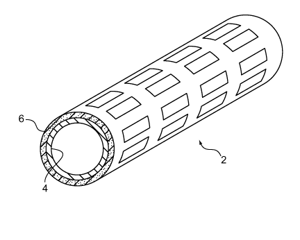
Referring to FIGS. lA and lB endoprosthesis 2 (as shown, a stent) includes a
bioerodible layer 4 and an antioxidant-containing layer 6 ("antioxidant layer
6") disposed
radially outward and on a surface of the bioerodible layer. Bioerodible layer
4, which can
include a bioerodible material (e.g., a metal) such as a magnesium alloy, is a
tubular body
capable of maintaining the patency of a bodily lumen after implantation and is
capable of
eroding within the bodily lumen. Antioxidant layer 6 provides therapeutic
benefits, such as
inhibiting restenosis as well as affecting (e.g., reducing or inhibiting) the
erosion of
bioerodible layer 4 to allow the endoprosthesis to maintain structural
integrity (e.g., patency)
for a longer duration. Examples of antioxidants in antioxidant layer 6 include
phenolic
compounds (e.g., isoeugenol, eugenol, and acetyl eugenol), polyphenols,
phenols, and any
mixtures thereof. As shown, antioxidant layer 6 is disposed radially outward
of bioerodible
layer 4, but alternatively or additionally, the antioxidant layer can be
disposed radially inward
of the bioerodible layer.
Antioxidants can inhibit or reduce oxidative processes caused by oxygen or
free
radicals. The use of an antioxidant in an erodible endoprosthesis can provide
a number of
advantages. The antioxidant can inhibit restenosis by inhibiting lipid
peroxidation.
Antioxidants such as eugenol compounds can have an inhibitory effect on LDL
suppression
of free radical cascade of lipid peroxidation and reduction of LDL to its
receptor, as well as
provide anti-inflammatory effects. In addition, the antioxidant presence on
its own as a
coating or in a carrier with another material acts as a barrier that modifies
the exposure of the
bioerodible endoprosthesis to body fluids and thus the degradation processes
which occur
upon exposure to body fluids. Moreover, the presence of an antioxidant can
chemically
inhibit corrosive degradation, particularly of metals. Without being bound by
theory, it is
believed that in a biological fluid, an antioxidant can reduce (e.g., inhibit)
free radical
reactions by decreasing the level of active products from oxygen reduction
and/or
sequestering (e.g., binding to a protein) a transition metal group such as Fe
and Cu to reduce
the formation of oxidants. Further discussion of antioxidants is provided in
Chaieb et al.,
-6-
CA 02663250 2009-03-11
WO 2008/034031 PCT/US2007/078450
Applied Surface Science, 2005, 246, 199; Lee et al., Journal of Dentistry,
2000, 28, 69;
Satoh et al., Anticancer Res., 1998, 18, 1549; Damiani et al., Vascular
Pharmal. 2003, 40,
59; Stoclet et al., European Journal of Pharmacology, 2005, 500, 461; Ito et
al., Food and
Chemical Toxicology, 2005, 43, 461; Naderi et al., Molecular and Cellular
Biochemistry,
2004, 267, 59; Molnar et al., International Immunopharmacology, 2005, 5, 849;
Kim et al.,
Circ. J., 2005, 69, 101; Andi6n et al., Corrosion Science, 2002, 44, 2805-
2816; and Ou et al.,
Food and Chemical Toxicology, 2006, 44, 1485-1495, the entire contents of each
of which is
hereby incorporated by reference.
As an example, an antioxidant can be low-molecular weight compounds (e.g.,
isoeugenol, eugenol, acetyl eugenol, polyphenols, phenols (including
antioxidants of the
phenolic class of compounds such as phenols, polyphenols, and phenolic
compounds),
tocopherols, anethol, geraniol, limonene, linalool, p-cymol, pulegone, thymol,
ubiquitol-l0,
ascorbic acid, (3-carotene, lycopene, glutathione, uric acid, bilirubin,
carvediol, Curcuma
longa, and Ocimum sanctum. Classes of antioxidants can include phenols,
phenolic acids,
flavonoids, anthocyanins, catechins, flavones, flavonols, flavanones,
isoflavones, lignins,
proanthocyanidins, procyanidins, stilbenes, tannins, spice antioxidants, and
plant-derived
antioxidants. In some embodiments, an antioxidant is a high-molecular weight
compound
such as a protein (e.g., albumin, transferrin, haptoglobin, haemopexin,
caeruloplasmin,
ferritin, superoxide dismutase, catalase, glutation reductase, glutathione
peroxidase, etc.)
and/or a polymer (e.g., polymeric phenols). In some embodiments, the
antioxidant is
polymeric. The polymeric antioxidant can be provided as a layer directly on
the bioerodible
layer. In embodiments, the polymeric antioxidant layer is directly deposited
onto an
endoprosthesis by electropolymerization, and/or the polymeric antioxidant
layer is dissolved
in a solvent and applied to the endoprosthesis. A plurality of different
antioxidants can be
used.
The antioxidant compound can be provided as a layer directly on the
bioerodible
layer or incorporated into the bioerodible layer, or incorporated into a
bioerodible or
nonbioerodible carrier layer on the bioerodible material. The antioxidant can
be released
from the carrier by diffusion through the carrier and/or erosion of the
carrier in the case
where a bioerodible carrier is used. The antioxidant can be noncovalently
bonded, e.g.
adsorbed, or covalently bonded to the carrier or the bioerodible material,
e.g. by
-7-
CA 02663250 2009-03-11
WO 2008/034031 PCT/US2007/078450
copolymerization with the carrier. Further examples of antioxidants are
described, for
example, in Ivanova et al., Experimental Pathology and Parasitology, 2000, 4,
49; Frei, B.,
Proceedings - Society for Experimental Biology and Medicine, 1999, 222, 196;
Mohanty et
al., BMC Complementary and Alternative Medicine, 2006, 6:3; Suhaj, M., Journal
of Food
Composition and Analysis, 2006, 19, 531-537; Ratnam et al., Journal of
Controlled Release,
2006, 113, 189-207; Gurib-Fakim, A., Molecular Aspects of Medicine, 2006, 27,
1-93; Arts
et al., Am. J. Clin. Nutr., 2005, 81(1), 317S-325S; Wallerath et al., Nitric
Oxide, 2005, 12(2),
97-104; Grassi et al., Am. J. Clin. Nutr., 2005, 81(3), 611-614; Kim et al.,
Crit. Rev. Food
Sci. Nutr., 2004, 44(4), 253-273; Lambert et al., Am. J. Clin. Nutr., 2005,
81(1), 284S-291S;
Moskaug et al., Am. J. Clin. Nutr., 2005, 81(1), 277S-283S; and Williamson et
al., Am. J.
Clin. Nutr., 2005, 81(1), 243S-255S.
In FIGS. lA and 1B, antioxidant layer 6 has an antioxidant (shading)
distributed
uniformly within a matrix of a biocompatible carrier 7. Suitable carriers
include, for
example, bioerodible or non bioerodible polymers or metals. A bioerodible
carrier (e.g., a
bioerodible polymer) can erode over time and expose the incorporated
antioxidant for
gradual release. A bioerodible carrier can inhibit direct contact of body
fluids with
bioerodible layer 4 and reduce the bioerosion rate of the endoprosthesis.
Suitable bioerodible
polymer carriers include polylactic acid (PLA), polylactic glycolic acid
(PLGA),
polyanhydrides (e.g., poly(ester anhydride)s, fatty acid-based polyanhydrides,
amino acid-
based polyanhydrides), polyesters, polyester-polyanhydride blends,
polycarbonate-
polyanhydride blends, and/or combinations thereof. Bioerodible polymers such
as
polyanhydrides are described, for example, in Kumar et al., Advanced Drug
Delivery
Reviews, 2002, 54, 889. Bioerodible polymers are also described in U.S.S.N.
10/958,435
(U.S. Patent Application Publication No. 2005/0216074), filed October 5, 2004.
The
antioxidant and the polymer can be dissolved in a solvent and applied to
bioerodible layer 4,
the antioxidant and the polymer can be blended together and applied to the
bioerodible layer,
and/or the antioxidant and the polymer can be formed into a composite in a
solvent and
applied to the bioerodible layer. The antioxidant can be applied (e.g.,
adsorbed) to
antioxidant layer using, for example, vapor phase adsorption and solution
phase adsorption
methods (such as solution impregnation). Varying amounts of the antioxidant
can be
dispersed (uniformly or non-uniformly) within antioxidant layer 6. For
example, the
-8-
CA 02663250 2009-03-11
WO 2008/034031 PCT/US2007/078450
antioxidant can be present from about 0.5 percent by weight of the antioxidant
layer 6 (e.g.,
from about 1 percent by weight, from about 2 percent by weight, from about 5
percent by
weight, from about 10 percent by weight, from about 15 percent by weight, from
about 20
percent by weight, from about 25 percent by weight) to about 30 percent by
weight of the
antioxidant layer (e.g., to about 25 percent by weight, to about 20 percent by
weight, to about
percent by weight, to about 10 percent by weight, to about 5 percent by
weight, to about 2
percent by weight). The carrier can include one or more bioerodible materials
and/or one or
more non-bioerodible materials that has a different chemical composition than
a composition
of material in bioerodible layer 4.
10 Referring to FIGS. 2A and 2B, endoprosthesis 2' includes a bioerodible
layer 4' and
an antioxidant layer 6' radially outward of the bioerodible layer 4'. The
antioxidant layer 6'
includes (e.g., is formed of) a bioerodible or non-bioerodible carrier 7'
having a plurality of
pores 8. The antioxidant is dispersed (e.g., sorbed) in the pores in
antioxidant layer 6'. Pores
8 increase the total free volume and surface area of antioxidant layer 6', and
allow more
15 antioxidant to be loaded in and delivered from antioxidant layer 6'. The
antioxidant layer
can be formed of a bioerodible or non-bioerodible metal, polymer or ceramic in
which pores
are created. For example, the carrier can be formed of the same material or a
different
material as the bioerodible layer 4'. For example, carrier and the bioerodible
layer can be
formed of the same metal. Antioxidant layer 6' can be made by forming pores 8
and
applying the antioxidant to the porous outer surface. In some embodiments, a
first layer of
carrier material is formed on the surface of the bioerodible layer and pores
are formed by
creating a number of holes (e.g., by laser ablation) and the holes are filled
or partially filled
with an antioxidant 6. A second layer of a same or different polymer can be
coated (e.g., by
spraying) onto the endoprosthesis. Pores can also be formed during the coating
process by
techniques discussed below. The pores can be formed directly into the surface
of the
bioerodible layer 4' without the use of a carrier. Pores 8 can have an average
diameter of
from about 10 nm (e.g., from about 20 nm, from about 50 nm, from about 100 nm,
from
about 200 nm, from about 500 nm, from about 700 nm, from about 1 m, from
about 1.5 m,
from about 2 m, from about 2.5 m, from about 3 m, from about 3.5 m, from
about 4 m,
from about 4.5 m) to about 10 m (e.g., to about 9 m, to about 8 m, to
about 7 m, to
about 6 m, to about 5 m, to about 4.5 m, to about 4 m, to about 3 m, to
about 2.5 m,
-9-
CA 02663250 2009-03-11
WO 2008/034031 PCT/US2007/078450
to about 2 m, to about 1.5 m, to about 1 m, to about 750 nm, to about 500
nm, to about
250 nm, to about 100 nm, to about 75 nm, to about 50 nm, to about 25nm). Pores
8 can have
an average surface area of from about 300 nm2 (e.g. from about 1,000 nm2, from
about 5,000
nm2 , from about 30,000 nm~, from about 0.5 m~, from about 6 m~, from about
10 m~,
from about 20 m2, from about 30 m2, from about 40 m2, from about 50 m2,
from about
65 m2) to about 350 m2 (e.g., to about 300 m2, to about 250 m2, to about
200 m2, to
about 150 m2, to about 100 m2, to about 70 m2, to about 65 m2, to about 50
m2, to
about 40 m2, to about 30 m2, to about 20 m2, to about 10 m2, to about 6
m2, to about
0.5 m2, to about 30,000 nm2, to about 5,000 nm2, to about 1000 nm2). Pores 8
can also be
expressed by average volume. In some embodiments, pores 8 can be from about
500 nm3
(e.g., from about 0.00005 m3, from about 0.0005 m3, from about 0.005 m3,
from about
0.05 m3, from about 0.5 m3, from about 1 m3, from about 5 m3, from about
35 m3,
from about 50 m) to about 550 m3 (e.g., to about 450 m3, to about 300 m3,
to about 200
m3, to about 100 m3, to about 75 m3, to about 40 m3, to about 10 m3, to
about 5 m3,
to about 1 m3, to about 0.5 m3, to about 0.05 m3, to about 0.005 m3, to
about 0.00005
m3). Pores can occupy a portion of antioxidant layer 6'. In some embodiments,
pores range
from about 1% by volume of the antioxidant layer (e.g., from about 5% by
volume, from
about 10% by volume, from about 25% by volume, from about 50% by volume) to
about
75% by volume of the antioxidant layer (e.g., to about 60% by volume, to about
50% by
volume, to about 40% by volume, to about 30% by volume, to about 25% by
volume, to
about 20% by volume, to about 10% by volume, to about 5% by volume. The
antioxidant
can be applied (e.g., adsorbed) to antioxidant layer 6' using, for example,
vapor phase
adsorption and solution phase adsorption methods (such as solution
impregnation). The
antioxidant can be sorbed (uniformly or non-uniformly) within antioxidant
layer 6' from
about 0.5% by weight of the antioxidant layer (e.g., from about 1% by weight,
from about
5% by weight, from about 10% by weight, from about 20% by weight, from about
30% by
weight, from about 40% by weight) to about 50% by weight of the antioxidant
layer (e.g., to
about 45% by weight, to about 40% by weight, to about 30% by weight, to about
25% by
weight, to about 15% by weight, to about 10% by weight, to about 5 % by
weight, to about
2% by weight, to about 1% by weight).
-10-
CA 02663250 2009-03-11
WO 2008/034031 PCT/US2007/078450
Referring to FIGS. 3A, 3B, and 3C, endoprosthesis 2" and 2"' include particles
10,
10', which carry one or more antioxidants. Particles 10, 10' can be dispersed
throughout an
endoprosthesis, or can be dispersed in an antioxidant layer including a
carrier of the types
discussed above on an endoprosthesis. Referring to FIGS. 3A and 3B,
endoprosthesis 2"
includes a bioerodible layer 4", and an antioxidant layer 6" including
particles 10 dispersed
in a carrier 7" of the types described above. Referring to FIG. 3C,
endoprosthesis 2"'
includes particles 10' dispersed throughout the erodible layer 4' of the
endoprosthesis. In
other embodiments, the particles are absorbed or bonded to the surface of the
erodible layer.
The particles can include (e.g., is formed of) a bioerodible material, such as
zinc oxide,
poly(y-benzyl-L-glutamate) (PBLG), poly((3-benzyl-L-aspartate) (PBLA), poly-
D,L-lactide-
co-glycolide (PLGA), and polylactic acid (PLA), that encapsulates the
antioxidant and allows
the antioxidant to be delivered to the body. Particles 10 (e.g.,
nanoparticles) can have an
average diameter of from about 100 nm (from about 200 nm, from about 400nm,
from about
600 nm, from about 1 m, from about 2 m, from about 3 m, from about 4 m) to
about 5
m (to about 4.5 m, to about 4 m, to about 3.5 m, to about 3 m, to about 2
m, to about
1 m, to about 800 nm, to about 500 nm, to about 300 nm, to about 200 nm).
Particles 10
(e.g., nanoparticles) can also be expressed by volume. In some embodiments,
particles 10
can have a volume of from about 0.0005 m3 (e.g., from about from about 0.005
m3, from
about 0.05 m3, from about 0.5 m3, from about 5 m3, from about 50 m) to
about 70 m3
(e.g., to about 60 m3, to about 50 m3, to about 5 m3, to about 0.5 m3, to
about 0.05 m3,
to about 0.005 m3, to about 0.0025 m) . The antioxidant can be present in
varying amounts
within the particles. For example, the antioxidant can be present from about 5
weight percent
of particles 10 (e.g., from about 10 weight percent, from about 15 weight
percent, from about
20 weight percent, from about 25 weight percent) to about 30 weight percent of
particles 10
(e.g., to about 25 weight percent, to about 20 weight percent, to about 15
weight percent, to
about 10 weight percent, to about 7 weight percent). Prior to implantation,
particles 10 can
be present from about 0.5 weight percent of antioxidant layer 6" (e.g., from
about 1 weight
percent, from about 2 weight percent, from about 5 weight percent, from about
10 weight
percent, from about 15 weight percent) to about 20 weight percent of
antioxidant layer 6"
(e.g., to about 17 weight percent, to about 15 weight percent, to about 10
weight percent, to
about 5 weight percent, to about 3 weight percent, to about 2 weight percent).
Particles 10
-11-
CA 02663250 2009-03-11
WO 2008/034031 PCT/US2007/078450
can be substantially spherical or any other shape. Suitable processes for
making particles
include spraying (e.g., electrospraying), emulsion processes, and dispersion
polymerization.
Further processes for making particles are described, for example, in Jiang ,
S.B., Materials
Science and Engineering, 2006, 418, 199.
Referring now to FIG. 4, the thicknesses for bioerodible layer 4, 4', 4" and
antioxidant layer 6, 6', 6" is illustrated. In some embodiments, bioerodible
layer 4, 4', 4"
has a total thickness (Tb) that is from about 5 m (e.g., from about 10 m,
from about 20 m,
from about 30 m, from about 40 m, from about 50 m, from about 60 m from
about 80
m, from about 100 m) to about 200 m (e.g., to about 175 m, to about 150 m,
to about
100 m, to about 85 m, to about 75 m, to about 50 m, to about 35 m, to
about 20 m, to
about 15 m). In some embodiments, antioxidant layer 6, 6', 6" has a total
thickness (Ta)
that is from about 0.5 m (e.g., from about 1 m, from about 2 m, from about
3 m, from
about 4 m, from about 5 m, from about 6 m, from about 7 m, from about 8
m) to about
10 m (e.g., to about 9 m, to about 8 m, to about 7 m, to about 6 m, to
about 5 m, to
about 4 m, to about 3 m, to about 2 m, to about 1 m). Total Tt can be from
about 10 m
(e.g., from about 20 m, from about 30 m, from about 40 m, from about 50 m,
from
about 60 m from about 80 m, from about 100 m) to about 200 m (e.g., to
about 150 m,
to about 100 m, to about 85 m, to about 75 m, to about 50 m, to about 35
m, to about
m, to about 15 m).
20 The thicknesses for bioerodible layer 4, 4', 4" and antioxidant layer 6,
6', 6" can also
be expressed relative to the total thickness (Tt) of endoprosthesis 2, 2', 2".
In some
embodiments, bioerodible layer 4, 4', 4" has a total thickness Tb that is from
about 10
percent of Tt (e.g., from about 35 percent, from about 60 percent, from about
70%, from
about 80 percent) to about 90% of Tt (e.g., to about 80%, to about 70%, to
about 50%. to
about 35%, to about 15%, to about 10%). In some embodiments, antioxidant layer
6, 6', 6"
has a total thickness Ta that is from about 10 percent of Tt (e.g., from about
35 percent, from
about 60 percent, from about 80 percent) to about 90 percent of Tt (e.g., to
about 80%, to
about 75 percent, to about 50 percent, to about 45%, to about 35%, to about 25
percent, to
about 15%, to about 10%, to about 5%).
Referring to Figs. 5 and 6, within an antioxidant layer, the antioxidant can
be equally
distributed throughout or unequally distributed. For example, the antioxidant,
such as that
-12-
CA 02663250 2009-03-11
WO 2008/034031 PCT/US2007/078450
located near the outer peripheral region of an endoprosthesis, can be
distributed in a gradient
manner along the radial direction of the endoprosthesis. Referring to FIG. 5,
the antioxidant
(shading) can increase in concentration toward an outer periphery 12 of an
endoprosthesis 2,
2', 2". Greater release of the antioxidant can be achieved during the early
stages of the
endoprosthesis lifetime following implantation. Referring to FIG. 6, the
antioxidant
(shading) can decrease in concentration toward an outer periphery 14 of an
endoprosthesis 2,
2', 2". The antioxidant release can increase during the endoprosthesis
lifetime within a
vessel. A decrease or increase in concentration of an antioxidant within an
endoprosthesis
can occur linearly, non-linearly (e.g., exponentially), and/or in a stepwise
manner in order to
tailor the release of the antioxidant. In some embodiments, an antioxidant
layer includes one
or more zones having an equal distribution of antioxidant throughout, and one
or more zones
having an unequal distribution of antioxidant, in any combination.
Referring to FIGS. 7-9, similarly, in embodiments in which the antioxidant
layer is
radially inward of the bioerodible layer, the antioxidant can be equally
distributed throughout
or unequally distributed. FIG. 7 shows an endoprosthesis 20 including an
antioxidant layer
4"' located radially inwardly of a bioerodible layer 6"', for example, to
avoid direct contact
of the antioxidant with a vessel. The antioxidant can be uniformly dispersed
within
antioxidant layer 4"', which defines an inner circumferential region of
endoprosthesis 20. In
some embodiments, the antioxidant in antioxidant layer 4"' can be dispersed in
a gradient
manner along a radius of endoprosthesis 20 to tune the release of the
antioxidant within a
vessel. For example, as shown in FIG. 8, the antioxidant can increase in
concentration
radially outward, or as shown in FIG. 9, the antioxidant can decrease in
concentration radially
outward. A decrease or increase in concentration of the antioxidant within an
endoprosthesis
can occur linearly, exponentially, or in a stepwise manner in order to tailor
the release of the
antioxidant. In some embodiments, an antioxidant layer includes one or more
zones having
an equal distribution of antioxidant throughout, and one or more zones having
an unequal
distribution of antioxidant, in any combination.
Referring to FIGS. 10 and 11, the antioxidant layer be on selected portion(s)
of the
bioerodible layer, for example, to tune the release of antioxidant, to treat
specific locations in
a vessel, or to create a desirable degradation pattern. For example, FIG. 10
shows an
endoprosthesis 22 having a bioerodible layer 4"", and an antioxidant layer 6""
including an
-13-
CA 02663250 2009-03-11
WO 2008/034031 PCT/US2007/078450
antioxidant 24 located in strips extending along the length of. As another
example, FIG. 11
shows an endoprosthesis 26 having a bioerodible layer 4""', and an antioxidant
28 applied as
circular bands on the bioerodible layer. Referring to FIG. 12, an
endoprosthesis 30 includes a
series of generally circumferential interconnected struts 32 , and an
antioxidant 34 can be
applied to selected struts to reduce the degradation rate of the struts to
maintain structural
features of the struts compared to struts not including the antioxidant. An
antioxidant can
have a patterned distribution on the bioerodible layer, and/or along the
length of an
endoprosthesis
Referring to FIG. 13, a method 100 of making an endoprosthesis as described
herein
is shown. Method 100 includes forming a bioerodible tube (step 102), forming a
pre-
endoprosthesis from the bioerodible tube (step 104), and applying one or more
antioxidants
to the pre-endoprosthesis (step 106) to form an endoprosthesis. In other
embodiments, one or
more antioxidants are applied to the bioerodible tube, and the tube with the
applied
antioxidant(s) is subsequently formed into an endoprosthesis.
The bioerodible tube can be formed (step 102) by manufacturing a tubular
member
including (e.g., is formed of) one or more bioerodible materials and capable
of supporting a
bodily lumen. For example, a mass of bioerodible material can be machined into
a rod that is
subsequently drilled to form the tubular member. As another example, a sheet
of bioerodible
material can be rolled to form a tubular member with overlapping portions, or
opposing end
portions of the rolled sheet can be joined (e.g., welded) together to form a
tubular member. A
bioerodible material can also be extruded to form a tubular member. The
bioerodible or
erodible material can be a substantially pure metallic element, or an alloy.
The alloy can
include metal and non-metal components, for example, the alloy can be a
metallic alloy, a
ceramic, or a metal matrix composite. Examples of metallic elements include
iron and
magnesium. Examples of alloys include iron alloys having, by weight, 88-99.8%
iron, 0.1-
7% chromium, 0-3.5% nickel, and less than 5% of other elements (e.g.,
magnesium and/or
zinc); or 90-96% iron, 3-6% chromium and 0-3% nickel plus 0-5% other metals.
Other
examples of alloys include magnesium alloys, such as, by weight, 50-98%
magnesium, 0-
40% lithium, 0-5% iron and less than 5% other metals or rare earths; or 79-97%
magnesium,
2-5% aluminum, 0-12% lithium and 1-4% rare earths (such as cerium, lanthanum,
neodymium and/or praseodymium); or 85-91% magnesium, 6-12% lithium, 2%
aluminum
-14-
CA 02663250 2009-03-11
WO 2008/034031 PCT/US2007/078450
and 1% rare earths; or 86-97% magnesium, 0-8% lithium, 2% -4% aluminum and 1-
2% rare
earths; or 8.5-9.5% aluminum, 0.15%-0.4% manganese, 0.45-0.9% zinc and the
remainder
magnesium; or 4.5-5.3% aluminum, 0.28%-0.5% manganese and the remainder
magnesium;
or 55-65% magnesium, 30-40% lithium and 0-5% other metals and/or rare earths.
Magnesium alloys are also available under the names AZ91D, AM50A, and AE42.
Other
erodible materials are described in Bolz, U.S. 6,287,332 (e.g., zinc-titanium
alloy and
sodium-magnesium alloys); Heublein, U.S. Patent Application 2002000406; and
Park,
Science and Technology of Advanced Materials, 2, 73-78 (2001), all of which
are hereby
incorporated by reference herein in their entirety. In particular, Park
describes Mg-X-Ca
alloys, e.g., Mg-Al-Si-Ca, Mg-Zn-Ca alloys. The bioerodible tube can include
more than one
bioerodible material, such as different bioerodible materials physically mixed
together,
multiple layers of different bioerodible materials, and/or multiple sections
of different
bioerodible materials along a direction (e.g., length) of the tube. In other
embodiments, the
bioerodible material is a bioerodible polymer.
As shown in FIG. 13, after the bioerodible tube is formed, the tube is formed
into a
pre-endoprosthesis (step 104). For examples, selected portions of the tube can
be removed to
form bands and connectors by laser cutting, as described in U.S. Patent No.
5,780,807,
hereby incorporated by reference in its entirety. Other methods of removing
portions of the
tube can be used, such as mechanical machining (e.g., micro-machining, grit
blasting or
honing), electrical discharge machining (EDM), and photoetching (e.g., acid
photoetching).
The pre-endoprosthesis can be etched and/or electropolished to provide a
selected finish. In
certain embodiments, such as jelly-roll type endoprostheses, step 104 is
omitted.
Prior to apply the antioxidant, selected surfaces (e.g., inner surface) or
portions (e.g.,
portion between the end portions of the endoprosthesis) of the pre-
endoprosthesis can be
masked so that the antioxidant will not be applied to the masked surfaces or
portions.
In some embodiments, prior to applying the antioxidant, pores are formed on/in
the
pre-endoprosthesis, the bioerodible tube, and/or a coating layer. Pores can be
formed by a
variety of methods (e.g., micro-arc surface modification, sol-gel templating
process, plasma
spraying, adding foaming structures into a melt or liquid metal, melting a
powder compact
containing a gas evolving element or a space holder material, incorporating a
removable
scaffold (e.g., polyurethane) in a metal powder/slurry prior to sintering,
sintering hollow
-15-
CA 02663250 2009-03-11
WO 2008/034031 PCT/US2007/078450
spheres, sintering fibers, combustion synthesis, powder metallurgy, bonded
fiber arrays, wire
mesh constructions, vapor deposition, three-dimensional printing, and/or
electrical discharge
compaction). In some embodiments, pores can be formed by incorporating
embedded
microparticles and/or compounds (e.g., a salt) within the antioxidant layer
(e.g., a
polymerizable monomer, a polymer, a metal alloy), forming the antioxidant
layer, and
removing (e.g., dissolving, leaching, burning) the microparticles and/or
compounds to form
pores at locations where the microparticles and/or compounds were embedded.
Removable
(e.g., dissolvable) microparticles can be purchased, for example, from
MicroParticles GmbH.
In some embodiments, pores are formed by using a gas as a porogen, bonding
fibers, and/or
phase separation in materials such as polymers, metals, or metal alloys.
Methods for forming
porous structures are described, for example, in Ryan et al., Biomaterials,
2006, 27, 2651;
Liao et al., Journal of Biomedical Materials Research, 2001, 59(4), 676; Mikos
et al.,
Electronic Journal of Biotechnology, 2000, 3(2), 1; Widmer et al.,
Biomaterials, 1998, 19,
1945; and Gomes et al., Materials Science and Engineering C, 2002, 20, 19.
Next, the antioxidant(s) can applied to the pre-endoprosthesis (step 106) to
form an
endoprosthesis. The antioxidant and a polymer (e.g., polylactic acid (PLA),
polylactic
glycolic acid (PLGA), polyanhydrides (e.g., poly(ester anhydride)s, fatty acid-
based
polyanhydrides, amino acid-based polyanhydrides), polyesters, polyester-
polyanhydride
blends, polycarbonate-polyanhydride blends, and/or combinations thereof) can
be dissolved
in a solvent and applied to the pre-endoprosthesis, the antioxidant and the
polymer can be
blended together (e.g., in a manner that the antioxidant is mixed, embedded or
encapsulated
in a polymer matrix) and applied to the pre-endoprosthesis, and/or the
antioxidant and the
polymer can be formed into a composite in a solvent and applied to the pre-
endoprosthesis.
In some embodiments, the antioxidant layer is directly deposited onto an
endoprosthesis
(e.g., by electropolymerization). Methods for depositing an antioxidant is
described, for
example, in Andi6n et al., Corrosion Science., 2002, 44, 2805-2816. The
antioxidant can be
applied (e.g., adsorbed on the surfaces defining the pores, adsorbed on a
substantially pore-
free surface, or dispersed within the pores) directly to the pre-
endoprosthesis using vapor
phase adsorption, solution phase adsorption methods (e.g., solution
impregnation). The
antioxidant can also be incorporated with (e.g., encapsulated in) particles
including a second,
different bioerodible material than the bioerodible material in the pre-
endoprosthesis, the
-16-
CA 02663250 2009-03-11
WO 2008/034031 PCT/US2007/078450
second bioerodible material with the antioxidant can be applied to the pre-
endoprosthesis.
The second bioerodible material can also be combined with the bioerodible
material and co-
extruded with a bioerodible material free of the second bioerodible material.
In some
embodiments, more than one method of applying an antioxidant to a pre-
endoprosthesis can
be used. As an example, a pre-endoprosthesis may be coated with an antioxidant
in a
polymer matrix, and impregnated with a bioerodible material-encapsulated
antioxidant.
Methods for incorporating one material in another are described, for example,
in Jiang, S.B.,
Materials Science and Engineering, 2006, 418, 199.
In certain embodiments, the antioxidant can be applied to a pre-endoprosthesis
in one
layer, or in multiple layers (e.g., at least two layers, at least three
layers, at least four layers,
at least five layers) in order, for example, to provide greater control over
the amount and
variety of the antioxidant. For example, the layers can have different
concentrations of one
or more antioxidants (e.g., to provide a gradient or other profiles of
antioxidants), and/or the
layers can have different compositions of antioxidants. Within an antioxidant
layer, the
concentrations and/or compositions of the antioxidant can be the same or
different to provide
a selected antioxidant profile. For example, the end portions of the
endoprosthesis can have
a greater concentration of antioxidant than the intermediate portion of the
endoprosthesis to
provide reduced restenosis. The antioxidant layers can be applied the same way
or in
different ways. For example, a first, innermost antioxidant layer can be
sorbed to a porous
surface of the pre-endoprosthesis, and a second, outer antioxidant layer can
include an
antioxidant and a polymer that are applied to the first layer.
As indicated above, in some embodiments, the antioxidant(s) is applied to the
bioerodible tube prior to forming the bioerodible tube into an endoprosthesis
(if necessary).
As a result, the endoprosthesis can have its outer and inner surfaces coated
with the
antioxidant(s), and the side surfaces of the endoprosthesis can be free of the
antioxidant(s).
Prior to applying the antioxidant(s), the inner surface or the outer surface
of the bioerodible
tube can be masked to apply the antioxidant(s) to only selected portion(s) of
the tube.
The endoprosthesis can be made of a desired shape and size (e.g., coronary
stents,
aortic stents, peripheral vascular stents, gastrointestinal stents, urology
stents, and neurology
stents). Depending on the application, the endoprosthesis can have a diameter
of between,
for example, 1 mm to 46 mm. In certain embodiments, a coronary stent can have
an
-17-
CA 02663250 2009-03-11
WO 2008/034031 PCT/US2007/078450
expanded diameter of from about 2 mm to about 6 mm. In some embodiments, a
peripheral
stent can have an expanded diameter of from about 5 mm to about 24 mm. In
certain
embodiments, a gastrointestinal and/or urology stent can have an expanded
diameter of from
about 6 mm to about 30 mm. In some embodiments, a neurology stent can have an
expanded
diameter of from about 1 mm to about 12 mm. An abdominal aortic aneurysm (AAA)
stent
and a thoracic aortic aneurysm (TAA) stent can have a diameter from about 20
mm to about
46 mm.
The endoprostheses described herein can be configured for non-vascular lumens.
For
example, they can be configured for use in the esophagus or the prostate.
Other lumens
include biliary lumens, hepatic lumens, pancreatic lumens, urethral lumens and
ureteral
lumens.
In use, the endoprosthesis can be used, e.g., delivered and expanded, using a
catheter
delivery system, such as a balloon catheter system. Catheter systems are
described in, for
example, Wang U.S. 5,195,969, Hamlin U.S. 5,270,086, and Raeder-Devens, U.S.
6,726,712.
Endoprosthesis delivery, such as stent delivery, are also exemplified by the
Radius or
Symbiot systems, available from Boston Scientific Scimed, Maple Grove, MN.
The endoprostheses described herein can be a covered stent or a stent-graft.
For
example, the stent described herein can include and/or be attached to a
biocompatible, non-
porous or semi-porous polymer matrix including polytetrafluoroethylene (PTFE),
expanded
PTFE, polyethylene, urethane, or polypropylene.
The endoprostheses can further include a releasable therapeutic agent, drug,
or a
pharmaceutically active compound, such as described in U.S. Patent No.
5,674,242, U.S.S.N.
09/895,415, filed July 2, 2001, U.S.S.N. 11/111,509, filed Apri121, 2005, and
U.S.S.N.
10/232,265, filed August 30, 2002. The therapeutic agents, drugs, or
pharmaceutically active
compounds can include, for example, anti-thrombogenic agents, antioxidants,
anti-
inflammatory agents, anesthetic agents, anti-coagulants, and antibiotics. The
therapeutic
agent, drug, or a pharmaceutically active compound can be dispersed in a
polymeric coating
carried by the stent. The polymeric coating can include more than a single
layer. For
example, the coating can include two layers, three layers or more layers,
e.g., five layers.
The therapeutic agent can be a genetic therapeutic agent, a non-genetic
therapeutic agent, or
cells. Therapeutic agents can be used singularly, or in combination.
Therapeutic agents can
-18-
CA 02663250 2009-03-11
WO 2008/034031 PCT/US2007/078450
be, for example, nonionic, or they may be anionic and/or cationic in nature.
An example of a
therapeutic agent is one that inhibits restenosis, such as paclitaxel. The
therapeutic agent can
also be used, e.g., to treat and/or inhibit pain, encrustation of the stent or
sclerosing or
necrosing of a treated lumen. Any of the above coatings and/or polymeric
portions can be
dyed or rendered radio-opaque.
In other embodiments, an endoprosthesis includes one or more filaments or
wires
including one or more bioerodible materials and one or more antioxidants
applied to the
bioerodible material(s) as described above. The filaments or wires can be
knitted, woven, or
braided to form an endoprosthesis. All the filaments or only selected
filaments can include
bioerodible material and the antioxidant. The bioerodible material and/or the
antioxidant can
be the same or different.
All references, such as patent applications, publications, and patents,
referred to
herein are incorporated by reference in their entirety.
Other embodiments are within the scope of the claims.
-19-