Note: Descriptions are shown in the official language in which they were submitted.
CA 02663439 2009-03-12
WO 2008/036504 PCT/US2007/077481
MEDZCAL DEVICE WITH POROC7S SURFACE
CROSS-REFERENCE `I'O RELATED APPLICATIONS
This application claims priority under 35 USC; .l 19(c) to U.S. Provisional
Patent
Application Serial No. 60/825;965, filed on September.l8, 2006, the entire
contents of
which are hereby incorporated by reference herein:
TECHNTCAL FIELD
The invcntion relates to medical devices, sucti as .endoprostheses (e.g.,
stents).
BACKGROUND
The body defines various passageways such as arteries, other blood vessels,
and
other body lumens. These passageways sometimes become occluded or weakened.
For
exainple, the passageways can be occluded by a tumor, restricted by plaque, or
weakened
by an aneurysm. When this occurs, the passageway can be reopened or
reinforced, or
even replaced, with a medical endoprosthesis. An endoprosthcsis is typically a
tubular
tnember that is placed in a lumen in the body. Exalnples ofendoprostheses
includ.e.stents,
covered stents, and stent-grafts.
Endoprostlieses can be delivered inside the body by a cathcter that supports.
the
endoprosthesis in :a compacted or reduced-size form as the endoprosthesis is
transported
to a desired site. Upon reaching the site, the endoprosthesis is expanded, for
exainple, or
allowed to expand, into contact witli the walls of the.lurnen.
Thc cxpansion mechanism may include forcing the endoprosthesis to expand
radially. For exwnple, the expansion mechanism can include the catheter
carrying a
balloon, which carries a balloon-expandable. endoprosthesis. The balloon can
be intlated
to deform and to fix_ the expanded endoprosthesis at a predetermined
position_in contact
with the lumen wall. The balloon can then be deflated, and the catheter
withdrawn.
In another delivery technique, the endoprosthesis is fonned of an elastic
tnaterial
that can be reversibly compacted and expanded, e.g., elastically or. through a
material
phase transition. During introduction.into the_body, the endoprosthesis is
restrained in a
compacted condition. Upon.reaching the.desired implantation site, the
restraint:is
CA 02663439 2009-03-12
WO 2008/036504 PCT/US2007/077481
removed, for example, by retracting a restraining device such as,an outer
sheath, enabling
the endoprosthesis to self-cxpand by its own intcrnal elastic restoring force.
SUMMARY
The invention relates to medical devices, such as endoprostheses.
According to one aspect of the invention, a medical device in the form of a:
stent
has a tubular body with an outer wall surface, and an inner wall surface
defining a stent
central lulnen, with one or more regions.of the outer andinnerwall surfaces
being fornlcd
by a porous, sintered metal layer. The porous, sintered metal layer provides a
porous
reservoir or media for drug material, and provides relatively reduced
friction, increased
hardness and lower tack, as compared to excipient polymeric coating material:
for stents,
the one or more regions of porous, sintered metal layer being positioned to
facilitate
improved device tracking and relatively:lower resistance to withdrawal of a
stent delivery
device from the stcnt central lumen.
Implementations of this aspect of the invention may include one or more of the
following additional, features. The porous, sintered metal layer in one or
more regions
comprises a porous, sintered metal coating. Preferably, the porous, sintered
metal coating
comprise a very thin, porous, sintered metal coating, e.g., with a thickness
in the ratige of
about 5 micron to about 50 micron. The very thin, porous, sintered metal
coating is
bonded to the surface of the tubular metal body of'the stent: The porous,
sintered metal
forms the tubular metal body of the stent.. The tubular metal body of the
stent is forrned of
woven wire. The tubular metal.body is formed of porous, sintered metal mesh.
According to another aspect of the invention, a method for introducing a
medical
device in the fonn.of a stent into a lumen of apatietit's body includes the
steps of:
mounting a stent delivery device within a stent central lumen, the stent
having a tubular
body with an outer wall surface, and an inner wall surface defining the stent
central
lumen, with.one or more regions of the outer wall surface and the and inner
wall surface
formed of a porous, sintered metal layer, the stent as mounted disposed in a
condition
having a first outer diamcter; at a'site of delivery of the stent within the
lumen of the
patient's body, acting to enlarge the stent to:a second, relatively
larger.outer diameter and
into engagement with surrounding surfaces of the lumen of the patient's body;
and
2
CA 02663439 2009-03-12
WO 2008/036504 PCT/US2007/077481
withdrawing the stent delivery device from the stent central lunien, the
porous, sintered
metal coating of one or more regionsof the outer wall surface andthe inner
Wall surtace
providing relatively reduced friction,:increased hardness.and lowc,'rtack, as
compared to
excipicnt polymeric coating material for stents, facilitating improved
device.tracking and
relatively lower resistance to withdrawal of the stcntdelivery device from the
stent
central luinen.
Implementations of this aspect of the invention may include the following
additional features. The porous, sintered metal layer of one or more regions
of the outer
wall surface and the inner wall surface provides a porous rescrvoir or media
for drug
material, and the method comprises the further step of delivering the drug
material from
the porous reservoir or media into the lumen of the. patient's body at the
site of delivery.
The stent delivery device is a balloon catheter, and the method further
coinprises
expanding the catheter balloon within the stenC central lumen to cause the
stent to enlarge
to a second, relatively larger outer diameter and into engagement with
surrounding
surfaces of the lumen of the patient's body. The stent is self-expanding, and
the method
further comprises releasing the stent from the stent delivery device to allow
the stent to
enlarge to a second, relatively larger outer diameter and into engagement with
surrounding surfaces of the lumen ofthe patient's body.
Implementations may also include one or moreofthe following advantages: The
implantable stent drug delivery system provides improved frictional, hardness,
tack and,
drug delivery properties for improved device tracking, lower resistance to
balloon
withdrawal, and improved diffusion of drug, resulting in improved SDS delivery
and
complete drug release, and possibly; although not yet proven; improved or
faster
neointirnal growth (endothelialization) resulting in.improved healing.
Unless otherwise defined, all technical and scientific terms:used herein have
the
same meaning as coinmonly understood by one of ordinary skill in the art to
which this
disclosure belongs. Although methods and materials:similar or equivalent to
those
described herein can be used in the practice or testing of the present
disclosure, suitable
methods and materials are described below. All publications, patent
applications,patents,
and other references mentioned hcrein are incorporated by reference in their
entirety: in
case of conflict, the present specification will control. In addition, the
materials, methods,
3
CA 02663439 2009-03-12
WO 2008/036504 PCT/US2007/077481
and examples are illustrative only'and not intended to be limiting. Other
features and
ttdvantages.of the invention will be apparent from the following detailed
deseription, and
from the claims.
DESCRIPTION OF DRAWIiYGS
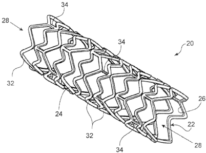
The FIGURE is a perspective view of an implementation of an expanded stent.
DETAILED - DESCI2I1'TION
Referring to FIG. 1, a stent 20 has the form of a tubular body 22 defining an
outer
wall surface 24 and an inner wall surface 26. '1'he iyuicr wall surface
defines a central
lumen 28. The stent tubular body member 22 is formed by a plurality of bands
32 and a
plurality of conncctors 34 that extend between and connect adjaccnt bands.
During use,
bands 32 are expanded from an initial, small outer diameter to a relatively
larger outer
diameter to contact the outer wall surface 24 of stent 20 against a
surrounding wall of a
vessel, thereby maintaining the patency of the vessel. Connectors 34
provide.stent 20
with flexibility anci conformability that allow the stent to adapt to the
contours of the
vessel.
Stent 20 caii include (e.g., be manufactured from) one or more biocompatible
materials with mechanical properties that allow a stent including a compositc
material to
be compacted, and subsequently expandedto support a vessel. In some
impleinentations,
.stent 20 can have an ultimatc tensile yield strength (YS) of about 20-150
ksi, greater than
about 15% elongation to failure, and.a modulusof.elasticity of about 10-60
msi. When
stent 20 is expanded, the material can be stretched to strains on the order of
about 0.3.
Examples of suitable materials for the tubular body of stent 20 include
stainless steel
(e.g., 316L, BioDurO 108 (UNS S29108), and 304L stainless steel, and an alloy
including stainless steel and 5=60 1o by weight of one or more radiopaque
eletnents (e.g.,
Pt, Ir, Au, W) (PERSS ) as described in US-2003-0018380-Al, US-2002-0144757-
AI,
and US-2003-0077200-A1), Nitinol (a nickel-titanium alloy); cobalt alloys
suclz as
Elgiloy,L.605 alloys,lVIP35N, titanium, titanium alloys (e.g., Ti-6Al-4V, Ti-
50Ta, Ti-
10Ir), platinum, platinum alloys, niobium,.niobium alloys (e.g., Nb-1Zr) Co-
28Cr-6Nlo,
tantalum, and tantalum alloys: Other examples of materials are described in
commonly
4
CA 02663439 2009-03-12
WO 2008/036504 PCT/US2007/077481
assignied U.S. Application No. 10/672;891, frled September 26, 2993;,and
entitled
"Medical Devices and Methods of Making Same;" and U.S. Application No.
11/035,316,
filed January 3, 2005, and entitled."Medical Devices and Methods of Making
Same."
Other Ynaterialsinclude elastic biocompatible metals such as a superelastic or
pseudo-
elastic,metal alloy, as.described, for cxample, in Schetsky, L. McDonald,
"Sliape
Memory Alloys," Encyclopedia of Chemical Technology (3rd ed;), John Wiley &
Sons,
1982, :vol. 20. pp. 726-736; and commonly assigned U.S. Application No.
10/346,487,
filed January 17, 2003.
In some implementations, the tubular metal body 22 forming stent 20 includes
oiie or more materials that enhancevisibility by MRI: Examples ofMRI materials
include
non-ferrous metals (e.g., copper, silver, platinum, or gold) and non-ferrous
metal-alloys
containing.paramagnetic elernents (e.g., dysprosium or gadolinium) such.as
terbium-
dysprosium, dysprosium, and gadolinium. Alternutively or additionally, the
metallic
matrix can include one or more materials having low magnetic susceptibility to
reduce
magnetic susceptibility artifacts, which during imaging can interfere with
imaging of
tissue, e.g., adjacent to and/or surrounding the.stent: Low magnetic
susceptibility
materials include those described above, such as,tantalum, platinum,
titanium,,niobium,
copper, and alloys containing these elements.
The bands 32 and connectors 34 defining the tubular metal body 22 of the stent
20
are formed, as shown, by cutting the tube. Selected portions of the tube can
be removed
to form bands 32 and connectors 34. by lascr cutting, as described in'Saunders
U.S. Patent
No. 5,780,807. In certain implementations, during laser cutting, a
liquid:carrier, such as a
solvent or an oil, may be flowed through the lumen of the tube. The carrier
can prevent
dross formed on one portion of the tube from re-depositing on another portion,
and/or
reduce formation of recast material on the tube. Other incthods of removing
portions of
the tube can be used, such as mechanical machining (e.g., micro-machining),
electrical.
discharge machining (EDM), andphotoetching (e.g., acidphotoetching).
As an example, while stent 20 is described above as.being.formed wholly of
composite rnaterial, in other implementations, the composite material :forms
one or.more
sclected portions of the medical device. For example, stent 20 can include
multiple layers
in which one or more layers include a composite material, and one or more
layers do not
5
CA 02663439 2009-03-12
WO 2008/036504 PCT/US2007/077481
include a composite material: The layer or layers including a composite
material can
include the same composite material or different composite materials. The
layer or layers
not ineluding a composite material may include one or more. of the
biocompatible matrix
matcrials: listed above. The layering of the composite material provides yet
another way
to tailor and tune the properties of the medical device. Stents including
multiple layers
are described, for exatnple, in U.S. Patent Publication No. 2004-0044397 and
in Heath
U.S. Patent No. 6,287;331.
In some implementations, after bands 32 and connectors 34 are formed, areas of
the tube affected by the cutting operation above: can be removed. For example,
laser
machining ofbands 32 and connectors 34 can leave a surface layer ofinelted and
resolidified material and//or oxidized metal that can adversely afTect the
mechanical
properries and perfonnance of stent 20. The affected areas can be re,~moved
mechanically
(such as by grit blasting or honing) and/or chemically (such as by etching or
electropolishing). In some implementations, the tubular member can be near net
size and
confguration at this stage. "Near-net size" means that the tube has a
relatively thin
envelope of material that is next removed to provide a semi=fiiiished stent,
c:g: for
receiving the porous, sintered metal coating to be bonded to the surface, as
discussed
below. In some implementtitions, the tube is formed less than about 25%
oversized, e.g.,
less than about 15%, 10%, or 5% oversized.
The unfinished stent is then finishedto form stent 20. Since the unfinished
stent
can be fornied .to near-net size, relatively little of the unfinishcxl stent
must be reinoved to
finish the stent. As a result, further processing (which could damage the
stent) and
discard. of costly materials can be.reduced. In some implementations, about
0.0001 inch
of the stent material can.be removed by chemical milling and/or
electropolishing to yield
a scmi-tinishedstent.
Stent 20 can be of a desired shape and size (e.g., coronary stents, aortic,
stents,
peripheral vascular stents, gastrointestinal stents, urology stents, and
neurology stents).
Dc;pending on the intended application, stent 20 can have an outer diameter of
between,
for example, about l mm to about 46 mm: In certain`implementations, a coronary
stent
can have an expanded outer diaineter of from about 2 mm to about 6 mm. In some
implementations, a peripheral stent can have an expanded outer diameter of
from about 5
6
CA 02663439 2009-03-12
WO 2008/036504 PCT/US2007/077481
mm to about 24 mm. In certain implementations, a gastrointestina7 and/or
urology stent;
can have an expanded outer diameter of from about 6 mm to about 30 mm. In some
implementations, a neurology stent can have an expanded outer diameter of from
about I
mnrto:about 12 mm. An abdominal aortic aneurysm (AAA) stent and a thoracic
aortic
aneurysm (TAA) stent can have. an outer diameter from about 20 mm to about 46
mm.
Stent 20 can be balloon-expandable, self-expandable, or a combination of both
(e.g.,
Andersen et a1. U.S. Patent No. 5,366,504).
Also, current,. conventional, block copolymer-based implantable stent drug
delivery technology utilizes a 16.5 mole% polystyrene, linear, triblock,
styrcnic polymer
system, commonly referred to as SIBS, as the.exeipient material. With current,
known
paclitaxel / SIBS stent coatings, the ezcipient material is soft, elastomeric,
and possesses
some inherent tack. These inherent properties of SIBS provide excellent
elastic recovery
and resistance to fatigue in stent regions of high strain but may result in
low occurrence
instances of resistance to balloon withdrawal after the: BE. stent is
deployed. Resistance to
withdrawal is being demonstrated to-be a key factor in DE stent delivery. The
very thin,
porous, sintered metal coating.of the outer w.all surface and.the iiuier wall
surface of the
stent 20 addresses these issues.
In one particular implementation, the improved stent 20 of the.rIGU1Z.E is
provided with a non-polymeric, very thin, porous sintered rrietalcoating, e:g.
with
thickness in the range of about 5 micron.to about 50.micron, bonded to one or
more
regions of the, outer wall surface and the inner wall surface of the stent to
provide a
porous reservoir, or media, for drug material. This.thin, porous, sintercd
metal material
can be manipulated in.terms of density, porosity, e.g. down.to 2 micron size,
or
tortuosity, to control drug clution rates and duration. In other
implementations, the stent
20, may be a seamless. stent produced entirely from sintered metal, sintered
mesh, woven
wire, etc.
Inparticular, the described implantable stent drug delivery system provides
improved frictional, hardness, tack and drug delive'ry properties for
lower.resistance to
balloon withdrawal and improved di'ffusion of drug, resulting in improved SDS
delivery
and complete drug release.
7
CA 02663439 2009-03-12
WO 2008/036504 PCT/US2007/077481
Porous. sintered metal powders, fibers, or wires are utilized in many
industries as
very high performance, complex, filter material of virtualty any shape with
near-exact
dimensional tolerances. Furnace sintering is an established metallurgical
method of
bonding every contact point of very small rnetal species to produce strong,
porous,
ductile.laminates or material objects with porosity 4own to 2 micron size.
The porous reservoir formed by the:sinter metal coating or body of the stent
20
preferably includes a releasable therapeutic agent, drug, or a
pharmaccutically active
compound, such as described in U.S. PatentNo. 5,674,242, U.S. Application No.
09/895,415, filed July 2, 2001, and U.S. Application No. 10/232,265, filed
August 30,
2002. The therapeutic agents, drugs, or pharmaceutically active compounds can
include,
for example, anti-thrombogenic agents, antioxidants, anti-inflammatory agents,
anesthetic
agents; anti-coabulants, and antibiotics.
In current, conventional SIBS-based sterit drug delivery technology employing
known paclitaxel / SIBS stent coatings, the drug exposed on the surface of the
excipient.
coating is guickly solubilized into the tissue during the initial stage of
drug release.:This
initial "spike" or "burst" of release constitutes a substantial portion of the
total
cumulative device drug release, while a.large portion of the total drug
c:ontent remains
within the coating for extended periods of time. The ability to control
release kinetics.and
to provide complete drug release may be linked to, late successful healing and
resistance
to thrombosis.
ln use, stent 20 can be employed, e.g., delivered and expanded, using a
catheter
delivery system. Catheter systems are described in, for example, Wang U.S.
Patent No.
5,195,969, Hamlin U.S. :Patent No. 5,270,086, and. Raeder-Devens U.S. Patent
No.
6,726,712. Stents and stent delivery are also exemplified by the Radius or
Symbiot
systems, available from Boston Scientific Scimed, Maple Grove, MN.
OTHER EMBODIMENTS
While a number of implementations have been describcd above, the invention is
not so limited. For example, in some implementations, stent 20 canbe formed by
fabricating a wire including.the coinposite material, and knitting and/or
weaving the wire
8
CA 02663439 2009-03-12
WO 2008/036504 PCT/US2007/077481
iaito a tubular member. The coinposite materials described hercin can also be
used:to
form other medical devices.
Other implementations are within the claims.
9