Note: Descriptions are shown in the official language in which they were submitted.
CA 02663535 2009-03-16
WO 2008/016685 PCT/US2007/017269
A SOFT TISSUE FIXATION DEVICE
BACKGROUND OF THE INVENTION
Field of the Invention. This invention relates in general to apparatus and
methods to assist in the fixation of soft tissue to bone, and more
particularly to human
anterior cruciate ligament (ACL) and canine cranial cruciate ligament (CrCL)
reconstruction grafts.
Prior Art. Rupture of the cranial cruciate ligament (CrCL) and subsequent
osteoarthritis is a leading cause of canine hind limb lameness. Numerous
techniques
have been described to stabilize the canine knee or stifle following CrCL
rupture to
inhibit or prevent osteoarthirits. One such technique is intra-articular
CrCL graft
reconstruction for stabilization of CrCL deficient stifles. Various devices
for initial
surgical graft fixation have been utilized. These include the EndoButton CL,
the Bone
Mulch Screw, the RigidFix, Interference Screws, the BioScrew, the RCI screw,
the
SmartScrew ACL, a Synthes 6.5 cancellous screw with a spiked plastic washer or
a soft
tissue fixation plate, as well as various type staples, including stone and
barbed staples.
In CrCL reconstruction it is necessary to obtain a desired graft tension and
then
to secure the tensioned graft at the desired position on the bone. There
remains in the
current surgical procedure problems with obtaining the necessary graft
fixation strength
quickly to prevent loss of the desired graft tension. A second problem relates
to the
damage of the graft as it is being tensioned over the rough or sharp surfaces
of the bone
or fixation device used. In addition the fixation device must have the ability
to maintain
the graft tension during the normal activity of the person or animal during
the recovery
period.
In the surgical application of these devices it is necessary that the graft to
be
1
CA 02663535 2009-03-16
WO 2008/016685 PCT/US2007/017269
affixed to the bone have the desired tension. The current devices are more
difficult to
employ during the surgical procedures than is desired. Therefore, there
remains a need
for a soft tissue fixation device that can be used with known arthroscopic or
open
surgical techniques, does not require unique application equipment, can be
used with
various CrCL reconstruction materials, and can affix the graft to the bone
quickly and
easily while still permitting the desired graft tensioning.
Still further there remains a need to provide a device that is simple in
construction and would allow the CrCL reconstruction graft to be tensioned and
secured
to the bone in a single step.
2
CA 02663535 2009-03-16
WO 2008/016685 PCT/US2007/017269
OBJECTS AND SUMMARY OF THE INVENTION
Therefore, one object of this invention is to provide a soft tissue fixation
device
that can be used with known arthroscopic or open surgical techniques.
Another object of this invention is to provide a soft tissue fixation device
that
does not require unique application equipment.
Still another object of this invention is to provide a soft tissue fixation
device
that can be used with various ACL or CrCL reconstruction materials, such as
from
material that will over time become absorbed.
Another object of this invention is to provide a soft tissue fixation device
that is
resistant to axial and rotational movement during the tensioning and securing
of the graft
to the fixation device.
A further object of this invention is to provide a soft tissue fixation device
that
permits easier and quicker attachment of the graft to the bone while
permitting the
desired graft tensioning.
Another object of this invention is to provide a soft tissue fixation device
that
when attached to the bone maintains a low profile to reduce impingement on
surrounding structure, as well as reduce the visibility of its presence under
the skin.
Another further object is to construct a soft tissue fixation device that is
simple
in construction, and can be manufactured from absorbable materials by
injection
molding.
A still further object of this invention is to provide improved CrCL
reconstruction procedures that permit the reconstruction graft to be tensioned
and
secured to the femur in one step without screws or staples.
Other objects and advantages of this invention shall become apparent from the
3
CA 02663535 2009-03-16
WO 2008/016685 PCT/US2007/017269
ensuing descriptions of the invention.
Accordingly, this invention in one embodiment comprises a soft tissue fixation
device for use in ACL or CrCL reconstruction comprising a base member having a
top
and bottom surface. The base member is provided with a passageway extending
from
the top surface through the bottom surface and sized to allow the soft tissue
to be
inserted into the passageway and extend out the opposite passageway end. The
bottom surface of the base member is shaped to be attachable to the bone. In a
preferred embodiment the top surface is shaped to present no sharp edges that
a graft
will contact during the surgical procedure. The fixation device also comprises
an
affixing member pivotally attached or otherwise attachable to the base member.
The
affixing member is constructed having a graft fixation section shaped to
secure a
tensioned graft between the base member and the affixing member.
In a preferred embodiment, the base member has a notched section in the top
surface extending from the passageway to a first perimeter section of the base
member.
The notched section is sized to accommodate at least a portion of the graft in
order for
the fixation device to present a lower profile when used. In one preferred
embodiment
surgical grade tissue glue is spread on the bottom surface of the base member
to secure
the base member to the bone when the glue has dried. In another preferred
embodiment
the base member is disc-shaped with its substantially flat bottom surface
having at least
one perpendicularly extending spike shaped to permit it to be driven into the
bone to
which the graft will be affixed. The spikes are shaped to hold the base member
rotationally and axially in position during and after the tensioning of the
graft. In
another preferred embodiment a sleeve whose interior wall surfaces form a part
of the
passageway extends perpendicularly from the base member bottom surface. The
sleeve
4
CA 02663535 2009-03-16
WO 2008/016685 PCT/US2007/017269
is sized to permit it to be inserted into the opening drilled into the bone to
provide, along
with the spikes, additional translational stability to the base member when
the graft is
being tensioned, as well as to prevent damage to the graft by the sharp edges
of the bone
tunnel.
In another preferred embodiment the affixing member is a clip having a
generally arched shaped and provided with a series of teeth members extending
downward from the lower surface of the clip member. The teeth members are
positioned so that when the clip member is attached to the base member the
teeth
members will extend across and into the notched section of the top surface of
the base
member. In a more preferred embodiment the teeth members will extend beyond
the
notched section to better secure any portion of the graft that may lap out
from the
notched section. In this preferred embodiment the opposite ends of the clip
member are
shaped to fit into aligned notches positioned along perimeter sections of the
bottom
surface of the base member for attaching the clip member to the base member.
This is
accomplished by placing one of the clip member ends into its base member
notch.
Then through the use of a single, fluid type motion the affixing member is
levered
downward to force the opposite end into its base member notch thus securing
the
affixing member to the base member. In this embodiment it is preferred that
the clip
member be constructed from flexible material, such as an acetal copolymer or
other
material having similar flexibility characteristics.
In an alternate embodiment an improved surgical procedure for cranial cruciate
ligament reconstruction is provided. In this embodiment the femur is prepared
for
receipt of the soft tissue fixation device in a conventional manner. This
includes
drilling a bone tunnel from the intra-articular origin of the CrCL to the
center of the
CA 02663535 2009-03-16
WO 2008/016685 PCT/US2007/017269
lateral aspect of the femoral condyle. The base member is aligned with the
bone tunnel
so that its sleeve is over the pilot hole. The base member is then tapped into
place with
an osteotomy mallet. If the base member is not provided with a sleeve, then
the base
member passageway is positioned over the pilot hole and secured in place with
the use
of surgical grade tissue glue. The graft is passed through the femoral tunnel
and the
sleeve. That portion of the graft extending from the sleeve is positioned
across the top
surface notch of the base member and pulled to achieve the desired tension.
The
pivoting end of the clip member is placed in one of the bottom surface
notches. The clip
member is then in one motion pivoted downward until the attaching end of the
clip
member is secured in the opposite bottom surface notch. This action causes the
teeth of
the clip member to grab the graft and secure the graft between the base member
and the
clip member sufficiently to maintain the desired tension.
6
CA 02663535 2009-03-16
WO 2008/016685 PCT/US2007/017269
BRIEF DESCRIPTION OF THE DRAWINGS
The accompanying drawings illustrate a preferred embodiment of this invention.
However, it is to be understood that these embodiments are not intended to be
exhaustive, nor limiting of the invention. They are but examples of some of
the forms in
which the invention may be practiced.
Figure 1 is three-quarter top perspective view of a preferred embodiment of
the
soft tissue fixation device of this invention illustrated having the pivoting
end of the clip
member in position to be pivoted by the surgeon into an attached or locked
position.
Figure 2 is a three-quarter bottom perspective view of the base member of the
soft tissue fixation device of Figure 1.
Figure 3 is a bottom view of the base member of the soft tissue fixation
device
of Figure 1.
Figure 4 is a three-quarter perspective view of the clip member forming the
soft
tissue fixation device of Figure 1.
Figure 5 is a cross-sectional view taken along Section Lines I-I of Figure 4.
Figure 6 is a photograph of soft tissue affixed. in position by a preferred
embodiment of the fixation device of this invention attached to a human femur.
Figure 7 is an exploded view of an alternate embodiment of the soft tissue
fixation device of this invention illustrating the affixing member attached to
the base
member by hinge means.
Figure 7A is a side view of the base member and affixing member in connecting
relationship of Figure 7.
Figure 7B is a cross-sectional view taken along Section Lines 11-11 of Figure
7A.
Figure 7C is a bottom view of Figure 7A.
7
CA 02663535 2013-10-21
Figure 8 is a three-quarter perspective view of another alternate embodiment
of
the soft tissue fixation device of this invention illustrating the affixing
member pivotally
attachable to the base member.
Figure 8A is a three-quarter perspective view of Figure 8 wherein the affixing
member is engaged with the base member 8.
Figure 8B is a top three-quarter perspective view of the base member of Figure
8
Figure 8C is a top view of the base member of Figure 8 without the affixing
member attached.
Figure 8D is side view of the base member of Figure 8 without the affixing
member attached.
Figure 8E is a bottom three-quarter perspective view of the affixing member of
Figure 8.
Figure 8F is across-sectional view taken along lines of Figure 8E.
Figure 9 is a top three-quarter perspective view of an alternate fixation
device of
this invention.
Figure 9A is a top three-quarter perspective view of the affixing member of
Figure 9 illustrated in a closed position.
Figure 9B is a top three-quarter perspective view of the affixing member of
Figure 9 illustrated in an open position.
Figure 9C is a side view of the affixing member of Figure 9B.
Figures 9D and 9E are top views of an alternate fixation deViee having
multiple
closing positions.
8
CA 02663535 2009-03-16
WO 2008/016685 PCT/US2007/017269
PREFERRED EMBODIMENTS OF THE INVENTION
Without any intent to limit the scope of this invention, reference is made to
the
figures in describing the preferred embodiments of the invention.
Although the
preferred embodiments of the invention will be described utilizing the
invention in
CrCL reconstruction, this in no way is meant to limit the invention to such
use, as it will
be appreciated it has use in ACL reconstruction and other human and animal
applications.
The device of this invention is particularly useful in ACL or CrCL
reconstruction to fix a soft tissue graft to the bone of a human or dog
suffering from an
ACL or CrCL tear. Referring now to Figure I, a preferred embodiment of the
soft
tissue fixation device 1 includes a base member 2 and a graft affixing member
3. The
primary function of base member 2 is to provide a stable platform to allow the
soft
tissue graft to be tensioned during the procedure to attach the graft to the
bone. More
particularly, base member 2 should be constructed to minimize the axial and
rotational
movement of the base member during the graft tensioning step. Base member 2
should
further be constructed to minimize potential tearing of the graft during the
tensioning
step. On the other hand the primary function of affixing member 3 is to affix
and
maintain the soft tissue graft in the desired tensioned position on the base
member 2.
That portion of base member 2 that will extend above the bone surface when
attached to the bone is preferably is constructed to have a low profile. In a
preferred
embodiment base member 2 will be constructed having top surface 4 with a
curved
perimeter top surface section 5 surrounding a flat top surface center section
6 and a
substantially flat bottom surface 7. The height of base member 2 must be such
to
permit the attachment of affixing member 3. Base member 2 may be secured in
the
9
CA 02663535 2009-03-16
WO 2008/016685 PCT/US2007/017269
desired position to the femur bone by the use of known surgical grade tissue
glue. In
another embodiment extending downward from bottom surface 7 is at least one
securing
member, such as spike 8. It is preferred that there be at least three spikes 8
equally
spaced about the perimeter edge of bottom surface 7 to provide greater
stability against
rotation and lift forces on base member 2 during the tensioning of the graft.
It is further
preferred that spikes 8 be shaped to be easily driven into the bone, hold base
member 2
in place during the tensioning of the graft, as well as minimize rotational
and axial
movement of base member 2 during the tensioning and securing of the graft to
the
fixation device 1. One preferred shape of spikes 8 is a tubular or solid
shaped spike
having a pyramidal shaped bottom section. Other shapes of spikes 8 include a
tubular
shaped spike with triangular cross-sectional shape bottom section. Spikes
having a star-
shaped tubular section can also be employed. If desired known surgical grade
tissue
glue can also be used in conjunction with a spiked base member 2.
The structural design of spikes 8 are preferably selected to provide ease of
attachment to the bone while providing the desired stability to the base
member 2 during
the graft tensioning process. If desired there could be multiple rows of
spikes 8. The
shape of spike 8 must permit their insertion into the femur bone and to resist
shearing
caused by rotational forces on base member 3. In addition the shape of spike 8
should
be resistant to upward forces that might cause base member 2 to become
detached from
the bone during the graft tensioning process. In one preferred embodiment
these
objectives are achieved utilizing spikes 8 having a substantially rectangular
base 9
attached to bottom surface 7 with side 10 of base 9 tracking a portion of the
perimeter of
the bottom surface 7. The opposite side 11 of the base is provided with an arc-
shaped
portion 12 that with side 10 culminates to form a shape edge 13. Other known
shapes
CA 02663535 2009-03-16
WO 2008/016685 PCT/US2007/017269
can be utilized that will provide the desired objectives.
In another preferred embodiment illustrated in Figure 2, a sleeve 14 will
extend
downward from the center section 15 of bottom surface 7. Sleeve 14 will have
an
outside diameter that permits its snug insertion into a channel drilled into
the femur
bone. Sleeve 14 will also form a passageway 16 that will provide protection to
the graft
being attached to the bone. It is preferred that all edges of sleeve 14 that
may be
contacted with the graft be rounded and smooth to prevent cutting or tearing
of the graft.
Passageway 16 extends along the vertical center axis of base member 2 and
sleeve 14.
Passageway 16 is shaped to permit the graft to pass through the passageway 16,
but
preferably has no sharp comers that might damage the graft that is held
against the
passageway wall 17. The top surface 4 is also provided with a notch 18 that
extends
from passageway 16 to the perimeter 19 of top surface 4. The notch 18 should
have a
width to accommodate at least a substantial portion of the graft. Preferably,
notch 18
will also have a depth to accommodate at least a substantial portion of the
graft to permit
a lower profile design of fixation device 1. It is also preferred that the
upper edge
sections 20 and 21 of the side walls 22 and 23, respectively, will be rounded
and smooth
so as to present no sharp edges that would injure the graft when the graft is
pressing
against the walls forming notch 18.
As illustrated in Figures 2 and 3, bottom surface 7 of base member 2 is
provided
with two aligned notches 24 and 25, respectively. Notches 24 and 25 are
constructed to
accommodate the attachment of affixing member 3, and more preferably the
positioning
of affixing member 3 over at least a portion of top surface notch 18.
As illustrated in Figures 4 and 5, affixing member 3 is constructed having
11
CA 02663535 2009-03-16
WO 2008/016685 PCT/US2007/017269
opposing curved end sections 26 and 27. End section 26 includes with a leg
member 28
having a flat upper surface 29 that can be positioned on the flat floor
surface 30 of notch
24 and of a length to prevent leg member 28 from slipping out of notch 24. The
second leg member 31 of end section 26 forms an acute angle "a" with leg
member 28.
In a more preferred embodiment angle "42" is less than 600. In a more
preferred
embodiment a rounded notch 32 is formed by cutting into the interior surfaces
29 and 33
where both leg members 28 and 31 are joined. This construction permits easier
flexing
of end section 26, yet provides sufficient strength that the end section 26
will not crack
when the two leg members 28 and 31 are pressed toward one another. Opposing
end 27
is similar constructed except that its leg member 34 is provided with a
rounded end 35
to permit easier attachment of affixing member 3.
Affixing member 3 has a middle section 36 provided with teeth 37, or .other
similar known grabbing elements, that will extend across and into notch 18
when
affixing member 3 is secured to base member 2 to hold the graft in the desired
tensioned
position. In a preferred embodiment teeth 37 will extend on either side of
notch 18 to
hold any portion of the graft that may overlap notch 18. In order to
facilitate clip closure
and/or minimize damage to the soft tissue graft, it is also preferred that the
length of the
teeth 37 be decreased as they are positioned away from the center teeth 37a.
In the surgical procedure utilizing fixation device 1, a tunnel is first
drilled from
the intra-articular origin of the CrCL to the center of the lateral aspect of
each femoral
condyle. For a mid-sized dog (approximately 70 lbs.) this canal will be
approximately
4.5 mm in diameter. Base member 2 is aligned with the bone tunnel so that its
sleeve 14
is over the pilot hole. Base member 2 is then tapped into place with an
osteotomy
mallet. The graft is passed through the femoral tunnel and sleeve 9. Pivoting
end 26 of
12
CA 02663535 2009-03-16
WO 2008/016685 PCT/US2007/017269
affixing member 3 is placed in bottom surface notch 24. That portion of the
graft
extending from the sleeve 14 is positioned across top surface notch 18 of base
member 2
and pulled to achieve the desired tensioning. Member 3 is then in one motion
pivoted
downward until latching end 27 of affixing member 3 is secured in opposite
bottom
surface notch 19. This action causes teeth 37 of affixing member 3 to grab the
graft and
secure the graft between base member 2 and affixing member 3 sufficiently to
maintain
the desired tensioning. Figure 6 is a photograph of fixation device 1
positioned on a
human femur bone 41 with a graft 42 secured to the bone 41 by a fixation
device 1
similar to that illustrated in Figures 1-5.
Figures 7-9 illustrate alternate embodiments of the combination of base member
2 and affixing member 3. In Figures 7, 7A, 78 and 7C, base member 38 is
similarly
constructed as base member 2, except there are no notches 24 and 25.
Affixing
member 39 is shaped to fit over base member 38 and is constructed of material
that will
permit member 39 to be flexed so that its lower perimeter edge 40 will expand
to fit into
a groove 38A formed along the lower perimeter area of interior surface 38B of
base
member 38 to provide a positive attachment between base member 38 and affixing
member 39. When spikes 8 are positioned at the perimeter of the base member
38, then
affixing member 39 will be provided with corresponding notches 45A to
facilitate
closure over spikes 8.
Teeth 43 extend downward from the bottom surface 44 of affixing member 39
for positioning in notch 45 shaped in the upper surface of base member 38.
Notch 44 is
similarly shaped as notch 18 illustrated in Figures 1-5. In a preferred
embodiment teeth
43 will also be shaped similarly as teeth 37 in Figures 1-5.
Figures 8, 8A-8F illustrates another embodiment wherein affixing member 45 is
13
CA 02663535 2009-03-16
WO 2008/016685 PCT/US2007/017269
pivotally attached to base member 46 by a conventional pivoting construction.
More
particularly, base member 46 is provided with parallel separated shoulder
members 47
and 48 with each having a connecting aligned passageway 49 and 50,
respectively, to
accept a pivot pin 51. End section 52 of affixing member 45 is shaped to pass
between
separated shoulder members 47 and 48. End section 52 is also provided with a
passageway 53 that can be aligned with passageways 49 and 50 to permit a pivot
pin 51
to extend through each of the passageways to permit affixing member 45 to
pivot about
pin 51. In a preferred embodiment two pads 54 and 55 extend from opposite
sides 56 -
and 57, respectively, of affixing member 45 to provide a larger surface for
the surgeon's
hand to contact and provide the force necessary to attach affixing member 45
to base
member 46. Shoulder members 47 and 48 are positioned so that the teeth
58 of
affixing member 54 will be positioned over notch 59 in the top surface 60 of
base
member 46 in similar fashion as described with respect to the Figures 1-5
embodiment.
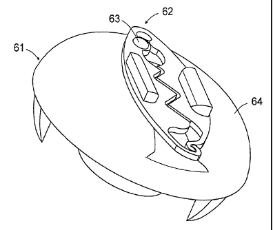
Figure 9 and Figures 9A-9C illustrate another embodiment for affixing member
62 to grasp and secure the graft to the bone. In this embodiment affixing
member 62 is
pivotally secured horizontally to base member 61 by a peg or screw 63
extending
upward from the top surface 64 of base member 61 and through opening 65.
Affixing
member 62 is formed by two leg sections 66 and 67 that are affixed at one of
their ends
in a manner to be biased to form a gap 68 between the facing serrated edge
surfaces 69
and 70 of leg sections 66 and 67, respectively. Each serrated edge surface 69
and 70 is
shaped having a series of teeth 71 and 72, respectively, that mate in
corresponding valley
areas 73 and 74, respectively when leg sections 66 and 67 are forced toward
one another
as illustrated in Figure 9A. When leg sections 66 and 67 are separated as
illustrated in
Figure 9B, the graft will be extended through gap 68. In a preferred
embodiment
14
CA 02663535 2013-10-21
grasping pads 75 and 76 are positioned on the top surfaces of leg sections 66
and 67,
respectively, to assist the surgeon in placing the leg sections 66 and 67 in a
closed
position to secure the graft at the desired tension. To lock leg sections 66
and 67 in a
closed position, opposite end 77 of leg section 66 is configured to form a
keeping
structure 78 for retaining opposite end 79 of leg section 67. Opposite end 79
is
configured to form a latch 80 that operatively fits into keeping structure 78
to reduce gap
68 sufficiently to permit teeth 71 and 72 to hold the graft at the desired
tension.
Figures 9D-9E illustrate an embodiment of Figure 9 wherein there are multiple
keeping structures 78A, 78B and 78C to permit leg section 67 to be secured to
latch 80.
This permits variation in the width of gap 68 between teeth 71 and valley 74
in order to
facilitate the use of grafts of different thickness. When leg section 67 is in
the position
shown as 67A, gap 68 will be reduced to ensure that a thinner graft will be
securely held ,
in the correct tension. There can of course be more than three different
keeping
structures 78 to permit different ratcheting positions for securing latch 79.
The scope of the claims should not be limited by the embodiments set out
herein but should be given the broadest interpretation consistent with the
description as
a whole.
=