Note: Descriptions are shown in the official language in which they were submitted.
CA 02663853 2009-03-17
WO 2008/048361 PCT/US2007/006776
TITLE
Venous Access Port Assembly With Radiopaque Indicia
FIELD OF THE INVENTION
[0001] This relates to the field of medical devices and more particularly to
venous access
ports for the infusion of fluids into the patient and/or withdrawal of fluids
from the patient.
BACKGROUND OF THE INVENTION
[0002] Venous access ports for the infusion and/or withdrawal of fluids from a
patient
are well-known, secured to the proximal end of an implanted catheter. These
ports are typically
used for drug infusion or for withdrawal of small amounts of blood, where
large flows of fluid
are not required. The ports are assemblies of a needle-impenetrable housing
with a discharge
port in fluid conununication with the catheter and the reservoir within the
port housing, and
provide a subcutaneous self-sealing septum that defines an access site for
multiple needle sticks
through the covering skin tissue of the patient, through the septum and into
the reservoir, without
the need to continuously search for new access sites. Examples of such ports
are disclosed, for
example, in U.S. Patents Nos. 4,704,103; 4,762,517; 4,778,452; 5,185,003;
5,213,574 and
5,637,102.
[0003] It is desired to provide a venous access port assembly that provides
for a
radiologist, radiology technologist, nurse and ultimately a medical
practitioner to be able to
discern an important property of the port assembly after the port assembly has
been implanted
into a patient.
CA 02663853 2009-03-17
WO 2008/048361 PCT/US2007/006776
BRIEF SUMMARY OF THE INVENTION
[0004] The present invention is related to a venous access port having a
housing and a
septum, providing an interior reservoir and a passageway extending from the
reservoir through a
stem of a discharge port to establish fluid communication with a proximal end
of a catheter
lumen to which the port assembly is secured prior to placement of the assembly
into a patient.
The port may optionally have more than one reservoir and associated septum.
The invention is
the application of radiopaque indicia onto a venous access port that is
discernible under X-ray
examination to provide information concerning the nature or key attribute of
the venous access
port, so that the practitioner, subsequent to the date of implantation
thereof, can determine that
nature or key attribute under X-ray examination. One such key attribute in
particular would be
for example that the venous access port is rated to be used for power
injection such as of contrast
fluid, wherein for example the letters "CT" (for "computed tomography", or
"contrast enhanced
computed tomography") would be provided that are of radiopaque material,
optionally
positioned within radiopaque circles. The attribute in this example is the
property of the port's
being adapted to withstand high pressures that are used for injection of
contrast fluid into a
patient, and the letters "CT" would be understood in medical practice to
indicate that the port is
suitable for the high pressure injection of contrast fluid. The radiopaque
indicia could for
example be applied in a mirror-image orientation on the bottom housing
surface, and would
appear on the X-ray as right-side up and easily readable by the radiologist,
technologist or
practitioner.
2
CA 02663853 2009-03-17
WO 2008/048361 PCT/US2007/006776
BRIEF DESCRIPTION OF THE DRAWINGS
[0005] The accompanying drawings, which are incorporated herein and constitute
part of
this specification, illustrate the presently preferred embodiments of the
invention, and, together
with the general description given above and the detailed description given
below, serve to
explain the features of the invention. In the drawings:
[0006] Fig. 1 is an isometric view of the venous access port of the present
invention;
[0007] Fig. 2 is a plan view of the port of Fig. 1;
[0008] Figs. 3 and 4 are cross-section views of the port of Figs. 1 and 2
taken along lines
3-3 and lines 4-4 of Fig. 1, respectively;
[0009] Fig. 5 is an isometric view of the needle-impenetrable housing base of
the
venous access port of Fig. 1;
[0010] Figs. 6 and 7 are transverse cross-sectional and longitudinal cross-
sectional views
of the housing base of Fig. 5;
[0011] Fig. 8 is an isometric view from below of the housing base of Figs. 6
and 7,
showing the radiopaque indicia applied on the housing base bottom surface; and
[0012] Figs. 9 and 10 are bottom and top views of the housing base of Fig. 8
having
radiopaque indicia thereon, with the top view being analogous to the X-ray
view of the venous
access port by the radiologist, and the indicia being shown in dashed lines in
Fig. 10.
DETAILED DESCRIPTION OF THE INVENTION
[0013] Certain terminology is used herein for convenience only and is not to
be taken as
a limitation on the present invention. The terms "distal" and "proximal"
refer, respectively, to
directions closer to and away from the insertion tip of a catheter in an
implantable catheter
3
CA 02663853 2009-03-17
WO 2008/048361 PCT/US2007/006776
assembly. The terminology includes the words specifically mentioned,
derivatives thereof and
words of similar import. The embodiments illustrated below are not intended to
be exhaustive or
to limit the invention to the precise form disclosed. These embodiments are
chosen and
described to best explain the principle of the invention and its application
and practical use and
to enable others skilled in the art to best utilize the invention.
[0014] Venous access port assembly 10 of Figs. 1 to 4 includes a housing 12
and a
septum 14, with a discharge port 16 extending from a distal end 18 of the port
assembly 10 to be
attached securely and sealingly to the proximal end of a catheter (not shown).
A passageway 20
extends from the interior reservoir 22 to the distal tip opening 24 of
discharge port 16. A recess
26 is seen to be provided along both sides of discharge port 16, facilitating
insertion of the
discharge port 16 into the catheter lumen and providing a clearance for a
locking sleeve or clamp
(not shown) utilized to compress the catheter lumen wall against the exterior
surface of the
discharge port 16 for assured sealed connection of the catheter with the port
assembly 10.
[0015] With reference now to Figures 3 to 7, the interior of the port assembly
10 is
shown to provide an interior reservoir 22. Housing 12 is shown to include a
housing base 28 of
needle-impenetrable material that includes a well 30 having a bottom floor 32
and side walls 34
that define the interior reservoir 22 beneath septum 14. Bottom floor 32 may
be convex or
elevated (not shown) toward the center of the reservoir, if desired. Housing
base 28 includes a
base flange 36 extending radially outwardly from the bottom of well 30, and
base flange 36
includes openings 38,40 that serve to enable suturing to the patient upon
placement of the venous
access port and the attached catheter into the patient.
[0016] As shown in Figures 3 and 4, a skirt 42 is overmolded about housing
base 28
4
CA 02663853 2009-03-17
WO 2008/048361 PCT/US2007/006776
and may be of silicone elastomer. It is seen that skirt 42 encapsulates the
outer surfaces of the
bottom wall 44 and the bottom portion of the side walls 46 of housing base 28,
and is shown to
fill in the suture holes 38,40; but since the material is silicone elastomer,
suturing is possible
since the suturing needle can easily be inserted through the material of skirt
42 and through the
suture holes, and thereafter the filled openings provide minimal opportunity
for ingrowth of
patient tissue into the openings.
[0017] Also seen in Figures 1 to 4 is cap 48, which secures to housing base 28
to in turn
secure septum 14 in position in the port assembly 10. Preferably, skirt 42 is
insert molded onto
base flange 36 of housing base 28 after cap 48 is secured to the upper portion
of housing base 28
to secure the septum in position. It is seen in Figures 4 and 7 that discharge
port 16 is integral
with housing base 28 as is preferable. Discharge port 16 is shown to have a
pair of annular
ridges 50 that facilitate with the mechanical connection of the catheter
proximal end with the
port assembly 10. Housing base 28 includes a septum seat 52 extending into the
top of well 30,
into which a flange of the septum will be seated, preferably under radially
inward compression.
Housing base 28 has a bottom outer surface 54.
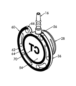
[00181 Radiopaque markings 60 of the present invention are shown in Figures 8
to 10. A
larger outer circle 62 is seen provided on the outermost periphery of bottom
base surface 54, and
a smaller inner circle 64 is seen provided within the area circumscribed by
the suture openings
38 and holes 40 through base flange 36. Adjacent to discharge port 16, a
recess 56 is provided in
the skirt of the housing base to provide a clearance for use of a connection
sleeve that will be
used to secure the catheter (not shown), and outer circle 62 is shown to have
a gap 66 at the
recess. Outer and inner circles or rings 62,64 circumscribe radiopaque indicia
70.
CA 02663853 2009-03-17
WO 2008/048361 PCT/US2007/006776
[0019] Radiopaque indicia 70 are provided on bottom outer surface 54 within
the region
directly beneath the reservoir and septum. In the example shown, indicia 70
comprise the letters
"CT" (Fig. 10) representing the tenn "computed tomography." The meaning of
this particular
example of indicia is that the venous access port assembly 10 is rated for
high pressure injection
such as is necessary for infusion into the patient of contrast medium that is
used in computed
tomography. Other indicia may of course be used that indicate some other
attribute or
characteristic of the venous access port assembly. The radiopaque markings and
indicia would
appear on an X-ray of the patient, and the indicia are provided in a mirror-
image orientation- on
the bottom outer surface of the housing base (Figs. 7 and 8) so that the
indicia would appear as
"CT" when the X-ray is viewed (Fig. 9), easily discerned by the radiologist or
technologist.
Centering of the indicia within the region (identified as "30,22" in Fig. 10)
directly beneath the
reservoir and septum minimizes any obscuring by the structure of the venous
access port
assembly, and the indicia may also be easily discemable should the port
assembly be at an angle
from the horizontal plane of the X-ray; the outer and inner circles 62,64
would appear oval or
elliptical should the port assembly be at such an angle. Gap 66 in outer
circle 62 would also
appear and would indicate the location of the discharge port stem 16.
[0020] The radiopaque markings may constitute marking fluid that is embossed
or
imprinted or otherwise applied onto the surface of the housing base 28, such
as black radiopaque
ink Part No. C11002 Rev A formulated by Creative Imprinting of Erie,
Pennsylvania, from
Marabu Tampapur TPU 910 clear with tungsten added, available from Marabuwerke
GmbH &
Co. KG of Stuttgart, Germany, and may be applied on plasma-treated surfaces.
At least the
housing base 28, the septum 14 and the skirt 42 are of radiotransparent or
radiolucent material as
6
CA 02663853 2009-03-17
WO 2008/048361 PCT/US2007/006776
is well known in implanted medical devices, and the housing base may be molded
of polysulfone
resin.
[0021] The radiopaque markings may altematively applied to the inwardly facing
surface
of the bottom wall of the housing base, or may constitute foil or film (such
as a decal) of
radiopaque material embedded within the housing base, these alternatives not
being shown in the
drawings.
7