Note: Descriptions are shown in the official language in which they were submitted.
CA 02664105 2009-03-20
WO 2008/036674 PCT/US2007/078784
Intraocular Lenses for Managing Glare, Adhesion, and Cell Migration
Background of the Invention
Field of the Invention
This invention relates generally to an intraocular lens and more specifically
to an
intraocular lens configured to reduce glare, improve adhesion to the eye,
and/or mitigate
unwanted cell migration such as posterior capsule pacification (PCO).
Description of the Related Art
The implantation of intraocular lenses represents one of the safest surgical
procedures currently conducted and enjoys an extremely high success rate. One
common
use of intraocular lenses is for the replacement of natural lenses that have
become clouded
due to the formation of cataracts. Intraocular lenses have also found other
uses, for example
in the form of anterior chamber lenses that are implanted just behind the
cornea in order to
restore vision to patients that are extremely myopic or hyperopic.
One set of problems that are frequently encountered in the use of intraocular
lenses is
that of glare and posterior capsule pacification (PCO). Glare problems can
occur due to
edge effects from the implanted optic, which is typically much smaller than
the natural lens
being replaced. For example peripheral light entering the eye can be
redirected by the edges
of the optic, or even haptic portions connected to the optic, back toward the
central portion
of the field of view to create annoying and even dangerous glare images that
are
superimposed with the normal image formed by the center of the optic.
PCO typically occurs as a result of cells (epithelial cells) that migrate from
the
equatorial regions of the capsular bag into the optic portion of the
intraocular lens. When
this occurs, the result can be a loss of vision that is similar to that caused
by the cataractous
material that precipitated the surgery in the first place.
Another problem that may occur when an intraocular lens is implanted into an
eye is
that of poor adhesion of the intraocular lens to the eye, for example, poor
adhesion to the
capsule walls of a capsular bag into which the intraocular lens is placed.
Good adhesion
between the intraocular lens and the capsular bag can, for example, help
maintain centration
of the lens about the optical axis. In addition, good adhesion about the
periphery of an optic
may, at least in part, be important for reducing migration of epithelial cells
toward the center
of the optic. Adhesion can be particularly important in accommodating
intraocular lenses,
CA 02664105 2014-02-26
since these types of lenses typically require that force from the ciliary
muscles and the
capsular bag be effectively transferred to the intraocular lens so that the
lens can translate or
deform when changing between accommodative and disaccommodative states.
Various methods and device designs have been used to handle the duo of
maladies
common to intraocular lens implants. Examples include those disclosed in U.S.
Patent
Numbers 6,162,249; 6,468,306; and 6,884,262, and U.S. Patent Application
Number
2005/033422.
In some cases a solution for one of these two problems may actually exacerbate
the
other. For example, sharp corner edges about the periphery have been found to
generally
reduce the problem of PCO; however, such discontinuities may also have the
unwanted
effect of increasing glare due to the scatter of entering the intraocular lens
from the
peripheral field of view.
Further improvements and design options are needed for reducing the problems
of
both glare and PCO in patients receiving intraocular lens implants, as well as
increase the
adhesion of intraocular lens implants to the capsular bag.
Summary of the Invention
The present invention is broadly directed to devices and methods that may be
used to
reduce the problems of glare and PCO common to intraocular lenses and/or other
ophthalmic
devices such as capsular rings. Embodiments of the present invention are also
generally
directed to structures that enhance the ability of an intraocular lens to
adhere or bond to the
eye, for example, to the capsule walls of a capsular bag. Using embodiments of
the current
invention, each of these problems may be addressed in such a way that the
solution to one of
these problems does not exacerbate or augment the other problem. For instance,
an
intraocular lens comprising an optic and a support structure coupled to the
optic may be
configured with one or more textured surfaces comprising a plurality of
periodically-spaced
protrusions, each protrusion having a smooth distal face and at least one
sharp corner edge
configured to engage a capsule wall of the capsular bag and/or at least one
cell disposed
along the capsule wall. In certain embodiments, the textured surface may be
configured to
reduce glare effects produced by light interacting with the peripheral edge of
an optic or a
portion of a haptic. For example, the dimensions and/or spacing of the
protrusions may be
selected to diverge or scatter incident light and/or to produce optical
interference.
In some embodiments, the texture surface comprises a plurality of channels or
grooves separated by a plurality of smooth ridges. In other embodiments, the
textured
-2-
CA 02664105 2009-03-20
WO 2008/036674 PCT/US2007/078784
surface comprises a plurality of pillars that are periodically disposed along
the surface in one
or two dimensions. In yet other embodiments, the textured surface comprises a
plurality of
rings that are concentrically disposed about an optical axis of the
intraocular lens. In some
embodiments, the textured surface comprises a contiguous smooth surface with a
plurality of
periodically-spaced wells disposed along the smooth surface, wherein a
plurality of sharp
corner edges are formed at a plurality of intersections between the smooth
surface and the
wells. The textured surface may be configured to control or maintain cells
(e.g., epithelial
cells) that come into contact with the textured surface in a favorable state.
A favorable cell
state of the cells may include a state in which the cells closely adhere to
the textured surface
or a state in which cell proliferation or propagation is mitigated by
maintaining the cell in a
form in which they are more contented and less likely to divide to produce
more cells (e.g.,
when the cells are in a more spindle-like form, and not in a more spherical
form). In
addition, the textured surface may be configured to provide adhesion directly
between the
capsular bag and the textured surface, even where no epithelial cells are
present. The
improved adhesion provided by the textured surface, either directly or
indirectly (e.g., via
epithelial cells remaining on the capsule walls), may provide enhanced
stabilization and
centration of the intraocular lens. In some embodiments, improved adhesion is
used to
enhance the so-called "shrink wrap" effect produced as the capsular walls
adhere to one
another in the vicinity of the intraocular lens. This improved adhesion and
the tendency of
cells in contact with the textured surface to not proliferate, either alone or
in combination,
advantageously permits the textured surface to be used to reduce the problem
of PCO. Also,
the improved adhesion provided by the textured surface may be of particular
importance in
accommodating intraocular lenses in which forces of the entire capsular bag
need to be
transmitted to the intraocular lens in an evenly distributed manner.
The textured surface may be disposed along any portion of the intraocular lens
where
attachment to the capsular bag or cell growth management is desired. The
textured surface
may be used in conjunction with mono-focal lenses, multi-focal lenses, or
accommodating
lenses, for example, to cause a structural element of the intraocular lens to
remain attached
to the capsular bag during accommodative movement thereof. In some
embodiments, a
cellular mono-layer is formed that is able to impede or prevent the migration
of cells beyond
the mono-layer.
In certain embodiments, the intraocular lens is alternatively or additionally
configured with a subsurface layer that is disposed within an interior region
of the
-3-
CA 02664105 2009-03-20
WO 2008/036674 PCT/US2007/078784
intraocular lens that is configured to reduce glare effects produced by
incident light. The
subsurface layer may be located, for example, within a periphery of the optic
between a top
surface and a bottom surface or inside a portion of a haptic that is attached
to the optic.
Preferably, the subsurface layer is configured to scatter light, for example,
to scatter an
amount of light that is at least twice the amount of light scattered by
material adjacent the
subsurface layer. In some embodiments, the subsurface layer is a subsurface
mark that may
be, for example, a symbol, one or more alphanumeric characters, or reticle.
Such a
subsurface mark may be used to show an orientation and/or position of the
intraocular lens,
to identify the intraocular lens, and/or to provide one or more
characteristics of the
intraocular lens (e.g., the focal length of the intraocular lens).
The subsurface layer may be produced using a plasma that is generated within
the
internal region of the intraocular lens and that forms a plurality of
localized micro-
discontinuities having refractive indices differing from the refractive index
of material
adjacent the subsurface layer. The plasma may be created, for example, by
using a laser to
create a laser-induced optical breakdown (LIOB) condition.
Since the subsurface layer is located inside the intraocular lens and is
isolated from
the outer surfaces of the intraocular lens, it may be specifically structured
to address glare
issues with no negative impact on cell migration. Conversely, the channels
discussed above
may be configured independent of their potential impact on glare, since a
subsurface layer
may be configured to scatter or redirect light impinging on the channels.
Thus, embodiments of the present invention may be used, in effect, to decouple
the
solutions to the problems of PCO and glare. In certain embodiments, only one
of the two
solutions discussed above need be incorporated, since the remaining problem in
such cases
either is not particularly critical or is solved using a different approach or
solution.
Additional aspects, features, and advantages of the present invention are set
forth in
the following description and claims, particularly when considered in
conjunction with the
accompanying drawings in which like parts may bear like reference numbers.
Brief Description of the Drawings
Embodiments of the present invention may be better understood from the
following
detailed description when read in conjunction with the accompanying drawings.
Such
embodiments, which are for illustrative purposes only, depict the novel and
non-obvious
aspects of the invention. The drawings include the following figures:
-4-
CA 02664105 2009-03-20
WO 2008/036674 PCT/US2007/078784
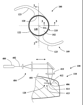
FIG. 1 is a top view of an intraocular lens according to an embodiment of the
present
invention illustrating an anterior side of an optic and a peripheral region
that includes a
subsurface layer disposed below a surface of the intraocular lens.
FIG. 2 is a cross-sectional side view of the intraocular lens illustrated in
FIG. 1
across a section 2-2.
FIG. 3 is a magnified side view of the intraocular lens illustrated in FIG. 1
across a
section 3-3.
FIG. 4 is a further magnified side view of the intraocular lens illustrated in
FIG. 3
illustrating the details of a structured surface for promoting capsular
adhesion, optical
control, and/or control of cellular growth.
FIG. 5 is a top view of an intraocular lens according to another embodiment of
the
invention.
FIG. 6 is a magnified side view of the intraocular lens illustrated in FIG. 5
across a
section 6-6.
FIG. 7 is a magnified side view of the intraocular lens illustrated in FIG. 5
across a
section 7-7.
FIG. 8 is a bottom view of the intraocular lens illustrated in FIG. 7.
FIG. 9 is a perspective view of an accommodating intraocular lens according to
an
embodiment of the present invention.
FIGS. 10a-e are side views of intraocular lenses illustrating various
embodiments of
a subsurface layer or layers for scattering incident light.
FIG. 11 is a side view of an intraocular lens showing a laser configured to
produce a
subsurface layer within the intraocular lens.
Detailed Description of the Drawings
Embodiments of the invention are generally directed to intraocular lenses for
implantation within the posterior chamber or capsular bag of an eye; however,
novel
embodiments of the invention may also be applied, where appropriate, to
intraocular lenses
in general (e.g., a phakic intraocular lens located in the anterior chamber or
a corneal implant
located within the cornea) or to other ophthalmic devices (e.g., contact
lenses or a capsular
ring).
Referring to FIGS. 1-4, an intraocular lens 100 according to an embodiment of
the
present invention is illustrated that advantageously addresses the dual
problems of unwanted
cell migration (e.g., PCO) and glare. The intraocular lens 100 comprises an
optic 102
-5-
CA 02664105 2009-03-20
WO 2008/036674 PCT/US2007/078784
disposed about an optical axis OA and has an anterior surface 104 and an
opposing posterior
surface 108. The surfaces 104, 108 are configured to focus light onto the
retina of an eye
into which the intraocular lens 100 is placed. The intraocular lens 100
further comprises a
support structure 109 and a periphery or peripheral region 110 disposed about
the optical
axis OA that includes a top surface 112, a bottom surface 114, and a
subsurface layer 120
disposed between the top surface and bottom surfaces 112, 114. As discussed in
greater
detail below, the subsurface layer 120 may be configured to advantageously
scatter or
otherwise redirect incident light so as to reduce glare on the retina of an
eye into which the
intraocular lens 100 is placed. The subsurface layer 120 may also be
configured for other
uses such as for marking the intraocular lens 100 for identification or
providing a
practitioner information regarding the orientation or position of the
intraocular lens 100.
The peripheral region 110 may also include an outer surface 122 that is
disposed
substantially parallel to the optical axis OA. The outer surface 122 may be
straight, arcuate,
or some combination thereof when viewed in cross-section in a plane congruent
with the
optical axis OA. In some embodiments, the outer surface 122 is also configured
to reduce
glare and/or PCO, for example, as disclosed in U.S. Patent Number 6,884,262.
In the illustrated embodiment, the support structure 109 comprises two haptics
123.
The hapties 123 may be used to center the intraocular lens 100 within the eye
of a subject
and are generally constructed to minimize damage to eye. In some embodiments,
the
support structure is more complex than that shown in the FIG. 1. In certain
embodiments,
the support structure includes a structure that is configured to fill or
substantially fill a
capsular bag and/or to provide accommodative action.
Referring to FIGS. 3-4, the intraocular lens 100 further comprises a textured
surface
128 disposed over a surface portion 129. The textured surface 128 may be
advantageously
configured to address the problems of cell migration and/or glare. For
example, the textured
surface 128 may be configured to maintain cells coming into contact with the
textured
surface 128 in a favorable state that prevents or reduces proliferation and/or
propagation of
cells beyond the boundary of the textured surface 128. Alternatively or
additionally, the
textured surface 128 may be advantageously configured to adhere to the walls
of a capsular
bag by adhering to the epithelial cells that remain on the capsule surface
after the natural lens
of the eye has been removed. In certain embodiments, the structured surface
128 is
configured to provide adhesion directly with the capsule wall, even where no
or few
epithelial cells are present. While the textured surface is located on the
periphery 110 of the
-6-
CA 02664105 2009-03-20
WO 2008/036674 PCT/US2007/078784
optic 102, it may be disposed on any surface of the intraocular lens 100,
including the optic
102.
The textured surface 128 comprises a plurality of periodically-spaced
protrusions
130, each protrusion 130 having a smooth distal face 132 and at least one
sharp corner edge
134 configured to engage a wall of the capsular bag (not illustrated) of a
subject and/or at
least one cell disposed along the capsule wall. The protrusions extend from
the surface
portion by an amount that is between about 0.1 micrometer and about 2
micrometers,
preferably between 0.3 micrometers and 1 micrometer, more preferably by about
0.5
micrometers,
In certain embodiments, the textured surface 128 is configured to reduce glare
effects
produced by light interacting with the optic 102, the periphery 110, and/or
the support
structure 109. For example, the dimensions and/or spacing of the protrusions
130 may be
selected to diverge or scatter incident light and/or to produce optical
interference. Also, in
some embodiments, while the smooth distal faces 132 are generally smooth, the
roughness
or structure of the surface portion 129 may be selected to be rough or
otherwise structured to
produce a predetermined characteristic, for example, to scatter or redirect
light incident
thereon so as to reduce glare.
The sharp corner edges 134 preferably have a radius that is less than about
200
nanometers, more preferably less than 100 nanometers, and even more preferably
less than
20 nanometers. The radius of the corners formed between the support structure
109 and the
protrusions 130 may be substantially equal to those of the sharp edge corners
134. However,
the radius of these corners may be greater than those of the sharp edge
corners 134 without
adverse affect, for example, in order to increase the manufacturability of the
structured
surface 128.
The smooth distal faces 132 generally have an RA surface roughness that is
less than
about 200 nanometers, preferably less than 50 nanometers, even more preferably
less than
about 20 nanometers. The roughness of the other surfaces of the textured
surface 128 (e.g.,
the surface portion 129) may be greater than that of the smooth distal faces
132.
In the illustrated embodiment, the plurality of protrusions 130 comprises a
plurality
of pillars and the smooth distal faces 132 are circular; however, other shapes
and
configurations of the protrusions 130 are possible (e.g., smooth distal faces
132 may be
rectangular, oval, or some other shape; the protrusions 130 may be configured
to form
concentric rings, as discussed below herein). Each protrusion 130 may further
comprise a
-7-
CA 02664105 2009-03-20
WO 2008/036674 PCT/US2007/078784
side wall 136, such that the sharp corner edge 134 is formed along an
intersection of the side
wall 136 and the smooth distal face 132. The sharp corner edges 134 are
generally
substantially perpendicular to the smooth distal face 132. The side walls 136
and the smooth
distal faces 132 form an angle that is generally between about 60 degrees and
about 120
degrees and is preferably about 90 degrees.
Each smooth distal face 132 has a width w and is disposed along the surface
portion
129 with a center-to-center spacing L between adjacent distal faces 132. The
width w is
generally between about 1 micrometer and about 10 micrometers, preferably
between 1
micrometer and 5 micrometers, and even more preferably between 1 micrometer
and 4
micrometers. The ratio of the width w to the center-to-center spacing L is
generally between
about 0.4 and about 0.7, with a ratio of about 0.5 being preferable in certain
embodiments.
In some embodiments, for example, when the center-to-center spacing is
relatively large, the
ratio of the width w to the center-to-center spacing L may be as great as 0.8
or more.
In some embodiments, the textured surface 128 comprises an essentially inverse
pattern to that illustrated in FIG. 4. That is to say, the textured surface
128 may comprise a
contiguous smooth surface with a plurality of periodically-spaced wells or
voids disposed
along the smooth surface in one or more directions. In such embodiments, a
plurality of
sharp corner edges are formed at the intersections between the smooth surface
and the wells.
The textured surface 128 may be disposed at various locations upon an
intraocular
lens according to embodiments of the present invention. For example, referring
to FIGS. 5-
8, an intraocular lens 200 comprises an optic 202, a pair of haptics 223, and
a periphery or
peripheral region 210. The intraocular lens 200 further comprises a textured
surface 228 that
may be disposed both within the peripheral region 210 and along at least a
portion of the
haptics 223 adjacent the peripheral region 210. The textured surface 228 may
run
contiguously between the peripheral region 210 and haptics 223, as illustrated
in FIG. 8.
Alternatively or additionally, one or more textured surfaces 228' (not shown)
may be formed
on one or more portions of the haptics 223 that are separate from the textured
surface 228
formed within the peripheral region 210. In some embodiments, the textured
surface 228' is
formed on the haptics 223 and there is no textured surface formed within the
peripheral
region 210.
The intraocular lens 200 further comprises a textured surface 228a disposed on
outer
surface 222 of the periphery 210 and a textured surface 228b disposed on an
anterior surface
212 of the optic 202. The additional textured surfaces 228a, 228b may be used
to further
-8-
CA 02664105 2009-03-20
WO 2008/036674 PCT/US2007/078784
provide adhesion between the capsular bag and the intraocular lens 200, for
example, by
causing the anterior capsule to adhere to the anterior surface of the
peripheral region 210.
The textured surfaces 228, 228a, and/or 228b may be separated from one another
(as
illustrated in FIG. 6) or be contiguous with one another to form a single
textured surface.
One or more of the textured surfaces 228, 228a, 228b may form an annular ring
that
completely surrounds the center of the optic 202. Alternatively, one or more
of the textured
surfaces 228, 228a, 228b form an annular ring that is broken at predetermined
locations.
Referring to FIGS. 7-8, textured surface 228 comprises a plurality of equally-
spaced
channels or grooves 240 separated by a plurality of smooth ridges 242. The
smooth ridges
242 are generally smooth so as to maintain cells in a favorable state, reduce
glare, and/or to
provide adhesion between the intraocular lens 200 and a capsular bag. The
textured surface
228 may be used alone or in combination with a subsurface layer such as the
subsurface
layer 120 in order to reduce or eliminate both PCO on the optic 202 and the
formation of
glare patterns on the retina of the eye due to light entering the eye from
peripheral fields of
view.
In some embodiments, the textured surface 228 completely surrounds the central
portion 248 of the optic 202. In such embodiments, the textured surface 228
may form a
mono-layer of cells that may act as a barrier that is effective in impeding or
completely
preventing the migration of epithelial cells inside the optic 202 when the
intraocular lens 200
is implanted into the eye of a subject. Alternatively, the channels 240 may be
configured
radially or with some orientation or pattern, while the overall shape of the
textured surface
228 is disposed circumferentially about the optic 202.
In the illustrated embodiment shown in FIGS. 7-8, the textured surface 228 is
circumferentially disposed about the optic 202 and has an over all radial
length L2 that is
greater than about 100 micrometers and less than about 1 millimeter. In some
embodiments,
the radial length L2 is less than 100 micrometer or greater than 1 millimeter.
For example,
the radial length L2 may be greater than 1 millimeter, so as to increase
adhesion or prevent
propagation of cellular growth onto the posterior surface 208 of the optic
202. In the
illustrated embodiment, the textured surface 228 is disposed entirely and
continuously about
the optic 202 on a surface portion 229 that follows the general form or
contour of the
intraocular lens 200 in the vicinity of the textured surface 228. The surface
portion 229 may
be flat, curved, or arcuate in shape.
-9-
CA 02664105 2014-02-26
The channels 240 have depth D, a width Wc, and may be disposed periodically
with
a period P. The depth D of the channels 240 is generally less than about 2
micrometer, in
some instances preferably less than or equal to about 0.5 micrometer. The
width Wc of the
channels 240 and a width Wit of the smooth ridges 242 is generally between
about 1
micrometer and about 10 micrometers, preferably between 1 micrometer and 5
micrometers,
and even more preferably between 1 micrometer and 4 micrometers. The ratio of
the width
WR of the smooth ridges 242 to the period spacing L is generally between about
0.4 and
about 0.7, with a ratio of about 0.5 being preferable in certain embodiments.
In some
embodiments, for example, when the center-to-center spacing is relatively
large, ratio of the
width w to the center-to-center spacing L may be as great as 0.8 or more.
The smooth ridges 242 generally have an RA surface roughness that is less than
about 200 nanometers, preferably less than 50 nanometers, even more preferably
less than
about 20 nanometers. The roughness of the other surfaces of the textured
surface 228 may
be greater than that of the smooth ridges 242.
The walls of the channels 240 preferably intersect the smooth ridges to form
sharp
edge corners 234. The sharp corner edges 234 preferably have a radius that is
less than
about 200 nanometers, more preferably less than 100 nanometers, and even more
preferably
less than 20 nanometers. The radius of the corners formed between the at the
bottom of the
channels 240 may be substantially equal to those of the sharp edge corners
234; however, the
radius of these comers may be greater than those of the sharp edge corners 134
without
adverse affect, for example, in order to increase the manufacturability of the
structured
surface 228.
It will be appreciated that the geometry and dimensions discussed in relation
to any
one of the textured surfaces 128, 228, 228a, or 228b may, where appropriate,
also be applied
to any one of the other textured surfaces 128, 228, 228a, or 228b, or any
other embodiment
of a textured surface according to the present invention.
Textured surfaces according to embodiments of the present invention may be
used in
accommodating intraocular lenses, for example, to provide adhesion between the
support
structure or positioning member of an intraocular lens and the walls of the
capsular bag.
Such accommodating intraocular lenses are disclosed, for example, in U.S.
Patent Numbers
6,488,708, 6,494,911, or 6,761,737, and in U.S. Patent Application Publication
Numbers
2004/0082994 and 2004/0111153. In an
exemplary embodiment illustrated in FIG. 9, a bag filling accommodating
intraocular lens
-10-
CA 02664105 2014-02-26
300 comprises a flexible positioning member 301 coupled to an optic 302. The
flexible
positioning member 301 has an outer surface 304 configured to engage the
capsular bag so
as to produce accommodation in response to an ocular force. As used herein,
the term
"ocular force" means any force produced by the eye of a subject that stresses,
moves, or
changes the shape of an optic or intraocular lens that is placed in the eye of
a subject. The
ocular force acting on a lens may be produced, for example, by the state or
configuration of
the ciliary body (e.g., contracted or retracted), changes in the shape of the
capsular bag of the
eye, stretching or contraction of one or more zonules, vitreous pressure
changes, and/or
movement of some part of the eye such as the ciliary body, zonules, or
capsular bag, either
alone or in combination.
The textured surface 328 may be disposed over substantially the entire outer
surface
304, as illustrated in FIG. 9. Alternatively, the textured surface may be
applied only over
predetermined portions of the outer surface 304, such that portions of the
outer surface 304
are able to slide against the capsular bag as it changes between accommodative
and
disaccommodative states. For example the textured surface 328 may be
selectively disposed
along an equatorial region 306.
The textured surface 328 is generally configured to produce adhesion between
the
capsular bag and the positioning member 301 so that ocular forces produced by
the eye (e.g.,
by the capsular bag) may be effectively transferred to the positioning member
301 in such a
way that optic 302 is translated and/or deformed to produce a predetermined
amount of
change in optical power. It will be appreciated that sufficient adhesion to
the capsular is
generally important for enabling and controlling both the amount of
accommodation and the
quality of resultant image produced as the optic 302 changes between
accommodative and
disaceommodative states.
A textured surface according to the present invention may also be applied to
at least
portions of the surface of an intraocular lens having essentially no haptics
or positioning
member. For example, as will be appreciated by one of ordinary skill in the
art, the textured
surface may be applied to at least a portion of an outer surface of a flexible
bag or bladder of
an intraocular lens, wherein the bladder is filled with a resilient fill
material. An example of
such an intraocular lens is illustrated in FIG. 14 of U.S. Patent Application
Publication
Number 2004/0082993. The textured surface
may be applied to specific portions of the outer surface, for example, about
an equatorial
portion of the flexible bag. Alternatively, the textured surface may be
applied over large
-11-
CA 02664105 2009-03-20
WO 2008/036674 PCT/US2007/078784
portions of the flexible bag, for example, over all areas of the outer surface
of the flexible
bag that are to contact the walls of a capsular bag into which the intraocular
lens is to be
placed. In any event, the textured surface generally covers a sufficient
portion of the flexible
bag to permit the intraocular lens to deform in conformance with deformations
of the
capsular bag as it changes between accommodative and disaccommodative states.
The textured surfaces 128, 228, 228a, 228b may be produced using one or more
of a
variety of known fabrication methods. For simplicity, fabrication methods
discussed herein
are with reference to the textured surface 128; however, it will be
appreciated that such
methods may also be applied in the formation of the textured surfaces 228,
228a, 228b, 328,
or other textured surfaces according to embodiments of the present invention.
In some
embodiments, the textured surface 128 is produced by chemically etching the
periodically-
spaced protrusions 130 along the surface portion 129. In such embodiments, a
mask may be
disposed over the surface potion 129 to provide a plurality of exposed areas
thereon. One or
more chemicals may be subsequently used to etch material from the exposed
areas. In other
embodiments, a protective film is disposed upon the mask and exposed areas of
the surface
portion 129. The mask may then be removed and a subsequent chemical treatment
used to
from the textured surface 128 by etching material from portions of the surface
portion 129
not protected by the protective film. In yet other embodiments, a laser
similar to that used in
forming the subsurface layer 120 is used to etch or form the textured surface
128.
Alternatively or in addition to etching material to from the surface portion
129,
material may be deposited onto the surface portion 129 in forming the textured
surface 128.
For example, the protrusions 130 illustrated in FIG. 4 may be formed by
applying one or
more layers onto the surface portion 129 (e.g., using a chemical vapor
deposition process).
In some embodiments, the textured surface 128 is formed by an embossing
process or by
machining the desired features from the surface portion 129, for example, by
using a CNC
lathe with milling capabilities. In other embodiments, the textured surface
128 is formed by
molding or by a combination of machining and molding.
When an intraocular lens according to embodiments of the present invention has
both a textured surface 128 and one or more subsurface layers 120, the
textured surface 128
may be formed either before or after formation of the subsurface layer 120. In
some
embodiments, the textured surface 128 is disposed directly above or below the
subsurface
layer 120, for example within the peripheral region 110 surrounding the optic
102.
-12-
CA 02664105 2009-03-20
WO 2008/036674 PCT/US2007/078784
Referring again to FIGS. 1-3, the subsurface layer 120 may be used to reduce
glare
potentially caused by light that might otherwise be reflected by the periphery
110 and
redirected toward the central field of view of the eye. As illustrated in FIG.
3, the subsurface
layer 120 is configured to produce diffuse or scattered light 134 when
illuminated by a beam
of light 146. In general, the amount of scattered light 134 may be
characterized by a
scattering cross-section that indicates the amount of light from an incident
beam that is
scattered by the subsurface layer 120. As shown in the illustrated embodiment,
the
subsurface layer 120 may be circumferentially disposed entirely about a
central portion 148
of the optic 102. In some embodiments, the periphery 110 comprises a single
material that,
apart from the subsurface layer 120, is homogeneous throughout. Alternatively,
the
subsurface layer 120 may form a separation between two different materials
that form the
periphery 110.
In some embodiments, the subsurface layer 120 is configured to scatter an
amount of
light that is at least twice the amount of light scattered by portions of the
material adjacent
the subsurface layer 120, more preferably at least 4 times the amount of light
scattered by
portions of the material adjacent the subsurface layer 120, and even more
preferably 10
times the amount of light scattered by portions of the material adjacent the
subsurface layer
120. In other embodiments, the subsurface layer 120 is configured to scatter
an amount of
light that is at least twice the amount of light scattered by an intraocular
lens that does not
have a subsurface layer such as the subsurface layer 120, but which is
otherwise substantially
equivalent to the intraocular lens 100. In yet other embodiments, the
subsurface layer 120 is
configured to scatter an amount of light that is at least 4 times, more
preferably 10 times the
amount of light scattered by an intraocular lens that does not have a
subsurface layer such as
the subsurface layer 120, but which is otherwise substantially equivalent to
the intraocular
lens 100. In some embodiments, the amount of light scattered by the subsurface
layer 120 is
determined by illuminating at least a portion of the subsurface layer 120 with
a beam of
light, such as a laser beam, and measuring the amount of light received by a
photodetector
having a predetermined area and disposed, for example, 10 centimeter to 1
meter or more
from the intraocular lens 100. The amount of light received by the
photodetector may then
be compared to the amount of light received by the photodetector under a
reference
condition, for example, by removing the intraocular lens 100 or replacing the
intraocular lens
100 by an intraocular lens that does not have a subsurface layer, but which is
otherwise
substantially equivalent to the intraocular lens 100.
-13-
CA 02664105 2009-03-20
WO 2008/036674 PCT/US2007/078784
As illustrated in FIG. 1, the subsurface layer 120 may form a contiguous strip
that
completely surrounds the central portion 148 of the optic 102. This
configuration of the
subsurface layer 120 advantageously scatters light intercepting the periphery
region 110 of
the intraocular lens 100. Alternatively, the subsurface layer 120 may be
circumferentially
broken along one or more regions.
In FIG. 3, the subsurface layer 120 is disposed within a plane that is
orthogonal to
the optical axis OA and has a radial width L 1 in a direction away from or
perpendicular to
the optical axis OA. In some embodiments, the radial width Li of the
subsurface layer 120
may be clearly delineated by distinct inner and outer edges. In other
embodiments, the radial
width Li may be estimated if inner and/or outer edges are less distinct, for
example, if the
subsurface layer 120 has a scattering cross-section that is a Gaussian in a
radial direction.
The thickness of the subsurface layer 120 in a direction along the optical
axis OA may be
relatively thin, as shown in FIG. 3, or may be thicker in order to increase
the scattering
cross-section of the subsurface layer 120. Generally, the radial width Li is
greater than
about four times the thickness. In certain embodiments, the radial width is at
least 100
micrometers, while in other embodiments, the radial width Li is at least 200
micrometers,
500 micrometers, or 1 millimeter or more.
The subsurface layer 120 may be disposed at or near the top surface 112 of the
peripheral region 110, as illustrated in FIG. 3. Alternatively, the subsurface
layer may be
disposed at other depths beneath the top surface 112, for example, at or near
the bottom
surface 114 or approximately equidistant between the surfaces 112, 114. The
location will
generally be predicated on such factors as ease of fabrication or scattering
characteristics as a
function of layer depth.
Other configurations and distributions of the subsurface layer 120 besides
that
illustrated in FIG. 3 are possible. For example, in FIG. 10a, an intraocular
lens 100a
comprises a peripheral region 110a having a subsurface layer 120a that forms a
conic section
in which the subsurface layer 120a is oriented at an angle relative to the
optical axis OA.
The angle 0 may be selected to provide a particular light scattering
characteristic (e.g.,
scattering cross-section or angular distribution of the light scattered) that
reduces the amount
of glare produced by peripheral light. Referring to FIG. 10b, an intraocular
lens 100b
comprises a peripheral region 110b having a subsurface layer 120b that is
disposed to form a
cylindrical surface that is oriented parallel to an optical axis or an outer
surface 122b.
Referring to FIG. 10e, an intraocular lens 100c comprises a peripheral region
110c having a
-14.
CA 02664105 2009-03-20
WO 2008/036674 PCT/US2007/078784
subsurface layer 120c that is disposed to form an arcuate shape when viewed in
cross-section
in a plane congruent with the optical axis OA.
Referring to FIG. 10d, in certain embodiments, an intraocular lens 100d
comprises a
peripheral region 110d having at least two subsurface layers 120d and 120d'
configured to
provide a predetermined scattering characteristic, for example, causing light
entering the
peripheral region 110d to be multiply scattered. In the illustrated
embodiment, the
subsurface layer 120d' is parallel to an optical axis of the intraocular lens
100d, while the
subsurface layer 120d is perpendicular to the optical axis. In such
embodiments, at least
some of the light directed toward an outer surface 122d of the peripheral edge
110d is
reflected and scattered by the subsurface layer 120d. At least some of the
reflected light is
subsequently diffusely scattered by the subsurface layer 120d'. Referring to
FIG. 10e, an
intraocular lens 100e comprises at least two subsurface layers 120e, 120e'
that are disposed
parallel to one another so that at least some of the light entering the
peripheral region 110 is
twice scattered, first by the subsurface 120d and then by the subsurface
120d'. Additional
subsurface layers may be used may be used to further increase the amount of
light scattered
and/or to increase the scattering cross-section for light entering the
peripheral region at one
or more specific angles or ranges of angles. For example, the subsurface
layers 120d, 120d'
or 120e, 120e' may be configured to scatter at least twice the amount of light
that would be
scattered by the surface 120d or 120e alone if illuminated by a beam of light.
In some
embodiments, two or more subsurface layers are configured at one or more
angles relative to
an optical axis (similar to the subsurface layer 120a in FIG. 10a) or have a
arcuate or more
complex shape (similar to the subsurface layer 120c in FIG. 10c).
The subsurface layer 120 may comprise a variety of characteristics and
mechanisms
for scattering light in a predetermined manner. In some embodiments, the
subsurface layer
120 comprises a variation in refractive index of the material within the
layer. The refractive
index variations may be random or pseudo-random in nature or may be more
systematically
structured to scatter light in one or more preferred directions or with a
predetermined angular
distribution. The subsurface layer 120 may be configured so that the
refractive index
variations are along one axis or along multiple axes, for example, in one or
two directions
along the subsurface layer 120 and/or in a direction normal to the subsurface
layer 120. The
variation in refractive index in one or more directions may be continuous
and/or
characterized by localized micro-discontinuities. For example, the refractive
index variation
in one or more directions may be in the form of a plurality of small voids,
opaque particles
-15-
CA 02664105 2014-02-26
or spots, and/or localized material changes in the intraocular lens material.
In. general, the
size of such discontinuities is preferably on the order of a wavelength of
visible light, for
example, about 2 micrometers or less, about 1 micrometer or less, or about 500
nanometers
or less.
In some embodiments, the subsurface layer 120 may be configured for
alternative or
additional purposes beside the purpose of preventing or reducing glare on the
retina. For
example, the subsurface layer 120 may be formed to produce one or more shapes
that may be
used to identify the intraocular lens 100. In such embodiments, the subsurface
layer 120
may be configured to form of one or more alphanumeric characters, symbols, or
geometric
shapes such as squares, rectangles, triangles, circles, or ellipses.
Alternatively or
additionally, one or more subsurface layers may be configured to assist a
practitioner to
orient the intraocular lens 100 prior to and/or after placement within the eye
of a subject.
One example of such features to orient an intraocular lens is found in US
Patent Application
Number 2005/149184.
Referring to FIG. 11, in certain embodiments, a method of producing the
subsurface
layer 120 comprises providing a laser 400 and using the laser to form a plasma
within an
interior portion 401 of the intraocular lens 100, for example, within the
peripheral region
110. The laser 400 may be any laser providing a beam that can be sufficiently
focused to
produce a plasma, for example, a near infrared (NIR), ultra-short pulse laser
such as the
experimental system described by Leander Zickler, et al. in "Femtosecond All-
Solid State
Laser for Refractive Surgery" (Commercial and Biomedical Applications of
Ultrafast Lasers
III, Proceedings of SHE, Vol. 4978 (2003)).
Alternatively, the laser 400 may comprise a commercial system such as the
Coherent RegA
9000/9050 Ti:Sapphire regenerative amplifier available from Coherent Inc.
(Santa Clara,
CA, USA) or the IMRA FCPA microjoule D-1000 Ytterbium fiber
oscillator/amplifier laser
system available from IMRA America Inc. (Ann Arbor, MI, USA) or high-
repetition rate,
cavity dumped, mode-locked ultrafast laser systems such as femtoNOVA available
from
High Q Laser Production GmbH (Hohenems, Austria). In certain embodiments, the
laser
400 is able to produce a pulse sequence of pulses having pulse widths of 10 to
100,000
femtoseconds, minimum pulse energies of 0.1 nJ to 100 micojoules, temporal
pulse
separations of 10 ns to 100 microseconds, at a laser wavelength of 200 urn to
2 microns.
The use of lasers for this type of material processing are described in
greater detail in, for
example, U.S. Patent No. RE 37,585.
-16-
CA 02664105 2009-03-20
WO 2008/036674 PCT/US2007/078784
The laser 400 may be used to produce a beam 402 of light that is expanded
using
expansion optics 404. Light from the beam 402 is directed to at least one
focus 406 within
the interior portion 401 using a lens 410. The focus 406 preferably has a spot
size ranging
from about 1 to about 100 microns. Alternatively, the single lens 410 may be
replaced by
some other optical element or optical system suitable for focusing laser light
such as a
mirror, a diffractive optical element, or some combination of lenses, mirrors,
and/or
diffractive optical elements that form a focus or a plurality of foci.
Preferably, the optical
systems used to create the focus 406 that creates a high energy density within
a relatively
small volume, for example, by configuring the optical system to have a high
numerical
aperture (NA). In certain embodiments, the NA is between about 0.25 and about
1.2,
preferably greater than 0.5 or greater than 0.8, even more preferably greater
than or equal to
about 1.
The laser light contained in the focus 406 provides an energy or power density
that is
sufficient to produce a plasma within the interior portion 401. An exemplary
laser system
for producing such a plasma is discussed by Leander Zickler in the Proceedings
of SPIE,
Vol. 4978 (2003) publication cited above herein. Generally, the subsurface
layer 120 is
formed as a condition of laser-induced optical breakdown occurs within the
material inside
the interior portion 401. As the laser 400 is a pulsed, the laser pulses
create a plurality 413
of localized micro-discontinuities 414, each of the micro-discontinuities 414
having an
overall or average refractive index or effective refractive index that is
different from that of
the surrounding material.
In some embodiments, each of the micro-discontinuities 414 is in the form of a
small
volume in which the refractive index is substantially constant, but is
different from the
refractive index of material adjacent the subsurface layer 120. In other
embodiments, the
refractive index within a micro-discontinuity 414 varies, for example, having
a higher
refractive index in the center and a refractive index at a periphery that
approaches or is
substantially equal to the refractive index of adjacent material. In yet other
embodiments,
the micro-discontinuities 414 comprise small cavities or voids that forms
within the interior
portion 401 of the intraoeular lens 100.
In general, the localized difference in refractive index or effective
refractive index of
the micro-discontinuities 414 causes light incident to refract in a different
direction or
directions. The combined effect of the plurality 413 of micro-discontinuities
414 is that at
least some of the light incident upon the subsurface layer 120 is scattered in
a different
-17-
CA 02664105 2009-03-20
WO 2008/036674 PCT/US2007/078784
direction. In some embodiments, the subsurface layer 120 is configured to
produce a
random or quasi-random scattering distribution of incident light by randomly
or quasi-
randomly varying one or more properties of different micro-discontinuities
414. The
variation in property may include, but not be limited to, the size of the
micro-discontinuities
414, the refractive index of the micro-discontinuities 414, and the spacing
between adjacent
micro-discontinuities 414. In addition, the plurality 413 of micro-
discontinuities 414 can be
distributed at varying depths within the interior portion 401 to produce
multiple scattering of
light incident upon the subsurface layer 120. In some embodiments, the
absorption or
transmissivity of the micro-discontinuities 414 may also be varied compared to
the
surrounding material or compared to one another.
The method of producing the subsurface layer 120 further comprises moving the
focus 406 within the interior portion 401 so as to from an extended area with
a
predetermined extent and scattering cross-section. The extent, shape, number
of the
subsurface layer(s) 120 formed by the focus 406 may be any of those
illustrated and
discussed herein, such as those illustrated in FIGS. 1, 3, and 10a-e, or any
other form suited
to provide a desired scattering characteristic or cross-section.
The subsurface layer 120 may be formed by moving the focus 406 and/or
intraocular
lens 100 relative to one another by using, for example, a scanning mirror,
translation stage,
and/or rotation stage that is under computer control to provide a
predetermined pattern. In
certain embodiments, hardware and control mechanisms similar to those used in
performing
a LASIK or similar surgical procedures may in adapted for use in the present
application of
forming the subsurface layer 120. As an example for such system, the IntraLase
Pulsion
FS60 available from IntraLase Inc. (Irvine, CA, USA), is cited. In the
illustrated
embodiment in FIG. 11, the focus 406 moves along a straight line portion 412
and then
indexed circumferentially along a new line 412' (not shown). Alternatively,
the focus 406
may be moved in a more complex pattern along the surface layer 120 being
formed by the
laser 400, for example in a pattern similar to those used in modifying the
corneal surface in a
LASIK surgical procedure. In some embodiments, several passes may be made over
the
same position or area in order to provide subsurface layer 120 with a
particular scattering
characteristic. In addition, several passes may be made at varying depths
within the interior
portion in order to increase the thickness of the subsurface layer 120.
In some embodiments, the micro-discontinuities 414 are evenly distributed, as
illustrated in FIG. 11. Alternatively, the micro-discontinuities 414 may be
randomly
-18-
CA 02664105 2014-02-26
distributed within the plane of the subsurface layer 120 and/or along the
thickness of the
subsurface layer 120. In addition, the density of the micro-discontinuities
414 may be either
constant throughout the subsurface layer 120 or may vary over portions of the
subsurface
layer 120. For example, the micro-discontinuities 414 may be evenly
distributed within a
central portion or along an annular portion of the subsurface layer 120, while
density of the
micro-discontinuities 414 near boundary portions of the subsurface layer 120
may decrease,
for example as a Gaussian function.
In certain embodiments, the subsurface layer 120 may be configured to
systematically vary the refractive index or transmissivity along the surface
in way that causes
incident light to produce an interference pattern that diffracts or scatters
at least some the
incident light in a predetermined manner. This variation may be constructed to
redirect a
predetermined portion of the light (e.g., light at a particular wavelength or
range of
wavelengths) in a particular direction so as to prevent or reduce the
formation of glare
patterns on the retina. Additionally or alternatively, the variation may be
configured to cause
incident light to scatter with a predetermined angular distribution.
The above presents a description of the best mode contemplated of carrying out
the
present invention, and of the manner and process of making and using it, in
such full, clear,
concise, and exact terms as to enable any person skilled in the art to which
it pertains to
make and use this invention. This invention is, however, susceptible to
modifications and
alternate constructions from that discussed above which are fully equivalent.
Consequently,
it is not the intention to limit this invention to the particular embodiments
disclosed. The
scope of the claims should not be limited by the preferred embodiments or the
examples but
should be given the broadest interpretation consistent with the description as
a whole.
-19-