Note: Descriptions are shown in the official language in which they were submitted.
= CA 02664157 2014-08-01
NOROVIRUS VACCINE FORMULATIONS
FIELD OF THE INVENTION
The invention is in the field of vaccines, particularly vaccines for
Noroviruses. In
addition, the invention relates to methods of preparing vaccine compositions
and methods of
inducing an immunogenic response.
STATEMENT OF GOVERNMENT SUPPORT
This invention was produced with government support from the US Army Medical
Research and Material Command, under contract numbers DAMD17-01-C-0400 and
W81XWH-
05-C-0135. The government may have certain rights to the invention.
BACKGROUND OF THE INVENTION
Noroviruses are non-cultivatable human Caliciviruses that have emerged as the
single
most important cause of epidemic outbreaks of nonbacterial gastroenteritis
(Glass et al., 2000;
Hardy et al., 1999). The clinical significance of Noroviruses was under-
appreciated prior to the
development of sensitive molecular diagnostic assays. The cloning of the
prototype genogroup I
Norwalk virus (NV) genome and the production of virus-like particles (VLPs)
from a
recombinant Baculovirus expression system led to the development of assays
that revealed
widespread Norovirus infections (Jiang et al. 1990; 1992).
Noroviruses are single-stranded, positive sense RNA viruses that contain a non-
segmented RNA genome. The viral genome encodes three open reading frames, of
which the
latter two specify the production of the major capsid protein and a minor
structural protein,
respectively (Glass et al. 2000). When expressed at high levels in eukaryotic
expression systems,
the capsid protein of NV, and certain other Noroviruses, self-assembles into
VLPs that
structurally mimic native Norovirus virions. When viewed by transmission
electron microscopy,
1
CA 02664157 2009-03-20
WO 2008/042789
PCT/US2007/079929
LIG0-017/01W0
the VLPs are morphologically indistinguishable from infectious virions
isolated from human
stool samples.
Immune responses to Noroviruses are complex, and the correlates of protection
are just
now being elucidated. Human volunteer studies performed with native virus
demonstrated that
mucosally-derived memory immune responses provided short-term protection from
infection and
suggested that vaccine-mediated protection is feasible (Lindesmith et al.
2003; Parrino et al.
1997; Wyatt et aL, 1974).
Although Norovirus cannot be cultivated in vitro, due to the availability of
VLPs and
their ability to be produced in large quantities, considerable progress has
been made in defining
the antigenic and structural topography of the Norovirus capsid. VLPs preserve
the authentic
confirmation of the viral capsid protein while lacking the infectious genetic
material.
Consequently, VLPs mimic the functional interactions of the virus with
cellular receptors,
thereby eliciting an appropriate host immune response while lacking the
ability to reproduce or
cause infection. In conjunction with the NIEL Baylor College of Medicine
studied the humoral,
mucosal and cellular immune responses to NV VLPs in human volunteers in an
academic,
investigator-sponsored Phase I clinical trial. Orally administered VLPs were
safe and
immunogenic in healthy adults (Ball et al. 1999; Tacket et al. 2003). At other
academic centers,
preclinical experiments in animal models have demonstrated enhancement of
immune responses
to VLPs when administered intranasally with bacterial exotoxin adjuvants
(Guerrero et al. 2001;
Nicollier-Jamot et al. 2004; Periwal et al. 2003). Collectively, these data
suggest that a vaccine
consisting of properly formulated VLPs represents a viable strategy to
immunize against
Norovirus infection.
SUMMARY OF THE INVENTION
The present invention provides antigenic and vaccine formulations comprising a
Norovirus antigen. In one embodiment, the formulations further comprise at
least one adjuvant.
The Norovirus antigen can be derived from genogroup I or genogroup II viral
sequences or a
consensus viral sequence. The Norovirus formulations comprise antigenic
peptides, proteins or
virus-like particles (VLPs). In one embodiment, the VLPs may be denatured. In
another
embodiment, the antigenic peptides and proteins are selected from the group
consisting of capsid
monomers, capsid multimers, protein aggregates, and mixtures thereof. In
another embodiment,
2
CA 02664157 2009-03-20
WO 2008/042789
PCT/US2007/079929
LIG0-017/01 WO
the Norovirus antigen is present in a concentration from about 0.01% to about
80% by weight.
The dosage of Norovirus antigen is present in an amount from about 1 1.1,g to
about 100 mg per
dose.
In another embodiment, the Norovirus VLPs are recombinant VLPs produced in an
expression system using a Norovirus nucleic acid sequence, which encodes at
least one capsid
protein or fragment thereof. The capsid protein is selected from the group
consisting of VP1 and
VP2 or a combination thereof. The expression system can be a recombinant
cellular expression
system such as a yeast, bacterial, insect, mammalian expression system, or a
baculovirus-infected
cellular expression system.
In still another embodiment, the composition further comprises a delivery
agent, which
functions to enhance antigen uptake by providing a depot effect, increase
antigen retention time
at the site of delivery, or enhance the immune response through relaxation of
cellular tight
junctions at the delivery site. The delivery agent can be a bioadhesive,
preferably a
mucoadhesive selected from the group consisting of glycosaminoglycans (e.g.,
chondroitin
sulfate, dermatan sulfate chondroitin, keratan sulfate, heparin, heparan
sulfate, hyaluronan),
carbohydrate polymers (e.g., pectin, alginate, glycogen, amylase, amylopectin,
cellulose, chitin,
stachyose, unulin, dextrin, dextran) , cross-linked derivatives of
poly(acrylic acid), polyvinyl
alcohol, polyvinyl pyrollidone, polysaccharides (including mucin and other
mucopolysaccharides)cellulose derivatives (e.g., hydroxypropyl
methylcellulose,
carboxymethylcellulose), proteins (e.g. lectins, fimbrial proteins), and
deoxyribonucleic acid.
Preferably, the mucoadhesive is a polysaccharide. More preferably, the
mucoadhesive is
chitosan, or a mixture containing chitosan, such as a chitosan salt or
chitosan base.
In yet another embodiment, the present invention provides a composition
further
comprising an adjuvant. The adjuvant may be selected from the group consisting
of toll-like
receptor (TLR) agonists, monophosphoryl lipid A (MPL ), synthetic lipid A,
lipid A mimetics
or analogs, aluminum salts, cytokines, saponins, muramyl dipeptide (MDP)
derivatives, CpG
oligos, lipopolysaccharide (LPS) of gram-negative bacteria, polyphosphazenes,
emulsions,
virosomes, cochleates, poly(lactide-co-glycolides) (PLG) microparticles,
poloxamer particles,
microparticles, endotoxins, for instance bacterial endotoxins and liposomes.
Preferably, the
adjuvant is a toll-like receptor (TLR) agonist. More preferably, the adjuvant
is MPL .
3
CA 02664157 2009-03-20
WO 2008/042789
PCT/US2007/079929
LIG0-017/01W0
The compositions of the present invention may be provided as a liquid
formulation or a
dry powder formulation. Dry power formulations of the present invention may
contain an
average particle size from about 10 to about 500 micrometers in diameter. In
one embodiment,
the composition is an antigenic formulation. In another embodiment, the
composition is
formulated for administration as a vaccine. Suitable routes of administration
include mucosal,
intramuscular, intravenous, subcutaneous, intradermal, subdermal, or
transdermal. In particular,
the route of administration may be intramuscular or mucosal, with preferred
routes of mucosal
administration including intranasal, oral, or vaginal routes of
administration. In another
embodiment, the composition is formulated as a nasal spray, nasal drops, or
dry powder, wherein
the formulation is administered by rapid deposition within the nasal passage
from a device
containing the formulation held close to or inserted into the nasal
passageway. In another
embodiment, the formulation is administrated to one or both nostrils.
The present invention also provides methods for generating an immune response
to
Norovirus in a subject, comprising administering to the subject an antigenic
formulation or a
vaccine comprising the Norovirus composition. In one embodiment, the antigenic
formulations
and vaccines comprising the Norovirus composition find use in generating
antibodies to one or
more Norovirus antigens. In another embodiment, the Norovirus vaccine
formulations may be
used to treat Norovirus infections.
BRIEF DESCRIPTION OF THE DRAWINGS
Figure 1 illustrates an in vitro antigen-specific proliferation assay of
murine cervical
lymph node cells following in vivo intranasal immunization with 10 lig VLP.
Figure 2 illustrates in vitro antigen-specific proliferation assay of
splenocytes following
in vivo intranasal immunization with 10 gig VLP.
Figure 3 illustrates in vitro antigen-specific proliferation assay of
splenocytes following
in vivo intraperitoneal immunization with 25 mg VLP.
Figure 4 illustrates VLP-specific IgG or IgA from antibody secreting cells
(ASCs)
measured by ELISPOT assay.
Figure 5 illustrates VLP-specific IgG measured by ELISA.
Figure 6 illustrates the result of a potency assay for serum IgG response
against Norwalk
VLPs.
4
CA 02664157 2009-03-20
WO 2008/042789
PCT/US2007/079929
LIG0-017/01 WO
Figure 7 depicts the results of a potency assay comparing serum IgG responses
against
Norwalk VLPs in mice immunized with either a liquid formulation of the antigen
or a
formulation reconstituted from dry powder. The graph shows potency versus
concentration of
Norwalk VLPs in the different formulations.
Figure 8 shows the serum IgG response in rabbits on day 21 (left panel) and
day 42
(right panel) following administration of different formulations of Norovirus
VLP vaccine.
Figure 9 illustrates the serum IgG response in rabbits immunized intranasally
with either
a liquid formulation or a dry powder formulation of Norwalk VLPs.
Figure 10 depicts the stability of dry powder formulation as measured by
quantitative
SDS-PAGE analysis and size exclusion chromatography (SEC). Regression analysis
indicates
no statistical trends in either the total or intact lAg VLP per 10 mg dry
powder over 1 year. The
percent aggregate is a calculation assuming that VLP protein not detected by
SEC, compared to
the total VLP protein by quantitative SDS-PAGE, is aggregated.
Figure 11 illustrates the results of an ELISA assay of anti-Norovirus antibody
response
in mice immunized i.p. with multiple Norovirus antigens. The thin arrows
indicate booster
injections with formulations containing only Norwalk VLPs. The thick arrows
denote booster
injections with formulations containing both Norwalk and Houston VLPs.
Figure 12 illustrates an ELISA assay of anti-Norovirus antibody response in
mice
immunized i.p. with either Norwalk VLPs, Houston VLPs, or a combination of
Norwalk and
Houston VLPs.
Figure 13 shows the presence of Norwalk VLP-specific long-lived plasma cells
in
splenocytes (A), cervical lymph nodes (B), and bone marrow (C) in mice 114
days after
intranasal immunization with Norwalk VLPs in mice.
Figure 14 depicts the Norwalk-specific memory B cell response in splenocytes
of mice
immunized intranasally with Norwalk VLPs. Panel A shows IgA antibody secreting
cells on
day 0 (left graph) and day 4 in culture with Norwalk VLPs (right graph). Panel
B shows the IgG
antibody secreting cells on day 0 (left graph) and day 4 in culture with
Norwalk VLPs (right
graph). The difference in the number of cells between day 0 and day 4
indicates the level of
memory B cell expansion and differentiation.
Figure 15 shows the ELISPOT assay results of peripheral blood mononuclear
cells
isolated from rabbits immunized intranasally with a Norwalk VLP vaccine
formulation. The left
5
CA 02664157 2009-03-20
WO 2008/042789
PCT/US2007/079929
LIG0-017/01W0
panel shows the number of Norwalk VLP-specific antigen secreting cells (ASCs)
at day 0 (day
of tissue harvest), while the right panel illustrates the number of Norwalk
VLP-specific ASCs
after 4 days in culture with Norwalk VLPs. The difference in the number of
cells between day 0
and day 4 indicates the memory B cell response.
Figure 16 shows the ELISPOT assay results of splenocytes harvested from
rabbits
immunized intranasally with a Norwalk VLP vaccine formulation. The left panel
shows the
number of Norwalk 'VLP-specific antigen secreting cells (ASCs) at day 0 (day
of tissue harvest),
while the right panel illustrates the number of Norwalk VLP-specific ASCs
after 4 days in
culture with Norwalk VLPs. The difference in the number of cells between day 0
and day 4
indicates the memory B cell response.
Figure 17 shows the ELISPOT assay results of bone marrow cells harvested from
the
tibias of rabbits immunized intranasally with a Norwalk VLP vaccine
formulation. The left
panel shows the number of Norwalk VLP-specific antigen secreting cells (ASCs)
at day 0 (day
of tissue harvest), while the right panel illustrates the number of Norwalk
VLP-specific ASCs
after 4 days in culture with Norwalk VLPs. The presence of ASCs at day 0
indicates the presence
of long-lived plasma cells. The difference in the number of cells between day
0 and day 4
indicates the memory B cell response.
Figure 18 shows the ELISPOT assay results of mesenteric lymph node cells
harvested
from rabbits immunized intranasally with a Norwalk VLP vaccine formulation.
Panel A shows
IgG positive antibody secreting cells (ASCs) specific for Norwalk VLPs. Panel
B shows IgA
positive ASCs specific for Norwallc VLPs. The left panels show the number of
Norwalk VLP-
specific ASCs at day 0 (day of tissue harvest), while the right panels
illustrate the number of
Norwalk VLP-specific ASCs after 4 days in culture with Norwalk VLPs. The
presence of ASCs
at day 0 indicates the presence of long-lived plasma cells. The difference in
the number of cells
between day 0 and day 4 indicates the memory B cell response.
Figure 19 illustrates in vitro antigen-specific proliferation assay of
splenocytes following
in vivo intranasal immunization in rabbits. The left panel shows T cell
proliferation upon
restimulation with Norwalk VLPs in unfractionated splenocytes, while the right
panel shows
CD4+ T cell proliferation upon restimulation with Norwalk VLPs.
6
CA 02664157 2009-03-20
WO 2008/042789
PCT/US2007/079929
LIG0-017/01 WO
DETAILED DESCRIPTION OF THE INVENTION
The present invention relates to Norovirus antigenic and vaccine compositions
and
methods of preparing the compositions. In particular, the present invention
provides a
composition that comprises a Norovirus antigen and at least one adjuvant.
Additionally or
alternatively, the composition may further comprise at least one delivery
agent. The invention
also provides methods of administering the composition to an animal to produce
an immune
response or generate antibodies to Norovirus antigens.
Norovirus Antigens
The invention provides a composition comprising one or more Norovirus
antigens. By
"Norovirus," "Norovirus (NOR)," "norovirus," and grammatical equivalents
herein, are meant
members of the genus Norovirus of the family Caliciviridae. In some
embodiments, a Norovirus
can include a group of related, positive-sense single-stranded RNA,
nonenveloped viruses that
can be infectious to human or non-human mammalian species. In some
embodiments, a
Norovirus can cause acute gastroenteritis in humans. Noroviruses also can be
referred to as
small round structured viruses (SRSVs) having a defined surface structure or
ragged edge when
viewed by electron microscopy. Included within the Noroviruses are at least
four genogroups
(GI-IV) defined by nucleic acid and amino acid sequences, which comprise 15
genetic clusters.
The major genogroups are GI and GII. GIII and GIV are proposed but generally
accepted.
Representative of GIII is the bovine, Jena strain. GIV contains one virus,
Alphatron, at this time.
For a further description of Noroviruses see Vinje et al. J. Clin. Micro.
41:1423-1433 (2003). By
"Norovirus" also herein is meant recombinant Norovirus virus-like particles
(rNOR VLPs). In
some embodiments, the recombinant Norovirus VLPs are produced in an expression
system
using a Norovirus nucleic acid sequence, which encodes at least one capsid
protein or fragment
thereof. In other embodiments, recombinant expression of at least the
Norovirus capsid protein
encoded by ORF2 in cells, e.g., from a baculovirus vector in Sf9 cells, can
result in spontaneous
self-assembly of the capsid protein into VLPs. In yet other embodiments,
recombinant
expression of at least the Norovirus proteins encoded by ORF1 and ORF2 in
cells, e.g., from a
baculovirus vector in Sf9 cells, can result in spontaneous self-assembly of
the capsid protein into
VLPs. The Norovirus nucleic acid sequence may also be a consensus sequence
comprising
various Norovirus strains or a synthetic construct modified to enhance yields
or stability, or
7
CA 02664157 2009-03-20
WO 2008/042789
PCT/US2007/079929
LIG0-017/01W0
improve antigenic or immunogenic properties of the encoded antigen. VLPs are
structurally
similar to Noroviruses but lack the viral RNA genome and therefore are not
infectious.
Accordingly, "Norovirus" includes virions that can be infectious or non-
infectious particles,
which include defective particles.
Non-limiting examples of Noroviruses include Norwalk virus (NV, GenBank
M87661,
NP056821), Southampton virus (SHV, GenBank L07418), Desert Shield virus (DSV,
U04469),
Hesse virus (HSV), Chiba virus (CHV, GenBank AB042808), Hawaii virus (HV,
GenBank
U07611), Snow Mountain virus (SMV, GenBank U70059), Toronto virus (TV, Leite
et al., Arch.
Virol. 141:865-875), Bristol virus (BV), Jena virus (JV, AJ01099), Maryland
virus (MV,
AY032605), Seto virus (SV, GenBank AB031013), Camberwell (CV, AF145896),
Lordsdale
virus (LV, GenBank X86557), Grimsby virus (GrV, AJ004864), Mexico virus
(1V1XV, GenBank
U22498), Boxer (AF538679), C59 (AF435807), VA115 (AY038598), BUDS (AY660568),
Houston virus (HoV), Minerva strain (EF126963.1), Laurens strain (EF126966.1),
MOH
(AF397156), Parris Island (PiV; AY652979), VA387 (AY038600), VA207 (AY038599),
and
Operation Iraqi Freedom (0IF, AY675554). The nucleic acid and corresponding
amino acid
sequences of each are all incorporated by reference in their entirety. In some
embodiments, a
cryptogram can be used for identification purposes and is organized: host
species from which the
virus was isolated/genus abbreviation/species abbreviation/strain name/year of
occurrence/country of origin. (Green et al., Human Caliciviruses, in Fields
Virology Vol. 1 841-
874 (Knipe and Howley, editors-in-chief, 4th ed., Lippincott Williams &
Wilkins 2001)). Use of
a combination of Norovirus genogroups such as a genogroup 1.1 (Norwalk virus)
and 11.4
(Houston virus) or other commonly circulating strains, or synthetic constructs
representing
combinations or portions thereof are preferred in some embodiments. New
strains of
Noroviruses are routinely identified (Centers for Disease Control, Morbidity
and Mortality
Weekly Report, 56(33):842-846 (2007)) and consensus sequences of two or more
viral strains
may also be used to express Norovirus antigens.
The Norovirus antigen may be in the form of peptides, proteins, or virus-like
particles
(VLPs). In a preferred embodiment, the Norovirus antigen comprises VLPs. As
used herein,
"virus-like particle(s) or VLPs" refer to a virus-like particle(s),
fragment(s), aggregates, or
portion(s) thereof produced from the capsid protein coding sequence of
Norovirus and
comprising antigenic characteristic(s) similar to those of infectious
Norovirus particles.
8
CA 02664157 2009-03-20
WO 2008/042789
PCT/US2007/079929
LIG0-017/01W0
Norovirus antigens may also be in the form of capsid monomers, capsid
multimers, protein or
peptide fragments of VLPs, or aggregates or mixtures thereof. The Norovirus
antigenic proteins
or peptides may also be in a denatured form, produced using methods known in
the art.
Norovirus antigens may also include variants of the said capsid proteins or
fragments
thereof expressed on or in the VLPs of the invention. The variants may contain
alterations in the
amino acid sequences of the constituent proteins. The term "variant" with
respect to a
polypeptide refers to an amino acid sequence that is altered by one or more
amino acids with
respect to a reference sequence. The variant can have "conservative" changes,
wherein a
substituted amino acid has similar structural or chemical properties, e.g.,
replacement of leucine
with isoleucine. Alternatively, a variant can have "nonconservative" changes,
e.g., replacement
of a glycine with a tryptophan. Analogous minor variations can also include
amino acid deletion
or insertion, or both. Guidance in determining which amino acid residues can
be substituted,
inserted, or deleted without eliminating biological or immunological activity
can be found using
computer programs well known in the art, for example, DNASTAR software.
General texts which describe molecular biological techniques, which are
applicable to the
present invention, such as cloning, mutation, and the like, include Berger and
Kimmel, Guide to
Molecular Cloning Techniques, Methods in Enzymology volume 152 Academic Press,
Inc., San
Diego, Calif. (Berger); Sambrook et al., Molecular Cloning--A Laboratory
Manual (3rd Ed.),
Vol. 1-3, Cold Spring Harbor Laboratory, Cold Spring Harbor, N.Y., 2000
("Sambrook") and
Current Protocols in Molecular Biology, F. M. Ausubel et al., eds., Current
Protocols, a joint
venture between Greene Publishing Associates, Inc. and John Wiley & Sons,
Inc., ("Ausubel").
These texts describe mutagenesis, the use of vectors, promoters and many other
relevant topics
related to, e.g., the cloning and mutating of capsid proteins of Norovirus.
Thus, the invention
also encompasses using known methods of protein engineering and recombinant
DNA
technology to improve or alter the characteristics of the proteins expressed
on or in the VLPs of
the invention. Various types of mutagenesis can be used to produce and/or
isolate variant
nucleic acids including concensus sequences that encode for protein molecules
and/or to further
modify/mutate the proteins in or on the VLPs of the invention. They include
but are not limited
to site-directed, random point mutagenesis, homologous recombination (DNA
shuffling),
mutagenesis using uracil containing templates, oligonucleotide-directed
mutagenesis,
phosphorothioate-modified DNA mutagenesis, mutagenesis using gapped duplex DNA
or the
9
CA 02664157 2009-03-20
WO 2008/042789
PCT/US2007/079929
LIG0-017/01W0
like. Additional suitable methods include point mismatch repair, mutagenesis
using repair-
deficient host strains, restriction-selection and restriction-purification,
deletion mutagenesis,
mutagenesis by total gene synthesis, double-strand break repair, and the like.
The VLPs of the present invention can be formed from either the full length
Norovirus
capsid protein such as VP1 and/or VP2 proteins or certain VP1 or VP2
derivatives using
standard methods in the art. Alternatively, the capsid protein used to form
the VLP is a truncated
capsid protein. In some embodiments, for example, at least one of the VLPs
comprises a
truncated VP1 protein. In other embodiments, all the VLPs comprise truncated
VP1 proteins.
The truncation may be an N- or C-terminal truncation. Truncated capsid
proteins are suitably
functional capsid protein derivatives. Functional capsid protein derivatives
are capable of raising
an immune response (if necessary, when suitably adjuvanted) in the same way as
the immune
response is raised by a VLP consisting of the full length capsid protein.
VLPs may contain major VP1 proteins and/or minor VP2 proteins. Preferably each
VLP
contains VP1 and/or VP2 protein from only one Norovirus genogroup giving rise
to a
monovalent VLP. As used herein, the term "monovalent" means the antigenic
proteins are
derived from a single Norovirus genogroup. For example, the VLPs contain VP1
and/or VP2
from a virus strain of genogroup I (e.g., VP1 and VP2 from Norwalk virus).
Preferably the VLP
is comprised of predominantly VP1 proteins. In one embodiment of the
invention, the antigen is
a mixture of monovalent VLPs wherein the composition includes VLPs comprised
of VP1 and/or
VP2 from a single Norovirus genogroup mixed with VLPs comprised of VP1 and/or
VP2 from a
different Norovirus genogroup taken from multiple viral strains (e.g. Norwalk
virus and Houston
virus). Purely by way of example the composition can contain monovalent VLPs
from one or
more strains of Norovirus genogroup I together with monovalent VLPs from one
or more strains
of Norovirus genogroup II. Preferably, the Norovirus VLP mixture is composed
of the strains of
Norwalk and Houston Noroviruses.
However, in an alternative embodiment of the invention, the VLPs may be
multivalent
VLPs that comprise, for example, VP1 and/or VP2 proteins from one Norovirus
genogroup
intermixed with VP1 and/or VP2 proteins from a second Norovirus genogroup,
wherein the
different VP1 and VP2 proteins are not chimeric VP1 and VP2 proteins, but
associate together
within the same capsid structure to form immunogenic VLPs. As used herein, the
term
"multivalent" means that the antigenic proteins are derived from two or more
Norovirus
CA 02664157 2009-03-20
WO 2008/042789
PCT/US2007/079929
LIG0-017/01 WO
genogroups. Multivalent VLPs may contain VLP antigens taken from two or more
viral strains.
Purely by way of example the composition can contain multivalent VLPs
comprised of capsid
monomers or multimers from one or more strains of Norovirus genogroup I
together with capsid
monomers or multimers from one or more strains of Norovirus genogroup II.
Preferably, the
multivalent VLPs contain capsid proteins from the strains of Norwalk and
Houston Noroviruses.
The combination of monovalent or multivalent VLPs within the composition
preferably
would not block the immunogenicity of each VLP type. In particular it is
preferred that there is
no interference between Norovirus VLPs in the combination of the invention,
such that the
combined VLP composition of the invention is able to elicit immunity against
infection by each
Norovirus genotype represented in the vaccine. Suitably the immune response
against a given
VLP type in the combination is at least 50% of the immune response of that
same VLP type
when measured individually, preferably 100% or substantially 100%. The immune
response
may suitably be measured, for example, by antibody responses, as illustrated
in the examples
herein.
Multivalent VLPs may be produced by separate expression of the individual
capsid
proteins followed by combination to form VLPs. Alternatively multiple capsid
proteins may be
expressed within the same cell, from one or more DNA constructs. For example,
multiple DNA
constructs may be transformed or transfected into host cells, each vector
encoding a different
capsid protein. Alternatively a single vector having multiple capsid genes,
controlled by a shared
promoter or multiple individual promoters, may be used. IRES elements may also
be
incorporated into the vector, where appropriate. Using such expression
strategies, the co-
expressed capsid proteins may be co-purified for subsequent VLP formation, or
may
spontaneously form multivalent VLPs which can then be purified.
A preferred process for multivalent VLP production comprises preparation of
VLP capsid
proteins or derivatives, such as VP1 and/or VP2 proteins, from different
Norovirus genotypes,
mixing the proteins, and assembly of the proteins to produce multivalent VLPs.
The capsid
proteins may be in the form of a crude extract, be partially purified or
purified prior to mixing.
Assembled monovalent VLPs of different genogroups may be disassembled, mixed
together and
reassembled into multivalent VLPs. Preferably the proteins or 'VLPs are at
least partially
purified before being combined. Optionally, further purification of the
multivalent VLPs may be
carried out after assembly.
11
CA 02664157 2014-08-01
=
Suitably the VLPs of the invention are made by disassembly and reassembly of
VLPs, to
provide homogenous and pure VLPs. In one embodiment multivalent VLPs may be
made by
disassembly of two or more VLPs, followed by combination of the disassembled
VLP
components at any suitable point prior to reassembly. This approach is
suitable when VLPs
spontaneously form from expressed VP1 protein, as occurs for example, in some
yeast strains.
Where the expression of the VP1 protein does not lead to spontaneous VLP
formation,
preparations of VP1 proteins or capsomers may be combined before assembly into
VLPs.
Where multivalent VLPs are used, preferably the components of the VLPs are
mixed in
the proportions in which they are desired in the final mixed VLP. For example,
a mixture of the
same amount of a partially purified VP1 protein from Norwalk and Houston
viruses (or other
Norovirus strains) provides a multivalent VLP with approximately equal amounts
of each
protein.
Compositions comprising multivalent VLPs may be stabilized by solutions known
in the
art, such as those of WO 98/44944, W00045841.
Compositions of the invention may comprise other proteins or protein fragments
in
addition to VP1 and VP2 proteins or derivatives. Other proteins or peptides
may also be
coadministered with the composition of the invention. Optionally the
composition may also be
formulated or co-administered with non-Norovirus antigens. Suitably these
antigens can provide
protection against other diseases.
The VP1 protein or functional protein derivative is suitably able to form a
VLP, and VLP
formation can be assessed by standard techniques such as, for example,
electron microscopy and
dynamic laser light scattering.
Antigen Preparation
The antigenic molecules of the present invention can be prepared by isolation
and
purification from the organisms in which they occur naturally, or they may be
prepared by
recombinant techniques. Preferably the Norovirus VLP antigens are prepared
from insect cells
such as Sf9 or H5 cells, although any suitable cells such as E. coli or yeast
cells, for example, S.
cerevisiae, S. pombe, Pichia pastori or other Pichia expression systems,
mammalian cell
expression such as CHO or HEK systems may also be used. When prepared by a
recombinant
method or by synthesis, one or more insertions, deletions, inversions or
substitutions of the
12
CA 02664157 2009-03-20
WO 2008/042789
PCT/US2007/079929
LIG0-017/01W0
amino acids constituting the peptide may be made. Each of the aforementioned
antigens is
preferably used in the substantially pure state.
The procedures of production of norovirus VLPs in insect cell culture have
been
previously disclosed in U.S. Patent No. 6,942,865, which is incorporated
herein by reference in
its entirety. Briefly, a cDNA from the 3' end of the genome containing the
viral capsid gene
(ORF2) and a minor structural gene (ORF3) were cloned. The recombinant
baculoviruses
carrying the viral capsid genes were constructed from the cloned cDNAs.
Norovirus VLPs were
produced in Sf9 or H5 insect cell cultures.
Adjuvants
The invention further provides a composition comprising adjuvants for use with
the
Norovirus antigen. Most adjuvants contain a substance designed to protect the
antigen from
rapid catabolism, such as aluminum hydroxide or mineral oil, and a stimulator
of immune
responses, such as Bordatella pertussis or Mycobacterium tuberculosis derived
proteins. Suitable
adjuvants are commercially available as, for example, Freund's Incomplete
Adjuvant and
Complete Adjuvant (Pifco Laboratories, Detroit, Mich.); Merck Adjuvant 65
(Merck and
Company, Inc., Rahway, N.J.); aluminum salts such as aluminum hydroxide gel
(alum) or
aluminum phosphate; salts of calcium, iron or zinc; an insoluble suspension of
acylated tyrosine;
acylated sugars; cationically or anionically derivatized polysaccharides;
polyphosphazenes;
biodegradable microspheres; and Quil A.
Suitable adjuvants also include, but are not limited to, toll-like receptor
(TLR) agonists,
monophosphoryl lipid A (MPL), synthetic lipid A, lipid A mimetics or analogs,
aluminum salts,
cytokines, saponins, muramyl dipeptide (MDP) derivatives, CpG oligos,
lipopolysaccharide
(LPS) of gram-negative bacteria, polyphosphazenes, emulsions, virosomes,
cochleates,
poly(lactide-co-glycolides) (PLG) microparticles, poloxamer particles,
microparticles, and
liposomes. Preferably, the adjuvants are not bacterially-derived exotoxins.
Preferred adjuvants
are those which stimulate a Thl type response such as 3DMPL or QS21.
Monophosphoryl Lipid A (MPL), a non-toxic derivative of lipid A from
Salmonella, is a
potent TLR-4 agonist that has been developed as a vaccine adjuvant (Evans et
al. 2003). In pre-
clinical murine studies intranasal MPL has been shown to enhance secretory, as
well as systemic,
humoral responses (Baldridge et al. 2000; Yang et al. 2002). It has also been
proven to be safe
13
CA 02664157 2009-03-20
WO 2008/042789
PCT/US2007/079929
LIG0-017/01 WO
and effective as a vaccine adjuvant in clinical studies of greater than
120,000 patients (Baldrick
et al., 2002; 2004). MPL stimulates the induction of innate immunity through
the TLR-4
receptor and is thus capable of eliciting nonspecific immune responses against
a wide range of
infectious pathogens, including both gram negative and gram positive bacteria,
viruses, and
parasites (Baldrick et al. 2004; Persing et al. 2002). Inclusion of MPL in
intranasal formulations
should provide rapid induction of innate responses, eliciting nonspecific
immune responses from
viral challenge while enhancing the specific responses generated by the
antigenic components of
the vaccine.
Accordingly, in one embodiment, the present invention provides a composition
comprising monophosphoryl lipid A (MPL ) or 3 De-O-acylated monophosphoryl
lipid A (3D-
MPL ) as an enhancer of adaptive and innate immunity. Chemically 3D-MPL is a
mixture of 3
De-O-acylated monophosphoryl lipid A with 4, 5 or 6 acylated chains. A
preferred form of 3
De-O-acylated monophosphoryl lipid A is disclosed in European Patent 0 689 454
B1
(SmithKline Beecham Biologicals SA), which is incorporated herein by
reference. In another
embodiment, the present invention provides a composition comprising synthetic
lipid A, lipid A
mimetics or analogs, such as BioMira's PET Lipid A, or synthetic derivatives
designed to
function like TLR-4 agonists.
The term "effective adjuvant amount" or "effective amount of adjuvant" will be
well
understood by those skilled in the art, and includes an amount of one or more
adjuvants which is
capable of stimulating the immune response to an administered antigen, i.e.,
an amount that
increases the immune response of an administered antigen composition, as
measured in terms of
the IgA levels in the nasal washings, serum IgG or IgM levels, or B and T-Cell
proliferation.
Suitably effective increases in immunoglobulin levels include by more than 5%,
preferably by
more than 25%, and in particular by more than 50%, as compared to the same
antigen
composition without any adjuvant.
Delivery Agent
The invention also provides a composition comprising a delivery agent which
functions
to enhance antigen uptake based upon, but not restricted to, increased fluid
viscosity due to the
single or combined effect of partial dehydration of host mucopolysaccharides,
the physical
properties of the delivery agent, or through ionic interactions between the
delivery agent and host
14
CA 02664157 2009-03-20
WO 2008/042789
PCT/US2007/079929
LIG0-017/01W0
tissues at the site of exposure, which provides a depot effect. Alternatively,
the delivery agent
can increase antigen retention time at the site of delivery (e.g., delay
expulsion of the antigen).
Such a delivery agent may be a bioadhesive agent. In particular, the
bioadhesive may be a
mucoadhesive agent selected from the group consisting of glycosaminoglycans
(e.g., chondroitin
sulfate, dermatan sulfate chondroitin, keratan sulfate, heparin, heparan
sulfate, hyaluronan),
carbohydrate polymers (e.g., pectin, alginate, glycogen, amylase, amylopectin,
cellulose, chitin,
stachyose, unulin, dextrin, dextran) , cross-linked derivatives of
poly(acrylic acid), polyvinyl
alcohol, polyvinyl pyrollidone, polysaccharides (including mucin and other
mucopolysaccharides) cellulose derivatives (e.g., hydroxypropyl
methylcellulose,
carboxymethylcellulose), proteins (e.g. lectins, fimbrial proteins), and
deoxyribonucleic acid.
Preferably, the mucoadhesive agent is a polysaccharide, such as chitosan, a
chitosan salt, or
chitosan base (e.g. chitosan glutamate).
Chitosan, a positively charged linear polysaccharide derived from chitin in
the shells of
crustaceans, is a bioadhesive for epithelial cells and their overlaying mucus
layer. Formulation
of antigens with chitosan increases their contact time with the nasal
membrane, thus increasing
uptake by virtue of a depot effect (Illum et al. 2001; 2003; Davis et al.
1999; Bacon et al. 2000;
van der Lubben et al. 2001; 2001; Lim et al. 2001). Chitosan has been tested
as a nasal delivery
system for several vaccines, including influenza, pertussis and diphtheria, in
both animal models
and humans (Blum et al. 2001; 2003; Bacon et al. 2000; Jabbal-Gill et al.
1998; Mills et al.
2003; McNeela et al. 2004). In these trials, chitosan was shown to enhance
systemic immune
responses to levels equivalent to parenteral vaccination. In addition,
significant antigen-specific
IgA levels were also measured in mucosal secretions. Thus, chitosan can
greatly enhance a nasal
vaccine's effectiveness. Moreover, due to its physical characteristics,
chitosan is particularly
well suited to intranasal vaccines formulated as powders (van der Lubben et
al. 2001; Milcszta et
al. 2005; Huang et al. 2004).
Accordingly, in one embodiment, the present invention provides an antigenic or
vaccine
composition adapted for intranasal administration, wherein the composition
includes antigen and
optionally an effective amount of adjuvant. In preferred embodiments, the
invention provides an
antigenic or vaccine composition comprising Norovirus antigen such as
Norovirus VLP, in
combination with at least one delivery agent, such as chitosan, and at least
one adjuvant, such as
MPL , CPGs, imiquimod, gardiquimod, or synthetic lipid A or lipid A mimetics
or analogs.
CA 02664157 2009-03-20
WO 2008/042789
PCT/US2007/079929
LIG0-017/01 WO
The molecular weight of the chitosan may be between 10 kDa and 800 kDa,
preferably
between 100 kDa and 700 kDa and more preferably between 200 kDa and 600 kDa.
The
concentration of chitosan in the composition will typically be up to about 80%
(w/w), for
example, 5%, 10%, 30%, 50%, 70% or 80%. The chitosan is one which is
preferably at least
-- 75% deacetylated, for example 80-90%, more preferably 82-88% deacetylated,
particular
examples being 83%, 84%, 85%, 86% and 87% deacetylation.
Vaccine and Antigenic Formulations
The compositions of the invention can be formulated for administration as
vaccines or
-- antigenic formulations. As used herein, the term "vaccine" refers to a
formulation which
contains Norovirus VLPs or other Norovirus antigens of the present invention
as described
above, which is in a form that is capable of being administered to a
vertebrate and which induces
an immune response sufficient to induce a therapeutic immunity to ameliorate
an infection
and/or to reduce at least one symptom of an infection and/or to enhance the
efficacy of another
-- dose of VLPs or antigen. As used herein, the term "antigenic formulation"
or "antigenic
composition" refers to a preparation which, when administered to a vertebrate,
e.g. a mammal,
will induce an immune response. As used herein, the term "immune response"
refers to both the
humoral immune response and the cell-mediated immune response. The humoral
immune
response involves the stimulation of the production of antibodies by B
lymphocytes that, for
-- example, neutralize infectious agents, block infectious agents from
entering cells, block
replication of said infectious agents, and/or protect host cells from
infection and destruction. The
cell-mediated immune response refers to an immune response that is mediated by
T-lymphocytes
and/or other cells, such as macrophages, against an infectious agent,
exhibited by a vertebrate
(e.g., a human), that prevents or ameliorates infection or reduces at least
one symptom thereof.
-- Vaccine preparation is generally described in Vaccine Design ("The subunit
and adjuvant
approach" (eds Powell M. F. & Newman M. J.) (1995) Plenum Press New York). The
compositions of the present invention can be formulated, for example, for
delivery to one or
more of the oral, gastro-intestinal, and respiratory (e.g. nasal) mucosa.
Where the composition is intended for delivery to the respiratory (e.g. nasal)
mucosa,
-- typically it is formulated as an aqueous solution for administration as an
aerosol or nasal drops,
or alternatively, as a dry powder, e.g. for rapid deposition within the nasal
passage.
16
CA 02664157 2009-03-20
WO 2008/042789
PCT/US2007/079929
LIG0-017/01W0
Compositions for administration as nasal drops may contain one or more
excipients of the type
usually included in such compositions, for example preservatives, viscosity
adjusting agents,
tonicity adjusting agents, buffering agents, and the like. Viscosity agents
can be microcrystalline
cellulose, chitosan, starches, polysaccharides, and the like. Compositions for
administration as
dry powder may also contain one or more excipients usually included in such
compositions, for
example, mucoadhesive agents, bulking agents, and agents to deliver
appropriate powder flow
and size characteristics. Bulking and powder flow and size agents may include
mannitol,
sucrose, trehalose, and xylitol.
In one embodiment, the Norovirus vaccine or antigenic formulation of the
present
invention may be formulated as a dry powder containing one or more Norovirus
genogroup
antigen(s) as the immunogen, an adjuvant such as MPL , a biopolymer such as
chitosan to
promote adhesion to mucosal surfaces, and bulking agents such as mannitol and
sucrose. For
example, the Norovirus vaccine may be formulated as 10 mg of a dry powder
containing one or
more Norovirus genogroup antigen(s) (e.g., Norwalk virus, Houston virus, Snow
Mountain
virus), MPL adjuvant, chitosan mucoadhesive, and mannitol and sucrose as
bulking agents and
to provide proper flow characteristics. The formulation may comprise about 7.0
mg (25 to 90%
w/w range) chitosan, about 1.5 mg mannitol (0 to 50% w/w range), about 1.5 mg
sucrose (0 to
50% w/w range), about 25 g MPL (0.1 to 5% w/w range), and about 100 lag
Norovirus antigen
(0.05 to 5% w/w range).
Norovirus antigen may be present in a concentration of from about 0.01% (w/w)
to about
80% (w/w). In one embodiment, Norovirus antigens can be formulated at dosages
of about 5 g,
about 15 g, and about 50 g per 10 mg dry powder formulation (0.025, 0.075
and 0.25% w/w)
for administration into both nostrils or about 10 g, about 30 g, and about
100 ug (0.1, 0.3 and
1.0% w/w) for administration into one nostril. The formulation may be given in
one or both
nostrils during each administration. There may be a booster administration 1
to 12 weeks after
the first administration to improve the immune response. The content of the
Norovirus antigens
in the vaccine and antigenic formulations may be in the range of 1 ,g to 100
mg, preferably in
the range 1-500 g, more preferably 5-200 g, most typically in the range 10-
100 lig. Total
Norovirus antigen administered at each dose will be either about 10 ug, about
30 g, or about
100 ug in a total of 20 mg dry powder when administered to both nostrils or 10
mg dry powder
when administered to one nostril. Dry powder characteristics are such that
less than 10% of the
17
CA 02664157 2009-03-20
WO 2008/042789
PCT/US2007/079929
LIG0-017/0 1 WO
particles are less than 10 am in diameter. Mean particle sizes range from 10
to 500 Jim in
diameter.
In another embodiment, the antigenic and vaccine compositions can be
formulated as a
liquid for subsequent administration to a subject. A liquid formulation
intended for intranasal
administration would comprise Norovirus genogroup antigen(s), adjuvant, and a
delivery agent
such as chitosan. Liquid formulations for intramuscular (i.m.) or oral
administration would
comprise Norovirus genogroup antigen(s), adjuvant, and a buffer, without a
delivery agent (e.g.,
chitosan).
Preferably the antigenic and vaccine compositions hereinbefore described are
lyophilized
and stored anhydrous until they are ready to be used, at which point they are
reconstituted with
diluent, if used in a liquid formulation. Alternatively, different components
of the composition
may be stored separately in a kit or device (any or all components being
lyophilized). The
components may remain in lyophilized form for dry formulation or be
reconstituted for liquid
formulations, and either mixed prior to use or administered separately to the
patient. For dry
powder administration the vaccine or antigenic formulation may be preloaded
into an intranasal
delivery device or topical (e.g., dermal) delivery patch and stored until
used. Preferably, such
delivery device and associated packaging would protect and ensure the
stability of its contents.
The lyophilization of antigenic formulations and vaccines is well known in the
art.
Typically the liquid antigen is freeze dried in the presence of agents to
protect the antigen during
the lyophilization process and to yield powders with desirable
characteristics. Sugars such as
sucrose, mannitol, trehalose, or lactose (present at an initial concentration
of 10-200 mg/mL) are
commonly used for cryoprotection and lyoprotection of protein antigens and to
yield lyophilized
cake or powders with desirable characteristics. Lyophilized compositions are
theoretically more
stable. Other drying technologies, such as spray drying or spray freeze drying
may also be used.
While the goal of most formulation processes is to minimize protein
aggregation and
degradation, the inventors have demonstrated that the presence of aggregated
antigen enhances
the immune response to Norovirus VLPs (see Examples 3 and 4 in animal models).
Therefore,
the inventors have developed methods by which the percentage of aggregation of
the antigen can
be controlled during the lyophilization process to produce an optimal ratio of
aggregated antigen
to intact antigen to induce a maximal immune response in animal models.
18
CA 02664157 2009-03-20
WO 2008/042789
PCT/US2007/079929
LIG0-017/01W0
Thus, the invention also encompasses a method of making Norovirus antigen
formulations comprising (a) preparing a pre-lyophilization solution comprising
Norovirus
antigen, sucrose, and chitosan, wherein the ratios of sucrose to chitosan are
from about 0:1 to
about 10:1; (b) freezing the solution; and (c) lyophilizing the frozen
solution for 30-72 hours,
wherein the final lyophilized product contains a percentage of said Norovirus
antigen in
aggregated form. The lyophilization may occur at ambient temperature, reduced
temperature, or
proceed in cycles at various temperatures. For illustration purposes only,
lyophilization may
occur over a series of steps, for instance a cycle starting at -69 C,
gradually adjusting to -24 C
over 3 hours, then retaining this temperature for 18 hours, then gradually
adjusting to -16 C over
1 hour, then retaining this temperature for 6 hours, then gradually adjusting
to +34 C over 3
hours, and finally retaining this temperature over 9 hours In one embodiment,
the pre-
lyophilization solution further comprises a bulking agent. In another
embodiment, said bulking
agent is mannitol.
Appropriate ratios of sucrose and chitosan to yield desired percentages of
aggregation
can be determined by the following guidelines. A pre-lyophilization mixture
containing mass
ratios of sucrose to chitosan in a range from about 2:1 to about 10:1 will
yield a range of about
50% to 100% intact Norovirus antigen (i.e. 0% to 50% aggregated antigen) post-
lyophilization
depending on pre-lyophilization solution concentrations (see Example 13). Mass
ratios of 0:1
sucrose to chitosan will produce less than 30% of intact Norovirus antigen
(i.e. greater than 70%
aggregated antigen). Omission of both sucrose and chitosan and use of only a
bulking agent,
such as mannitol, will produce less than 10% intact antigen (i.e. greater than
90% aggregated
antigen depending on pre-lyophilization solution concentrations). Using these
guidelines, the
skilled artisan could adjust the sucrose to chitosan mass ratios and
concentrations in the pre-
lyophilization mixture to obtain the desired amount of aggregation necessary
to produce an
optimal immune response.
In addition, the inclusion of sucrose, chitosan, and mannitol in the pre-
lyophilization
solution has no negative effect on the stability of the intact Norovirus
antigen over time, i.e. the
ratio of aggregated antigen/intact antigen in the formulation does not
increase when stored as a
dry powder for a period of about 12 months or greater (see Example 10). Thus,
this
lyophilization procedure ensures stable formulations with predictable and
controllable ratios of
aggregated to intact Norovirus antigen.
19
CA 02664157 2009-03-20
WO 2008/042789
PCT/US2007/079929
LIG0-017/01 WO
Methods of Stimulating an Immune Response
The amount of antigen in each antigenic or vaccine formulation dose is
selected as an
amount which induces a robust immune response without significant, adverse
side effects. Such
amount will vary depending upon which specific antigen(s) is employed, route
of administration,
and adjuvants used. In general, the dose administered to a patient, in the
context of the present
invention should be sufficient to effect a beneficial therapeutic response in
the patient over time,
or to induce the production of antigen-specific antibodies. Thus, the
composition is administered
to a patient in an amount sufficient to elicit an immune response to the
specific antigens and/or to
alleviate, reduce, or cure symptoms and/or complications from the disease or
infection. An
amount adequate to accomplish this is defined as a "therapeutically effective
dose."
For a substantially pure form of the Norovirus antigen, it is expected that
each dose will
comprise about 1 jig to 10 mg, preferably about 2-50 ug for each Norovirus
antigen in the
formulation. In a typical immunization regime employing the antigenic
preparations of the
present invention, the formulations may be administered in several doses (e.g.
1-4), each dose
containing 1-100 ug of each antigen. The dose will be determined by the
immunological activity
the composition produced and the condition of the patient, as well as the body
weight or surface
areas of the patient to be treated. The size of the dose also will be
determined by the existence,
nature, and extent of any adverse side effects that may accompany the
administration of a
particular composition in a particular patient.
The antigenic and vaccine formulations of the present invention may be
administered via
a non-mucosal or mucosal route. These administrations may include in vivo
administration via
parenteral injection (e.g. intravenous, subcutaneous, and intramuscular) or
other traditional direct
routes, such as buccal/sublingual, rectal, oral, nasal, topical (such as
transdermal and
ophthalmic), vaginal, pulmonary, intraarterial, intraperitoneal, intraocular,
or intranasal routes or
directly into a specific tissue. Alternatively, the vaccines of the invention
may be administered
by any of a variety of routes such as oral, topical, subcutaneous, mucosal,
intravenous,
intramuscular, intranasal, sublingual, transcutaneous, subdermal, intradermal
and via
suppository. Administration may be accomplished simply by direct
administration using a patch,
needle, catheter or related device, at a single time point or at multiple time
points.
CA 02664157 2009-03-20
WO 2008/042789
PCT/US2007/079929
LIG0-017/01W0
In a preferred embodiment, the antigenic and vaccine formulations of the
present
invention are administered to a mucosal surface. Immunization via the mucosal
surfaces offers
numerous potential advantages over other routes of immunization. The most
obvious benefits
are 1) mucosal immunization does not require needles or highly-trained
personnel for
administration, and 2) immune responses are raised at the site(s) of pathogen
entry, as well as
systemically (Isaka et al. 1999; Kozlowski et al. 1997; Mestecky et al. 1997;
Wu et al. 1997).
In a further aspect, the invention provides a method of eliciting an IgA
mucosal immune
response and an IgG systemic immune response by administering (preferably
intranasally or
orally) to a mucosal surface of the patient an antigenic or vaccine
composition comprising one or
more Norovirus antigens, at least one effective adjuvant and /or at least one
delivery agent.
The present invention also contemplates the provision of means for dispensing
intranasal
formulations of Norovirus antigens hereinbefore defined, and at least one
adjuvant or at least one
delivery agent as hereinbefore defined. A dispensing device may, for example,
take the form of
an aerosol delivery system, and may be arranged to dispense only a single
dose, or a multiplicity
of doses. Such a device would deliver a metered dose of the vaccine or
antigenic formulation to
the nasal passage. Other examples of appropriate devices include, but are not
limited to,
droppers, swabs, aerosolizers, insufflators (e.g. Valois Monopowder Nasal
Administration Device,
Bespak UniDose DP), nebulizers, and inhalers. The devices may deliver the
antigenic or vaccine
formulation by passive means requiring the subject to inhale the formulation
into the nasal
cavity. Alternatively, the device may actively deliver the formulation by
pumping or spraying a
dose into the nasal cavity. The antigenic formulation or vaccine may be
delivered into one or
both nostrils by one or more such devices. Administration could include two
devices per subject
(one device per nostril). Actual dose of active ingredient (Norovirus antigen)
may be about 5-
1000 mg. In a preferred embodiment, the antigenic or vaccine formulation is
administered to the
nasal mucosa by rapid deposition within the nasal passage from a device
containing the
formulation held close to or inserted into the nasal passageway.
The invention also provides a method of generating antibodies to one or more
Norovirus
antigens, said method comprising administration of a vaccine or antigenic
formulation of the
invention as described above to a subject. These antibodies can be isolated
and purified by
routine methods in the art. The isolated antibodies specific for Norovirus
antigens can be used in
the development of diagnostic immunological assays. These assays could be
employed to detect
21
CA 02664157 2009-03-20
WO 2008/042789
PCT/US2007/079929
LIG0-017/01W0
a Norovirus in clinical samples and identify the particular virus causing the
infection (e.g.
Norwalk, Houston, Snow Mountain, etc.). Alternatively, the isolated antibodies
can be
administered to subjects susceptible to Norovirus infection to confer passive
or short-term
immunity.
As mentioned above, the vaccine formulations of the invention may be
administered to a
subject to treat symptoms of a Norovirus infection. Symptoms of Norovirus
infection are well
known in the art and include nausea, vomiting, diarrhea, and stomach cramping.
Additionally, a
patient with a Norovirus infection may have a low-grade fever, headache,
chills, muscle aches,
and fatigue. The invention encompasses a method of inducing an immune response
in a subject
experiencing a Norovirus infection by administering to the subject a vaccine
formulation of the
invention such that at least one symptom associated with the Norovirus
infection is alleviated
and/or reduced. A reduction in a symptom may be determined subjectively or
objectively, e.g.,
self assessment by a subject, by a clinician's assessment or by conducting an
appropriate assay or
measurement (e.g. body temperature), including, e.g., a quality of life
assessment, a slowed
progression of a Norovirus infection or additional symptoms, a reduced
severity of Norovirus
symptoms or suitable assays (e.g. antibody titer and/or T-cell activation
assay). The objective
assessment comprises both animal and human assessments.
Examples
The invention will now be illustrated in greater detail by reference to the
specific
embodiments described in the following examples. The examples are intended to
be purely
illustrative of the invention and are not intended to limit its scope in any
way.
Example 1. Investigations into Immune Responses to Different Norovirus Antigen
Forms
To investigate the efficacy of the vaccine formulations, mice were immunized
intranasally (i.n.) with liquid suspension vaccine formulation by
micropipette. Mice received
only a single vaccine dose (prime).
For the experiment, three vaccine formulations were prepared. The first,
referred to as
100% aggregate, was prepared by lyophilization of VLPs under conditions that
disrupt the native
structure of the VLP and induce aggregation. The second, 100% intact, was
prepared with
rehydrated lyophilized placebo, spiked with 100% native monodisperse VLPs from
non-
lyophilized VLP stock. The third formulation, 50/50 Mix, is made either by
mixing the previous
22
CA 02664157 2009-03-20
WO 2008/042789
PCT/US2007/079929
LIG0-017/01 WO
two formulations at a ratio of 1:1, or by lyophilizing under conditions that
yield ¨ 50% intact and
50% aggregated VLPs. The structural state and concentration of the intact
native VLP was
assayed by size exclusion high performance liquid chromatography (SE-HPLC) and
ultraviolet
(UV) absorbance. The total protein concentration (which includes the
aggregate) of the
formulations was determined by quantitative staining of sodium dodecyl sulfate
polyacrylamide
gel electrophoresis (SDS-PAGE) -resolved proteins. Percent aggregated/intact
was calculated as
the ratio of intact native VLP to total protein.
Table 1. Mixtures shown below were prepared for Experiment 605.125, mouse i.n.
liquid
vaccination.
Norwalk
Group Chitosan Mannitol Sucrose MPL
VLP
number (mg/mL) (mg/mL) (mg/mL) (mg/mL)
(mg/mL)
1 3.5 0.750 0.750 1.0 1.0
2 3.5 0.750 0.750 1.0 1.0
3 3.5 0.750 0.750 1.0 1.0
4 3.5 0.750 0.750 1.0 0
Table 1. Prime for exp 605.125 (mouse i.n.) Values indicate final
concentrations of
the formulations.
Dose: 20 pt per mouse, 10 L per nare.
Group 1, 100% Agg: rehydrated 100% aggregated VLP
Group 2, 100% Intact: rehydrated lyophilized placebo, spiked with 100% intact
VLPs from non-
lyophilized VLP stock.
Group 3, 50/50 mix: 1:1 mixture of solutions from Groups 1 and 2.
Group 4, Naïve: rehydrated lyophilized placebo
This experiment measures the immune response in mice to different Norovirus
VLP
formulations. Groups of mice (5 per group) were vaccinated intranasally (i.n.)
once with
rehydrated dry powder formulations shown in Table 1. Animals vaccinated with
VLP-
containing formulations received the same amount of total protein. 100% Agg
(100%
aggregated VLP protein); 100% Intact (100% native, monodisperse VLPs); 50/50
Mix (1:1
mixture of monodisperse and aggregated VLP); Nave (no VLP protein). On day 14
following
i.n. immunization, mice were euthanized, the cervical lymph nodes and spleens
were harvested,
and a single cell suspension was prepared for in vitro antigen-specific cell
proliferation assays.
In these assays the response of cervical lymph node cells or splenocytes were
assessed to
determine immunogenic responses against the antigen following in vivo
immunization. Cervical
lymph node cells or splenocytes were restimulated with either native
monodisperse VLPs (native
23
CA 02664157 2009-03-20
WO 2008/042789
PCT/US2007/079929
LIG0-017/01 WO
VLP, black bars) or heat-denatured VLP protein (AVLP, white bars) and the
extent of cellular
proliferation from each antigen form (100% Agg, 100% Intact, 50/50 Mix, or
naïve) was
measured by tritiated thymidine incorporation as indicated on the ordinate
axis (CPM) (Figure 1,
cervical lymph node cells; Figure 2, splenocytes).
Example 2. In vitro Antigen-Specific Proliferation Assay
To further investigate the potency of the vaccine formulations, mice were
immunized
intraperitoneally (i.p.) with liquid suspension vaccine formulation. Mice
received only a single
vaccine dose (prime).
Similar to Example 1, groups of mice (5 per group) were vaccinated, but this
time
intraperitoneally (i.p.), once with rehydrated dry powder formulations shown
in Table 2. Again,
animals vaccinated with VLP-containing formulations received the same amount
of total protein.
100% Agg (100% aggregated VLP protein); 100% Intact (100% native, intact
VLPs); 50/50 Mix
(1:1 mixture of intact and aggregated VLP); Naïve (no VLP protein).
Table 2. Mixtures shown below were prepared for 605.127, mouse i.p. liquid
immunization.
Group Chitosan Marmitol Sucrose MPL Norwalk VLP
number (mg/mL) (mg/mL) (mg/mL) (mg/mL) (mg/mL)
1 7 1.475 1.475 0.025 0.025
2 7 1.475 1.475 0.025 0.025
3 7 1.475 1.475 0.025 0.025
4 7 1.475 1.475 0.025 0
Prime for exp 605.127 (mouse i.p.) Values indicate final concentrations of the
formulations
and are equivalent to a single 10 mg delivery device.
Dose: 1 mL per mouse i.p.
Group 1, 100% Agg: rehydrated 100% aggregated VLP
Group 2, 100% Intact: rehydrated lyophilized placebo, spiked with 100% intact
'VLPs from non-
lyophilized VLP stock.
Group 3, 50/50 mix: rehydrated from lyophilized 50/50 intact VLP/Aggregate
Group 4, Naïve: rehydrated lyophilized placebo
In this assay, response of different murine cells to VLPs following in vivo
immunization was
measured. On day 14 following immunization, mice were euthanized, the spleens
were
harvested, and a single cell suspension was prepared. Splenocytes were
restimulated with either
intact, native VLPs (native VLP, dotted bars) or heat-denatured VLP protein
(AVLP, white bars)
and the extent of cellular proliferation from each antigen form (100% Agg,
100% Intact, 50/50
Mix, or naïve) was measured by tritiated thymidine incorporation as indicated
on the ordinate
24
CA 02664157 2009-03-20
WO 2008/042789
PCT/US2007/079929
LIG0-017/01W0
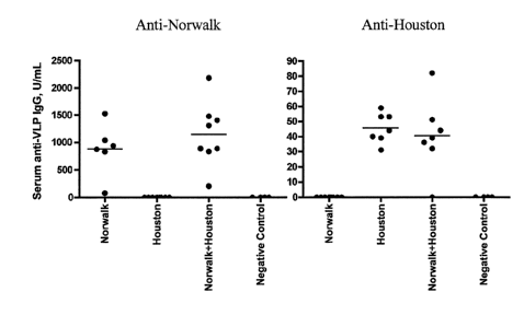
axis (CPM) (Figure 3). These data indicate that different biophysical forms of
the VLPs
prepared in the vaccine formulations elicit comparable T cell responses.
Example 3. VLP-Specific ELISPOT Assay
VLP-specific antibody-secreting cell (ASC) responses were measured from mice
immunized intraperitoneally with different NV-VLP formulations described in
Example 2.
Groups of mice (5 per group) were vaccinated i.p. once with rehydrated dry
powder formulations
shown in Table 2 (Example 2). Animals vaccinated with VLP-containing
formulations received
the same amount of total protein. 100% Agg (100% aggregated VLP protein); 100%
Intact
(100% native, intact VLPs); 50/50 Mix (1:1 mixture of intact and aggregated
VLP); Nave (no
VLP protein). On day 14, the mice were euthanized and the cervical lymph nodes
were
harvested. The cervical lymph node cells were cultured overnight on native,
intact VLP-coated
ELISPOT plates and were developed for either IgG or IgA-specific ELISPOTS
using the
appropriate HRP-conjugated secondary antibodies (Figure 4). These data show
that the three
VLP antigen formulations all elicit an antigen-specific B cell response. The
group immunized
with 100% Agg VLPs exhibited the greatest immune response.
Example 4. VLP-Specific ELISA
Serum IgG levels were measured from mice immunized i.p. with different NV-VLP
formulations. Groups of mice (5 per group) were vaccinated i.p. once with
rehydrated dry
powder formulations shown in Table 2 (Example 2). Animals vaccinated with VLP-
containing
formulations received the same amount of total protein. 100% Agg (100%
aggregated VLP
protein); 100% Intact (100% native, intact VLPs); 50/50 Mix (1:1 mixture of
intact and
aggregated VLP); Naïve (no VLP protein). On day 14, serum was collected and
assayed by
ELISA for anti-VLP-specific serum IgG (Figure 5). These data correlate with
the results shown
in Example 3, indicating that the three VLP antigen formulations all elicit an
antigen-specific B
cell response. Again, the group immunized with 100% Agg VLPs showed the
greatest immune
response.
25
CA 02664157 2009-03-20
WO 2008/042789
PCT/US2007/079929
LIG0-017/01W0
Example 5. Vaccine Formulations in Rabbits.
Formulations were administered intranasally (i.n.) in rabbits using the Valois
Monopowder Nasal Administration Device. The dry powder formulations are shown
in Tables 3
and 4.
Table 3. Formulations described below were prepared for 605.129, rabbit i.n.
dry powder (DP)
vaccination.
Prime formulations for exp 605.129 (Rabbit i.n.) (final amounts for DP
vaccines).
Group Chitosan Mannitol Sucrose MPL Norwalk VLP
number (mg/10mg DP) (mg/10mg) (mg/10mg) (mg/10mg) (mg/10mg)
1 7 1.475 1.475 0.025 0.025
2 7 1.475 1.475 0.025 0.025
3 7 1.475 1.475 0.025 0.025
4 7 1.475 1.475 0.025 0
Values indicate final concentrations of the formulations based on a single
device (10 mg
DP) which is 1/2 total dose.
Dose: 20 mg DP per animal, 10 mg per nare.
Group 1, 100% Agg: 100% aggregated lyophilized VLP
Group 2, 100% Intact: 100% intact lyophilized VLP
Group 3, 50/50 mix: 50/50 intact/aggregate lyophilized VLP (not a mixture of 1
& 2)
Group 4, Naive: placebo
Table 4. Formulations shown below were prepared for 605.129, rabbit i.n. dry
powder (DP)
vaccination.
Boost formulations for exp 605.129 (Rabbit i.n.) (final amounts for DP
vaccines).
Group Chitosan Mannitol Sucrose MPL Norwalk VLP
number (mg/10mg DP) (mg/10mg) (mg/10mg) (mg/10mg) (mg/10mg)
1 7 1.475 1.475 0.025 0.025
2 7 0 2.95 0.025 0.025
3 7 1.475 1.475 0.025 0.025
4 7 1.475 1.475 0.025 0
Values indicate final concentrations of the formulations based on a single
device (10 mg
DP) which is 1/2 total dose.
Dose: 20 mg DP per animal, 10 mg per nare.
Group 1, 100% Agg: 100% lyophilized aggregated VLP
Group 2, 100% Intact: 100% intact** VLP*
Group 3, 50/50 mix: 50/50 intact /aggregate lyophilized VLP (not a mixture of
1 & 2)
Group 4, Naive: lyophilized placebo
*Formulated without marmitol to increase amount of intact VLP post
lyophilization.
**Preparation yielded only ¨80% intact VLP.
26
CA 02664157 2009-03-20
WO 2008/042789
PCT/US2007/079929
LIG0-017/0 1 WO
Example 6. Potency Assay of Norovirus Vaccine Formulation in Mice
Female C57B16 mice were immunized intraperitoneally (i.p.) on day 0 with
different
dilutions of a reconstituted Norwalk VLP dry powder vaccine (containing
Norwalk VLP, MPL
and chitosan). Each animal was injected with 100 p,L of the formulations
indicated. Serum was
collected weekly and serum anti-VLP IgG measured by ELISA. Values for serum
collected 3
weeks following immunization are shown in Figure 6.
The value for each individual mouse is represented, with bars indicating the
group mean.
Serum anti-VLP IgG values correlated with the dose of vaccine indicated. This
experimental
design has been refined and developed as a potency assay required for the
release of GMP
manufactured vaccines for human clinical trials (Figure 6).
Example 7. Potency of Liquid vs. Reconstituted Norovirus Formulations in Mice
Female C57B16 mice were immunized i.p. on day 0 with formulations that
contained
chitosan, mannitol, MPL, and various concentrations of Norwalk VLP (Table 5)
in a volume of
100 L. An internal standard curve was generated (groups 1-5) by solubilizing
10 mg/mL of dry
powder matrix (mannitol, MPL, and chitosan) in purified water and adding the
specified amounts
of liquid Norwalk VLP. In contrast, the GMP VLP lots were previously
lyophilized and then
solubilized in 1.0 ml of purified water (groups 6-8). Serum was collected from
mice on days 14,
21 and 30, and serum anti-Norwalk VLP IgG was measured by ELISA.
Table 5. Liquid and Reconstituted Norwalk Formulations used to immunize mice
(i.p.).
Calculated Potency
Group Treatment 95% Cl Potency Min Max
1 5 pg VLP in Placebo 0.173 58.0 39.0
86.3
2 2.5 pg VLP in Placebo 0.192
23.3 15.0 36.3
3 1.25 pg VLP in Placebo 0.182
11.2 7.4 17.0
4 0.63 pg VLP in Placebo 0.287 5.4
2.8 10.4
5 0.31 pg VLP in Placebo 0.114 3.8
2.9 4.9
6 2.5 pg GMP lot 0.276 11.3 6.0
21.3
7 7.5 pg GMP lot 0.221 96.8 58.2
161.0
8 25 pg GMP lot 0.147 113.6 80.9
159.5
27
CA 02664157 2009-03-20
WO 2008/042789
PCT/US2007/079929
LIG0-017/01 WO
The relative potency for each formulation was calculated using the following
formula:
Inv Log (Ave.- Y intercept/slope). Potency is plotted against VLP
concentration in the
formulations and reported in relation to the standard curve generated using
known amounts of
VLP spiked into the matrix background (Figure 7). The results shown are
representative of 3
separate serum collection time points. These data indicate that the Norwalk
VLP formulation
reconstituted from dry powder has an overall higher potency than the liquid
formulations.
Example 8. Potency of Dry Powder Formulation in Rabbits
Forty-three female New Zealand White rabbits were intranasally (i.n.)
immunized using
the Valois Monopowder Nasal Administration Device with either 5 jig (Low) or
25 gg (Hi) of
Norwalk VLPs MPL and chitosan formulated into dry powders. One group
received the Hi
dose of VLPs and MPL formulated as a liquid and administered intramuscularly
(i.m.). Rabbits
were vaccinated on days 0 and 21. MPL, when used, was used at the same dose as
the VLPs
(i.e. , 5 lig Norwalk VLPs and 5 j.tg MPL). Chitosan, when used, was 7
mg/dose.
Serum IgG specific for the Norwalk VLPs (as determined by ELISA) is shown in
Figure
8. Mean values for each treatment groups are shown for day 21 (left panel,
collected just prior to
administration of the booster immunization) and day 42 (right panel). Values
are reported in
U/mL of VLP-specific IgG, with 1 U approximating 1 gg. Standard deviations are
indicated by
bars. All treatment groups had 6 animals, except the negative control group (3
rabbits) and the
intramuscularly immunized group (4 animals). These data show that generally
the higher VLP
dose results in greater serum anti-VLP IgG levels. Chitosan, in particular,
enhances responses to
intranasal vaccines. The i.m. immunized group showed the greatest responses.
However, VLP-
specific IgG levels in the intranasally immunized groups were also quite
robust.
Example 9. Potency of Liquid vs. Dry Norovirus Formulations given Intranasally
in Rabbits
Female New Zealand White rabbits were intranasally immunized using the Valois
Monopowder Nasal Administration Device with 50 lig of Norwalk VLPs + 50 j.tg
MPL + 14 mg
chitosan formulated into either a dry powder or a liquid. The vaccine content
was identical,
except for the physical state. Immunizations were on days 0 and 21 (weeks 0
and 3), with serum
collected prior to the boost at 3 weeks, and again at 6 weeks following the
initial vaccination.
28
CA 02664157 2009-03-20
WO 2008/042789
PCT/US2007/079929
LIG0-017/0 1 WO
Serum IgG specific for Norwalk VLPs was measured by ELISA, and the results are
shown in
Figure 9.
Group means are indicated, with the bars representing standard deviations. The
dry
powder immunization group had 6 rabbits, and the liquid immunization group had
10 rabbits.
Eight negative control rabbits are represented. Little difference was seen
between the liquid and
dry powder immunization groups at 3 weeks; however, at 6 weeks following the
initial
immunization, rabbits immunized with the dry powder formulation had superior
serum anti-VLP
IgG responses compared to the liquid immunization group.
Example 10. Stability of Norovirus Dry Powder Formulations
To investigate the stability of the dry powder VLP formulation, bulk drug
product was
prepared by mixing (per 10 mg drug product) 25 vtg of a Genogroup I VLP in
solution with 25
ug MPL, 700 ug chitosan glutamate, 1.475 mg mannitol, and 1.475 mg sucrose.
The solution
was lyophilized, blended with an additional 6.3 mg chitosan glutamate (per 10
mg drug product),
filled into Bespak unidose devices at a nominal 10 mg of dry powder, and
stored in sealed foil
pouches with desiccant capsules. Total VLP content was measured using Imperial
stained SDS-
PAGE and scanning densitometry, while size exclusion chromatography (SEC) was
used to
quantify intact VLP content. These measurements indicated that, within
experimental error, no
change in either total or intact VLP was detectable over the 12 month period
(Figure 10).
Assuming that the lower VLP protein recovery by SEC, when compared to SDS-PAGE
results,
was due to aggregation, the calculated % aggregate did not increase with time
but rather
remained constant or decreased throughout the 12 months of storage. One of the
more common
stability issues with proteins is increased aggregation with storage. Based on
the results in
Figure 10, it can be concluded that the formulation results in a stable
percentage of intact VLPs
allowing the product to be manufactured and used over at least a one year
period.
Example 11. Multiple Norovirus Antigens
Eight C57B1/6 mice (female, 9 weeks of age) were immunized intraperitoneally
(IP) on
days 0 and 14 with 2.5 tig Norwalk VLP formulated with 0.7 mg chitosan, 2.5 g
MPL and 0.3
mg mannitol brought to 0.1 mL with water. Two control mice were immunized with
saline. On
days 28 and 49, they were immunized again IP with 2.5 lig Norwalk VLP + 2.5 ug
Houston VLP
29
CA 02664157 2009-03-20
WO 2008/042789
PCT/US2007/079929
LIG0-017/01 WO
formulated with 0.7 mg chitosan, 2.5 g MPL and 0.3 mg mannitol brought to 0.1
mL with
water. The control mice again received saline. Serum samples were collected
weekly beginning
on week 5 (day 35) and analyzed by ELISA for reactivity with Norwalk VLPs or
Houston VLPs.
The time of boost with the Norwalk only mixtures are indicated by the thin
arrows, and the
Norwalk + Houston VLP mixtures are indicated by thick arrows. Individual serum
IgG responses
specific for Norwalk VLPs (top panel) or Houston VLPs (bottom panel) in U/mL
(with 1 U
approximating 1 tig of IgG) are shown. Means are indicated by bars. Note that
the Y-axis scales
are different, as the anti-Norwalk responses were much more robust due to two
previous
immunizations on days 0 and 14 (weeks 0 and 2). However, the responses against
Houston
VLPs are quite robust, with a large increase appearing in the second week
after the boost. These
data demonstrate that specific immune responses can be generated against
different antigenic
strains of Norovirus VLPs in the same immunizing mixture. (Figure 11).
Example 12. Immune Response to Different Norovirus Antigens
Female C57B16 mice were immunized intraperitoneally (IP) on days 0 and 14 with
25 lig
Norwalk VLP, 25 ttg Houston VLP, or a combination of 25 jig of each Norwalk
and Houston
VLP. Serum was collected weekly and serum anti-VLP IgG measured by ELISA.
Values for
serum collected 4 weeks following immunization are shown in Figure 12.
VLP content of the immunizations is indicated on the X axis. The value for
each
individual mouse is represented, with bars indicating the group mean. Antibody
levels are
represented in U/mL, with 1 U approximating 1 g of serum IgG. Values in the
left panel were
determined using Norwalk VLPs as the capture agent, while Houston VLPs were
used to coat
ELISA plates in order to measure the values on the right panel. These data
show that
immunization with Norwalk VLP does not lead to serum antibodies that are able
to recognize
Houston VLPs, or vice versa.
Example 13. Mixtures of Sucrose and Chitosan Preserve Norovirus VLP Structure
in Dry
Powder Formulations
The following experiments examined the effects of sucrose, chitosan, and
mannitol, alone
or in combination, in pre-lyophilization solutions on the native Norwalk VLP
quaternary
structure during lyophilization. Table 6 is a composite of several experiments
showing pre-
CA 02664157 2009-03-20
WO 2008/042789
PCT/US2007/079929
LIG0-017/01W0
lyophilization solution concentrations of the constituents of interest, the
total volume of the
mixture, and the corresponding mass ratios. All solutions were manually
swirled and gently
vortexed to homogeneity, then shell frozen in liquid nitrogen and lyophilized
external to the unit
using side-arm vessels for times ranging from about 30 to 60 hours.
Table 6. Pre-lyophilization solution mixtures used for testing the effects of
different
concentrations and combinations of sucrose, chitosan glutamate (chitosan) and
mannitol on the
structure of the quaternary structure the Norwalk VLP.
Mass equivalents
Solution concentrations of constituents pre- Total S = sucrose
Experiment
lyophilization (mg/mL) Volume C = chitosan
and Sample (mL) M
= mannitol
Sucrose Chitosan Mannitol VLP (protein) S C
M
LEI 0 0 100 0.83 0.30
LE2 0 0 75.0 0.62 0.40
LE3 0 0 50.0 0.42 0.60 0 0
1
LE4 0 0 25.0 0.21 1.20
LE5 0 0 10.0 , 0.08 3.00
LG1-LG3 0 7.83 0 0.20 1.28
LG4-LG6 0 5.06 0 0.13 1.98 0 1
0
LG7-LG9 0 2.09 0 0.05 4.78
LGIO 19.32 1.93 0 0.05 5.18 10 1
LG11 10.05 2.01 0 0.05 4.98 5 1
0
LG12 5.13 2.05 0 0.05 4.88 2.5 1
LG13 9.52 0.00 0 0.09 2.63 1 0
,
LJ1-02 5.29 2.51 0 0.09 2.79
LJ3- LJ4 4.17 1.98 0 0.07 3.54
LJ5- LJ6 3.65 1.73 0 0.06 4.04
LJ7- LJ8 2.93 1.39 0 0.05 5.04 2 1
0
LJ9- LJ10 5.25 2.49 0 0.09 2.81
LJ11-LJ12 4.14 1.97 0 0.07 3.56
LJ13- LJ14 3.63 1.72 0 , 0.06 4.06
LIGld - Sa 2.98 1.42 0.00 1.12 4.94 2 1
0
LIG1d-S1 12.89 6.12 0.00 1.12 2.29 2 1
0
LIGld - S2 12.26 5.82 12.26 0.67 2.41 1 0.5
1
LIGld - Sb 2.95 1.40 2.95 0.67 5.00 1 0.5
1
LIGld - S3 29.32 0.00 29.32 0.83 1.01 0 0
1
Table 7 shows the results from size exclusion-high performance liquid
chromatography
(SE-HPLC) analysis of the lyophilized samples shown in Table 6. Lyophilized
samples were
reconstituted with water and analyzed by SE-HPLC. Unprocessed NV-VLPs,
analyzed
concurrently, were used as a reference standard to quantify the NV-VLP content
of the
reconstituted test samples. Both UV and fluorescence detectors were used for
quantification
31
CA 02664157 2009-03-20
WO 2008/042789
PCT/US2007/079929
LIG0-017/01 WO
(data shown are from the fluorescence detector). The SE-HPLC was conducted
using a
SuperoseTM 6 10-300 column, with mobile phase consisting of 10 mM sodium
phosphate, 10
mM citric acid, pH 5, and 500 mM NaC1, at a flow rate of 0.5 mL/min. Protein
concentrations
were quantified using integrated areas of elution peaks. "VLP" is the peak
that eluted at about
15 min from the column, and any preceding shoulder and/or peak tail within the
approximate
elution time window of the reference standard NV-VLP analyzed concurrently.
The VLP
fragment that elutes from the column at around 32 min is a highly stable
single species that
results from destabilization and consequent disassembly of the VLP.
Intermediate and smaller
fragments were not observed.
The results show that combinations of sucrose and chitosan produced a wide
range of
native monodisperse NV-VLP recoveries including the highest (approximately
100% recovery)
post-lyophilization (samples LG10-LG12). Moreover, the NV-VLP elution peak
shapes from
these samples were identical to the unprocessed NV-VLP reference standard
indicating high
preservation of native structure. Samples containing sucrose only exhibited
peak shapes similar
to the reference standard, though NV-VLP recoveries were lower (approx. 60%
recovery (sample
LG13) Samples that contained only mannitol resulted in nearly completely
aggregated VLPs
(samples LE1-LE6 and LIG1d-S3). The deleterious effects of mannitol on NV-VLP
structure
were counteracted by the presence of chitosan and sucrose (samples LIGld ¨ S2
and LIG1d-SB).
Table 7. Experiment and sample identification, and results for testing the
effect of sucrose,
chitosan, and mannitol or combinations thereof on stability of NV-VLP
structure during freezing
and lyophilization.
32
CA 02664157 2009-03-20
WO 2008/042789
PCT/US2007/079929
LIG0-017/01 WO
Measured SE-BPLC
Mean percent values of
mean protein
recovered protein as Mass
equivalents
concentration and peak
Theoreticalpercent of theoretical S = sucrose
Experiment elution time
VLP conc N C = chitosan
and Sample VLP
(mg/mL) "VLP" Total,,M = mannitol
Fragment "VLP
- 15 min
- 32 min protein
(mg/mL) (/o) (mg/mL) S C M
LE1- LE5 0.25 5 0.02 0.12 56.0 6.3 0 0 1
LG1 - LG9 0.25 9 0.06 0.00 24.0 24.0 0 1 0
LGIO 0.25 1 0.25 0.00 101 101 10 1
0
LG11 0.25 1 0.25 0.00 101 101 5 1 0
LG12 0.25 1 0.25 0.00 100 100 2.5 1
0
LG13 0.25 1 0.16 0.00 65 65 1 0 0
LJ1 -LJ14 0.25 14 0.22 0 _ 85.4 85.4 2 1 0
LIGld - S1 0.25 1 0.21 0 88 88 2 1 0
LIGld - Sa 0.25 1 0.12 0 50 50 2 I 0
LIGld - S2 0.25 1 92 0 92 92 1 0.5
1
LIGld - Sb 0.25 1 60 0 60 60 1 0.5
1
LIGld - S3 0.25 1 < 1 < 1 < 1 < 1 0 0 1
Example 14. Induction of Norovirus-Specific Long-Lived Plasma Cells and Memory
B Cells in
Mice Immunized Intranasally
A. Norwalk VLP-specific long-lived plasma cells
BALB/c mice were immunized intranasally with Norovirus VLPs and an adjuvant.
Naïve controls were administered the adjuvant alone. At 114 days after
immunization, spleen,
cervical lymph nodes, and bone marrow were harvested from both groups of mice.
On the day
of harvesting the tissues (day 0), cells were assayed using an ELISPOT assay
for the presence of
antigen-specific antibody-secreting cells (ASCs). The results are presented in
Figure 13A-C for
the different tissues. The detection of immunoglobulins (IgG, IgA, and IgM) in
these tissues
indicates the presence of Norovirus-specific long-lived plasma cells.
B. Norwalk VLP-specific memory B cells
An in vitro assay was developed to detect the presence of Norwalk VLP-specific
memory
B-cells from mice immunized intranasally with Norwalk VLPs. Various lymphoid
tissues or
whole blood (peripheral blood mononuclear cells, splenocytes, lymph node
cells, etc.) can serve
as the source of cells that can be assayed for the presence of memory B-cells
using this assay.
In this experiment, the spleen was harvested and processed from immunized and
naïve
animals (controls), and splenocytes were cultured for four days in the
presence or absence
33
CA 02664157 2009-03-20
WO 2008/042789
PCT/US2007/079929
LIG0-017/01 WO
(controls) of Norwalk VLPs (20 [tg/m1). An initial VLP-specific ELISPOT assay
was performed
on the day of tissue harvest (day 0) to establish background levels of ASCs
(see Section A
above). After four days in culture the cells were harvested and assayed again
in an ELISPOT
assay to quantify the number of VLP-specific ASCs. The difference in VLP-
specific ASC
numbers between the day 0 and the day 4 assays represent the antigen-specific
memory B-cell
population. The results of this experiment are shown in Figure 14A and B.
Example 15. Norovirus Memory B Cell Responses in Rabbits
Two female New Zealand White rabbits were immunized intranasally with a dry
powder
formulation consisting of 25 tig Norwalk VLP, 251.T MPL, 1.5 mg mannitol, 1.5
mg sucrose,
and 7.0 mg chitosan per 10 mg of dry powder loaded into Valois Mark 4
intranasal delivery
devices. The two rabbits received a total of three immunizations at 14 day
intervals. For these
experiments, a non-immunized female rabbit was used as a naive control.
A. Collection and processing of rabbit tissues
Peripheral blood mononuclear cells (PBMCs): Whole blood (-50 mL) was obtained
from
rabbits in collection tubes containing EDTA to prevent coagulation. The whole
blood was diluted
1:3 with sterile D-PBS and ¨35 mL of diluted whole blood was layered onto 15
mL of
Lympholyte Separation Medium in a sterile 50-mL centrifuge tube. The tubes
were centrifuged
at 800 x g for 20 minutes at room temperature. The buffy coat layer containing
the PBMCs was
carefully removed using a sterile 5 mL pipette and the cells were washed twice
with D-PBS. If
necessary, contaminating red blood cells were removed by ACK lysis. The cells
were
resuspended in RPMI-1640-10% FBS (1640-C) and counted in a hemocytometer using
a Trypan
exclusion method.
Mesenteric lymph node cells: The lymph nodes were aseptically collected from
each
rabbit separately following euthanasia. The tissues were maintained in a
sterile plastic Petri dish
containing ¨10 mL of RPMI-1640-No Serum (1640-NS). The lymph nodes were
pressed through
a sterile mesh screen using a sterile pestle to disperse the tissue and obtain
a single cell
suspension of lymph node cells. The cells were collected, washed twice with
1640-NS, and
finally filtered through a sterile 701.1m filter to remove clumps and debris.
The cells were
resuspended in 1640-C and counted in a hemocytometer using a Trypan blue
exclusion method.
34
CA 02664157 2009-03-20
WO 2008/042789
PCT/US2007/079929
LIG0-017/01W0
Splenocytes: Spleens were aseptically obtained from each rabbit following
euthanasia.
The spleens were placed in sterile Petri dishes containing approximately 10 mL
of 1640-NS.
Using a sterile 22-guage needle and syringe the media was repeatedly injected
into the tissue to
disrupt the splenic capsule and elaborate the cells. Sterile forceps were then
used to tease apart
the remaining tissue fragments. The contents of the Petri dish were
transferred to a sterile
centrifuge tube and the cell suspension and disrupted splenic tissue was
allowed to sit for 6-8
minutes to allow for the settling of large tissue fragments. The single cell
suspension was
transferred to a second sterile centrifuge tube and the cells were washed once
with 1640-NS. The
red blood cells in the splenocyte prep were removed by an ACK lysis (8 mL ACK
buffer, 8
minutes, room temperature) and the cells were washed one more time with 1640-
NS and finally
filtered through a sterile 70 [tm filter to remove clumps and debris. The
final cell pellet was
resuspended in 1640-Complete and counted in a hemocytometer using a Trypan
blue exclusion
method.
Bone marrow cells: The tibia bones in the lower legs were removed from
individual
rabbits following euthanasia. To remove the bone marrow cells the ends of the
bones were
aseptically cut off using a bone saw and the contents of the bone were flushed
out by repeated
injections of 1640-NS medium. The bone marrow cells were pipetted up and down
repeatedly to
break up and disperse clumps of cells. The cells were washed once with 1640-
NS; the red blood
cells were lysed with ACK, and the cells were washed one more time with 1640-
NS. Finally, the
cells were filtered through a sterile 70 J.Lm filter to remove clumps and
debris. The final cell
pellet was resuspended in 1640-Complete and counted in a hemocytometer using a
Trypan blue
exclusion method.
B. ELISPOT assays
Following pre-wetting and washing, 96-well Millipore PVDF filter plates were
coated
with a sterile solution of native Norwalk VLPs at a concentration of 40 lig/m1
in a final volume
of 501.11/well. The plates were incubated overnight at 4 C, washed with D-
PBS, and blocked
with the addition of 1640-C. Mesenteric lymph node cells, splenocytes, and
bone marrow cells
from the immunized rabbits and from the naïve control rabbit were added to the
wells at varying
concentrations (1x10⁶, 5x10⁵, 2x10⁵, and 1x10⁵ cells/well) and the plates were
incubated
overnight at 37 C. The plates were washed thoroughly with PBS-Tween and
secondary reagents
CA 02664157 2009-03-20
WO 2008/042789
PCT/US2007/079929
LIG0-017/01 WO
specific for rabbit IgG and IgA were added to the wells and incubated for an
additional 2 hours at
room temperature. Following extensive washing the plates were developed with
DAB
chromagen/substrate and read in an ELISPOT plate reader. Spots appearing on
wells from naïve
control animals were subtracted from the experimental groups. The data is
expressed as Norwalk
VLP-specific antibody-secreting cells (ASCs) and is normalized per lx106
cells.
C. Norwalk VLP-Specific Memory B-cell Assay
Isolated lymphoid cells from the various tissues described above were
resuspended in
1640-C medium in the presence of Norwalk VLPs (101.1.g/rnL) at a density of
5x10⁶ cells per
mL. The cells were incubated in 24-well plates in 1-mL volumes for four days
at 37 C. VLP-
specific ELISPOT assays were performed on these cells at the time of
culturing. After four days
in culture the cells were harvested, washed twice with 1640-NS medium,
resuspended in 1640-
Complete, and counted in a hemocytometer using a Trypan blue exclusion method.
The cells
were tested once again in a Norwalk VLP-specific ELISPOT assay. The data
obtained from the
ELISPOT assays performed on the day of tissue harvest is referred to as day 0
(background)
ASC activity. Any spots detected at the day 0 time point are assumed to be
actively-secreting
plasma cells or long-lived plasma cells (LLPCs).The data obtained from the
ELISPOT assay
performed on the 4-day cultured cells is referred to as day 4 ASC activity,
and the memory B-
cell activity is represented by the difference between day 4 ASC activity and
day 0 ASC activity.
D. Norwalk VLP-Specific Memory B-Cells are Present in the Peripheral Blood of
Intranasally
Immunized Rabbits
Whole blood was obtained from two immunized rabbits (RB735, RB1411) 141 days
following the last of three intranasal immunizations with a dry powder
formulation vaccine
containing Norwalk VLPs as described above. Blood was also obtained from an
non-immunized,
naïve rabbit. The blood was processed to obtain peripheral blood mononuclear
cells (PBMCs)
and the PBMCs were placed in a Norwalk VLP-Specific memory B-Cell assay
(section C
above). The results are shown in Figure 15. The left panel shows results of
the initial ELISPOT
assay at the time of tissue harvest (day 0 ASCs). The right panel shows the
results of the
ELISPOT assay after 4 days in culture with Norwalk VLPs (day 4 ASCs).
36
CA 02664157 2009-03-20
WO 2008/042789
PCT/US2007/079929
LIG0-017/0 1 WO
The day 0 ELISPOT results (Fig. 15, left panel) illustrate that there are no
VLP-specific
plasma cells remaining in the peripheral blood approximately 140 days after
the last boost with
Norwalk VLP dry powder vaccine. The right panel of Figure 15 shows the ELISPOT
assay
results from PBMCs cultured for four days in vitro with Norwalk VLPs. In the
two immunized
rabbits, a significant number of PBMCs, presumably a subpopulation of memory B-
cells, have
matured into active IgG-secreting Norwalk VLP-specific plasma cells. Although
assays for IgA-
secreting memory B-cells were conducted, only IgG-secreting memory B-cells
were detected in
the PBMC population. As expected, the naïve animal showed no antigen-specific
memory B-
cells. Thus, VLP-specific memory B-cells were found in the peripheral
circulation of rabbits
140+ days following the last of three intranasal immunizations.
E. Norwalk VLP-Specific Memory B-Cells are Present in the Spleen of
Intranasally Immunized
Rabbits
Splenocytes were obtained from the spleens of the two vaccine immunized
rabbits and
the non-immunized control rabbit. Norwalk VLP-specific memory B-cell assays
(described
above) were performed on these cells and the results are shown in Figure 16.
As observed for
the PBMC population the day 0 ELISPOT assay shows that there are no antigen-
specific plasma
cells present in the spleen (Fig. 16, left panel). However, following a four
day in vitro
incubation with Norwalk VLPs, IgG-secreting Norwalk VLP-specific memory B-
cells are
apparent in the splenocyte population. Thus, the spleen represents one site
for the migration of
memory B-cells following intranasal immunization.
F. A Population of Norwalk VLP-Specific Long-lived Plasma Cells is Found in
the Bone
Marrow but No Memory B-cells Are Present
Bone marrow cells were obtained from the tibias of the experimental rabbits
and assayed
for the presence of long-lived plasma cells and memory B-cells. The results
are presented in
Figure 17. The left panel of Figure 17 shows that rabbit 1411 still had a
significant population of
antigen-specific plasma cells in the bone marrow. Plasma cells that migrate to
the bone marrow
and reside there for a significant period of time following immunization are
referred to as long-
lived plasma cells (LLPCs). Rabbit 735 did not show a high number of LLPCs. No
LLPCs were
found in the bone marrow of the nave rabbit. The bone marrow cells were
cultured in a memory
37
CA 02664157 2009-03-20
WO 2008/042789
PCT/US2007/079929
LIG0-017/01W0
B-cell assay and re-tested for the presence of memory B-cells. The right panel
of Figure 17
shows that there are essentially no antigen-specific memory B-cells present in
the bone marrow.
Thus, long-lived plasma cells migrate to the bone marrow but no memory B-cells
are found
there.
G. Both IgG-Secreting and IgA-Secreting Norwalk VLP-Specific Memory B-Cells
are Present in
the Mesenteric Lymph Nodes of Intranasally Immunized Rabbits
The mesenteric lymph nodes were obtained from all of the experimental rabbits
and the
isolated cells were assayed for LLPCs and memory B-cells. The results from
this assay are
shown in Figure 18A. As with most of the lymphoid tissue analyzed, except bone
marrow, no
LLPCs (Fig. 18A left panels) were found in the mesenteric nodes. Following in
vitro incubation
with Norwalk VLPs, a very high number of IgG-secreting VLP-specific memory B-
cells were
evident in the mesenteric lymph node population (Fig. 18A, right panel). The
numbers of
memory B-cells observed in the mesenteric lymph nodes were significantly
higher than those
observed for the other lymphoid tissues assayed.
Numerous researchers have shown that immunization at a mucosal inductive site,
such as
the nasal passages or the gut, is capable of eliciting a so-called mucosal
immune response. This
response has generally been characterized by the presence of IgA+ B-cells and
IgA-secreting
plasma cells localized in the mucosal lymphoid tissue. For this reason the
mesenteric lymph node
cells were also assayed for the presence of IgA-secreting LLPCs or memory B-
cells. The results
from these assays are shown in Figure 18B. Once again, no IgA+ LLPCs were
found in the
mesenteric lymph node population (Figure 18B, left panel). However, IgA-
secreting memory B-
cells were detected in this tissue (Figure 18B, right panel). Thus, intranasal
immunization with a
dry powder Norwalk VLP vaccine formulation elicited a mucosal immune response
that resulted
in the migration of both IgG+ and IgA+ antigen-specific memory B-cells to the
gut-associated
lymphoid tissue. The production of antigen-specific memory B cells induced by
immunization
with the Norwalk vaccine formulation is a possible indicator of vaccine
effectiveness. The
presence of memory B cells is one marker of long-lasting immunity.
H. VLP-specific CD4+ Memory T cells
38
. CA 02664157 2014-08-01
_
,
Splenocytes harvested from immunized rabbits were restimulated with intact
Norwalk
VLPs and the extent of cellular proliferation was measured by tritiated
thymidine incorporation
as indicated on the ordinate axis (CPM) (Figure 19). The left panel shows
cellular proliferation
of an unfractionated population of splenocytes, while the right panel shows
cellular proliferation
of CD4+ T cells.
The present invention is not to be limited in scope by the specific
embodiments described
which are intended as single illustrations of individual aspects of the
invention, and functionally
equivalent methods and components are within the scope of the invention.
Indeed, various
modifications of the invention, in addition to those shown and described
herein, will become
apparent to those skilled in the art from the foregoing description and
accompanying drawings
using no more than routine experimentation. Such modifications and equivalents
are intended to
fall within the scope of the appended claims.
Citation or discussion of a reference herein shall not be construed as an
admission that
such is prior art to the present invention.
39
CA 02664157 2009-03-20
WO 2008/042789
PCT/US2007/079929
LIG0-017/01W0
References
1. Glass, RI, JS Noel, T Ando, RL Fankhauser, G Belloit, A Mounts, UD
Parasher, JS Bresee
and SS Monroe. The Epidemiology of Enteric Caliciviruses from Human: A
Reassessment
Using New Diagnostics. J Infect Dis 2000; 181 (Sup 2): S254-S261.
2. Hardy, ME. Norwalk and "Norwalk-like Viruses" in Epidemic Gastroenteritis.
Clin Lab
Med 1999;19(3): 675-90.
3. Jiang, X, DY Graham, KN Wang, and MK Estes. Noralk Virus Genome Cloning and
Characterization. Science 1990; 250: 1580-1583.
4. Jiang, X, M Want, DY Graham, and MK Estes. Expression, Self-Assembly, and
Antigenicity of the Norwalk Virus Capsid Protein. J Virol 1992; 66: 6527-6532.
5. Glass, P, LJ White, JM Ball, I Leparc-Goffart, ME Hardy, and MK Estes.
Norwalk Virus
Open Reading Frame 3 Encodes a Minor Structural Protein. J Virol 2000; 74:
6581-6591.
6. Lindesmith, L, C Moe, S Marionneau, N Ruvoen, X Jiang, L Lindblad, P
Stewart, J
LePendu, and R Baric. Human Susceptiblity and Resistance to Norwalk Virus
Infection.
Nat Med 2003; 9: 548-553.
7. Parrino, TA, DS Schreiber, JS Trier, AZ Kapikian, and NR Blacklow. Clinical
Immunity in
Acute Gastroenteritis Caused by Norwalk Agent. N Engl J Med 1977; 297: 86-89.
8. Wyatt, RG, R Dolin, NR Blacklow, HL DuPont, RF Buscho, TS Thornhill, AZ
Kapikian,
and RM Chanock. Comparison of Three Agents of Acute Infectious Nonbacterial
Gastroenteritis by Cross-challenge in Volunteers. J Infect Dis 1974; 129: 709.
9. Ball, JM, DY Graham, AR Opekum, MA Gilger, RA Guerrero, and MK Estes.
Recombinant Norwalk Virus-like Particles Given Orally to Volunteers: Phase I
Study.
Gastroenterology 1999; 117: 40-48.
10. Tacket, CO, MB Sztein, GA Losonky, SS Wasserman, and MK Estes. Humoral,
Mucosal,
and Cellular Immune Responses to Oral Nowalk Virus-like Particles in
Volunteers. Clin
Immunol 2003; 108: 241.
11. Guerrero, RA, JM Ball, SS Krater, SE Pacheco, JD Clements, and MK Estes.
Recombinant
Norwalk Virus-like Particles Administered Intranasally to Mice Induce Systemic
and
Mucosal (Fecal and Vaginal) Immune Responses. J Virol 2001; 75: 9713.
CA 02664157 2009-03-20
WO 2008/042789
PCT/US2007/079929
LIG0-017/01 WO
12. Nicollier-Jamot, B, A Ogier, L Piroth, P Pothier, and E Kohli. Recombinant
Virus-like
Particles of a Norovirus (Genogroup II Strain) Administered Intranasally and
Orally with
Mucosal Adjuvants LT and LT(R192G) in BALB/c Mice Induce Specific Humoral and
Cellular Thl/Th2-like Immune Responses. Vaccine 2004; 22:1079-1086.
13. Periwal, SB, KR Kourie, N Ramachandaran, SJ Blakeney, S DeBruin, D Zhu, TJ
Zamb, L
Smith, S Udem, JH Eldridge, KE Shroff, and PA Reilly. A Modified Cholera
Holotmdn
CT-E29H Enhances Systemic and Mucosal Immune Responses to Recombinant Norwalk
Virus- like Particle Vaccine. Vaccine 2003; 21: 376-385.
14. Isaka, M, Y Yasuda, S Kozuka, T Taniguchi, K Matano, J Maeyama, T Komiya,
K
Ohkuma, N Goto, and K Tochikubo. Induction of systemic and mucosal antibody
responses
in mice immunized intranasally with aluminium-non-adsorbed diphtheria toxoid
together
with recombinant cholera toxin B subunit as an adjuvant. Vaccine 1999; 18: 743-
751.
15. Kozlowski, PA, S Cu-Uvin, MR Neutra, and TP Flanigan. Comparison of the
oral, rectal,
and vaginal immunization routes for induction of antibodies in rectal and
genital tract
secretions of women. Infect Immun 1997; 65: 1387-1394.
16. Mestecky, J, SM Michalek, Z Moldoveanu, and MW Russell. Routes of
immunization and
antigen delivery systems for optimal mucosal immune responses in humans.
Behring Inst
Mitt 1997; 33-43.
17. Wu, HY, and MW Russell. Nasal lymphoid tissue, intranasal immunization,
and
compartmentalization of the common mucosal immune system. Immunol Res 1997;
16:
187-201.
18. Evans, JT, CW Cluff, DA Johnson, MJ Lacy, DH Persing, and JR Baldridge.
Enhancement
of antigen-specific immunity via the TLR4 ligands MPL adjuvant and Ribi 529.
Expert Rev
Vaccines 2003; 2: 219-229.
19. Baldridge, JR, Y Yorgensen, JR Ward, and JT Ulrich. Monophosphoryl lipid A
enhances
mucosal and systemic immunity to vaccine antigens following intranasal
administration [In
Process Citation]. Vaccine 2000; 18: 2416-2425.
20. Yang, QB, M Martin, SM Michalek, and J Katz. Mechanisms of monophosphoryl
lipid A
augmentation of host responses to recombinant HagB from Porphyromonas
gingivalis.
Infect Immun 2002; 70: 3557-3565.
41
CA 02664157 2009-03-20
WO 2008/042789
PCT/US2007/079929
LIG0-017/01W0
21. Baldrick, P, D Richardson, G Elliott, and AW Wheeler. Safety evaluation of
monophosphoryl lipid A (MPL): an immunostimulatory adjuvant. Regul Toxicol
Pharmacol 2002; 35: 398-413.
22. Baldridge, JR, P McGowan, JT Evans, C Cluff, S Mossman, D Johnson, and D
Persing.
Taking a toll on human disease: Toll-like receptor 4 agonists as vaccine
adjuvants and
monotherapeutic agents. Expert Opin Biol Ther 2004; 4: 1129-1138.
23. Persing, DH, RN Coler, MJ Lacy, DA Johnson, JR Baldridge, RM Hershberg,
and SG Reed.
Taking toll: lipid A mimetics as adjuvants and immunomodulators. Trends
Microbiol 2002;
10: S32-37.
24. Illum, L. Nasal drug delivery--possibilities, problems and solutions. J
Control Release
2003; 87: 187-198.
25. Illum, L, I Jabbal-Gill, M Hinchcliffe, AN Fisher, and SS Davis. Chitosan
as a novel nasal
delivery system for vaccines. Adv Drug Deliv Rev 2001; 51: 81-96.
26. Davis, SS. Delivery of peptide and non-peptide drugs through the
respiratory tract. Pharm
Sci Technol Today 1999; 2: 450-456.
27. Bacon, A, J Makin, P J Sizer, I Jabbal-Gill, M Hinchcliffe, L Illum, S
Chatfield, and M
Roberts. Carbohydrate biopolymers enhance antibody responses to mucosally
delivered
vaccine antigens. Infect Immun 2000; 68: 5764-5770.
28. van der Lubben, IM, JC Verhoef, G Borchard, and HE Junginger. Chitosan for
mucosal
vaccination. Adv Drug Deliv Rev 2001; 52: 139-144.
29. van der Lubben, IM, JC Verhoef, G Borchard, and HE Junginger. Chitosan and
its
derivatives in mucosal drug and vaccine delivery. Eur J Pharm Sci 2001; 14:
201-207.
30. Lim, S T, B Forbes, GP Martin, and MB Brown. In vivo and in vitro
characterization of
novel microparticulates based on hyaluronan and chitosan hydroglutamate. AAPS
Pharm
Sci Tech 2001; 2: 20.
31. Jabbal-Gill, I, AN Fisher, R Rappuoli, SS Davis, and L Illum. Stimulation
of mucosal and
systemic antibody responses against Bordetella pertussis filamentous
haemagglutinin and
recombinant pertussis toxin after nasal administration with chitosan in mice.
Vaccine 1998;
16: 2039-2046.
32. Mills, KH, C Cosgrove, EA McNeela, A Sexton, R Giemza, I Jabbal-Gill, A
Church, W Lin,
L Illum, A Podda, R Rappuoli, M Pizza, GE Griffin, and DJ Lewis. Protective
levels of
42
CA 02664157 2009-03-20
WO 2008/042789
PCT/US2007/079929
LIG0-017/01W0
diphtheria-neutralizing antibody induced in healthy volunteers by unilateral
priming-
boosting intranasal immunization associated with restricted ipsilateral
mucosal secretory
immunoglobulin. A Infect Immun 2003; 71: 726-732.
33. McNeela, EA., I Jabbal-Gill, L Illum, M Pizza, R Rappuoli, A Podda, DJ
Lewis, and KH
Mills. Intranasal immunization with genetically detoxified diphtheria toxin
induces T cell
responses in humans: enhancement of Th2 responses and toxin-neutralizing
antibodies by
formulation with chitosan. Vaccine 2004; 22: 909-914.
34. Mikszta, JA., VJ Sullivan, C Dean, AM Waterston, JB Alarcon, JP Dekker,
3rd, JM
Brittingham, J Huang, CR Hwang, M Ferriter, G Jiang, K Mar, KU Saikh, BG
Stiles, CJ
Roy, RG Ulrich, and NG Harvey. Protective immunization against inhalational
anthrax: a
comparison of minimally invasive delivery platforms. J Infect Dis 2005; 191:
278-288.
35. Huang, J, RJ Garmise, TM Crowder, K Mar, CR Hwang, AJ Hickey, JA Mikszta,
and VJ
Sullivan. A novel dry powder influenza vaccine and intranasal delivery
technology:
induction of systemic and mucosal immune responses in rats. Vaccine 2004; 23:
794-801.
36. GSK Press Room. www.gsk.com/media/archive.htm
37. Corixa Press Room. www.corixa.com/default.asp?pid= release
detai1&id=248&year=2004
38. BioMira Web Site. http://www.biomira.com/business/outLicensing/
39. Centers for Disease Control, Morbidity and Mortality Weekly Report 2007;
56(33):842-846.
43