Note: Descriptions are shown in the official language in which they were submitted.
CA 02664351 2009-03-24
1
DESCRIPTION
IMPLANTATION GUIDE MAKING METHOD AND GUIDE BLOCK
TECHNICAL FIELD
[0001] The present invention relates to a method of
producing an implant implantation guide for implanting
a dental implant (artificial tooth root) and,
particularly, to a method of producing an implant
implantation guide by utilizing a CAD/CAM system and
to a guide block to be used in the producing method.
BACKGROUND ART
[0002] Dental implant (artificial tooth root)
treatments are widely utilized in the dental field. In
order to improve the functionality (occlusal balance)
and the aesthetic appearance of an artificial tooth to
be fitted on a dental implant and to stably maintain
the dental implant in a jawbone, it is important to
properly design the implantation position and the
implantation direction (implantation angle) of the
dental implant through a diagnosis, and precisely
implant the dental implant based on the design.
[0003] In recent years, an attempt is made to properly
determine the implantation position and the
CA 02664351 2009-03-24
2
implantation direction of the dental implant through
a diagnosis utilizing a three-dimensional medical image
obtained by CT imaging and produce an implant
implantation guide for effecting the implantation
position and the implantation direction determined
through the diagnosis by means of a CAD/CAM system for
clinical application (see, for example, Patent
Documents 1 and 2).
[0004] However, it is difficult to produce a highly
precise implant implantation guide that permits
accurate positioning of the dental implant in an oral
cavity based on CT imaging data alone. This is because:
the CT imaging data includes data of
several-hundred-micron voxels; a metal fixture
attached to a tooth causes a noise called "metal
artifact" in the image; a prosthetic device composed
of a non-imageable material such as a resin cannot be
imaged; the imaging range, the imaging depth and the
size and shape of the image vary depending on CT values;
and the three-dimensional image formed based on the CT
imaging data has a simplified shape with reduced
geometrical and dimensional accuracies.
[0005] In other words, the implant implantation guide
produced based on the CT imaging data is not as precise
as that produced based on a dental arch model formed
CA 02664351 2009-03-24
3
of a plaster (a dental arch model of a plaster obtained
by taking an impression of a dental arch in a patient's
oral cavity).
Patent Document 1: Japanese Unexamined Patent
Publication No. 2003-245289
Patent Document 2: Japanese Unexamined Patent
Publication No. 2001-170080
DISCLOSURE OF THE INVENTION
PROBLEMS TO BE SOLVED BY THE INVENTION
[0006] It is difficult to produce the highly precise
implant implantation guide based on the CT imaging data
alone. Therefore, it is conceivable to substitute data
obtained from the highly precise dental arch model for
a corresponding data portion of the three-dimensional
image formed based on the CT imaging data, and produce
the implant implantation guide based on the substituted
data by means of the CAD/CAM system. However, it is
necessary to scan the dental arch model to obtain
geometrical data of the dental arch model.
Disadvantageously, the data obtained by the scanning
is less precise than the original dental arch model at
this stage. Further, it is difficult to eliminate an
error occurring when the image is correlated with the
model data for the substitution.
CA 02664351 2009-03-24
4
[0007] In view of the foregoing, it is a principal
object of the present invention to provide a method of
producing a highly precise implant implantation guide
for safely and precisely performing a dental implant
treatment.
MEANS FOR SOLVING THE PROBLEMS
[0008] According to the present invention, there is
provided a method of producing an implant implantation
guide for CAD/CAM, the method including the steps of:
(1) preparing a guide block including an attachment
portion to be fitted on a dental arch of a patient, and
a processing portion having a mark of a processing
reference coordinate system required for processing;
(2) acquiring patient's CT image data with the guide
block being fitted on the patient's dental arch; (3)
transforming information of an implant implantation
position and an implant implantation direction
(implantation angle) determined through diagnosis on
a three-dimensional image formed based on the CT image
data into coordinate information based on the
processing reference coordinate system on the guide
block; and (4) setting the guide block in a cutting
processor, and cutting the guide block into a guide
shape that reflects the coordinate information based
CA 02664351 2009-03-24
on the processing reference coordinate system.
[0009] The attachment portion of the guide block is
preferably composed of a non-imageable material, and
the processing portion of the guide block is preferably
composed of an imageable material.
[0010] According to the present invention, there is
provided a method of producing an implant implantation
guide for CAD/CAM, the method including the steps of:
(1) separately preparing a guide base including an
attachment portion to be fitted on a dental arch of a
patient and an imaging marker specifying at least three
points, and a processing portion attachable to the guide
base; (2) acquiring patient's CT image data with the
guide base being fitted on the patient's dental arch;
(3) providing a guide block by attaching the processing
portion to the guide base for unification; (4)
transforming information of an implant implantation
position and an implant implantation direction
(implantation angle) determined through diagnosis on
a three-dimensional image formed based on the CT image
data into coordinate information based on a processing
reference coordinate system to be utilized for
processing the processing portion via a coordinate
system defined by the imaging marker; and (5) setting
the guide block in a cutting processor, and cutting the
CA 02664351 2009-03-24
6
guide block into a guide shape that reflects the
coordinate information based on the processing
reference coordinate system.
[0011] According to the present invention, there is
provided a guide block for use in the implant
implantation guide producing methods described above,
the guide block including an attachment portion to be
fitted on a patient's dental arch, and a processing
portion having a mark of a processing reference
coordination system required for a cutting process.
[0012] According to the present invention, the
processing portion of the guide block is composed of
an imageable material.
EFFECTS OF THE INVENTION
[0013] According to the present invention, the guide
block is first prepared. The guide block unitarily
includes the processing portion to be milled (cut) into
a predetermined shape in a step to be described later,
and the attachment portion for attaching the processing
portion to the patient's dental arch.
[0014] The attachment portion is, for example, a
dental impression of a plaster or the like directly
taken from the patient's oral cavity, or formed to
conform to the patient's dental arch. Therefore, when
CA 02664351 2009-03-24
7
the guide block is thereafter worn by the patient, the
attachment portion is perfectly fitted on the patient's
dental arch without displacement in the oral cavity.
[0015] With the guide block being fitted in the
patient's oral cavity, the patient's oral cavity is
imaged through the CT imaging to provide CT image data.
[0016] The image data thus provided is a
three-dimensional image including images of patient's
jawbones, a patient's dental arch, a patient's tooth
deficient site and the like. The three-dimensional
image also includes an image of the processing portion
of the guide block fitted in the patient's oral cavity.
That is, the image data includes raw image data of the
patient as well as image data of the guide block.
[0017] The implant implantation position and the
implant implantation direction (implantation angle)
are determined on the three-dimensional image through
the diagnosis. The implantation position and the
implantation direction (implantation angle) are
defined, for example, in the form of a straight line
on the three-dimensional image.
[0018] The straight line representing the
implantation position and the implantation direction
(implantation angle) is data based on a
three-dimensional image display coordinate system.
CA 02664351 2009-03-24
8
[0019] On the other hand, the guide block is processed
based on the processing reference coordinate system
defined on the processing portion of the guide block
in the guide block cutting step to be described later.
[0020] The data indicating the implant implantation
position and the implant implantation direction
(implantation angle) determined through the diagnosis
on the three-dimensional image based on the
three-dimensional image display coordinate system is
transformed into the data based on the processing
reference coordinate system.
[0021] Then, the guide block is set in the cutting
processor, and cut into the guide shape that reflects
the implant implantation position and the implant
implantation direction (implantation angle) obtained
through the transformation based on the processing
reference coordinate system, i.e., that reflects the
data of the implant implantation position and the
implant implantation direction, by the CAD/CAM system.
[0022] An implant implantation guide thus produced by
the cutting process is configured such that the
attachment portion to be fitted on the dental arch has
a shape conformal to the dental arch model. Therefore,
when the implant implantation guide is fitted in the
patient's oral cavity, the implant implantation guide
CA 02664351 2009-03-24
9
is perfectly fitted on the patient's dental arch without
a gap. Thus, the implant implantation guide is free
from wobble in the patient's oral cavity, and serves
as a guide for forming a hole for implantation of the
dental implant in the patient's oral cavity.
[0023] The implant implantation guide produced by the
inventive producing method is properly fitted on the
dental arch in the patient's oral cavity without wobble
in the patient's oral cavity.
[0024] Since the implant implantation guide is
properly and precisely fitted in the patient's oral
cavity, a dental treatment can be properly performed
on the patient with reference to the guide.
[0025] In the present invention, the guide base and
the processing portion of the guide block may be
provided as separate members rather than as a unitary
member. Where the patient who wears the guide block
has a smaller mouth or suffers from a sensitive vomiting
reflex, it is often difficult to fit the guide block
in the patient's oral cavity for the CT imaging. In
this case, it is desirable to use the guide block
including the guide base and the processing portion
provided as separate members. This is because the
guide base is a smaller and thinner member including
the attachment portion and the imaging marker, thereby
CA 02664351 2009-03-24
alleviating a burden on the patient who wears the guide
base during the CT imaging.
[0026] By utilizing an existing technique, the
imaging marker specifying the at least three points on
the guide base makes it possible to transform the data
based on the three-dimensional image display coordinate
system into the data based on the processing reference
coordinate system with the use of the coordinate system
defined by the three points.
[0027] The inventive guide block is advantageously
used for a dental implant surgery on the patient.
[0028] Particularly, where the processing portion of
the guide block is composed of the imageable material,
the implant implantation guide is produced by
processing the guide block, and then the CT imaging is
carried out with the implant implantation guide being
fitted in the patient's oral cavity for confirmation.
Thus, a guide surface of the implant implantation guide
is clearly imaged. As required, the implant
implantation guide may be modified with reference to
the resulting image.
BRIEF DESCRIPTION OF THE DRAWINGS
Fig. 1 is a diagram illustrating a dental arch
model of a plaster to show a method of producing an
CA 02664351 2009-03-24
11
implant implantation guide according to one embodiment
of the present invention.
Fig. 2 is a perspective view illustrating an
exemplary processing portion 11 to show the implant
implantation guide producing method according to the
embodiment of the present invention.
Fig. 3 is a diagram showing the implant
implantation guide producing method according to the
embodiment of the present invention, particularly, for
explaining a method of producing a guide block 10 from
the dental arch model.
Fig. 4 is a diagram showing the implant
implantation guide producing method according to the
embodiment of the present invention, particularly, for
explaining the step of acquiring CT image data.
Fig. 5 is a diagram illustrating an exemplary
three-dimensional image based on the resulting CT image
data to show the implant implantation guide producing
method according to the embodiment of the present
invention.
Fig. 6 is a diagram showing the implant
implantation guide producing method according to the
embodiment of the present invention, particularly, for
explaining how to carry out a coordinate
transformation.
CA 02664351 2009-03-24
12
Fig. 7 is a diagram showing the implant
implantation guide producing method according to the
embodiment of the present invention, particularly, for
explaining the cutting of the guide block 10.
Fig. 8 is a perspective view illustrating an
exemplary implant implantation guide 100.
Fig. 9 is a diagram for explaining another
exemplary guide block 10 to be used for the implant
implantation guide producing method according to the
embodiment of the present invention.
DESCRIPTION OF REFERENCE CHARACTERS
[0029]
10: GUIDE BLOCK
11: PROCESSING PORTION
12: ATTACHMENT PORTION
50: CUTTING PROCESSOR
52: CUTTING MACHINE
100: IMPLANT IMPLANTATION GUIDE
BEST MODE FOR CARRYING OUT THE INVENTION
[0030] Specific embodiments of the present invention
will hereinafter be described with reference to the
drawings.
[0031] Figs. 1 to 7 show a method of producing an
CA 02664351 2009-03-24
13
implant implantation guide according to one embodiment
of the present invention.
[0032] First, a dental arch model of a patient who is
to be subjected to an implant treatment is produced.
The production of the dental arch model is achieved,
for example, by taking a patient's dental impression
with a plaster by a conventionally known method.
[0033] Fig. 1 shows the dental arch model thus
produced. The dental arch model faithfully replicates
a lower dental arch on a patient's lower jaw. In the
dental arch model, three left molar teeth are missing
by way of example.
[0034] The dental arch model may replicate a dental
arch which is restored with dummy teeth DT1, DT2, DT3
of an imageable material (e.g., aluminum, apatite or
the like) disposed at deficient sites. The dummy teeth
DT1, DT2, DT3 replicate teeth to be disposed at the
deficient sites in a proper arrangement ashavingproper
sizes. In order to maintain the replicated teeth in
this state, artificial tooth roots (dental implants)
for supporting the replicated teeth are required.
Therefore, the implantation positions and the
implantation directions of the dental implants for the
replicated teeth are determined through diagnosis in
a step to be described later.
CA 02664351 2009-03-24
14
[0035] The step of positioning the dummy teeth at the
deficient sites in the dental arch model is not
necessarily required, but the subsequent step may be
performed without positioning the dummy teeth in the
dental arch model.
[0036] Next, a guide block 10 to be fitted on the dental
arch model is produced. The guide block 10 includes
a processing portion 11 and an attachment portion 12.
As shown in Fig. 2, the processing portion 11, for
example, has a rectangular plan shape and a
predetermined thickness h (as measuredvertically), and
is composed of an imageable material (e.g., aluminum,
apatite or the like).
[0037] The processing portion 11 has, for example, a
corner CO defined by three orthogonal edges. These
three edges are defined as X-, Y- and Z-axes on the
processing portion 11. The X-, Y- and Z-axes define
a processing reference coordinate system for processing
the processing portion 11.
[0038] The attachment portion 12 serves to attach the
processing portion 11 to the dental arch model. The
attachment portion 12 is composed of a non-imageable
material such as an acryl resin (see Fig. 3).
[0039] The processing portion 11 is positioned with
respect to the dental arch model. For example, the
CA 02664351 2009-03-24
processing portion 11 is positioned generally
horizontally with respect to the dental arch model as
covering the deficient sites. In order to fix the
position of the processing portion 11 with respect to
the dental arch model, an acryl resin gel is filled in
a space defined between a lower surface of the
processing portion 11 and the dental arch model, more
specifically, in a space inside the dental arch, and
properly shaped.
[0040] The filled acryl resin is solidified with time
to serve as the attachment portion 12. The solidified
acryl resin is bonded to the lower surface of the
processing portion 11 to be unified with the processing
portion 11. On the other hand, the solidified acryl
resin is not bonded to the dental arch model, but is
removable from the dental arch model. The attachment
portion 12 of the acryl resin thus solidified and
removed from the dental arch model has an attachment
surface that is conformal to the geometry of the inner
side of the dental arch.
[0041] Where the dummy teeth are disposed in the
dental arch model in this case, the dummy teeth may be
covered with the acryl resin and contained as a part
of the attachment portion 12 in the guide block 10.
[0042] Referring to Fig. 4, the guide block 10
CA 02664351 2009-03-24
16
produced by utilizing the dental arch model is removed
from the dental arch model after the acryl resin 12 is
solidified. The removal of the guide block 10 is
facilitated, for example, by preliminarily applying a
releasing agent or the like onto the dental arch model.
Then, the removed guide block 10 is fitted in the
patient's oral cavity.
[0043] The attachment portion 12 of the guide block
is conformal to the dental arch model prepared based
on the patient's oral cavity and, particularly, the
attachment surface of the attachment portion 12 is
conformal to the geometry of the inner side of the dental
arch. Therefore, the guide block 10 is perfectly
fitted in the patient's oral cavity without wobble.
[0044] With the guide block 10 being fitted in the
patient's oral cavity, the CT imaging is carried out
to provide CT image data. A three-dimensional image
of the patient's oral cavity formed based on the
resulting CT image data is shown in Fig. 5.
[0045] The three-dimensional image shown in Fig. 5 is
displayed on a display of a computer system. The
three-dimensional image can be rotated in a desired
direction. Further, a slice of a desired portion can
be displayed. With this arrangement, an optimum
implant implantation position and an optimum implant
= CA 02664351 2009-03-24
17
implantation direction (angle) are determined on the
three-dimensional image through diagnosis.
[0046] Meanwhile, the implant implantation position
and the implant implantation direction (implantation
angle) determined on the three-dimensional image
through the diagnosis are data specified based on a
three-dimensional image display coordinate system.
[0047] For example, it is herein assumed that the
implant implantation positions and the implant
implantation directions (implantation angles) are
specified on the three-dimensional image as shown in
Fig. 6.
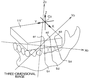
[0048] In Fig. 6, the three-dimensional image is
displayed based on the display coordinate system (X0,
Y0, ZO) . Points al, bl, a2, b2, a3, b3 for specifying
the implant implantation positions and the implant
implantation directions are represented based on the
display coordinate system (X0, Y0, ZO) as follows:
al=(xOal, y0al, zOal)
bl=(xObl, y0bl, zObl)
a2=(xOa2, yOa2, zOa2)
b2=(xOb2, yOb2, zOb2)
a3=(xOa3, yOa3, zOa3)
b3=(xOb3, yOb3, zOb3)
[0 049] A line segment extending between the points al
CA 02664351 2009-03-24
18
and bl is represented as follows:
x x0a1 XObl
y -~l-t) yoa] +r yonl , o<r<1
Z Zopi Zoni
[0050] On the other hand, an image 11' of the
processing portion 11 of the guide block 10 is also shown
in the three-dimensional image of Fig. 6. The corner
CO is also shown. In the three-dimensional image of
Fig. 6, the coordinates of the corner CO are represented
as follows:
C0=(xOcO, y0c0, zOcO)
The X-, Y- and Z-axes defined by the three edges of the
processing portion 11 as extending through the corner
CO are also defined based on the three-dimensional
display coordinate system (X0, Y0, ZO).
[0051] That is, the coordinates (X, Y, Z) are
represented as follows:
(X, Y, Z)=(8C0X0+C0, 6COYO+CO, 6COZO+CO)
wherein ACO is a difference between X0 and X, between
Y0 and Y or between ZO and Z.
[0052] Thus, in the three-dimensional image, the
implant implantation positions, the implant
implantation directions (implantation angles), the
position of the corner CO of the processing portion 11
of the guide block 10 and the orientation of the
CA 02664351 2009-03-24
19
processing portion 11 (directions of the X-, Y- and
Z-axes) are defined as data based on the
three-dimensional image display coordinate system.
[0053] Next, the data based on the three-dimensional
image display coordinate system is transformed into
data based on the processing reference coordinate
system defined by the corner CO of the processing
portion 11 of the guide block 10 and the X-, Y- and Z-
axes.
[0054] The transformation is carried out, for example,
in the following manner.
[0055] Provided that the coordinates of the origin are
represented by (XcO, YcO, ZcO) in the processing
reference coordinate system on the guide block 10, the
coordinates of the origin are represented by (XOcO, YOcO,
ZOcO) in the three-dimensional display coordinate
system.
[0056] On the other hand, provided that coordinates
associated with the implant implantation positions and
the implant implantation directions (implantation
angles) determined on the three-dimensional image
through the diagnosis are represented by (XOal, YOal,
ZOa1) on the three-dimensional image, the coordinates
(based on the three dimensional display coordinate
system) are transformed into coordinates based on the
CA 02664351 2009-03-24
processing reference coordinate system in the following
manner:
(XOal, YOal, ZOal)x(XcO, YcO, Zc0)=(XOcO, YOcO, ZOcO)
=(Xal, Yal, Zal)
[0057] Next, as shown in Fig. 7, the guide block 10
is set in a cutting processor 50, and fixed in position
by a fixing device 51. Then, the guide block 10 is
processed into a shape such as to guide the dental
implants by a cutting machine 52.
[0058] In the cutting process, the reference
coordinate system on the guide block 10 and the
coordinate data of the implantation positions and the
implantation directions based on the reference
coordinate system (obtained through the
transformation) are provided, so that the cutting
machine 52 can automatically cut the guide block 10 into
a shape such as to properly guide the dental implants.
The cutting may be carried out semi-automatically,
semi-manually, or manually with reference to the data,
rather than automatically.
[0059] The resulting guide block 10 serves as the
implant implantation guide 100.
[0060] Fig. 8 illustrates one example of the implant
implantation guide 100. The implant implantation
guide 100 includes a generally U-shaped portion 11'
= CA 02664351 2009-03-24
21
produced by processing the processing portion 11 and
covering the dental arch, guide grooves Gl, G2, G3
formed in the U-shaped portion 11' and an attachment
portion 12.
[0061] The guide grooves Gl, G2, G3 are each
dimensioned such as to guide a drill (a drill shaft or
a bar), serving as a drill (drill shaft or bar) guide
groove. Alternatively, the guide grooves G1, G2, G3
may each serve as a head guide groove for guiding a head
of a hand piece in which the drill is chucked (a groove
having a greater size than the drill guide groove).
[0062] The attachment portion 12 of the implant
implantation guide 100 to be fitted on the dental arch
is composed of the acryl resin. The attachment portion
12 is perfectly fitted on the patient's dental arch
without a gap or a play. Therefore, the implant
implantation guide 100 fitted on the patient's dental
arch makes it possible to properly drill implant
implantation holes at the positions previously
determined through the diagnosis in the patient's
jawbone. That is, the drill is operated according to
the implant implantation guide 100, whereby the implant
implantation holes can be properly and speedily drilled
in the directions at the positions previously
determined through the diagnosis. Then, the dental
CA 02664351 2009-03-24
22
implants are implanted at these positions.
[0063] In the aforementioned embodiment, the
processing reference coordinate system for the
processing of the processing portion 11 of the guide
block 10 is defined by the single corner CO and the three
edges defined as the three orthogonal lines. However,
how to define the processing reference coordinate
system for the processing of the processing portion 11
is not limited to the aforementioned method.
[0064] For example, as disclosed in Japanese Patent
Application No. 2004-334936 previously filed by the
applicant of the present invention, a processing
reference plane or a processing reference coordinate
system may be defined based on three points
preliminarily specified.
[0065] In this case, the processing portion 11 is not
necessarily required to be composed of the imageable
material, but may be composed of a material such that
the at least three points are imageable on the CT image.
[0066] More specifically, the processing portion 11
of the guide block 10 may be configured such that, when
the processing portion 11 is imaged through the CT
imaging to provide CT image data, the at least three
points required for defining the processing reference
plane or the processing reference coordinate system are
CA 02664351 2009-03-24
23
shown on the three*-dimensional image formed based on
the resulting CT image data. For example, the
processing portion 11 may be entirely composed of the
non-imageable material, and the three points required
for specifying the position of the processing portion
11 may be composed of an imageable material.
Alternatively, straight lines required for defining the
processing reference coordinate system may be drawn
with an imageable material on the processing portion
11.
[0067] In the embodiment described above, the guide
block 10 prepared for the production of the implant
implantation guide includes the processing portion 11
and the attachment portion 12 provided as a unitary
member. However, at the initial stage, the processing
portion 11 and the attachment portion 12 of the guide
block 10 may be provided as separate members.
[0068] Fig. 9 illustrates the guide block 10 having
such a structure.
[0069] Referring to Fig. 9, a guide base (resin base)
12 serving as the attachment portion is fitted on the
dental arch model of the plaster. The guide base 12
is composed of, for example, an acryl resin
(non-imageable material), and unitarily includes an
imaging marker 114 including at least three balls 111,
CA 02664351 2009-03-24
24
112, 113. The three balls 111, 112, 113 each have an
imageable member which defines a center thereof.
[0070] The processing portion 11 may have the same
construction as that described with reference to Fig.
2. For example, the processing portion 11 has a
rectangular plan shape and a predetermined thickness
h, and is composed of an imageable material (e.g.,
aluminum, apatite or the like).
[0071] The guide base (resin base) 12 including the
imagingmarker 114, and the processing portion 11 having
the processing coordinate system required for the
processing are separately prepared, and only the guide
base (resin base) 12 is fitted in the patient's oral
cavity. Then, the CT imaging is carried out to provide
the CT image data.
[0072] If it is difficult to fit the guide block 10
shown in Fig. 3 in the patient's oral cavity because
the patient has a smaller mouth or suffers from a
sensitive vomiting reflex, the fitting of the resin base
12 shown in Fig. 9 alleviates a burden on the patient
during the CT imaging.
[0073] In this case, the imaging marker 114 including
the at least three balls 111, 112, 113 is provided
unitarily with the resin base 12, so that a relationship
between a marker coordinate system defined by the three
CA 02664351 2009-03-24
centers of the three balls 111, 112, 113 and the
processing reference coordinate system of the
processing portion 11 to be attached after the imaging
can be determined by means of a three-dimensional
measurement apparatus by utilizing an existing
technique. Therefore, information of the implant
implantation positions and the implant implantation
directions determined on the CT image through the
diagnosis can be transformed into the coordinate
information required for the processing of the
processing portion 11 of the guide block 10 via the
marker coordinate system defined by the imaging marker
114. The coordinate information is used for cutting
the guide block 10 through CAD/CAM.
[0074] The present invention is not limited to the
embodiments described above, but various modifications
may be made within the scope of the present invention
defined by the following claims.