Note: Descriptions are shown in the official language in which they were submitted.
CA 02664879 2013-07-08
- 1 -
BIODEGRADABLE OCULAR IMPLANTS AND METHODS FOR
TREATING OCULAR CONDITIONS
Cross-Reference to Related Application
This application claims the benefit of U.S. Provisional Patent Application
Serial Number 60/848,563, filed September 29, 2006, entitled OCULAR
IMPLANTS INCLUDING NATURAL BIODEGRADABLE
POLYSACCHARIDES AND METHODS FOR TREATING OCULAR
CONDITIONS.
Technical Field
The present invention relates to ocular implants comprising a biodegradable
material and a bioactive agent. The bioactive agent can provide a therapeutic
effect
to treat on ocular condition.
Background
In recent years, much attention has been given to site-specific delivery of
drugs within a patient. Although various drugs have been developed for
treatment of
a wide variety of ailments and diseases of the body, in many instances, such
drugs
cannot be effectively administered systemically without risk of detrimental
side
effects. Site-specific drug delivery focuses on delivering the drugs locally,
i.e., to
the area of the body requiring treatment. One benefit of the local release of
bioactive
agents is the avoidance of toxic concentrations of drugs that are at times
necessary,
when given systemically, to achieve therapeutic concentrations at the site
where
they are required.
Site-specific drug delivery can be accomplished by injection and/or
implantation of an article or device that releases the drug to the treatment
site.
Injection of drugs can have limitations, for example, by requiring multiple
administrations, increasing risk of complications (such as infection), and
patient
discomfort. Implantation of an article or device that delivers drug to the
treatmentsite has therefore gained much interest in recent years.
Further, site-specific drug delivery has been enhanced by technologies that
allow controlled release of one or more drugs from an implanted device or
article.
CA 02664879 2009-03-30
WO 2008/060359
PCT/US2007/020959
- 2 -
Controlled release can relate to the duration of time drug is released from
the device
or article, and/or the rate at which the drug is released.
Several challenges confront the use of medical devices or articles that
release
bioactive agents into a patient's body. For example, treatment may require
release
of the bioactive agent(s) over an extended period of time (for example, weeks,
months, or even years), and it can be difficult to sustain the desired release
rate of
the bioactive agent(s) over such long periods of time. Further, the device or
article
surface is preferably biocompatible and non-inflammatory, as well as durable,
to
allow for extended residence within the body.
10. Generally
speaking, a bioactive agent can be associated with the surface of a
medical device or article by surface modification, embedded, and released from
within polymeric materials (matrix-type), or surrounded by and released
through a
carrier (reservoir-type). The polymeric materials in such applications should
optimally act as a biologically inert barrier and not induce further undesired
tissues
responses within the body, such as a strong inflammatory response. However,
many polymers used in association with medical devices do not provide ideal
properties when placed in the body.
Synthetic biodegradable polymers, such as polyglycolide-type molecules,
have been used for the construction of implantable medical devices and for
delivery
of bioactive agents. While there has been an abundance of prior art relating
to these
devices, some concerns exist that regard the use of synthetic materials which
degrade into materials that are not typically found in the body, or that are
found at
particularly low levels in the body. These types of biodegradable materials
have the
potential to degrade into products that cause unwanted side effects in the
body by
virtue of their presence or concentration in vivo. These unwanted side effects
can
include immune reactions, toxic buildup of the degradation products in the
body, or
the initiation or provocation of other adverse effects on cells or tissue in
the body.
Another problem is that preparations of some biodegradable materials may
not be obtained at consistent purity due to variations inherent in natural
materials.
This is relevant at least with regard to biodegradable materials derived from
animal
sources. Inconsistencies in preparations of biodegradable materials can result
in
problematic implantable devices.
CA 02664879 2009-03-30
WO 2008/060359
PCT/US2007/020959
- 3 -
Additional concerns are that preparations from animal sources may provide
other unwanted contaminants, such as antigenic factors. These antigenic
factors
may promote a localized immune response in the vicinity of the implanted
article
and foul its function. These factors may also cause infection as well as local
inflammation.
In addition, the delivery of bioactive agents within limited access regions of
the body can present additional challenges. Limited access regions of the body
can
be characterized in terms of physical accessibility as well as therapeutic
accessibility. For example, the relatively small size and sensitive tissues
-
surrounding the eye can contribute to physical accessibility difficulties. In
addition,
ocular absorption of systemically administered pharmacologic agents is limited
by
the blood ocular barrier, namely the tight junctions of the retinal pigment
epithelium
and vascular endothelial cells. These can make accessing the eye with
therapeutics
difficult. High systemic doses of bioactive agents can penetrate this blood
ocular
barrier in relatively small amounts, but expose the patient to the risk of
systemic
toxicity. Intravitreal injection of bioactive agents (such as drugs) is an
effective
means of delivering a drug to the posterior segment of the eye in high
concentrations. However, these repeated injections carry the risk of such
complications as infection, hemorrhage, and retinal detachment. Patients also
often
find this procedure somewhat difficult to endure.
Because description of the invention will involve treatment of the eye as an
illustrative embodiment, basic anatomy of the eye will now be described in
some
detail with reference to Figure 1, which illustrates a cross-sectional view of
the eye.
Beginning from the exterior of the eye, the structure of the eye includes the
iris 38
that surrounds the pupil 40. The iris 38 is a circular muscle that controls
the size of
the pupil 40 to control the amount of light allowed to enter the eye. A
transparent
external surface, the cornea 30, covers both the pupil 40 and the iris 38.
Continuous
with the cornea 30, and forming part of the supporting wall of the eyeball, is
the
sclera 28 (the white of the eye). The pars plana is a region of the eye
approximately
4 mm posterior to the point on the globe where the colored iris 38 meets the
white
sclera 28. The pars plana encircles the iris and is not constant in width, but
rather
typically varies between 2-3 mm in width around the iris (with the largest
width of
CA 02664879 2009-03-30
WO 2008/060359
PCT/US2007/020959
- 4 -
the pars plana typically lying on the temporal side and measuring about 3 mm
in
width).
The conjunctiva 32 is a clear mucous membrane covering the sclera 28.
Within the eye is the lens 20, which is a transparent body located behind the
iris 38.
The lens 20 is suspended by ligaments attached to the anterior portion of the
ciliary
body 21. Light rays are focused through the transparent cornea 30 and lens 20
upon
the retina 24. The central point for image focus (the visual axis) in the
human retina
is the fovea (not shown in the figures). The optic nerve 42 is located
opposite the
lens.
There are three different layers of the eye, the external layer, formed by the
sclera 28 and cornea 30; the intermediate layer, which is divided into two
parts,
namely the anterior (iris 38 and ciliary body 21) and posterior (the choroid
26); and
the internal layer, or the sensory part of the eye, formed by the retina 24.
The sclera
28 is composed of dense, fibrous tissue and is composed of collagen fiber.
Scleral
thickness is approximately 1 mm posteriorly near the optic nerve and
approximately
0.3 mm anteriorly. At the pars plana, the eye tissues are composed of sclera
only;
there is no choroidal or retinal tissue layer within this region. For this
reason, the
avascular pars plana is typically selected for implantation and/or injection
of
materials into the interior (vitreous) of the eye.
The lens 20 divides the eye into the anterior segment (in front of the lens)
and the posterior segment (behind the lens). More specifically, the eye is
composed
of two chambers of fluid: the anterior chamber 34 (between the cornea 30 and
the
iris 38), and the vitreous chamber 22 (between the lens 20 and the retina 24).
The
anterior chamber 34 is filled with aqueous humor whereas the vitreous chamber
22
is filled with a more viscous fluid, the vitreous humor.
The vitreous chamber 22 is the largest chamber of the eye, consisting of
approximately 4.5 ml of fluid. The vitreous chamber is filled with a
transparent gel
composed of a random network of thin collagen fibers in a highly dilute
solution of
salts, proteins and hyaluronic acid (the vitreous humor comprises
approximately
98% water).
CA 02664879 2009-03-30
WO 2008/060359
PCT/US2007/020959
- 5 -
Summary of the Invention
In one aspect, the present invention provides biodegradable implants that are
particularly useful for delivering bioactive agents to a treatment site within
a body.
In particular, the biodegradable implants can be configured for placement and
release of the bioactive agent in the interior of the eye. Upon implantation,
bioactive
agent can be released from the implant and provide a therapeutic effect at the
treatment site. In particular, the biodegradable implants can be placed in a
portion
of the eye and are herein referred to as ocular implants.
According to experimental studies associated with the invention, small
biodegradable ocular implants having a polypeptide agent were prepared and
placed
in the inner eye of a mammal in a minimally invasive manner. Pharmacokinetic
analysis revealed that these implants were capable of releasing polypeptide to
the
vitreal fluid in amounts suitable for the treatment of ocular conditions.
Notably,
analysis also revealed that the implants released the polypeptide over a
prolonged
period of time after placement of implant in the eye (i.e., for periods of
time of one
about month or greater following implantation).
Explant analysis from the experimental studies also revealed that bioactive
agent activity was maintained in the implant over the period of treatment. In
view of
this result, the implant not only provides a suitable matrix for the retention
and
release of a bioactive agent over these longer time periods, but also prevents
loss of
bioactive agent activity over the course of treatment.
Experimental studies also showed that implant formulations could be altered
to adjust the delivery rate and the delivery period of the polypeptide from
the
implant, without compromising the bioactivity of the polypeptide. This
"tunability"
of the implant system provides great advantages for the treatment of ocular
conditions requiring administration of bioactive agent over prolonged periods
of
time, and accommodates the preparation of implants having a wide variety of
bioactive agents and bioactive agent release profiles.
In one aspect, the invention provides a biodegradable implant for delivery of
a bioactive agent to the interior of the eye, wherein the implant comprises a
matrix
comprising a biodegradable polymer and a bioactive agent and is capable
of releasing a therapeutically effective amount of bioactive agent in the
interior of
CA 02664879 2009-03-30
WO 2008/060359
PCT/US2007/020959
- 6 -
the eye after a period of about 30 days from implantation. In another aspect,
the
ocular implant is configured for delivery of a bioactive agent to the eye,
wherein at
least a portion of the bioactive agent is released from the implant after a
period of
implantation of about three months or greater.
The ocular implant can have certain dimensions desirable for delivering
and/or immobilizing the implant to and/or at a target location in the eye. In
many
cases, the ocular implant of the invention can be delivered to the eye in a
minimally
invasive manner. In some aspects, the implant is sized so that the method of
insertion does not require additional procedures to be performed during or
after the
insertion process, such as suturing of the sclera. Therefore, the implant is
configured so that it can be placed at a location in the inner eye using a
sutureless
procedure. In some aspects the ocular implant is configured for placement
within a
needle having a size of 25 gauge or smaller.
In some aspects, the implant has an elongate shape. The elongate shape can
be that of a rod, cylinder, or filament. In one specific embodiment, the
ocular
implant comprises a length of about 5 mm or less. In another specific
embodiment,
the ocular implant comprises a diameter of about 0.35 mm or less. For example,
the
ocular implant can have a cylindrical or rod-like shape, and the diameter of
the
implant is about 0.35 mm or less. In one specific embodiment, the ocular
implant
comprises a diameter of about 0.35 mm or less, and a length of about 5 mm or
less.
These dimensions can provide advantages for the insertion of the implant into
a
portion of the eye.
In another specific embodiment, the ocular implant has a weight of about 6
mg or less. In another specific embodiment, the ocular implant has a weight of
about 2.5 mg or less.
The ocular implants can have a defined structure and can be formed by any
suitable process, including molding, extruding, shaping, cutting, casting, and
the
like.
In some aspects, the biodegradable implants include a matrix of natural
biodegradable polysaccharides and a bioactive agent. In preparing these types
of
ocular implants, a plurality of natural biodegradable polysaccharides are
crosslinked
to each other via coupling groups that are pendent from the natural
biodegradable
CA 02664879 2009-03-30
WO 2008/060359
PCT/US2007/020959
- 7 -
polysaccharide (i.e., one or more coupling groups are chemically bonded to the
polysaccharide). In some aspects, the coupling group on the natural
biodegradable
polysaccharide is a polymerizable group. In a free radical polymerization
reaction
the polymerizable group can crosslink natural biodegradable polysaccharides
together in the composition, thereby forming a natural biodegradable
polysaccharide
matrix. A bioactive agent useful for treating an ocular condition of
indication is
included within the matrix. The matrix can be in the form of an implant having
a
size and configuration for placement in a portion of the eye.
Ocular implants formed of natural biodegradable polysaccharides can be
enzymatically degraded within a portion of the eye. These types of ocular
implants
also offer the advantage of being generally non-enzymatically hydrolytically
stable.
This is particularly advantageous for bioactive agent delivery since the
bioactive
agent can be released from the implant under conditions of enzyme-mediated
degradation. The kinetics of bioactive agent release from the ocular implant
of the
present invention can provide an advantage over the release of drugs retained
within
systems prepared from synthetic biodegradable materials, such as
poly(lactides).
Natural biodegradable polysaccharides include polysaccharide and/or
polysaccharide derivatives that are obtained from natural sou'rces, such as
plants or
animals. Exemplary natural biodegradable polysaccharides include amylose,
maltodextrin, amylopectin, starch, dextran, hyaluronic acid, heparin,
chondroitin
sulfate, dermatan sulfate, heparan sulfate, keratan sulfate, dextran sulfate,
pentosan
polysulfate, and chitosan. Preferred polysaccharides are low molecular weight
polymers that have little or no branching, such as those that are derived from
and/or
found in starch preparations, for example, amylose, maltodextrin, and
polyalditol.
Because of the particular utility of the amylose, maltodextrin, and
polyalditol
polymers, in some aspects natural biodegradable polysaccharides are used that
have
an average molecular weight of 500,000 Da or less, 250,000 Da or less, 100,000
Da
or less, or 50,000 Da or less. In some aspects the natural biodegradable
polysaccharides have an average molecular weight of 500 Da or greater. In some
aspects the natural biodegradable polysaccharides have an average molecular
weight
in the range of about 1000 Da to about 10,000 Da. Natural biodegradable
polysaccharides of particular molecular weights can be obtained commercially
or
CA 02664879 2009-03-30
WO 2008/060359
PCT/US2007/020959
- 8 -
can be prepared, for example, by acid hydrolysis and/or enzymatic degradation
of a
natural biodegradable polysaccharide preparation, such as starch. The decision
of
using natural biodegradable polysaccharides of a particular size range may
depend
on factors such as the desired physical characteristics of the ocular implant,
the
desired rate of degradation of the implant, and the type of bioactive agent
present in
the implant.
The natural biodegradable polysaccharides that are used in accordance with
the methods and compositions of the invention are readily available at \a low
cost
and/or can be prepared easily using established techniques. This allows for a
cost
effective method of fabricating ocular implants.
The use of natural biodegradable polysaccharides, such as maltodextrin or
amylose, provides many advantages when used for the formation of an ocular
implant. Degradation of a natural biodegradable polysaccharide-containing
ocular
implant can result in the release of, for example, naturally occurring mono-
or
disaccharides, such as glucose, which are common components of bodily fluids,
such as the vitreous humor. Furthermore, the use of natural biodegradable
polysaccharides that degrade into common components found in bodily fluids,
such
as glucose, can be viewed as more acceptable than the use of synthetic
biodegradable polysaccharides that degrade into non-natural compounds, or
compounds that are found at very low concentrations in the body.
In some aspects of the invention, this advantageous feature is reflected in
the
use of natural biodegradable polysaccharides which are non-animal derived,
such as
amylose and maltodextrin, and that degrade into products that present little
or no
immunogenic or toxic risk to the individual. The invention provides improved,
cost-
efficient, natural biodegradable polysaccharide compositions for articles that
can be
used in a variety of treatments for the eye.
Another advantage of the invention is that the natural biodegradable
polysaccharide-based ocular implant are more resistant to hydrolytic
degradation
than other biodegradable polymers, such as poly(lactides). Degradation of the
matrices prepared from natural biodegradable polysaccharides of the invention
are
primarily enzyme-mediated, with minimal or no hydrolysis of the natural
biodegradable polysaccharide occurring when a natural biodegradable
CA 02664879 2009-03-30
WO 2008/060359
PCT/US2007/020959
- 9 -
polysaccharide-containing composition is prepared under ambient conditions.
This
allows the natural biodegradable polysaccharide-based ocular implant to remain
substantially stable (for example, resistant to degradation) prior to placing
the
implant into a portion of the eye. For example, a natural biodegradable
polysaccharide ocular implant can be manipulated in a non-biological, aqueous-
based-medium without risk that the implant will prematurely degrade due to non-
enzyme-meditated hydrolysis. Systems that are based on biodegradable polymers
such as poly(lactide) or poly(lactide-co-glycolide) are subject to hydrolysis
even at
relatively neutral pH ranges (e.g., pH 6.5 to 7.5) and therefore do not offer
this
advantage. The properties of the polymer systems of the present invention
provide
ocular implant with improved storage characteristics.
In some aspects, the invention provides a bioactive agent-releasing
biodegradable ocular implant comprising (i) a matrix of natural biodegradable
polysaccharides (ii) and a bioactive agent within the matrix. The implant is
configured to reside in a portion of the eye and comprises an amount of
bioactive
agent useful for treating an ocular condition or indication. The implant is
prepared
having a matrix of natural biodegradable polysaccharides that includes
bioactive
agent, wherein the matrix is slowly degradable in the presence of ocular
fluids
and/or tissues.
The ocular implant can be prepared having any suitable bioactive agent for
the treatment of an ocular condition or indication. Illustrative bioactive
agents
include antiproliferative agents, anti-inflammatory agents, anti-angiogenic
agents,
hormonal agents, antibiotics, neurotrophic factors, or combinations thereof.
In some aspects, the implant includes a larger hydrophilic bioactive agent,
such as a polypeptide, nucleic acid, polysaccharide, or combinations thereof
Viral
particles and cells can also be included in the ocular implant. The implant
provides
a distinct advantage for delivering these larger bioactive agents.
Comparatively, use
of non-degrading drug delivery matrices may not allow delivery of these larger
bioactive agents if too large to diffuse out of the matrix. However, an ocular
implant
that includes a matrix of natural biodegradable polysaccharides allows release
of the
bioactive agent upon degradation of the matrix. In some aspects of the
invention,
CA 02664879 2009-03-30
WO 2008/060359
PCT/US2007/020959
- 10 -
the ocular implant comprises a bioactive agent having a molecular weight of
about
10,000 Da or greater.
In some aspects the ocular implant comprises a bioactive agent that is a high
molecular weight compound and that is an inhibitor of angiogenesis. For
example,
the inhibitor can be selected from angiostatin, thrombospondin, anti-VEGF
antibody, and anti-VEGF fragment. In some aspects the ocular implant comprises
a
bioactive agent that is a high molecular weight compound and a hormonal agent.
For example, the bioactive agent could be ciliary neurotrophic factor or
pigment
endothelium derived growth factor.
The ocular implant can also include lower molecular weight compounds. In
some aspects these compounds are held within the matrix of the implant in
particulate form. For example, the bioactive agent can be present in the form
of
microparticles that are immobilized in the matrix of natural biodegradable
polysaccharide. In some aspects the bioactive agent is an antiproliferative
agent,
such as 13-cis retinoic acid, retinoic acid derivatives, 5-fluorouracil,
taxol, sirolimus
(rapamycin), analogues of rapamycin, tacrolimus, ABT-578, everolimus,
paclitaxel,
taxane, or vinorelbine. In some aspects the bioactive agent is an anti-
inflammatory
agent such as hydrocortisone, hydrocortisone acetate, dexamethasone 21-
phosphate,
fluocinolone, medrysone, methylprednisolone, prednisolone 21-phosphate,
prednisolone acetate, fluoromethalone, betamethasone, triamcinolone, or
triamcinolone acetonide. In some aspects the bioactive agent is an inhibitor
of
.angiogenesis such as anecortave acetate or a receptor tyrosine kinase
antagonist.
A bioactive agent can also be included in an ocular implant prepared using a
natural biodegradable polysaccharide that is modified with a hydrophobic
moiety.
The hydrophobic moiety can be used to provide a biodegradable matrix having
hydrophobic properties. The hydrophobic moieties can be pendent from the
polysaccharide chain. Exemplary hydrophobic moieties include fatty acids and
derivatives thereof, and C2-C18 alkyl chains.
In some aspects of the invention, the bioactive agent can be coupled to and
cleavable from the polysaccharide. For example, a bioactive agent can be
covalently
attached to the polysaccharide via an ester bond. Upon implantation into a
portion
of the eye, the bond can be hydrolyzed resulting in the release of the
bioactive agent
CA 02664879 2009-03-30
WO 2008/060359
PCT/US2007/020959
- 11 -
which provides a therapeutic effect. Illustrative therapeutically useful
bioactive
agents include butyric acid, valproic acid, retinoic acid, and the like.
The invention also provides a method for delivery of a bioactive agent, or
more than one bioactive agent, to a subject for the treatment of an ocular
condition
or indication.
In one aspect, the invention provides a method for administering a bioactive
agent to the inner eye, the method comprising the steps of (a) providing a
biodegradable implant comprising a matrix comprising a biodegradable polymer
and
a bioactive agent, wherein the implant is configured so that it can be placed
at a
location in the inner eye, (b) implanting the implant in the inner eye, and
(c)
maintaining the implant in the inner eye, wherein the implant releases a
therapeutically effective amount of bioactive agent in the inner eye after a
period of
30 days from the step of implanting.
In some aspects, the step of implanting comprises implanting the implant in
the inner eye using a sutureless procedure.
In another aspect, the invention provides a method comprising the steps of
providing an ocular implant comprising (a) a matrix of natural biodegradable
polysaccharides and (b) a bioactive agent within the matrix to a portion of
the eye.
The method also comprises a step of maintaining the implant in the portion of
the
eye for a period of time sufficient for the treatment of an ocular condition
of
indication.
Within the eye the ocular implant is exposed to a carbohydrase that promotes
the degradation of the matrix and release of the bioactive agent. For example,
an
ocular implant including amylose and/or maltodextriri polymers can be exposed
to
an a-amylase to promote degradation of the implant and release of the
bioactive
agent. During the step of maintaining the ocular implant generally is eroded
on its
surface and releases bioactive agent. Release of bioactive agent occurs until
the
implant is completely degraded.
Desirably, the ocular implant releases the bioactive agent over a prolonged
period of time to treat the ocular condition or indication. For example, the
ocular
implant can be maintained in the eye for a period of about three months or
greater to
provide treatment to the eye. This means that a portion of the ocular implant
CA 02664879 2009-03-30
WO 2008/060359
PCT/US2007/020959
- 12 -
remains in the eye and is able to release bioactive agent after a period of
three
months. The lifetime of the ocular implant may be greater than three months,
in the
range of three to eighteen months, in the range of three to twelve months, or
in the
range of three to six months.
The ocular condition or indication can be one or more selected from retinal
detachment; vascular occlusions; retinitis pi'gmentosa; proliferative
vitreoretinopathy; diabetic retinopathy; inflammations such as uveitis,
choroiditis,
and retinitis; degenerative disease (such as age-related macular degeneration,
also
referred to as AMD); vascular diseases; and various tumor-related conditions,
including those associated with neoplasms.
In yet further embodiments, the biodegradable medical article can be used
post-operatively, for example, as a treatment to reduce or avoid potential
complications that can arise from ocular surgery. In one such embodiment, the
medical article can be provided to a patient after cataract surgical
procedures, to
assist in managing (for example, reducing or avoiding) post-operative
inflammation.
In some aspects, the step of providing comprises placing the implant in
contact with retinal tissue. For example, the method can include providing the
implant to a subretinal location. In another aspect, the step of providing
comprises
placing the implant in the vitreous.
In some aspects, the method of treatment of an ocular condition or indication
comprises delivering the ocular implant to a target location in the eye via an
implant
delivery instrument. In some desired modes of practice, the ocular implant is
releasably associated with a distal end of the implant delivery instrument.
The step
of providing can include the sub-steps of (i) providing a system comprising a
delivery instrument and the ocular implant releasably associated with a
portion of
the instrument (ii) inserting the ocular implant and a portion of the
instrument into
the eye, and (iii) actuating the instrument to release the ocular implant at a
target
= location in the eye.
In some aspects of the invention, the implant is delivered to a portion of the
eye using an implant delivery instrument having a distal end with an outer
diameter
of about 0.5 mm or less. This can be particularly beneficial when it is
desirable to
CA 02664879 2009-03-30
WO 2008/060359
PCT/US2007/020959
- 13 -
minimize the size of any incision in the body, thereby reducing or avoiding
the use
of sutures or other closure-devices.
In other aspects, the invention provide a kit for placing a biodegradable
implant in the interior of the eye, the kit comprising a biodegradable implant
comprises a matrix comprising a biodegradable polymer and a bioactive agent
which
is capable of releasing a therapeutically effective amount of bioactive agent
in the
interior of the eye after a period of 30 days from implantation, and an
insertion
instrument to provide the implant to a target site within the eye.
Brief Description of the Drawings
Figure 1 is an illustration of a cross-sectional view of the eye.
Figure 2 is a graph of cumulative BSA release from maltodextrin-acrylate
filaments treated with amylase, over a period of time.
Figure 3 is a graph of cumulative absorbance values of active and total IgG
Fab fragment release from maltodextrin-acrylate filaments treated with
amylase,
over a period of time.
Figure 4 is a graph of cumulative absorbance values of active and total IgG
release from a maltodextrin-acrylate filament treated with amylase and percent
degradation of the filament, over a period of time.
Figure 5 is a graph of mass loss of biodegradable implants after periods of
time in vitro and in vivo.
Figure 6 is a graph of mass loss of biodegradable implants after periods of
time in vitro and in vivo.
Figure 7 is a graph of amounts of active F(Ab) fragment from explanted
biodegradable implants after periods of time in vivo.
Figure 8 is a graph of amounts of active F(Ab) fragment released from
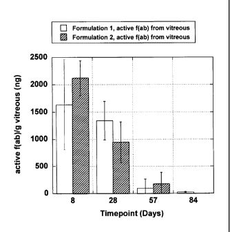
biodegradable implants in the vitreous after periods of time in vivo.
Figure 9 is a graph of amounts of total and active F(Ab) fragment released
from biodegradable implants after periods of time in vitro.
Figure 10 is a graph of amounts of active F(Ab) fragment from explanted
biodegradable implants after periods of time in vivo.
Figure 11 is a graph of mass of biodegradable implants remaining after
periods of time in vivo.
CA 02664879 2013-07-08
- 14 -
Detailed Description
The embodiments of the present invention described herein are not intended
to be exhaustive or to limit the invention to the precise forms disclosed in
the
following detailed description. Rather, the embodiments are chosen and
described
so that others skilled in the art can appreciate and understand the principles
and
practices of the present invention.
The publications and patents disclosed herein are provided solely for their
disclosure. Nothing herein is to be construed as an admission that the
inventors are
not entitled to antedate any publication and/or patent, including any
publication
and/or patent cited herein.
In some aspects, the polymeric compositions can be utilized in to form an
ophthalmic article, such as an ocular implant. The ocular implant can be
configured
for placement at an internal site of the eye. Suitable ocular implants in
accordance
with these aspects can provide bioactive agent to any desired area of the eye.
In
some aspects, the ocular implant is utilized to deliver bioactive agent to a
posterior
segment of the eye (behind the lens). The biodegradable polysaccharide
compositions described herein can be used for the formation of an ophthalmic
article, such as an ocular implant.
In some aspects, the ocular implant can be configured for placement at a
subretinal area within the eye. In some aspects the ocular implant is used in
association with an ophthalmic devices. Ophthalmic devices are described in
U.S.
Patent Publication No. 2005/0143363 ("Method for Subretinal Administration of
Therapeutics Including Steroids; Method for Localizing Pharmacodynamic Action
at
the Choroid and the Retina; and Related Methods for Treatment and / or
Prevention
of Retinal Diseases," de Juan et al.); U.S. Application No. 11/175,850
("Methods
and Devices for the Treatment of Ocular Conditions," de Juan et al.); and
related
applications.
In some aspects, the invention provides a biodegradable implant that is
formed from the biodegradable polysaccharide and that includes a bioactive
agent,
such as a high molecular weight bioactive agent useful for treating an ocular
condition.
CA 02664879 2009-03-30
WO 2008/060359
PCT/US2007/020959
- 15 -
As referred to herein, a "natural biodegradable polysaccharide" refers to a
non-synthetic polysaccharide that is capable of being enzymatically degraded
but
that is generally non-enzymatically hydrolytically stable. Natural
biodegradable
polysaccharides include polysaccharide and/or polysaccharide derivatives that
are,
obtained from natural sources, such as plants or animals. Natural
biodegradable
polysaccharides include any polysaccharide that has been processed or modified
from a natural biodegradable polysaccharide (for example, maltodextrin is a
natural
biodegradable polysaccharide that is processed from starch). Exemplary natural
biodegradable polysaccharides include hyaluronic acid, starch, dextran,
heparin,
chondroitin sulfate, dermatan sulfate, heparan sulfate, keratan sulfate,
dextran
sulfate, pentosan polysulfate, and chitosan. Preferred polysaccharides are low
molecular weight polymers that have little or no branching, such as those that
are
derived from and/or found in starch preparations, for example, amylose and
maltodextrin. Therefore, the natural biodegradable polysaccharide can be a
substantially non-branched or non-branched poly(glucopyranose) polymer.
Because of the particular utility of the amylose and maltodextrin polymers, it
is preferred that natural biodegradable polysaccharides having an average
molecular
weight of 500,000 Da or less, 250,000 Da or less, 100,000 Da or less, or
50,000 Da
or less. It is also preferred that the natural biodegradable polysaccharides
have an
average molecular weight of 500 Da or greater. A particularly preferred size
range
for the natural biodegradable polysaccharides is in the range of about 1000 Da
to
about 10,000 Da. Natural biodegradable polysaccharides of particular molecular
weights can be obtained commercially or can be prepared. The decision of using
natural biodegradable polysaccharides of a particular size range may depend on
factors such as the physical characteristics of the composition (e.g.,
viscosity) used
to form the implant, the desired rate of degradation of the implant, the
presence of
other components in the composition used to form the implant, for example,
bioactive agents, etc.
As used herein, "amylose" or "amylose polymer" refers to a linear polymer
having repeating glucopyranose units that are joined by a-1,4 linkages. Some
amylose polymers can have a very small amount of branching via a-1,6 linkages
(about less than 0.5% of the linkages) but still demonstrate the same physical
CA 02664879 2009-03-30
WO 2008/060359
PCT/US2007/020959
- 16 -
properties as linear (unbranched) amylose polymers do. Generally amylose
polymers derived from plant sources have molecular weights of about I X 106 Da
or
less. Amylopectin, comparatively, is a branched polymer having repeating
glucopyranose units that are joined by a-1,4 linkages to form linear portions
and the
linear portions are linked together via a-1,6 linkages. The branch point
linkages are
generally greater than I% of the total linkages and typically 4% - 5% of the
total
linkages. Generally amylopectin derived from plant sources have molecular
weights
of 1 X 10⁷ Da or greater.
= Amylose can be obtained from, or is present in, a variety of sources.
Typically, amylose is obtained from non-animal sources, such as plant sources.
In
some aspects, a purified preparation of amylose is used as starting material
for the
preparation of the amylose polymer having coupling groups. In other aspects,
as
starting material, amylose can be used in a mixture that includes other
polysaccharides.
For example, in some aspects, starch preparations having a high amylose
content, purified amylose, synthetically prepared amylose, or enriched amylose
preparations can be used in the preparation of amylose having the coupling
groups.
In starch sources, amylose is typically present along with amylopectin, which
is a
branched polysaccharide. According to the invention, it is preferred to use
compositions that include amylose, wherein the amylose is present in the
composition in an amount greater than amylopectin, if present in the
composition.
For example, in some aspects, starch preparations having high amylose content,
purified amylose, synthetically prepared amylose, or enriched amylose
preparations
can be used in the preparation of amylose polymer having the coupling groups.
In
some embodiments the composition includes a mixture of polysaccharides
including
amylose wherein the amylose content in the mixture of polysaccharides is 50%
or
greater, 60% or greater, 70% or greater, 80% or greater, or 85% or greater by
weight. In other embodiments the composition includes a mixture of
polysaccharides including amylose and amylopectin and wherein the amylopectin
content in the mixture of polysaccharides is 30% or less, or 15% or less.
In some cases it may be desirable to use non-retrograding starches, such as
waxy starch, in the current invention. The amount of amylopectin present in a
starch
CA 02664879 2009-03-30
WO 2008/060359
PCT/US2007/020959
- 17 -
may also be reduced by treating the starch with amylopectinase, which cleaves
a-1,6
linkages resulting in the debranching of amylopectin into amylose.
In some cases a synthesis reaction can be carried out to prepare an amylose
polymer having pendent coupling groups (for example, amylose with pendent
ethylenically unsaturated groups) and steps may be performed before, during,
and/or
after the synthesis to enrich the amount of amylose, or purify the amylose.
Amylose of a particular size, or a combination of particular sizes can be
used. In some embodiments amylose having an average molecular weight of
500,000 Da or less, 250,000 Da or less, 100,000 Da or less, 50,000 Da or less,
preferably greater than 500 Da, or preferably in the range of about 1000 Da to
about
10,000 Da is used. Amylose of particular molecular weights can be obtained
commercially or can be prepared. For example, synthetic amyloses with average
molecular masses of 70, 110, 320, and 1,000 kDa can be obtained from Nakano
Vinegar Co., Ltd. (Aichi, Japan). The decision of using amylose of a
particular size
range may depend on factors such as the physical characteristics of the
composition
(e.g., viscosity) used to form the implant, the desired rate of degradation of
the
implant, the presence of other component in the composition used to form the
implant (for example, bioactive agents, etc.), etc.
Maltodextrin is typically generated by hydrolyzing a starch slurry with heat-
stable a-amylase at temperatures at 85 - 90 C until the desired degree of
hydrolysis
is reached and then inactivating the a-amylase by a second heat treatment. The
maltodextrin can be purified by filtration and then spray dried to a final
product.
Maltodextrins are typically characterized by their dextrose equivalent (DE)
value,
which is related to the degree of hydrolysis defined as: DE = MW
dextrose/number-
averaged MW starch hydrolysate x 100.
A starch preparation that has been totally hydrolyzed to dextrose (glucose)
has a DE of 100, where as starch has a DE of about zero. A DE of greater than
0 but
less than 100 characterizes the mean-average molecular weight of a starch
hydrolysate, and maltodextrins are considered to have a DE of less than 20.
Maltodextrins of various molecular weights, for example, in the range of about
500
¨ 5000 Da are commercially available (for example, from CarboMer, San Diego,
CA).
CA 02664879 2009-03-30
WO 2008/060359
PCT/US2007/020959
- 18 -
In some aspects, the ocular implant can include a natural biodegradable non-
reducing polysaccharide. The ocular implant can include a matrix having a
plurality
of natural biodegradable non-reducing polysaccharides along with a bioactive
agent,
such as a polypeptide. A non-reducing polysaccharide can provide an inert
matrix
thereby improving the stability of sensitive bioactive agents, such as
proteins and
enzymes. A non-reducing polysaccharide refers to a polymer of non-reducing
disaccharides (two monosaccharides linked through their anomeric centers) such
as
trehalose (a-D-glucopyranosyl a-D-glucopyranoside) and sucrose (13-D-
fructofuranosyl a-D-glucopyranoside). An exemplary non-reducing polysaccharide
comprises polyalditol which is available from GPC (Muscatine, Iowa). In
another
aspect, the polysaccharide is a glucopyranosyl polymer, such as a polymer that
includes repeating (1¨>3)0-13-D-glucopyranosyl units. Biodegradable non-
reducing
polysaccharides can be useful for formulating ocular implants that release the
bioactive agent over a prolonged period of time, such as about three months or
greater.
Refinement of the molecular weight of a polysaccharide preparation can be
carried out using diafiltration. Diafiltration of polysaccharides such as
maltodextrin
can be carried out using ultrafiltration membranes with differing pore sizes.
As an
example, use of one or more cassettes with molecular weight cut-off membranes
in
the range of about 1K to about 30 K can be used in a diafiltration process to
provide
polysaccharide preparations with average molecular weights in the range of
less than
K Da, in the range of about 5 K Da to about 30 K Da, in the range of about 10
K
Da to about 30 K Da, or in the range of about 1 K Da to about 10 K Da.
In some aspects, the ocular implant can include natural biodegradable
25 polysaccharides that include chemical modifications other than the
pendent coupling
group. To exemplify this aspect, modified amylose having esterified hydroxyl
groups can be prepared and used in compositions in association with the
implants
and methods of the invention. Other natural biodegradable polysaccharides
having
hydroxyl groups may be modified in the same manner. These types of
modifications
30 can change or improve the properties of the natural biodegradable
polysaccharide
making for an implant composition that is particularly suitable for a desired
CA 02664879 2009-03-30
WO 2008/060359
PCT/US2007/020959
- 19 -
application. Many chemically modified amylose polymers, such as chemically
modified starch, have at least been considered acceptable food additives.
As used herein, "modified natural biodegradable polysaccharides" refers to
chemical modifications to the natural biodegradable polysaccharide that are
different
than those provided by the coupling group or the initiator group. Modified
amylose
polymers having a coupling group (and/or initiator group) can be used to form
the
ocular implants of the invention.
To exemplify this aspect, modified amylose is described. By chemically
modifying the hydroxyl groups of the amylose, the physical properties of the
amylose can be altered. The hydroxyl groups of amylose allow for extensive
hydrogen bonding between amylose polymers in solution and can result in
viscous
solutions that are observed upon heating and then cooling amylose-containing
compositions such as starch in solution (retrograding). The hydroxyl groups of
amylose can be modified to reduce or eliminate hydrogen bonding between
molecules thereby changing the physical properties of amylose in solution.
Therefore, in some embodiments the natural biodegradable polysaccharides,
such as amylose, can include one or more modifications to the hydroxyl groups
wherein the modifications are different than those provided by coupling group.
Modifications include esterification with acetic anhydride (and adipic acid),
succinic
anhydride, 1-octenylsuccinic anhydride, phosphoryl chloride, sodium
trimetaphosphate, sodium tripolyphosphate, and sodium monophosphate;
etherification with propylene oxide, acid modification with hydrochloric acid
and
sulfuric acids; and bleaching or oxidation with hydrogen peroxide, peracetic
acid,
potassium permanganate, and sodium hypochlorite.
Examples of modified amylose polymers include carboxymethyl amylose,
carboxyethyl amylose, ethyl amylose, methyl amylose, hydroxyethyl amylose,
hydroxypropyl amylose, acetyl amylose, amino alkyl amylose, ally' amylose, and
oxidized amylose. Other modified amylose polymers include succinate amylose
and
oxtenyl succinate amylose.
In another aspect of the invention, the natural biodegradable polysaccharide
is modified with a hydrophobic moiety in order to provide a biodegradable
matrix
having hydrophobic properties. Exemplary hydrophobic moieties include those
CA 02664879 2009-03-30
WO 2008/060359 PCT/US2007/020959
- 20 -
previously listed, fatty acids and derivatives thereof, and C2-C18 alkyl
chains. A
polysaccharide, such as amylose or maltodextrin, can be modified with a
compound
having a hydrophobic moiety, such as a fatty acid anhydride. The hydroxyl
group of
a polysaccharide can also cause the ring opening of lactones to provide
pendent
open-chain hydroxy esters.
In some aspects, the hydrophobic moiety pendent from the natural
biodegradable has properties of a bioactive agent. The hydrophobic moiety can
be
hydrolyzed from the natural biodegradable polymer and released from the matrix
to
provide a therapeutic effect. One example of a therapeutically useful
hydrophobic
moiety is butyric acid, which has been shown to elicit tumor cell
differentiation and
apoptosis, and is thought to be useful for the treatment of cancer and other
blood
diseases. Other illustrative hydrophobic moieties include valproic acid and
retinoic
acid. Retinoic acid is known to possess antiproliferative effects and is
thought to be ,
useful for treatment of proliferative vitreoretinopathy (PVR). The hydrophobic
moiety that provides a therapeutic effect can also be a natural compound (such
as
butyric acid, valproic acid, and retinoic acid). Therefore, degradation of the
matrix
having a coupled therapeutic agent can produce natural degradation products.
In further aspects, the natural biodegradable polysaccharide can be modified
with a corticosteroid. In these aspects, a corticosteroid, such as
triamcinolone, can
be coupled to the natural biodegradable polymer. One method of coupling
triamcinolone to a natural biodegradable polymer is by employing a
modification of
the method described in Cayanis, E. et al., Generation of an Auto-anti-
idiotypic
Antibody that Binds to Glucocorticoid Receptor, The Journal of Biol. Chem.,
261(11): 5094-5103 (1986). Triamcinolone hexanoic acid is prepared by reaction
of
triamcinolone with ketohexanoic acid; an acid chloride of the resulting
triamcinolone hexanoic acid can be formed and then reacted with the natural
biodegradable polymer, such as maltodextrin or polyalditol, resulting in
pendent
triamcinolone groups coupled via ester bonds to the natural biodegradable
polymer.
Optionally, when the natural biodegradable polymer includes a pendent
hydrophobic moiety and/or corticosteroid, an enzyme, such as lipase, can be
used in
=
association with the implant to accelerate degradation of the bond between the
hydrophobic moiety and the polysaccharide (e.g., ester bond).
CA 02664879 2009-03-30
WO 2008/060359
PCT/US2007/020959
- 21 -
According to the invention, a natural biodegradable polysaccharide that
includes a coupling group is used to form the ocular implant. Other
polysaccharides
can also be present in the composition. For example, the two or more natural
biodegradable polysaccharides are used to form the ocular implant. Examples
include amylose and one or more other natural biodegradable polysaccharide(s),
and
maltodextrin and one or more other natural biodegradable polysaccharide(s); in
one
aspect the composition includes a mixture of amylose and maltodextrin,
optionally
with another natural biodegradable polysaccharide.
In one preferred embodiment, amylose or maltodextrin is the primary
polysaccharide. In some embodiments, the composition includes a mixture of
polysaccharides including amylose or maltodextrin and the amylose or
maltodextrin
content in the mixture of polysaccharides is 50% or greater, 60% or greater,
70% or
greater, 80% or greater, or 85% or greater by weight.
Purified or enriched amylose preparations can be obtained commercially or
can be prepared using standard biochemical techniques such as chromatography.
In
some aspects, high-amylose cornstarch can be used.
As used herein, "coupling group" can include (1) a chemical group that is
able to form a reactive species that can react with the same or similar
chemical
group to form a bond that is able to couple the natural biodegradable
polysaccharides together (for example, wherein the formation of a reactive
species
can be promoted by an initiator); or (2) a pair of two different chemical
groups that
are able to specifically react to form a bond that is able to couple the
natural
biodegradable polysaccharides together. The coupling group can be attached to
any
suitable natural biodegradable polysaccharide, including the amylose and
maltodextrin polymers as exemplified herein.
Contemplated reactive pairs include Reactive Group A and corresponding
Reactive Group B as shown in the Table 1 below. For the preparation of an
implant
composition, a reactive group from group A can be selected and coupled to a
first set
of natural biodegradable polysaccharides and a corresponding reactive group B
can
be selected and coupled to a second set of natural biodegradable
polysaccharides.
Reactive groups A and B can represent first and second coupling groups,
respectively. At least one and preferably two, or more than two reactive
groups are
CA 02664879 2009-03-30
WO 2008/060359
PCT/US2007/020959
- 22 -
coupled to an individual natural biodegradable polysaccharide polymer. The
first
and second sets of natural biodegradable polysaccharides can be combined and
reacted, for example, thermochemically, if necessary, to promote the coupling
of
natural biodegradable polysaccharides and the formation of a natural
biodegradable
polysaccharide matrix.
Table 1
Reactive group A Reactive group B
amine, hydroxyl, sulfhydryl ..... N-oxysuccinimide ("NOS")
amine ........................... .Aldehyde
amine ........................ .1sothiocyanate
amine, sulfhydryl ............... Bromoacetyl
amine, sulfhydryl ............... Chloroacetyl
amine, sulfhydryl ................ Iodoacetyl
amine, hydroxyl ................. .Anhydride
aldehyde ..................... .Hydrazide
amine, hydroxyl, carboxylic acid .. Isocyanate
amine, sulfhydryl ............... Maleimide
sulfhydryl ...................... Vinylsulfone
Amine also includes hydrazide (R-NH-NH2)
For example, a suitable coupling pair would be a natural biodegradable
polysaccharide having an electrophilic group and a natural biodegradable
polysaccharide having a nucleophilic group. An example of a suitable
electrophilic-
nucleophilic pair is N-hydroxysuccinimide-amine pair, respectively. Another
suitable pair would be an cdirane-amine pair.
In some aspects, the natural biodegradable polysaccharides of the invention
include at least one, and more typically more than one, coupling group per
natural
biodegradable polysaccharide, allowing for a plurality of natural
biodegradable
polysaccharides to be coupled in linear and/or branched manner. In some
preferred
embodiments, the natural biodegradable polysaccharide includes two or more
pendent coupling groups.
In some aspects, the coupling group on the natural biodegradable
polysaccharide is a polymerizable group. In a free radical polymerization
reaction
the polymerizable group can couple natural biodegradable polysaccharides
together
in the composition, thereby forming a biodegradable natural biodegradable
polysaccharide matrix:
CA 02664879 2009-03-30
WO 2008/060359
PCT/US2007/020959
- 23 -
A preferred polymerizable group is an ethylenically unsaturated group.
Suitable ethylenically unsaturated groups include vinyl groups, acrylate
groups,
methacrylate groups, ethacrylate groups, 2-phenyl acrylate groups, acrylamide
groups, methacrylamide groups, itaconate groups, and styrene groups.
Combinations of different ethylenically unsaturated groups can be present on a
natural biodegradable polysaccharide, such as amylose or maltodextrin.
In preparing the natural biodegradable polysaccharide having pendent
coupling groups any suitable synthesis procedure can be used. Suitable
synthetic
schemes typically involve reaction of, for example, hydroxyl groups on the
natural
biodegradable polysaccharide, such as amylose or maltodextrin. Synthetic
procedures can be modified to produce a desired number of coupling groups
pendent
from the natural biodegradable polysaccharide backbone. For example, the
hydroxyl groups can be reacted with a coupling group-containing compound or
can
be modified to be reactive with a coupling group-containing compound. The
number and/or density of acrylate groups can be controlled using the present
method, for example, by controlling the relative concentration of reactive
moiety to
saccharide group content.
In some modes of practice, the biodegradable polysaccharides have an
amount of pendent coupling groups of about 0.7 moles of coupling group per
milligram of natural biodegradable polysaccharide. In a preferred aspect, the
amount of coupling group per natural biodegradable polysaccharide is in the
range
of about 0.3 moles/mg, or about 0.4 moles/mg, to about 0.7 moles/mg. For
example, amylose or maltodextrin can be reacted with an acrylate groups-
containing
compound to provide an amylose or maltodextrin macromer having a acrylate
group
load level in the range of about 0.3 moles/mg, or about 0.4 moles/mg, to
about 0.7
moles/mg.
As used herein, an "initiator" refers to a compound, or more than one
compound, that is capable of promoting the formation of a reactive species
from the
coupling group. For example, the initiator can promote a free radical reaction
of
natural biodegradable polysaccharide having a coupling group. In one
embodiment
the initiator is a photoreactive group (photoinitiator) that is activated by
radiation.
In some embodiments, the initiator can be an "initiator polymer" that includes
a
CA 02664879 2009-03-30
WO 2008/060359
PCT/US2007/020959
- 24 -
polymer having a backbone and one or more initiator groups pendent from the
backbone of the polymer.
In some aspects the initiator is a compound that is light sensitive and that
can
be activated to promote the coupling of the polysaccharides via a free radical
polymerization reaction. These types of initiators are referred to herein as
"photoinitiators." In some aspects it is preferred to use photoinitiators that
are
activated by light wavelengths that have no or a minimal effect on a bioactive
agent
if present in the composition. A photoinitiator can be present in a
composition
independent of the polysaccharides or pendent from the polysaccharides.
In some embodiments, photoinitiation occurs using groups that promote an
intra- or intermolecular hydrogen abstraction reaction. This initiation system
can be
used without additional energy transfer acceptor molecules and utilizing
nonspecific
hydrogen abstraction, but is more commonly used with an energy transfer
acceptor,
typically a tertiary amine, which results in the formation of both aminoalkyl
radicals
and ketyl radicals. Examples of molecules exhibiting hydrogen abstraction
reactivity and useful in a polymeric initiating system, include analogs of
benzophenone, thioxanthone, and camphorquinone.
In some preferred embodiments the photoinitiator includes one or more
charged groups. The presence of charged groups can increase the solubility of
the
photoinitiator (which can contain photoreactive groups such as aryl ketones)
in an
aqueous system and therefore provide for an improved composition. Suitable
charged groups include, for example, salts of organic acids, such as
sulfonate,
phosphonate, carboxylate, and the like, and onium groups, such as quaternary
ammonium, sulfonium, phosphonium, protonated amine, and the like. According to
this embodiment, a suitable photoinitiator can include, for example, one or
more aryl
ketone photogroups selected from acetophenone, benzophenone, anthraquinone,
anthrone, anthrone-like heterocycles, and derivatives thereof; and one or more
charged groups, for example, as described herein. Examples of these types of
water-
soluble photoinitiators have been described in U.S. Patent No. 6,077,698.
In some aspects the photoinitiator is a compound that is activated by long-
wavelength ultraviolet (UV) and visible light wavelengths. For example, the
initiator includes a photoreducible or photo-oxidizable dye. Photoreducible
dyes can
CA 02664879 2009-03-30
WO 2008/060359
PCT/US2007/020959
- 25 -
also be used in conjunction with a compound such as a tertiary amine. The
tertiary
amine intercepts the induced triplet producing the radical anion of the dye
and the
radical cation of the tertiary amine. Examples of molecules exhibiting
photosensitization reactivity and useful as an initiator include acridine
orange,
camphorquinone, ethyl eosin, eosin Y, erythrosine, fluorescein, methylene
green,
methylene blue, phloxime, riboflavin, rose bengal, thionine, and xanthine
dyes. Use
of these types of photoinitiators can be particularly advantageous when a
light-
sensitive bioactive agent is included in the implant.
Thermally reactive initiators can also be used to promote the polymerization
of natural biodegradable polymers having pendent coupling groups. Examples of
thermally reactive initiators include 4,4' azobis(4-cyanopentanoic acid), 2,2-
.
azobis[2-(2-imidazolin-2-y1) propane] dihydrochloride, and analogs of benzoyl
peroxide. Redox initiators can also be used to promote the polymerization of
the
natural biodegradable polymers having pendent coupling groups. In general,
combinations of organic and inorganic oxidizers, and organic and inorganic
reducing
agents are used to generate radicals for polymerization. A description of
redox
initiation can be found in Principles of Polymerization, 2nd Edition, Odian
G., John
Wiley and Sons, pgs 201-204, (1981).
The ocular implant can also be formed using an initiator that includes an
oxidizing agent/reducing agent pair, a "redox pair," to drive polymerization
of the
biodegradable polysaccharide. In this case, polymerization of the
biodegradable
polysaccharide is carried out upon combining one or more oxidizing agents with
one
or more reducing agents. Other compounds can be included in the composition to
promote polymerization of the biodegradable polysaccharides.
In order to promote polymerization of the biodegradable polysaccharides in a
composition to form an ocular implant, the oxidizing agent is added to the
reducing
agent in the presence of the one or more biodegradable polysaccharides. For
example, a composition including a biodegradable polysaccharide and a reducing
agent is added to a composition including an oxidizing agent, or a composition
including a biodegradable polysaccharide and an oxidizing agent is added to a
composition containing a reducing agent. One desirable method of preparing an
ocular implant is to combine a composition including a biodegradable
CA 02664879 2009-03-30
WO 2008/060359
PCT/US2007/020959
- 26 -
polysaccharide and an oxidizing agent with a composition including a
biodegradable
polysaccharide and a reducing agent. For purposes of describing this method,
the
terms "first composition" and "second composition" can be used.
The oxidizing agent can be selected from inorganic or organic oxidizing
agents, including enzymes; the reducing agent can be selected from inorganic
or
organic reducing agents, including enzymes. Exemplary oxidizing agents include
peroxides, including hydrogen peroxide, metal oxides, and oxidases, including
glucose oxidase. Exemplary reducing agents include salts and derivatives of
electropositive elemental metals such as Li, Na, Mg, Fe, Zn, Al, and
reductases. In
one mode of practice, the reducing agent is present at a concentration of
about 2.5
mM or greater when the reducing agent is mixed with the oxidizing agent. Prior
to
mixing, the reducing agent can be present in a composition at a concentration
of, for
example, 5 mM or greater.
Other reagents can be present in the composition to promote polymerization
of the biodegradable polysaccharide. Other polymerization promoting compounds
can be included in the composition, such as metal or ammonium salts of
persulfate.
Optionally, the compositions and methods of the invention can include
polymerization accelerants that can improve the efficiency of polymerization.
Examples of useful accelerants include N-vinyl compounds, particularly N-vinyl
pyrrolidone and N-vinyl caprolactam. Such accelerants can be used, for
instance, at
a concentration of between about 0.01% and about 5%, and preferably between
about 0.05% and about 0.5%, by weight, based on the volume of the composition.
In some aspects, a natural biodegradable polysaccharide that includes a
coupling group is used to form an ocular implant. Other polysaccharides can
also be
present in the ocular implant. For example, the ocular implant can include two
different natural biodegradable polysaccharides, or more than two different
natural
biodegradable polysaccharides. For example, in some cases the natural
biodegradable polysaccharide (such as amylose or maltodextrin) can be present
in
the ocular implant along with another biodegradable polymer (i.e., a secondary
polymer), or more than one other biodegradable polymer. An additional polymer
or
polymers can be used to alter the properties of the matrix, or serve as bulk
polymers
to alter the volume of the matrix. For example, other biodegradable
polysaccharides
CA 02664879 2009-03-30
WO 2008/060359
PCT/US2007/020959
- 27 -
can be used in combination with the amylose polymer. These include hyaluronic
acid, dextran, starch, amylose (for example, non-derivitized), amylopectin,
cellulose,
xanthan, pullulan, chitosan, pectin, inulin, alginates, and heparin.
The invention also provides methods of preparing ocular implants. The
ocular implants can function as bioactive agent-releasing implants or depots.
In
some aspects, the ocular implants of the invention biodegrade within a period
that is
acceptable for the desired application.
The concentration of the natural biodegradable polysaccharide in the
composition can be chosen to provide an ocular implant having a desired
density of
crosslinked natural biodegradable polysaccharide. In some embodiments, the
concentration of natural biodegradable polysaccharide in the composition can
depend on the type or nature of the bioactive agent that is included in the
composition.
For example, in forming a implant, the concentration of the natural
biodegradable polysaccharide may be higher to provide a more structurally
rigid
implant. Also, wherein it is desired to prepare an ocular implant having a
prolonged
rate of degradation, a composition having a relatively high concentration of
polysaccharide is prepared.
In some embodiments, the natural biodegradable polysaccharide having the
coupling groups is present in a composition used to form the ocular implant at
a
concentration of at least about 4.8% solids (50 mg polysaccharide + 1 mL
solution).
In more specific aspects the ocular implant is prepared using a composition
having a concentration of polysaccharide of about 50% solids or greater, about
52.4% solids or greater, about 54.5% or greater, about 56.5% solids or
greater, about
58.3% solids or greater, or about 60% solids.
In some aspects the ocular implant comprises a matrix prepared from a
natural biodegradable polysaccharide comprising a molecular weight of about 50
KDa or less. In some aspects the ocular implant comprises a matrix prepared
from a
natural biodegradable polysaccharide having coupling groups pendent from the
polysaccharide in an amount of about 0.4 mmol/mg polysaccharide or greater. In
some aspects the implant is prepared using a composition having a
concentration of
CA 02664879 2009-03-30
WO 2008/060359
PCT/US2007/020959
- 28 -
polysaccharide of about 48.7% solids (950 mg polysaccharide + 1 mL solution)
or
greater.
Other polymers or non-polymeric compounds can be included in the
composition that can change or improve the properties of the ocular implant.
These
optional compounds can change the elasticity, flexibility, wettability, or
adherent
properties, (or combinations thereof) of the ocular implant.
Exemplary optional components include a mixture one or a combination of
plasticizing agpnts. Suitable plasticizing agents include glycerol, diethylene
glycol,
sorbitol, sorbitol esters, maltitol, sucrose, fructose, invert sugars, corn
syrup, and
mixtures thereof. The amount and type of plasticizing agents can be readily
determined using known standards and techniques.
The ocular implant of the present invention can also have can also be
prepared by assembling an article having two or more "parts" wherein at least
one of
the parts has a matrix of biodegradable material. All or a portion of the
ocular
implant can be biodegradable. Desirably, for many applications, the ocular
implant
is entirely degradable.
The term "bioactive agent" refers to a peptide, protein, carbohydrate, nucleic
acid, lipid, polysaccharide, synthetic inorganic or organic molecule, viral
particle,
cell, or combinations thereof, that causes a biological effect when
administered in
vivo to an animal, including but not limited to birds and mammals, including
humans. Nonlimiting examples are antigens, enzymes, hormones, receptors,
peptides, and gene therapy agents. Examples of suitable gene therapy agents
include
(a) therapeutic nucleic acids, including antisense DNA, antisense RNA, and
interference RNA, and (b) nucleic acids encoding therapeutic gene products,
including plasmid DNA and viral fragments, along with associated promoters and
excipients.
Although not limited to such, the ocular implants of the invention are
particularly useful for delivering bioactive agents that are large hydrophilic
molecules, such as polypeptides (including proteins and peptides), nucleic
acids
(including DNA and RNA), polysaccharides (including heparin), as well as
particles,
such as viral particles, and cells. In one aspect, the bioactive agent has a
molecular
weight of about 10,000 or greater.
CA 02664879 2009-03-30
WO 2008/060359
PCT/US2007/020959
- 29 -
Classes of bioactive agents which can be incorporated into biodegradable
implant (both the natural biodegradable matrix and/or the biodegradable
microparticles) of this invention include, but are not limited to: ACE
inhibitors, actin
inhibitors, analgesics, anesthetics, anti-hypertensives, anti polymerases,
antisecretory agents, anti-AIDS substances, antibiotics, anti-cancer
substances, anti-
cholinergics, anti-coagulants, anti-convulsants, anti-depressants, anti-
emetics,
antifungals, anti-glaucoma solutes, antihistamines, antihypertensive agents,
anti-
inflammatory agents (such as NSAIDs), anti metabolites, antimitotics,
antioxidizing
agents, anti-parasite and/or anti-Parkinson substances, antiproliferatives
(including
antiangiogenesis agents), anti-protozoal solutes, anti-psychotic substances,
anti-
pyretics, antiseptics, anti-spasmodics, antiviral agents, calcium channel
blockers,
cell response modifiers, chelators, chemotherapeutic agents, dopamine
agonists,
extracellular matrix components, fibrinolytic agents, free radical scavengers,
growth
hormone antagonists, hypnotics, immunosuppressive agents, immunotoxins,
inhibitors of surface glycoprotein receptors, microtubule inhibitors, miotics,
muscle
contractants, muscle relaxants, neurotoxins, neurotransmitters, opioids,
photodynamic therapy agents, prostaglandins, remodeling inhibitors, statins,
steroids, thrombolytic agents, tranquilizers, vasodilators, and vasospasm
inhibitors.
Antibiotics are art recognized and are substances which inhibit the growth of
or kill microorganisms. Examples of antibiotics include penicillin,
tetracycline,
chloramphenicol, minocycline, doxycycline, vancomycin, bacitracin, kanamycin,
neomycin, gentamycin, erythromycin, cephalosporins, geldanamycin, and analogs
thereof. Examples of cephalosporins include cephalothin, cephapirin,
cefazolin,
cephalexin, cephradine, cefadroxil, cefamandole, cefoxitin, cefaclor,
cefuroxime,
cefonicid, ceforanide, cefotaxime, moxalactam, ceftizoxime, ceftriaxone, and
cefoperazone.
Antiseptics are recognized as substances that prevent or arrest the growth or
action of microorganisms, generally in a nonspecific fashion, e.g., by
inhibiting their
activity or destroying them. Examples of antiseptics include silver
sulfadiazine,
chlorhexidine, glutaraldehyde, peracetic acid, sodium hypochlorite, phenols,
phenolic compounds, iodophor compounds, quaternary ammonium compounds, and
chlorine compounds.
CA 02664879 2009-03-30
WO 2008/060359
PCT/US2007/020959
- 30 -
Anti-viral agents are substances capable of destroying or suppressing the
replication of viruses. Examples of anti-viral agents include a-methyl-P-
adamantane methylamine, hydroxy-ethoxymethylguanine, adamantanamine, 5-iodo-
2'-deoxyuridine, trifluorothymidine, interferon, and adenine arabinoside.
Enzyme inhibitors are substances that inhibit an enzymatic reaction.
Examples of enzyme inhibitors include edrophonium chloride, N-
methylphysostigmine, neostigmine bromide, physostigmine sulfate, tacrine HC1,
tacrine, 1-hydroxymaleate, iodotubercidin, p-bromotetramisole, 10-(a-
diethylaminopropiony1)-phenothiazine hydrochloride, calmidazolium chloride,
hemicholinium-3, 3,5-dinitrocatechol, diacylglycerol kinase inhibitor I,
diacylglycerol kinase inhibitor II, 3-phenylpropargylamine, N-monomethyl-L-
arginine acetate, carbidopa, 3-hydroxybenzylhydrazine HC1, hydralazine HC1,
clorgyline HC1, deprenyl HC1, L(-), deprenyl HC1, D(+), hydroxylamine HC1,
iproniazid phosphate, 6-Me0-tetrahydro-9H-pyrido-indole, nialamide, pargyline
HC1, quinacrine HC1, semicarbazide HC1, tranylcypromine HC1, N,N-
diethylaminoethy1-2,2-diphenylvalerate hydrochloride, 3-isobuty1-1-
methylxanthine,
papaverine HC1, indomethacin, 2-cycloocty1-2-hydroxyethylamine hydrochloride,
2,
3-dichloro-a-methylbenzylamine (DCMB), 8,9-dichloro-2,3,4,5-tetrahydro-1H-2-
benzazepine hydrochloride, p-aminoglutethimide, p-aminoglutethimide tartrate,
R(+), p-aminoglutethimide tartrate, S(-), 3-iodotyrosine, alpha-
methyltyrosine, L(-)
alpha-methyltyrosine, D L(-), cetazolamide, dichlorphenamide, 6-hydroxy-2-
benzothiazolesulfonamide, and allopurinol.
Anti-pyretics are substances capable of relieving or reducing fever. Anti-
inflammatory agents are substances capable of counteracting or suppressing
inflammation. Examples of such agents include aspirin (salicylic acid),
indomethacin, sodium indomethacin trihydrate, salicylamide, naproxen,
colchicine,
fenoprofen, sulindac, diflunisal, diclofenac, indoprofen and sodium
salicylamide.
Local anesthetics are substances that have an anesthetic effect in a localized
region.
Examples of such anesthetics include procaine, lidocaine, tetracaine and
dibucaine.
Cell response modifiers are chemotactic factors such as platelet-derived
growth factor (pDGF). Other chemotactic factors include neutrophil-activating
protein, monocyte chemoattractant protein, macrophage-inflammatory protein,
SIS
CA 02664879 2009-03-30
WO 2008/060359
PCT/US2007/020959
- 31 -
(small inducible secreted) proteins, platelet factor, platelet basic protein,
melanoma
growth stimulating activity, epidermal growth factor, transforming growth
factor
(alpha), fibroblast growth factor, platelet-derived endothelial cell growth
factor,
insulin-like growth factor, nerve growth factor, and bone growth/cartilage-
inducing
factor (alpha and beta). Other cell response modifiers are the interleukins,
interleukin inhibitors or interleukin receptors, including interleukin 1
through
interleukin 10; interferons, including alpha, beta and gamma; hematopoietic
factors,
including erythropoietin, granulocyte colony stimulating factor, macrophage
colony
stimulating factor and granulocyte-macrophage colony stimulating factor; tumor
necrosis factors, including alpha and beta; transforming growth factors
(beta),
including beta-1, beta-2, beta-3, inhibin, activin, and DNA that encodes for
the
production of any of these proteins.
Examples of statins include lovastatin, pravastatin, simvastatin, fluvastatin,
atorvastatin, cerivastatin, rousvastatin, and superstatin.
Imaging agents are agents capable of imaging a desired site, e.g., tumor, in
vivo, can also be included in the implant. Examples of imaging agents include
substances having a label which is detectable in vivo, e.g., antibodies
attached to
fluorescent labels. The term antibody includes whole antibodies or fragments
thereof.
Exemplary ligands or receptors include antibodies, antigens, avidin,
streptavidin, biotin, heparin, type IV collagen, protein A, and protein G.
Exemplary antibiotics include antibiotic peptides.
The bioactive agent can provide antirestenotic effects, such as
antiproliferative, anti-platelet, and/or antithrombotic effects. In some
embodiments,
the bioactive agent can include anti-inflammatory agents, immunosuppressive
agents, cell attachment factors, receptors, ligands, growth factors,
antibiotics,
enzymes, nucleic acids, and the like. Compounds having antiproliferative
effects
include, for example, actinomycin D, angiopeptin, c-myc antisense, paclitaxel,
taxane, and the like.
Representative examples of bioactive agents having antithrombotic effects
include heparin, heparin derivatives, sodium heparin, low molecular weight
heparin,
hirudin, lysine, prostaglandins, argatroban, forskolin, vapiprost,
prostacyclin and
CA 02664879 2009-03-30
WO 2008/060359
PCT/US2007/020959
- 32 -
prostacyclin analogs, D-ph-pr-arg-chloromethylketone (synthetic antithrombin),
dipyridamole, glycoprotein Ilb/Illa platelet membrane receptor antibody,
coprotein
IIb/Illa platelet membrane receptor antibody, recombinant hirudin, thrombin
inhibitor (such as commercially available from Biogen), chondroitin sulfate,
modified dextran, albumin, streptokinase, tissue plasminogen activator (TPA),
urokinase, nitric oxide inhibitors, and the like.
The bioactive agent can also be an inhibitor of the GPIlb-IIIa platelet
receptor complex, which mediates platelet aggregation. GPIlb/Illa inhibitors
can
include monoclonal antibody Fab fragment c7E3, also know as abciximab
(ReoProTm), and synthetic peptides or peptidomimetics such as eptifibatide
(IntegrilinTM) or tirofiban (AgrastatTm).
The bioactive agent can be an immunosuppressive agent, for example,
cyclosporine, CD-34 antibody, everolimus, mycophenolic acid, sirolimus,
tacrolimus, and the like.
Other exemplary therapeutic antibodies include trastuzumab (HerceptinTm), a
humanized anti-HER2 monoclonal antibody (moAb); alemtuzumab (CampathTm), a
humanized anti-CD52 moAb; gemtuzumab (MylotargTm), a humanized anti-CD33
moAb; rituximab (RituxanTm), a chimeric anti-CD20 moAb; ibritumomab
(ZevalinTm), a murine moAb conjugated to a beta-emitting radioisotope;
tositumomab (BexxarTm), a murine anti-CD20 moAb; edrecolomab (PanorexTm), a
murine anti-epithelial cell adhesion molecule moAb; cetuximab (ErbituxTm), a
chimeric anti-EGFR moAb; bevacizumab (AvastinTm), a humanized anti-VEGF
moAb, ranibizumab (LucentisTm), an anti-vascular endothelial growth factor mAb
fragment, satumomab (OncoScintTM) an anti-pancarcinoma antigen (Tag-72) mAb,
pertuzumab (OmnitargTM) an anti-HER2 mAb, and daclizumab (ZenapaxTM) an anti
IL-2 receptor mAb.
Additionally, the bioactive agent can be a surface adhesion molecule or cell-
cell adhesion molecule. Exemplary cell adhesion molecules or attachment
proteins
(such as extracellular matrix proteins including fibronectin, laminin,
collagen,
elastin, vitronectin, tenascin, fibrinogen, thrombospondin, osteopontin, von
Willibrand Factor, bone sialoprotein (and active domains thereof), or a
hydrophilic
polymer such as hyaluronic acid, chitosan or methyl cellulose, and other
proteins,
CA 02664879 2009-03-30
WO 2008/060359
PCT/US2007/020959
- 33 -
carbohydrates, and fatty acids. Exemplary cell-cell adhesion molecules include
N-
cadherin and P-cadherin and active domains thereof.
Exemplary growth factors include fibroblastic growth factors, epidermal
growth factor, platelet-derived growth factors, transforming growth factors,
vascular
endothelial growth factor, bone morphogenic proteins and other bone growth
factors, and neural growth factors.
The bioactive agent can be also be selected from mono-2-(carboxymethyl)
hexadecanamidopoly (ethylene glycol)200 mono-4-benzoylbenzyl ether, mono-3-
carboxyheptadecanamidopoly (ethylene glycol)200 mono-4-benzoylbenzyl ether,
mono-2-(carboxymethyl) hexadecanamidotetra (ethylene glycol) mono-4-
benzoylbenzyl ether, mono-3-carboxyheptadecanamidotetra (ethylene glycol) mono-
4-benzoylbenzyl ether, N-[2-(4-benzoylbenzyloxy) ethyl]-2-(carboxymethyl)
hexadecanamide, N42-(4-benzoylbenzyloxy)ethy1]-3-carboxyheptadecanamide, N-
[12-(benzoylbenzyloxy) dodecyI]-2-(carboxymethyl) hexadecanamide, N-[12-
(benzoylbenzyloxy) dodecy1]-3-carboxy-heptadecanamide, N-[3-(4-
benzoylbenzamido) propy1]-2-(carboxymethyl) hexadecanamide, N-[3-(4-
benzoylbenzamido) propy1]-3-carboxyheptadecanamide, N-(3-benzoylpheny1)-2-
(carboxymethyl) hexadecanamide, N-(3-benzoylpheny1)-3-
carboxyheptadecanamide, N-(4-benzoylpheny1)-2-(carboxymethyl)
hexadecanamide, poly(ethylene glycol)200 mono-15-carboxypentadecyl mono-4-
benzoylbenzyl ether, and mono-15-carboxypentadecanamidopoly (ethylene
glycol)200 mono-4-benzoylbenzyl ether.
Additional examples of contemplated bioactive agents and/or bioactive agent
include analogues of rapamycin ("rapalogs"), ABT-578 from Abbott,
dexamethasone, betamethasone, vinblastine, vincristine, vinorelbine, poside,
teniposide, daunorubicin, doxorubicin, idarubicin, anthracyclines,
mitoxantrone,
bleomycins, plicamycin (mithramycin), mitomycin, mechlorethamine,
cyclophosphamide and its analogs, melphalan, chlorambucil, ethylenimines and
methylmelamines, alkyl sulfonates-busulfan, nitrosoureas, carmustine (BCNU)
and
analogs, streptozocin, trazenes-dacarbazinine, methotrexate, fluorouracil,
floxuridine, cytarabine, mercaptopurine, thioguanine, pentostatin, 2-
chlorodeoxyadenosine, cisplatin, carboplatin, procarbazine, hydroxyurea,
mitotane,
CA 02664879 2009-03-30
WO 2008/060359
PCT/US2007/020959
- 34 -
estrogen, ticlopidine, clopidogrel, abciximab, breveldin, cortisol, cortisone,
fludrocortisone, prednisone, prednisolone, 6U-methylprednisolone,
triamcinolone,
acetaminophen, etodalac, tolmetin, ketorolac, ibuprofen and derivatives,
mefenamic
acid, meclofenamic acid, piroxicam, tenoxicam, phenylbutazone,
oxyphenthatrazone, nabumetone, auranofin, aurothioglucose, gold sodium
thiomalate, azathioprine, mycophenolate mofetil; angiotensin receptor
blockers;
nitric oxide donors; and mTOR inhibitors.
Viral particles and viruses include those that may be therapeutically useful,
such as those used for gene therapy, and also attenuated viral particles and
viruses
which can promote an immune response and generation of immunity. Useful viral
particles include both natural and synthetic types. Viral particles include,
but are not
limited to, adenoviruses, baculoviruses, parvoviruses, herpesviruses,
poxviruses,
adeno-associated viruses, vaccinia viruses, and retroviruses.
Other bioactive agents that can be used for altering gene function include
plasmids, phages, cosmids, episomes, and integratable DNA fragments, antisense
oligonucleotides, antisense DNA and RNA, modified DNA and RNA, iRNA,
ribozymes, siRNA, and shRNA.
Other bioactive agents include cells such as platelets, stem cells, T
lymphocytes, B lymphocytes, acidophils, adipocytes, astrocytes, basophils,
hepatocytes, neurons, cardiac muscle cells, chondrocytes, epithelial cells,
dendrites,
endrocrine cells, endothelial cells, eosinophils, erythrocytes, fibroblasts,
follicular
cells, ganglion cells, hepatocytes, endothelial cells, Leydig cells,
parenchymal cells,
lymphocytes, lysozyme-secreting cells, macrophages, mast cells,
megakaryocytes,
melanocytes, monocytes, myoid cells, neck nerve cells, neutrophils,
oligodendrocytes, oocytes, osteoblasts, osteochondroclasts, osteoclasts,
osteocytes,
plasma cells, spermatocytes, reticulocytes, Schwann cells, Sertoli cells,
skeletal
muscle cells, and smooth muscle cells. Bioactive agents can also include
genetically
modified, recombinant, hybrid, mutated cells, and cells with other
alterations.
Additives such as inorganic salts, BSA (bovine serum albumin), and inert
organic compounds can be used to alter the profile of bioactive agent release,
as
known to those skilled in the art.
CA 02664879 2009-03-30
WO 2008/060359
PCT/US2007/020959
- 35 -
The concentration of the bioactive agent or agents dissolved or suspended in
the mixture can range from about 0.01 to about 90 percent, by weight, based on
the
weight of the final composition.
The particular bioactive agent, or combination of bioactive agents, can be
selected depending upon one or more of the following factors: the area of
application of the ocular implant, the medical condition to be treated, the
anticipated
duration of treatment, characteristics of the implantation site, the number
and type of
bioactive agents to be utilized, and the like.
A comprehensive listing of bioactive agents can be found in The Merck
Index Thirteenth Edition, Merck & Co. (2001). Bioactive agents are
commercially
available from Sigma Aldrich Fine Chemicals, Milwaukee, WI.
The bioactive agent can be present in the matrix of the ocular implant in
- particulate form. The particulates of bioactive agent can be from a powdered
composition of the bioactive agent. In some cases, powders of bioactive agent
can
be formed from processes including precipitation and/or crystallization, and
spray
drying. Small particulates, such as microparticles, can also be formed by
processes
such as micronizing, milling, grinding, crushing, and chopping.
In some aspects of the invention, a microparticle is used to deliver the
bioactive agent from the natural biodegradable polysaccharide-based ocular
implant.
The microparticles of bioactive agent can comprise any three-dimensional
structure
that can be immobilized in the matrix formed by the biodegradable
polysaccharide.
The term "microparticle" is intended to reflect that the three-dimensional
structure is very small but not limited to a particular size range, or not
limited to a
structure that has a particular shape. According to the invention,
microparticles
typically have a size in the range of 5 nm to 100 gm in diameter. In some
embodiments, the microparticles have a size in the range of 100 nm to 20 1.1M
in
diameter, and even more preferable in the range of 400 nm to 20 gm in
diameter.
In some aspects, the ocular implants can have two, or more than two,
different bioactive agents present in the matrix of biodegradable
polysaccharides.
The bioactive agents may be mutually incompatible in a particular environment,
for
example, as hydrophobic and hydrophilic drugs are incompatible in either a
polar or
non-polar solvent. Different bioactive agents may also demonstrate
incompatibility
CA 02664879 2009-03-30
WO 2008/060359
PCT/US2007/020959
- 36 -
based on protic/aprotic solvents or ionic/non-ionic solvents. For example, the
invention allows for the preparation of one set of microparticles containing a
hydrophobic drug and the preparation of another set of microparticles
containing a
hydrophilic drug; the mixing of the two different sets of microparticles into
a
polymeric material used to form the matrix; and then forming an ocular
implant.
Both hydrophobic and hydrophilic drugs can be released from the ocular implant
at
the same time or the natural biodegradable polysaccharide matrix can be
altered so
that one bioactive agent is released at a different rate or time than the
other one.
Implants of the invention are typically designed to minimize interference
with the functions of the eye and discomfort and damage to the eye. In some
embodiments, the implant is rod-like or filament-like in shape. In some
embodiments, the implant may have a distal end that is beveled, tapered, or
sharpened. Alternatively, the implant may have a distal end that is blunt or
rounded.
In some embodiments, the implant has a total diameter that is no greater than
about 1000 p.m, in other embodiments no greater than about 900 m, in other
embodiments no greater than about 800 m, in other embodiments no greater than
about 700 ,m, in other embodiments no greater than about 600 ,m, in other
embodiments no greater than about 500 m, in other embodiments no greater than
about 400 m, in other embodiments no greater than about 300 m, in other
embodiments no greater than about 200 pm, in other embodiments no greater than
about 100 jam, in other embodiments no greater than about 50 m. In some
embodiments, the total diameter of the implant ranges from about 200 tim to
about
500 ?Am.
In some embodiments, the implants of the invention have a length that is no
greater than about 5 mm, in 'other embodiments no greater than about 4.5 mm,
in
other embodiments no greater than about 4 mm, in other embodiments no greater
than about 3.5 mm, in other embodiments no greater than about 3.0 mm, in other
embodiments no greater than about 2.9 mm, in other embodiments no greater than
about 2.8 mm, in other embodiments no greater than about 2.7 mm, in other
embodiments no greater than about 2.6 mm, in other embodiments no greater than
about 2.5 mm, in other embodiments no greater than about 2.4 mm, in other
embodiments no greater than about 2.3 mm, in other embodiments no greater than
CA 02664879 2009-03-30
WO 2008/060359
PCT/US2007/020959
- 37 -
about 2.2 mm, in other embodiments no greater than about 2.1 mm, in other
embodiments no greater than about 2 mm. In some embodiments, the length of the
implant ranges from about 2.25 mm to about 2.75 mm.
In some aspects of the invention the natural biodegradable polymer is used to
_ 5 form the body member of an ocular implant, wherein the body member
has a dry
weight of about 6 mg or less. In some aspects the body member has a dry weight
of
about 2.5 mg or less. In some aspects the body member has a dry weight of
about
2.3 mg or less. In some aspects the body member has a dry weight of about 2.0
mg
or less. In some aspects the body member has a dry weight of about 1.8 mg or
less.
In some aspects the body member has a dry weight of about 1.5 mg or less.
The ocular implants can have a defined structure and can be formed by any
suitable process, including molding, extruding, shaping, cutting, casting, and
the
like.
A molding process exemplifies a process for forming the ocular implants of
the present invention. A composition including acrylated maltodextrin, a high
molecular weight bioactive agent (such as a polypeptide), and a
photoactivatable
polymerization initiator is prepared. The composition is disposed in a plastic
mold
that allows UV light to pass through the mold material and then sealed. The
mold
can be plastic tubing having inner dimensions in the desired size and shape of
the
ocular implant. The mold is then treated with UV light to initiate
polymerization
and matrix formation, thereby forming the implant. The mold is then unsealed
and
the implant is removed.
In some aspects of the invention, the natural biodegradable polysaccharide
compositions can be used to form an ocular implant with an optically clear
matrix.
For example, maltodextrin and polyalditol can be formed into optically clear
matrices using either redox or photoinitiation. Factors that can affect the
ability of
the formed matrix to be optically clear include the water solubility of the
macromers
utilized to form the matrix, and/or transparency of the initiating reagents.
It will be
readily appreciated that optically clear matrices formed in accordance with
the
invention can provide significant benefits, since such matrices can form
implants
that will not adversely impact the patient's vision (e.g., by creating blind
spots by
virtue of interference from the implant material). In turn, this can allow
more
CA 02664879 2009-03-30
WO 2008/060359
PCT/US2007/020959
- 38 -
flexibility as to the size and/or location of an ocular implant located within
the
interior of the eye.
The implant can also be dehydrated, or de-liquefied, prior to implantation in
a subject. Typically, the composition includes a certain amount of water, or a
polar
liquid, which remains in the matrix following its formation. The matrix can be
air-
dried or vacuum dried to remove some of or most all of the liquid present in
the
matrix. Upon dehydration, the matrix may also shrink somewhat.
The implant in a substantially or fully dehydrated form can have a certain
amount of components, as conveyed as a percentage weight of the implant. In
some
aspects, the percentage of biodegradable polymer by total weight of the
implant is
about 80 wt % or greater, about 85 wt % or greater, about 87.5 wt % or
greater,
about 90 wt % or greater, about 92.5 wt % or greater, or about 95 wt % or
greater.
In the partially or fully dehydrated implant, and in some aspects, the
percentage of bioactive agent (or combination of bioactive agents) by total
weight of
the implant is up to about 15 wt %, up to about 12.5 wt %, such as in the
range of
about 0.1 wt % to about 15 wt %, in the range of about 2.5 wt % to about 12.5
wt %,
or in the range of about 5 wt % to about 11 wt %.
The ocular implant can be sterilized before insertion into the eye. In some
aspects the ocular implant can be contacted with an aqueous sterilization
solution.
The implant can be provided to an individual that performs the implantation
procedure in a partially dehydrated or fully dehydrated form. After the
implant has
been inserted into the inner eye, such as in the vitreous, it can undergo
partial or full
rehydration. The rehydration may cause some swelling of the implant, and an
increase in size may be observed.
In accordance with the invention, the biodegradable ocular implant can be
implanted into a portion of the eye using any suitable method. Typically, the
implant is administered by using an insertion instrument to provide the
implant to a
target site within the eye. The term "implantation site" refers to the site
within a
patient's body at which the ocular implant is located during a treatment
course
according to the invention.
The ocular can be placed at an implantation site within the eye tissues.
Suitable ocular implants can perform a function and/or provide bioactive agent
to
CA 02664879 2009-03-30
WO 2008/060359
PCT/US2007/020959
- 39 -
any desired area of the eye. In some aspects, the ocular implant can be
utilized to
deliver bioactive agent to an anterior segment of the eye (in front of the
lens), and/or
a posterior segment of the eye (behind the lens). Suitable ocular implant can
also be
utilized to provide bioactive agent to tissues in proximity to the eye, when
desired.
Ocular implants configured for placement at an internal site of the eye can
reside within any desired area of the eye. In some aspects, the ophthalmic
article
can be configured for placement at an intraocular site, such as the vitreous
or
subretinal space.
As mentioned, the vitreous chamber is the largest chamber of the eye and
contains the vitreous humor or vitreous. Generally speaking, the vitreous is
bound
interiorly by the lens, posterior lens zonules and ciliary body, and
posteriorly by the
retinal cup. The vitreous is a transparent, viscoelastic gel that is 98% water
and has
a viscosity of about 2-4 times that of water. The main constituents of the
vitreous
are hyaluronic acid (HA) molecules and type II collagen fibers, which entrap
the HA
molecules. The viscosity is typically dependent on the concentration of HA
within
the vitreous. The vitreous is traditionally regarded as consisting of two
portions: a
cortical zone, characterized by more densely arranged collagen fibrils, and a
more
liquid central vitreous.
Therefore, in some aspects, the invention provides method for placing an
ocular implant at a site within the body, the site comprising a gel-like
material, such
as viscoelastic gel.
In many aspects of the invention, the ocular implant is placed in the
vitreous.
In some aspects, the ocular implant can be delivered through the scleral
tissue (trans-
scleral injection). Typically, intravitreal delivery will be accomplished by
using an
insertion instrument utilizing a 25 to 30-gauge needle (or smaller) having a
length of
about 0.5 inches to about 0.62 inches.
This methodology also yields a technique that can be implemented in an
outpatient clinic setting. According to this embodiment, a insertion
instrument or
device is provided (e.g., a cannula or syringe), a portion of which is
configured and
arranged such that when the instrument is inserted into the eye, the opening
formed
in the sclera to receive the instrument is small enough so as to not require
sutures to
seal or close the opening in the sclera. In other words, the opening is small
enough
CA 02664879 2009-03-30
WO 2008/060359
PCT/US2007/020959
- 40 -
that the wound or opening is self-sealing, thereby preventing the vitreous
humor
from leaking out of the eye.
In addition, the step of inserting can further include inserting the
insertable
portion of the insertion instrument or device transconjunctivally so the
operable end
thereof is within the vitreous. In this regard, transconjunctival shall be
understood to
mean that the instrument's operable end is inserted through both the
conjunctiva and
through the sclera into the vitreous. More particularly, inserting the
insertable
portion that forms an opening in the sclera and the conjunctiva that is small
enough
so as to not require sutures or the like to seal or close the opening in the
sclera. In
conventional surgical techniques for the posterior segment of the eye, the
conjunctiva is routinely dissected to expose the sclera, whereas according to
the
methodology of this embodiment, the conjunctiva need not be dissected or
pulled
back.
Consequently, when the instrument is removed from the eye, the surgeon
does not have to seal or close the opening in the sclera with sutures to
prevent
leaking of the aqueous humor, since such an opening or wound in the sclera is
self-
sealing. In addition, with the transconjunctival approach, the surgeon does
not have
to reattach the dissected conjunctiva. These features can further simplify the
surgical procedure, as well as reduce (if not eliminate) suturing required
under the
surgical procedure.
It will be understood that the inventive methods do not require dissection of
the conjunctiva. However, if such additional step is desired in a particular
treatment,
such conjunctival dissection could be performed.
The insertion procedure can be performed without vitrectomy and results in a
self-sealing sclerotomy, eliminating the need for sutures and minimizing risk
of
infection. In some aspects, the small sclerotomy is leakage-free, thereby
reducing
risk of leakage of vitreous from the implantation site. Advantageously, the
inventive
methods can be performed as an office-based procedure.
In some aspects, the ocular implant in placed at a subretinal area within the
eye. An insertion instrument can be advanced transconjunctivally and trans-
retinally, to reach the subretinal space within the eye to deliver the
implant. Once
the tip of the instrument has reached the subretinal space, a limited or
localized
CA 02664879 2009-03-30
WO 2008/060359
PCT/US2007/020959
- 41 -
retinal detachment (e.g., a bleb detachment) can be formed using any of a
number of
devices and/or techniques known to those skilled in the art, thereby defining
or
forming a subretinal space. The implant can then be placed in the subretinal
space
formed by the retinal detachment. The limited or local dome-shaped subretinal
detachment is created in such a fashion that the detachment itself generally
does not
have an appreciable or noticeable long-term effect on the vision of the
patient.
In some cases, a grasping member (such as forceps) can be used to locate
(for example, by pulling) the ocular implant at the desired implantation site.
The
ocular implant can then reside at the implantation site during a treatment
course.
In some aspects, the invention provides a method for delivering a bioactive
agent from ocular implant by exposing the ocular implant to an enzyme that
causes
the degradation of the implant. In performing this method ocular implant is
provided to a subject. The ocular implant is then exposed to a carbohydrase
that can
promote the degradation of the ocular implant.
The carbohydrase that contacts the ocular implant can specifically degrade
the natural biodegradable polysaccharide causing release of the bioactive
agent.
Examples of carbohydrases that can specifically degrade natural biodegradable
polysaccharide implants include a-amylases, such as salivary and pancreatic a-
amylases; disaccharidases, such as maltase, lactase and sucrase;
trisaccharidases;
and glucoamylase (amyloglucosidase).
Serum concentrations for amylase are estimated to be in the range of about
50 ¨100 U per liter, and vitreal concentrations also fall within this range
(Varela,
R.A., and Bossart, G.D. (2005) J Am Vet Med Assoc 226:88-92).
=
In some aspects, the carbohydrase can be administered to a subject to
increase the local concentration, for example in the serum or the tissue
surrounding
the implanted device, so that the carbohydrase may promote the degradation of
the
implant. Exemplary routes for introducing a carbohydrase include local
injection,
intravenous (IV) routes, and the like. Alternatively, degradation can be
promoted by
indirectly increasing the concentration of a carbohydrase in the vicinity of
the
implant, for example, by a dietary process, or by ingesting or administering a
compound that increases the systemic levels of a carbohydrase. In some cases a
carbohydrase can be delivered to a portion of the eye, by, for example,
injection.
CA 02664879 2009-03-30
WO 2008/060359
PCT/US2007/020959
= - 42 -
In other cases, the carbohydrase can be provided on a portion of the ocular
implant. For example the carbohydrase may be eluted from a portion of the
ocular
implant. In this aspect, as the carbohydrase is released it locally acts upon
the ocular
implant to cause its degradation and promote the release of the bioactive
agent.
Alternatively, the carbohydrase can be present in a particle in one or more
portions
the ocular implant. As the carbohydrase is released from the particle, it
causes
degradation and promotes the release of the bioactive agent.
The invention will be further described with reference to the following non-
limiting Examples. It will be apparent to those skilled in the art that many
changes
can be made in the embodiments described without departing from the scope of
the
present invention. Thus the scope of the present invention should not be
limited to
the embodiments described in this application, but only by embodiments
described
by the language of the claims and the equivalents of those embodiments. Unless
otherwise indicated, all percentages are by weight.
Example 1
Synthesis of acrylated-amylose
Amylose having polymerizable vinyl groups was prepared by mixing 0.75g
of amylose (A0512; Aldrich) with 100 mL of methylsulfoxide (JT Baker) in a 250
mL amber vial, with stirring. After one hour, 2 mL of triethylamine (TEA;
Aldrich)
= 20 was added and the mixture was allowed to stir for 5 minutes at room
temperature.
Subsequently, 2 mL of glycidyl acrylate (Polysciences) was added and the
amylose
and glycidyl acrylate were allowed to react by stirring overnight at room
temperature. The mixture containing the amylose-glycidyl acrylate reaction
product
was dialyzed for 3 days against DI water using continuous flow dialysis. The
resultant acrylated-amylose (0.50g; 71.4% yield) was then lyophilized and
stored
desiccated at room temperature with protection from light.
Example 2 =
Synthesis of MTA-PAAm
A polymerization initiator was prepared by copolymerizing a
methacrylamide having a photoreactive group with acrylamide.
= A methacrylamide-oxothioxanthene monomer (N43-(7-Methy1-9-
oxothioxanthene-3-carboxamido) propyl]methacrylamide (MTA-APMA)) was first
CA 02664879 2009-03-30
WO 2008/060359
PCT/US2007/020959
-43 -
prepared. N-(3-aminopropyl)methacrylamide hydrochloride (APMA), 4.53 g (25.4
mmol), prepared as described in U.S. Patent No. 5,858,653, Example 2, was
suspended in 100 mL of anhydrous chloroform in a 250 mL round bottom flask
equipped with a drying tube. 7-methyl-9-oxothioxanthene-3-carboxylic acid
(MTA)
was prepared as described in U.S. Patent No. 4,506,083, Example D. MTA-
chloride
(MTA-C1) was made as described in U.S. Patent No. 6,007,833, Example 1. After
cooling the slurry in an ice bath, MTA-C1 (7.69 g; 26.6 mmol) was added as a
solid
with stirring to the APMA-chloroform suspension. A solution of 7.42 mL (53.2
mmol) of TEA in 20 mL of chloroform was then added over a 1.5 hour time
period,
followed by a slow warming to room temperature. The mixture was allowed to
stir
16 hours at room temperature under a drying tube. After this time, the
reaction was
washed with 0.1 N HCI and the solvent was removed under vacuum after adding a
small amount of phenothiazine as an inhibitor. The resulting product was
recrystallized from tetrahydrofuran (THF)/toluene (3/1) and gave 8.87 g (88.7%
yield) of product after air drying. The structure of MTA-APMA was confirmed by
NMR analysis.
MTA-APMA was then copolymerized with acrylamide in DMSO in the
presence of 2-mercaptoethanol (a chain transfer agent), N,N,N',N'-tetramethyl-
ethylenediamine (a co-catalyst), and 2,2'-azobis(2-methyl-propionitrile) (a
free
radical initiator) at room temperature. The solution was sparged with nitrogen
for
20 minutes, sealed tightly, and incubated at 55 C for 20 hours. The solution
was
dialyzed for 3 days against DI water using continuous flow dialysis. The
resultant
MTA-PAAm was lyophilized, stored desiccated, and protected from light at room
temperature.
Example 3
Preparation of 4-bromomethylbenzophenone (BMBP)
4-Methylbenzophenone (750 g; 3.82 moles) was added to a 5 liter Morton
flask equipped with an overhead stirrer and dissolved in 2850 mL of benzene.
The
solution was then heated to reflux, followed by the dropwise addition of 610 g
(3.82
moles) of bromine in 330 mL of benzene. The addition rate was approximately
1.5
mL/min and the flask was illuminated with a 90 watt (90 joule/sec) halogen
spotlight to initiate the reaction. A timer was used with the lamp to provide
a 10%
CA 02664879 2009-03-30
WO 2008/060359
PCT/US2007/020959
- 44 -
duty cycle (on 5 seconds, off 40 seconds), followed in one hour by a 20% duty
cycle
(on 10 seconds, off 40 seconds). At the end of the addition, the product was
analyzed by gas chromatography and was found to contain 71% of the desired 4-
bromomethylbenzophenone, 8% of the dibromo product, and 20% unreacted 4-
methylbenzophenone. After cooling, the reaction mixture was washed with 10 g
of
sodium bisulfite in 100 mL of water, followed by washing with 3X 200 mL of
water. The product was dried over sodium sulfate and recrystallized twice from
1:3
toluene:hexane. After drying under vacuum, 635 g of 4-bromomethylbenzophenone
was isolated, providing a yield of 60%, having a melting point of 112 C - 114
C.
Nuclear magnetic resonance ("NMR") analysis (1H NMR (CDC13)) was consistent
with the desired product: aromatic protons 7.20-7.80 (m, 9H) and methylene
protons
4.48 (s, 2H). All chemical shift values are in ppm downfield from a
tetramethylsilane internal standard.
Example 4
Preparation of ettivIenebis(4-benzovlbenzyldimethylammonium) dibromide
N,N,N',N'-Tetramethylethylenediamine (6 g; 51.7 mmol) was dissolved in
225 mL of chloroform with stirring. BMBP (29.15 g; 106.0 mmol), as described
in
Example 3, was added as a solid and the reaction mixture was stirred at room
temperature for 72 hours. After this time, the resulting solid was isolated by
filtration and the white solid was rinsed with cold chloroform. The residual
solvent
was removed under vacuum and 34.4 g of solid was isolated for a 99.7% yield,
melting point 218 C - 220 C. Analysis on an NMR spectrometer was consistent
with the desired product: 1H NMR (DMSO-d6) aromatic protons 7.20-7.80 (m,
18H), benzylic methylenes 4.80 (br. s, 4H), amine methylenes 4.15 (br. s, 4H),
and
methyls 3.15 (br. s, 12H).
Example 5
Preparation of 1-(6-oxo-6-hydroxyhexyl)maleimide (Mal-EACA)
A maleimide functional acid was prepared in the following manner, and was
used in Example 6. EACA (6-aminocaproic acid), (100 g; 0.762 moles), was
30. dissolved in 300 mL of acetic acid in a three-neck, three liter flask
equipped with an
overhead stirrer and drying tube. Maleic anhydride, (78.5 g; 0.801 moles), was
dissolved in 200 mL of acetic acid and added to the EACA solution. The mixture
CA 02664879 2009-03-30
WO 2008/060359
PCT/US2007/020959
- 45 -
was stirred one hour while heating on a boiling water bath, resulting in the
formation
of a white solid. After cooling overnight at room temperature, the solid was
collected by filtration and rinsed two times with 50 mL of hexane each rinse.
After
drying, the yield of the (z)-4-oxo-5-aza-undec-2-endioic acid (Compound 1) was
in
the range of 158-165 g (90-95%) with a melting point of 160-165 C. Analysis on
an
NMR spectrometer was consistent with the desired product: 1H NMR (DMSO-d6,
400 MHz) 8 6.41, 6.24 (d, 2H, J = 12.6 Hz; vinyl protons), 3.6-3.2 (b, 1H;
amide
proton), 3.20-3.14 (m, 2H: methylene adjacent to nitrogen), 2.20 (t, 2H, J =
7.3;
methylene adjacent to carbonyl), 1.53-1.44 (m, 4H; methylenes adjacent to the
central methylene), and 1.32-1.26 (m, 2H; the central methylene).
(z)-4-oxo-5-aza-undec-2-endioic acid, (160 g; 0.698 moles), zinc chloride,
280 g (2.05 moles), and phenothiazine, 0.15 g were added to a two liter round
bottom flask fitted with an overhead stirrer, condenser, thermocouple,
addition
funnel, an inert gas inlet, and heating mantle. Chloroform (CHC13), 320 mL was
added to the 2 liter reaction flask, and stirring of the mixture was started.
Triethylamine (480 mL, 348 g, 3.44 moles (TEA)) was added over one hour.
Chlorotrimethyl silane (600 mL; 510 g, 4.69 moles) was then added over two
hours.
The reaction was brought to reflux and was refluxed overnight (-16 hours). The
reaction was cooled and added to a mixture of CHC13 (500 mL), water (1.0
liters),
ice (300 g), and 12 N hydrochloric acid (240 mL) in a 20 liter container over
15
minutes. After 15 minutes of stirring, the aqueous layer was tested to make
sure the
pH was less than 5. The organic layer was separated, and the aqueous layer was
extracted three times with CHC13 (700 mL) each extraction. The organic layers
were combined and evaporated on a rotary evaporator. The residue was then
placed
in a 20 liter container. A solution of sodium bicarbonate (192 g) in water
(2.4 liters)
was added to the residue. The bicarbonate solution was stirred until the
solids were
dissolved. The bicarbonate solution was treated with a solution of
hydrochloric
acid, (26 liters of 1.1 N) over 5 minutes to a pH of below 2. The acidified
mixture
was then extracted with two portions of CHC13, (1.2 liters and 0.8 liters)
each
extraction. The combined extracts were dried over sodium sulfate and
evaporated.
The residue was recrystallized from toluene and hexane. The crystalline
product
was then isolated by filtration and dried which produced 85.6 g of white N-(6-
oxo-6-
CA 02664879 2009-03-30
WO 2008/060359
PCT/US2007/020959
- 46 -
hydroxyhexyl)maleimide (Mal-EACA; Compound 2). Analysis on an NMR
spectrometer was consistent with the desired product: 1H NMR (CDC13, 400 MHz)
8
6.72 (s, 2H; maleimide protons), 3.52 (t, 2H, J = 7.2 Hz; methylene next to
maleimide), 2.35 (t, 2H, J = 7.4; methylene next to carbonyl), 1.69 ¨ 1.57 (m,
4H;
methylenes adjacent to central methylene), and 1.39 1.30 (m, 2H; the central
methylene). The product had a DSC (differential scanning calorimator) melting
point peak at 89.9 C.
9
N OH
OH
0
0
Compound 1
0
OH
0
0
Compound 2
Example 6
Preparation of N-(5-isocyanatopentyl)maleimide (Mal-05-NCO)
Mal-EACA from Example 5 (5.0 g; 23.5 mmole) and CHC13 (25 mL) were
placed in a 100 mL round bottom flask and stirred using a magnetic bar with
cooling
in an ice bath. Oxalyl chloride (10.3 mL; ¨15 g; 118 mmole) was added and the
reaction was brought to room temperature with stirring overnight. The
volatiles
were removed on a rotary evaporator, and the residue was azetroped with three
times
with 10 mL CHCI3 each time. The intermediate Mal-EAC-Cl [N-(6-oxo-6-
chlorohexyl)maleimide] (Compound 3) was dissolved in acetone (10 mL) and
added to a cold (ice bath) stirred solution of sodium azide (2.23 g; 34.3
mmole) in
CA 02664879 2009-03-30
WO 2008/060359
PCT/US2007/020959
=
- 47 -
water (10 mL). The mixture was stirred one hour using an ice bath. The organic
layer was set aside in an ice bath, and the aqueous layer was extracted three
times
with 10 mL CHC13. All operations of the acylazide were done at ice bath
temperatures. The combined organic solutions of the azide reaction were dried
for
an hour over anhydrous sodium sulfate. The N-(6-oxo-6-azidohexyl)maleimide
(Compound 4) solution was further dried by gentle swirling over molecular
sieves
over night. The cold azide solution was filtered and added to refluxing CHC13,
5 mL
over a 10 minute period. The azide solution was refluxed for 2 hours. The
weight
of Mal-05-NCO (Compound 5) solution obtained was 55.5 g, which was protected
from moisture. A sample of the isocyanate solution, 136 mg was evaporated and
treated with DBB (1,4-dibromobenzene), 7.54 mg and chloroform-d, 0.9 mL: 1H
NMR (CDC13, 400MHz) 8 6.72 (s,2H), 3.55 (t, 2H, J = 7.2 Hz), 3.32 (t, 2H, J =
6.6
Hz), 1.70-1.59 (m, 4H), 1.44-1.35 (m, 2H). The NMR spectra was consistent with
desired product. The DBB internal standard 8 at 7.38 (integral value was 2.0,
4H;
per mole of product) was used to estimate the moles of Mal-05-NCO in solution.
The calculated amount of product in solution was 23.2 mmole for a yield of 98%
of
theory. NCO reagent (concentration was 0.42 mmole/g) was used to prepare a
macromer in Example 12.
0
CI
=
0=
0
Compound 3
CA 02664879 2009-03-30
WO 2008/060359
PCT/US2007/020959
-48-
0
0
0 W
Compound 4
0
0
N
0
Compound 5
Example 7
Preparation of 3-(acryloyloxy)propanoic acid (2-carboxyethyl acrylate; CEA)
Acrylic acid (100 g; 1.39 mole) and phenothiazine (0.1 g) were placed in a
500 mL round bottom flask. The reaction was stirred at 92 C for 14 hours. The
excess acrylic acid was removed on a rotary evaporator at 25 C using a
mechanical
vacuum pump. The amount of residue obtained was 51.3 g. The CEA (Compound
6) was used in Example 7 without purification.
0
/\O
0 0 H
Compound 6
Example 8
Preparation of 3-chloro-3-oxopropyl acrylate (CEA-CI)
CEA from Example 7 (51 g; 0.35 mole) and dimethyl formamide (DMF;
0.2 mL; 0.26 mmole) were dissolved in CH2C13 (100 mL). The CEA solution was
added slowly (over 2 hours) to a stirred solution of oxalyl chloride (53 mL;
0.61
CA 02664879 2009-03-30
WO 2008/060359 PCT/US2007/020959
- 49 -
mole), DMF (0.2 mL; 2.6 mmole), anthraquinone (0.5 g; 2.4 mmole),
phenothiazine .
(0.1 g, 0.5 mmole), and CH2C13 (75 mL) in a 500 mL round bottom flask in an
ice
bath at 200 mm pressure. A dry ice condenser was used to retain the CH2C13 in
the
reaction flask. After the addition was complete the reaction was stirred at
room
temperature overnight. The weight of reaction solution was 369 g. A sample of
the
CEA-C1 (Compound 7) reaction solution (124 mg) was treated with 1,4-
dibromobenzene (DBB, 6.85 mg) evaporated and dissolved in CDC13: 1H NMR
(CDC13, 400 MHz) 67.38 (s, 4H; DBB internal std.), 6.45 (d, 1H, J = 17.4 Hz),
6.13
(dd, 1H, J = 17.4, 10.4 Hz), 5.90 (d, 1H, J = 10.4 Hz), 4.47 (t, 2H, J = 5.9
Hz), 3.28
(t, 2H, J = 5.9). The spectra was consistent with the desired product. There
was
0.394 mole DBB for 1.0 mole CEA-CI by integration, which gave a calculated
yield
of 61%. Commercially available CEA (426 g; Aldrich) was reacted with oxalyl
chloride (532 mL) in a procedure similar to the one listed above. The residue
CEA-
Cl (490 g) was distilled using an oil bath at 140 C at a pressure of 18 mm Hg.
The
distillate temperature reached 98 C and 150 g of distillate was collected. The
distillate was redistilled at 18 mm Hg at a maximum bath temperature of 120 C.
The temperature range for the distillate was 30 C to 70 C which gave 11 g of
material. The distillate appeared to be 3-chloro-3-oxopropyl 3-
chloropropanoate.
The residue of the second distillation (125 g; 26 % of theory) was used in
Example
9.
0
oocI
Compound 7
Example 9
Preparation of 3-azido-3-oxopropvl acrvlate (CEA-N3)
CEA-C1 from Example 7 (109.2 g; 0.671 mole) was dissolved in acetone
(135 mL). Sodium azide (57.2 g; 0.806 mole) was dissolved in water (135 mL)
and
chilled. The CEA-C1 solution was then added to the chilled azide solution with
vigorous stirring in an ice bath for 1.5 hours. The reaction mixture was
extracted
CA 02664879 2009-03-30
WO 2008/060359
PCT/US2007/020959
- 50
two times with 150 mL of CHC13 each extraction. The CHC13 solution was passed
through a silica gel column 40 mm in diameter by 127 mm. The 3-azido-3-
oxopropyl acrylate (Compound 8) solution was gently agitated over dried
molecular
sieves at 4 C overnight. The dried solution was used in Example 10 without
purification.
o0/\\
Compound 8
Example 10
Preparation of 2-isocyanatoethyl acrylate (EA-NCO)
The dried azide solution (from Example 9) was slowly added to refluxing
CHC13, 75 mL. After the addition was completed, refluxing was continued 2
hours.
The EA-NCO (Compound 9) solution (594.3 g) was protected from moisture. A
sample of the EA-NCO solution (283.4 mg) was mixed with DBB (8.6 mg) and
evaporated. The residue was dissolved in CDC13: 1HNMR (CDC13, 400 MHz) 8
7.38 (s, 4H; DBB internal std.), 6.50 (d, 1H, J = 17.3 Hz), 6.19 (dd, 1H, J =
17.3,
10.5 Hz), 5.93 (d, 1H, J = 10.5 Hz), 4.32 (t, 2H, J = 5.3 Hz), 3.59 (t, 2H, J
= 5.3).
The spectra was consistent with the desired EA-NCO. There was 0.165 mole DBB
for 1.0 mole EA-NCO by integration, which gave a calculated concentration of
110
mg EA-NCO/g of solution. The EA-NCO solution was used to prepare a macromer
in Example 11.
o\o/
0
Compound 9
CA 02664879 2009-03-30
WO 2008/060359
PCT/US2007/020959
- 51 -
Example 11
Preparation of Maltodextrin-acrylate macromer (MD-Acrylate)
Maltodextrin (MD; Aldrich; 9.64 g; ¨3.21 mmole; DE (Dextrose
Equivalent): 4.0 - 7.0) was dissolved in dimethylsulfoxide (DMSO) 60 mL. The
size of the maltodextrin was calculated to be in the range of 2,000 Da - 4,000
Da. A
solution of EA-NCO from Example 10 (24.73 g; 19.3 mmole) was evaporated and
dissolved in dried DMSO (7.5 mL). The two DMSO solutions were mixed and
heated to 55 C overnight. The DMSO solution was placed in dialysis tubing
(1000
MWCO, 45 mm flat width x 50 cm long) and dialyzed against water for 3 days.
The
macromer solution was filtered and lyophilized to give 7.91 g white solid. A
sample
of the macromer (49 mg), and DBB (4.84 mg) was dissolved in 0.8 mL DMSO-d6:
1H NMR (DMSO-d6, 400 MHz) 8 7.38 (s, 4H; internal std. integral value of
2.7815),
6.50, 6.19, and 5.93 (doublets, 3H; vinyl protons integral value of 3.0696).
The
calculated acrylate load of macromer was 0.616 umoles/mg of polymer.
Example 12
Preparation of Maltodextrin-maleimide macromer (MD-Mal)
A procedure similar to Example 11 was used to make the MD-Mal macromer. A
solution of Mal-05-NCO from Example 6(0.412 g; 1.98 mmole) was evaporated
and dissolved in dried DMSO (2 mL). MD (0.991 g; 0.33 mmole) was dissolved in
DMSO (5 mL). The DMSO solutions were combined and stirred at 55 C for 16
hours. Dialysis and lyophilization gave 0.566 g product. A sample of the
macromer
(44 mg), and DBB (2.74 mg) was dissolved in 00.8 mL DMSO-d6: 1H NMR
(DMSO-d6, 400 MHz) 8 7.38 (s, 4H; internal std. integral value of 2.3832), 6.9
(s,
2H; Maleimide protons integral value of 1.000). The calculated acrylate load
of
macromer was 0.222 umoles/mg of polymer. The macromer was tested for its
ability to make a matrix (see Example 15)
Example 13
Formation of Maltodextrin-acrylate biodegradable matrix using MTA-PAAm
250 mg of MD-Acrylate as prepared in Example 11 was placed in an 8 mL
amber vial. To the MD-Acrylate was added 3 mg of MTA-PAAm (lyophilized), 2
ut of 2-N VP, and 1 mL of 1X phosphate-buffered saline (1X PBS), providing a
CA 02664879 2009-03-30
WO 2008/060359
PCT/US2007/020959
- 52 -
composition having MD-Acrylate at a 20% solids content. The reagents were then
mixed for one hour on a shaker at 37 C. The mixture in an amount of 50 L was
placed onto a glass slide and illuminated for 40 seconds with an EFOS 100 SS
illumination system equipped with a 400-500 nm filter. After illumination the
polymer was found to form a semi-firm gel having elastomeric properties.
Example 14
Formation of MD-Acrylate biodegradable matrix using cam phorquinone
250 mg of MD-acrylate as prepared in Example 11 was placed in an 8mL
amber vial. To the MD-Acrylate was added 14 mg of camphorquinone-10-sulfonic
acid hydrate (Toronto Research Chemicals, Inc.), 3 tL of 2-N VP, and 1 mL of
distilled water. The reagents were then mixed for one hour on a shaker at 37
C. The
mixture in an amount of 50 !IL was placed onto a glass slide and illuminated
for 40
seconds with a SmartlitelQTM LED curing light (Dentsply Caulk). After
illumination
the polymer was found to form a semi-firm gel having with elastomeric
properties.
Example 15
Formation of MD-Mal biodegradable matrix using MTA-PAAm
250 mg of MD-Mal as prepared in Example 12 was placed in an 8 mL amber
vial. To the MD-Mal was added 3 mg of MTA-PAAm (lyophilized), 2 piL of 2-
NVP, and 1 mL of 1X phosphate-buffered saline (1X PBS). The reagents were then
mixed for one hour on a shaker at 37 C. The mixture in an amount of 50 1_,
was
placed onto a glass slide and illuminated for 40 seconds with an EFOS 100 SS
illumination system equipped with a 400-500 nm filter. After illumination the
polymer was found to form a semi-firm gel having elastomeric properties.
Example 16
Bioactive agent incorporation/release from a MD-Acrylate Matrix
500 mg of MD-Acrylate as prepared in Example 11 was placed in an 8 mL
amber vial. To the MD-Acrylate was added 3 mg of MTA-PAAm (lyophilized), 2
1., of 2-N VP, and 1 mL of 1X phosphate-buffered saline (1X PBS). The reagents
were then mixed for one hour on a shaker at 37 C. To this mixture was added
either
5 mg 70kD FITC-Dextran or 5mg 10kD FITC-Dextran (Sigma) and vortexed for 30
seconds. The mixture in an amount of 200 pt was placed into a Teflon well
plate
(8mm diameter, 4mm deep) and illuminated for 40 seconds with an EFOS 100 SS
CA 02664879 2009-03-30
WO 2008/060359
PCT/US2007/020959
- 53 -
illumination system equipped with a 400-500 nm filter. The formed matrix was
loose, and not as well crosslinked as the formed MD-acrylate matrix in Example
15.
After illumination, the matrix was transferred to a 12 well plate (Falcon) and
placed
in a well containing 0.6 mL PBS. At daily intervals for 6 days, 150 !IL of PBS
was
removed from each well and placed into a 96 well plate. The remaining 850 tit
were removed from the samples, and replaced with 1 mL fresh PBS. The 96 well
plate was analyzed for FITC-Dextran on a spectrophotometer (Shimadzu) at 490
absorbance. Results showed that at least 70% of the detectable 10kd or 70kD
FITC-
Dextran was released from the matrix after 2 days. Visual observation showed
that
an unquantified amount of 10 kD or 70kD FITC-Dextran remained within the
matrix
after 6 days.
Example 17
Polyalditol-acrylate synthesis
Polyalditol (PA; GPC; 9.64 g; ¨3.21 mmole) was dissolved in
dimethylsulfoxide (DMSO) 60 mL. The size of the polyalditol was calculated to
be
in the range of 2,000 Da - 4,000 Da. A solution of EA-NCO from Example 10
(24.73 g; 19.3 mmole) was evaporated and dissolved in dried DMSO (7.5 mL). The
two DMSO solutions were mixed and heated to 55 C overnight. The DMSO
solution was placed in dialysis tubing (1000 MWCO, 45 mm flat width x 50 cm
long) and dialyzed against water for 3 days. The polyalditol macromer solution
was
filtered and lyophilized to give 7.91 g white solid. A sample of the macromer
(49
mg), and DBB (4.84 mg) was dissolved in 0.8 mL DMSO-d6: 1H NMR (DMSO-d6,
400 MHz) 8 7.38 (s, 4H; internal std. integral value of 2.7815), 6.50, 6.19,
and 5.93
(doublets, 3H; vinyl protons integral value of 3.0696). The calculated
acrylate load
of macromer was 0.616 moles/mg of polymer.
Example 18
Maltodextrin-acrylate Filaments
1,100 milligrams of MD-Acrylate as prepared in Example 11 was placed in
an 8 mL amber vial. To the MD-Acrylate was added 1 mg of a photoinitiator 4,5-
bis(4-benzoylphenyl-methyleneoxy) benzene-1,3-disulfonic acid (5 mg) (DBDS)
and 1 mL of 1X phosphate-buffered saline (PBS). The reagents were then mixed
for
one hour on a shaker at 37 C. The mixture in an amount of 10 uL was injected,
CA 02664879 2009-03-30
WO 2008/060359
PCT/US2007/020959
- 54 -
using a 23 gauge needle, into a 22 mm length opaque silicone tube (P/N 10-447-
01;
Helix Medical, Carpinteria, CA). The tubing was placed into a Dymax Lightweld
PC-2 illumination system (Dymax Corp.; light intensity 6.5 mW/cm2), 15 cm from
light source, illuminated for 270 seconds, and then removed. After
illumination, the
filament was removed from the silicone tubing by rolling a pencil over the
tubing,
starting from the back. The filament was firm, which indicated complete
polymerization of the MD-Acrylate. No excess liquid was observed. The filament
was manipulated with forceps. Maltodextrin filaments were also made from a MD-
acrylate solution having concentration of 16.7% solids content (200 mg + 1
mL).
These are physically firm and same as the composition with MD-acrylate at
52.4%
solids content (1,100mg + 1 mL).
Example 19
Polyalditol-acrylate Filaments
1,500 milligrams of polyalditol-acrylate as prepared in Example 17 was placed
in an
8m1 amber vial. To the polyalditol-acrylate was added 1 mg of DBDS
(lyophilized),
15 mg Bovine Serum Albumin, and 200 uL of IX phosphate-buffered saline ( PBS).
The reagents were then mixed for one hour on a shaker at 37 C. The mixture in
an
amount of 10 uL was injected, using a 23 gauge needle, into a 22 mm length
opaque
silicone tube (P/N 10-447-01; Helix Medical, Carpinteria, CA). The tubing was
placed into a Dymax Lightweld PC-2 illumination system (Dymax Corp.; light
intensity 6.5 mW/cm2), 15 cm from light source, illuminated for 270 seconds,
and
then removed. After illumination, the filament was removed from the silicone
tubing by rolling a pencil over the tubing, starting from the back. The
filament was
firm, which indicated complete polymerization of the polyalditol-acrylate. No
excess liquid was observed. The filament was manipulated with forceps
Example 20
Amylase Degradation of Maltodextrin-acrylate Filaments
Maltodextrin-acrylate filaments were synthesized using the 16.7% solids
content (200 mg + 1 mL) composition and 52.4% solids content (1,100mg + 1 mL)
composition as described in Example 18 and were tested for degradation in
Amylase
solutions. These filaments were placed in microcentrifuge tubes containing 1
mL of
either 1X PBS (control), 1X PBS containing alpha-Amylase at 0.121 [tg/mL
(Sigma;
CA 02664879 2009-03-30
WO 2008/060359
PCT/US2007/020959
- 55 -
catalog # A6814), or 1X PBS containing alpha-Amylase at 24 lig/mL. The tubes
were then placed in an incubator at 37 C.
After 2 days in the PBS with the 0.121 ps/mL alpha-Amylase solution the
filament formed from the 16.7% solids content composition filament was
completely
degraded, and no trace of the filament was observable. The filament formed
from
the 16.7% solids content composition in PBS (control) showed no signs of
degradation.
After 33 days in the 1X PBS containing alpha-Amylase at 0.121 i.tg/mL, the
filament formed from the 52.4% solids content composition had lost some of its
initial firmness (as noted by the slightly curled appearance of the filament),
but was
still completely intact. The filament formed from the 52.4% solids content
composition in the PBS with 24 ug Amylase had completely degraded after 48
hours. The filament formed from the 52.4% solids content composition in the
PBS
showed no signs of degradation.
Example 21
Maltodextrin-acrylate Filaments with Bioactive Agent and Release
MD-Acrylate in an amount of 1,100 milligrams of as prepared in Example 11
was placed in an 8m1 amber vial. To the MD-Acrylate was added 1 mg of DBDS
(lyophilized), 15 mg Bovine Serum Albumin (representing the bioactive agent;
and
1 mL of 1X phosphate-buffered saline (1X PBS). The reagents were then mixed
for
one hour on a shaker at 37 C. The mixture in an amount of 10 uL was injected,
using a 23 gauge needle, into a 22 mm length opaque silicone tube (P/N 10-447-
01;
Helix Medical, Carpinteria, CA). The tubing was placed into a Dymax Lightweld
PC-2 illumination system (Dymax Corp.; light intensity 6.5 mW/cm2), 15 cm from
light source, illuminated for 270 seconds, and then removed. After
illumination, the
filament was removed from the silicone tubing by rolling a pencil over the
tubing,
starting from the back. The filament was firm, which indicated complete
polymerization of the MD-Acrylate. No excess liquid was observed.
The filament was placed in a 1.7 ml microcentrifuge tube with 1 ml 1X PBS.
At daily intervals for 6 days, 150 111_, of PBS was removed from each well and
placed into a 96 well plate for subsequent analysis. The remaining 850 1.11_,
was
removed from the sample, and to the tube was added 1 ml of 1X PBS. After 6
days,
CA 02664879 2009-03-30
WO 2008/060359 PCT/US2007/020959
- 56 -
the filament was placed in a 1.7 ml microcentrifuge tube with 1X PBS
containing
alpha-Amylase at 0.121 [tg/mL. At daily intervals for 35 days, 150 !IL of PBS
was
removed from each well and placed into a 96 well plate for subsequent
analysis.
The remaining 850 1., was removed from the sample, and to the tube was added
1
ml of fresh 1X PBS containing alpha-Amylase at 0.121 1.1g/mL. The 96-well
plate
was analyzed for BSA using the Quanitpro Assay Kit (Sigma). For the first 6
days,
there was an initial burst of BSA, followed by a very slow release. After the
addition of PBS + Amylase, the rate of BSA release significantly increased,
and was
relatively constant over the next 35 days. Results are shown in Table 2 and
Figure
2.
Table 2
Cumulative BSA release (% Cumulative BSA release ( /0
Timepoint of Total BSA) Timepoint of Total BSA)
1 4.8 22 25.35
2 5.35 23 26.31
3 5.7 24 26.91
4 5.98 25 27.51
5 6.19 26 28.63
6 6.36 27 29.19
7 9.46 28 29.75
8 10.7 29 30.44
9 11.82 30 31.11
10 12.94 31 31.43
11 14.01 32 31.63
12 15.06 33 31.83
13 . 16.11 34 32.07
14 17.23 35 32.31
18.11 36 32.72
16 19.04 37 32.95
17 19.92 38 33.27
18 21.26 39 33.83
19 22.15 40 34.15
23.04 41 34.43
21 24.06 42 34.71
Example 22
15 Polvalditol-acrylate Filaments with Bioactive Agent and Release
Polyaldtiol-acrylate in an amount of 1,500 mg of as prepared in Example 17
was placed in an 8m1 amber vial. To the PA-Acrylate was added 1 mg of DBDS
(lyophilized), 15 mg Bovine Serum Albumin, and 1 mL of 1X phosphate-buffered
saline (IX PBS). The reagents were then mixed for one hour on a shaker at 37
C.
CA 02664879 2009-03-30
WO 2008/060359
PCT/US2007/020959
- 57 -
The mixture in an amount of 10 uL was injected, using a 23 gauge needle, into
a 22
mm length opaque silicone tube (P/N 10-447-01; Helix Medical, Carpinteria,
CA).
The tubing was placed into a Dymax Lightweld PC-2 illumination system (Dymax
Corp.; light intensity 6.5 mW/cm2), 15 cm from light source, illuminated for
270
seconds, and then removed. After illumination, the filament was removed from
the
silicone tubing by rolling a pencil over the tubing, starting from the back.
The
filament was firm, which indicated complete polymerization of the polyalditol-
acrylate. No excess liquid was observed. The filament was manipulated with
forceps.
The filament was placed in a 1.7 ml microcentrifuge tube with 1 ml PBS
containing alpha-Amylase at 0.121 g/mL. At daily intervals for 15 days, 150
1 of
PBS was removed from each well and placed into a 96 well plate for subsequent
analysis. The remaining 8501.LL was removed from the sample, and to the tube
was
added 1 ml of fresh PBS containing alpha-Amylase at 0.121 g/mL. The 96-well
plate was analyzed for BSA using the Quanitpro Assay Kit (Sigma).
Example 23
Maltodextrin-acrvlate Filaments with Bioactive Agent and Release
Maltodextrin filaments were synthesized using a 52.4% solids content
(1,100mg + 1 mL) composition as described in Example 21 using an anti-
horseradish peroxidase antibody (P7899; Sigma) instead of BSA. The filament
contained 800 ug of the anti-horseradish peroxidase antibody. The filament was
placed in a 1.7 ml microcentrifuge tube containing 1 ml of 1X PBS containing
alpha-Amylase at 0.121 1.1g/mL. At daily intervals for 5 days, 100 I of PBS
was
removed from the sample, placed into a 96 well plate and incubated for 60
minutes
at 37 C. The remaining 850 p.L was removed from the sample, and replaced with
1
ml fresh 1X PBS containing alpha-Amylase at 0.121 g/mL. After 1 hour, the
plate
was washed three times with 1 ml PBS/Tween (Sigma). 150 ul StabilCoatTM
Immunoassay Stabilizer (SurModics, Eden Prairie, MN) was added to the well and
incubated for 30 minutes at room temperature. After 30 minutes, the 96-well
plate
was washed three times with PBS/Tween. A solution of 0.5 mg/ml Horseradish
Peroxidase (Sigma) in 1X PBS (100 uL) was added to the well and incubated for
60
minutes. After 60 minutes, the 96-well plate was washed six times with
PBS/Tween.
CA 02664879 2009-03-30
WO 2008/060359
PCT/US2007/020959
- 58 -
A chromogenic assay was then performed. After 15 minutes, the 96 well plate
was
analyzed for HRP conjugate on a spectrophotometer (Tecan) at 560 nm
absorbance.
Detectable Antibody was found at each time point.
Example 24
Degradation of MD-Acrylate filament in Vitreal Fluid
A circumferential dissection of the anterior segment (cornea, aqueous
humour, lens) of porcine eye was performed, and the vitreous was squeezed out
from the globe into a 20 mL amber vial; approx 10 mL total was retrieved from
a
total of four eyes. Maltodextrin filaments, formed in Example 17, were placed
into 2
mL of the vitreous solution, and placed at 37 C on a rotator plate. The
filament
formed from the 16.7% solids content (200 mg + 1 mL) composition had
completely
dissolved after 24 hours. The filament formed from the 52.4% solids content
(1,100mg + 1 mL) completely degraded after 30 days in the vitreous.
= Example 25
Formation of a Maltodextrin-acrylate biodegradable
matrix using REDOX chemistry
Two solutions were prepared. Solution #1 was prepared as follows: 250 mg
of MD-acrylate as prepared in Example 11 was placed in an 8 mL vial. To the MD-
acrylate was added 15 mg ferrous gluconate hydrate (Sigma), 30 mg Ascorbic
Acid
(Sigma), 67 uL AMPS (Lubrizol) and 1,000 uL deionized water. Solution #2 was
prepared as follows: 250 mg of MD-acrylate as prepared in Example 11 was
placed
in a second 8 mL vial. To this MD-acrylate was added 30 uL AMPS, 80 uL
Hydrogen Peroxide (Sigma) and 890 uL 0.1 M Acetate buffer (pH 5.5).
50 uL of Solution #1 was added to a glass slide. 50 uL of solution #2 was
added to Solution #1 with slight vortexing. After mixing for 2 seconds, the
mixture
polymerized and formed a semi-firm gel having elastomeric properties.
Example 26
Bioactive agent incorporation into a MD-Acrylate Matrix
Two solutions were prepared. Solution #1 was prepared as follows: 250 mg
of MD-acrylate (as prepared in Example 13) was placed in an 8 ml vial. To the
MD-
acrylate was added 15 mg Iron (II) Acetate (Sigma), 30 mg Ascorbic Acid
(Sigma),
67 ul AMPS (Lubrizol), 75 mg Bovine Serum Albumin (BSA; representing the
CA 02664879 2009-03-30
WO 2008/060359
PCT/US2007/020959
- 59 -
bioactive agent) and 1,0001AL deionized water. Solution #1 was prepared as
follows: 250 mg of MD-acrylate was placed in a second 8 ml vial. To this MD-
acrylate was added 30 L AMPS, 80 l.tL Hydrogen Peroxide (Sigma), 75 mg BSA
and 890 lit Acetate buffer (pH 5.5).
50 pt of Solution #1 was added to a glass slide. 504 of solution #2 was
added to Solution #1 with slight vortexing. After mixing for 2 seconds, the
mixture
polyinerized and formed a semi-firm gel having elastomeric properties.
Example 27
Enzyme Degradation of a MD-Acrylate Matrix formed by REDOX
Maltodextrin-acrylate filaments were prepared using the reagents at
concentrations as described in Example 25. These filaments were placed in
microcentrifuge tubes containing 1 ml either Phosphate Buffered Saline (PBS)
or IX
PBS containing alpha-Amylase at 0.121 ug/mL. The tubes were then placed in an
incubator at 37 C.
After 4 days in the 1X PBS containing alpha-Amylase at 0.121 g/mL, the
filament formed from the 20% solids composition (250 mg + 1 mL) had completely
degraded, leaving no trace of the matrix. The matrix in PBS showed no signs of
degradation.
Example 28
FAB fragment incorporation and release from a MD-Acrylate Filament
600 milligrams of MD-Acrylate as prepared in Example 11 was placed in an
8 mL amber vial. To the MD-Acrylate was added 5 mg of DBDS (lyophilized), 10
mg Rabbit Anti-Goat Fragment Antibody (catalog # 300-007-003; Jackson
Immunological Research, West Grove, PA) and 1 mL of 1X phosphate-buffered
saline (PBS). The reagents were then mixed for one hour on a shaker at 37 C.
The
mixture in an amount of 104 was pipetted into a 22 mm length opaque silicone
tube (P/N 10-447-01; Helix Medical, Carpinteria, CA). The tubing was placed
into
a Dymax Lightweld PC-2 illumination system (Dymax Corp.; light intensity 6.5
mW/cm2), 15 cm from light source, illuminated for 270 seconds, and then
removed.
After illumination, the filament was removed from the silicone tubing by
rolling a
pencil over the tubing, starting from the back. The filament was firm and
completely crosslinked, with no excess liquid.
CA 02664879 2009-03-30
WO 2008/060359
PCT/US2007/020959
- 60 -
The filament was placed in a 1.7 mL microcentrifuge tube with 0.5 ml 1X PBS
containing alpha-Amylase at 0.121 i.tg/mL (eluent solution). At predetermined
intervals for 17 days, 200 pit of the eluent solution was removed from each
tube,
and 100 fit was placed into two 96 well plates. The remaining 300 mt were
removed from the samples, and replaced with 0.5 mL fresh 1X PBS containing
alpha-Amylase at 0.121 pig/mL. The 96 well plates were analyzed for total FAB
molecule release and FAB activity Wing an Enzyme-Linked Immunosorbent Assay
(ELISA). Briefly, the 100 L eluent solution was incubated at 37 C for one
hour
and then washed 3x with 2 ml PBS/Tween 20 (Sigma). The wells were blocked
with,
100 !IL StabilCoatTM for 1 hour at room temperature and then washed 3x with
2 mL
PBS/Tween 20. 100 uL of either 0.1 ug/mL (in PBS/Tween) HRP-labeled Goat IgG
(Jackson Immunological; catalog #005-030-003)-for molecule activity or 0.08
ug/mL (in PBS/Tween) HRP-labeled Goat anti-Rabbit IgG (Jackson Immunological;
catalog #111-305-003) was incubated for 1 hour at 37 C. The wells were washed
6x
with 2 mL PBS/Tween 20. 100 [iL of TMB Microwell Peroxidase Substrate
System
(KPL, Catalog #50-76-00; Gaithersburg, MD) as added to each well. After 15
minutes, the 96 well plate was analyzed for HRP conjugate on a
spectrophotometer
(Tecan) at 650 nm absorbance. Detectable Antibody was found at each timepoint.
Results are shown in Table 3 and Figure 3.
Table 3: Fab Fragment release ABS values
µCumulative Active FAB Abs
Cumulative Total Fab Abs at
Timepoint (Day) at 650 nm 650 nm
1 1.37 1.97
3 3.12 4.07
4 4.54 5.87
6 5.69 7.54
7 6.12 8.60
8 6.53 9.01
10 6.94 9.79
13 7.34 10.64
15 7.54 11.18
17 7.71 11.62
19 7.81 11.92
21 7.90 12.28
23 8.00 12.68
26 8.09 13.11
CA 02664879 2009-03-30
WO 2008/060359
PCT/US2007/020959
- 61 -
Example 29
Rabbit Antibody incorporation and release from a MD-Acrylate Filament
600 milligrams of MD-Acrylate as prepared in Example 11 was placed in an
8m1 amber vial. To the MD-Acrylate was added 5 mg of DBDS (lyophilized), 16
mg Rabbit Antibody Anti-HRP (Sigma; catalog # P7899) and l ml of 1X phosphate-
buffered saline (PBS). The reagents were then mixed for one hour on a shaker
at
37 C. The mixture in an amount of 10 1.11, was pipetted into a 22 mm length
opaque
silicone tube (P/N 10-447-01; Helix Medical, Carpinteria, CA). The tubing was
placed into a Dymax Lightweld PC-2 illumination system (Dymax Corp.; light
intensity 6.5 mW/cm2), 15 cm from light source, illuminated for 270 seconds,
and
then removed. After illumination, the filament was removed from the silicone
tubing by rolling a pencil over the tubing, starting from the back. The
filament was
firm and completely crosslinked, with no excess liquid.
The filament was placed in a 1.7 ml microcentrifuge tube with 0.5 ml 1X
PBS containing alpha-Amylase at 0.121 g/mL (eluent solution). At
predetermined
intervals for 25 days, 200 pl of the eluent solution was removed from each
tube, and
100 L was placed into two 96 well plates. The remaining 300 pl were removed
from the samples, and replaced with 0.5 ml fresh 1X PBS containing alpha-
Amylase
at 0.121 g/mL. The 96 wellplates were analyzed for total Rabbit Antibody
molecule release and activity using an Enzyme-Linked Immunosorbent Assay
(ELISA). Briefly, the 100 L eluent solution was added to the wells and
incubated
at 37 degrees C for one hour and then washed 3x with 2 ml PBS/Tween 20
(Sigma).
The wells were blocked with 1001AL StabilCoatTM (SurModics) for 1 hour at room
temperature and then washed 3x with 2 ml PBS/Tween 20. 100 L of either 0.1
ug/ml (in PBS/Tween) HRP (Sigma; catalog # P8375 ) for molecule activity or
0.08
ug/ml (in PBS/Tween) HRP-labeled Goat anti-Rabbit IgG (Jackson Immunological;
catalog # 111-305-003) was incubated for 1 hour at 37 degrees C. The wells
were
, washed 6x with 2 ml PBS/Tween 20. 1004 of TMB Microwell Peroxidase
Substrate System (KPL, Catalog # 50-76-00; Gaithersburg, MD) was added to each
well. After 15 minutes, the 96 well plate was analyzed for HRP conjugate on a
spectrophotometer (Tecan) at 650 nm absorbance. Detectable Antibody was found
at each time point.
CA 02664879 2009-03-30
WO 2008/060359
PCT/US2007/020959
- 62 -
Results are shown in Table 4 and Figure 4.
Table 4
Cumulative Active Cumulative Total MD-acrylate Maximum
Timepoint IgG release (%) IgG release (%) coating
(theoretical total
(Day) (ELISA) (ELISA) remaining.(%) IgG
release (c/o)
1 5.56 5.31
2 12.13 11.94
4 18.38 19.13
6 27.75 22.88
7 83 17
8 33.50 25.44
37.63 27.44
12 39.50 28.31
14 40.75 28.57 59 31
17 41.75 28.76
19 42.75 28.98
21 40 60
22 43.44 29.67
25 44.31 30.67
5 Example 30
Preparation of Acylated Maltodextrin (Butyrylated-MD)
Maltodextrin having pendent butyryl groups were prepared by coupling
butyric anhydride at varying molar ratios.
To provide butyrylated-MD (1 butyl/4 glucose units, 1:4 B/GU) the
10 following procedure was performed. Maltodextrin (MD; Aldrich; 11.0 g;
3.67
mmole; DE (Dextrose Equivalent): 4.0 - 7.0) was dissolved in dimethylsulfoxide
(DMSO) 600 mL with stirring. The size of the maltodextrin was calculated to be
in
the range of 2,000 Da - 4,000 Da. Once the reaction solution was complete, 1-
methylimidazole (Aldrich; 2.0g, 1.9m1s) and butyric anhydride (Aldrich; 5.0 g,
5.2
mls) was added with stirring. The reaction mixture was stirred for four hours
at
room temperature. After this time, the reaction mixture was quenched with
water
and dialyzed against DI water using 1,000 MWCO dialysis tubing. The
butyrylated
starch was isolated via lyophylization to give 9.315 g (85 % yield). NMR
confirmed
a butyrylation of 1:3 B/GU (1.99mmoles butyl/g sample).
To provide butyrylated-MD (1:8 B/GU), 2.5g (2.6 mL) butyric anhydride
was substituted for the amount of butyric anhydride described above. A yield
of
79% (8.741 g) was obtained. NMR confirmed a butyrylation of 1:5 B/GU (1.31
mmoles butyl/g sample).
CA 02664879 2009-03-30
WO 2008/060359
PCT/US2007/020959
- 63 -
To provide butyrylated-MD (1:2B/GU), 10.0g (10.4 mL) butyric anhydride
was substituted for the amount of butyric anhydride described above. A yield
of
96% (10.536 g) was obtained. NMR confirmed a butyrylation of 1:2 B/GU (3.42
mmoles butyl/g sample).
Example 31
Preparation of Acrylated Acylated Maltodextrin (Butyrylated-MD-Acrylate)
Preparation of an acylated maltodextrin macromer having pendent butyryl
and acrylate groups prepared by coupling butyric anhydride at varying molar
ratios.
To provide butyrylated-MD-acrylate (1 butyl/4 glucose units, 1:4 B/GU) the
following procedure was performed. MD-Acrylate (Example 11; 1.1 g; 0.367
mmoles) was dissolved in dimethylsulfoxide (DMSO) 60 mL with stirring. Once
the reaction solution was complete, 1-methylimidazole (0.20g, 0.19m1s) and
butyric
anhydride (0.50 g, 0.52 mls) was added with stirring. The reaction mixture was
stirred for four hours at room temperature. After this time, the reaction
mixture was
quenched with water and dialyzed against DI water using 1,000 MWCO dialysis
tubing. The butyrylated starch acrylate was isolated via lyophylization to
give 821
mg (75% yield, material lost during isolation). NMR confirmed a butyrylation
of
1:3 B/GU (2.38 mmoles butyl/g sample).
Example 32
Preparation of Acrylated Acylated Maltodextrin (Butyrylated-MD-Acrylate)
Maltodextrin having pendent butyryl and acrylate groups prepared by
coupling butyric anhydride at varying molar ratios.
To provide butyrylated-MD-acrylate the following procedure is performed.
Butyrylated-MD (Example 31; 1.0 g; 0.333 mmole) is dissolved in
dimethylsulfoxide (DMSO) 60 mL with stirring. Once the reaction solution is
complete, a solution of EA-NCO from Example 10 (353 mg; 2.50 mmole) is
evaporated and dissolved in dried DMSO 1.0 mL). The two DMSO solutions are
mixed and heated to 55 C overnight. The DMSO solution is placed in dialysis
tubing (1000 MWCO) and dialyzed against water for 3 days. The macromer
solution is filtered and lyophilized to give a white solid.
CA 02664879 2009-03-30
WO 2008/060359
PCT/US2007/020959
- 64 -
Example 33
Preparation of Biodegradable Ocular Implants, FAb fragment incorporation,
release, and detection from a MD-acrylate Filament
1,500 milligrams (Formulation 1) of MD-acrylate as prepared in Example 11
was placed in an 8 mL amber vial. To the MD-acrylate was added 5 mg of a
photoinitiator 4,5-bis(4-benzoylphenyl-methyleneoxy) benzene-1,3-disulfonic
acid
(DBDS), 65 mg of Rabbit anti Goat IgG, F(ab) (F(ab); Lampire Biological
Laboratories; Pipersville, PA) and 1 mL of modified PBS (0.01M Phosphate,
0.015M NaC1). The reagents were then mixed for 4 hours on a shaker at room
temperature. The mixture in an amount of 20 L was injected, using a 1 ml
syringe,
into a 18 mm length of UV-transmissive silicone tubing (0.64 mm ID; P/N 60-011-
03; Helix Medical, Carpinteria, CA). The tubing was capped on both ends using
binder clips and placed into a Dymax Lightweld PC-2 illumination system (Dymax
Corp.; light intensity 1.5 mW/cm2), 15 cm from light source, illuminated for
75
seconds, flipped 180 degree, illuminated for an additional 75 seconds, and
then
removed. After illumination, the tubing was cut in lengths of 0.65 cm. The
filaments were pushed from the tubing using a 0.018" stainless steel rod into
a 1.5
ml eppendorf (VWR). The filaments were firm, which indicated complete
polymerization of the MD-Acrylate. No excess liquid was observed. The
filaments
were manipulated with forceps. The filaments were completely dried at 4 C
overnight, weighed on a microbalance (UMX2, Mettler Toledo, Columbus, OH),
and stored at 4 C until use.
Maltodextrin filaments containing F(ab) were also made from a MD-acrylate
solution having concentration of solids content of 52.4 % (1,100 mg + lmL)
(formulation 2). These are physically firm and the same as those made from
solution having a solids content of 60% (1,500mg + 1 mL).
Maltodextrin filaments without F(ab) were also made from MD-acrylate
solution having concentration of solids content of 52.4 % (1,100 mg + lmL;
Control
1) or 60% (1,500mg + 1 mL; Control 2).
To evaluate in vitro F(ab) elution, filaments were placed in 0.6 mL
microcentrifuge tubes (VWR) with 0.5 ml 1X PBS containing alpha-Amylase
(Catalog # A6380; Sigma) at 0.121 g/mL and bovine serum albumin (BSA;
CA 02664879 2009-03-30
WO 2008/060359
PCT/US2007/020959
- 65 -
Catalog # A7906Sigma,) (eluent solution). At predetermined intervals for 133
days
(formulation 1) or 79 days (formulation 2), 200 L of the eluent solution was
removed from each tube, diluted with a known volume of 1xPBS, and analyzed for
either total F(ab) molecule release or F(ab) molecule activity using an Enzyme-
Linked Immunosorbent Assay (ELISA). The results of the elution over the time
interval is represented in Figure 9.
Briefly, the wells of 96-well plates were first coated with either a goat IgG
(Sigma, St. Louis, MO; catalog# 15256) solution for F(ab) activity or donkey
anti-
rabbit IgG (Rockland Immunochemicals, Gilbertsville, PA; catalog# 611-703-127)
solution for total F(ab) detection. The solutions incubated for 90 minutes at
room
temperature, and then washed 3x with 300 L PBS/Tween 20 (Sigma). The wells
were blocked with 200 L StabilCoat (SurModics, Eden Prairie, MN) for 1 hour
at
room temperature and then washed 3x with 300 I PBS/Tween 20. A 100111 aliquot
of elution solution (from the elution of F(ab) from the MD filament) was added
to _
the appropriate wells and incubated for 1 hour at room temperature, and then
washed
3x with PBS/Tween 20. A 100 pl sample of donkey anti-rabbit IgG HRP
(Rockland Immunochemicals, Gilbertsville, PA; catalog# 611-703-127) was added
to each well and incubated for 1 hour at room temperature. The wells were
washed
4x with 3004 PBS/Tween 20. A 100 L of TMB Microwell Peroxidase Substrate
System (KPL, catalog# 50-76-00; Gaithersburg, MD) was added to each well. For
kinetic assays, the TMB substrate produces a blue color upon reaction with
peroxidase. After 15 minutes, the 96-well plate was analyzed for HRP conjugate
on
a spectrophotometer (Molecular Devices) at 650 nm absorbance. For endpoint
analysis, addition of an acidic stop solution will halt color development and
turn the
TMB substrate yellow. Alternatively, after 15 minutes, 100 L of a IN HC1
solution was added to the well to stop the reaction. Absorption was then
measured
at 450 nm.
For in vitro filament mass loss evaluation, filaments were placed in 0.6 mL
microcentrifuge tubes (VWR) with 0.5 ml 1X PBS containing alpha-Amylase
(Catalog # A6380; Sigma) at 0.121 g/rnL and bovine serum albumin (BSA; Sigma,
catalog # A7906) (eluent solution). At predetermined timepoints through 84
days
for both formulations, all of the eluent solution was removed from each tube,
the
CA 02664879 2009-03-30
WO 2008/060359
PCT/US2007/020959
- 66 -
filaments washed with 5004 deionized water, and the water removed. The
filaments were completely dried and then weighed on a microbalance (UMX2,
Mettler Toledo, Columbus, OH). Percent mass remaining was calculated by
dividing the filament weight at each timepoint by the initial weight (not
exposed to
alpha-amylase) of the same filament (n=5/timepoint). The results of the mass
loss
over the time interval is represented in Figure 5 (formulation 1) and 6
(formulation
2).
Example 34
Implantation of Biodegradable Ocular Implants
Dutch-belted rabbits were used as animal models for implantation of the
biodegradable ocular implants. The study provided information on the
pharmacokinetics and safety of different maltodextrin-based ocular implants up
to 12
weeks following intravitreal implantation.
Test implants were formulated with either 1500 mg/mL maltodextrin (Test
Article 1 (formulation 1), as prepared in Example 33) and Rabbit anti goat IgG
Fab
(F(ab)), or 1000 mg/mL maltodextrin (Test Article 2 (formulation 2), as
prepared in
Example 33) and F(ab). Rods not containing F(ab) fragments were used as the
corresponding control articles (Control 1 and Control 2, as prepared in
Example 33).
Test Article 1 and Test Article 2 were intravitreally implanted in the left
and
right eyes, respectively, of 26 female rabbits. Control 1 and Control 2 were
intravitreally implanted in the right and left eyes, respectively, of four
female
rabbits. Ophthalmic examinations (slit lamp and indirect ophthalmoscopy) and
intraocular pressure measurements (I0P) were conducted on Days 3, 8, 29,
56/57/58,
and 84/85. All four rabbits implanted with control articles and two of the
rabbits
implanted with test articles were euthanized on Day 29 or Day 84/85; their
globes
were histopathologically evaluated. The other 24 rabbits implanted with test
articles
were euthanized on Day 8, 29, 57, or 84; the implanted articles, vitreous
humor, and
sclera/retina/choroid complexes were collected from their eyes and used for
pharmacokinetics analyses.
There was no mortality of the rabbits in the study. Following implantation of
the biodegradable rods (in studies involving rabbits with and without the
presence of
antibody in the rod) the rabbit eyes were assessed for the following
physiological
CA 02664879 2009-03-30
WO 2008/060359
PCT/US2007/020959
- 67 -
responses: conjunctival discharge, conjunctival congestion, conjunctival
swelling,
aqueous flare, pupil response, vitreal opacity, vitreal hemorrhage, retinal
detachment, and retinal scarring. Pathological analysis from both the Day 29
and
Day 84/85 durations revealed that these physiological responses were quite
limited.
Treatment efficacy for bioactive agents delivered from the implants can also
be measured in a VEGF-induced model of retinal vascular leakage in rabbits.
Details of the animal study are as follows.
Animals
Thirty female Dutch Belted rabbits were obtained from Covance (Denver,
PA). Animals were 12-13 months old and weighed 1.88-2.80 kg at the time of
dosing. Animal husbandry was carried out using approved protocols. Prior to
placement on study, a physical examination was performed on each animal. Each
animal underwent a pre-treatment ophthalmic examination (slit lamp and
indirect
ophthalmoscopy), performed by a board-certified veterinary ophthalmologist.
Prior
to dosing, 30 animals were weighed and randomly assigned to eight treatment
groups.
Animals were fasted at least two hours prior to implantation.
Pharmaceutical administration
Neomycin/Polymyxin/Bacitracin (NPB) Ophthalmic Ointment was placed in
both eyes of each animal once daily on the day of intravitreal implantation
(Day 1)
and two days after intravitreal implantation (Days 2 and 3). Animals were
anesthetized with an injection of ketamine (100 mg/mL) at 35 mg/kg plus
xylazine
(100 mg/mL) at 7 mg/kg either via intramuscular or intravenous injection. Both
eyes of each animal were prepared for implantation as follows: Approximately
20 minutes prior to surgery, two drops of 1% tropicamide were placed into each
eye.
Ten minutes prior to surgery, two drops of phenylephrine hydrochloride 2.5%
were
placed into each eye. Eyes were moistened with an ophthalmic Betadine
solution.
After five minutes, the Betadine was washed out of the eyes with sterile
saline.
Finally, proparacaine hydrochloride 0.5% (1-2 drops) was delivered to each
eye.
Eyes were positioned under the operating microscope with a wire lid speculum
and
draped using Steridrape. For analgesia, animals were administered
Buprenorphine
at 0.02 mg/kg subcutaneously prior to implantation.
CA 02664879 2009-03-30
WO 2008/060359
PCT/US2007/020959
- 68 -
Implantation
For the intravitreal implantation procedure, a small peritomy was made at the
superior temporal quadrant of one eye. A sclerotomy was created with a 20-
gauge
MVR blade, 1-2 mm posterior to the limbus in the superior temporal quadrant.
The
test or control article was inserted through the sclerotomy, close to the
vitreal base,
using surgical microforceps. Once the article was fully implanted, the
sclerotomy
and conjunctival opening were closed with Vicryl 7-0 absorbable sutures. The
animal
was repositioned and the opposite eye was similarly implanted with the
appropriate
article. NPB Ophthalmic Ointment was applied to the eye following the
implantation
procedure.
In an alternative implantation method, the ocular implant is placed within the
= hollow bore of a 20-25 gauge needle for delivery to the eye. The piercing
action of
the needle creates a transconjunctival sclerotomy. A plunger is placed into
the
needle bore proximal to the implant and expels the implant from the needle
bore into
the vitreous. The needle is then withdrawn from the eye. Using the 25 gauge
needle
(or smaller), the wound is self-sealing and requires no sutures.
Ophthalmic observations
Ophthalmic observations (slit lamp and indirect ophthalmoscopy) were
performed on both eyes of each remaining animal on Days 3, 8, 29, 56/57, and
84/85.
Eyes were dilated with a mydriatic agent (1% tropicamide solution) to
sufficiently
view the retina and vitreous. 1ntraocular pressure (I0P) was determined for
both
eyes of each remaining animal on Days 3, 8, 29, 57/58, and 84/85. 10P was
evaluated
with a Medtronic Solan Model 30 classic pneumatonometer.
Tissue preparation and analysis
Animals were euthanized with an intravenous injection of commercial
euthanasia solution according to a standard protocol. Eyes designated for
safety
analysis were prepared as follows: Both globes were enucleated and placed into
Davidson's solution for approximately 24 hours. Following the 24-hour period,
eyes
were transferred to 70% ethanol. The time that eyes were placed into
Davidson's
solution and the time of removal were recorded. Globes were then submitted for
histopathological evaluation. Eyes designated for pharmacokinetics analysis
were
prepared as follows: Both globes were enucleated and frozen at approximately -
70 C
CA 02664879 2009-03-30
WO 2008/060359
PCT/US2007/020959
- 69 -
in liquid nitrogen. The following tissues were collected from all eyes and
their
weights recorded: The vitreous humor was collected with care not to
contaminate
with ciliary body or retina cells. The sclera, retina, and choroid were
collected as a
single complex. For each eye, the time that necropsy/tissue collection was
completed
was recorded. All tissue samples were stored at approximately -70 C. During
the
collection of vitreous humor from each eye, the test article was explanted
from the
eye and placed in a dry, labeled eppendorf tube. Test articles explanted were
either
stored at 4 C or in the dark at -70 C prior to analysis.
Explant analysis
At 7, 28, 56 and 84 day timepoints, the devices ((Formulation 1, with 53 g
of F(ab); Formulation 2 with 73 g F(ab); n=6/formulation/timepoint) were
explanted and assayed for remaining active and total F(ab) using ELISA (as
described in Example 33), the data which is represented in Figure 7. Vitreous
.
samples at these time points were similarly assayed via ELISA for active
F(ab), the
data which is represented in Figure 8.
For explanted filament mass loss evaluation, explanted filaments were
completely dried, excess adherent tissue was removed via a razor blade, and
then
weighed on a microbalance (UMX2, Mettler Toledo, Columbus, OH). Percent mass
remaining was calculated by dividing the filament weight at each timepoint by
the
initial weight of the filament, with the data represented in Figure 5
(formulation 1)
and 6 (formulation 2).
Example 35
Preparation of molecular weight fractionated
maltodextrin (fractionated MD)
Maltodextrin having molecular weight ranges were prepared by diafiltration
of the maltodextrin using ultrafiltration membranes with differing pore sizes.
To provide fractionated MD the following procedure was performed.
Maltodextrin (MD; Grain Processing Corp, Muscatine, IA; 1 kg; DE (Dextrose
Equivalent): 9 - 12) was dissolved in 9000 mL deionized water with stirring.
The
maltodextrin can be diafiltered using one cassette holder or via a dual
diafiltration
system ran simultaneously. The ten liters of MD solution was kept at a
constant
volume of ten liters (retentate) and diafiltered versus a 30 K cassette. A
total of 100
CA 02664879 2009-03-30
WO 2008/060359
PCT/US2007/020959
- 70 -
liters of permeate was collected. The 100 liters of permeate that was
collected was
concentrated versus 1 K cassettes down to a retentate volume of 10 liters. The
fractionated MD (1-30K) was isolated via lyophilization to give 546.3 g (55%
yield). GPC-MALLS confirmed a MWAvE 7900, starting material MWAvE 11,300.
To provide fractionated MD (<30K): 30 g was fractionated as above; 3 liters
permeate was collected and lyophilized. A yield of 79% (23.6 g) was obtained.
GPC-MALLS confirmed a MWAvE 6,876, starting material MWAvE 15,400.
To provide fractionated MD (1-10K): 100 g was fractionated as above using
a 10K membrane; 10 liters permeate was concentrated vs. 1K cassette to 1 liter
retentate volume. A yield of 64% (63.9 g) was obtained. GPC-MALLS confirmed a
MWAvE 7,300, starting material MWAvE 15,400
To provide fractionated MD (5-30K): 100 g was fraationated as above; 10
liters permeate was concentrated vs. 5K cassette to 1 liter retentate volume.
A yield
of 49% (48.6 g) was obtained. GPC-MALLS confirmed a MWAvE 17,860, starting
material MWAvE 11,300.
To provide fractionated MD (10-30K): 300 g was fractionated as above; 30
liters permeate was concentrated vs. 10K cassette to 3 liter retentate volume.
A
yield of 20% (60.5 g) was obtained. GPC-MALLS confirmed a MWAvE 25,000,
starting material MWAvE 11,300.
Example 36
Preparation of Maltodextrin-methacrylate macromer (MD-methacrylate)
Maltodextrin-methacrylate was prepared as follows: 1-30K maltodextrin or
5-30K maltodextrin (as prepared in example 35) was dissolved in
dimethylsulfoxide
(DMSO) 1,000 mL with stirring. Once the reaction solution was complete, 1-
methylimidazole (Aldrich; 2.0g, 1.9mL) followed by methacrylic-anhydride
(Aldrich; 38.5 g) were added with stirring. The reaction mixture was stirred
for one
hour at room temperature. After this time, the reaction mixture was quenched
with
water and dialyzed against DI water using 1,000 MWCO dialysis tubing. The MD-
methacrylate was isolated via lyophilization to give 63.283 g (63 % yield).
The
calculated methacrylate load of macromer was 0.56 umoles/mg of polymer for the
I-
30K MD-methacrylate, and 0.54 moles/mg of polymer for the 5-30K MD-
methacrylate.
CA 02664879 2009-03-30
WO 2008/060359
PCT/US2007/020959
- 71 -
Example 37
Preparation of Biodegradable Ocular Implants, FAB fragment incorporation,
release, and detection from a MD-methacrylate Filament
1,300 milligrams of 1-30K (formulation 1) or 5-30K (formulation 2) MD-
methacrylate, as prepared in Example 36, was placed in an 8 mL amber vial. To
the
MD-methacrylate was added 5 mg of a photoinitiator 4,5-bis(4-benzoylphenyl-
methyleneoxy) benzene-1,3-disulfonic acid (DBDS), 90 mg of Rabbit anti Goat
IgG,
F(ab) (F(ab); Lampire Biological Laboratories; Pipersville, PA) and 1 mL of
modified PBS (0.01M Phosphate, 0.015M NaCl). The reagents were then mixed for
4 hours on a shaker at room temperature. The mixture in an amount of 20111_,
was
injected, using a 1 ml syringe, into a 18 mm length opaque silicone tube (0.64
mm
ID; P/N 60-011-03; Helix Medical, Carpinteria, CA). The tubing was capped on
both ends using binder clips and placed into a Dymax Lightweld PC-2
illumination
system (Dymax Corp.; light intensity 1.5 mW/cm2), 15 cm from light source,
illuminated for 60 seconds, flipped 180 degree, illuminated for an additional
60
seconds, and then removed. After illumination, the tubing was cut in lengths
of 0.65
cm. The filaments were pushed from the tubing using a 0.018" stainless steel
rod
into a 1.5 ml eppendorf (VWR). The filaments were firm, which indicated
complete
polymerization of the MD-methacrylate. No excess liquid was observed. The
filaments were manipulated with forceps. The filaments were allowed to dry at
4 C
overnight, weighed on a microbalance (UMX2, Mettler Toledo, Columbus, OH),
and stored at 4 C until use.
Example 38
Implantation of Biodegradable Ocular Implants
In another animal study similar to that described in Example 34, Dutch-
belted rabbits were used as animal models for implantation of the
biodegradable
ocular implants prepared according to Example 37.
Explant analysis
At 3, 7, 14, 28, and 84 day timepoints, the devices (Formulation's 1 and 2,
with ¨75[tg F(ab)/device; n=5/formulation/timepoint) were explanted and
assayed
for remaining active and total F(ab) using ELISA (as described in example 33),
the
data which is represented in Figure 10.
CA 02664879 2009-03-30
WO 2008/060359
PCT/US2007/020959
- 72 -
For explanted filament mass loss evaluation, explanted filaments were
completely dried, gross excess adherent tissue was removed via a razor blade,
and
then weighed on a microbalance (UMX2, Mettler Toledo, Columbus, OH). Percent
mass remaining was calculated by dividing the filament weight at each
timepoint by
the initial weight of the filament, with the data represented in Figure 11.