Note: Descriptions are shown in the official language in which they were submitted.
CA 02664919 2009-02-06
WO 2008/021908 PCT/US2007/075516
MULTISTAGE DELIVERY OF ACTIVE AGENTS
STATEMENT REGARDING FEDERALLY SPONSORED
RESEARCH OR DEVELOPMENT
The U.S. Government has a paid-up license in this invention and the right in
limited
circumstances to require the patent owner to license others on reasonable
terms as provided for
by the terms of DOD Grant No. W81XWH-04-2-0035 Project 16; NASA Grant No. SA23-
06-
017; andNIH GrantNo. NCl 1R21CA1222864-01.
BACKGROUND
Technical Field
The present inventions relate generally to the field of nanotechnology and, in
particular,
to compositions utilizing micro and/or nanoparticles for delivery active
agents, such as
therapeutic and imaging agents, and methods of making and methods of using
such
compositions.
Description of Related Art
is The past quarter century's progress in the fundamental understanding of
health and
disease has not translated into comparable advances in clinical medicine.
Inadequacies in the
ability to administer therapeutic moieties so that they will selectively reach
desired targets with
marginal or no collateral damage has largely accounted for the discrepancy,
see, e.g., Langer,
R. Nature 392, 5-10(1998); and Duncan, R. Nature Rev. Drug Discov. 2, 347-360
(2003).
Ideally, an active agent, such as a therapeutic or imaging agent, should
travel through
vasculature, reach the intended target at full concentration, then act
selectively on diseased cells
and tissues only, without creating undesired side effects. Unfortunately, even
the best current
therapies fail to attain this ideal behavior by a wide margin.
Nano-scale and micro-scale drug delivery systems, also known as `nanovectors',
are
promising candidates for providing solutions to the problem of optimizing
therapeutic index for
a treatment, i.e. maximizing efficacy, while reducing health-adverse side
effects. Even modest
amounts of progress towards this goal have historically engendered substantial
benefits across
multiple fields of medicine, with the translatability from, for example, a
subfield of oncology to
a field as distant as the treatment of infectious disease being granted by the
fact that the
progresses had a single common denominator in the underlying technological
platform. For
example, liposomes, the first nanovector therapy to reach health-care fruition
over 10 years ago
for treatment of Kaposi's sarcoma, have also yielded advances in the treatment
of breast and
ovarian cancers, as well as fungal infections.
Today, many hundreds, if not thousands, of different nanovector technology
platforms
have joined liposomes, each with different properties, strengths, and
weaknesses. Various
1
CA 02664919 2009-02-06
WO 2008/021908 PCT/US2007/075516
nanovector platforms include polymer-based platforms, dendrimers, gold nano-
shells,
semiconductor nano-crystals, fullerenes, biologically derived nano-constructs,
silicon- and
silica-based nanosystems and superparamagnetic nanoparticulates are described
in the
literature49 71
SUMMARY
In certain embodiments a composition is provided which comprises at least one
first
stage particle that is a micro or nanoparticle and that has (i) a body, (ii)
at least one surface, and
(iii) at least one reservoir inside the body, such that the reservoir contains
at least one second
stage particle that comprises at least one active agent.
In certain embodiments a method is provided which comprises administering to a
subject a composition comprising: at least one first stage particle, that is a
micro or nanoparticle
and that has (i) a body, (ii) at least one surface and (iii) at least one
reservoir inside the body,
such that the reservoir contains at least one second stage particle that
comprises at least one
active agent.
is In still another embodiment a method of making a multistage delivery system
is
provided which comprises(A) providing at least one first stage particle, that
is a micro or
nanoparticle and that has (i) a body, (ii) at least one surface (iii) at least
one reservoir inside the
body; (B) providing at least one second stage particle and (C) loading the at
least one second
stage particle inside the reservoir of the first stage particle. These and
other embodiments,
features and advantages will be apparent with reference to the following
description and
drawings
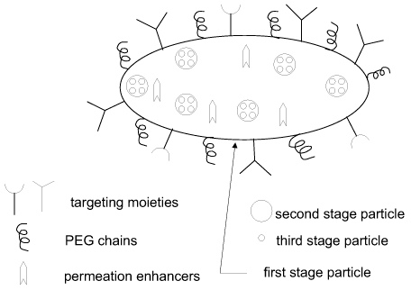
BRIEF DESCRIPTION OF THE DRAWINGS
FIG. 1 illustrates a multistage delivery vehicle, in accordance with an
embodiment of
the invention. The first stage particle contains inside second stage
particles. The second stage
particles may comprise at least one active agent, such as a therapeutic agent
or an imaging
agent. The first stage particle also contains inside an additional agent, such
as a permeation
enhancer or an additional active agent, which may be an imaging agent or a
therapeutic agent.
Optionally, the second stage particles contain third stage particles.
Targeting moieties, such as
antibodies, aptamers or ligands, attached to the surface of the first stage
particle, facilitate
localization at the selected body site.
FIG. 2 illustrates the principle of operation of a multistage delivery vehicle
administered intravascularly, in accordance with an embodiment. The first
stage particle
localizes at the targeted vasculature location. Upon the localization, the
particle releases
permeation enhancers that generate a fenestration in the vasculature. Second
stage particles
2
CA 02664919 2009-02-06
WO 2008/021908 PCT/US2007/075516
carry targeting moieties, such as antibodies. The second stage particles may
permeate through
the fenestration and target specific cells, that carry surface marker
antigens, using the
antibodies.
FIG. 3, Panel A, depicts time dependence of amino and carboxy modified quantum
dots
s (q-dots) in APTES modified "large pore" LP and "small pore" nanoporous
silicon first stage
particle. FIG. 3, Panel B, demonstrates an effect of second stage PEG-FITC-
SWNT
nanoparticles concentration on their loading into nanoporous silicon first
stage particles. In
Panels A and B, Y-axis reads mean fluorescence.
FIG. 4, Panels A-D, demonstrate time dynamics of second stage nanoparticles
loading
into nanoporous silicon first stage particles. Panel A for "large pore" (LP)
oxidized silicon first
stage particles; Panel B for LP APTES modified silicon first stage particles;
Panel C for "small
pore" (SP) oxidized silicon first stage particle; Panel D for SP APTES
modifies silicon first
stage particles. In Panels A-D, Y-axis reads mean fluorescence.
FIG. 5 demonstrates time dynamics of second stage nanoparticles release from
LP
is oxidized nanoporous silicon first stage particles (Panel A) and LP APTES
modified nanoporous
silicon first stage particles (Panel B). In Panel A and Panel B, Y-axis reads
released payload
N.
FIG. 6A, Panel A, presents the concentration effect of loading carboxy
modified
quantum dots and FITC-conjugated single wall carbon nanotubes (SWNTs). Y axis
in Panel A
reads mean fluorescence (%). FIG. 6A, Panel B, demonstrates fluorescence
quenching of
Fluorescein Isothiocyanate (FITC) conjugated Single Wall Carbon Nanotubes
(SWNT).
FIG. 6B relates to optimization of chemical condition for loading of second
stage
particles into nanoporous silicon particles. Nanoporous silicon particles were
mixed with
second stage nanoparticles (Q-dots in FIG. 6B, Panel C and Panel D; PEG-FITC-
SWNTs in
Panel E and Panel F) in the presence of increasing concentration of TRIS. High
concentration
of TRIS helped in increasing the amount of Q-dots loaded into the first stage
silicon particles
(Panel C and Panel D). On the contrary, loading efficiency of PEG-FITC-SWNTs
reached its
peak at 20Mm TRIS and then decreased at higher TRIS concentrations, Panel E
and Panel F. Y
axis in Panels B-F reads mean fluorescence.
FIG. 7 demonstrates data for loading and release respectively FITC conjugated
with
SWNT second stage particles into LP nanoporous silicon first stage particles.
Panel A presents
load columns, corresponding to the amount of FITC-SWNT initially loaded in the
nanoporous
silicon first stage particles after exposure to a FITC-SWNT solution prior to
washing. Wash
columns in Panel A corresponds to the amount of FITC-SWNT after washing the
first stage
3
CA 02664919 2009-02-06
WO 2008/021908 PCT/US2007/075516
particles. The actual load of the FITC-SWNT in the first stage particles is
the amount of FITC-
SWNT retained in the first stage particles after washing, i.e., a difference
between the
respective values in the load column and in the wash column. Panel B shows
data for release of
FITC-SWNT from the first stage particles over time. The total amount of FITC-
SWNT
released from the first stage particles over time, i.e., a sum of all the
columns in Panel B,
substantially matches the difference between respective load and wash columns
in Panel A. Y
axis in Panels A and B reads amount of FITC-SWNTs (ng).
FIGS. 8A-B present data for lipid based second stage particles loading into
nanoporous
silicon first stage particles. In FIG. 8A, Panels A-C show data for cationic
and neutral
liposomes loaded into 1 micron nanoporous silicon first stage particles. Panel
A shows
confocal microscopy images of neutral liposomes (left) and cationic liposomes
(right). Panels
B and C present respectively FACS analysis and Excel quantification of neutral
and cationic
liposome loading into 1 micron nanoporous silicon first stage particles. Y
axis in Panel B reads
particle number and Y axis in Panel C reads green fluorescence (logarithmic
values). In FIG.
is 8B, Panel D shows time dynamics of loading liposomes containing Alexa 555
labeled SiRNA
into 3.5 micron nanoporous oxidized silicon first stage particles. Y axis in
Panel D reads mean
fluorescence. Panel E and Panel F present fluorescent microscopy images
visualizing
fluorescence associated with liposomes containing Alexa 555 labeled SiRNA into
3.5 micron
nanoporous silicon first stage particles.
FIG. 9, Panels A-D, demonstrate Scanning Electron Microscopy (SEM) images of
"large pore" (LP) nanoporous silicon first stage particles.
FIG. 10, Panels A-D, demonstrate Scanning Electron Microscopy (SEM) images of
"small pore" (SP) nanoporous silicon first stage particles.
FIG. 11, Panels A-D, demonstrate degradation of nanoporous silicon particles
measured
using Z2 Coulter Particle Counter. Y axis in Panels A and B reads number of
particles. Y
axis in Panels C and D reads volume of particles.
FIGS. 12A-B demonstrate degradation of nanoporous silicon particles measured
using
Inductive Coupled Plasma -Atomic Emission Spectrometry. FIG. 12A, Panel A, for
LP
oxidized silicon particles; Panel B for SP oxidized silicon particles; FIG.
12B, Panel C, for LP
APTES modified silicon particles. Panel D for SP APTES modified silicon
particles. Y axis in
FIGS. 12A-12B reads concentration of silicon (ng/mL).
FIG. 13 demonstrates biocompatability of nanoporous silicon first stage
particles by
presenting bright field microscopy images of selected nanoporous silicon
particles and Human
Umbelical Vein Endothelial Cells (HUVEC) cells. In particular, Panel A
demonstrates images
4
CA 02664919 2009-02-06
WO 2008/021908 PCT/US2007/075516
for small pore oxidized silicon nanoparticles; Panel B demonstrates images for
small pore
APTES modified silicon nanoparticles; Panel C demonstrates images for large
pore oxidized
silicon nanoparticles; Panel D demonstrates images for large pore APTES
modified silicon
nanoparticles. In each Panel A-D, from left to right: first image day 0 (2 hrs
after particles
addition), second image day 1; third image day 2; fourth image day 3.
FIGS. 14A-B demonstrate biocompatibility of nanoporous silicon particles by
presenting data for Lactate Dehydrogenase (LDH) toxicity assay on HUVEC cells
incubated
with nanoporous silicon nanoparticles. Y axis in Panels A-F reads absorbance
at 490 nm.
FIGS. 15A-B present data for MTT proliferation assay on HUVEC cells incubated
with
nanoporous silicon nanoparticles. Y axis in FIG. 15A, Panels A-B and FIG. 15B,
Panels C-F
reads absorbance at 570 nm.
FIG. 16, Panels A-C, present FACS 3D Profiles of HUVEC cells incubated with
Nanoporous Silicon Particles and analyzed for their size and shape.
FIG. 17A-17D present FACS 3D Profiles of HUVEC cells incubated with Nanoporous
is Silicon First Stage Particles and stained with propidium iodide to study
cell cycle.
FIGS. 18A-C present statistical analysis of different phases of the cell cycle
of cells
exposed to nanoporous silicon first stage particles. Y axis in FIG. 18A,
Panels A-C, FIG. 18B,
Panels D-G, and FIG. 18C, Panels H-K reads % of total cell population.
FIG. 19, Panels a and b, demonstrate SEM images of a porous silicon particle.
"Large
pore" (LP, FIG. 19a) and "small pore" (SP, Panel b) particle images showing
(from left to
right) the back side, front side, a cross-section, a closer view of the pores
on the back side and
of the pores in the cross-section. The size and shape of the LP and SP
particles are the same,
the size and structure of the pores are significantly different.
FIG. 20 presents results of flow cytometric and fluorescence microscopy
analysis of
loading of fluorescently labeled Q-dots and PEG-FITC-SWNTs into nanoporous
silicon
particles. An increase in the amount of nanoparticles in the loading solution
resulted in an
increase in the mean fluorescence of silicon particles (1 LP APTES + Carboxyl
Q-dots = LP
oxidized + Amino Q-dots ^ SP APTES + Carboxyl Q-dots = SP oxidized + Amino Q-
dots)
measured by flow cytometry (Panels A and B). Fluorescent microscopy (Panels C
and D)
confirmed that the fluorescence associated to first stage particle was dimmer
when the amount
of nanoparticles used was lower. Y axis in Panels A and B reads mean
fluorescence.
FIG. 21 presents time dependent loading and releasing of second stage
particles in
nanoporous silicon first stage particles. Four different types of nanoporous
silicon first stage
particles (LP oxidized (Panel a), LP APTES (Panel b), SP oxidized (Panel c),
and SP APTES
5
CA 02664919 2009-02-06
WO 2008/021908 PCT/US2007/075516
(Panel d) were loaded with different second stage nanoparticles (= Carboxyl q-
dots, ^Amino q-
dots, = PEG-FITC-SWNTs) and their fluorescence measured by flow cytometry.
Histograms
in Panels e-h represent a percentage of second stage particles released from
the first stage
silicon particles with the passage of time.
FIG. 22 presents confocal microscopy images demonstrating concentrated loading
of Q-
dots in a highly porous region of the back side of silicon particle. Panel a
shows confocal
microscopy images reconstructed in a series of 3 dimensional projections
showing a single
porous silicon particle rotated to display different vantage points. Panel b
shows computer
generated 3 dimensional models illustrating the rotation of the particle as
shown in Panel a.
FIGS. 23A-23C illustrate simultaneous loading and releasing of Q-dots and PEG-
FITC-
SWNTs second stage particles in nanoporous silicon first stage particles. Flow
cytometry
analysis showing background green and red fluorescence of LP APTES particles
(Unloaded;
FIG. 23A, Panel A and Panel B respectively) and the shifts of the fluorescence
signals after the
incubation with PEG-FITC-SWNTs (+SWNTs), Q-dots (+Q-dots) and both (+Q-
is dots+SWNTs). Flow cytometry analysis show that PEG-FITC-SWNTs load rapidly
and
stabilize, while Q-dots gradually load before reaching a plateau, see FIG.
23B, Panel C. The
release profiles of the Q-dots and PEG-FITC-SWNTs are both unaltered by the
presence of
another type of nanoparticle and are both sustained along time, FIG. 23 B,
Panel D. Confocal
microscopy images show the localization of the Q-dots (red) and PEG-FITC-SWNTs
(green) in
a single porous silicon particle. FIG. 23C, Panel E, Panel F and Panel G show
bright field,
green and red fluorescence respectively, while Panel h shows overlay of the 3
channels are
shown. Yellow display showed co-localization of green and red fluorescent
signals. Y axis in
FIG 23 B, Panel D, reads mean fluorescence; Y axis in Panel D reads released
payload.
FIG. 24, Panels a-m, show fluorescent spectroscopy images related to
incubating
nanoporous silicon particles loaded with second stage particles with HUVEC
cells for lh at
37C. In FIG. 24, Panels a-d, the second stage particles are Q-dots; in Panels
e-h, the second
stage particles are PEG-FITC-SWNTs; in Panels j-m, the second stage particles
are Q-dots and
PEG-FITC-SWNTs. FIG. 24, Panels d, h and m are bright field images showing the
details of
particle morphology.
FIG. 25, Panels A-C, present computer models representing the three major
physical,
chemical and electrostatic mechanisms responsible for loading and release of
second stage
nanoparticles in first stage silicon carrier. (Panel A) Size Dependency: The
size of the pores
determines the type of nanoparticles that are preferably loaded into the
silicon particle. (Panel
B) Dose Dependency: A larger number of nanoparticles in the loading solution
cause an
6
CA 02664919 2009-02-06
WO 2008/021908 PCT/US2007/075516
increase in the number of particles that are loaded into the pores. (Panel B)
Charge
Dependency: Compatibly charged nanoparticles will be attracted into the pores,
whereas
incompatible charges will partially repel the nanoparticles and thus prevent
them to enter the
pores.
FIG. 26, Panels A-B, present data for liposomes containing SiRNA loaded into
nanoporous silicon first stage particles. Panel A: Alexa 555 fluorescently
labeled siRNAs were
encapsulated into nano-liposomes and loaded into the 1 st stage nano-vectors.
The data show
that the fluorescence associated with the porous Silicon carrier increased
with the amount of
nanoliposomes. Y axis reads mean fluorescence in Panel A. FIG. 26, Panel B,
presents a graph
io showing relative amount of liposomes released from first stage nanoporous
silicon particles. Y
axis reads % of total amount of released liposomes. To test the release of
nano-liposome from
1 st stage carriers, the assembled multistage particles were incubated with
10% fetal bovine
serum (pH 7.4) and release of nanoliposomes from 1 st stage particles was
followed along time
using fluorimetry. Complete unloading was achieved in about 36h.
is FIG. 27 demonstrates optimization of physical condition for loading of
quantum dots
and PEG-FITC-SWNTs. Y axis in Panel a and Panel b reads mean fluorescence.
Porous
silicon particles were desiccated in a desiccator over night and then mixed
with the loading
solution containing the second stage nanoparticles. Loading efficiency in dry
condition was not
significantly different than loading efficiency in a wet environment (p
value>0.5).
20 DEFINITIONS
Unless otherwise specified "a" or "an" means one or more.
"Biochemical environment of the target body site" refers to one or more
intrinsic
physiological conditions at the target site, such as pH, salt conditions,
temperature, or the
presence of target specific moieties, effective to initiate and promote
release of the particle
25 content.
"Biodegradable" refers to a material that may dissolve or degrade in a
physiological
medium or a biocompatible polymeric material that may be degraded under
physiological
conditions by physiological enzymes and/or chemical conditions.
"Mucoadhesive" refers to the capability of the particle to adhere to the
mucosal layer,
30 which lines the entire surface in the small and large intestine. Adherence
is mediated by
ligands grafted to the surface of the particles, which bind to chemical
receptors present in
mucin or the surface of the intestinal epithelial cells.
"Targeting moiety" is any factor that may facilitate targeting of a specific
site by a
particle. For example, the targeting moiety may be a chemical targeting
moiety, a physical
7
CA 02664919 2009-02-06
WO 2008/021908 PCT/US2007/075516
targeting moiety, a geometrical targeting moiety or a combination thereof. The
chemical
targeting moiety may be a chemical group or molecule on a surface of the
particle; the physical
targeting moiety may be a specific physical property of the particle, such as
a surface such or
hydrophobicity; the geometrical targeting moiety includes a size and a shape
of the particle.
"Microparticle" means a particle having a maximum characteristic size from 1
micron
to 1000 microns, or from 1 micron to 100 microns. "Nanoparticle" means a
particle having a
maximum characteristic size of less than 1 micron.
"Biocompatible" refers to a material that, when exposed to living cells, will
support an
appropriate cellular activity of the cells without causing an undesirable
effect in the cells such
as a change in a living cycle of the cells; a change in a proliferation rate
of the cells and a
cytotoxic effect.
DETAILED DESCRIPTION
In embodiments of the invention, a composition that includes two or more
stages of
particles, such that particles of a later stage (smaller size particles) are
contained in particles of
is an earlier stage (larger particles), will potentially provide one or more
advantages for treating,
preventing and/imaging a physiological condition, such as a disease, in a
subject, which may be
any animal with a blood system (e.g., the subject may be a warm blooded
animal, such as
mammal including human being). Embodiments of such multistage composition
provide one
or more of the following features or advantages: (1) an active agent, such as
a therapeutic agent
or an imaging agent, is preferentially delivered and/or localized to a
particular target site in a
body of a subject. Preferential delivery and/or localization means that an
amount or
concentration of the active agent delivered to and/or localized at the target
site is higher than an
amount or concentration of the active agent delivered to and/or localized at
other sites in the
body of the subject; (2) a multistage composition sequentially overcomes
multiple biological
barriers in a body of the subject; and (3) a multistage composition allows for
simultaneous
delivery and localization at the same or different target site of multiple
active agents.
Biological barriers
Following administration, an active agent, such as a therapeutic or imaging
agent,
formulated conventionally or in a nanovector, encounters a multiplicity of
biological barriers
that adversely impact the agent's ability to reach an intended target at a
desired concentration.
The biological barrier may be, for example, an epithelial or endothelial
barrier, such as a blood-
brain barrier or intestinal lumen endothelium, that are based on tight
junctions, that prevent or
limit para-cellular transport of an active agent. Each of the endo/epithelial
barrier includes a
plurality of sequential subbarriers, such as tight junction barriers, that owe
their molecular
8
CA 02664919 2009-02-06
WO 2008/021908 PCT/US2007/075516
discrimination to one or more zonula occluden proteins, and one or more
additional biological
membranes, such as vascular endothelial basement membrane or a musocal layer
of the
intestinal endothelium. Cells of the reticulo-endothelial system may also act
as a biological
barrier against an active agent encapsulated inside nanoparticles, as such
cells sequester/uptake
the nanoparticles. The biological barrier may be also represented by a cell
membrane or a
nuclear membrane in a cell that an active agent has to come through.
Multistage delivery vehicle
Since the biological barriers are sequential, overcoming or bypassing such
barriers has
to be sequential too. Accordingly, a delivery vehicle has been developed that,
in embodiments,
acts in multiple stages. Each stage of the vehicle is defined by a particle
having a separate
intended function, which may be different from an intended function of a
particle of another
stage. For example, a particle of one stage is designed to target a specific
body site, which may
be different from a site targeted by a particle of another stage, and thus to
overcome or bypass a
specific biological barrier, which is different from a biological barrier
being overcome or
is bypassed by a particle of another stage. A particle of each subsequent
stage is contained inside
a particle of an immediately preceding stage. A particle of any particular
stage may contain an
active agent, such as a therapeutic agent or an imaging agent, intended for
use at this particular
stage.
In a preferred embodiment, a particle of the last stage is an active agent
formulated as a
nanoparticle or alternatively the last stage particle contains the active
agent inside, while a
particle of any earlier stage per se may or may not comprise an active agent.
In some
embodiments, in addition to targeting a specific body site, a particle of each
stage is designed in
such a way that it is capable to perform targeted release of its content. In
embodiments, the
number and type of stages in the multistage delivery vehicle depends on
several parameters,
including administration route and an intended final target for the active
agent. An
embodiment of a multistage delivery vehicle is illustrated on Fig. 1.
First Stage Particle
In some embodiments, the particle of the first stage is a micro or
nanoparticle. In some
embodiments, the first stage particle has a characteristic size of at least
500 microns or at least 1
mm. Such a particle may be configured to contain inside at least one micro or
nanoparticle,
which in turn may contain inside at least one particle of a smaller size. The
first stage particle
is any particle that is capable of containing inside particles of a smaller
size.
In some embodiments, the first stage particle is a top-down fabricated
particle, i.e., a
particle prepared by top-down microfabrication or nanofabrication methods,
such as
9
CA 02664919 2009-02-06
WO 2008/021908 PCT/US2007/075516
photolithography, electron beam lithography, X-ray lithography, deep UV
lithography or
nanoprint lithography. A potential advantage of using the top-down fabrication
methods is that
such methods provide for a scaled up production of particles that are uniform
in dimensions.
The particle of the first stage may be configured to overcome at least one of
the
following biological barriers: a hemo-rheology barrier, a Reticulo-Endothelial
System barrier,
an endothelial barrier, a blood brain barrier, a tumor-associated osmotic
interstitial pressure
barrier, an ionic and molecular pump barrier, a cell membrane barrier, an
enzymatic
degradation barrier, a nuclear membrane barrier or a combination thereof.
The first stage particle may have a body that is defined by a size and a shape
of the
particle and one or more reservoirs inside the body. One or more second stage
particles may be
contained inside the reservoir.
The body of the first stage particle comprises any appropriate material.
Preferably, the
material of the body of the first stage particle is biocompatible. In some
embodiments, the
body of the first stage particle comprises an oxide material such as silicon
oxide, aluminum
is oxide, titanium oxide, or iron oxide; a semiconductor material, such as
silicon; a polymer or a
polymer oxide material; or a ceramic material. In some embodiments, the body
of the first
stage particle comprises a biodegradable material, such as, for example,
nanoporous silicon.
The biodegradable material may be such that it degrades when exposed to a
physiological
medium such as silicic acid.
In some embodiments, a material of the body of the first stage particle is
substantially
the same in different regions of the body. The shape of the first stage
particle may depend on
the administration route. For example, the shape may be configured to maximize
the contact
between the first stage particle and a surface of the target site, such as
endothelium surface for
intravascular administration or intestinal epithelium for oral administration.
Accordingly, for
oral and intravascular administration, the first stage particle may have a
selected non-spherical
shape configured to maximize the contact between the particle and endothelium
surface.
Examples of appropriate shapes include, but are not limited to, an oblate
spheroid or a disc. For
pulmonary administration, i.e., administration to lungs of the subject, the
first stage particles
may also have a selected non-spherical shape configured to maximize a contact
between the
particle and one of the epithelial tissues in lungs.
For pulmonary administration, i.e., an administration route, which involves
passing of
the particle through lungs of a subject, the first stage particle may also
have a spicular shape,
which may facilitate entering of the particle from the lungs into a body
tissue, not necessarily
through the blood circulation.
CA 02664919 2009-02-06
WO 2008/021908 PCT/US2007/075516
Although top-down fabrication allows manufacturing particles having size in a
wide
range from 50 nm up to several millimeters, for certain administration routes
particular particle
sizes may be preferred. For example, for intravascular administration, a
maximum
characteristic size of the particle, e.g., a diameter for a disc-shaped
particle, is preferably
sufficiently smaller than a radius of the smallest capillary. In humans, such
a radius is about 4
or 5 microns. Accordingly, the maximum characteristic size of the particle
are, in some
embodiments, less than about 3 microns, less than about 2 microns or less than
about 1 micron.
In embodiments, the maximum characteristic size of the first stage particle is
from 500
nm to 3 microns, or from 700 nm to 2 microns. Yet in some embodiments for
intravascular
administration in oncological applications, the maximum characteristic size of
the first stage
particle is such that the first stage particle could localize at the targeted
vasculature site without
penetrating a fenestration in vascular cancer endothelium. For such
applications, the maximum
characteristic size of the first stage particle is greater than about 100 nm,
or greater than about
150 nm, or greater than about 200 nm.
is Yet in some embodiments for intravascular administration, the size of the
first stage
particle is such so that the particle may penetrate the fenestration.
Accordingly, the maximum
characteristic size in such applications is preferably less than about 200 nm,
or less than about
150 nm, or less than about 200 nm. In some embodiments, one may select a size
of the first
stage particle that is selected to be a critical radius of normal non-
fenestrated vasculature for
targeting fenestrated vasculature as detailed in PCT patent application No.
PCT/US2006/038916 "Methods and Compositions for Targeting Fenestrated
Vasculature" filed
September 27, 2006 to Paolo Decuzzi and Mauro Ferrari.
For oral administration, it may be preferable to use the first stage particle
that has a
maximum characteristic size greater than about 2 microns or greater than about
5 microns or
greater than about 10 microns. One advantage of using the first stage particle
of such size for
oral administration is that such particle may be too large to be endocytosed
by intestinal
epithelial cells. The endocytosis by intestinal epithelial cells has at least
two potential
disadvantages: 1) the content of the first stage particle may be deactivated
as it is processed by
the endothelial cell before it reaches the desired target; 2) the potential
toxicity of particular
carrier, e.g. of the material of the particle, is of greater concern if it is
endocytosed than if it is
cleared through the gastrointestinal tract.
In some embodiments, for oral administration, the first stage particle has a
size ranging
from 500 microns to several centimeters, or from 1 mm to 2 cm.
11
CA 02664919 2009-02-06
WO 2008/021908 PCT/US2007/075516
For pulmonary administration, the maximum characteristic size of the first
stage
particle is preferably less than about 20 microns but greater than about 5
microns, if a targeted
site is located in the lungs' air passages. For targeting a site in alveoli,
the maximum
characteristic site may be less than about 5 microns.
In some embodiments, for subcutaneous administration, a characteristic size of
the first
stage particle is from 50 microns to 1 mm; or from 100 microns to 1 mm.
One of the functions of the first stage particle, in embodiments, is
localization at a
particular target site. For intravascular administration, such target site may
be a particular
vasculature site. For example, in anticancer applications, the targeted
vasculature site may be a
tumor vasculature, such as angiogenesis vasculature, coopted vasculature or
renormalized
vasculature. The localization of the first stage particle at the targeted site
may be facilitated by
geometrical factors, such as the size and the shape of the particle.
For intravascular administration, the localization at the targeted site may be
also
facilitated by one or more recognition factors on the surface of the first
stage particle. The
is recognition factor may be a chemical targeting moiety, such as a dendrimer,
an antibody, an
aptamer, which may be a thioaptamer, a ligand or a biomolecule that binds a
particular receptor
on the targeted site. For oral delivery, the chemical moiety may comprise one
or more
mucoadhesive ligands, as described in Table 1 of U.S. Patent No. 6,355,270.
The selectivity of the targeting may be tuned by changing chemical moieties of
the
surface of the particles. For example, coopted vasculature is specifically
targetted using
antibodies to angiopoietin 2; angiogenic vasculature is recognized using
antibodies to vascular
endothelial growth factor (VEGF), basic fibroblast growth factor (FGFb) or
endothelial
markers such as av(33 integrins, while renormalized vasculature are recognized
using
carcinoembionic antigen-related vell adhesion molecule 1(CEACAM1), endothelin-
B receptor
(ET-B), vascular endothelial growth factor inhibitors gravin/AKAPl2, a
scallofldoing protein
for protein kinase A and protein kinase C, see, e.g., Robert S. Korbel
"Antiangiogenic Therapy:
A Universal Chemosensitization Strategy for Cancer?", Science 26 May 2006, vol
312, no.
5777, 1171-1175.
For targeting to non-circulating vasculature cells, the binding between the
first stage
particle and the molecular marker of the targeted vasculature site should be
sufficiently strong
to overcome the drag force exerted by the flowing blood. This objective may be
satisfied by
having a relatively large planar surface area for specific binding and a
relatively low profile in a
12
CA 02664919 2009-02-06
WO 2008/021908 PCT/US2007/075516
capillary's blood flow space, i.e., by having the particle of the selected non-
spherical shape,
such as an oblate spheroid or a disc.
The recognition factor may be also a physical recognition moiety, such as a
surface
charge. The charge may be introduced during the fabrication of the particle by
using a
chemical treatment such as a specific wash. For example, immersion of porous
silica or
oxidized silicon surface into water may lead to an acquisition of a negative
charge on the
surface, see, e.g., Behrens and Grier, J. Chem. Phys. 115(14), (2001). P. 6716-
6761. The
surface charge may be also provided by an additional layer or by chemical
chains, such as
polymer chains, on the surface of the particle. For example, polyethylene
glycol chains may be
a source of a negative charge on the surface. Polyethylene glycol chains may
be coated or
covalently coupled to the surface as described in P.K. Jal, S. Patel, B.K.
Mishra, Talanta 62
(2004) P1005-1028; S.W. Metzger and M. Natesan, J. Vac. Sci. Technol. A 17(5),
(1999)
P2623-2628; and M. Zhang, T. A. Desai and M. Ferrari, Biomaterials, 19,
(1998), p 953. The
positive charge is introduced, for example, by coating the surface with a
basic polymer, such as
is polylysine or by covalently linking to the surface an amino containing
molecule, such as 3-
aminopropyltriethoxysilane.
In some embodiments, modeling methods are applied for selecting geometrical
factors,
such as a size and a shape, and/or surface modification, such as chemical
modification and
electrostatic modification, of the particle based on one or more properties of
the selected target
site. Such modeling methods are disclosed, for example, in 1) U.S. Provisional
Patent
Application No. 60/829,075 "Particles for Cell Targeting" filed October 11,
2006 to Paolo
Decuzzi and Mauro Ferrari; 2) U.S. Provisional Patent Application No.
60/891,584
"Endocytotic particle" filed February 26, 2007 to Paolo Decuzzi and Mauro
Ferrari; 3)
Decuzzi, P., Causa, F., Ferrari, M. & Netti, P. A. The effective dispersion of
nanovectors within
the tumor microvasculature. Ann Biomed Eng 34, 633-41 (2006); 4) Decuzzi, P. &
Ferrari, M.
The adhesive strength of non-spherical particles mediated by specific
interactions. Biomaterials
27, 5307-14 (2006); 5) Decuzzi, P. & Ferrari, M. The role of specific and non-
specific
interactions in receptor-mediated endocytosis of nanoparticles. Biomaterials
28, 2915-22
(2007); 6) Decuzzi, P., Lee, S., Bhushan, B. & Ferrari, M. A theoretical model
for the
margination of particles within blood vessels. Ann Biomed Eng 33, 179-90
(2005); Decuzzi, P.,
Lee, S., Decuzzi, M. & Ferrari, M. Adhesion of microfabricated particles on
vascular
endothelium: a parametric analysis. Ann Biomed Eng 32, 793-802 (2004).
In some embodiments, the first stage particle is configured to release the
second stage
particles from its reservoir at the target site. The release of the second
stage particles may be
13
CA 02664919 2009-02-06
WO 2008/021908 PCT/US2007/075516
performed according to a variety of mechanisms, including, but not limited to,
diffusion of the
second stage particles through the channels connecting the reservoir and the
surface of the first
stage particle and degradation or erosion of the body the first stage
particle. For such a
purpose, the first stage particle is configured to have a characteristic
release time longer than a
characteristic delivery time of the first stage particle to its target site
when administered to the
subj ect.
In some embodiments, the first stage particle is configured to perform a
release of the
second stage particles from its reservoir that is sustained over a period of
time longer than a
characteristic delivery time of the first stage particle to its target site
when administered to the
subject. In some embodiments, the first stage particle is configured to
release the second stage
particles over a time period longer than at least 0.5 hr; or longer than at
least 1 hr; or longer
than at least 2 hr; or longer than at least 8 hr; or longer than at least 15
hr; or longer than 30 hr.
In some embodiments, physical localization and/or targeted release of the
first stage
particle is initiated by one or more intrinsic physiological conditions at the
target site such as
is pH, salt concentrations or temperature. In some embodiments, physical
localization and/or
targeted release of the first stage particle is initiated by exogenous
stimulation. Examples of
exogeneous stimulations include mechanical activation, such activation by
ultrasound;
electromagnetic activation, such an activation by visible, ultraviolet, near-
infrared or infrared
light, radiofrequency or X-ray radiation; magnetic radiation. For example, the
first stage
particle may comprise a smart polymer, i.e., a polymer that contracts or
expand in a response to
a specific stimulus, such as light, temperature or pH. Smart polymers are
described, for
example, in "In Situ Activation of Microencapsulated Drugs (MSC-22866)," NASA
Tech
Briefs, Vol. 24, No. 9 (September 2000), page 64; "Externally Triggered
Microcapsules
Release Drugs In Situ (MSC-22939), NASA Tech Briefs, (April 2002), page 50;
and U.S.
Patent No. 6,099,864 issued to Morrison and Mosier on August 8, 2000. In some
cases, more
than one exogeneous stimulation is used together for activating release.
To increase the localization /targeting efficiency, the first stage particle
may utilize
multiple, i.e., more than one recognition/localization/targeting factors,
which preferably do not
interfere with each other. For example, the first particle may have the
selected non-spherical
shape as discussed above and at the same time carry tumor-targeting antibodies
on its surface.
In some embodiments, the surface of the first stage particle may be coated
with polymer
chains partially or completely. The polymer chains may be added after the
fabrication of the
intravascular stage particle. The polymer chains may be hydrophilic chains,
such as
polyethylene glycol (PEG) chains or synthetic glycocalix chains, which may be
used for
14
CA 02664919 2009-02-06
WO 2008/021908 PCT/US2007/075516
overcoming the uptake of the particle by cell of the reticulo-endothelial
system and, thus,
extending the blood circulation of the intravascular stage particle. The
hydrophilic groups may
also serve for enhancement of solubility of the multistage delivery devices.
A variety of materials may be derivatized with polymer chains. For example,
when the
particle's surface material comprises a polymer, polymer chains may be
attached by linking
carboxylic groups of the polymer and amine or hydroxyl groups in the polymer
chains; if the
particle's surface material comprises metal such as gold, the polymer chains
may be attached to
the surface via thiol chemistry; when the particle's surface material
comprises an oxide, such
silicon oxide, titanium oxide or aluminum oxide, the polymer chains may be
attached using
silane chemistry.
In addition to one or more second stage particles, the reservoir of the first
stage particle
may contain one or more additional agents. Such additional agent may include
an additional
active agent, such as a therapeutic agent or an imaging agent, a targeting
agent, one or more
penetration enhancers or any combination thereof. The penetration enhancer may
include one
is or more compounds listed in Table 1.
TABLE 1
Penetration Enhancers
Class of Enhancer Specific Examples
Bile Salts Glyo-deoxycholate
Taruro-dexoycholate
Tauro-chenodeoxycholate
Glyco-chenodexycholate
Taurocholate
Glycocholate
Glycoursocholate
Tauroursocholate
Dexoycholate
Chenodeoxycholate
Cholate
Ursocholate
Non-ionic Surfactants Polyoxyethylene (POE) ethers (e.g., Brij,
Texaphor)
Alkylphenoxy-POEs (Triton, Igepal,
Surfonic)
Anionic Surfactants sodium dodecyl sulfate
dioctyl sodium sulfosuccinate
Lecithins Lysolecithin
Medium chain glycerides mono-, di-, or triglycerides of C8, C10,
or C 12 fatty acids
Medium chain fatty acids sodium caprylate
sodium caprate
sodium laurate
CA 02664919 2009-02-06
WO 2008/021908 PCT/US2007/075516
Salicylates sodium salicylate
Acylcarnitines decylcarnitine
laurylcarnitine
myristoylcarnitine
Acylcholines Laurylcholine
Palmitoylcholine
Acyl amino acids N-laurylphenylglycine
N-palmitoylglycine
Calcium chelators Ethylenediaminetetraacetic acid (EDTA)
Peptides PZ-peptide
Zonula occludens toxin (ZOT)
For intravascular administration, the penetration enhancers may include a
basement
membrane penetration enhancer that may act against the basement membrane,
and/or one or
is more penetration enhancers, that may act against tight junction proteins
(TJPs). An example of
the basement membrane penetration enhancer is metalloproteinase, such as
Collagenase IV,
MMP-2, and MMP-9. An example of the anti-TJP penetration enhancer is zonula
occluden
toxin. Anti-TJP penetration enhancers and strategies for modulating tight
junction permeability
are detailed, for example, in Gonzalez-Mariscal, L., Nava, P. and Hemandez, S.
J. Membr.
Biol., 2005. 207(2): p. 55-68.
Figs. 2A-B illustrate the action of penetration enhancer upon localization of
the first
stage particle at the targeted vasculature site, in accordance with an
embodiment. The first
stage particle releases the permeation enhancers that generate in the targeted
vasculature one or
more fenestrations, through which the second stage particles penetrate into
the vasculature.
In some embodiments, the first stage particle has one or more channels
fluidically
connecting the reservoir with the surface, that may be in contact with endo or
epithelial cells.
For intravascular administration, such first stage particle may be a micro or
nano fabricated
particle, such as those detailed in U.S. Patent Application Publication No
2003/0114366 and
U.S. Patent No. 6,107,102, and for oral administration, such first stage
particle may be a micro
or fabricated particle, such as the ones disclosed in U.S. Patent No.
6,355,270.
In some embodiments, the reservoir and the channels are pores in the body of
first stage
particle. In such case, the first stage particle may comprise a porous or
nanoporous material.
Preferably, the pores of the porous or nanoporous material may be controlled
to achieve a
desired load of the next stage particles and a desired release rate. The
nanoporous material with
controllable pore size may be an oxide material, such as silicon oxide,
aluminum oxide,
titanium oxide, or iron oxide. Fabrication of nanoporous oxide particles, also
known as sol gel
particles, is detailed, for example, in Paik J. A. et. al. J. Mater. Res.,
Vol. 17, Aug 2002. The
nanoporous material with controllable pore size may be also nanoporous
silicon. For details of
16
CA 02664919 2009-02-06
WO 2008/021908 PCT/US2007/075516
fabrication of nanoporous silicon particles, see Cohen M. H. et. al.
Biomedical Microdevices
5:3, 253-259, 2003. Control of pore density, size, shape and/or orientation
may be
accomplished by changing electrical current and etching time during formation
of nanoporous
silicon from non-porous silicon. Control of pore density, size, shape and/or
orientation may be
also accomplished by varying doping in non-porous silicon used for formation
of nanoporous
silicon. Thus, pore size, density, size, shape and /or orientation of
nanoporous may be
configured for efficient loading of the second stage particles.
In some embodiments, pore size in nanoporous first stage particles may be, for
example, from about 1 nm to about 200 nm; or from about 2 nm to about 100 nm.
In some
cases, pore size in nanoporous first stage particle may be from about 3 to
about 10 nm or from
about 5 to about 7 nm. Yet in some cases, pore size in nanoporous first stage
particle may be
from about 10 to about 60 nm, or from about 20 to about 40 nm.
In some embodiments, to facilitate loading of the second stage particles into
pores of
the porous or nanoporous material may be modified chemically and/or
electrostatically to make
is it compatible with chemical and/or electrostatical surface properties of
the second stage
particles. For example, for loading negatively charged second stage particles,
it may be
preferable to use positively charged pore surface. The positive charge may be
achieved, for
example, by depositing on pore surface an amino-containing molecule, such as 3-
aminopropyltriethoxysilane. For loading positively charged second stage
particles, it may be
preferable to use negatively charged pore surface. The negative pore surface
charge may be
accomplished, for example, by oxidizing pore surface with water.
In some embodiments, the first stage particle has no channels at all. Such
particle may
comprise, for example, a biodegradable material. For oral administration, the
material may be
designed to erode in the GI tract. As examples, the first stage particle may
be formed of metals,
such as iron, titanium, gold, silver, platinum, copper, and alloys and oxides
thereof. The
biodegradable material may be also a biodegradable polymer, such as
polyorthoesters,
polyanhydrides, polyamides, polyalkylcyanoacrylates, polyphosphazenes, and
polyesters.
Exemplary biodegradable polymers are described, for example, in U.S. Pat. Nos.
4,933,185,
4,888,176, and 5,010,167. Specific examples of such biodegradable polymer
materials include
poly(lactic acid), polyglycolic acid, polycaprolactone, polyhydroxybutyrate,
poly(N-palmitoyl-
trans-4-hydroxy-L-proline ester) and poly (DTH carbonate).
In some embodiments, the body of the first stage particle includes two or more
regions
configured to contain different populations of second stage particles. For
example, the body of
the first stage particle may have a first region configured to contain a first
population of second
17
CA 02664919 2009-02-06
WO 2008/021908 PCT/US2007/075516
stage particles and a second region configured to contain a second population
of second stage
particles. For instance, the first stage particle formed of porous nanoporous
material may be
such that its body has two or more porous regions that differ from each other.
The body of the
nanoporous first stage particle may include a first porous region and second
porous region that
differ from each other in at least one property such as a pore density, pore
geometry; pore
charge, pore surface chemistry, pore orientation or any combination thereof.
Such first and
second regions may be configured respectively to contain a first and a second
population of
second stage particles.
In some embodiments, the first and second populations of second stage
particles differ
in at least one property such as size; shape; surface chemical modification;
surface charge or a
combination thereof.
In some embodiments, the first and second populations of second stage
particles contain
the same active agent. Yet, in some embodiments, the first and second
population of second
stage particles contain respectively a first active agent and a second active
agent that are
is different from each other.
The first and second populations of second stage particles may be configured
to
perform different functions. For example, in some embodiments, the first and
second
populations may be configured to target respectively a first and second target
sites that are
different from each other.
In some embodiments, the first population are configured to target a
particular site in a
body of the subject, while the second population is configured to free
circulate in the blood
system of the subject.
In some embodiments, the first and the second population target the same
target site in a
body of the subject but perform a different function at the body site. For
example, the first
population contains a therapeutic agent to be delivered to the target site,
while the second
population contains an imaging agent to be delivered to the target site for
imaging or
visualizing the target site.
The first and the second regions of the body of the first stage particle may
be such that
at least one of them is a biodegradable region. Preferably, both the first and
the second regions
of the body of the first stage particle is biodegradable.
The first and the second regions of the body of the first stage particle may
have different
characteristic time for releasing the first and the second population of
second stage particles. In
some embodiments, both characteristic time for releasing the first population
of second stage
particles from the first region and characteristic time for releasing the
second population of
18
CA 02664919 2009-02-06
WO 2008/021908 PCT/US2007/075516
second stage particles from the second region may be greater that a
characteristic for delivery
and/or localization of the first stage particle at its target site when
administered to the subject.
In some embodiments, the first stage particle is configured to separate into a
first
component that includes the first region and a second component that includes
the second
region when being exposed to a physiological medium, such as a medium that may
be present
at a target site of the first stage particle. Such exposure may occur, for
example, when the
particle is administered to the subject.
In some embodiments, the first and the second regions of the body of the first
stage
particle are configured to perform a different function when the particle is
administered to the
io subject. For example, the first and the second region of the body of the
first stage particle may
be configured to overcome respectively the first and the second biological
barriers that are
different from each other. Such biological barriers may be each selected, for
example, from a
hemo-rheology barrier, a Reticulo-Endothelial System barrier, an endothelial
barrier, a blood
brain barrier, a tumor-associated osmotic interstitial pressure barrier, an
ionic and molecular
is pump barrier, a cell membrane barrier, an enzymatic degradation barrier, a
nuclear membrane
barrier or a combination thereof.
Second stage particle
The second stage particle may be any micro or nanoparticle that may fit inside
the
reservoir of the first stage particle. For example, in certain embodiments,
for oral or pulmonary
20 administration, the second stage particle is the same as the first stage
particle for intravascular
administration.
The particle of the second stage may be configured to overcome at least one
barrier
selected from a hemo-rheology barrier, a Reticulo-Endothelial System barrier,
an endothelial
barrier, a blood brain barrier, a tumor-associated osmotic interstitial
pressure barrier, an ionic
25 and molecular pump barrier, a cell membrane barrier, an enzymatic
degradation barrier, a
nuclear membrane barrier or a combination thereof.
The composition, size and shape of the second stage particle are not
particularly limited.
For example, for many administration routes, the second stage particle may be
a lipid based
particle, such as a liposome, a micelle or lipid encapsulated perfluorocarbon
emulsion; an
30 ethosome; a carbon nanotube, such as single wall carbon nanotube; a
fullerene nanoparticle; a
metal nanoparticle, such gold nanoshell or triangular silver nanoparticle; a
semiconductor
nanoparticle, such as quantum dot or boron doped silicon nanowire; a polymer
nanoparticle,
such as particles made of biodegradable polymers and ion doped polyacrylamide
particles; an
oxide nanoparticle, such as iron oxide particle, a polymer coated iron oxide
nanoparticle or a
19
CA 02664919 2009-02-06
WO 2008/021908 PCT/US2007/075516
silicon oxide particle; a viral particle, such as an engineered viral particle
or an engineered
virus-polymer particle; a polyionic particle, such as leashed polycations; a
ceramic particle,
such as silica based ceramic nanoparticles, or a combination thereof.
In some embodiments, the second stage particle is configured to target a
particular
target site in a body of the subject. Such target site may be the same or
different from the target
site targeted by the first stage particle.
For example, the surface of the second stage particle may have one or more
antibodies
that may conjugate with surface marker antigens of certain types of cells.
Thus, the second
stage particle may selectively target cells that carry such marker antigens.
The examples of
cells that carry surface marker antigens include stem or clonogenic cells and
tumor cells. The
surface marker antigens on stem or clonogenic cells may be targeted by CD33
antibody. A
number of monoclonal antibodies to tumor specific antigens are available, see,
e.g., pp. 301-
323 of CANCER, 3rd Ed., De Vita, et. al. eds; Janeway et. al. Immunology 5th
Edition,
Garland Press, New York, 2001; A. N. Nagappa, D. Mukherjee & K. Anusha
"Therapeutic
is Monoclonal Antibodies", PharmaBiz.com, Wednesday, September 22, 2004. Table
2 presents
FDA approved monoclonal antibodies for treatment of cancer.
TABLE 2
....... ........ ........ ........ .. ..... ........ ........ ........
........ ........ ........ ........ ........ ........ ..........
. . . . . . . . . . . . . . . . . . . . . . . . . . . . . . . . . . . . . . .
. . . . . . . . . . . . . . . . . . . . . . . . . . . . . . . . . . . . . . .
. . . . . . . . . . . . . . . . . . . . . . . . . . . . . . . . . . . . . . .
. . . . . . . . . . . . . . . . . . . . . . . . . . . . . . . . . . . . . . .
. . . . . . . . . . . . . . . . . . . . . . . . . . . . . . . . . . . .
..........................................
MAb Name Trade Used to Treat: Approved in
Name
Rituximab Rituxan Non-Hodgkin lymphoma 1997
Trastuzumab Herceptin Breast cancer 1998
Gemtuzumab ozogamicin* Mylotarg Ac~L) myelogenous leukemia 2000
Alemtuzumab Campath ~L~~ic lymphocytic leukemia 2001
Ibritumomab tiuxetan* Zevalin Non-Hodgkin lymphoma 2002
Tositumomab* Bexxar Non-Hodgkin lymphoma 2003
Cetuximab Erbitux Colorectal cancer 2004
Head & neck cancers 2006
Bevacizumab Avastin Colorectal cancer 2004
In some embodiments, the second stage particle is configured to freely
circulate in a
blood system of the subject upon being released from the first stage particle.
In some cases,
CA 02664919 2009-02-06
WO 2008/021908 PCT/US2007/075516
such second stage particle may have a surface free of targeting moieties, such
as antibodies.
The free circulating second stage particle may be used, for example, as to
report of therapeutic
action a therapeutic agent associated with a first stage particle per se.
In some embodiments, the surface of the second stage particle does not have
hydrophilic polymer chains, such as polyethylene glycol (PEG) disposed on it.
In certain cases,
this may be an advantage of the multistage delivery vehicle as PEG chains are
usually attached
to liposomes and other nanoparticles to delay recognition and sequestration by
macrophages of
the reticulo-endothelial system. Unfortunately, the PEG chains may also hide
antibodies on the
nanoparticles surfaces and thus inhibit the targeting/localization ability of
the antibodies. In the
multistage delivery vehicle, PEGs attached to the first stage particle may
perform shielding
from the RES macrophages. Although the PEGs may hide antibodies on the first
stage particle,
the recognition/localization capability of the first stage particle is not
limited to the antibodies,
as other factors such as the particle's size and shape also may contribute to
such capability. In
some embodiments, the surface of the second stage particle has hydrophilic
polymer chains,
is such as PEG chains, disposed on it.
In some embodiments, the surface of second stage particle is modified, for
example, to
facilitate the second stage particle's ability to load into a reservoir of the
first stage particle
and/or to facilitate the second stage particle's ability to reach its target
site. The surface
modification may include a chemical modification of the surface of the second
stage particle
and/or electrostatic modification of the surface of the second stage particle.
For example, to
facilitate loading of the second stage particles into a porous or nanoporous
first stage particle,
the surface of the second stage particles may be modified so that its
properties are compatible
with surface properties of pores of the porous or nanoporous first stage
particle. For example,
when the pores of the porous or nanoporous first stage particle are positively
charged it may be
preferably to modify the surface of the second stage particles so that they
are electrostatically
neutral or have a negative surface charge; while when the pores of the porous
or nanoporous
first stage particle are negatively charged, it may be preferably to modify
the surface of the
second stage particles so that they are electrostatically neutral or have a
positive surface charge.
The chemical and/ or electrostatic surface modification of the second stage
particles may be
performed using the same methods as detailed above to the first stage
particles.
For lipid containing second stage particles, such as liposomes or micelles,
the
electrostatic modification may be performed by incorporating in their lipid
layers lipids that
may affect the electrostatic charge of the liposome. For example, to form a
positively charged
cationic lipid containing particle, one may use cationic lipids, such as 1,2-
Dioleyl-3-
21
CA 02664919 2009-02-06
WO 2008/021908 PCT/US2007/075516
trymethylammoniumpropane (DOTAP); to form a negatively charged anionic lipid
containing
particle, anionic lipids, such as dioleoylphosphatidyl glycerol (DOPG); and to
form a neutral
lipid containing particle one may use, neutral lipids, such as DOPC.
In some embodiments, upon binding to the targeted cell or cells, the second
stage
particle may release its content into the cell's cytoplasm. In some
embodiments, such release is
activated by exogenous factors, such as electromagnetic radiation. For
fullerene nanoparticles
and carbon nanotubes, such radiation may be radio-frequency radiation, while
for gold-shell
nanoparticles, the radiation may be a near-infrared radiation. Activation of
nanoparticulates by
exogenous radiation is detailed, for example, Hirsch, L . R., Halas, N. J. &
West, J. L. Anal.
Chem. 75, 2377-2381 (2003); Hirsch, L. R., Halas, N. J. & West, J. L. Proc.
Natl Acad. Sci.
USA 100, 13549-13554 (2003); and O'Neal, D. P., Halas, N. J. & West, J. L.
Cancer Lett. 209,
171-176 (2004).
In some embodiments, the content of the second stage particle is one or more
active
agents per se. In some embodiments, the second stage particle contains inside
a third stage
is particle, which itself contains inside one or more active agents. The third
stage particle may be,
for example, a particle, that is small enough to be able to cross a nuclear
membrane of the
targeted cell. Thus, the third stage particle may serve for delivering to the
cell's nucleus an
active agent, that may be an agent acting against nucleic acids or a gene
therapeutic agent. To
be able to cross the nuclear membrane, the third stage particle may range from
about 3 nm to
about 10 nm. The ability to deliver nanoparticles in 3 nm to 10 nm size range
may be one of
the advantages of the multistage delivery vehicle as a conventional
administration of such
particles via injection usually results in their immediate globular clearance.
In some embodiments, the multistage vehicle includes a third stage. The third
stage
may be any nanoparticle that may fit inside the second stage particle. As with
the second stage
particle, the third stage particle may be a lipid based particle, such as a
liposome, a micelle or
lipid encapsulated perfluorocarbon emulsion; an ethosome; a carbon nanotube,
such as single
wall carbon nanotube; a fullerene nanoparticle; a metal nanoparticle, such
gold nanoshell or
triangular silver nanoparticle; a semiconductor nanoparticle, such as quantum
dot or boron
doped silicon nanowire; a polymer nanoparticle, such as particles made of
biodegradable
polymers and ion doped polyacrylamide particles; an oxide nanoparticle, such
as iron oxide
particle, a polymer coated iron oxide nanoparticle or a silicon oxide
particle; a viral particle,
such as an engineered viral particle or an engineered virus-polymer particle;
a polyionic
particle, such as leashed polycations; a ceramic particle, such as silica
based ceramic
22
CA 02664919 2009-02-06
WO 2008/021908 PCT/US2007/075516
nanoparticles, or a combination thereof. In some embodiments, the third stage
particle is a
nucleic acid nanoparticle, such as a small interfering RNA (siRNA) particle.
Loading later stage particles into a reservoir of earlier stage particle
[0101] Later stage particles may be introduced into a earlier stage particle
of an earlier stage by
any appropriate technique. In some embodiments, one may soak nanoporous
earlier stage
particles fabricated in a solution containing a carrying fluid and the later
stage particles, which
may enter pores of the earlier stage particle via capillary action and/or
diffusion. The carrying
fluid may be a liquid that is biologically non-harmful and that is neutral
with respect to the
earlier stage particle's material. An example of the carrying fluid may be
phosphate buffer
saline (PBS) or a deionized water. The solution may also contain one or more
additional
agents, such one or more additional therapeutic agents and one or more
appropriate penetration
enhancers, desired to be introduced in the first stage particle. To maximize a
load of the later
stage particles, one may use a solution that has a saturated concentration of
the later stage
particles.
is The earlier stage particles may be introduced into the solution in a form
of suspension.
Preparation of nanoporous particles suspension is detailed, for example, in
U.S. Patent
Application No. 2003/0114366. Pores of the nanoporous particles may be dried
prior their
submerging in the solution containing the later stage particles.
In some embodiments, the solution containing the later stage particles pores
may be
degassed prior to the introduction of the earlier stage particles. Then, the
earlier stage particles
may be submerged in the degassed solution in a sealed chamber. The earlier
stage particles
may be subjected to reduced pressure to ensure that trapped air is forced from
the pores in the
particles. Then the earlier stage particles may be fully immersed in the
solution and the pressure
in the sealed chamber may be elevated slightly above atmospheric to make sure
that the
solution enters the pores of the earlier stage particles. The earlier stage
particles may then be
trapped on a filter and dried using one of the three methods described below.
In some embodiments, to remove any trapped air within the reservoirs in the
submerged
earlier stage particles, the pressure within the chamber is reduced and then
raised slightly above
atmospheric pressure.
In some embodiments, after filling the solution into the pores of the earlier
stage
particles, drying is achieved by one or more of the following three methods.
Water may be
removed by evaporation under reduced pressure in a vacuum chamber, or by
passage of a
stream of warm air or an inert gas such as nitrogen over the surface particles
collected on a
filter, or by freeze drying. In the case of freeze drying, a flat heat
exchanger may be placed in
23
CA 02664919 2009-02-06
WO 2008/021908 PCT/US2007/075516
good thermal contact, e.g. directly below, the filter, on which the earlier
stage particles have
been collected. Refrigerant fluid at temperatures ranging from -20 C to -60 C,
such as Freon,
or a cold liquid, such as liquid nitrogen, may be passed through the heat
exchanger flowing into
port and passing out port in order to freeze any water remaining within the
pores. The pressure
may be then reduced until all the water sublimes.
In some embodiments, for loading later stage particles into nanoporous earlier
stage
particles, a solution containing the earlier stage nanoporous particles, the
later stage particles
and a carrier liquid is prepared. The carrier liquid may be a physiological
buffer, such as Tris-
HC1. A concentration of the carrier liquid may be selected by using standard
techniques to
io maximize loading of the later stage particle into the nanoporous earlier
stage particles. For
example, in some embodiments, for Tris-HC1, the optimal concentration may be
selected from
1 to 500 mM.
Geometrical properties of the later stage particles, such as size and shape,
may be
selected to be compatible pore properties of the nanoporous earlier stage
particles, such as pore
is density, pore size, and pore orientation.
In some embodiments, loading of the later stage particle into the nanoporous
earlier
stage particles is facilitated by agitating the solution containing both the
later stage and earlier
stage particles. Such agitation may be performed by spinning the solution in a
rotating wheel.
Agitation conditions, such as a rotation speed of the rotating wheel, may be
optimized using
20 standard techniques to achieve a desired loading degree and/or loading
time.
In some embodiments, loading of the later stage particles into the earlier
stage
nanoporous particles is controlled by varying a concentration of the later
stage particles in the
solution. In some embodiments, the higher load may be achieved by using a
higher
concentration of the later stage particles in the solution. Yet, in some
embodiments, one may
25 achieve a higher load of the later stage particles by using a concentration
of the later stage
particles, which is lower than the highest possible concentration of the later
stage particles in
the solution. Determining a concentration maximizing loading of the later
stage particles in the
nanoporous earlier stage particles may be performed using standard methods.
In some embodiments, loading of the later stage particles into the earlier
stage
30 nanoporous particles is controlled by modifying a pore surface of the
nanoporous earlier stage
particles and/or a surface of the later stage particles in order to make them
more compatible.
Such modifying may be performed by modifying surface chemical groups on either
surface
and/or by modifying an electrical charge on either surface. For example, in
some
24
CA 02664919 2009-02-06
WO 2008/021908 PCT/US2007/075516
embodiments, to achieve a higher load of the later stage particle, one may use
negatively
charged surface of the later stage particles and positively charged porous
surface of the earlier
stage nanoporous particles or positively charged surface of the later stage
particles and
negatively charged porous surface of the earlier stage nanoporous particles.
In some
embodiments, a pore surface of the nanoporous earlier stage particles and a
surface of the later
stage particles are modified with chemical groups compatible with each other.
For example,
one of the pore surface of the nanoporous earlier stage particles and the
surface of the later
stage particles may be modified with carboxy groups; while the other may be
modified with
amino groups.
Active agent
The active agent may be a therapeutic agent, an imaging agent or a combination
thereof.
The active agent may be any appropriate agent that may be released from a
particle containing
it. The selection of the active agent depends on the application.
When the active agent is not a particle of any stage per se, it may be
introduced into
is particle using any appropriate technique. For example, when the active
agent is doxorubicin, it
may be introduced in a liposome particle using a protocol detailed in Working
Example 4.
When the active agent is a particle of one of the stages of multistage
delivery vehicle,
the active agent may introduced using one of the methods disclosed above.
Therapeutic agent
The therapeutic agent may be any physiologically or pharmacologically active
substance that may produce a desired biological effect in a targeted site in
an animal, such as a
mammal or a human. The therapeutic agent may be any inorganic or organic
compound,
without limitation, including peptides, proteins, nucleic acids, and small
molecules, any of
which may be characterized or uncharacterized. The therapeutic agent may be in
various
forms, such as an unchanged molecules, molecular complex, pharmacologically
acceptable salt,
such as hydrochloride, hydrobromide, sulfate, laurate, palmitate, phosphate,
nitrite, nitrate,
borate, acetate, maleate, tartrate, oleate, salicylate, and the like. For
acidic therapeutic agent,
salts of metals, amines or organic cations, for example, quatemary ammonium,
may be used.
Derivatives of drugs, such as bases, esters and amides also may be used as a
therapeutic agent.
A therapeutic agent that is water insoluble may be used in a form that is a
water soluble
derivative thereof, or as a base derivative thereof, which in either instance,
or by its delivery, is
converted by enzymes, hydrolyzed by the body pH, or by other metabolic
processes to the
original therapeutically active form.
CA 02664919 2009-02-06
WO 2008/021908 PCT/US2007/075516
The therapeutic agent may be a chemotherapeutic agent, an immunosuppressive
agent,
a cytokine, a cytotoxic agent, a nucleolytic compound, a radioactive isotope,
a receptor, and a
pro-drug activating enzyme, which may be naturally occurring or produced by
synthetic or
recombinant methods, or any combination thereof.
Drugs that are affected by classical multidrug resistance, such as vinca
alkaloids (e.g.,
vinblastine and vincristine), the anthracyclines (e.g., doxorubicin and
daunorubicin), RNA
transcription inhibitors (e.g., actinomycin-D) and microtubule stabilizing
drugs (e.g., paclitaxel)
may have particular utility as the therapeutic agent.
A cancer chemotherapy agents may be a preferred therapeutic agent. Useful
cancer
io chemotherapy drugs include nitrogen mustards, nitrosorueas, ethyleneimine,
alkane sulfonates,
tetrazine, platinum compounds, pyrimidine analogs, purine analogs,
antimetabolites, folate
analogs, anthracyclines, taxanes, vinca alkaloids, topoisomerase inhibitors
and hormonal
agents. Exemplary chemotherapy drugs are Actinomycin-D, Alkeran, Ara-C,
Anastrozole,
Asparaginase, BiCNU, Bicalutamide, Bleomycin, Busulfan, Capecitabine,
Carboplatin,
is Carboplatinum, Carmustine, CCNU, Chlorambucil, Cisplatin, Cladribine, CPT-
11,
Cyclophosphamide, Cytarabine, Cytosine arabinoside, Cytoxan, Dacarbazine,
Dactinomycin,
Daunorubicin, Dexrazoxane, Docetaxel, Doxorubicin, DTIC, Epirubicin,
Ethyleneimine,
Etoposide, Floxuridine, Fludarabine, Fluorouracil, Flutamide, Fotemustine,
Gemcitabine,
Herceptin, Hexamethylamine, Hydroxyurea, Idarubicin, Ifosfamide, Irinotecan,
Lomustine,
20 Mechlorethamine, Melphalan, Mercaptopurine, Methotrexate, Mitomycin,
Mitotane,
Mitoxantrone, Oxaliplatin, Paclitaxel, Pamidronate, Pentostatin, Plicamycin,
Procarbazine,
Rituximab, Steroids, Streptozocin, STI-571, Streptozocin, Tamoxifen,
Temozolomide,
Teniposide, Tetrazine, Thioguanine, Thiotepa, Tomudex, Topotecan, Treosulphan,
Trimetrexate, Vinblastine, Vincristine, Vindesine, Vinorelbine, VP- 16, and
Xeloda.
25 Useful cancer chemotherapy drugs also include alkylating agents, such as
Thiotepa and
cyclosphosphamide; alkyl sulfonates such as Busulfan, Improsulfan and
Piposulfan; aziridines
such as Benzodopa, Carboquone, Meturedopa, and Uredopa; ethylenimines and
methylamelamines including altretamine, triethylenemelamine,
trietylenephosphoramide,
triethylenethiophosphaoramide and trimethylolomelamine; nitrogen mustards such
as
30 Chlorambucil, Chlomaphazine, Cholophosphamide, Estramustine, Ifosfamide,
mechlorethamine, mechlorethamine oxide hydrochloride, Melphalan, Novembiehin,
Phenesterine, Prednimustine, Trofosfamide, uracil mustard; nitroureas such as
Cannustine,
Chlorozotocin, Fotemustine, Lomustine, Nimustine, and Ranimustine; antibiotics
such as
Aclacinomysins, Actinomycin, Authramycin, Azaserine, Bleomycins, Cactinomycin,
26
CA 02664919 2009-02-06
WO 2008/021908 PCT/US2007/075516
Calicheamicin, Carabicin, Carminomycin, Carzinophilin, Chromoinycins,
Dactinomycin,
Daunorubicin, Detorubicin, 6-diazo-5-oxo-L-norleucine, Doxorubicin,
Epirubicin, Esorubicin,
Idambicin, Marcellomycin, Mitomycins, mycophenolic acid, Nogalamycin,
Olivomycins,
Peplomycin, Potfiromycin, Puromycin, Quelamycin, Rodorubicin, Streptonigrin,
Streptozocin,
Tubercidin, Ubenimex, Zinostatin, and Zorubicin; anti-metabolites such as
Methotrexate and 5-
fluorouracil (5-FU); folic acid analogues such as Denopterin, Methotrexate,
Pteropterin, and
Trimetrexate; purine analogs such as Fludarabine, 6-mercaptopurine,
Thiamiprine, and
Thioguanine; pyrimidine analogs such as Ancitabine, Azacitidine, 6-azauridine,
Carmofur,
Cytarabine, Dideoxyuridine, Doxifluridine, Enocitabine, Floxuridine, and 5-FU;
androgens
io such as Calusterone, Dromostanolone Propionate, Epitiostanol,
Rnepitiostane, and
Testolactone; anti-adrenals such as aminoglutethimide, Mitotane, and
Trilostane; folic acid
replenisher such as frolinic acid; aceglatone; aldophosphamide glycoside;
aminolevulinic acid;
Amsacrine; Bestrabucil; Bisantrene; Edatraxate; Defofamine; Demecolcine;
Diaziquone;
Elfomithine; elliptinium acetate; Etoglucid; gallium nitrate; hydroxyurea;
Lentinan;
is Lonidamine; Mitoguazone; Mitoxantrone; Mopidamol; Nitracrine; Pentostatin;
Phenamet;
Pirarubicin; podophyllinic acid; 2-ethylhydrazide; Procarbazine; PSK ;
Razoxane; Sizofrran;
Spirogermanium; tenuazonic acid; triaziquone; 2, 2',2"-trichlorotriethylamine;
Urethan;
Vindesine; Dacarbazine; Mannomustine; Mitobronitol; Mitolactol; Pipobroman;
Gacytosine;
Arabinoside ("Ara-C"); cyclophosphamide; thiotEPa; taxoids, e.g., Paclitaxel
(TAXOL ,
20 Bristol-Myers Squibb Oncology, Princeton, NJ) and Doxetaxel (TAXOTERE ,
Rhone-
Poulenc Rorer, Antony, France); Chlorambucil; Gemcitabine; 6-thioguanine;
Mercaptopurine;
Methotrexate; platinum analogs such as Cisplatin and Carboplatin; Vinblastine;
platinum;
etoposide (VP-16); Ifosfamide; Mitomycin C; Mitoxantrone; Vincristine;
Vinorelbine;
Navelbine; Novantrone; Teniposide; Daunomycin; Aminopterin; Xeloda;
Ibandronate; CPT-11;
25 topoisomerase inhibitor RFS 2000; difluoromethylomithine (DMFO); retinoic
acid;
Esperamicins; Capecitabine; and pharmaceutically acceptable salts, acids or
derivatives of any
of the above. Also included are anti-hormonal agents that act to regulate or
inhibit hormone
action on tumors such as anti-estrogens including for example Tamoxifen,
Raloxifene,
aromatase inhibiting 4(5)-imidazoles, 4 Hydroxytamoxifen, Trioxifene,
Keoxifene,
30 Onapristone, And Toremifene (Fareston); and anti-androgens such as
Flutamide, Nilutamide,
Bicalutamide, Leuprolide, and Goserelin; and pharmaceutically acceptable
salts, acids or
derivatives of any of the above.
Cytokines may be also used as the therapeutic agent. Examples of such
cytokines are
lymphokines, monokines, and traditional polypeptide hormones. Included among
the cytokines
27
CA 02664919 2009-02-06
WO 2008/021908 PCT/US2007/075516
are growth hormones such as human growth hormone, N-methionyl human growth
hormone,
and bovine growth hormone; parathyroid hormone; thyroxine; insulin;
proinsulin; relaxin;
prorelaxin; glycoprotein hormones such as follicle stimulating hormone (FSH),
thyroid
stimulating hormone (TSH), and luteinizing hormone (LH); hepatic growth
factor; fibroblast
growth factor; prolactin; placental lactogen; tumor necrosis factor-a and -0;
mullerian-
inhibiting substance; mouse gonadotropin-associated peptide; inhibin; activin;
vascular
endothelial growth factor; integrin; thrombopoietin (TPO); nerve growth
factors such as NGF-
0; platelet growth factor; transforming growth factors (TGFs) such as TGF-a
and TGF-(3;
insulin-like growth factor-I and -II; erythropoietin (EPO); osteoinductive
factors; interferons
io such as interferon-a, -0 and -y; colony stimulating factors (CSFs) such as
macrophage-CSF (M-
CSF); granulocyte-macrophage-CSF (GM-CSF); and granulocyte-CSF (GCSF);
interleukins
(ILs) such as IL-l, IL-la, IL-2, IL-3, IL-4, IL-5, IL-6, IL-7, IL-8, IL-9, IL-
1l, IL-12, IL-15; a
tumor necrosis factor such as TNF-a or TNF-(3; and other polypeptide factors
including LIF
and kit ligand (KL). As used herein, the tern cytokine includes proteins from
natural sources or
is from recombinant cell culture and biologically active equivalents of the
native sequence
cytokines.
Imaging agent
The imaging agent may be any substance that provides imaging information about
a
targeted site in a body of an animal, such as a mammal or a human being. The
imaging agent
20 may comprise magnetic material, such as iron oxide, for magnetic resonance
imaging. For
optical imaging, the active agent may be, for example, semiconductor
nanocrystal or quantum
dot. For optical coherence tomography imaging, the imaging agent may be metal,
e.g., gold or
silver, nanocage particles. The imaging agent may be also an ultrasound
contrast agent, such as
a micro or nanobubble or iron oxide micro or nanoparticle.
25 Administration
The multistage delivery vehicle may be administered as a part of a
composition, that
includes a plurality of the vehicles, to a subject, such as human, via any
suitable administration
method in order to treat, prevent and/or monitor a physiological condition,
such as a disease.
The particular method employed for a specific application is determined by the
attending
30 physician. Typically, the composition may be administered by one of the
following routes:
topical, parenteral, inhalation/pulmonary, oral, vaginal and anal.
Embodiments of the multistage delivery vehicles may be particularly useful for
oncological applications, i.e. for treatment and/or monitoring cancer or a
condition, such as
tumor associated with cancer. For example, skin cancer may be treated and/or
monitored by
28
CA 02664919 2009-02-06
WO 2008/021908 PCT/US2007/075516
topical application of, preferably, a viscous suspension; lung cancer may be
treated and/or
monitored by inhalation of an aerosolized aqueous microdevice suspension;
cervical cancer
may be treated and/or monitored by vaginal administration of a microdevice
suspension; and
colon cancer may be treated and/or monitored by rectal administration of such
a suspension.
The majority of therapeutic applications may involve some type of parenteral
administration,
which includes intravenous (i.v.), intramuscular (i.m.) and subcutaneous
(s.c.) injection.
Administration of the multistage delivery vehicles may be systemic or local.
The non-
parenteral examples of administration recited above, as well as i.m. and s.c.
injections, are
examples of local administration. Intravascular administration may be either
local or systemic.
Local intravascular delivery may be used to bring a therapeutic substance to
the vicinity of a
known lesion by use of guided catheter system, such as a CAT-scan guided
catheter. General
injection, such as a bolus i.v. injection or continuous/trickle-feed i.v.
infusion are typically
systemic.
For intravenous administration, the multistage delivery vehicle may be
formulated as a
is suspension that contains a plurality of the vehicles. Preferably, the
vehicles are uniform in their
dimensions and their content. To form the suspension, the vehicles as
described above may be
suspended in any suitable aqueous carrier vehicle. A suitable pharmaceutical
carrier is one that
is non-toxic to the recipient at the dosages and concentrations employed and
is compatible with
other ingredients in the formulation. Preparation of suspension of
microfabricated particles is
disclosed, for example, in US patent application publication No. 20030114366.
For oral administration, the multistage delivery vehicle may administered as a
part of an
oral composition made up of a plurality of the vehicles. Preferably, the
vehicles in the
composition are uniform in dimensions, i.e. the first stage particles of each
vehicle have the
same or substantially the same dimension; the second stage particles of each
vehicle have the
same or substantially the same dimensions. The composition may be made by
mixing the
vehicles with suitable non-aqueous carriers, such as oil or micronized powder,
and filled in unit
dose amounts into standard enteric-coated capsules or, alternatively,
compressed into tablets
and coated with an enteric coating material. The enteric coating ensures that
the vehicles are
transported in dry form through the low (acidic) pH environment of the stomach
and released at
pre-selected regions of the small or large intestine.
The composition may be coated with a protective polymer. This material may be
applied by film coating technology to either tablets or capsules to protect
the product from the
effects of, or prevent release of drugs in, the gastric environment. Such
coatings are those,
which remain intact in the stomach, but may dissolve and release the contents
of the dosage
29
CA 02664919 2009-02-06
WO 2008/021908 PCT/US2007/075516
form once it reaches the small intestine. The purpose of an enteric coating
may be to delay
release of the first stage particle's content.
One of the most extensively used enteric polymer may be cellulose acetate
phthalate
(CAP). Another useful polymer may be polyvinyl acetate phthalate (PVAP), which
is less
permeable to moisture, more stable to hydrolysis and able to ionize at a lower
pH than is CAP.
These properties may allow more reliable release in the duodenum. Another
example of
currently used polymers may be those based on methacrylic acid-methacrylic
acid ester
copolymers with acid ionizable groups. Represented among these are polymers
having the
tradename Eudragit available through Rohm Pharma. Generally, the enteric
coating may be
applied from about 0.5% by weight to about 10% by weight of the tablet or
capsule.
In some embodiments, a less water-soluble polymer, such as methylcellulose,
could be
used to delay release of the drug from the chambers following release of the
particles in the
duodenum. ChronSetRTM technology developed by ALZA Corporation (Mt. View,
Calif.)
may be used to release a bolus of the multistage delivery vehicles at
designated times and at
is targeted sites after passage from the stomach into the small intestine. In
this case, a suspension
of the vehicles may be loaded into ChronSet capsules. After swallowing, the
capsules pass
intact through the stomach. The shell may be engineered to regulate the rate
of water
imbibition the osmotically permeable portion of the system. The osmotic engine
may expand
to push and separate two halves of the capsule. The length of the capsule
halves may be
specifically designed to produce separation at pre-selected times. The
contents of each capsule
may be expelled into the intestinal lumen at 2 to 20 hours after
administration. Greater than
80% of contents (in this case a suspension of drug-filled microfabricated
particles) may be
expelled within 15 minutes time frame. This approach may provide a means of
releasing the
suspension of the vehicles at preselected areas of the small or large
intestine. Such a system
may be used to release the multistage delivery vehicles at sites in the small
or large intestine
that are optimal for binding, such as areas, which contain receptors for the
muco-adhesive
ligand grafted to the particles, and/or absorption, such as regions of the
intestinal epithelium
that are sensitive to the permeation enhancers contained inside the first
stage particle.
Embodiments described herein are further illustrated by, though in no way
limited to,
the following working examples.
CA 02664919 2009-02-06
WO 2008/021908 PCT/US2007/075516
WORKING EXAMPLE 1
The following experiments were conducted to study loading and release of
selected
second stage particles into nanoporous silicon first stage microparticles.
Biodegradation and
biocompatibility of the nanoporous silicon first stage particles was also
studied.
Materials and Methods
Z2 analysis
Particles were counted in a Z2 Coulter Particle Counter and Size Analyzer
(Beckman
Coulter). The aperture size used for particle analysis was 50 m. The lower
and upper size
limits for analysis were set at 1.8 and 3.6 m. For analysis,particles were
suspended in the
balanced electrolyte solution of the instrument (ISOTON II Diluent) and
counted. The total
volume of original suspension of particles did not exceed 0.3% of the final
analysis volume.
Oxidation of Silicon microparticles
Silicon microparticles in IPA were dried in a glass beaker kept on a hot plate
(80-90 C).
is Silicon particles were oxidized in piranha (1 volume H202 and 2 volumes of
H2SO4). The
particles sonicated after H202 addition and then acid was added. The
suspension was heated to
100-110 C for 2 hours with intermittent sonication to disperse the particles.
The suspension
was then washed in DI water till the pH of the suspension is - 5.5 - 6.
Particles were then
transferred to appropriate buffer, IPA or stored in water and refrigerated
till further use.
Surface modification of Si particles with APTES
Prior to the silanization process, the oxidized particles were hydroxylated in
1.5 M
HNO3 acid for approximately 1.5 hours (room temperature). Particles were
washed 3-5 times
in DI water (washing includes suspending in water and centrifuging, followed
by the removal
of supernatant and the repeating of the procedure).
The particles were suspended in IPA (isopropyl alcohol) by washing them in IPA
twice.
They were then suspended in IPA containing 0.5% (v/v) of APTES (3-
aminopropyltriethoxysilane) for 45 minutes at room temperature. The particles
were then
washed with IPA 4-6 times by centrifugation and stored in IPA refrigerated.
Alternatively, they
were aliquoted, dried and stored under vacuum and dessicant till further use.
Attaching PEG to APTES modified particles
PEG was attached to the microparticles to provide a spacer for further
coupling with
anti-VEGFR2 antibody. Fmoc-PEG-NHS, which provided the NHS ester for rapid
coupling to
amine groups and an Fmoc group to protect the amine on the PEG, was useed. The
106 - 109
particles/ml of APTES modified microparticles were resuspended in PBS (pH-7.2)
and Fmoc-
3s PEG-NHS at a concentration of 1-10 mM added to the particles.
31
CA 02664919 2009-02-06
WO 2008/021908 PCT/US2007/075516
Coupling was carried out for 30 min to 1 h at room temperature, then unreacted
Fmoc-
PEG-NHS groups were washed by centrifugation 3-5 times in PBS. The Fmoc group
on the
distal end of PEG coupled to the silicon microparticles were deprotected with
piperidine (20%
v/v for 30 min) to provide a free amine on the PEG for further coupling with
antibodies.
Fluorescent tagging of anti- VEGFR2
The antibody to VEGFR2 was conjugated with Alexa 488 using an antibody
labeling
kit from Invitrogen Corp. The fluorescent tag was conjugated to the antibody
by following the
procedure provided in the manual of the kit. 1 mg/ml of antibody was used for
tagging. The
amount of antibody conjugated to the fluorescent tag was determined by
analyzing the antibody
with a DU 730 UV/Vis Spectrophotometer (Beckman Coulter Inc., CA, USA) at 280
and 494
nm. The amount of antibody conjugated was found to be 407 g/ml.
Coupling anti-VEGFR2 to PEG modified particles
Silicon particles, resuspended in PBS, were treated with the
heterobifunctional
crosslinker ANB-NOS (dissolved in DMF and added to a final concentration of 10
mM) for 30
is min to 1 h. The reaction was carried out in the dark to prevent the
photolysis of the nitrophenyl
azide group on the crosslinker. The particles were then washed with PBS to
remove unreacted
crosslinker and mixed with the fluorescently labeled anti-VEGFR2. 4.6 x 10⁶
particles were
used for each experimental point. The different amounts of antibody used, were
0.814, 0.407,
and 0.163 g, corresponding to 2.7, 1.35, and 0.54 g/ml respectively, and
exposed to UV light
for 10-15 min to couple the amine groups present on the antibody to the
attached crosslinker.
Determining buffer concentration for loading of Amino-PEG quantum-dots (Q-
dots) into
Porous Silicon Microparticles
In a low binding micro centrifuge tube, 3.Ox105 large pore (LP) and small pore
(SP)
oxidized silicon (stored at the concentration of 300x 106 particles/ml) or
APTES modified
microparticles (stored at the concentration of 200x106 particles/ml), and 2 M
Amino-PEG Q-
dots or Carboxyl Q-dots were combined in a solution containing 10, 20, 50, 100
or 200 mM of
TRIS-HC1 at pH 7.3. Samples were incubated in a rotating wheel (20 rpm) for 15
minutes at
25 C in a final volume of 20 1 for each experimental point. After incubation,
the samples were
diluted with Tris 20mM, pH 7.3 to 150 1 and promptly read at a FACScalibur
flow cytometer
for fluorescence intensity. FIG. 3, Panel A, presents results of time dynamics
for loading
carboxy modified quantum dots and amino modified quantum dots into APTES
modified LP
and SP silicon particles. FIG. 4, Panels A-D present results of time dynamics
for loading
carboxy modified quantum dots and amino modified dots into (Panel A) LP
oxidized silicon
particles; (Panel B) LP APTES modified silicon particles; (Panel C) SP
oxidized silicon
32
CA 02664919 2009-02-06
WO 2008/021908 PCT/US2007/075516
particles and (Panel D) SP APTES modified silicon particles. Each of Panels A-
D also presents
time dynamics for loading PEG-FITC single wall carbon nanotubes, as described
below.
Determining concentration of Amino-PEG and Carboxyl Q-dots for Porous Silicon
Microparticles loading
In a low binding micro centrifuge tube, 3.0x10⁵ LP or SP oxidized silicon or
APTES
modified microparticles, were combined with 0.01, 0.1, 1, 10, 100, 1000 and
2000nM Amino-
PEG or Carboxyl Q-dots respectively, in 200 mM TRIS-HC1 pH 7.3. Samples were
incubated
in a rotating wheel (20 rpm) for 15 minutes at 25 C in a final volume of 20 1
for each
experimental point. After incubation, the samples were diluted with Tris 20mM,
pH 7.3 to
150 1 and promptly read at a FACScalibur flow cytometer for fluorescence
intensity.
Determining time for loading of Amino-PEG and Carboxyl Q-dots into Porous
Silicon
Microparticles
In a low binding micro centrifuge tube, 1.2x10⁶ LP and SP oxidized silicon or
APTES
modified microparticles, were combined with 2 M Amino-PEG Q-dots or Carboxyl Q-
dots in
is 200 mM TRIS-HC1 pH 7.3 in a 80 1 final volume. Particles and Q-dots were
incubated in a
rotating wheel (20 rpm) and sampled out from the vial after 15, 30, 45 and 60
minutes at 25 C.
After incubation, the samples were diluted with Tris 20mM, pH 7.3 to 150 1 and
promptly read
at a FACScalibur flow cytometer for fluorescence intensity.
Loading of Amino-PEG and of Carboxyl Q-dots into oxidized and APTES modified
Porous
Silicon Microparticles
2.1x10⁶ LP silicon microparticles either oxidized or APTES modified were
combined
with 2 M Amino-PEG Q-dots or Carboxyl Q-dots respectively, in a TRIS-HC1200mM
pH7.3
solution. Final incubation volume was 140 1. Samples were incubated in a
rotating wheel (20
rpm) for 15 minutes at 25 C. The microparticles were then washed in 1.4mL
deionized water
(10 folds dilution), and centrifuged 5 minutes at 4200 RPM in a Beckman
Coulter Allegra X-22
centrifuge. Particles pellet was resuspended in 70 1 of deionized water and 10
1 were taken
out the vial, diluted with a TRIS 20mM, pH 7.3 solution to 150 1 final volume.
Sample's
fluorescence was immediately read with Becton Dickinson FACScalibur flow
cytometer and
recorded as time 0 or loading fluorescence.
Release of Amino-PEG and of Carboxyl Q-dots from oxidized and APTES modified
Porous
Silicon Microparticles
The residua160 1 were diluted 10 times into 600 L of TRIS-HCL 20mM NaC10.9%
release buffer. 100 1 were taken out this solution and additionally diluted 5
times by aliquoting
the sample into tubes pre filled with 400uL TRIS-HCL 20mM NaC10.9% release
buffer. Final
dilution at this point was 500 folds. Samples were put in a rotating wheel (20
rpm) for the
given amount of time (15, 45, 90, 180, 360 and 1200 minutes) minutes at 37 C.
At each time
33
CA 02664919 2009-02-06
WO 2008/021908 PCT/US2007/075516
point the aliquots were centrifuged 5 minutes at 4200 RPM in a Beckman Coulter
Allegra X-22
centrifuge. Each pellet was then resuspended in 150 1 of TRIS 20mM and
fluorescence was
immediately read with a Becton Dickinson FACScalibur flow cytometer. FIG. 5A
presents
time dynamics of release of amino-modified quantum dots from LP oxidized
silicon particles.
FIG. 5B presents time dynamics of release of carboxy modified quantum dots
from APTES
modified LP silicon particles.
Determining buffer concentration for loading of Poly(ethylene glycol) (PEG)
Fluorescein
Isothiocyanate (FITC) conjugated Single Walled Carbon Nanotubes (SWNTs) into
Porous
Silicon Microparticles
In a low binding micro centrifuge tube, 3.0x10⁵ large pore oxidized silicon or
APTES
modified microparticles, were combined with 20ng/ l PEG-FITC-SWNTs in a
solution
containing different molarities (20, 100, 200 mM) of TRIS-HC1 pH 7.3. Final
volume was
1 for each experimental point. Samples were incubated in a rotating wheel (20
rpm) for 15
minutes at 25 C. After incubation, the samples were resuspended in 150 1 of
TRIS 20mM and
is immediately read at a FACScalibur flow cytometer for fluorescence
intensity.
Determining time for loading of PEG-FITC-SWNTs into Porous Silicon
Microparticles
In a low binding micro centrifuge tube, 1.2x10⁶ large pore oxidized silicon or
APTES
modified microparticles, were combined with 20ng/ l PEG-FITC-SWNTs in 20 mM
TRIS-
HC1 pH 7.3 in a 80 1 final volume. Particles and SWNTs were incubated in a
rotating wheel
20 (20 rpm) and sampled out from the vial after 15, 30, 45 and 60 minutes at
25 C. After
incubation, the samples were diluted with Tris 20mM, pH 7.3 to 150 1 and
promptly read at a
FACScalibur flow cytometer for fluorescence intensity.
Determining concentration of PEG-FITC-SWNTs for Porous Silicon Microparticles
loading
In a low binding micro centrifuge tube, 3.0x10⁵ LP or SP oxidized silicon or
APTES
modified microparticles, were combined with 1, 10, 20 and 50ng/ l PEG-FITC-
SWNTs in 20
mM TRIS-HC1 pH 7.3 in a final volume of 20 1 for each experimental point.
After incubation,
the samples were diluted with Tris 20mM, pH 7.3 to 150 1 and promptly read at
a FACScalibur
flow cytometer for fluorescence intensity.
Determining fluorescence intensity and quenching effect of PEG-FITC-SWNTs
using
fluorimetry
A concentration curve for PEG- FITC SWNTs in 20mM TRIS-HCL pH 7.3 was
determined using a SPECTRAmax fluorimeter. A serial dilution was performed,
starting with
ng/ l and proceeding by a factor of 1:2 down to minimum detectable amount of
107 pg/ 1.
The aliquots were placed in a 96 well plate and the fluorescence was read
using excitation at
3s 485, and emission at 520. Fluorescence quenching was observed at the higher
concentrations
of SWNTs, as shown by their lower fluorescence.
34
CA 02664919 2009-02-06
WO 2008/021908 PCT/US2007/075516
Loading of PEG-FITC-SWNTs into oxidized and APTES modified Porous Silicon
Microparticles
1.8 x10⁶ "large pore" (LP, approximately 30nm) silicon microparticles were
combined
with 20ng/ l PEG-FITC-SWNTs in a 20mM TRIS-HC1 pH7.3 solution. Final
incubation
volume was 120 1. Samples were incubated in a rotating wheel (20 rpm) for 15
minutes at
25 C. The microparticles were then washed in 1.2 mL deionized water (10 folds
dilution), and
centrifuged 5 minutes at 4200 RPM in a Beckman Coulter Allegra X-22
centrifuge. After
incubation, the supematants were removed and placed in a 96 well plate for
fluorescence
reading using the SPECTRAmax plate reader.
Particles pellet was re-suspended in 60 1 of deionized water and l0 1 were
taken out
the vial, diluted with a TRIS 20mM, pH 7.3 solution to 150 1 final volume.
Sample's
fluorescence was immediately read with Becton Dickinson FACScalibur flow
cytometer and
recorded as time 0 or loading fluorescence.
Release of PEG-FITC-SWNTs from oxidized and APTES modified Porous Silicon
is Microparticles
The residua150 l were diluted 10 times into 500 L of TRIS-HCL 20mM NaC10.9%
release buffer. 100 1 were taken out this solution and additionally diluted 5
times by aliquoting
the sample into tubes pre filled with 400uL TRIS-HCL 20mM NaC10.9% release
buffer. Final
dilution at this point was 500 folds. Samples were put in a rotating wheel (20
rpm) for the
given amount of time (15, 45, 120, 240 and 1200 minutes) minutes at 37 C. At
each time point
the aliquots were centrifuged 5 minutes at 4200 RPM in a Beckman Coulter
Allegra X-22
centrifuge. The supematants were removed and placed in a 96 well plate for
fluorescence
reading using the SPECTRAmax plate reader. Each pellet was then resuspended in
150 1 of
TRIS 20mM and fluorescence was promptly read with a Becton Dickinson
FACScalibur flow
cytometer. FIG. 5A presents time dynamics of release of PEG-FITC-SWNTs from LP
oxidized
silicon particles. FIG. 5B presents time dynamics of release of PEG-FITC-SWNTs
from
APTES modified LP silicon particles.
Loading of nanoliposomes into oxidized and APTES modified Porous Silicon
Microparticles
1.5 x10⁶ LP and SP oxidized silicon microparticles were combined with lOng/ 1
of
liposomes, containing an Alexa fluo 555 conjugated si_RNA, in a 20mM TRIS-HC1
pH7.3
solution. Particles and nanoliposomes were incubated in a rotating wheel (20
rpm) and
sampled out from the vial after 15, 30 and 60 minutes at 25 C. After
incubation, the samples
were diluted with Tris 20mM, pH 7.3 to 150 1 and promptly read at a
FACScalibur flow
cytometer for fluorescence intensity.
CA 02664919 2009-02-06
WO 2008/021908 PCT/US2007/075516
Degradation
5x10⁶ LP and SP oxidized silicon particles were either mixed in a solution
containing
2.5mM TRIS and 0.9% NaC1 at pH 7.3 (Saline) or in cell culture media
supplemented with
10% FBS (CCM). The mixture was incubated in a rotating wheel (8 rpm) at 37 C
and sampling
for SEM, Z2 and ICP was performed at time 0 and after 6, 18 and 24 hours.
The experiment was designed in order to have enough material to perform the
three
types of analysis for the degradation study in parallel at the same time, thus
avoiding
differences due to uncontrolled or unnoticed variables. This protocol
therefore applied to the
samples obtained for SEM, Z2 and ICP.
An additional degradation experiment was performed to provide material for the
toxicity experiment with HUVEC cells. For each experimental point. we
incubated 3x10⁶ LP,
oxidized or Aptes modified, or SP, oxidized or Aptes modified, silicon
particles in CCM in a
rotating wheel (8 rpm) at 37 C for 24.
ICP
is To understand whether the porous Silicon particles disappeared from the
solutions, they
were incubated in, the porous Silicon particles were dissolved in Silicic Acid
and studied by a
technique called Inductive Coupled Plasma Atomic Emission Spectrometry (ICP-
AES).
This methodology allows quantifying the absolute amount of any given element
present
in a solution. Amples were withdrawn at the same time points, the Z2 and the
SEM analysis
and centrifuged them to pellet down the particles that were not degraded yet.
The sumatants
filtered through a 0.450 m filter unit were analyzed with ICP.
Cell culture
Half a million freshly isolated Human Umbelical Vein Endothelial Cells (HUVEC)
(CloneticsTM Cambrex Bio Science Walkersville, Inc) were plated in M199 medium
(Gibco/Life Technologies Inc.) supplemented with 10% foetal calf serum (FCS,
Gibco),
containingl00 IU/ml penicillin (Sigma), 100 g/ml streptomycin (Sigma), 7.5
IU/ml heparin
(Sigma), 2 ng/ml epidermal growth factor (EGF) (R&D system) and 250 pg/ml
endothelial cell
growth factor (-ECGF) (BioSource, USA), referred to as complete medium.
Confluent cells
were detached with trypsin (Sigma) for subculturing. The cells were expanded
until passage 6
and used for the biocompatibility studies.
Microscopy
Cells were analyzed in bright field contrast with an Olympus CKX41 microscope
with a
40X magnification lens. Images were taken with an SP-350 Olympus True-Pic
TURBO Image
Processor camera.
36
CA 02664919 2009-02-06
WO 2008/021908 PCT/US2007/075516
LDH toxicity assay
For the calibration of the toxicity assay, different amount of cells (125,
250, 500, 1000,
2000) were seeded in three 96 well plates. After 12, 24 and 48 hours the cell
culture media was
replaced with fresh media containing 1% Triton. The cells were incubated at
room temperature
s (RT) for 10 minutes then spun down for 3 minutes at 1200 rpm and 100 1 of
cell culture media
were transferred into a replica plate kept on ice. 100 1 of a solution
containing the
reconstituted catalyst and the dye (Biovision LDH Cytotoxicity Assay Kit) was
added to each
well and the plate was transferred on an orbital shaker, covered with foil and
shaked at RT for
20 minutes. The plate was then transferred in a SPECTRAmax fluorimeter and
absorbance at
490nm was read. All the experiments were done in triplicate and results
expressed as mean
values with standard deviations.
For the experiment, 1000 cells were seeded and, after the cells attached on
the surface
(approximately 6 hours), different amount of LP oxidized, SP oxidized, LP
APTES and SP
APTES Silicon particles (1000, 5000 and 10000, corresponding to a 1:1, 1:5,
1:10
is cells:particles ratio) resuspended in 2 l of sterile, deionized H20 were
added to the media. As
a positive control, 2 1 of sterile, deionized H20 were added to the cells
while as a negative
control we added 50 g/ml of Cis-platinum to cell culture media.
The plates were incubated at 37 C and, at the time points of 12, 24, 48 and 72
hours, the
toxicity assay was performed as described for the calibration assay with the
exception of Triton
addition. All the experiments were done in triplicate and results expressed as
mean values with
standard deviations.
MTT assay
For the calibration of the MTT assay, different amount of cells (125, 250,
500, 1000,
2000) were seeded in three 96 well plates. After 12, 24 and 48 hours the cell
culture media was
replaced with fresh media containing 50 1 of MTT dye. The cells were incubated
at 37 C for 4
hours then the media and MTT were removed and 200 1 of DMSO and 25 1 of a 0.1M
glycine,
0.1 M NaC1, pH 10.5, solution were added to each well and the plate was
incubated at RT for
10 minutes. The plate was then transferred in a SPECTRAmax fluorimeter and
absorbance at
570nm was read. All the experiments were done in triplicate and results
expressed as mean
values with standard deviations.
For the experiment 1000 cells were seeded and, after the cells attached on the
surface
and generated a 50% confluent cell bed (approximately 6 hours), different
amount of LP
oxidized, SP oxidized, LP APTES and SP APTES Silicon particles (1000, 5000 and
10000,
corresponding to a l:l, 1:5, 1:10 cells:particles ratio) resuspended in 2 l
of sterile, deionized
37
CA 02664919 2009-02-06
WO 2008/021908 PCT/US2007/075516
H20 were added to the media. As a positive control, 2 l of sterile, deionized
H20 were added
to the cells while as a negative control we added 50 g/ml of Cis-platinum to
cell culture media.
The plates were incubated at 37 C and, at the time points of 12, 24, 48 and 72
hours, the
medium was removed from the well and the cells extensively washed with sterile
PBS to
remove Silicon particles. The proliferation assay was then performed as
described for the
calibration assay. All the experiments were done in triplicate and results
expressed as mean
values with standard deviations.
Propidium Iodide Staining
0.6 x10⁶ cells were seeded in 25 cm2 flasks and when the cells were attached
io (approximately 6 hours later, when the cells generated a 60% confluent cell
bed), 3x10⁶ of
either LP oxidized, SP oxidized, LP APTES or SP APTES Silicon particles (1:5
ratio)
resuspended in 20 1 of sterile, deionized H20, were added to the medium of
each flask. As a
positive control 20 1 of sterile, deionized H20 were added to the cells while
as a negative
control we added 50 g/ml of Cis-platinum to cell culture media. As an
additional control, the
is same amount of HUVEC cells was seeded in 75 cm2 flasks to allow them to
freely grow
without reaching confluency during the experiment.
After 12, 24, 48 and 72 hours cells were washed 3 times with 5 ml of sterile
PBS then
were trypsinized, harvested and centrifuged. After an additional washing step
with PBS, each
cell pellet was resuspended in 250 1 of PBS and 750 1 of ice-cold methanol
were added to cell
20 suspensions while gently vortexing. Fixation was carried on for 20 minutes
then the cells were
spun down and washed twice with PBS. A solution containing (50 g/ml propidium
iodide in
mM Tris, 5 mM MgC12, pH 7.3 and 75 g/ml RNAse) was then added to the cell
pellet and
the suspension was incubated in dark on a rotating wheel (5rpm) for 60
minutes. Samples were
then transferred into FACS tubes and immediately read with a Becton Dickinson
flow
25 cytometer.
Flow Cytometry Setup
Particles were assessed for fluorescence using a FACS Calibur (Becton
Dickinson).
Bivariate dot-plots defining log SSC versus log FSC were used to evaluate the
size and shape
of the Silicon particles (3 microns in diameter, 1.5 in height) used and to
exclude from the
30 analysis non-specific events. Control rainbow BD CalibriteTM beads (3.5
micron in size) were
analyzed as a standard.
A polygonal region (Rl) was defined around the centre of the population
excluding the
events that were to close to the noise signal of the instrument. For each
sample, the number of
particles detected in the Rl region was above 80%.
38
CA 02664919 2009-02-06
WO 2008/021908 PCT/US2007/075516
For mean fluorescence intensity (MFI) determination, log FLl and FL2 versus
log FSC
dot-plots were created by gating on the events falling within the defined
region (Rl). The
peaks generated by each of the samples were analyzed in the corresponding
fluorescent
histogram and the geometric mean values recorded.
For particles detection, detectors used were forward scatter (FSC) E- 1 and
side scatter
(SSC) with 474V voltage set. The fluorescent detector FLl was set at 800V.
Green fluorescence (FITC and Q-dots525) was detected using FLl, 530/30 nm band-
pass filter. Orange and red fluorescence (Q-dots D565 and Propidium Iodide)
was detected
using FL2. As single color detection only was being analyzed compensation was
set at zero.
Instrument calibration was carried out before, in between and after each
series of acquisition
using rainbow BD CalibriteTM beads.
For cells detection, detectors used were linear forward scatter (FSC) E00 with
an
amplification gain of 4.59 and linear side scatter (SSC) with the voltage set
at 474V with an
amplification gain of 1.07. The fluorescent detectors FL2 was set at 449V with
the
amplification gain set at 1.32.
Cell and particle debris were electronically gated from the analysis on basis
of scatter
properties. In all the experiments at least 20,000 particles or cells were
analyzed. All the
experiments were performed in triplicate. The results are presented as mean
fluorescence
intensity of intact particles or viable cells only. Data analysis was carried
out with CellQuest
software (BD Biosciences).
Fluorescent Microscopy
Fluorescent imaging of particles was performed with a Nikon Eclipse TE2000-E
with a
DQC-FS Nikon CCD Camera kept at -30.1 C. All the samples were mounted
immediately
before the analysis and the images acquired with a 63X immersion oil
objective. The
microscope settings were kept constant throughout all the experiments.
Numerical Aperture
was set at 1.4, Refractive Index at 1.515, Exposure Time at 500 ms, Readout
Speed at 10 MHz
and Conversion Gain at 1/6 x. The images were analyzed and measured with the
NIS Elements
AR 2.3 Software.
Results
Antibody conjugation
The total number of particles used for conjugating APTES modified particles
with the
anti-VEGFR2 was - 7.03 X 10⁶. Two different amounts of anti-VEGFR2 were used
for the
conjugation as listed in Table 3 below.
39
CA 02664919 2009-02-06
WO 2008/021908 PCT/US2007/075516
Table 3.
Amount of anti -VEGFR2 added to particles
Particles ID Anti-VEGFR2 Anti-VEGFR2 per 106
added ( g) particles ( g)
APTES 1 2.04 0.29
APTES2 0.407 0.058
In a different experiment, after conjugation, the particles were washed and
centrifuged
in phosphate buffer containing 0.5% Triton X-100 6 times followed by 4 washes
in plain
phosphate buffer and then read on the plate reader. Two different amounts of
anti-VEGFR2
were used for the conjugation as listed in Table 4 below.
Table 4.
Amount of anti-VEGFR2 conjugated to particles
io as detected by plate reader based on fluorescence assay of the anti-VEGFR2.
Particles ID No. of particles Amt. of Anti-VEGFR2 Anti-VEGFR2 per 106
particles
APTESI 7.42 x 10⁵ 0.065 0.088
APTES2 7.42 x 10⁵ 0.022 0.03
As indicated by Table 4, there was a significant reduction in the number of
particles in
all the cases during the conjugation. This might be due to losses incurred
during the numerous
washing steps involved in removing unbound antibody from the solution.
is Table 5.
Zeta Potential of microparticles and nanoparticles used in loading experiments
Particle Size of Zeta potential Buffer conidition at
nanopores (nm) (mV) pH - 7.3
LP oxidized Si 20 - 40 -10.1 20 mM Tris
1.31 200 mM Tris
LP APTES modified Si 20 - 40 6.52 20 mM Tris
5.19 200 mM Tris
LP MPTMS modified Si 20 - 40 -16.9 20 mM Tris
-6.17 200 mM Tris
SP oxidized Si 5-7 -11.15 20 mM Tris
-13 200 mM Tris
SP APTES modified Si 5-7 6.42 20 mM Tris
-7.42 200 mM Tris
SP MPTMS modified Si 5-7 -18.3 20 mM Tris
-8.21 200 mM Tris
Amino qDots NA -1.21 20 mM Tris
1.3 200 mM Tris
Carboxy qDots NA -32.8 20 mM Tris
-10.1 200 mM Tris
SWNTs NA -9.21 20 mM Tris
CA 02664919 2009-02-06
WO 2008/021908 PCT/US2007/075516
Load and Release
Quantum dots (Qdots) and single-wall carbon nanotubes (SWNT) were used as
second
stage nanovectors for their distinct and excellent properties. Q-dots are
light-emitting
semiconductor nanocrystal (CdSe or CdTe), and many research has been conducted
on in-vitro
and in-vivo molecular and cellular imaging because of their high brightness,
photostability and
the capacity of multiplexed color coding. Q-dots were coated with hydrophilic
polyethylene
glycol (PEG) to increase biocompability. The zeta potential was -lmV for Amino-
PEG Qdots
in 20mM Trs buffer, and -32.8mV for carboxyl PEG Q-dots. The hydrodynamic size
was
measured by the diffusion coefficient of colloidals in solution using
scattering light dynamic
light scattering technique. Amino and Carboxyl Q-dots show similar
hydrodynamics size (13
and 17 nm respectively).
SWNT is well-ordered hollow "rolled-up" structures, and has high aspect ratio,
high
surface area, high mechanical strength, ultra light weight, excellent
chemical/ thermal stability.
Many protocols have been studied to functionalize the SWNT surface with
peptides, proteins
is and drugs. In this study, SWNT was of diameter 2-4nm and lengths between 30
and 100nm,
the surface was functionalized with PEG-amine, providing solubility in water
and physiological
saline and further linked to FITC for fluorescent imaging. The zeta potential
measurement
shows -9.2mV in 20nM TRIS buffer.
The effect of Tris concentration on the loading of quantum dots in large pores
particles
was first investigated. Tris was selected as a buffering agent because, it may
give the highest
stability to the quantum dots (Q-dots) compared to other buffering agents. The
mean
fluorescence of Q-dots loaded particles was studied as a function of Tris
concentration. The
fluorescence signal on the particles increased as the Tris concentration
increases. The
concentration of 200mM Tris gave the best effect in terms of loading and was
therefore
selected for all the subsequent experiments.
The loading capacity of "large porous" silicon particles was evaluated by
using "small
porous" silicon particles as the control. The hydrodynamic size of Q-dots is
13-15nm, and
should not be able to get into the small pores (<5nm) but stay on their
surface. The
concentration effect of Q-dots for the loading was performed by changing the
amount of Q-dots
from 10 picomolar to 2 micromolar and incubating all the reactions in the same
final volume
(20 1). Sample's loading was analyzed at FACS and the quantum dot
concentration of 2 M
resulted in the highest level of loading and therefore used for all the
subsequent experiments.
The loading time/dynamics of particles with Q-dots were studied by different
incubation time (15min, 30min and 60min). The loading of Q-dots in the
particles was a very
41
CA 02664919 2009-02-06
WO 2008/021908 PCT/US2007/075516
fast process that reached its maximum within 15min. After that, the
fluorescence signal drops
slowly with time as shown in FIG. 21, Panels a-d.
The surface properties of particles also proved to be important in the loading
kinetics.
As shown in FIG 21, Panels a-d, carboxyl PEG Q-dots showed little loading into
the oxidized
particles. This may be attributed to the electrostatic force since both
particles were negatively
charged. When the surface of the silicon particles was modified with amino
groups (APTES),
the zeta potential became positive (+5 - +6mV) as shown in Table 5. The
loading of carboxyl
Q-dots significantly increased into APTES treated particles. The charge of
amino-PEG Q-dots
is almost neutral (zeta potential -1 - +lmV), and the APTES treatment of
silicon particles had
only a minor impact on the loading (zeta potential changed from -1 lmV to
+6mV).
The kinetics of amino quantum dots release from LP oxidized particles were
exponential like with a 33% decrease of the amount of loaded nanoparticles
after 20 minutes
and of 67.5% after 45 minutes. After 20 hours only a residual 1% of the
initial fluorescence
was found associated to the particles demonstrating that all the quantum dots
could be released
is from the nanoporous silicon particles.
Fluorescence quenching
The fluorescence of a solution containing 35 g/ml of FITC conjugated single
wall
carbon nanotubes (FITC-SWNTs) and of a serial dilution of them (ranging from
1:2 to 1:2048)
in milliq pure water was measured (FIG. 6A, Panel A). The fluorescence values
of the first 3
and more concentrated samples (respectively 35, 17.5 and 8.725 g/ml) were
lower than the
values of less concentrated solution. This is a phenomenon known as
fluorescence quenching.
As a control the same readings were performed with a series of solutions of
carboxy pegylated
quantum dots ranging from 8 M to 4 nM that showed a linear decrease of the
fluorescence
value along with the dilution. The fluorescence quenching dynamics in the
loading of APTES
modified LP particles with FITC-SWNTs was evaluated. The experiments were
performed as
previously described. The profiles of the curves suggested that the loading of
the higher
amount of FITC-SWNTs induced fluorescence quenching thus resulting in
decreased
fluorescence of the silicon particles. The concentration of 20 g/ml was
selected for all the
subsequent experiments (FIG. 6A, Panel B and FIG. 20, Panel B).
The effect of Tris concentration on the loading of PEG-FITC-SWNTs and q-dots
in
large pores particles was also investigated, see FIG. 6B, Panels C-F. Panel E
and Panel F show
the mean fluorescence of PEG-FITC-SWNTs loaded particles in LP APTES modified
and LP
oxidized silicon particles as a function of Tris concentration. The
fluorescence signal on the
particles decreased as the Tris concentration increased. The concentration of
20mM Tris gave
42
CA 02664919 2009-02-06
WO 2008/021908 PCT/US2007/075516
the best effect in terms of loading and was therefore selected for all the
subsequent
experiments.
Carbon nanotubes loading
The dynamics of the loading of both oxidized and APTES modified LP and SP
particles
with FITC-SWNTs was evaluated. The experiments were performed as previously
described
using 20 g/ml of fluorescent nanoparticles (see FIG. 4, Panels A-D). In this
experimental
setting, the loading time dynamics were fast for the LP APTES particles that
showed a similar
decrease at later time points (FIG. 4, Panel B, and FIG. 21). A different
kinetic was observed
for the LP oxidized particles loading that increased during the whole duration
of the experiment
and reached its maximum at the 60 minutes time point (FIG. 4, Panel A, and
FIG. 21).
The same loading patterns were occurring during the loading of small pore
particles
(SP). The APTES modified particles loaded very quickly and then lost some of
their associated
fluorescence through time (FIG. 4, Panel D, and FIG. 21), while the oxidized
SP loading
increased over time as happened with the LP particles suggesting that APTES
modification
is played a crucial role in the loading process (FIG. 4, Panel C, and FIG.
21).
The kinetics of PEG-FITC-SWNTs release from LP oxidized particles were
exponential
like in the first time points. The release slowed down in later time points.
After 20 hours only a
residual 20% of the initial fluorescence was found associated to the particles
suggesting that
some of the payload could be retained into the nanoporous silicon particles
(FIG. 7B and FIG.
21)
Nanoliposomes also loaded into LP particles with the same dynamics as
described
above. The fluorescence associated with the Silicon particles were visualized
by means of
fluorescence microscopy (FIG. 8A, Panels A and B, FIG. 8B, Panels E and F).
Degradation
Porous Silicon (PSi) is known to be fully biodegradable (Mayne 2000; Low,
Williams
et al. 2006) and biocompatible (Canham 1995). The oxidation process, that was
performed
according with protocols already described for Silicon wafers (Canham 1997),
introduced
hydroxil groups (OH) on the exposed Silicon surface and these groups, in the
presence of
water, are subjected to hydrolysis thus converting solid Silicon into highly
soluble Silicic acid.
SEM
The study of the modifications in the size, shape and overall aspect of the
particles was
performed by Scanning Electron Microscopy (SEM) (FIGS. 9 and 10). The images
showed
that the particles degraded along time starting from the areas with high
porosity and then
degradation spread to particles edges where the porosity is lower and the size
of the pores
3s smaller. It is suprising to notice that the overall shape of the particles
remained almost intact
43
CA 02664919 2009-02-06
WO 2008/021908 PCT/US2007/075516
until the end of the degradation process occurred. Although the present
inventions arenot
limited by a theory of their operation, this surprising observation may be
explained by the fact
that corrosion of oxidized Silicon surfaces progressively consumed the walls
in between the
pores thus creating bugs and holes in the center of the particles. The same
process took more
time to dissolve the sides of the particles, where the number of pores is
smaller and the walls
between the pores are thicker.
Z2 Coulter
The characterization of nanoporous Silicon particles degradation was performed
through several techniques. The measurement of a decrease in the number of
particles and in
their volume was performed with Z2 Coulter Particle Counter and Size Analyzer
(Beckman
Coulter). The particles were kept for 24 hours in a rotating wheel (8 rpm) at
37 C in two
different solution: TRIS 2.5 mM, 0.9% NaC1 at pH7.3 (Saline) and Cell Culture
Media with
10% FBS (CCM). To understand, if the size of the pores had a role in the
degradation kinetics,
both Silicon particles with smaller pores of 7-10 (SP) nanometers and
particles with larger
is pores of 20-30 nanometers (LP) were used.
The particles incubated in Saline did not show any significant decrease in
their median
size distribution, either in the case of LP and in the SP. On the contrary,
their number went
progressively down throughout time (FIG. 11, Panel A). These results combined
together may
mean an overall loss of Silicon particles mass of 50% or more for both LP and
SP particles at
24hrs, as indicated in FIG. 11, Panel C.
In the experiment performed with particles incubated in CCM, a decrease in
both their
median size and number (FIG. 11, Panel B). These results combined together
indicated a loss
of particles mass of 60% for the LP particles and more than 90% for SP
particles.
ICP
The readings at the ICP showed a linear increase in the amount of silicon
present after
degradation. These data confirmed the ability of the ICP-AES method for
verification of
degradation. The data were regular, with low standard deviations, and had a
strong correlation
with the Z2 readings of particles count and size during the test. The
increased degradation that
occurred in cell culture media as opposed to TRIS buffer suggested high
biodegradability of
these particles in cell culture conditions (FIG. 12A, Panels A-B and FIG. 12B,
Panels C-D).
44
CA 02664919 2009-02-06
WO 2008/021908 PCT/US2007/075516
Biocompatbility
Silicon nanoporous particles induced cell toxicity by means of Lactate
Dehydrogenase
(LDH) toxicity assay (Biovision Inc.), cell proliferation by means of MTT
assay (Promega) and
apoptosis and cell cycle by means of Propidium Iodide staining.
Bright field Microscopy
Cells grown in the presence of porous Silicon particles did not show any
significant or
abnormal type of morphological change (FIG. 13, Panels A-D) during time. The
cells were at
50% confluency after 12 hours and they reached almost 100% confluency after 24
hours. Some
signs of apoptosis or, more generally, of cell death were visible at 48 hours
and even more at 72
hours (bright, rounded, reflectant cells or cells with large, multilobular
nuclei or with high
narrowing of the cell body) as indicated by the white arrows in the FIG. 13,
Panels A-D. This
was mainly due to the fact that the cells had already reached confluency and
could not find any
space to grow further and thus underwent some processes of cell degradation.
LDH Toxicity assay
is LDH is a cytoplasmic enzyme that is released into the cytoplasm upon cell
lysis. The
LDH assay, therefore, is a measure of membrane integrity. The basis of the LDH
assay are
oxidization of lactate to pyruvate by LDH, pyruvate reaction with the
tetrazolium salt to form
formazan, and the spectrophotometrical detection of the water-soluble
formazan. (Decker, T.
& Lohmann Matthes, M.L. (1988) J. Immunol Methods 15:61-69; Korzeniewski, C. &
Callewaert, D.M. (1983) J. Immunol Methods 64:313-320).
A toxicity calibration curve (FIG. 14A, Panel A) was made to correlate the
absorbance
values read at fluorimeter with the number of cells that were lysed by 1%
Triton incubation.
From this calibration curve, the amount of cells that were releasing the LDH
enzyme during
particles incubation was derived. At 12 and 24 hours, the toxicity signal was
very low and
could be mainly due to unbound cells that did not recover from trypsinization
and seeding
(background toxicity). This was confirmed by comparing the toxicity values
coming from the
experimental plates with those measured in the control plates (FIG. 14A, Panel
B). The
toxicity of HUVEC cells grown in the 96 well plates without any particles
(positive control)
showed at 72 hours a mean absorbance of 0.496 at 470nm. That was almost the
same value as
from cells incubated with a l:l ratio (0.507). At the higher cells:particles
ratios (1:5 and 1:10)
the mean values for toxicity were 0.55 and 0.57 respectively (FIG. 14A, Panel
B and FIG. 14B,
Panels C-F). The same toxicity pattern was observed in all the experimental
conditions thus
showing that porosity and surface chemistry did not either increased or
decreased cell toxicity.
All together these results did not suggest any massive toxicity due to
exposition of cells to
particles.
CA 02664919 2009-02-06
WO 2008/021908 PCT/US2007/075516
MTT proliferation assay
MTT is a yellow water-soluble tetrazolium dye that is reduced by live cells to
a water
insoluble purple formazan. The amount of formazan may be determined by
solubilizing it in
DMSO and measuring it spectrophotometrically. Comparisons between the spectra
of treated
and untreated cells may give a relative estimation of cytotoxicity. (Alley et
al. (1988) Cancer
Res. 48:589-601). A toxicity calibration curve (FIG. 15, Panel A) to correlate
the absorbance
values read at fluorimeter with the number of growing cells. From this
calibration curve, the
amount of cells that were metabolically active was derived.
At 12, 24 and 48 hours, the proliferation of cells incubated with particles
was exactly
the same that in the control plate (FIG. 15, Panel B). At 72 hours the mean
absorbance at 570
nm was 1.635 for HUVEC cells cultured without any particles (positive control)
while it was
1.623 from cells incubated with a 1:1 ratio. At higher cells:particles ratios
(1:5 and 1:10)
HUVEC gave an absorbance value equal to 1.46 and 1.41 respectively (FIG. 15,
Panels B-F).
These lower values correlated with a slight decreased in cell proliferation
when the cells were
is cultured in presence of a high number of particles. This was in accordance
with the little
increase in toxicity measured with the LDH assay at 72 hours (FIG. 14A, Panel
B and FIG.
14B, Panels C-F).
The same proliferation pattern was observed in all the experimental conditions
thus
showing that porosity and surface chemistry did not either increased or
decreased cell growth.
All together these results did not suggest any significant change in cell
viability due to
exposition of cells to particles.
Cell cycle and apoptosis
HUVEC cells exposed for 12, 24, 48 and 72 hours to a 1:5 ratio of particles
(LP and SP
either oxidized or APTES modified) were analyzed after fixation and propidium
iodide staining
at FACS. The forward scattering (FSC) and side scattering (SSC) in a dot plot
and in a contour
plot were first analyzed (FIG. 16, Panels A-C). FSC parameter provided an
information of the
size of the cells while SSC provided an information of the shape of the cells.
The 3D plot on
the bottom of each panel indicated on the z-axis the counts or events and
provided an
information on the overall distribution of the cells analyzed at FACS.
The peaks of necrotic/apoptotic cells that were characterized by a smaller and
less
homogeneous shape are indicated with the red arrows. The data reported in FIG.
16, Panels A-
C, are relative to HUVEC cells incubated with Saline (control) and with LP and
SP oxidized
Silicon particles. Cells distribution along time changed significantly, with a
major
accumulation in the low SSC, low FSC region at 48 and 72 hours. No significant
difference in
46
CA 02664919 2009-02-06
WO 2008/021908 PCT/US2007/075516
the distribution of cells among the 3 groups was noticed and the same results
were obtained
from the cells treated with LP and SP APTES modified particles. Although the
present
inventions are not bound by their theory of operation, the induction of cell
death at later time
points could be attributed to overconfluency. This could be expected since the
seeded cells
were at 60% confluency at day 0 in order to keep the experiment as close as
possible to a
physiologic solution, in which the particles would contact the endothelial
walls of capillaries.
The distribution of the treated HUVEC cells in the different phases of the
cell cycle was
also measured quantitatively (FIG. 17A-17D). By comparing the data coming from
control
cells (FIG. 16, Panel A, HUVEC + Saline) with the ones coming from LP and SP
treated cells
it was confirmed that exposure to Nanoporous Silicon Particles was not toxic
and did not
induce any significant increase in the amount of cell death. It was also
demonstrated that there
were no alterations in the cell cycle phases between treated and untreated
cells.
In order to demonstrate that not only the intact particles, but also their
degradation
products did not induce any toxicity, HUVEC cells were incubated with CCM
containing the
is degradation product of all the particles type and confirmed the results
described so far (FIG.
17D). A slight increase was measured in the amount of cells in the apoptotic
region when the
cells were incubated for 72 hours with the degradation product of SP oxidized
and SP and LP
APTES modified particles. It may be possible that smaller Silicon fragments
produced during
the degradation process induced some kind of toxicity45-4s
WORKING EXAMPLE 2
A multi-stage delivery system based on biodegradable silicon particles
containing
nanopores of specific size as first stage carriers that may load, carry,
release and deliver into
cells multiple types of nanoparticles with a precise control was developed.
The first stage
silicon nanoporous particles may be simultaneously loaded with different types
of second stage
nanoparticles, which are released in a sustained fashion over time. The major
physical,
chemical, and electrostatic mechanisms that control the loading and release of
second stage
nanoparticles were defined. Finally, it was shown that the porous silicon
carriers are able to
locally delivery the second stage nanoparticles into the cytoplasm. Taken
together, these
studies provide evidence that silicon nanoporous particles may be used as
cargoes for the
simultaneous deliver of different types of nanovectors into cells. This system
may offer
unprecedented methods to achieve intracellular delivery of multiple
therapeutics and/or
imaging agents.
47
CA 02664919 2009-02-06
WO 2008/021908 PCT/US2007/075516
Introduction
Since its development in the last decade, nanotechnology has being used in a
wide
variety of applications in biomedical research for disease detection,
diagnosis, and treatment1-3
The objectives of many studies involving the development and refinement of
nanoparticles
s have focused on their use as agents for delivery of therapeutic or imaging
molecules to organs,
tissues, and cells4's. A number of different nanovectors have been evaluated
for therapeutic
uses, including liposomes of various sizes and composition6'7 , quantum dots
(Q-dots)8'9, iron
oxide10, single wall carbon nanotubes (SWNTs)", gold nanoshellsi2'i3 and
several other types
of nanoparticles14. The progress thus far has given rise to the field defined
as "molecularly
targeted therapeutics"is-ig however the accomplishment of the original
objective, which is to
selectively deliver a therapeutic agent using a nanovector-based delivery
system, has not been
fully realized19'2o
The integration of nanovectors into nanoporous silicon particles may allow the
nanovectors to evade the natural biological barriers that may normally retard
their therapeutic
is effects. Such approach may provide protection of therapeutic agents from
surrounding fluids
and molecules that may normally degrade them prematurely and from uptake by
the Reticulo
Endothelial System (RES). This may lead to a greater stability of the active
agents, while
providing increased delivery and concentrated dosage of therapeutic agents to
a targeted tissue.
The multiple and simultaneous loading of second stage nanoparticles into first
stage
silicon nanoporous particles was achieved. The second stage particles (Q-dots
and SWNTs)
were slowly released over time periods sufficient for the first stage particle
to reach a specific
biological target site. The main physical, chemical and electrostatic
mechanisms that govern
the loading and release processes were. The nanodelivery system was able to
locally release its
payload into cells.
Results
First And Second Stage Particles
Silicon microparticles were characterized by Z2 Coulter Particle Counter and
Size
Analyzer to measure their volume, size and concentration and by Scanning
Electron
Microscopy (SEM) to evaluate their morphology in detail. FIG. 19, Panels a and
b, show
representative SEM images of the back and front sides and of the cross
sections of "large pore"
(LP) and "small pore" (SP) silicon particles respectively. These particles are
highly porous and
hemispherical in shape. The diameter of LP particles is approximately 3.5 m,
with pores of
straight profile, which cross the particles perpendicularly to their external
surface and have pore
size ranging from 20 to 30 nm (FIG. 19, Panel a). SP silicon particles have a
mean diameter of
3.2 m, and have a flatter shape than LP silicon particles as a consequence of
anodization and
48
CA 02664919 2009-02-06
WO 2008/021908 PCT/US2007/075516
electropolishing processes used to produce them, with pore sizes less than 10
nm (FIG. 19,
Panel b). Both LP and SP silicon particles have a thickness of 0.5 - 0.6 m.
According to BET
analysis, the surface area is 156 m2 g-1 for LP particles and 294 m2 g i for
SP particles. Pore
volume is 0.542 cm3 g i for LP particles and 0.383 cm3 g-1 for SP particles.
The pore density,
size, shape and profile may be finely tuned by changing electrical current,
etching time, and
doping, which is reproducible from batch to batch. Non-oxidized silicon is a
hydrophobic
material but a variety of surface treatment protocols are available for
silicon-based materials to
be stabilized and further functionalized with oligonucleotides2',
biomolecules2g, antibodies29'3o,
and polyethylene glycol (PEG) chains31. Oxidized silicon particles have a
negative zeta
potential value (-10.1 for LP; -11-25 for SP) due to the presence of the
hydroxyl groups on the
surface of the particles32. The particles that were modified with APTES have a
positive zeta
potential value (6.52 for LP ; 6.45 for SP) as a consequence of the
introduction of amino groups
on their surface. The hydrodynamic size of Q-dots was measured by the
diffusion coefficient
of colloidal in solution using dynamic light scattering technique. Diameter
was 13 nm for
is Amino-PEG Q-dots, and 16 nm for Carboxyl Q-dots. Size of PEG-FITC-SWNTs was
evaluated by AFM microscopy. The diameter of non PEGylated SWNTs is around 1
nm but
due to the PEG surrounding the nanotubes, PEG-FITC-SWNTs used for this study
had a mean
diameter of 4 nm, and a mean length of 30 nm. The SWNTs were functionalized
using PEG-
amine through carboxylic acids obtained via the SWNT cutting process and were
conjugated to
FITC to allow for their fluorescent imaging. Second stage nanoparticles with
fluorescent
properties were selected to quantify loading into and release from silicon
nanoporous particles
using established techniques, such as flow cytometry, fluorimetry, as well as
both fluorescence
and confocal microscopy. Physical and chemical of first and second stage
particles are
summarized in Table 6.
Table 6.
1 st Stage Particle Chemical Modification Pore size, nm Zeta potential, mV
"Large pore" (LP) oxidized 20-40 -10.1
LP APTES 20-40 6.52
"Small pore" (SP) oxidized 5-7 -11.15
SP APTES 5-7 6.42
2"a Stage Particle Size, nm
Q-dots Amino PEG 13 -1.21
Q-dots Carboxyl PEG 16 -32.8
49
CA 02664919 2009-02-06
WO 2008/021908 PCT/US2007/075516
SWNTs PEG-FITC Diameter 4 -9.21
Length 30
Loading Second Stage Nanoparticles Into First Stage Silicon Particles
Protocols to efficiently load nanoparticles inside the pores of first stage
silicon particles
were developed. One of the factors affecting the loading process was the
amount of second
stage particles in the media surrounding the particles. The analyzed
fluorescent intensity of
silicon particles loaded with increasing amounts of both Q-dots (FIG. 20,
Panel A) and PEG-
FITC-SWNTs (FIG. 20, Panel B) and demonstrated that particles loading was
directly
correlated with the second stage particles concentration. By raising the
concentration of the
second stage particles, progressively higher levels of loading were achieved
as assessed by flow
cytometry (FIG. 20, Panels A and B) and by direct visualization with
fluorescent microscopy
(FIG. 20, Panels C and D).
These studies also demonstrated that surface chemical properties of both the
first stage
silicon particles and second stage particles affected loading efficiency and
stability of the
assembled multi-stage nanoparticulate carrier system. These observations were
confirmed by
the results shown in FIG. 21, Panel a and Panel c, in which Carboxyl Q-dots,
which had a
is negative surface charge (zeta potential -32.8 mV), and PEG-FITC-SWNTs, with
a negative
surface charge (zeta potential -9.21 mV) could be more efficiently loaded into
LP APTES
modified silicon particles, than into the LP oxidized silicon particles, which
had a negative
surface charge. Given the wide range of protocols available to modify porous
silicon2''29 35, it
is possible to efficiently load any kind of second stage particles by
exploiting first and second
stage surface chemistries.
Loading of second stage nanoparticles was a very rapid process. FIG. 21,
Panels a and c
illustrate the results obtained when LP oxidized silicon particles and LP
APTES modified
silicon particles were incubated with a fixed amount of both Amino and
Carboxyl Q-dots and
PEG-FITC-SWNTs. Complete loading of the first stage porous silicon particles
occurred
within 15 min if the combination of first stage porous silicon particles and
second stage
particles matched the electrostatic criteria described above. The loading of
SP oxidized silicon
particles and SP APTES modified silicon particles with second stage
nanoparticles was also
evaluated (FIG. 21, Panels c and d, respectively). Q-dots size was between 13
and 16 nm so
they could not be loaded into the pores of SP silicon particles (5-7 nm). On
the contrary, the
pores of LP silicon particles (20-40 nm) were sufficiently large to allow Q-
dots loading. As a
consequence, SP particle mean fluorescence was only 6% of LP particles.
Conversely, the
CA 02664919 2009-02-06
WO 2008/021908 PCT/US2007/075516
smaller size of the PEG-FITC-SWNTs was compatible with SP particle pores,
resulting in
increased loading (25% of the PEG-FITC-SWNTs loaded into the LP silicon
particles).
Releasing Second Stage Nanoparticles From First Stage Silicon Particles
To evaluate the multistage delivery system as a tool for drug delivery, the
kinetics of
release of the second stage particles from the nanoporous silicon first stage
particles was
investigated. In order to mimic physiologic conditions, all the experiments
were performed at
37 C in buffered saline and the release solution was replaced at each time
point. FIG. 21,
Panels e-h, demonstrate that both types of second stage particles were
released over time from
the first stage silicon particles. Surprisingly, the release process was
sustained, with complete
release reached after 20 hours. In addition, the release kinetics
significantly differed between
Q-dots and PEG-FITC-SWNTs, with Q-dots being released significantly faster
than SWNTs.
Control experiments performed with SP oxidized silicon particles (FIG. 21,
Panel f) and
SP APTES modified silicon particles (FIG. 21, Panel h) confirmed that Q-dots
were not loaded
inside the pores. Detachment of Q-dots from the surfaces of SP silicon
particles was massive
is and rapid (>80% in 15 min) suggesting that they were loosely interacting
with the silicon
particle surface. In contrast, the kinetics of release of the PEG-FITC-SWNTs
second stage
nanovectors from LP and SP silicon particles were comparable, confirming that
PEG-FITC-
SWNTs were loaded into the pores of first stage SP particles.
Confocal microscopy was used to further characterize loading of the second
stage
nanoparticles into the pores of the silicon carriers. FIG. 22, Panels a-b,
show a series of 3
dimensional projections and rotations of LP APTES silicon particles loaded
with Carboxyl Q-
dots. A more intense fluorescence signal was detected in the central region of
the back face of
the silicon particles, where larger pores are, see FIG. 19, Panel a. The less
intense signal
coming from the surrounding areas was due either to partial loading into
smaller pores or by the
interaction of Q-dots with the surface of the silicon particle.
Multiple Loading
Both red fluorescent Q-dots (emission 565 nm) and green fluorescent PEG-FITC-
SWNTs, (emission 510 nm) were simultaneously loaded into the same nanoporous
silicon
particles (FIG. 23A, Panels A and B). These studies demonstrated that both
types of second
stage particles could be loaded into the same nanoporous silicon particle
carriers. The
dynamics of multiple loading were different from the ones described for single
loading and
reached a stable plateau after 60 minutes. The PEG-FITC-SWNTs were the first
to lodge
inside the pores probably as a result of their smaller size, while larger Q-
dots took more time
but reached a higher level of loading (FIG. 23B, Panel C). The release
profiles of both types of
51
CA 02664919 2009-02-06
WO 2008/021908 PCT/US2007/075516
second stage particles from the silicon carrier remained almost unaltered
(FIG. 23B, Panel D)
compared to the observations of release, shown in FIG 21 Panel B. Confocal
microscopy
images shown in FIG. 23C, Panels E-H, demonstrate that the two different
second stage
nanovectors preferentially localized to different areas of the silicon
particle. Given their larger
s size, Q-dots were exclusively found in the central area in association with
larger pores. PEG-
FITC-SWNTs were found around the whole particle, with primary accumulation
along the
borders of the particle, in association with smaller pores.
When loaded silicon particles were incubated with HUVEC cells for 1 h at 37 C,
second stage particles were released locally and selectively internalized by
the cells that were
reached by the first stage particles. Q-dots (FIG. 24, Panels a-d) and PEG-
FITC-SWNTs (FIG.
24, Panels e-h) entered the cytosol of the cells and resulted in significant
accumulation inside
cytoplasmic vesicles. It was next attempted to deliver both nanoparticles
after having loaded
them into the first stage silicon particle. Internalization of both
nanoparticles occurred with
similar kinetics and through the same mechanisms as suggested by the co-
localization of the
is fluorescent signals (FIG. 24, Panels j-m). In all cases, the 3.5 m silicon
particles on their own
were not internalized by the cells within the 1 h period. The bright field
images in FIG. 24
Panels d, h and m show the details of particle morphology suggesting that they
were associated
with the external cell surface rather than internalized by the cell, which was
also verified by
confocal imaging.
Discussion
Silicon is widely used in the microelectronics industry, and has demonstrated
its
capacity for mass production of microchips as well as for the development of
biosensors,
implantable devices, and drug delivery systems36 39 While bulk silicon remains
inert in
physiological buffer, porous silicon has a high degree of biodegradability
with degradation
kinetics that may be finely tuned between hours to days according to pore size
density40'41
Porous silicon particles degrades to silicic acid, which is harmless to the
body and we have
found that neither whole intact silicon particles nor their degradation
products are cytotoxic for
cells. The fabrication protocols may allow the manufacturing of silicon
particulates of any
desired shape, size (dimensions from 100 nm to hundreds of m), pore size (5-
100 nm), and/or
pore density.
In addition, protocols to chemically modify the particles, as attaching PEG to
the
surface of silicon particles in order to reduce plasma protein adsorption, RES
uptake, and to
increases the circulation time of the particle as a drug delivery systems42
(manuscript in
preparation) have been developed. Successful conjugation of silicon particles
to specific
52
CA 02664919 2009-02-06
WO 2008/021908 PCT/US2007/075516
antibodies directed against ligands of therapeutic importance such as vascular
endothelial
growth factor receptor 2 (VEGFR2) and epidermal growth factor receptor (EGFR)
and have
evaluated their targeting and selective cytotoxicity using an in-vitro cell
culture model of
primary HUVEC, as described above in Working Example 1.
The present studies provide evidence of the ability of efficient loading of
nanoporous
silicon particles with second stage nanoparticles of different natures, sizes,
and shapes. The
loading process may be regulated by adjustment of the concentration of
nanoparticles and/or by
taking advantage of the chemical surface properties of either or both first
and second stage
particles. The dynamics of nanoparticle release and explored the ability of
the nanoporous
silicon particles to be loaded with multiple types of nanoparticles
simultaneously has been
shown in the present studies. These studies have clearly demonstrated that the
Q-dots and
PEG-FITC-SWNTs used as second stage particles may localize to different
compartments of
the first stage silicon nanoporous particle carriers according to the size and
chemical properties
of the second stage particles. Furthermore, the key mechanisms and the driving
forces that are
is responsible for the loading and release processes have been identified and
ranked. The relative
size of the pores of the silicon particles and of the second stage particles
together may
determine the most successful configuration of the combination of first and
second stage
particles (FIG. 25, Panel A). Concentration of the second stage nanovectors
influences the
efficiency of the loading process, which may be regulated both by passive
diffusion and
capillary convection of the second stage particles into the nanopores of the
silicon particles.
Hydrodynamic interactions of the second stage nanovectors with the nanopores
of the silicon
particles and Brownian motion affects stability of the assembled system and
the time that the
second stage nanovectors remain loaded inside the nanopores of the first stage
silicon particles
(FIG. 25, Panel B). Finally, a third mechanism that may be considered is the
electrostatic
interaction between the first stage silicon particle carrier and the second
stage nanovectors
(FIG. 25, Panel C). Taken together, each of these features may allow the fine
tuning of the
amount of second stage nanovectors that may be loaded into the first stage
nanoporous silicon
particles, which may be matched to a specific pharmacological need in future
applications of
drug delivery.
The original and unique properties of the multi-stage nanocarrier system may
allow
multiple therapeutic agents, penetration enhancers or imaging contrast agents
to be delivered
for the simultaneous detection and then destruction of tumors or for imaging
and then treatment
of other pathological conditions.
53
CA 02664919 2009-02-06
WO 2008/021908 PCT/US2007/075516
METHODS
Fabrication ofporous silicon particles
A heavily doped p++ type Si(100) wafer with a resistivity of 0.005 ohm cm-1
(Silicon
Quest International, Santa Clara, CA, USA) was used as the substrate. A 200 nm
layer of
silicon nitride was deposited by low-pressure chemical vapor deposition
(LPCVD) system.
Standard photolithography was used to pattern using a contact aligner (EVG 620
aligner).
Silicon particles of small diameter (100-500 nm) were obtained by flash
imprint lithography.
The nitride was then selectively removed by reactive ion etching (RIE). The
photoresist was
removed with piranha (H2S04:H202=3:1 (v/v)). The wafer was then placed in a
home-make
Teflon cell for electrochemical etching. The silicon particles with large
pores (LP) were
formed in a mixture of hydrofluoric acid (49% HF) and ethanol (3:7 v/v) by
applying a current
density of 80 mA cm -2 for 25 s. A high porosity layer was formed by applying
a current density
of 320 mA cm -2 for 6 s. For fabrication of silicon particles with small pores
(SP), a solution of
HF and ethanol was used with a ratio of 1:1 (v/v), with a current density
applied of 6 mAcrri ~
is for 1.75 min. The high porosity layers were formed by applying a current
density of 320 mA
crri ~ for 6 s in an HF:ethanol mixture with a ratio of 2:5 (v/v). After
removing the nitride layer
by HF, particles were released by ultrasound in isopropyl alcohol (IPA) for 1
min. The IPA
solution containing porous silicon particles was collected and stored at 4 C.
BET Measurement
The nitrogen adsorption desorption volumetric isotherms obtained at 77 K were
measured on a Quantachrome Autosorb-3b BET Surface Analyzer. The samples were
baked at
150 C in vacuum overnight. Particle surface area was obtained by BET
linearization in the
pressure range 0.05 to 1 P/PO. For the LP particles, the BET surface area was
156 m2 g i and
pore volume was 0.54 cm3 g-1 using nitrogen adsorption-desorption isotherm
measurements.
The average pore size was estimated to be about 24.2 nm. For SP particles, the
BET surface
area was measured 294 m2 g-l; pore volume was 0.38 cm3 g i and the average
pore size was
7.4 nm.
Analysis of Particle Size and Concentration in Solution
Particles were counted using a Z2 Coulter Particle Counter and Size Analyzer
(Beckman Coulter, Fullerton, CA, USA), with a 50 m aperture size. The upper
and lower size
limits for analysis were set at 1.8 and 3.6 m. For analysis, particles were
suspended in the
balanced electrolyte solution (ISOTON II Diluent, Beckman Coulter Fullerton,
CA, USA)
and counted. The total volume of original suspension of particles did not
exceed 0.3% of the
final analysis volume. The particle counter displays the concentration of
particles in the
solution as well an analysis of the particle size distribution.
54
CA 02664919 2009-02-06
WO 2008/021908 PCT/US2007/075516
Oxidation of Silicon Microparticles
Silicon microparticles in IPA were dried in a glass beaker by heating (80-90
C) and
then oxidized them in a piranha solution (1:2 H202: concentrated H2SO4 (v/v)).
The particles
were added to a solution of H202 (30%), sonicated in a 5510R-MT ultrasonic
cleaner
(BRANSONIC, Danbury, CT, USA) for 30 s and then H2SO4 (95-98 %) was added and
the
suspension was heated to 100-110 C for 2 h, with intermittent sonication to
disperse the
particles. The particles were then washed with deionized (DI) water. Washing
of the particles
in DI water involves centrifugation of the particulate suspension at 4,200 rpm
for 5 min
followed by removal of the supernatant and resuspension of the particles in DI
water. The
io oxidized silicon particles were stored at 4 C in DI water until further
use.
Surface Modification of Silicon Particles with 3-Aminopropyltriethoxysilane
(APTES)
Prior to silanization, the oxidized silicon particles were hydroxylated in 1.5
M HNO3
for approximately 1.5 h. The particles were then washed 3-5 times with DI
water followed by
2 washes with IPA. The silicon particles were then suspended in IPA containing
0.5% (v/v)
is APTES for 45 min at room temperature. The particles were then washed with
IPA (4,200 g for
5 min, 5 times), and stored in IPA at 4 C.
Q-dots and Water Fluorescently Labeled Water-Soluble SWNTs.
Q-dots were purchased from Invitrogen (Carlsbad, CA, USA). In this study, we
used
Amino-PEG with 525 nm and 565 nm emission wavelength (catalogue number
Q21541MP and
20 Q21531MP respectively) and Carboxyl Q-dots with 525 nm and 565 nm emission
wavelength
(catalogue number Q21341MP and Q21331MP respectively). To produce ultra-short
(US)-
SWNTs 43 that are heavily sidewall carboxylated, oleum (20 % free SO3, 25 mL)
was added to
purified HiPco SWNTs (0.100 g) under nitrogen in round bottom flask and the
mixture was
stirred overnight to disperse the SWNTs (HiPco SWNTs were obtained from the
Rice
25 University HiPco laboratory. For the purification established procedures
were followed.44 In a
separate flask, oleum (25 mL, 20% free SO3) was slowly added to concentrated
nitric acid (18
mL). This mixture was immediately added to the SWNTs and the SWNTs were heated
to 60
C for 2 h. The solution was then slowly poured over ice and filtered over a
0.22 m
polycarbonate membrane. The filter cake was thoroughly washed with water. The
vacuum
30 was removed and the cake was dissolved in a minimal amount of methanol
after which ether
was added leading to flocculation of the US-SWNTs and the vacuum was
reapplied. Ether was
continually added until a neutral pH was achieved from the washings. The US-
SWNT cake
was dried under vacuum. To obtain PEGylated SWNTs, US-SWNTs (0.034 g, 3.1 meq
C) were
dispersed in dry DMF (30 mL) using sonication (bath or cup-horn, and Model)
for 30 min in a
CA 02664919 2009-02-06
WO 2008/021908 PCT/US2007/075516
round bottom flask. To this, Dicyclohexylcarbodiimide (DCC ) (0.32 g, 1.5
mmol), and 5,200
MW of hydroxyethyl phthalimide terminated PEG (0.32 g) were added and the
mixture stirred
under nitrogen for 24 h. The solution was transferred to a dialysis bag
(CelluSep Hl, 50,000
MWCO, Part #: 1-5050-34, Membrane Filtration Products) for 5 d and the product
filtered
through glass wool to remove any particulate that formed in the dialysis bag.
The contents of
the dialysis bag were subjected to removal of the phthalimide moiety revealing
the PEG-
terminated amine (H2H-PEG-US-SWNT). Hydrazine monohydrate (10 mL) was added to
and
the solution was heated to reflux under nitrogen for 12 h to give a terminal
primary amine on
the end of the PEGylated US-SWNT. The product was purified by dialysis in DI
water for 5
days. The solution was then transferred to a round bottom flask wrapped in
foil and FITC (0.12
g) predissolved in a small amount of DMF was added to the H2H-PEG-US-SWNT
solution.
The reaction was stirred at room temperature for 12 h. The solution was
transferred to a
dialysis bag and was kept in continuous dialysis with DI water in the dark for
5 days followed
by filtration through glass wool to remove the undissolved particulates. The
final product
is FITC-PEG-US-SWNT contained some FITC physisorbed to the PEG-US-SWNTs
instead of
being covalently attached. Most of the physisorbed FITC still remains
associated with the
SWNTs after months of dialysis in water.
Measurement of Zeta Potential of Silicon Particles, Q-dots, and SWNTs
The zeta potential of the silicon particles, Q-dots, and FITC-PEG-US-SWNTs
were
analyzed using a Zetasizer nano ZS (Malvem Instruments Ltd., Southborough, MA,
USA). The particles were suspended in 20 mM Tris(hydroxymethyl)aminomethane
(TRIS-
HCL) buffer (pH - 7.3) for the analysis.
Loading of Q-dots and SWNTs into first stage silicon particles
The silicon nanoporous particles that were used in development of the multi-
stage
nanodevice include LP oxidized porous silicon, SP oxidized porous silicon and
APTES
modified LP and SP particles. The second stage particles include Amino-PEG Q-
dots,
Carboxyl Q-dots, and PEG-FITC-SWNTs. Initial experiments were performed to
titrate the
loading of the second stage particles into the first stage silicon particles.
For each experimental
point of the titration experiments, 3.Ox105 silicon particles were used, which
were resuspended
in low binding polypropylene micro centrifuge tubes (VWR International, West
Chester, PA,
USA) containing 3 1 DI water. For every experiment, the given molarity of
TRIS-HC1
solution was adjusted at pH 7.3. Either 2 M Q-dots (5 l) or 20 ng/ l PEG-
FITC-SWNTs (9
l) were added to the TRIS-HC1 solution, with 20 l as the final volume for the
loading
experiments. Samples were incubated by being placed on a rotating wheel (20
rpm) for 15 min
56
CA 02664919 2009-02-06
WO 2008/021908 PCT/US2007/075516
at 25 C. After incubation, the samples were diluted with 20 mM TRIS-HC1, pH
7.3 to a
volume of 150 l and promptly examined for fluorescence intensity using a
FACScalibur
(Becton Dickinson) flow cytometer. To evaluate the concentration dependent
loading of Q-
dots and PEG-FITC-SWNTs into silicon particles, we used 3.0x10⁵ silicon
particles and an
incubation time of 15 min, with either 0.01, 0.1, 1, 10, 100, 1,000 and 2,000
nM Q-dots or 0.05,
0,1, 2.5, 10 and 20 ng/ l of PEG-FITC-SWNTs. To evaluate the time dependent
loading,
3.0x10⁵ first stage particles, 2,000nM Q-dots or 20 ng/ l of PEG-FITC-SWNTs
with
incubation times of 15, 30, 45 and 60 minutes were used. For evaluation of
loading of silicon
particles with both Q-dots and PEG-FITC-SWNTs, 3.0x10⁵ silicon particles,
1,000 nM Q-dots,
and 10 ng/ l of PEG-FITC-SWNTs were used with the same final volume of
incubation.
Studies of release of Q-dots and SWNTs from first stage silicon particles.
LP silicon particles (2.1x10⁶), which were either oxidized or APTES modified,
were
combined with 2 M Amino-PEG Q-dots or Carboxyl Q-dots in a 200 mM TRIS-HC1
solution
at pH 7.3 or with 20 ng/ l PEG-FITC-SWNTs in a 20 mM TRIS-HC1 solution at pH
7.3 or
is with both 1 M Q-dots and 10 ng/ l PEG-FITC-SWNTs in a 50 mM TRIS-HC1
solution at pH
7.3. The final incubation volume for all studies was 140 1. Samples of first
stage silicon
particle carriers and second stage particles were incubated using a rotating
wheel (20 rpm) for
min at 25 C. The solution containing the first stage silicon particles and
second stage
particles were then washed in 1.4 mL DI H20, and then centrifuged for 5 min at
4,200 rpm in a
Beckman Coulter Allegra X-22 centrifuge. Pellets present after centrifugation
were then
resuspended in 70 1 of DI H20 and 10 l was removed from each vial to assess
the
fluorescence of the samples using flow cytometry. Fluorescence was recorded at
time 0 and
then over 6 time points, which included 30, 60, 90, 180, 360, and 1,200 min.
The residual 60
l left in each vial was diluted to 3 ml in 20 mM TRIS-HC10.9% NaC1 release
buffer, followed
by incubation using a rotating wheel (20 rpm) for the given amount of time
(30, 60, 90, 180,
360 and 1,200 min) at 37 C. At each time point, samples were centrifuged for 5
min at 4,200
rpm and the fluorescence evaluated using flow cytometry.
Flow Cytometry Setup
Particles were assessed for fluorescence using a FACScalibur (Becton
Dickinson).
Bivariate dot-plots defining logarithmic side scatter (SSC) versus logarithmic
forward scatter
(FSC) were used to evaluate the size and shape of the silicon particles (3 m
in diameter, 1.5
m in height) and to exclude non-specific events from the analysis. Control
rainbow BD
CalibriteTM beads (3.5 m in size) were used to calibrate the instrument. A
polygonal region
(Rl) was defined as an electronic gate around the centre of the major
population of interest,
57
CA 02664919 2009-02-06
WO 2008/021908 PCT/US2007/075516
which excluded events that were too close to the signal-to-noise ratio limits
of the cytometer.
For each sample, the number of particles detected within the Rl region was
above 90%. For
analysis of the geometric mean fluorescence intensity (GMFI), dot-plots were
created which
compared fluorescence channel 1(FLl) and FL2 versus log FSC with analysis of
events falling
within the gated region defined as Rl. The peaks identified in each of the
samples were
analyzed in the corresponding fluorescent histogram and the geometric mean
values recorded.
For particle detection, the detectors used were FSC E- 1 and SSC with a
voltage setting of 474
volts (V). The fluorescent detector FLl was set at 800 V. Green fluorescence
(FITC and Q-
dots 525) was detected with FLl using a 530/30 nm band-pass filter. Red
fluorescence (Q-dots
565) was detected using FL2. When single color detection only was analyzed,
color
compensation was set at zero, and when dual red-green color detection was
performed, FLl
compensation was set at 25% of FL2 and FL2 compensation was set at 35% of FLl.
Instrument calibration was carried out before, in between, and after each
series of experiments
for data acquisition using BD CalibriteTM beads.
is Scanning Electron Microscopy
The morphology of the silica particles was acquired using scanning electron
microscopy (SEM, model LE01530). Particles were directly placed on SEM samples
stage
and dried. For the particles tested in the high salt buffers, a mild washing
step in DI water was
performed before putting on the stage. The acceleration voltage was 10 kV.
Tapping mode Atomic Force Microscopy (AFM)
AFM samples were prepared by deposition from DMF onto a freshly cleaved mica
surface. Samples were spin coated and sectional analysis was used to determine
the heights of
each sample. AFM samples were obtained using tapping mode.
Bright Field Microscopy
Particles were analyzed in bright field contrast with an Olympus CKX41
microscope
with a 40x magnification lens. Images were taken with an SP-350 Olympus True-
Pic TURBO
Image Processor camera.
Fluorescent Microscopy
Fluorescent imaging of particles was performed with a Nikon Eclipse TE2000-E
with a
DQC-FS Nikon CCD Camera kept at -30.1 C. All the samples were mounted
immediately
before the analysis and the images acquired with a 63x immersion oil
objective. The
microscope settings were kept constant throughout all the experiments.
numerical aperture was
set at 1.4, refractive index at 1.515, exposure time at 500 ms, readout Speed
at 10 MHz and
conversion gain at 1/6 x.
The images were analyzed and measured with the NIS Elements AR 2.3 Software.
58
CA 02664919 2009-02-06
WO 2008/021908 PCT/US2007/075516
Confocal Microscopy
Confocal imaging of particles was performed with a LEICA DM6000 microscope.
All
the samples were mounted immediately before the analysis and the images
acquired with a
HCX PL APO CS 63x immersion oil objective with a 1.4 numerical aperture and
1.52
refraction index. For all the acquisitions, the pinhole was set at 95.6 m (1
Airy unit), both 488
nm argon and 561 nm lasers were at 15% of their capacity; the scan speed was
set at 400 Hz.
The single green fluorescence imaging photomultiplier voltage was set at 750
V. For dual
color imaging, the PMT for the red channel was set at 600 V while the PMT for
the green
channel was set at 1000 V. To improve the image quality, 2-frame accumulation,
2-line
io accumulations and 4-frame averages were performed during acquisitions.
Final voxel width
and height were 19.8 nm. The images were magnified digitally (l Ox) and a
median adjustment
(3 pixels, 2 rounds) was used during the post-processing using the LAS AF
1.6.2 software.
WORKING EXAMPLE 3
Liposomes In 3.5 Micron Silicon Particles
is Fluorescently labeled siRNA loaded 1,2-dioleoyl-sn-glycero-3-
phosphaticholine
(DOPC) liposomes were prepared as detailed in C. N. Landen Jr., et al. Cancer
Res. 2005,
65(15), 6910-6918, and J. Clin. Cancer Res. 2006; 12(16), 4916-4924.
Fluorescently labeled siRNA loaded DOPC liposomes (original siRNA
concentration:
100ng/ l ), as second stage particles, were mixed with 1 st stage large pore
"LP" oxidized
20 silicon particles (3.5 micron). Incubation was performed in 20mM Tris pH
7.3 for 30 min at
room temperature. The solution was spun down at 4,200 rpm for 1 min at room
temperature.
The supematant was recovered and the fluorescence of the sample was measured
by
fluorimetry using 544nm/590nm (excitation/emission) settings. The particle
pellet comprising
the lst stage particles that had incorporated the fluorescent liposomes were
resuspended in 100
25 1 of deionized water and fluorimetric readings were taken. The loading
time dynamics of
Fluorescently labeled siRNA loaded DOPC liposomes into 3.5 micron LP and SP
particles is
shown in FIG. 8B, Panel D. In FIG. 8B, Panels E and F show fluorescent
microscopy images
visualizing second stage liposomes containing Alexa 555 labeled SiRNA into 3.5
micron
nanoporous silicon first stage particles LP in white field and red field
respectively
30 Alexa 555 fluorescently labeled siRNAs were encapsulated into liposomes and
loaded
into the 1 st stage porous silicon particles. The data in FIG. 26, Panel A,
show that the
fluorescence associated with the porous Silicon carrier increased with the
amount of
nanoliposomes.
59
CA 02664919 2009-02-06
WO 2008/021908 PCT/US2007/075516
To test the release of liposome from 1 st stage particles, the assembled
multistage
systems were incubated with 10% fetal bovine serum (pH 7.4) and release of
nanoliposomes
from the lst stage particles was followed along time using fluorimetry.
Complete unloading
was achieved in about 36 h, see FIG. 26, Panel B.
WORKING EXAMPLE 4
Liposomes In Silicon Particles
Preparation of liposomes:
Liposomes with a lipid composition of 58:40:2 (Mol%) DPPC: Cholesterol: DSPE-
Methoxy PEG(2000) respectively may be made by the extrusion process as
follows: Briefly,
the lipids may be dissolved in ethanol at 55 oC. The dissolved lipids may then
be hydrated
with 300mM ammonium sulfate solution (for 15-30 minutes) to facilitate active
loading of
doxorubicin, see Li, et al. Biochim et Biophys Acta 1415 (1998). Liposomes may
be extruded
through a series of Nuclepore track-etched polycarbonate membranes of
decreasing pore sizes.
The liposomes may then be extruded 5 times through a 0.2 m membrane. This may
be
is followed by an extrusion through 0.1 m membrane (5 times), then through a
0.05 m
membrane (5 times). The final extrusion may be through a 0.03 m membrane (10
times). The
extrusions may be carried out at 55 oC.
Produced fluorescently labeled liposomes contained 30% of
DiPalmitoylPhosphatidylCholine( DPPC) lipids, 30% of cholesterol, 10% of
fluorescently
labeled lipid N-[7-nitrobenz-2-oxa-1,3-diazol-4-yl] dipalmitoyl-L-a.-
phosphatidylethanolamine
(NBD-PE) lipid mixed with either 30% 1,2-Dioleyl-3-trymethylammoniumpropane
(DOTAP)
(for cationic liposomes), or 30% dioleoylphosphatidyl glycerol (DOPG) (for
anionic
liposomes), or 30% dioleoylphosphatidyl choline DOPC (for neutral liposomes).
Neutral liposomes had initial mean diameter 47.4 nm and mean diameter after 3
days
78.2 nm as determined by Dynamic Light Scattering (DLS). Zeta potential of
neutral
liposomes was -13.04 0.77 mV.
Cationic liposomes had initial mean diameter 49.6 nm and mean diameter after 3
days
58.2 nm as determined by DLS. Zeta potential of cationic liposomes was 30.30
2.55 mV.
Neutral and cationic liposomes were loaded in 1 micron "large pore" (LP)
oxidized
nanoporous silicon particles and 1 micron "large pore" (XLP) oxidized and
APTES modified
silicon particles. FIG. 8A, Panel A, shows confocal microscopy images of
neutral (left) and
cationic (right) fluorescently labeled liposomes loaded into 1 micron XLP
APTES modified
silicon particles. FIG. 8A, Panel B, shows FACS analysis for neutral and
cationic fluorescently
labeled liposomes loaded into 1 micron XLP oxidized and APTES modified silicon
particles
CA 02664919 2009-02-06
WO 2008/021908 PCT/US2007/075516
and 1 micron LP oxidized particles. FIG. 8A, Panel C shows Excel
quantification of
fluorescently labeled liposome loading.
Loading of Doxorubicin as an active agent:
The liposomes may be dialyzed overnight against 150 mM NaC1 to remove
unencapsulated ammonium sulfate to generate a trans-membrane proton gradient.
Doxorubicin
(-10 mg/ml) may be added to the liposomes at 60 C for 1 hr. The drug:lipid
ratio will be
0.2:1.0 and the final lipid concentration will be -25 mM. The resulting
liposomal formulation
may be kept on ice for 15 minutes to stop the remote loading process. The
liposomes may be
dialyzed overnight against 150 mM NaC1 to remove unencapsulated doxorubicin.
The final
encapsulated Doxorubicin concentration may be determined by lysis with
methanol (30% of
final volume) and measuring the UV absorbance at 480nm.
References
The following references are cited in the foregoing text:
l. Cheng, M. M. et al. Nanotechnologies for biomolecular detection and medical
is diagnostics. Curr Opin Chem Biol 10, 11-9 (2006).
2. Ferrari, M. Cancer nanotechnology: opportunities and challenges. Nat Rev
Cancer 5, 161-
71 (2005).
3. Moghimi, S. M., Hunter, A. C. & Murray, J. C. Nanomedicine: current status
and future
prospects. Faseb J 19, 311-30 (2005).
4. Sullivan, D. C. & Ferrari, M. Nanotechnology and tumor imaging: seizing an
opportunity.
Mol Imaging 3, 364-9 (2004).
5. Wang, M. D., Shin, D. M., Simons, J. W. & Nie, S. Nanotechnology for
targeted cancer
therapy. Expert Rev Anticancer Ther 7, 833-7 (2007).
6. Lasic, D. D. Doxorubicin in sterically stabilized liposomes. Nature 380,
561-2 (1996).
7. Uziely, B. et al. Liposomal doxorubicin: antitumor activity and unique
toxicities during
two complementary phase I studies. J Clin Oncol 13, 1777-85 (1995).
8. Kim, S. et al. Near-infrared fluorescent type II quantum dots for sentinel
lymph node
mapping. Nat Biotechnol 22, 93-7 (2004).
9. So, M. K., Xu, C., Loening, A. M., Gambhir, S. S. & Rao, J. Self-
illuminating quantum
dot conjugates for in vivo imaging. Nat Biotechnol 24, 339-43 (2006).
10. Corot, C., Robert, P., Idee, J. M. & Port, M. Recent advances in iron
oxide nanocrystal
technology for medical imaging. Adv Drug Deliv Rev 58, 1471-504 (2006).
11. Liu, Z. et al. In vivo biodistribution and highly efficient tumour
targeting of carbon
nanotubes in mice. Nature Nanotechnology 2, 47-52 (2007).
61
CA 02664919 2009-02-06
WO 2008/021908 PCT/US2007/075516
12. Hirsch, L. R. et al. Nanoshell-mediated near-infrared thermal therapy of
tumors under
magnetic resonance guidance. Proc Natl Acad Sci USA 100, 13549-54 (2003).
13. Bradbury, J. Nanoshell destruction of inoperable tumours. Lancet Oncol 4,
711 (2003).
14. Torchilin, V. P. Multifunctional nanocarriers. Adv Drug Deliv Rev 58, 1532-
55 (2006).
15. Bianco, R., Daniele, G., Ciardiello, F. & Tortora, G. Monoclonal
antibodies targeting the
epidermal growth factor receptor. Curr Drug Targets 6, 275-87 (2005).
16. Ciardiello, F. & Tortora, G. A novel approach in the treatment of cancer:
targeting the
epidermal growth factor receptor. Clin Cancer Res 7, 295 8-70 (2001).
17. Hobday, T. J. & Perez, E. A. Molecularly targeted therapies for breast
cancer. Cancer
Control 12, 73-81 (2005).
18. Druker, B. J. Perspectives on the development of a molecularly targeted
agent. Cancer
Cell 1, 31-6 (2002).
19. Sakamoto, J., Annapragada, A., Decuzzi, P. & Ferrari, M. Anti-Biological
Barrier
Nanovector Technology for Cancer Applications Expert Opin Drug Deliv (In
Press:
is Accepted May 2007) (2007).
20. Ferrari, M. Nanovector therapeutics. Curr Opin Chem Biol 9, 343-6 (2005).
21. Decuzzi, P. & Ferrari, M. Fantastic voyages. Mechanical Engineering 128,
24-27 (2006).
22. Decuzzi, P., Causa, F., Ferrari, M. & Netti, P. A. The effective
dispersion of nanovectors
within the tumor microvasculature. Ann Biomed Eng 34, 633-41 (2006).
23. Decuzzi, P. & Ferrari, M. The adhesive strength of non-spherical particles
mediated by
specific interactions. Biomaterials 27, 5307-14 (2006).
24. Decuzzi, P. & Ferrari, M. The role of specific and non-specific
interactions in receptor-
mediated endocytosis of nanoparticles. Biomaterials 28, 2915-22 (2007).
25. Decuzzi, P., Lee, S., Bhushan, B. & Ferrari, M. A theoretical model for
the margination
of particles within blood vessels. Ann Biomed Eng 33, 179-90 (2005).
26. Decuzzi, P., Lee, S., Decuzzi, M. & Ferrari, M. Adhesion of
microfabricated particles on
vascular endothelium: a parametric analysis. Ann Biomed Eng 32, 793-802
(2004).
27. Corrie, S. R., Lawrie, G. A. & Trau, M. Quantitative analysis and
characterization of
biofunctionalized fluorescent silica particles. Langmuir 22, 2731-7 (2006).
28. Mansur, H. S., Orefice, R. L., Vasconcelos, W. L., Lobato, Z. P. &
Machado, L. J.
Biomaterial with chemically engineered surface for protein immobilization. J
Mater Sci
Mater Med 16, 333-40 (2005).
29. Shriver-Lake, L. C. et al. Antibody immobilization using
heterobifunctional crosslinkers.
Biosens Bioelectron 12, 1101-6 (1997).
62
CA 02664919 2009-02-06
WO 2008/021908 PCT/US2007/075516
30. Starodub, N. F., Pirogova, L. V., Demchenko, A. & Nabok, A. V. Antibody
immobilisation on the metal and silicon surfaces. The use of self-assembled
layers and
specific receptors. Bioelectrochemistry 66, 111-5 (2005).
31. Lan, S., Veiseh, M. & Zhang, M. Surface modification of silicon and gold-
patterned
silicon surfaces for improved biocompatibility and cell patterning
selectivity. Biosens
Bioelectron 20, 1697-708 (2005).
32. Wang, Y. C. & Ferrari, M. Surface modification of micromachined silicon
filters. Journal
ofMaterials Science 35, 4923-4930 (2000).
33. Buriak, J. M. Organometallic chemistry on silicon and germanium surfaces.
Chem Rev
102, 1271-308 (2002).
34. Desai, T. A. et al. Microfabricated immunoisolating biocapsules.
Biotechnol Bioeng 57,
118-20 (1998).
35. Schreiber, F. Structure and growth of self-assembling monolayers. Progress
in Surface
Science 65, 151-256 (2000).
is 36. Foraker, A. B. et al. Microfabricated porous silicon particles enhance
paracellular
delivery of insulin across intestinal Caco-2 cell monolayers. Pharm Res 20,
110-6 (2003).
37. Lin, V. S., Motesharei, K., Dancil, K. P., Sailor, M. J. & Ghadiri, M. R.
A porous silicon-
based optical interferometric biosensor. Science 278, 840-3 (1997).
38. Desai, T. A., Hansford, D. J., Leoni, L., Essenpreis, M. & Ferrari, M.
Nanoporous anti-
fouling silicon membranes for biosensor applications. Biosens Bioelectron 15,
453-62
(2000).
39. Gardner, P. Microfabricated nanochannel implantable drug delivery devices:
trends,
limitations and possibilities. Expert Opin Drug Deliv 3, 479-87 (2006).
40. Prestidge, C. A. et al. Mesoporous silicon: a platform for the delivery of
therapeutics.
Expert Opin Drug Deliv 4, 101-10 (2007).
41. Canham, L. T. Bioactive silicon structure fabrication through nanoetching
techniques.
Advanced Materials 7, 1033-& (1995).
42. Zhang, M., Desai, T. & Ferrari, M. Proteins and cells on PEG immobilized
silicon
surfaces. Biomaterials 19, 953-60 (1998).
43. Chen, Z. et al. Soluble ultra-short single-walled carbon nanotubes. JAm
Chem Soc 128,
10568-71 (2006).
44. Saini, R. K. et al. Covalent sidewall functionalization of single wall
carbon nanotubes. J
Am Chem Soc 125, 3617-21 (2003).
63
CA 02664919 2009-02-06
WO 2008/021908 PCT/US2007/075516
45. Canham, L. T. (1995). "Bioactive silicon structure fabrication through
nanoetching
techniques." Advanced Materials 7(12): 1033-1037.
46. Canham, L. (1997). Properties of Porous Silicon. London, INSPEC -
Institution of
Electrical Engineers.
47. Mayne, A. H., Bayliss, S.C., Barr, P., Tobin, M., Buckberry, L.D. (2000).
"Biologically
Interfaced Porous Silicon Devices." Physica Status Solidi 182(1): 505-513.
48. Low, S. P., K. A. Williams, et al. (2006). "Evaluation of mammalian cell
adhesion on
surface-modified porous silicon." Biomaterials 27(26): 4538-46.
49. Becker M.L., et. al. Chem. Commun. (Camb) 2003:180-181
50. Duncan R. Nat Rev Drug. Discov. 2003, 2:347-360.
51. La Van D.A., et. al. Nat. Biotechnol. 21, 10:184-1191, 2003.
52. Nakanishi T., et. al.: J. Control. Release 2001, 74:295-302.
53. Soppimath K.S., et. al. J. Control. Release 2001, 70:1-20.
54. Cloninger M.J.Curr. Opin. Chem. Biol. 2002, 6:742-748.
is 55. Gilles E.R., et. al. J. Am. Chem. Soc 2002, 124:14137-14146.
56. Quintana A, et. al. Pharm. Res. 2002, 19:1310-1316.
57. Chamay C, et. al. J. Phys. Chem. B 2003, 107:7327-7333.
58. Hirsch L.R., et. al. Proc. Natl. Acad. Sci. USA 2003, 100:13549-13554.
59. Loo C, et. al. Technol. Cancer Res. Treat. 2004, 3:33-40.
60. O'Neal DP, et. al. Cancer Lett. 2004, 209:171-176.
61. Akerman M.E., et. al. Proc. Natl. Acad. Sci. USA 2002, 99:12617-12621.
62. Derfus A.M., Nano Lett. 2004, 4:11-18.
63. Ishii D, et. al. Nature 2003, 423:628-632.
64. Raja K.S., et. al. Biomacromolecules 2003, 4:472-476.
65. He X.X., et. al. J. Am. Chem. Soc. 2003, 125:7168-7169.
66. Vijayanathan V., et. al. Biochemistry 2002, 41:14085-14094.
67. Cohen M.H., et. al. Biomedical Microdevices 2003, 5:253-259.
68. Ferrari M, US Patent No. 6,107,102, issued August 22, 2000.
69. Yan F., et. al. Photoch. Photobiol. 2003, 78:587-591.
70. Yan F., et. al. J. Nanosci. Nanotechnol. 2004, 4:72-76.
71. Oyewumi M.O., et. al. Bioconjug. Chem. 2002, 13:1328-1335.
72. Ferrari M et. al.: US Patent No. 6,355,270, issued March 12, 2002.
73. Martin FJ, et. al. US Patent Application Publication No. 2003/0114366,
published June
19, 2003.
64
CA 02664919 2009-02-06
WO 2008/021908 PCT/US2007/075516
74. Ferrari M. Current Opinion in Chemical Biology 2005, 9:343-346.
75. Ferrari M., Nat. Rev. Cancer. 2005 5(3):161-71.
Although the foregoing refers to particular preferred embodiments, it will be
understood
that the present invention is not so limited. It will occur to those of
ordinary skill in the art in
light of this disclosure that various modifications may be made to the
disclosed embodiments
and that such modifications are intended to be within the scope of the present
invention. All of
the publications, patent applications and patents cited in this specification
are incorporated
herein by reference to the extent that they provide details and description
consistent with and
supplemental to this disclosure.