Note: Descriptions are shown in the official language in which they were submitted.
CA 02664972 2009-03-30
WO 2008/041183
PCT/1B2007/054001
1
MICROTUBES AND METHODS OF PRODUCING SAME
FIELD AND BACKGROUND OF THE INVENTION
Fabrication of nanoscopic and microscopic hollow structures such as polymer
tubes receives increasing attention due to the potential application of tubes
in
microfluidics, catalysis, drug release, nerve guidance and oxygenerators. The
electrospinning process is well-known for producing nanofibers and polymeric
nanofibers in particular (Reneker DH., et al., 2006; Ramakrishna S., et al.,
2005; Li D,
et al., 2004; PCT WO 2006/106506 to the present inventors).
There are two known approaches for fabricating tubes using electrospinning.
One approach, also known as the TUFT process (Bognitzki et al. 2000) is based
on
using the electrospun nanofibers as templates. In this case polymeric
nanofibers are
produced by electrospinning and then coated by various deposition methods with
a
precursor material from which the tubes are made. Subsequently, the inner
electrospun fiber is removed by selective dissolution or thermal degradation
and tubes
with nanometric and controlled inner diameter are gained. Modification of this
process using the sol-gel procedure to coat the template nanofiber was
employed for
fabricating titanium dioxide tubes with special morphologies (Caruso et al.,
2001).
The second approach uses the co-electrospinning process in which two different
solutions are spun simultaneously using a two co-axial capillaries spinneret
to
produce core-shell nanofibers (Sun Z, et al., 2003; Yu JH, et al., 2004; Huang
ZM, et
al., 2006; Jiang H., et al., 2005; Zhang YZ., et al., 2006). The core is then
selectively
removed and hollow fibers are formed. This method was used to fabricate
ceramic
hollow fibers by co-electrospinning viscous mineral oil as the core and a
mixture of
Polyvinylpyrrolidone (PVP) and Ti(OiPr)4 in ethanol as shell (Li D., et al.,
2004; Li
D., et al., 2005). The mineral oil was subsequently extracted and finally,
after
calcination, hollow fibers made of titania were obtained. Turbostratic carbon
nano-
tubes were also obtained by co-electrospinning of Polyacrylonitrile (PAN) /
Polymethyl methacrylate (PMMA) with a subsequent thermal degradation of the
PMMA core and finally carbonization of the PAN shell (Zussman E, et al.,
2006).
Both of these tube fabrication approaches were mainly used to produce ceramic,
carbon or metallic tubes.
CA 02664972 2009-03-30
WO 2008/041183
PCT/1B2007/054001
2
Studies show that co-electrospinning of two polymeric solutions which are
sufficiently viscous, spinnable and immiscible can result in solid core-shell
fibers
(i.e., filled fibers and not hollow fibers) (Li D., et al., 2006; Loscertales
IG., et al.,
2002; Loscertales IG., et al., 2004). Another study by the present inventors
(Sun Z et
al., 2003) showed that although core-shell nanofibers made of miscible
solutions can
be achieved this process is less controllable since mutual diffusion can take
place in
the Taylor cone and during the jet stretching.
Small blood vessels (10 - 2000 microns) including capillaries, arteriols and
venules connect arteries to veins and provide essential functions of the
circulatory
system such as exchange of nutrients and gases with the tissue and
distribution of
blood flow. Tissue damage (e.g., Atherosclerosis diseases, ischemia diseases)
due to
disruption of blood flow can be corrected in the case of large arterial (4 mm -
30 mm)
using artificial or autologous conduits. The most common form of treatment is
coronary artery bypass graft (CABG) surgery. The current used grafts have met
with
success, but when used in the coronary system, where diameters are 0.01 mm ¨ 2
mm,
thrombotic events rapidly close them off Hence, in many laboratories, tissue
engineering moved toward engineering a blood vessel substitute that exhibit
all the
functional characteristics of a normal blood vessel. This requires that the
engineered
substitute not only be non-thrombogenic, it also must exhibit vasoactivity and
possess
appropriate mechanical properties.
SUMMARY OF THE INVENTION
According to one aspect of the invention there is provided a method of
producing a microtube, the method comprising: co-electrospinning two polymeric
solutions through co-axial capillaries to thereby produce the microtube,
wherein a first
polymeric solution of the two polymeric solutions is for forming a shell of
the
microtube and a second polymeric solution of the two polymeric solutions is
for
forming a coat over an internal surface of the shell, the first polymeric
solution is
selected solidifying faster than the second polymeric solution and a solvent
of the
second polymeric solution is selected incapable of dissolving the first
polymeric
solution.
CA 02664972 2009-03-30
WO 2008/041183
PCT/1B2007/054001
3
According to another aspect of the invention there is provided a microtube
comprising an electrospun shell and an electrospun coat over an internal
surface of the
shell.
According to further features in the embodiments of the invention described
below, the electrospun shell is formed of a first polymeric solution and the
electrospun coat is formed of a second polymeric solution.
According to still further features in the described embodiments the first
polymeric solution solidifies faster than the second polymeric solution.
According to still further features in the described embodiments a solvent of
the second polymeric solution is incapable of dissolving the first polymeric
solution.
According to still further features in the described embodiments the
electrospun shell comprises a polymer selected from the group consisting of:
poly (e-
caprolactone) (PCL), polyamide, poly(siloxane), poly(silicone),
poly(ethylene),
poly(vinyl pyrrolidone), poly(2-hydroxy ethylmethacrylate), poly(N-vinyl
pyrrolidone), poly(methyl methacrylate), poly(vinyl alcohol), poly(acrylic
acid),
poly(vinyl acetate), polyacrylamide, poly(ethylene-co-vinyl acetate),
poly(ethylene
glycol), poly(methacrylic acid), polylactide, polyglycolide, poly(lactide-
coglycolide),
polyanhydride, polyorthoester, poly(carbonate), poly(acrylo nitrile),
poly(ethylene
oxide), polyaniline, polyvinyl carbazole, polystyrene, poly(vinyl phenol),
polyhydroxyacid, poly(caprolactone), polyanhydride, polyhydroxyalkanoate,
polyurethane, collagen, albumin, alginate, chitosan, starch, hyaluronic acid,
and
whereas the electrospun coat comprises a polymer selected from the group
consisting
of poly(acrylic acid), poly(vinyl acetate), polyacrylamide, poly(ethylene-co-
vinyl
acetate), poly(ethylene glycol), poly(methacrylic acid), polylactide
polyglycolide,
poly(lactide-coglycolide), polyanhydride, polyorthoester, poly(carbonate),
poly(ethylene oxide), polyaniline, polyvinyl carbazole, polystyrene,
poly(vinyl
phenol), polyhydroxyacid, alginate, starch, hyaluronic acid.
According to still further features in the described embodiments a solvent of
the first polymeric solution evaporates faster than a solvent of the second
polymeric
solution.
According to still further features in the described embodiments the
electrospinning is effected using a rotating collector.
CA 02664972 2009-03-30
WO 2008/041183
PCT/1B2007/054001
4
According to still further features in the described embodiments a solvent of
the second polymeric solution is capable of evaporating through the internal
surface of
the shell.
According to still further features in the described embodiments the second
polymeric solution is selected capable of wetting the internal surface of the
shell.
According to still further features in the described embodiments a thickness
of
the shell is from about 100 nm to about 20 micrometer.
According to still further features in the described embodiments an internal
diameter of the microtube is from about 50 nm to about 20 micrometer.
According to still further features in the described embodiments the first and
the second polymeric solutions are selected from the group consisting of: 10 %
poly
(e-caprolactone) (PCL) in chloroform (CHC13) and 20 dimethylforamide (DMF)
(80:20 by weight) as the first polymeric solution and 4 % poly(ethylene oxide)
(PEO)
in water (H20) and ethanol (60:40 by weight) as the second polymeric solution,
10 %
PCL in CHC13 and DMF (80:20 by weight) as the first polymeric solution and 6 %
PEO in H20 and ethanol (60:40 by weight) as the second polymeric solution, 9 %
PCL
in CHC13 and DMF (90:10 by weight) as the first polymeric solution and 7 % PEO
in
H20 as the second polymeric solution, and 10 % PCL in CHC13 and DMF (800:20 by
weight) as the first polymeric solution and 9 % poly(vinyl alcohol) (PVA) in
water and
ethanol (50:50 by weight) as the second polymeric solution.
According to still further features in the described embodiments the first
polymeric solution comprises polyethylene glycol (PEG).
According to still further features in the described embodiments the shell
comprises pores.
According to still further features in the described embodiments the microtube
is filled with a liquid.
According to still further features in the described embodiments the liquid is
blood.
According to still further features in the described embodiments the first and
the second polymeric solutions are biocompatible.
Unless otherwise defined, all technical and scientific terms used herein have
the same meaning as commonly understood by one of ordinary skill in the art to
which this invention belongs. Although methods and materials similar or
equivalent
CA 02664972 2014-11-17
WO 2008/041183
PCT/1B2007/054001
to those described herein can be used in the practice or testing of the
present
invention, suitable methods and materials are described below. In case of
conflict, the
patent specification, including definitions, will control. In addition, the
materials,
methods, and examples are illustrative only and not intended to be limiting.
5
BRIEF DESCRIPTION OF THE DRAWINGS
The invention is herein described, by way of example only, with reference to
the accompanying drawings. With specific reference now to the drawings in
detail, it
is stressed that the particulars shown are by way of example and for purposes
of
illustrative discussion of the preferred embodiments of the present invention
only, and
are presented in the cause of providing what is believed to be the most useful
and
readily understood description of the principles and conceptual aspects of the
invention. In this regard,
the description taken with the drawings making apparent to those skilled in
the art how the several forms of the invention may be embodied in practice.
In the drawings:
FIG. 1 is an image of a compound pendant drop of PCL solution as a shell,
and PEO solution as core (shell 1, core 1 as described in Table 2 of the
Examples
section which follows). The white arrow points to the protrusion of the inner
capillary
outside the shell capillary. The black arrow points to the Gelled interface
between
solutions. Shell flow rate = 4 ml/hour, core flow rate = 0.5 ml/hour;
FIGs. 2a-d are photomicrographs of co-eleetrospun PCL-shell PEO-corc (shell
1, core 1, as described in Table 2 of the Examples section which follows)
nanofibers:
Figure 2a - light microscope (LM) micrograph; Figures 2b-d - HRSEM (high
resolution scanning electron microscope) micrographs at different
magnifications.
Shell flow rate = 4 ml/hour, core flow rate = 0.5 ml/hour;
FIGs. 3a-c are HRSEM photomicrographs of co-electrospun fibers: Figures
3a-b - PCL- shell, PVA-core (shell 1, core 2, as described in Table 2 of the
Examples
section which follows), shell flow rate = 3.5 ml/hour, core flow rate = 0.5
ml/hour;
Figure 3c - PCL- shell PEO-core (shell 1, core 3, as described in Table 2 of
the
Examples section which follows), shell flow rate = 3.5 ml/hour, core flow rate
= 0.5
ml/hour;
CA 02664972 2009-03-30
WO 2008/041183
PCT/1B2007/054001
6
FIG. 4 is an HRSEM micrograph of fibers' cross-sections made of PCL only
(at the same composition as shell 1, as described in Table 2 of the Examples
section
which follows)
FIGs. 5a-b are images depicting contact angles: Figure 5a - drop of PEO
solution (core 1, as described in Table 2 of the Examples section which
follows) on
PCL film; Figure 5b - drop of water on PCL film;
FIGs. 6a-c are HRSEM micrographs of fibers made of PCL (shell 1) PEO
(core 1): Figures 6a-b - core 4 - conductivity of 200 uS/cm, shell flow rate =
3
ml/hour and core flow rate = 0.3 ml/hour; Figure 6c - core 1 - conductivity of
13
uS/cm, shell flow rate = 3 ml/hour, core flow rate = 0.3 ml/hour (b); The core
and
shell polymers are described in Table 2 of the Examples section which follows.
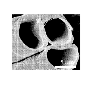
FIGs. 7a-b are HRSEM photomicrographs of extensively porous micro tubes:
Figure 7a - shell 2, core 1; Figure 7b - shell 3, core 1; The core and shell
polymers are
described in Table 2 of the Examples section which follows.
FIG. 8 is a schematic illustration of the core solvent evaporation process
from
the co-electrospun fiber;
FIGs. 9a-i depict the evaporation process of PCL/PEO (shell 1, core 1) micro-
tubes. Figures 9a-d ¨ Images taken at 2 seconds intervals [t = 0 seconds
(Figure 9a), t
= 2 seconds (Figure 9b), t = 4 seconds (Figure 9c) and t = 6 seconds (Figure
9d)]
depict the evaporation process of the PCL/PEO microtube with the following
diameter and slugs' length: X0= 0.7 mm, a = 5 um, b = 6 um, (test ii). The
meniscus
position is marked by arrows; Figures 9e-h - Images taken at t = 0 (Figure
9e), t = 1.3
(Figure 9f), t = 2.6 (Figure 9g) and t = 3.9 (Figure 9h) depict evaporation of
the
PCL/PEO microtube with the following diameter and slugs' length: X0 = 0.2 mm,
a =
7 um, b = 8 um, (test iii), the distance from the center of the slug to the
outlet was L =
0.22 mm. The meniscus position is marked by arrows; Figure 9i ¨ a graph
depicting
the displacement (measured in mm) of the meniscus (presented as Ax/x0) as
function
of time, experimental (dots) and calculated (solid line) results where tests i
and ii were
calculated according to Equation (2) and test iii was calculated according to
linear
combination of Equations (2) and (3). Test iii refers to X0 = 1.1 mm, a = 5
um, b = 6
um. The relative humidity in these experiments was 40 % (H = 0.4), and AX = Xo-
X;
CA 02664972 2009-03-30
WO 2008/041183
PCT/1B2007/054001
7
The core and shell polymers are described in Table 2 of the Examples section
which
follows.
FIGs. 10a-b are HRSEM micrographs of the inner pattern of PCL/PEO micro-
tubes (shell 1, core 1). The core and shell polymers are described in Table 2
of the
Examples section which follows.
FIGs. 1 1 a-e depict silicon oil capillary filling of microtubes. Figures 1l a-
d ¨
Sequence of video frames of silicon oil capillary filling of micro-tubes
PCL/PEO
(shell 1, core 1) taken at t = 0 seconds (Figure 11a), t = 2.4 (Figure 1 lb),
t = 3 (Figure
11c) and t = 3.5 (Figure 11d). The meniscus position is marked by arrows;
Figure lie
¨ a graph depicting the displacement of the meniscus as function of time of
two
experiments (i, ii) with micro-tubes (shell 1, core 1), and (iii) Washburn
capillary rise
model x ¨= 0.31Vi .
The core and shell polymers are described in Table 2 of the Examples section
which
follows.
FIGs. 12a-b depict a microfluid construct. Figure 12a ¨ An optical image of
the vascular microfluidic network connected with Teflon micro tubing. Shown
are
the inlet and outlets of the microfluidic network. The construct is mounted on
a sheet
of plastic for handling purposes; Figure 12b - Scanning electron microscope
image of
the cross-section of the micro fluidic.
FIG. 13 is an optical still image, depicting top view of the vascular
microfluidic network, showing individual blood red cells. Size bar = 40 micro-
meter.
DESCRIPTION OF EMBODIMENTS OF THE INVENTION
Some embodiments of the present invention provide microtubes and methods
of producing same. More specifically, microtubes of the present invention are
formed
by electrospinning.
The principles and operation of the method of producing a microtube
according to the present invention may be better understood with reference to
the
drawings and accompanying descriptions.
Before explaining at least one embodiment of the invention in detail, it is to
be
understood that the invention is not limited in its application to the details
set forth in
the following description or exemplified by the Examples. The invention is
capable
CA 02664972 2009-03-30
WO 2008/041183
PCT/1B2007/054001
8
of other embodiments or of being practiced or carried out in various ways.
Also, it is
to be understood that the phraseology and terminology employed herein is for
the
purpose of description and should not be regarded as limiting.
As used herein, the terms "comprising" and "including" or grammatical
variants thereof are to be taken as specifying the stated features, integers,
steps or
components but do not preclude the addition of one or more additional
features,
integers, steps, components or groups thereof This term encompasses the terms
"consisting of' and "consisting essentially of'.
The phrase "consisting essentially of' or grammatical variants thereof when
used herein are to be taken as specifying the stated features, integers, steps
or
components but do not preclude the addition of one or more additional
features,
integers, steps, components or groups thereof but only if the additional
features,
integers, steps, components or groups thereof do not materially alter the
basic and
novel characteristics of the claimed composition, device or method.
The term "method" refers to manners, means, techniques and procedures for
accomplishing a given task including, but not limited to, those manners,
means,
techniques and procedures either known to, or readily developed from known
manners, means, techniques and procedures by practitioners of the chemical and
physics art.
While reducing some embodiments of the invention to practice, the present
inventors uncovered a one-step procedure for producing microtubes.
As is shown in Figures 1, 2a-d, 3a-b, 10a-b, and described in Examples 1-8 of
the Examples section which follows, the present inventors were able to produce
hollow polymeric fibers i.e., microtubes, by electrospinning through co-axial
capillaries two polymeric solutions carefully selected to produce microtubes
characterized by a strong microtube shell of an even width (of about 100 nm to
about
20 micrometer) which does not collapse and an internal diameter of about 200
nm to
about 50 micrometer. In addition, as shown in Figures 7a-b and described in
Example
4 of the Examples section which follows, the presence and size of pores in the
microtube shell can be easily controlled by the selection of solvents of the
shell
polymeric solution and/or the inclusion of water-soluble polymers such as PEG.
Moreover, the thickness of the microtube shell and the tube diameter can be
controlled by the relative flow rate of the shell or coat polymeric solutions
(Figures
CA 02664972 2009-03-30
WO 2008/041183
PCT/1B2007/054001
9
6a-c, Example 3 of the Examples section which follows). Thus, a pair of
biocompatible and biodegradable polymers (e.g., PEO as a coat polymer and PCL
as
shell polymer, Table 2, hereinbelow) was used to form bio-microtubes. As is
further
described in Examples 6 and 8 of the Examples section which follows, these
microtubes can be filled with various liquids such as silicon oil (See Figures
lla-e) or
blood (Figures 12a-b and 13) which are capable of flowing therein,
demonstrating the
possible usage of these tubes as microfluidics.
Thus, according to one aspect of the invention there is provided a method of
producing a microtube. The method is effected by co-electrospinning two
polymeric
solutions through co-axial capillaries to thereby produce the microtube,
wherein a first
polymeric solution of the two polymeric solutions is for forming a shell of
the
microtube and a second polymeric solution of the two polymeric solutions is
for
forming a coat over an internal surface of the shell, the first polymeric
solution is
selected solidifying faster than the second polymeric solution and a solvent
of the
second polymeric solution is selected incapable of dissolving the first
polymeric
solution.
As used herein the term "microtube" refers to a hollow tube having an inner
diameter of e.g., about 200 nm to about 50 um and an outer diameter of e.g.,
about 0.5
um to 100 um.
As used herein the phrase "co-electrospinning" refers to a process in which at
least two polymeric solutions are electrospun from co-axial capillaries (i.e.,
at least
two capillary dispensers wherein one capillary is placed within the other
capillary
while sharing a co-axial orientation) forming the spinneret within an
electrostatic field
in a direction of a collector. The capillary can be, for example, a syringe
with a metal
needle or a bath provided with one or more capillary apertures from which the
polymeric solution can be extruded, e.g., under the action of hydrostatic
pressure,
mechanical pressure, air pressure and high voltage.
The collector serves for collecting the electrospun element (e.g., the
electrospun microtube) thereupon. Such a collector can be a rotating collector
or a
static (non rotating) collector. When a rotating collector is used, such a
collector may
have a cylindrical shape (e.g., a drum), however, it will be appreciated that
the rotating
collector can be also of a planar geometry (e.g., an horizontal disk). The
spinneret is
CA 02664972 2009-03-30
WO 2008/041183
PCT/1B2007/054001
typically connected to a source of high voltage, preferably of positive
polarity, while
the collector is grounded, thus forming an electrostatic field between the
dispensing
capillary (dispenser) and the collector. Alternatively, the spinneret can be
grounded
while the collector is connected to a source of high voltage, preferably with
negative
5
polarity. As will be appreciated by one ordinarily skilled in the art, any of
the above
configurations establishes motion of positively charged jet from the spinneret
to the
collector. Reverse polarity for establishing motions of a negatively charged
jet from
the spinneret to the collector are also contemplated.
At a critical voltage, the charge repulsion begins to overcome the surface
10 tension
of the liquid drop. The charged jets depart from the spinneret and travel
within
the electrostatic field towards the collector. Moving with high velocity in
the inter-
electrode space, the jet stretches and solvent therein evaporates, thus
forming fibers
which are collected on the collector.
As mentioned above, microtubes of some embodiments of the present
invention are formed by electrospinning. Thus, the first polymeric solution is
injected
into the outer capillary of the co-axial capillaries while the second
polymeric solution
is injected into the inner capillary of the co-axial capillaries. In order to
form a
microtube (i.e., a hollow structure, as mentioned above), the first polymeric
solution
(which is for forming a shell of the microtube) solidifies faster than the
second
polymeric solution (also referred herein as a core polymeric solution, and is
for
forming a coat over an internal surface of the shell). In addition, the
formation of a
microtube also requires that the solvent of the second polymeric solution is
incapable
of dissolving the first polymeric solution.
Thus, the solidification rates of the first and second polymeric solutions are
critical for forming the microtube. For example, for a microtube of about 100
gm, the
solidification of the first polymer (of the first polymeric solution) can be
within about
milliseconds (ms) while the solidification of the second polymer (of the
second
polymeric solution) can be within about 10-20 seconds. It will be appreciated
that
solidification may be a result of polymerization rate and/or evaporation rate.
30
According to an embodiment of the invention, the solvent of the first
polymeric solution evaporates faster than the solvent of second polymeric
solution
CA 02664972 2009-03-30
WO 2008/041183
PCT/1B2007/054001
11
(e.g., the solvent of the first polymeric solution exhibits a higher vapor
pressure than
the solvent of the second polymeric solution).
According to an embodiment of the invention, the rate of evaporation of the
solvent of the first polymeric solution is at least about 10 times faster than
that of the
solvent of second polymeric solution. Thus, the evaporation rate of the
solvent of the
first polymeric solution can be at least about 100 times faster or at least
about 1000
times faster than the evaporation rate of the solvent of second polymeric
solution. For
example, the evaporation of chloroform is significantly faster than the
evaporation of
an aqueous solution (water) due to the high vapor pressure at room temperature
of the
chloroform (195 mmHg) vs. that of the aqueous solution (23.8 mmHg).
It will be appreciated that by selecting a solvent of the second polymeric
solution which is incapable of dissolving the first polymeric solution, the
polymer of
the first polymeric solution can solidify and form a strong microtube shell
which does
not collapse, and is characterized by an even width. Thus, the first polymeric
solution
(e.g., the solvent of the first polymer) is substantially immiscible in the
solvent of the
second polymer.
As used herein the phrase "polymeric solution" refers to a soluble polymer,
i.e., a liquid medium containing one or more polymers, co-polymers or blends
of
polymers dissolved in a solvent. The polymer used by the invention can be a
natural,
synthetic, biocompatible and/or biodegradable polymer.
The phrase "synthetic polymer" refers to polymers that are not found in
nature,
even if the polymers are made from naturally occurring biomaterials. Examples
include, but are not limited to, aliphatic polyesters, poly(amino acids),
copoly(ether-
esters), polyalkylenes oxalates, polyamides, tyrosine derived
polycarbonates,
poly(iminocarbonates), polyorthoesters, polyoxaesters, polyamidoesters,
polyoxaesters containing amine groups, poly(anhydrides), polyphosphazenes, and
combinations thereof
Suitable synthetic polymers for use by the invention can also include
biosynthetic polymers based on sequences found in collagen, elastin, thrombin,
fibronectin, starches, poly(amino acid), poly(propylene fumarate), gelatin,
alginate,
pectin, fibrin, oxidized cellulose, chitin, chitosan, tropoelastin, hyaluronic
acid,
polyethylene, polyethylene terephthalate, poly(tetrafluoroethylene),
polycarbonate,
CA 02664972 2009-03-30
WO 2008/041183
PCT/1B2007/054001
12
polypropylene and poly(vinyl alcohol), ribonucleic acids, deoxyribonucleic
acids,
polypeptides, proteins, polysaccharides, polynucleotides and combinations
thereof
The phrase "natural polymer" refers to polymers that are naturally occurring.
Non-limiting examples of such polymers include, silk, collagen-based
materials,
chitosan, hyaluronic acid, albumin, fibrinogen, and alginate.
As used herein, the phrase "co-polymer" refers to a polymer of at least two
chemically distinct monomers. Non-limiting examples of co-polymers include,
polylactic acid (PLA)-polyethyleneglycol (PEG), polyethylene glycol
terephthalate
(PEGT) / polybutylene terephthalate (PBT), PLA-polyglycolic acid (PGA), PEG-
polycaprolactone (PCL) and PCL-PLA.
As used herein, the phrase "blends of polymers" refers to the result of mixing
two or more polymers together to create a new material with different physical
properties.
The phrase "biocompatible polymer" refers to any polymer (synthetic or
natural) which when in contact with cells, tissues or body fluid of an
organism does
not induce adverse effects such as immunological reactions and/or rejections
and the
like. It will be appreciated that a biocompatible polymer can also be a
biodegradable
polymer.
According to an embodiment of the invention, the first and the second
polymeric solutions are biocompatible.
Non-limiting examples of biocompatible polymers include Polyesters (PE),
PCL, Calcium sulfate, PLA, PGA, PEG, polyvinyl alcohol, polyvinyl pyrrolidone,
Polytetrafluoroethylene (PTFE, teflon), Polypropylene (PP), Polyvinylchloride
(PVC), Polymethylmethacrylate (PMMA), Polyamides, segmented polyurethane,
polycarbonate-urethane and thermoplastic polyether urethane, silicone-
polyether-
urethane, silicone-polycarbonate-urethane Collagen, PEG-DMA, Alginate,
Hydroxyapatite and Chitosan, blends and copolymers thereof.
The phrase "biodegradable polymer" refers to a synthetic or natural polymer
which can be degraded (i.e., broken down) in the physiological environment
such as
by proteases. Biodegradability depends on the availability of degradation
substrates
(i.e., biological materials or portion thereof which are part of the polymer),
the
presence of biodegrading materials (e.g., microorganisms, enzymes, proteins)
and the
availability of oxygen (for aerobic organisms, microorganisms or portions
thereof),
CA 02664972 2009-03-30
WO 2008/041183
PCT/1B2007/054001
13
carbon dioxide (for anaerobic organisms, microorganisms or portions thereof)
and/or
other nutrients. Examples of biodegradable polymers/materials include, but are
not
limited to, collagen (e.g., Collagen I or IV), fibrin, hyaluronic acid,
polylactic acid
(PLA), polyglycolic acid (PGA), polycaprolactone (PCL), polydioxanone (PDO),
trimethylene carbonate (TMC), polyethyleneglycol (PEG), Collagen, PEG-DMA,
Alginate, chitosan copolymers or mixtures thereof.
According to an embodiment, the polymeric solution can be made of one
polymer or more, each can be a polymer or a co-polymer such as described
hereinabove.
According to an embodiment of the invention, the polymeric solution of the
invention is a mixture of at least one biocompatible polymer and a co-polymer
(either
biodegradable or non-biodegradable).
According to an embodiment of the invention, the electrospun shell can be
made of a polymer such as poly (e-caprolactone) (PCL), polyamide,
poly(siloxane),
poly(silicone), poly(ethylene), poly(vinyl pyrrolidone), poly(2-hydroxy
ethylmethacrylate), poly(N-vinyl pyrrolidone), poly(methyl methacrylate),
poly(vinyl
alcohol), poly(acrylic acid), poly(vinyl acetate), polyacrylamide,
poly(ethylene-co-
vinyl acetate), poly(ethylene glycol), poly(methacrylic acid), polylactide,
polyglycolide, poly(lactide-coglycolide),
polyanhydride, polyorthoester,
poly(carbonate), poly(acrylo nitrile), poly(ethylene oxide), polyaniline,
polyvinyl
carbazole, polystyrene, poly(vinyl phenol), polyhydroxyacid,
poly(caprolactone),
polyanhydride, polyhydroxyalkanoate, polyurethane, collagen, albumin,
alginate,
chitosan, starch, hyaluronic acid, and blends and copolymers thereof
According to an embodiment of the invention, the electrospun coat can be
made of a polymer such as poly(acrylic acid), poly(vinyl acetate),
polyacrylamide,
poly(ethylene-co-vinyl acetate), poly(ethylene glycol), poly(methacrylic
acid),
polylactide polyglycolide, poly(lactide-coglycolide), polyanhydride,
polyorthoester,
poly(carbonate), poly(ethylene oxide), polyaniline, polyvinyl carbazole,
polystyrene,
poly(vinyl phenol), polyhydroxyacid, alginate, starch, hyaluronic acid, and
blends and
copolymers thereof
It will be appreciated that in order to form a hollow microtube, the solvent
of
the second polymeric solution may evaporate while the polymer forms a thin
layer on
the internal surface of the shell.
CA 02664972 2009-03-30
WO 2008/041183
PCT/1B2007/054001
14
According to an embodiment of the invention, the solvent of the second
polymeric solution is capable of evaporating through the internal surface of
the shell.
In addition, it will be appreciated that during the formation of the microtube
shell (i.e., the solidification of the first polymeric solution) the second
polymeric
solution flows within the internal surface of the shell.
According to an embodiment of the invention, the second polymeric solution
is selected capable of wetting the internal surface of the shell.
Various polymeric solutions which are known in the art as capable of wetting
other polymeric surfaces (forming the shell) can be used. Following is a non-
limiting
list of pairs of polymeric solutions in which the second polymeric solution is
capable
of wetting the internal surface of the shell formed by the first polymeric
solution.
Table I
Pairs of polymeric solutions for producing the microtube of the invention
First polymeric solution forming the Second
polymeric solution capable of
shell
wetting the internal surface of the shell
10 % poly (e-caprolactone) (PCL) in 4 % poly(ethylene oxide) (PEO) in water
chloroform (CHC13) and 20 (H20) and ethanol (60:40 by weight)
dimethylforamide (DMF) (80:20 by
weight %)
Nylon 6,6 in formic acid 7 to 12 wt % 4 % poly(ethylene oxide) (PEO) in
water
(H20) and ethanol (60:40 by weight)
Poly(L-lactide-co-glycolide)
(PLGA 4 % poly(ethylene oxide) (PEO) in water
10:90) in hexafluroisopropanol (HFIP) (H20) and ethanol (60:40 by weight)
concentrations ranging from 2 to 7 weight
% solution.
Poly(L-lactide-co-glycolide)
(PLGA 4 % poly(ethylene oxide) (PEO) in water
15:85) hexafluroisopropanol (HFIP) (H20) and ethanol (60:40 by weight)
concentrations ranging from 2 to 7
weight% solution.
poly(lactide-co-glycolide) (PLGA; 4 % poly(ethylene oxide) (PEO) in
water
1-lactide/glycolide 50/50) (H20) and ethanol (60:40 by weight)
1,1,1,3,3,3-hexafluoro-2-propanol (HFIP)
concentrations ranging from 2 to 7
weight% solution.
polyglycolide (PGA) in chloroform 3-10 9 % poly(vinyl alcohol) (PVA) in water
weight % solution. and ethanol (50:50 by weight)
poly(1-lactide) (PLA) in chloroform 3-10 9 % poly(vinyl alcohol) (PVA) in
water
weight % solution. and ethanol (50:50 by weight)
Segmented polyurethane in DMF and 9 % poly(vinyl alcohol) (PVA) in water
THF (80:20 by weight %) and ethanol (50:50 by weight)
CA 02664972 2009-03-30
WO 2008/041183
PCT/1B2007/054001
First polymeric solution forming the
Second polymeric solution capable of
shell
wetting the internal surface of the shell
Polyurethane in DMF and
9 % poly(vinyl alcohol) (PVA) in water
tetrahydrofuran, THF (80:20 by weight and ethanol (50:50 by weight)
%)
PLGA (poly lactic-co-glycolic acid) in 9 % poly(vinyl alcohol) (PVA) in water
chloroform and DMSO (dimethyl and ethanol (50:50 by weight)
sulfoxide) in chloroform and DMSO
(80:20 by weight %).
10 % PCL in CHC13 / DMF (80:20 by 6 % PEO in H20 / Et0H (60:40 by
weight) weight)
9 % PCL in CHC13 / DMSO (90:10 by 7 % PEO in H20
weight)
10 % PCL in CHC13 / DMF (80:20 by 9 % PVA in ethanol/water (50:50 by
weight) weight)
Table 1 (cont.)
According to an embodiment of the invention, the first and the second
polymeric solutions are selected from the group of: 10 % poly (e-caprolactone)
(PCL)
in chloroform (CHC13) and 20 dimethylforamide (DMF) (80:20 by weight) as the
first
5 polymeric solution and 4 % poly(ethylene oxide) (PEO) in water (H20) and
ethanol
(60:40 by weight) as the second polymeric solution, 10 % PCL in CHC13 and DMF
(80:20 by weight) as the first polymeric solution and 6 % PEO in H20 and
ethanol
(60:40 by weight) as the second polymeric solution, 9 % PCL in CHC13 and DMF
(90:10 by weight) as the first polymeric solution and 7 % PEO in H20 as the
second
10 polymeric solution, and 10 % PCL in CHC13 and DMF (800:20 by weight) as
the first
polymeric solution and 9 % poly(vinyl alcohol) (PVA) in water and ethanol
(50:50 by
weight) as the second polymeric solution.
As described in Example 3 of the Examples section which follows, the
thickness and internal diameter of the microtube can be controlled during the
15 electrospinning process. For example, the ratio between the flow rates
of the first and
second polymeric solutions can determine the fiber outer diameter and whether
the
resulting fiber is hollow or solid (see also Figures 6a-c).
According to an embodiment of the invention the thickness of the microtube
shell of the invention can vary from a few nanometers to several micrometers,
such as
from 100 nm to 20 ilm, e.g., from 200 nm to 10 ilm, from 100 nm to 5 ilm, from
100
nm to 1 ilm, e.g., about 500 nm.
CA 02664972 2009-03-30
WO 2008/041183
PCT/1B2007/054001
16
According to an embodiment of the invention the internal diameter of the
microtube shell of the invention can vary from a few nanometers to several
micrometers, such as from 50 nm to 50 um, e.g., from 100 nm to 20 um, from 200
nm
to 10 um, from 500 nm to 5 um, from 1 um to 5 um, e.g., about 3 um.
It will be appreciated that the microtube shell can be extensively porous thus
creating a "breathing" tube, or on the other hand can be totally sealed thus
forming a
one-axial flow system. A "breathing" microtube, i.e., a microtube which
comprises
pores in the shell thereof, can be formed by including a high percent of a
volatile
component in the first polymeric solution forming the shell. For example, a
high
percentage of chloroform (e.g., at least 80 %) within the polymeric solution
can result
in a porous shell (see for example, Figure 7a and Example 4 of the Examples
section
which follows).
According to an embodiment of the invention, in order to form a porous shell
the first polymeric solution includes a volatile solvent such as
Tetrahydrofuran
(THF), Chloroform, acetone, or trifluoroethanol (TFE).
Additionally or alternatively, the pores in the shell of the microtube of the
invention can be formed by including a water-soluble polymer such as
polyethylene
glycol (PEG) in the first polymeric solution. Thus, following wetting of the
microtube in a water-based solution, the water-soluble polymer is dissolved
and pores
are formed. For example, the first polymeric solution may include a blend of
polymers in which one is water-soluble and the other is water-insoluble. For
example, as is shown in Figure 7b and described in Example 4 of the Examples
section which follows, a blend of PEG and PCL was used as a first polymeric
solution
for forming a porous shell.
According to an embodiment of the invention, the first polymeric solution
comprises PEG for poring the shell. For example, to generate pores of > 150 nm
diameter, the first polymeric solution may include about 4 % PEG Mw 35 kDa
Similarly, to generate pores of < 150 nm diameter, the first polymeric
solution may
include about 2 % Mw PEG 6 kDa.
It will be appreciated that the pores in the electrospun shell can be also
generated after completion of the electrospinning process by passing an
electrical
spark through the electrospun shell, essentially as described in PCT WO
2006/106506
CA 02664972 2009-03-30
WO 2008/041183
PCT/1B2007/054001
17
to the present inventors. Such an electrical spark can be generated by any
electrical
spark producing element, such as, but not limited to, a needle-like electrode.
The
electrical spark can vary depending on the applied voltage, its duration and
the
distance between the electrode and the electrospun shell.
The electrical spark is produced with an electric field which is sufficient to
generate air breakdown. At normal conditions, such breakdown occurs at about
30
kV/cm. The electric field can be generated by a potential difference of at
least 10 kV,
e.g., at least 15 kV. Thus, the breakdown field is generated by positioning
the
electrode at a distance of about 10 mm, e.g., at a distance of 5 mm or 1 mm
from the
electrospun shell. The voltage used to generate the electrical spark can be
provided for
a time period of about 5 seconds, e.g., about 1 second, or 0.1 second
Additionally or alternatively, it will be appreciated that the pores in the
electrospun shell can be also generated by passing a heated puncturing element
through the electrospun shell.
As used herein, the phrase "puncturing element" refers to any sharp and
pointed element, e.g., a metal implement which is capable of being heated and
thus
puncturing (i.e., making a hole) the electrospun element. Non-limiting
examples of
such puncturing elements include, a metal needle and a metal pin.
Thus, the puncturing element is heated to a temperature of at least 90 C,
e.g.,
at least 91 C, 92 C, 93 C, 94 C, 95 C, 96 C, 97 C, 98 C, 99 C, 100
C, say
about 100 C, about 101 C, about 102 C and the passing of the puncturing
element
through the electrospun shell can be effected for a time period of 0.1-10
seconds, e.g.,
for a time period of 1-5 seconds.
Additionally or alternatively, it will be appreciated that the pores in the
electrospun shell can be also generated by a pulsed or continuous laser beam.
The
laser beam can be generated by any laser device capable of providing laser
radiation
which ablate or melt the polymer fibers to some extent. These include, but are
not
limited to, the following laser devices: Excimer laser device, Kr based laser
device,
Xe based laser device, Er based laser device, Ho:YAG laser device, carbon-
dioxide
laser device, Nd based laser device and laser diode device. Kr based laser
devices
include, but are not limited to krypton-fluoride (KrF) laser devices. Xe based
laser
devices include, but are not limited to xenon-fluoride (XeF) laser devices. Er
based
CA 02664972 2012-09-28
GAL115-1CA
18
laser devices include, but are not limited to, Er:YAG, Er:YSGG, Er:glass and
the like. Nd based
laser devices include, but are not limited to, Nd:YAG, Nd:YLF, Nd:glass and
the like. Also
contemplated are CO2 and Dye laser devices.
For example, perforation of an electrospun shell is performed using a pulsed
laser beam at a
specific energy (e.g., 200 Watt) which is provided at a specific rate (e.g.,
200 Hz), using several
pulses for each hole.
It will be appreciated that in order to enable flow of a liquid within the
microtube, i.e.,
along the coat polymer covering the internal surface of the shell, the surface
(thin film) formed by
the coat polymer should be designed such that it can be wetted by the liquid-
of-interest. The
wettability of polymer films by liquids are known in the art. For example,
silicone oil or water can
wet a surface made of a PEO polymer. It will be appreciated that the
wettability of the coat
polymer covering the internal surface of the shell can be controlled (e.g.,
improved) for example by
attaching functional groups such as hydroxyl group (OH) which increase the
hydrophilicity of the
coat by a plasma treatment [see Thurston RM, Clay JD, Schulte MD, Effect of
atmospheric plasma
treatment on polymer surface energy and adhesion, Journal of Plastic Film &
Sheeting 23 (1): 63-
78 JAN 2007].
Thus, the present invention provides a microtube which comprises an
electrospun shell and
an electrospun coat over an internal surface of the shell.
As used herein, the phrase "electrospun shell" refers to a hollow element of a
tubular shape,
made of one or more polymers, produced by the process of electrospinning as
detailed above.
As used herein the phrase "electro spun coat" refers to a thin layer covering
the internal
surface of the shell of the microtube of the invention which is made of one or
more polymers by
the process of electro spinning as detailed above.
One of ordinary skill in the art will know how to distinguish an electrospun
object from
objects made by means which do not comprise electrospinning by the high
orientation of the
macromolecules, the skin (e.g., shell) morphology, and the typical dimensions
of the microtube
which are unique to electrospinning.
It will be appreciated that the microtube produced by the method of the
invention can form
an individual (e.g., single or separated) microtube or can form part of a
plurality (e.g., an aligned
CA 02664972 2012-09-28
GAL115-1CA
19
array) of microtubes which can be either connected to each other or separated
(as single, not-
connected microtubes).
Thus, for the production of a single microtube a fork like clip is attached to
the edge of the
rotating disk. The disk is rotating for 1-2 seconds and individual microtubes
are collected between
the fork tooth. In a similar way individual electrospun fibers were collected
(see E. Zussman, M.
Burman, A.L. Yarin, R. Khalfin, Y. Cohen, "Tensile Deformation of Electrospun
Nylon 6,6
Nanofibers," Journal of Polymer Science Part B: Polymer Physics, 44, 1482-
1489, 2006).
Alternatively, when using a rotating collector, a plurality of microtubes can
be formed and
collected on the edge of the collector as described elsewhere for electrospun
fibers (A. Theron, E.
Zussman, A.L. Yarin, "Electrostatic field-assisted alignment of electrospun
nanofibers",
Nanotechnology J., 12, 3: 384-390, 2001).
The plurality of microtubes can be arranged on a single layer, but, more
preferably, the
plurality of microtubes define a plurality of layers hence form a three
dimensional structure. The
microtubes can have a general random orientation, or a preferred orientation,
as desired e.g., when
the fibers are collected on a cylindrical collector such as a drum, the
microtubes can be aligned
predominantly axially or predominantly circumferentially. Different layers of
the electrospun
microtubes can have different orientation characteristics. For example,
without limiting the scope
of the present invention to any specific ordering or number of layers, the
microtubes of a first layer
can have a first predominant orientation, the microtubes of a second layer can
have a second
predominant orientation, and the microtubes of third layer can have general
random orientation.
According to an embodiment of the invention a liquid fills the microtube. The
liquid may
be blood or blood components, e.g., plasma, red blood cells, coagulation
factors, white blood cells,
leukocytes, neutrophils, or any physiological solution which includes water
and physiological
concentrations of salts (e.g., phosphate buffered saline) and/or proteins.
It will be appreciated that the microtube of the invention may be configured
as or in a
microfluidics device. "Lab-on-a-chip" are described in a series of review
articles [see for example,
Craighead, H. Future lab-on-a-chip technologies for interrogating individual
molecules. Nature
442, 387-393 (2006); deMello, A. J. Control and detection of chemical
reactions in microfluidic
systems. Nature 442, 394-402 (2006); El-Ali, J., Sorger, P. K. & Jensen, K. F.
Cells on chips.
Nature 442,403-411 (2006); Janasek, D., Franzke, J. & Manz, A. Scaling and the
design of
CA 02664972 2012-09-28
GAL115-1CA
miniaturized chemical-analysis systems. Nature 442, 374-380 (2006); Psaltis,
D., Quake, S. R. &
Yang, C. H. Developing optofluidic technology through the fusion of
microfluidics and optics.
Nature 442, 381-386 (2006); Whitesides, G. M. The origins and the future of
microfluidics. Nature
442, 368-373 (2006); Yager, P. et al. Microfluidic diagnostic technologies for
global public health.
5 Nature 442, 412-418 (2006)].
Thus, the microtube of the invention (or a micro fluidic device comprising
same) can be
used as a graft of the desired length, width and internal diameter to replace
a damaged, injured or
diseased blood vessel (e.g., in a coronary artery bypass graft (CABG) surgery,
or for treating other
atherosclerosis or ischemic diseases).
As used herein the term "about" refers to 10 %.
Additional objects, advantages, and novel features of the present invention
will become
apparent to one ordinarily skilled in the art upon examination of the
following examples, which are
not intended to be limiting. Additionally, each of the various embodiments and
aspects of the
present invention as delineated hereinabove and as claimed in the claims
section below finds
experimental support in the following examples.
EXAMPLES
Reference is now made to the following examples, which together with the above
descriptions, illustrate the invention in a non limiting fashion.
Generally, the nomenclature used herein and the laboratory procedures utilized
III the
present invention include molecular, biochemical, microbiological and
recombinant DNA
techniques. Such techniques are thoroughly explained in the literature. See,
for example,
"Molecular Cloning: A laboratory Manual" Sambrook et al., (1989); "Current
Protocols in
Molecular Biology" Volumes I-III Ausubel, R. M.,
CA 02664972 2009-03-30
WO 2008/041183
PCT/1B2007/054001
21
ed. (1994); Ausubel et al., "Current Protocols in Molecular Biology", John
Wiley and
Sons, Baltimore, Maryland (1989); Perbal, "A Practical Guide to Molecular
Cloning",
John Wiley & Sons, New York (1988); Watson et al., "Recombinant DNA",
Scientific
American Books, New York; Birren et al. (eds) "Genome Analysis: A Laboratory
Manual Series", Vols. 1-4, Cold Spring Harbor Laboratory Press, New York
(1998);
methodologies as set forth in U.S. Pat. Nos. 4,666,828; 4,683,202; 4,801,531;
5,192,659 and 5,272,057; "Cell Biology: A Laboratory Handbook", Volumes I-III
Cellis, J. E., ed. (1994); "Current Protocols in Immunology" Volumes I-III
Coligan J.
E., ed. (1994); Stites et al. (eds), "Basic and Clinical Immunology" (8th
Edition),
Appleton & Lange, Norwalk, CT (1994); Mishell and Shiigi (eds), "Selected
Methods
in Cellular Immunology", W. H. Freeman and Co., New York (1980); available
immunoassays are extensively described in the patent and scientific
literature, see, for
example, U.S. Pat. Nos. 3,791,932; 3,839,153; 3,850,752; 3,850,578; 3,853,987;
3,867,517; 3,879,262; 3,901,654; 3,935,074; 3,984,533; 3,996,345; 4,034,074;
4,098,876; 4,879,219; 5,011,771 and 5,281,521; "Oligonucleotide Synthesis"
Gait, M.
J., ed. (1984); "Nucleic Acid Hybridization" Hames, B. D., and Higgins S. J.,
eds.
(1985); "Transcription and Translation" Hames, B. D., and Higgins S. J., Eds.
(1984);
"Animal Cell Culture" Freshney, R. I., ed. (1986); "Immobilized Cells and
Enzymes"
IRL Press, (1986); "A Practical Guide to Molecular Cloning" Perbal, B., (1984)
and
"Methods in Enzymology" Vol. 1-317, Academic Press; "PCR Protocols: A Guide To
Methods And Applications", Academic Press, San Diego, CA (1990); Marshak et
al.,
"Strategies for Protein Purification and Characterization - A Laboratory
Course
Manual" CSHL Press (1996); D. H. Reneker, A. Yarin, E. Zussman, S.
Koombhongse, and W. Kataphinan, "Nanofiber Manufacturing: Toward Better
Process Control", in: Polymeric Nanofibers, ACS Symposium Series, Vol. 918,
Ed.
Reneker, D. H.; Fong, H., ACS, Washington DC, 2005; A.L.Yarin, E. Zussman, A.
Greiner, J.H. Wendorff, "Material encapsulation and transport in core-shell
micro/nanofibers, polymer and carbon nanotubes and micro/nano channels", J. of
Materials Chemistry, 17, 2585 ¨ 2599, 2007; A.Greiner, J.H.Wendorff, A.L.
Yarin, E.
Zussman, "Biohybrid nanosystems with polymer nanofibers and nanotubes,"
Applied
Microbiology and Biotechnology, 71, 387-393, 2006; D.H. Reneker, A.L. Yarin,
E.
Zussman, H. Xu, "Electrospinning of nanofibers from polymer solutions,"
Advances
in Applied Mechanics (Review Paper), 41, 43-195, 2007; Z. M. Huang, Y. Z.
Zhang,
CA 02664972 2012-09-28
GAL115-1CA
22
M. Kotaki, S. Ramakrishna (2003) Composites Science and Technology 63:2223; S.
Ramakrishna, K. Fujihara, W.-e. Teo, Lim, T.C., Z. Ma, An Introduction to
Electrospinning and Nanofibers, World Scientific Publishing Company, 2005.
Other
general references are provided throughout this document. The procedures
therein are
believed to be well known in the art and are provided for the convenience of
the reader.
GENERAL MATERIALS AND EXPERIMENTAL METHODS
Polymer solutions and characterization - The polymers, poly (e-
caprolactone) (PCL) Mn 80 kDa, poly(ethylene oxide) (PEO) Mw 600 kDa,
poly(vinyl
alcohol) (PVA) Mw 100 kDa and polyethylene glycol (PEG) Mn 6 kDa were
purchased from Sigma-Aldrich and used without further treatment or
purification.
The solvents, chloroform, dimethylforamide (DMF), ethanol and the phosphate
buffer
saline (PBS- Dulbecco's) were also purchased from Sigma-Aldrich. Deionized
water
was used for the aqueous solution. The compositions of the core and shell
polymeric
solutions are given in Table 2, hereinbelow.
Table 2
Core and shell polymers used to produce electrospun microtubes
Polymer Solvent Polymer Conductivity Shear
content %(w1w) (mS cm-1) Viscosity (cP)
Shell PCL Chloroform/DMF
1 Mr, = 80 80/20 (by weight) 10 1 1300
KDa
Shell PCL Chloroform/DMF
2 Mõ = 80 90/10 (by weight) 10
KDa
Shell PCL:PEG Chloroform/DMF
3 Mr, (PCL) 80/20 (by weight) 9: 2
=80 KDa
Nin (PEG)
=6 KDa
Core 1 PEO Ethanol/water
Mw = 600 40/60 (by weight) 4 11-13 2700
KDa
Core 2 PVA Ethanol/water
Mw = 100 50/50 (by weight) 9 22 2000
KDa
Core 3 PEO DMF
Mw = 600 4 3.2 1400
KDa
CA 02664972 2009-03-30
WO 2008/041183
PCT/1B2007/054001
23
Core 4 PEO Ethanol/water
Mw = 600 40/60 (by weight) 4 200 2700
KDa lml PEO solution +50
ml PBS
Shear viscosities of the solutions were measured at different shear rates
using
a Couette viscometer (Brookfield DVII programmable viscometer). The reported
values are the results of extrapolation to zero shear rates. Conductivity was
measured
with an Oyster conductivity/temperature meter. The wetting angle of the fluids
was
measured with a home-made apparatus.
Electrospinning - Core-shell fibers were fabricated by a co-electrospinning
process using the set up described by Sun et al. (Z. Sun, E. Zussman, A. L.
Yarin, J.
H. Wendorff, A. Greiner, Adv. Mater. 2003, 15, 1929) and Zussman et al. (E.
Zussman, A. L. Yarin, V. Bazilevsky, R. Avrahami, M. Feldman, Adv. Mater.
2006,
18, 348). All experiments were conducted at room temperature and relative
humidity
of 50-60 %. For most of the runs the spinning parameters were as follow:
electrostatic field of about 0.5 kV/cm, distance between the spinneret and the
collector
plate between 16 and 20 cm. The flow rates of both the core and shell
solutions were
controlled by two syringe pumps and are given at each figure caption.
Imaging - Images of the fibers were obtained using Leo Gemini high
resolution scanning electron microscope (HRSEM) at acceleration voltage of 2-4
kV
and sample to detector distance of 2-5 mm. The specimens were coated with a
thin
gold film to increase their conductivity.
For imaging of the fibers' cross-section, the fibers were collected on a
rotating
wheel following Theron et al.'s approach (A. Theron, E. Zussman, A. L. Yarin,
Nanotechnology 2001, 12, 384) and the oriented mat was cut by a special blade
using
liquid nitrogen. The fibers were imaged by using a light microscope Olympus
BX51
(LM) and a digital camera Olympus DP 12 with a resolution of 3.34 million
pixels.
EXAMPLE I
FORMATION OF MICROTUBES USING ONE-STEP CO-ELECTROSPINNING
Experimental Results
Generation of a microtube using the core-shell set up for electrospinning
(core I: PEO, shell l: PCL, see Table 2, hereinabove) - Using the core-shell
set-up,
a stable compound drop, Taylor cone and subsequent jet were achieved as
presented
CA 02664972 2009-03-30
WO 2008/041183
PCT/1B2007/054001
24
in Figure 1. A well defined boundary between the core solution and the shell
solution
in the drop can be seen. In the present case the aqueous PEO solution of the
core and
the organic solution of PCL in the shell are immiscible and a core-shell
nanofiber with
a clear boundary between the core and the shell is achieved. It should be
mentioned
that in this case both solutions are spinnable, that is, can be electrospun as
individual
components. This is probably partially responsible for the remarkably stable
process
gained. Indeed, as can be seen by LM micrographs seen in Figure 2a, a core-
shell
nanofiber has been achieved with clear separation between the core and the
shell and
thickness uniformity of both. However, SEM micrographs of the cross-section of
these electrospun fibers reveal that the resulting fibers are hollow with a
relatively
thin and uniform shell and large hollow core as shown in Figure 2b-d. Thus,
the core
polymer is coating the inner surface of the shell (formed by the shell
polymer). The
outer diameter of the tubes is several microns while the thickness of the
walls is 0.5-1
pm. A longitudinal section seen in Figure 2d demonstrates undoubtedly that the
hollowness proceeds along the entire fiber. It is then substantiated that the
one-step
co-electrospinning results in polymeric micro-tubes. It is interesting to note
that most
of the fibers reserve their cylindrical shape without any catastrophic
collapse
indicating that the walls, although being relative thin, are sufficiently
robust.
A microtube formed using PVA and PCL as core and shell solutions,
respectively - Similar results were obtained with another system in which the
PEO
core solution was replaced by a solution of PVA at the concentration given in
Table 2
(core 2). The observations given above are valid for this system as well, as
can be
seen in Figure 3a-b.
A microtube formed using PEO dissolved in DMF as a core and PCL as a
shell - In Figure 3c a micrograph of the cross-sections of fibers made of PCL
shell
and PEO dissolved in DMF (core 3, see Table 2, hereinabove), is presented. It
can be
seen that micro-tubes are formed as well, however, the thickness of the wall
as well as
the diameter of the tubes are much less uniform. Also, adjacent tubes tend to
merge as
can be seen on the right side of the micrograph. Core 2 and core 3 differ in
their
miscibility with the shell solution. While core 2 is immiscible, core 3 is
miscible and
the difference between the resulting tubes is clearly seen.
CA 02664972 2009-03-30
WO 2008/041183
PCT/1B2007/054001
These cases are common in the fact that the shell (shell 1; Table 2,
hereinabove) solidifies first due to the relatively volatile solvent from
which the shell
solution is composed and that the pairs of solutions are immiscible (shell 1,
core 1, 2;
Table 2, hereinabove). The solidification of the core solution then takes
place once the
5 shell is already solid and the overall diameter of the fibers do not
change. The solvent
evaporation process from the core in the solidified tube is discussed below.
Without being bound by any theory, in order to achieve hollow tubes, the
solidification of the shell polymer shall be much faster than the evaporation
of the
core solvent (the solvent of the coat polymer).
10 Without being bound by any theory it seems that fast evaporation of the
shell
solvent and contact with a non-solvent, are responsible for the formation and
stabilization of the micro-tubes. Without being bound by any theory the
specific
process described herein can be considered as a dry/wet electrospinning. The
outer
surface of the shell experiences a dry spinning process as being exposed to
the
15 surrounding air and thus diffusion of the solvent and the evaporation
dominate the
solidification and the morphology of the outer surface. Since the solvent of
the shell
solution is rather volatile, the evaporation is fast. On the other hand, the
inner layer of
the shell experiences a wet spinning process as being in contact with the
relatively
nonvolatile core solution containing water/ethanol solvent which is a non-
solvent to
20 the shell polymer. The core solution then can be regarded as a
coagulation bath for the
shell. As the aqueous solution starts penetrating the shell an immediate
precipitation
of the inner layers of the shell takes place. As the affinity between the non-
solvent of
the core solution and the solvent of the shell solution is good, the
precipitation of the
polymer due to the inflow of the non-solvent into the shell is very fast (C.
C. Pereira,
25 R. Nobrega, C. P. Borges, Beaz. J. Chem. Eng. 2000, 17). Indeed, the
affinity
between the aqueous solution and DMF is strong while the chloroform tends to
separate from the DMF and evaporates first. The formation of a solid "gel
filament" at
the interface between the core and the shell probably takes place already at
the Taylor
cone (the residence time of a liquid in the Taylor cone is about 1 sec) (A. L.
Yarin, S.
Koombhongse, D. H. Reneker, Journal of Applied Physics 2001, 90, 4836). In
fact, in
such a case which can be generalized to all immiscible solutions co-
electrospinning
process, the coaxial flow is composed of a solid film enveloping the core
solution.
This is in contrast to two miscible solutions where the cone and the jet are
made of
CA 02664972 2009-03-30
WO 2008/041183
PCT/1B2007/054001
26
simply two flowing liquids. The overall process encourages a fast
solidification of the
shell by the two above- mentioned contributions, while the core solution
remains wet
for a long time after the shell has been solidified.
In the case presented in Figure 3c the shape of the tubes is relatively poor
with
a less uniform distinguished shell. Without being bound by any theory, this
may be
assigned to the fact that the core solvent is DMF (core 3) which is a good
solvent to
the shell polymer and thus doesn't contribute to fast precipitation of the
inner layers of
the shell. On the contrary, the DMF facilitates the polymer to remain in
solution.
Moreover, it is important to emphasize that the electrospinning of the same
shell
solution only (without core), i.e. an ordinary electrospinning process, didn't
result in
hollow fibers as can be seen in Figure 4, although the evaporation process was
still
fast. Thus, without being bound by any theory, in order to attain hollow tubes
both
the fast evaporation of the shell solvent and the existence of a slowly
solidifying core
are required.
EXAMPLE 2
THE CORE POLYMER PROVIDES WETTING OF THE INNER SURFACE OF
THE SHELL POLYMER
Experimental Results
The core polymer (e.g., PEO) reduces the surface tension and provides the
wetting of the solidified shell polymer (e.g., PCL) - The polymeric core
solution
being confined in the solid shell can solidify by either deposition of film
onto the
inner surface of the tubes or shrinkage to form a solid inner detached fiber.
Without
being bound by any theory, it appears that the wetting of the inner surface of
the shell
by the core solution is responsible for the way the core solidifies. In the
present case,
the PEO which is a surface active polymer (R. Nagarajan, Colloids and Surfaces
1985, 13, 1; D. Suss, Y. Cohen, Y. Talmon, Polymer 1995, 36, 1809), provides a
fair
wetting of the inner surface of the PCL shell and thus deposits a thin
adherent film
during the core evaporation as shown below, a process that ends with hollow
tubes.
This property can be represented by the contact angle. The contact angle
(wetting
angle) between PEO solution (core 1, Table 2, hereinabove) drop and a cast PCL
film
as was visually measured is ¨42 , while the contact angle between the water
and PCL
CA 02664972 2009-03-30
WO 2008/041183
PCT/1B2007/054001
27
film is much higher (¨ 90 ), see Figures 5a-b. Hence, the PEO reduces the
surface
tension and provides the wetting of the solidified PCL. The inner deposited
PEO
film also provides a supplementary mechanical strength beyond the inherent
strength
of the PCL shell.
EXAMPLE 3
THE RELATIVE FLOW RATES OF THE CORE AND SHELL SOLUTIONS
CAN DETERMINE THE FIBER DIAMETER AND THE FORMATION OF
HOLLOW FIBER
Experimental Results
The ratio between the flow rates of the core and shell solutions plays an
important role in determining both the fiber outer diameter and whether the
resulting fiber is hollow or solid - It is intuitively comprehensible that as
the fiber
gets thinner while the proportional core flow rate remains unchanged, the
fiber will be
solid since the core will fill the entire space within the tubes' walls.
Figures 6a-c
depict cross-sections of fibers made of same solutions with only difference in
their
electric conductivities. It is well known that the electric conductivity of
the spun
solution has an important influence on the resulting fiber diameter. As the
conductivity of the solution increases, the fibers get thinner (S. A. Theron,
E.
Zussman, A. L. Yarin, Polymer 2004, 45, 2017). Here, the conductivity of the
core
solution was modified by addition of PBS (core 4, Table 2, hereinabove) but
the flow
rates ratio was not adjusted accordingly. As visualized, the thinner fibers
(Figures 6 a
and b), 2.1 ilm in diameter on average, are mostly solid while the thicker
fibers
(Figure 6c), 6 ilm in diameter on average, made of core solution with the
regular low
conductivity are hollow.
EXAMPLE 4
MORPHOLOGY OF SHELL
Experimental Results
Controlling the shape of the shell morphology - The morphology of the shell
can be tailored according the applications requirements. For example the shell
can be
extensively porous thus creating a "breathing" tube, or on the other hand can
be totally
CA 02664972 2009-03-30
WO 2008/041183
PCT/1B2007/054001
28
sealed thus forming a one-axial flow system. To produce "breathing" tubes the
present inventors have used two approaches: In the first approach a higher
percent of
the volatile component (chloroform) for the shell solution (shell 2 in Table 2
hereinabove) was used, thus solidified even faster forming an extensively
porous shell
as shown in Figure 7a. The second approach exploits the phase's behavior in
polymer
blend. Here a blend of PEG and PCL was used as the shell (shell 3 in Table 2
hereinabove). The phase separation during the solidification ended with highly
porous
tubes as presented in Figure 7b.
EXAMPLE 5
THE EVAPORATION PROCESS OF THE CORE POLYMER
Experimental Results
The evaporation process of the core polymer - The evaporation of the PEO
core solution occurs once the shell is assumed to be fully dry. The
evaporation starts
with the nucleation of bubbles which soon grow to the size of the inner
diameter of
the micro-tubes. Subsequently, the bubbles continue to grow longitudinally
while
being radially confined by the tube's wall leaving a thin film of polymeric
solution.
The nucleation of the bubbles occurs at many distant sites along the fibers
consequently many slugs start to independently recede on both sides. The
bubbles
may nucleate at sites of defects or irregularities in the shell such as pores,
holes, local
thinning of the shell and more.
A schematic illustration of the solvent evaporation is shown in Figure 8. The
evaporation can take place at both the shell (region I) and the menisci
(region II). In
region I the solvent diffuses through the entire shell and evaporates. Region
II
describes the evaporation through the meniscus and the diffusion of the vapor
to the
outlet of the tubes. However, whenever the slugs are much longer than the
tubes'
diameter (as determined below), the evaporation through region II can be
neglected.
Since the vapor shall diffuse a long distance in order to reach the outlet,
the
concentration gradient along the vapor phase inside the tube is rather small
slowing
down the mass transport. On the other hand, due to the very large surface area
of the
micro-tubes and the negligible concentration of the solvent in the air
surrounding the
tubes, a large mass transport of solvent diffusing through the shell takes
place in spite
of the small diffusion coefficient
CA 02664972 2009-03-30
WO 2008/041183
PCT/1B2007/054001
29
"=:', 10-13 m2 Is
(Y. Peng, P. Y. Wu, Y. L. Yang, Journal of Chemical Physics 2003, 119, 8075).
This
transverse loss of mass causes the meniscus to recede at high velocity. A
typical
measured mean velocity of the meniscus is about U 50 lm/sec. Accordingly, the
Capillary number corresponding to this system is
Ca = ,uU I o- 10-3 ,
where ,u and a are the viscosity of core 1 solution and the surface tension of
water
(www.knovel.com) respectively.
For region I, knowing the diffusivity of the solvent across the shell, the
amount of solvent evaporated can be calculated and the meniscus receding
evolution
can be estimated by the following mass balance equation:
dmTr, dc 2 dX
Equation (1): ¨ = drr =a = c Tra ¨
dt dt
where the middle term describes the diffusion through the shell and the right
hand
side describes the movement of the meniscus where X is the half length of the
slug.
Assuming steady state, the evolution of the meniscus X with time t is
,
Equation (2): X= Xoe-t/t
where
c1a2 ln(b / a)
tI =
2D1(ci-c2)
X0 is the initial half length of the slug, a and b are the inner and outer
diameter of the
tube, and c1 and c2 are the concentration of the solvent at the inner and the
outer sides
of the micro-tubes respectively.
For region II, assuming the concentration profile of the vapor is determined
by
one-dimensional linear diffusion process, the evolution of the meniscus as
function of
time is given as:
(L Al ___
Equation (3): - X) t
(L-X0) tH
where
ci(L- X0)2
tll = 2DH(cs-Hcs)
CA 02664972 2009-03-30
WO 2008/041183
PCT/1B2007/054001
cs is the saturated vapor density of the solvent, His the relative humidity,
DH is the
diffusion coefficient of the solvent vapor in the gas phase, and L is the
distance from
the center of the slug to the outlet.
It appears that for a short slug (X0 in the order of tenths of microns) the
5 characteristic times of the evaporation from region I and II are of the
same order, for
example when X0=50 ,um
tH z' ci4 / csDH = 12s
and
ti z a2/Di = lOs
10 (ci = 1000 kg/m3, c, = 0.023 kg/m3, and DH ¨10-5m21s)
(www.epa.gov/athens/learn2model/part-two/onsite/estdiffusion.htm). However,
for a
longer slug, tH is exceeding several minutes while ti is still in the order of
tenths of
seconds. For example for X0 = 500 ,um, 41¨ 20 minutes. Sequences of video
frames
showing the receding of several slugs during the evaporation process in a few
15 experiments are presented in Figures 9a-d and 9e-h. The experimental
results
showing the displacement of the meniscus as a function of time together with
the
calculated values are presented in Figure 9i. For both tests i and ii good
agreement
with equation (2) is found, namely massive evaporation occurs through the
shell.
However, for test iii where the slug is short, the mass flux evaporated from
the
20 meniscus can not be ignored, thus the evolution of the meniscus X with
time is
calculated as a linear combination of equations (2) and (3), resulting in a
good
agreement with the experimental results.
As was argued before, the meniscus moves due to the loss of mass through the
shell leaving behind a thin but macroscopic film of PEO solution with
thickness in the
25 order of a = Ca213 ¨100nm (J. Bico, D. Quere, J. Fluid. Mech. 2002, 467,
101). By
the end of the evaporation a layer of polymer is deposited forming a wavy
pattern as
shown in Figures 10a-b. The diffusion of the water/ethanol mixture through the
shell
is slow due to the relative hydrophobicity of the PCL. Yet the diffusion is
still
available due to the possible absorption of water at the carbonyl sites of the
ester
30 groups in PCL via hydrogen bonding as was already demonstrated by Peng
et al. (Y.
Peng, P. Y. Wu, Y. L. Yang, Journal of Chemical Physics 2003, 119, 8075) that
CA 02664972 2009-03-30
WO 2008/041183
PCT/1B2007/054001
31
detected the diffusion of water in PCL film. In the present case, besides the
porosity
of the shell, the presence of the PEO encourages the absorption of the water
and the
diffusion, since the PEO easily wets the surface and makes the surface
accessible for
the water/ethanol molecules. In addition, the PEO can be dissolved in DMF up
to a
certain level and thus may penetrate into the PCL film at least a few
nanometers
before final solidification. This also enhances the penetration of the water
and ethanol
molecules and affects the inner surface morphology.
EXAMPLE 6
FILLING OF MICROTUBES WITH LIQUID
Experimental Results
Filling of electrospun microtubes with liquid - An additional evidence for the
formation of hollow fibers which has also an important practical consequence
is the
filling of these micro-tubes. Drops of silicon oil were placed onto the micro-
tubes
non-woven mat. The silicon drop easily wets the surface was immediately sucked
by
capillary forces through the porous shell and a progressive movement of a
meniscus
was observed by LM as can be seen in Figures lla-d. Experimental results of
the
filling process show that the meniscus progresses as nearly a linear function
of time
and the whole process takes only a few seconds (Figure lie).
It should be emphasized that in contrast to regular capillary filling
experiment
where the drop is placed at the entrance of the tube, here the drop was placed
on top
of the micro-tube mat and the penetration was through the shell pores. As a
consequence, the pressure drop across the tube was uneven. Therefore, the
measured
filling rate varied between the tubes and the filling rates do not follow the
Washburn's
theory (E. W. Washburn, Physical Review 1921, 17, 273) for which progression
of
the meniscus as function of square root of time would have been expected.
EXAMPLE 7
EVALUATION OF ELETROSPUN MICROTUBES
Experimental Results
Table 3, hereinbelow, presents the co-electrospinning results of different
combinations of core/shell polymer solutions. The results refer to the ability
to form
CA 02664972 2009-03-30
WO 2008/041183 PCT/1B2007/054001
32
tubes and distinguish between three cases: (a) perfect tube, (b) good tube,
and
collapsed tube. Perfect tube situation is considered when very small
variations in the
tube geometrical parameters were observed (e.g., the inner diameter varies at
the most
+/- 0.1 gm). Good tube situation is related to tubes which were stable
however,
variation in the geometry was observed (e.g., the inner diameter varies at the
most +/-
2 gm). Collapsed situation refer to a complete destroy morphology of the tube.
Solid
fiber is referred to the situation when co-electrospinning results in a solid
core/shell
fiber unlike a tube.
Table 3
Electrospun micro-fibers
Sys System Flow rates Relative Results
No. (ml/hr) Humidity (%)
Shell 1 10% PCL in 4 Good tubes
1 CHC13 / DMF 80:20 62
Core 1 4% PEO in 0.5
H20 / Et0H 60:40
Shell 1 10% PCL in 3.5
CHC13 / DMF 80:20 59 Perfect tubes
2 core 6% PEO in 0.5
H20 / Et0H 60:40
Shell 1 10% PCL in 4 Collapsed fibers
3 CHC13 / DMF 80:20
core 8% PEO in 0.5-1 30
H20 / Et0H 60:40
Shell 1 10% PCL in 4 Good tubes,
4 CHC13 / DMF 80:20 62
core 4% PEO in 0.5
H20 / Et0H 60:40
Shell 1 10% PCL in 4 Collapsed fibers
5 CHC13 / DMF 80:20 40
core
4% PEO in H20 0.5 ¨ 1
shell 10% PCL +1% 4 Collapsed fibers.
6 PEG 6K in 56 PEG 6k Mw was
CHC13 / DMF 90:10 blended with the
core 4% PEO in 0.5 shell polymer to
H20 / Et0H 60:40 enhance its
porosity
shell 8% PCL in 4
9 CHC13 44 Solid round
core 4% PEO in 0.3 fibers.
H20 / Et0H 60:40
shell 8% PCL in 4
10 CHC13 45 Solid round
fibers
core 7% PEO in H20 0.3
CA 02664972 2009-03-30
WO 2008/041183 PCT/1B2007/054001
33
Sys System Flow rates Relative Results
No. (ml/hr) Humidity (%)
shell 9% PCL in 3.5 51 Good tubes.
11 CHC13 / DMSO Highly porous
90:10 shell surface
(pore
core 7% PEO in H20 0.5 size 0.3-1um).
Table 3: Shell Polymer: PCL 80 kDa, Core polymer PEO 600 kDa.
Ambient Temperature range was 22-25 C.
EXAMPLE 8
DEVELOPMENT OF ARTIFICIAL VASCULAR MICROFLUIDIC
NETWORKS
Here the present inventors report the development of microfluidic networks of
a biocompatible polymer. Microfluidic were produced by co-electrospinning of
polymer solutions. The network resembling a capillary bed. The network used in
this
study has a total surface area of about 40 mm2. The inner diameters of the
vessels
range from 7 ilm to 20 ilm with wall thickness of about 0.5 pm. An overview of
a
microfluidic network is presented in Figure 12a. The inlet and outlet of the
network
are connected with a Teflon medical tube (SCI Scientific Commodities Inc.,
I.D.
0,036", and 0.066" 0.D.). The network was slightly inserted into the Teflon
tube and
sealed with an adhesive (Pattex N27, Henkel adhesives, Spain). Images of the
cross-
section of electrospun microfluidic network are presented in Figure 12b. The
microtubes are oriented and attached together. The tube were made by co-
electrospinning of shelll and core 1 solutions (see Table 2, hereinabove).
Flow test was conducted with these microfluidics constructs as follow: blood
drop was diluted with an heparin solution (heparin concentration (100.
units/m1),1:1
blood). A micro drop (0.2 ml) was located at the inlet of the construct. A
capillary
flow was observed under an optical microscope (see Figure 13). Blood red cells
were
moving in a typical speed of 20 '1m/second.
Altogether, these results clearly demonstrate the use of the microtube of the
invention for blood flow.
Conclusions
The fabrication of biocompatible and biodegradable polymeric microtubes in a
one step procedure by co-electrospinning has been presented. These microtubes
serve
CA 02664972 2009-03-30
WO 2008/041183
PCT/1B2007/054001
34
as micro-fluidic systems which may offer many bio-medical applications. Few
conditions are required in order to achieve tubes by this approach:
(1) Fast solidification of the shell solution;
(2) Good wetting of the shell by the core solution. This can be achieved using
a core solution consisting of a non-solvent to the shell polymer. Thus a film
can be
instantaneously formed at the interface due to the precipitation of the shell
polymer.
This film assists the stabilization of the co-electrospinning process and the
formation
of tubes with uniform and strong walls.
(3) It is recommended to use a viscoelastic polymeric core solution in order
to
gain a stabilized co-electrospinning process and deposition of significant
polymeric
film on the inner surface of the shell. This provides additional mechanical
strength to
the tubes' walls;
(4) The shell to core flow rates ratio should be chosen according to the
diameter of the resulting fibers in order to achieve hollow structure.
The evaporation process of the core solvent occurred by both the diffusion of
the solvent through the large area of the shell and evaporation through the
meniscus.
The large mass loss induced the fast receding of the slugs accompanied by
deposition
of PEO (e.g., core polymer) film. Without being bound by any theory, it is
suggested
that although the PCL shell is relatively hydrophobic the diffusion of the
water and
ethanol was encouraged by the porous character of the shell and the presence
of the
PEO which permitted the penetration of the water/ethanol molecules. The
filling of
these micro-tubes was also demonstrated showing that silicon oil and blood
were
rapidly sucked into the tubes by capillary forces. The method of the invention
depends on the stability of the co-electrospinning step which is affected by
many
factors such as miscibility or non-miscibility of the pair of solutions,
viscosity ratio,
viscoelastic relaxation time ratio, relative permittivities and conductivities
ratios,
interfacial tension, the electric field strength as well as the degree of
protrusion of the
core nozzle outside of the shell nozzle (Reznik SN, et al., 2006).
It is appreciated that certain features of the invention, which are, for
clarity,
described in the context of separate embodiments, may also be provided in
combination in a single embodiment. Conversely, various features of the
invention,
CA 02664972 2012-09-28
GAL115-1CA
which are, for brevity, described in the context of a single embodiment, may
also be provided
separately or in any suitable subcombination.
Although the invention has been described in conjunction with specific
embodiments
thereof, it is evident that many alternatives, modifications and variations
will be apparent to those
skilled in the art. Citation or identification of any reference in this
application shall not be
construed as an admission that such reference is available as prior art to the
present invention.
CA 02664972 2009-03-30
WO 2008/041183
PCT/1B2007/054001
36
REFERENCES
(Additional References are cited in Text)
1. D. H. Reneker, A. L. Yarin, E. Zussman, H. Xu, Advances in Applied
Mechanics 2006, 40.
2. S. Ramakrishna, K. Fujihara, W.-e. Teo, Lim, T.C., Z. Ma, An
Introduction to
Electrospinning and Nanofibers, 1 ed., World Scientific Publishing Company,
2005.
3. D. Li, Y. N. Xia, Advanced Materials 2004, 16, 1151.
4. M. Bognitzki, H. Hou, M. Ishaque, T. Frese, M. Hellwig, S. C., A.
Schaper, J.
H. Wendorff, A. Greiner, Adv. Mater. 2000, 12, 637.
5. R. A. Caruso, J. H. Schattka, A. Greiner, Adv. Mater. 2001, 13, 1577.
6. Z. Sun, E. Zussman, A. L. Yarin, J. H. Wendorff, A. Greiner, Adv. Mater.
2003, 15, 1929.
7. J. H. Yu, S. V. Fridrikh, G. C. Rutledge, Adv. Mater. 2004, 16, 1562.
8. Z. M. Huang, C. L. He, A. Yang, Y. Zhang, X. J. Han, J. Yin, Q. Wu, J.
Biomedical Mterials Research, Part A 2006, 77A, 169.
9. H. Jiang, Y. Hu, Y. Li, P. Zhao, K. Zhu, W. Chen, J. Control. Release
2005,
108, 237.
10. Y. Z. Zhang, X. Wang, C. T. Lim, S. Ramakrishna, Biomacromolecules
2006,
7, 1049.
11. D. Li, Y. Xia, Nano Letters 2004, 4, 933.
12. D. Li, J. T. McCann, Y. Xia, Small 2005, 1, 83.
13. E. Zussman, A. L. Yarin, V. Bazilevsky, R. Avrahami, M. Feldman, Adv.
Mater. 2006, 18, 348.
14. S. N. Reznik, A. L. Yarin, E. Zussman, L. Bercovici, Physics of Fluids
2006,
18, 1.
15. D. Li, J. T. McCann, Y. Xia, J. Am. Ceram. Soc. 2006, 89, 1861.
16. I. G. Loscertales, A. Barrero, I. Guerrero, R. Cortijo, M. Marquez, A.
M.
Ganan-Calvo, Science 2002, 295, 1695.
17. I. G. Loscertales, A. Barrero, M. Marquez, R. Spretz, R. Velarde-Ortiz,
G.
Larsen, Journal of the American Chemical Society 2004, 126, 5376.