Note: Descriptions are shown in the official language in which they were submitted.
CA 02665687 2015-05-13
BIOPSY SUPPORT WITH SECTIONABLE RESILIENT CELLULAR
MATERIAL
[0001]
Technical Field
[0002] The present invention generally relates to supports for handling
and embedding tissue samples for pathological analysis and, more particularly,
to sectionable supports which can receive one or more tissue samples and be
embedded and subsequently microtomed with the tissue sample or samples.
Background
[0003] To accurately diagnose various tissue diseases and conditions,
medical personnel must remove one or more samples of tissue from the body
of a patient. This process of harvesting tissue from the body is known as a
biopsy. Once the tissue sample or samples are removed and sent to a
pathology laboratory, the tissue will go through a series of procedures
performed by a histotechnician and, ultimately, a pathologist, in order to
diagnose one or more conditions associated with the tissue. The present
invention generally relates to those procedures that are normally performed
by the histotechnician to prepare the tissue sample or samples into slides
that
may be analyzed under a microscope by the pathologist.
[0004] Although the singular term "sample" is used throughout this
specification, it should be understood that this term likewise encompasses
plural "samples" as well. Once a tissue sample is removed from the body of a
patient, it is typically placed into a specimen container containing a tissue
-1-
CA 02665687 2009-04-06
WO 2008/073387 PCT/US2007/025253
fixative solution and then the container is transported to a pathology
laboratory.
The tissue will undergo a process known as "grossing-in" in the pathology lab
during which a histotechnician will retrieve the tissue sample from the
container,
typically cut the tissue into appropriate sizes for tissue processing, place
individual samples into the appropriate sized small plastic tissue cassettes,
and
assign tracking numbers to each cassette. These tracking numbers are then
logged into a tracking system used in the laboratory. For the smallest tissue
samples, which may only be scrapings, the cassette includes fine mesh
openings on the sides and bottoms. In other situations involving very small
tissue samples, the samples are placed into a bag that resembles a tea bag
that prevents the smallest tissue samples from escaping. Larger tissue
samples are placed into cassettes having somewhat larger slotted openings
which are nevertheless smaller than the tissue sample inside the cassette.
[0005] The cassettes are then placed into a stainless steel perforated
basket
and run through a tissue processing machine, often overnight. This machine
uses a combination of vacuum, heat, and chemicals to remove the interstitial
fluids within the tissue. Once the fluids have been removed from the tissue
samples, the processing machine immerses the tissues samples in a bath of a
hardenable material such as molten paraffin (i.e., a form of wax) so that the
interstices in the tissue are replaced with paraffin. The histotechnician then
removes the basket from the machine and removes the individual tissue
cassettes. In a conventional procedure practiced for many years, the
histotechnician individually removes the tissue sample from each cassette. The
histotechnician must carefully orient the tissue sample, based on tissue type,
into a stainless steel base mold that is roughly the size of the tissue
cassette
and is partially filled with molten paraffin. The tissue sample must be
manually
-2-
CA 02665687 2009-04-06
WO 2008/073387 PCT/US2007/025253
held, typically using forceps, against the bottom of the mold. If it is not,
this
could compromise the ability to make proper slices of the tissue sample later
in
a microtome. The molten paraffin is then rapidly cooled on a refrigerated
plate,
which may be a thermal electric cooler (TEC), to partially solidify the
paraffin
thereby holding the tissue sample in the proper orientation against the bottom
of
the mold. The cassette is then placed on top of the base mold and an
embedding material, which is also typically paraffin wax, is poured through
the
opened top of the cassette into the base mold. The cassette changes its
function at this point in the procedure from a tissue holding component to a
fixture type device for mounting in the microtome and making shavings or
slices
from the solidified paraffin in the microtome. The base mold is chilled until
all of
the molten paraffin has hardened and the histotechnician removes the stainless
steel base mold from the block of embedded paraffin. The tissue sample is
thus embedded within a rectangular block of hard paraffin with a plastic
tissue
cassette on the opposite side. As mentioned, the cassette may then be used
as a holder or fixture in the chuck of the microtome. As with the tissue
processing machine, the embedding process is accomplished in a batch fashion
during which an average histotechnician may embed approximately 40 to 60
cassettes per hour.
[0006] The blocks of hardened paraffin containing the embedded tissue
samples are then ready to be sliced into extremely thin sections for placement
on a microscope slide. The histotechnician mounts the embedded tissue block
in a chuck on the microtome that is sized to accept the side of the block that
has the embedded plastic cassette. The histotechnician can then begin slicing
the paraffin block which has the tissue sample embedded opposite to the
plastic
cassette surface. This yields a ribbon of individual slices of the tissue
-3-
CA 02665687 2009-04-06
WO 2008/073387 PCT/US2007/025253
embedded in the hardened paraffin. The action of the microtome causes the
individual slices to stick together when done properly and, subsequently,
these
very thin ribbons of slices are floated into a water bath and a glass slide is
carefully placed underneath the slice. The slice, with the thin sectioned
tissue
sample embedded therein, is then adhered to the top of the slide.
[0007] When the histotechnician has enough slides from the tissue
sample,
the slides are placed into an automatic staining machine. The staining machine
goes through a series of infiltrating steps to stain the different tissue and
cells of
the slide different colors. This helps the pathologist identify different
structures
and makes it easier to find any abnormalities in the tissue. After the
staining
procedure is complete, the slides are cover slipped and prepared for the
pathologist to place under a microscope for analysis.
[0008] Based on the summary of the procedure provided above, it will be
appreciated that conventional tissue sample handling and processing is a very
labor-intensive process involving several manual steps performed by a
histotechnician. Thus, repetitive stress injuries such as carpal tunnel
syndrome
are prevalent. This is especially true with the tissue sample embedding
process. These multiple manual operations and repeated tissue handling
increase the likelihood of human error and, moreover, require highly trained
and
skilled histotechnicians to ensure that the tissue samples ultimately adhered
to
the slides for analysis by the pathologist are in an optimum condition and
orientation to make accurate diagnoses.
[0009] U.S. Patent Nos. 5,817,032 (the '032 patent) and 7,156,814, and
U.S.
Patent Application Publication Nos. 2005/0226770; 2005/0147538; and
2005/0084425 disclose various improvements to this area of technology,
including new manners of holding tissue samples during the grossing in,
-4-
CA 02665687 2015-05-13
embedding, and microtome or slicing procedures. For example, the '032
patent relates to a tissue trapping and supporting device, which may be a
cassette, and which may be successfully sectioned using a microtome. When
such a cassette is used, the tissue sample is immobilized within the cassette
and subjected to the process for replacing tissue fluids with paraffin. Then,
the tissue sample and the cassette are sliced at the same time for later
mounting on microscope slides. Because the tissue sample is never removed
from the cassette from the time it is processed in the tissue processing
machine to the time that it is cut or sliced with the microtome, a significant
amount of handling time is saved. Moreover, the chance for human error or
tissue loss is significantly reduced due to the elimination of separate tissue
handling steps. The '032 patent and the above-incorporated published
applications also generally disclose further improvements that help to
automate the overall process and, in conjunction with the novel tissue
supports (e.g., cassettes), can even further reduce the handling steps during
the entire procedure and make the procedure more reliable.
[0010] In spite of the various advances made in this field, there is an
increasing need for additional improvements related to increased production
capability and more consistent quality of embedded tissue samples and
resulting slices or ribbons of embedded tissue that will be subject to
diagnosis.
This can be especially important when handling smaller tissue sample sizes,
although the improvements to be disclosed herein are applicable to all tissue
sample sizes.
-5-
CA 02665687 2015-05-13
Summary
[0011] In one general embodiment, a histologic tissue sample support is
provided and may generally comprise a tissue support coupled with a resilient
cellular material. The resilient cellular material is a three dimensional,
microtome sectionable, deflectable structure that is an improvement upon the
microtome sectionable, deflectable structures disclosed in '032 patent
discussed
above. The tissue sample support device can more specifically include a tissue
support formed of material which can be successfully sectioned in a microtome
and is resistant to degradation from solvents and chemicals used to fix,
process
and stain tissue. The porosity of
the resilient cellular material allows infiltration of the solvents and
chemicals
used to fix, process and stain tissue, and of embedding material used to embed
the tissue while the tissue is retained by the resilient cellular material.
The
resilient cellular material has a thickness that is compressible and
configured to
engage and retain tissue in place during processing and embedding and is also
capable of successful sectioning in the microtome after having its interstices
or
pores filled with liquefied embedding material which subsequently hardens.
[0012] The resilient cellular material may further comprise an open cell
foam
material, such as a foam including at least one of a polyether or a
polyurethane.
In addition, the open cell foam may be a fully reticulated foam. This helps
ensure full infiltration of fluids used during processing and embedding
procedures. Other synthetic and natural materials may be used such as
polyesters, alginates, or other materials that may be infiltrated with the
embedding material and successfully sectioned and a microtome without
adverse effects on the resulting ribbon of tissue and embedding material.
-6-
CA 02665687 2015-05-13
[0013] The support may further include a tissue containment portion
including a recess or interior area surrounded by at least one side wall and
including a bottom wall. The recess or interior area may be configured to at
least partially contain the resilient cellular material either during the
manufacturing of the device or during insertion of the cellular material into
the
recess or interior area by the user in order to retain the tissue sample in
place
during processing and embedding procedures. The support can further
comprise a cassette having a lid configured to be connected to the containment
portion. In one embodiment, the resilient cellular material is coupled to the
lid
and is inserted at least partially into the recess upon connecting the lid to
the
containment portion.
[0014] The material forming the support may be at least translucent so as
to be non-distracting during tissue analysis. For example, the support may be
formed of any of the materials disclosed in the aforementioned patent and
patent
applications such as polymers including fluorinated polymers or fluoropolymers
(e.g., PFA).
[0015] An assembly may be constructed with the support and a separate
frame. In such an assembly, the tissue support is releasably retained on the
frame and the frame is further configured for releasable securement within a
microtome chuck. The frame can further include an interior and the tissue
support may be sized to fit and move within the interior between at least a
first
position and a second position. The first position is used during processing
of
the tissue sample, and the second position is used to expose the tissue
outward
of the frame in a position for allowing the tissue sample to be sectioned in
the
microtome.
-7-
CA 02665687 2009-04-06
WO 2008/073387
PCT/US2007/025253 =
[0016] Various methods are disclosed or will be apparent based on a
review
of the disclosed embodiments and features. For example, a method for
preparing one or more biopsy tissue samples for histological examination may
comprise:
positioning a tissue sample in close proximity to a microtome
sectionable support;
immobilizing the tissue sample on a the support by contacting the
tissue sample with a microtome sectionable, resilient cellular material
coupled
to the tissue support;
subjecting the microtome sectionable support, resilient cellular
material and the tissue sample to a process that replaces fluid in the tissue
sample with a hardenable material;
embedding the microtome sectionable support, resilient cellular
material and the tissue sample in an embedding material;
hardening the embedding material into a block; and
slicing the block with a microtome into thin slices of the
embedding material, the microtome sectionable support, the resilient cellular
material and the tissue sample.
[0017] The hardenable material and the embedding material may be the
same material, such as a wax (e.g., paraffin). The support may further
comprise a bottom portion configured to hold the tissue sample and a lid
holding the resilient cellular material. The step of immobilizing the tissue
sample can further comprise closing the lid on top of the tissue sample to
trap
the tissue sample between the resilient cellular material and the bottom
portion.
The bottom portion can include an interior space surrounded by at least one
side wall and the positioning and immobilizing steps and can further comprise
-8-
CA 02665687 2015-05-13
placing the tissue sample within the interior space, and inserting the
resilient
cellular material at least partially into the interior space and into contact
with
the tissue sample. The resilient cellular material may deform during the
immobilizing step to create a three dimensional space that receives the tissue
sample. This can help immobilize the tissue sample in a desired form flat
against the bottom of the support or cassette. The force of the resilient
cellular material against the tissue should be enough to immobilize and/or
flatten the tissue but not enough to induce artifacts in the sample. The
microtome sectionable support may be coupled to a frame prior to being
subjected to the process for replacing fluid in the tissue sample with the
hardenable material. The method can then further comprise securing the
frame in the microtome prior to slicing the block. Prior to embedding the
microtome sectionable support, resilient cellular material and the tissue
sample in the embedding material, the microtome sectionable support may be
moved from a first position within the frame to a second position in which the
support, resilient cellular material and tissue sample are exposed for
simultaneous sectioning in the microtome.
[0017.1] According to one aspect of the present invention there is
provided a histologic tissue sample support device comprising a tissue
cassette, the tissue cassette including a tissue containment portion having a
recess surrounded by at least one side wall and including a bottom wall, the
tissue containment portion formed of material which can be successfully
sectioned in a microtome and is resistant to degradation from solvents and
chemicals used to fix, process and stain tissue, the cassette further
including
a lid; and an open cell, fully reticulated foam material coupled to the tissue
cassette, the open cell, full reticulated foam material configured to engage
and retain tissue in place during processing and embedding, the lid being
couplable to the tissue containment portion and used to compress the open
cell, fully reticulated foam against the tissue, and the open cell, full
reticulated
foam material further being capable of successful sectioning in the microtome
and porous to allow infiltration of the solvents and chemicals used to fix,
-9-
CA 02665687 2015-05-13
process and stain tissue, and of embedding material used to embed the tissue
while the tissue is retained by the open cell, full reticulated foam material
in
the recess.
[0017.2] According to a further aspect of the present invention there is
provided a method for preparing one or more biopsy tissue samples for
histological examination, comprising positioning a tissue sample in close
proximity to a microtome sectionable support; immobilizing the tissue sample
against the support by contacting the tissue sample with a microtome
sectionable, open cell fully reticulated foam material; subjecting the
microtome
sectionable support, the open cell fully reticulated foam material and the
tissue sample to a process that prepares the tissue sample for embedding;
embedding the microtome sectionable support, the open cell fully reticulated
foam material and the tissue sample in an embedding material; hardening the
embedding material into a block; and slicing the block with a microtome into
thin slices of the hardened embedding material, the microtome sectionable
support, the open cell fully reticulated foam material and the tissue sample.
[0017.3] According to another aspect of the present invention there is
provided a method for preparing one or more biopsy tissue samples for
histological examination, comprising positioning a tissue sample in close
proximity to a microtome sectionable support; immobilizing the tissue sample
on a the support by contacting the tissue sample with a microtome
sectionable, resilient cellular material coupled to the tissue support;
subjecting
the microtome sectionable support, resilient cellular material and the tissue
sample to a process that replaces fluid in the tissue sample with a hardenable
material; embedding the microtome sectionable support, resilient cellular
material and the tissue sample in an embedding material; hardening the
embedding material into a block; and slicing the block with a microtome into
thin slices of the embedding material, the microtome sectionable support, the
resilient cellular material and the tissue sample.
-9a-
CA 02665687 2015-05-13
[0018] Various additional details, features, advantages and aspects of
the invention will become more readily apparent to those of ordinary skill in
the art on review of the following illustrative, more detailed description.
Brief Description of the Drawings
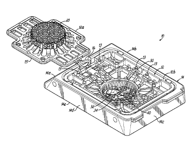
[0019] Fig. 1 is a perspective view of an assembly comprised of a
microtome sectionable tissue cassette received within a frame, with the lid of
the cassette shown in an open position
-9b-
CA 02665687 2009-04-06
WO 2008/073387 PCT/US2007/025253
[0020] Fig. 2 is a perspective view similar to Fig. 1, but illustrating
the lid of
the cassette in a closed position.
[0021] Fig. 3 is a cross sectional view generally taken along line 3-3
of Fig.
2.
[0022] Fig. 4 is a cross sectional view similar to Fig. 3, but showing
the
cassette/frame assembly embedded in paraffin and with the cassette in a
second, lower position within the frame for sectioning.
[0023] Fig. 5 is a cross sectional view taken generally along line 5-5
of Fig.
4.
[0024] Fig. 6 is a cross sectional view taken generally along line 6-6
of Fig.
4.
[0025] Fig. 7 is a perspective view of an assembly comprised of a
microtome
sectionable tissue cassette constructed according to another embodiment, and
received within a frame, with the lid of the cassette shown in an open
position.
[0026] Fig. 8 is a top view of the assembly shown in Fig. 7, but with
the lid in
a closed position, and the assembly in a mold.
[0027] Fig. 9 is a cross sectional view taken along line 9-9 of Fig. 8
and
schematically illustrating the lid being depressed into a closed position.
[0028] Fig. 10 is a cross sectional view similar to Fig. 9, but
illustrating the
cassette in a second, lower position within the frame such that a lower
portion
of the cassette extends into the mold.
Detailed Description
[0029] Figs. 1 and 2 generally illustrate an assembly 10 comprised of a
tissue sample cassette 12 carried within a frame 14. The connection of the
tissue cassette 12 to the frame 14 may be accomplished in many different
-10-
CA 02665687 2015-05-13
manners, such as any of the manners described in the aforementioned patent
and patent applications. It will be also be appreciated that the cassette 12
may
be configured in any suitable manner as a tissue support and the frame 14 may
be configured in any suitable manner. Any of the configurations, features,
characteristics and materials disclosed for the tissue supports (e.g.,
cassettes)
and frames in the above-incorporated patent and patent applications may be
employed for cassette 12 and frame 14. In the embodiment shown, the
cassette 12 is porous and is releasably retained in the frame 14 and the frame
14 is further configured to be releasably secured within a microtome chuck
(not
shown). The frame 14 generally includes an interior defined between
surrounding outer walls 14a, 14b, 14c, 14d and the cassette 12 is sized and
configured to frictionally or "snap" fit and move within the interior between
at
least first and second positions, again, as generally discussed in the
aforementioned patent and patent applications and for the same purposes.
The first position is shown in Fig. 3, while the second position is a position
(not
shown) in which the lower portion of the cassette 12 is exposed below the
bottom of the frame 14, as viewed in Fig. 3, for allowing the cassette 12 and
tissue sample to be sectioned in a microtome while the frame 14 is held in the
microtome chuck. The general procedure for processing, embedding, and
sectioning is discussed in the above-incorporated patent and patent
applications. The cassette may be formed from perfluoroalkoxyethylene (PEA)
in accordance with the above-incorporated patents and patent applications.
[0030] A lid 12a
of the cassette 12 may be coupled to a body 12b of the
cassette 12 by a hinge 16. The lid 12a may also snap fit into a closed
position
as shown in Fig. 2 through engagement of fingers or projecting connectors 13
on the cassette body 12b with an outer flange 15 of the lid 12a on each of the
-11-
CA 02665687 2009-04-06
WO 2008/073387 PCT/US2007/025253
four sides of the lid 12a. The lid 12a carries a resilient cellular material
20
which may, for example, be an open cell foam material, such as a foam
including at least one of a polyether or a polyurethane and which may be a
fully
reticulated foam. Here, "fully reticulated" means that at least substantially
all
cells of the foam are open. As shown in Fig. 1, one or more tissue samples
may be placed in a porous tissue containment portion 30 that may define a
recess or interior area surrounded by at least one sidewall 32 and including a
bottom wall 34. Although a circular recess is shown, it will be appreciated
that
any other shape may be used instead.
[0031] As further shown in Fig. 3, when the lid 12a is closed, the foam
material 20 will press against the tissue samples 40 and deform three
dimensionally around the tissue samples 40 creating three dimensional spaces
around each tissue sample 40 and essentially immobilizing each tissue sample
40 during the tissue processing and embedding procedures. This also ensures
that the tissue samples 40 are held flat against the bottom wall 34 of the
tissue
cassette 12 such that when microtome slices are made, complete and
continuous sections of the tissue sample 40 may be formed generally as shown
in Fig. 5. One specific type of foam structure suitable for the resilient
cellular
material 20 has a pore size of 50-60 ppi (pores per inch), with each pore
having
a diameter of between about 0.017 inch and 0.20 inch. The foam structure is
fully reticulated with a compression force deflection at 20% deflection of
0.55
lbslin2 and a density of 1.4 lbs./ft3. The foam material may be obtained from
Crest Foam of Moonachie, New Jersey under the name T-50. This is a
polyether/polyurethane foam and operates well with a thickness of 0.06 inch to
0.10 inch with a 0.075 inch thickness being a practical manufacturing example.
The foam should be constructed so as to shed or release processing fluid after
-12-
CA 02665687 2009-04-06
WO 2008/073387
PCT/US2007/025253
each reagent cycle of a tissue processing machine. If the foam is too dense or
too thick, or not fully reticulated, the reagents can become cross
contaminated
or the tissue may not be fully infiltrated with the fluids because each fluid
bath
must fully clear and exchange from one fluid bath to the next.
[00321 In use,
one or more tissue samples 40 are placed within the interior
space or recess and, specifically, on the bottom wall 34 as shown in Fig. 1.
The
cassette lid 12a is then closed and snapped into place such that the resilient
cellular material (e.g., foam) 20 bears against and traps the tissue samples
40
against the bottom wall 34 as shown in Fig. 3. At this point, the assembly 10
with the trapped tissue samples 40 may be subjected to a conventional tissue
processing operation that uses vacuum, heat and chemicals to remove the
interstitial fluids within the tissue and replace those fluids with a
hardenable
material, such as molten paraffin. As mentioned above, during these
processing steps, the porous nature of the foam or other resilient cellular
material 20 allows the fluids to reach and fully infiltrate into the tissue
samples
40. In addition, the foam 20 traps the tissue samples 40 flat against the
bottom
wall 34 without leaving artifacts or markings on the tissue that might
interfere
with subsequent analysis under a microscope. It will be appreciated that
different types of resilient cellular materials may be chosen based, for
example,
on the type of tissue to be processed and analyzed. For example, small
mucosal tissue samples may be held and processed with success using the T-
50 foam discussed above, while other types of tissue, such as fatty tissue,
may
be better served by another type of resilient cellular material.
[0033] It will
also be appreciated that the processing steps may take place
before assembling the tissue cassette 12 with the frame 14. After the tissue
processing is complete, the tissue cassette 12 may be moved to a second
-13-
CA 02665687 2015-05-13
position as shown in Fig. 4 exposing the containment portion 30 below the
bottom surface 14e of the frame 14. The cassette 12 and frame 14 are then
placed into a suitable mold (not shown) and embedded in paraffin 50, such that
the entire assembly including the lower exposed containment portion 30 are
embedded within a hardened block of paraffin wax 50. The mold may generally
follow the contour of the bottom of the cassette 12, although the portion of
the
mold surrounding the containment portion 30 is preferably square as opposed
to round. This assists with the subsequent production of ribbon slices. This
portion of the procedure may therefore be similar to that disclosed in the
aforementioned patent and patent applications. As discussed therein, the
frame 14 is then used as a fixture for mounting the embedded assembly 10 in a
microtome chuck and the necessary number of slices are taken of the exposed
underside until enough sections, similar to those shown in Fig. 5, are taken
and
appropriately mounted on a microscope slide, stained and cover slipped.
[0034] Figs. 7-10 illustrate an alternative embodiment. In this
embodiment,
the cassette is constructed with a somewhat different design than the first
embodiment as will be apparent from a review of the figures, as well as the
description below. Like reference numerals in Figs. 1-10 refer to like
structure
and, therefore, additional description with respect to Figs. 7-10 is not
necessary. Like reference numerals in Figs. 7-10 having prime marks (') refer
to analogous elements as shown and described in connection with Figs. 1-6 but
having differences that are either apparent by reviewing the drawings
themselves or by a combination of reviewing the drawings and the additional
description contained below.
[0035] The primary difference between assembly 10 and assembly 10' is
with respect to the lids 12a, 12a' and the manner that the lids 12a, 12a'
connect
-14-
CA 02665687 2009-04-06
WO 2008/073387
PCT/US2007/025253
with the bodies 12b, 12b' of the cassettes 12, 12'. In the first embodiment,
The
lid 12a is held down to the body 12b by a series of snap fit connectors 13 on
each of the four sides of the cassette body 12b. These connectors 13 engage
an outer flange section 15 of the lid 12a. Thus, the user typically would use
his
or her finger to depress each of the four sides of the cassette lid 12a
downward
to engage each of the sets of snap fit connectors 13. The cassette 12' shown
in
Figs. 7-10 instead uses a circular lid 12a' having three connecting elements
60
spaced apart by 1200. Elements 60 connect in a snap fit manner with
connectors 62 on the cassette body 12b' when the lid 12a' is folded over as
shown in Figs. 8 and 9. As further illustrated in Fig. 9, the lid 12a' may be
closed and locked in a snap fit manner utilizing the three mating connectors
60,
62 by depressing the lid 12a' with a single finger 64 of the user. As
previously
discussed, the cassette 12' may be depressed entirely as a unit from the upper
position in the frame 14, illustrated in Fig. 9, to the lower position shown
in Fig.
10. This extends the porous tissue containment portion 30 into the mold 68.
The mold may then be filled with embedding material such as paraffin 50. A
structure is thereby formed as previously described in connection with Fig. 4.
It
will further be appreciated that the basic perforated design of the cassette
12' is
changed relative to the perforated design of the cassette 12 illustrated in
Figs.
1-6. Essentially, the design of the molded cassette12' illustrated in Figs. 7-
10
reduces the amount of PFA material necessary to form the cassette 12'. All
other structural and functional aspects and uses of assembly 10' are as
described in connection with assembly 10 of the first embodiment.
[0036] While
the present invention has been illustrated by a description of
various illustrative embodiments and while these embodiments have been
described in some detail, it is not the intention of the Applicants to
restrict or in
-15-
CA 02665687 2015-05-13
,
any way limit the scope of the appended claims to such detail. Additional
advantages and modifications will readily appear to those skilled in the art.
The
various features of the invention may be used alone or any combinations
depending on the needs and preferences of the user. However, the invention
itself should only be defined by the appended claims.
-16-