Note: Descriptions are shown in the official language in which they were submitted.
CA 02666586 2009-04-16
WO 2008/051761 PCT/US2007/081606
ASSAY FOR CARDIAC TROPONIN AUTOANTIBODIES
FIELD OF THE INVENTION
The present invention relates generally to the area of assays for
autoantibodies
reactive with a cardiac troponin. In particular, the invention relates among
other things
to the use of such assays and kits for such assays in the assessment of
cardiac
pathologies or the risk thereof.
BACKGROUND OF THE INVENTION
Troponin complex is a heteromeric protein playing an important role in the
regulation of skeletal and cardiac muscle contraction. It consists of three
subunits:
troponin I(TnI), troponin T (TnT) and troponin C (TnC). Each subunit is
responsible
for part of troponin complex function; e.g., TnI inhibits the ATP-ase activity
of
actomyosin.
TnT and TnI are present in myocardium in different forms than in skeletal
muscles. Cardiac TnI (cTnl) is expressed only in myocardium. cTnl has been
widely
used as a marker of cardiac tissue injury. cTnl is considered to be more
sensitive and
significantly more specific in the diagnosis of myocardial infarction than CK-
MB,
myoglobin, and LDH isoenzymes.
cTnl can be detected in patient's blood 3 - 6 hours after onset of the chest
pain,
reaching peak level within 16 - 30 hours. cTnl is also useful for the late
diagnosis of
acute myocardial infarction, because elevated concentrations can be detected
in blood
even 5 - 8 days after onset.
During the incubation in the necrotic muscle after acute myocardial
infarction,
cTnl is cleaved by endogenous proteases. The most stable fragment resulting
from this
cleavage is located between 30 and 110 amino acid residues. For this reason,
cTnl
assays have employed antibodies that recognize this fragment.
In view of the importance of early detection of cardiac tissue injury, there
clearly remains a need for methods and kits to identify cardiac pathology, or
risk
thereof, either as an independent indicator, or which can be employed in
conjunction
with other assays.
1
CA 02666586 2009-04-16
WO 2008/051761 PCT/US2007/081606
This background information is provided for the purpose of making known
information believed by the applicant to be of possible relevance to the
present
invention. No admission is necessarily intended, nor should be construed, that
any of
the preceding information constitutes prior art against the present invention.
SUMMARY OF THE INVENTION
The invention provides among other things assays for autoantibodies reactive
with a cardiac troponin, kits for performing such assays, and the use of such
assays and
kits in the assessment of cardiac pathologies or the risk thereof.
In one embodiment, the present invention provides a method of determining the
reliability of a cardiac troponin assay result, e.g., where there is a chance
that the
amount of cardiac troponin measured by assay is impacted by the presence of
autoantibodies reactive with a cardiac troponin within a subject. The method
entails
assaying a biological sample for an autoantibody reactive with a cardiac
troponin,
wherein the presence of an elevated level of cardiac troponin-reactive
autoantibody
indicates that the cardiac troponin assay result is not reliable. This method
optionally
comprises the steps of: (a) obtaining a biological sample from the subject;
(b)
determining the level of autoantibody reactive with a cardiac troponin in the
sample
(e.g., using a cardiac troponin antigen); and (c) evaluating the reliability
of a cardiac
troponin assay result based on the level of autoantibody reactive with a
cardiac troponin
in the sample.
In another embodiment, the invention provides methods of assessing risk of a
cardiac pathology. This method entails assaying a biological sample for an
autoantibody reactive with a cardiac troponin, wherein the presence of an
elevated level
of cardiac troponin-reactive autoantibody indicates an elevated risk of a
cardiac
pathology. This method optionally comprises the steps of: (a) obtaining a
biological
sample from a subject; (b) determining the level of autoantibody reactive with
a cardiac
troponin in the sample (e.g., using a cardiac troponin antigen); and (c)
evaluating the
risk of a cardiac pathology result based on the level of autoantibody reactive
with a
cardiac troponin in the sample.
In a variation of this embodiment, a method of the invention entails assaying
a
biological sample for a cardiac troponin, and assaying a biological sample
from the
2
CA 02666586 2009-04-16
WO 2008/051761 PCT/US2007/081606
subject for an autoantibody reactive with a cardiac troponin. The assays can
be
conducted using one biological sample or different ones. The presence of an
elevated
level of cardiac troponin and/or an elevated level of cardiac troponin-
reactive
autoantibody indicates an elevated risk of a cardiac pathology. This method
optionally comprises the steps of: (a) obtaining one or more biological
samples from a
subject; (b) determining the level of a cardiac troponin in the one or more
biological
samples (e.g., using an antibody specific for the cardiac troponin antigen);
(c)
determining the level of autoantibody reactive with a cardiac troponin in the
one or
more biological samples (e.g., using a cardiac troponin antigen); and (d)
evaluating the
risk of a cardiac pathology result based on the presence of an elevated level
of cardiac
troponin and/or an elevated level of autoantibody reactive with a cardiac
troponin in the
sample. In this method, step (c) can be done before, after, concurrent with,
or in the
absence of step (b), optionally on either the same or a different biological
sample.
In particular embodiments, the above-described methods, and other methods
described herein can be carried out using a biological sample obtained from a
subject
with chest pain. In certain embodiments, the subject is suspected of having a
myocardial infarction.
Methods of assessing risk of cardiac pathology include screening for a subject
having, or at risk of having, myocarditis, cardiomyopathy, and ischemic heart
disease.
This method entails assaying a biological sample from the subject for an
autoantibody
reactive with a cardiac troponin. The presence of an elevated level of cardiac
troponin-
reactive autoantibody indicates the presence of, or risk of, the cardiac
pathology. This
method optionally comprises the steps of: (a) obtaining a biological sample
from a
subject; (b) determining the level of autoantibody reactive with a cardiac
troponin in
the sample (e.g., using a cardiac troponin antigen); and (c) evaluating the
risk of a
cardiac pathology result based on the level of autoantibody reactive with a
cardiac
troponin in the sample.
The invention also provides a method of determining whether a subject having,
or at risk for, a cardiac pathology is a candidate for immunosuppressive
therapy and/or
immunoabsorption therapy. The method entails assaying a biological sample from
the
subject for an autoantibody reactive with a cardiac troponin. The presence of
an
elevated level of cardiac troponin-reactive autoantibody indicates that the
subject is a
3
CA 02666586 2009-04-16
WO 2008/051761 PCT/US2007/081606
candidate for such therapy. This method optionally comprises the steps of: (a)
obtaining a biological sample from a subject; (b) determining the level of
autoantibody
reactive with a cardiac troponin in the sample (e.g., using a cardiac troponin
antigen);
and (c) evaluating whether a subject is a candidate for immunosuppressive
therapy
and/or immunoabsorption therapy based on the level of autoantibody reactive
with a
cardiac troponin in the sample.
In another embodiment, the invention provides a method of identifying a
subject
having, or at risk for, a cardiac pathology. The method entails assaying a
biological
sample from the subject for an autoantibody reactive with a cardiac troponin,
wherein
the subject has an autoimmune disease, or the subject is a first-degree
relative of an
individual having an autoimmune disease. This method optionally comprises the
steps
of: (a) obtaining a biological sample from a subject, wherein the subject has
an
autoimmune disease, and/or is a first-degree relative of an individual having
an
autoimmune disease; (b) determining the level of autoantibody reactive with a
cardiac
troponin in the sample (e.g., using a cardiac troponin antigen); and (c)
evaluating the
risk of a cardiac pathology result based on the level of autoantibody reactive
with a
cardiac troponin in the sample.
In each of the methods described herein: the biological sample can be obtained
from a subject that is a mammal (e.g., optionally human); the cardiac troponin
assayed
can include a cardiac troponin selected from the group consisting of a cardiac
troponin
I, T, C, and complexes thereof; and the autoantibody can be reactive with a
cardiac
troponin comprising a cardiac troponin selected from the group consisting of a
cardiac
troponin I, T, C, and complexes thereof.
Any of the methods described herein can conveniently be carried out using an
immunoassay. Suitable immunoassays include an agglutination assay. In an
exemplary agglutination assay, a biological sample is contacted with a cardiac
troponin
antigen affixed to a solid phase, under conditions sufficient for binding of
the cardiac
troponin antigen to any cardiac troponin-reactive autoantibody present in the
sample,
followed by measurement of any agglutination of the sample. The degree of
agglutination is positively correlated with the concentration of cardiac
troponin-reactive
autoantibody present in the sample.
4
CA 02666586 2009-04-16
WO 2008/051761 PCT/US2007/081606
In other embodiments, the biological sample is contacted with a cardiac
troponin antigen, under conditions sufficient for binding of the cardiac
troponin antigen
to any cardiac troponin-reactive autoantibody present in the sample, and
signal is
detected from one or more complex(es) comprising the cardiac troponin antigen
bound
to cardiac troponin-reactive autoantibody. Such immunoassays can be carried
out in a
non-competitive format, in which case the signal is positively correlated with
the
concentration of any cardiac troponin-reactive autoantibody present in the
sample.
In exemplary non-competitive immunoassays useful in the invention, the
method additionally entails contacting the biological sample with a species-
specific
antibody, wherein the species-specific antibody is specific for the species
from which
the biological sample was obtained, under conditions sufficient for specific
binding of
the species-specific antibody to any cardiac troponin-reactive autoantibody
present.
Signal detection entails detecting any complex including the cardiac troponin
antigen
bound to cardiac troponin-reactive autoantibody, which is itself bound to
labeled
species-specific antibody. The contact between the biological sample and the
cardiac
troponin antigen and the contact between the biological sample and the species-
specific
antibody can be carried out simultaneously or sequentially, in any order.
In exemplary non-competitive immunoassays useful in the invention, the
cardiac troponin antigen can be affixed to a solid phase. Binding of the
cardiac
troponin to any cardiac troponin-reactive autoantibody present in the sample
forms a
solid phase-affixed complex, and signal is detected from this complex. If
desired, the
species-specific antibody can be labeled (e.g., in a sandwich immunoassay).
Alternatively, the species-specific antibody can be affixed to a solid phase.
In this case,
binding of the species-specific antibody to any cardiac troponin-reactive
autoantibody
present in the sample forms a solid phase-affixed complex, and signal is
detected from
this complex. If desired, the cardiac troponin antigen can be labeled (e.g.,
in a
sandwich immunoassay).
Immunoassays useful in the invention can also be carried out in a competitive
format, in which case the signal is negatively correlated with the
concentration of
cardiac troponin-reactive autoantibody present in the sample. In particular
embodiments, the biological sample is contacted with a cardiac troponin
antigen, under
conditions sufficient for binding of the cardiac troponin antigen to any
cardiac
5
CA 02666586 2009-04-16
WO 2008/051761 PCT/US2007/081606
troponin-reactive autoantibody present in the sample. In addition the
biological sample
is contacted with a labeled cardiac troponin-reactive antibody under
conditions
sufficient for specific binding of the labeled cardiac troponin-reactive
antibody to the
cardiac troponin antigen. The contact between the biological sample and the
cardiac
troponin antigen and the contact between the biological sample and the labeled
cardiac
troponin-reactive antibody can be carried out simultaneously or sequentially,
in any
order.
In exemplary competitive immunoassays useful in the invention, the cardiac
troponin antigen can be affixed to a solid phase. Binding of the cardiac
troponin to any
cardiac troponin-reactive autoantibody present in the sample forms a solid
phase-
affixed complex, and signal is detected from this complex.
The invention also provides a test kit for assaying a biological sample for
cardiac troponin-reactive autoantibodies. In particular embodiments, the test
kit
includes an antibody reactive with cardiac troponin. For example, the test kit
can
include a humanized monoclonal antibody, wherein the humanized monoclonal
antibody is specific for a cardiac troponin. Alternatively or additionally,
the test kit can
include a labeled non-human monoclonal antibody, wherein the non-human
monoclonal antibody is specific for a cardiac troponin.
Test kits accordingly to the invention can include, if desired, a solid phase
and a
capture agent affixed to the solid phase. In exemplary embodiments, the
capture agent
is a cardiac troponin antigen or a species-specific antibody, wherein the
species-specific
antibody is specific for the species from which the biological sample is to be
obtained.
In particular embodiments, the species-specific antibody comprises a human-
specific
antibody.
Alternatively or additionally, test kits according to the invention can
include a
labeled detection agent wherein: (1) if the capture agent is a cardiac
troponin antigen,
the detection agent is a species-specific antibody; and (2) if the capture
agent is a
species-specific antibody, the detection agent is a cardiac troponin antigen.
Suitable solid phases useful in the methods and test kits of the invention can
include, for example, a microplate, an electrode, or a microparticle. Suitable
microparticles can be magnetic or paramagnetic. Labels useful in the methods
and test
kits of the invention include direct and indirect labels. For example,
acridinium-9-
6
CA 02666586 2009-04-16
WO 2008/051761 PCT/US2007/081606
carboxamide can be used as a direct label. In some embodiments, signal is
detected by
contacting the label with an indicator reagent.
BRIEF DESCRIPTION OF THE DRAWINGS
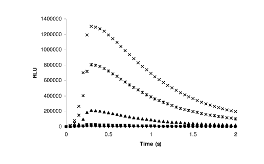
Figure 1 is a graph of cTnIC coating concentration effect on
chemiluminescence signal profile as described in Example 1. Figure 1 shows the
chemiluminescent profiles for cTnIC concentrations of 0 ng/mL (solid
diamonds), 80
ng/mL (solid squares), 400 ng/mL (solid triangles), 2000 ng/mL ("x" symbols),
and
10,000 ng/mL (asterisks).
Figure 2 is a graph showing cTnIC coating concentration effect on
chemiluminescent signal as described in Example 1.
Figure 3 is a box and whiskers plot showing cTnIC autoantibody response in
normal donors as described in Example 2.
Figure 4 is a box and whiskers plot showing cTnIC autoantibody response in
cTnl positive samples as described in Example 3.
Figure 5 is a box and whiskers plot showing cTnIC IgM autoantibody response
in cTnl positive samples as described in Example 4.
Figure 6 is a box and whiskers plot showing cTnl autoantibody response in
normal donors as described in Example 8.
Figure 7 is a box and whiskers plot showing cTnT autoantibody response in
normal donors as described in Example 9.
Figure 8 is a box and whiskers plot showing cTnC autoantibody response in
normal donors as described in Example 10.
Figure 9 is a box and whiskers plot showing cTnl autoantibody response in
cTnl positive samples as described in Example 11.
Figure 10 is a box and whiskers plot showing cTnT autoantibody response in
cTnl positive samples as described in Example 12.
Figure 11 is a box and whiskers plot showing cTnC autoantibody response in
cTnl positive samples as described in Example 13.
Figure 12 is a box and whiskers plot showing cTnIC autoantibody response in
anti-HBV positive samples as described in Example 14.
7
CA 02666586 2009-04-16
WO 2008/051761 PCT/US2007/081606
Figure 13 is a box and whiskers plot showing cTnl autoantibody response in
BNP positive samples as described in Example 15.
Figure 14 is a box and whiskers plot showing cTnl autoantibody response in
HCV disease positive samples as described in Example 16.
Figure 15 is a box and whiskers plot showing cTnl autoantibody response in
Chagas disease positive samples as described in Example 17.
Figure 16 is a graph of the cTnl autoantibody response in terms of
chemiluminescent signal (RLUmax, ordinate) of the normal donor population to
members of the cTnl peptide library (cTnI sequence residues, abscissa) as
described in
Example 19.
DETAILED DESCRIPTION
Cardiac troponin-reactive autoantibodies may arise via molecular mimicry of
pathogens such as viruses, bacteria, or toxins, genetic abnormalities, tissue
damage or
idiopathies. The presence of such autoantibodies leads to inflammatory
processes
which result in damage to cardiac tissue affecting normal cardiac function.
Further,
cardiac troponin-reactive autoantibodies provide an early indicator of risk
for
developing clinical manifestations of cardiac pathologies. Independently,
cardiac
troponin-reactive autoantibodies may interfere with the measurement of cTn1,
for
example, using conventional midfragment-specific immunoassays. This
interference
by endogenous antibodies can produce false negative results, such that
individuals fail
to be diagnosed in a timely fashion for acute myocardial infarction (AMI). The
present
invention includes methods based on the measurement of cardiac troponin
autoantibodies, in conjunction with cardiac troponin and as an independent
indicator of
cardiac pathology.
Definitions
Unless specifically defined otherwise as follows, all technical, scientific,
and
other terms used herein have the same meaning as commonly understood by one of
ordinary skill in the art to which this invention belongs.
The following terms encompass polypeptides that are identified in Genbank
(e.g., including but not limited to Accession Nos. P19429, P45379, P63316,
8
CA 02666586 2009-04-16
WO 2008/051761 PCT/US2007/081606
AAK9223 1, CAA56240, and CAA52818 ) and the scientific literature by the
following
designations, as well as polypeptides that are at least about 70% identical to
polypeptides identified in Genbank and the scientific literature by these
designations:
cardiac troponin I, cardiac troponin T, cardiac troponin C. In alternative
embodiments,
these terms encompass polypeptides identified in Genbank by these designations
and
polypeptides sharing at least about 80, 90, 95, 96, 97, 98, or 99% identity.
Percent
identity can, for example, be determined by a sequence alignment performed
using
BLASTP with default parameters set to measure the selected percent identity.
In
particular embodiments, these terms encompass full-length polypeptides, as
well as
fragments thereof.
As used herein, the term "cardiac troponin antigen" refers to any cardiac
troponin or fragment or complex thereof that is capable of binding to an
antibody
specific for a cardiac troponin or cardiac troponin complex.
The term "cardiac pathology" refers to any deviation from a healthy or normal
condition of the heart, including any structural or functional abnormality of
the heart, or
of the blood vessels supplying the heart, that impairs its normal functioning.
Examples
of cardiac pathologies include myocarditis, cardiomyopathy, and ischemic heart
disease.
The term "myocarditis" refers to inflammation of the myocardium. Myocarditis
can be caused by a variety of conditions such as viral infection, sarcoidosis,
rheumatic
fever, autoimmune diseases (such as systemic lupus, etc.), and pregnancy.
The term "cardiomyopathy" refers to a weakening of the heart muscle or a
change in heart muscle structure. It is often associated with inadequate heart
pumping
or other heart function abnormalities. Cardiomyopathy can be caused by viral
infections, heart attacks, alcoholism, long-term and severe high blood
pressure,
nutritional deficiencies (particularly of selenium, thiamine, and L-
carnitine), systemic
lupus erythematosus, celiac disease, and end-stage kidney disease. Types of
cardiomyopathy include dilated cardiomyopathy, hypertrophic cardiomyopathy,
and
restrictive cardiomyopathy.
As used herein, the term "dilated cardiomyopathy" refers to a global, usually
idiopathic, myocardial disorder characterized by a marked enlargement and
inadequate
function of the left ventricle. Dilated cardiomyopathy includes ischemic
9
CA 02666586 2009-04-16
WO 2008/051761 PCT/US2007/081606
cardiomyopathy, idiopathic cardiomyopathy, hypertensive cardiomyopathy,
infectious
cardiomyopathy, alcoholic cardiomyopathy, toxic cardiomyopathy, and peripartum
cardiomyopathy.
As used herein, the term "hypertrophic cardiomyopathy" refers to a condition
resulting from the right and left heart muscles growing to be different sizes.
As used herein, the term " restrictive cardiomyopathy" refers to a condition
characterized by the heart muscle's inability to relax between contractions,
which
prevents it from filling sufficiently.
The term "ischemic heart disease" refers to any condition in which heart
muscle
is damaged or works inefficiently because of an absence or relative deficiency
of its
blood supply; most often caused by atherosclerosis, it includes angina
pectoris, acute
myocardial infarction, and chronic ischemic heart disease.
"Angina pectoris" refers to chest discomfort caused by inadequate blood flow
through the blood vessels (coronary vessels) of the myocardium.
A "myocardial infarction" (heart attack) occurs when an area of heart muscle
dies or is damaged because of an inadequate supply of oxygen to that area.
The term "immunosuppressive therapy" is used herein to denote any therapy
aimed at decreasing the body's immune response, such as, for example, the
production
of autoantibodies.
As used herein, the term "immunoadsorption therapy" refers to any treatment
that removes antibodies from plasma by binding the target antibodies.
Typically,
plasma is removed from a subject, contacted with a solid phase-affixed binding
partner
for the target antibodies under conditions sufficient for binding, followed by
return of
the plasma to the subject.
As used herein, the term "autoimmune disease" refers to pathological
autoimmunity. "Autoimmunity" refers to one or more immune responses directed
against host antigens, characterized, for example, by the presence of
autoantibodies or
T lymphocytes reactive with host antigens.
A "first degree relative" is either a parent, child, or sibling.
"Biological samples" that can be assayed using the methods of the present
invention include biological fluids, such as whole blood, serum, plasma,
synovial fluid,
cerebrospinal fluid, bronchial lavage, ascites fluid, bone marrow aspirate,
pleural
CA 02666586 2009-04-16
WO 2008/051761 PCT/US2007/081606
effusion, urine, as well as tumor tissue or any other bodily constituent or
any tissue
culture supernatant that could contain the analyte of interest.
"Analyte," as used herein, refers to the substance to be detected, which may
be
present in the biological sample. The analyte can be any substance for which
there
exists a naturally occurring specific binding partner or for which a specific
binding
partner can be prepared. Thus, an analyte is a substance that can bind to one
or more
specific binding partners in an assay.
A "binding partner," as used herein, is a member of a binding pair, i.e., a
pair of
molecules wherein one of the molecules binds to the second molecule. Binding
partners that bind specifically are termed "specific binding partners." In
addition to the
antigen and antibody binding partners commonly used in immunoassays, other
specific
binding partners can include biotin and avidin, carbohydrates and lectins,
complementary nucleotide sequences, effector and receptor molecules, cofactors
and
enzymes, enzyme inhibitors and enzymes, and the like. Furthermore, specific
binding
partners can include partner(s) that is/are analog(s) of the original specific
binding
partner, for example, an analyte-analog. Immunoreactive specific binding
partners
include antigens, antigen fragments, antibodies and antibody fragments, both
monoclonal and polyclonal, and complexes thereof, including those formed by
recombinant DNA methods.
The term "specific binding" is defined herein as the preferential binding of
binding partners to another (e.g., two polypeptides, a polypeptide and nucleic
acid
molecule, or two nucleic acid molecules) at specific sites, as determined by
means
known in the art. The term "specifically binds" indicates that the binding
preference
(e.g., affinity) for the target molecule/sequence is at least 2-fold, more
preferably at
least 5-fold, and most preferably at least 10- or 20-fold over a non-specific
target
molecule (e.g. a randomly generated molecule lacking the specifically
recognized
site(s)).
As used herein with reference to cardiac troponin or cardiac troponin-reactive
autoantibody, the term "elevated level" refers to a level in a biological
sample that is
higher than a normal level or range. The normal level or range for cardiac
troponin and
cardiac troponin-reactive autoantibody is defined in accordance with standard
practice.
Thus, the level measured in a particular biological sample will be compared
with the
11
CA 02666586 2009-04-16
WO 2008/051761 PCT/US2007/081606
level or range of levels determined in similar samples of normal tissue. In
this context,
"normal tissue" is tissue from an individual with no detectable cardiac
pathology, and
"normal" (sometimes termed "control") patient or population is one that
exhibits no
detectable cardiac pathology. The level of an analyte is said to be "elevated"
where the
analyte is normally undetectable (e.g., the normal level is zero), but is
detected in a test
sample, as well as where the analyte is present in the test sample at a higher
than
normal level.
A "solid phase," as used herein, refers to any material that is insoluble, or
can
be made insoluble by a subsequent reaction. The solid phase can be chosen for
its
intrinsic ability to attract and immobilize a capture agent. Alternatively,
the solid phase
can have affixed thereto a linking agent that has the ability to attract and
immobilize the
capture agent. The linking agent can, for example, include a charged substance
that is
oppositely charged with respect to the capture agent itself or to a charged
substance
conjugated to the capture agent. In general, the linking agent can be any
binding
partner (preferably specific) that is immobilized on (attached to) the solid
phase and
that has the ability to immobilize the capture agent through a binding
reaction. The
linking agent enables the indirect binding of the capture agent to a solid
phase material
before the performance of the assay or during the performance of the assay.
The solid
phase can, for example, be plastic, derivatized plastic, magnetic or non-
magnetic metal,
glass or silicon, including, for example, a test tube, microtiter well, sheet,
bead,
microparticle, chip, and other configurations known to those of ordinary skill
in the art.
As used herein, term "microparticle" refers to a small particle that is
recoverable by ultracentrifugation. Microparticles typically have an average
diameter
on the order of about 1 micron or less.
The term "capture agent" is used herein to refer to a binding partner that
binds
to analyte, preferably specifically. Capture agents can be attached to a solid
phase. As
used herein, the binding of a solid phase-affixed capture agent to analyte
forms a "solid
phase-affixed complex."
The term "labeled detection agent" is used herein to refer to a binding
partner
that binds to analyte, preferably specifically, and is labeled with a
detectable label or
becomes labeled with a detectable label during use in an assay.
12
CA 02666586 2009-04-16
WO 2008/051761 PCT/US2007/081606
A "detectable label" includes a moiety that is detectable or that can be
rendered
detectable.
As used with reference to a labeled detection agent, a "direct label" is a
detectable label that is attached, by any means, to the detection agent.
As used with reference to a labeled detection agent, an "indirect label" is a
detectable label that specifically binds the detection agent. Thus, an
indirect label
includes a moiety that is the specific binding partner of a moiety of the
detection agent.
Biotin and avidin are examples of such moieties that are employed, for
example, by
contacting a biotinylated antibody with labeled avidin to produce an
indirectly labeled
antibody.
As used herein, the term "indicator reagent" refers to any agent that is
contacted
with a label to produce a detectable signal. Thus, for example, in
conventional enzyme
labeling, an antibody labeled with an enzyme can be contacted with a substrate
(the
indicator reagent) to produce a detectable signal, such as a colored reaction
product.
As used herein, an "antibody" refers to a protein consisting of one or more
polypeptides substantially encoded by immunoglobulin genes or fragments of
immunoglobulin genes. This term encompasses polyclonal antibodies, monoclonal
antibodies, and fragments thereof, as well as molecules engineered from
immunoglobulin gene sequences. The recognized immunoglobulin genes include the
kappa, lambda, alpha, gamma, delta, epsilon and mu constant region genes, as
well as
myriad immunoglobulin variable region genes. Light chains are classified as
either
kappa or lambda. Heavy chains are classified as gamma, mu, alpha, delta, or
epsilon,
which in turn define the immunoglobulin classes, IgG, IgM, IgA, IgD and IgE,
respectively.
A typical immunoglobulin (antibody) structural unit is known to comprise a
tetramer. Each tetramer is composed of two identical pairs of polypeptide
chains, each
pair having one "light" (about 25 kD) and one "heavy" chain (about 50 - 70
kD). The
N-terminus of each chain defines a variable region of about 100 to 110 or more
amino
acids primarily responsible for antigen recognition. The terms "variable light
chain
(VL)" and "variable heavy chain (VH)" refer to these light and heavy chains
respectively.
13
CA 02666586 2009-04-16
WO 2008/051761 PCT/US2007/081606
Antibodies exist as intact immunoglobulins or as a number of well-
characterized fragments produced by digestion with various peptidases. Thus,
for
example, pepsin digests an antibody below the disulfide linkages in the hinge
region to
produce F(ab')2, a dimer of Fab which itself is a light chain joined to VH-CH1
by a
disulfide bond. The F(ab')2 may be reduced under mild conditions to break the
disulfide linkage in the hinge region thereby converting the (Fab')2 dimer
into a Fab'
monomer. The Fab' monomer is essentially a Fab with part of the hinge region
(see,
Fundamental Immunology, W.E. Paul, ed., Raven Press, N.Y. (1993), for a more
detailed description of other antibody fragments). While various antibody
fragments
are defined in terms of the digestion of an intact antibody, one of skill will
appreciate
that such Fab' fragments may be synthesized de novo either chemically or by
utilizing
recombinant DNA methodology.
Thus, the term "antibody," as used herein also includes antibody fragments
either produced by the modification of whole antibodies or synthesized de novo
using
recombinant DNA methodologies. Preferred antibodies include single chain
antibodies
(antibodies that exist as a single polypeptide chain), more preferably single
chain Fv
antibodies (sFv or scFv), in which a variable heavy and a variable light chain
are joined
together (directly or through a peptide linker) to form a continuous
polypeptide. The
single chain Fv antibody is a covalently linked VH-VL heterodimer which may be
expressed from a nucleic acid including VH- and VL- encoding sequences either
joined
directly or joined by a peptide-encoding linker. Huston, et al. (1988) Proc.
Nat. Acad.
Sci. USA, 85: 5879-5883. While the VH and VL are connected to each as a single
polypeptide chain, the VH and VL domains associate non-covalently. The scFv
antibodies and a number of other structures converting the naturally
aggregated, but
chemically separated, light and heavy polypeptide chains from an antibody V
region
into a molecule that folds into a three dimensional structure substantially
similar to the
structure of an antigen-binding site are known to those of skill in the art
(see e.g., U.S.
Patent Nos. 5,091,513, 5,132,405, and 4,956,778).
An "autoantibody" is an antibody that binds to an analyte that is naturally
occurring in the individual in which the antibody is produced. A "cardiac
troponin-
troponin-reactive autoantibody" is an autoantibody that binds cardiac
troponin.
14
CA 02666586 2009-04-16
WO 2008/051761 PCT/US2007/081606
As used herein, a"species- specific antibody" refers to an antibody that
specifically binds target antibodies from a particular species, regardless of
the antigen-
binding specificity of the target antibodies.
A "human- specific antibody" is an antibody that specifically binds human
antibodies, e.g., human autoantibodies.
As used herein, a "cardiac troponin-reactive antibody or autoantibody" refers
to
an antibody or autoantibody, respectively that binds a cardiac troponin or
fragment or
complex thereof.
A "labeled cardiac troponin-reactive antibody" is a cardiac troponin-reactive
antibody that is labeled with a detectable label or that becomes labeled with
a
detectable label during immunoassay.
Sample Collection and Processing
The assay methods of the invention are generally carried out on biological
samples derived from an animal, preferably a mammal, and more preferably a
human.
The methods of the invention can be carried out using any sample that may
contain cardiac troponin-reactive autoantibodies. Convenient samples include,
for
example, blood, serum, and plasma.
The sample may be pretreated as necessary by dilution in an appropriate buffer
solution or concentrated, if desired. Any of a number of standard aqueous
buffer
solutions, employing any of a variety of buffers, such as phosphate, Tris, or
the like,
optionally at physiological pH, can be used.
Assay of Cardiac Troponin-Reactive Autoantibodies in Coniunction with Cardiac
Troponin
In particular embodiments, the invention provides a method of assessing the
reliability of a cardiac troponin assay result. The method entails assaying a
biological
sample for an autoantibody specific for a cardiac troponin, wherein the
presence of an
elevated level of cardiac troponin-reactive autoantibody indicates that the
cardiac
troponin assay result is not reliable.
In certain embodiments, a method of assessing risk of cardiac pathology
entails:
(a) assaying a biological sample for a cardiac troponin and (b) assaying a
biological
sample for an autoantibody specific for a cardiac troponin. The presence of an
elevated
CA 02666586 2009-04-16
WO 2008/051761 PCT/US2007/081606
level of cardiac troponin and/or an elevated level of cardiac troponin-
reactive
autoantibody indicates an elevated risk of cardiac pathology.
These methods can be carried out on samples from asymptomatic subjects or
subjects with one or more symptoms of cardiac pathology. For example, the
subject
may have chest pain or some other indication of myocardial infarction.
The assay for cardiac troponin-reactive autoantibody can be carried out
before,
simultaneously with, in the absence of, or after the cardiac troponin assay.
The assay
for cardiac troponin-reactive antibody can be carried out using the same
sample or a
different sample from the same subject. If a different sample is used, it will
generally
be of the same type (e.g., blood) and taken at approximately the same time as
the
sample for the cardiac troponin assay.
Cardiac troponin-reactive autoantibodies can be detected and quantified by any
of a number of methods well known to those of skill in the art. These may
include any
of a number of immunological methods such as fluid or gel precipitin
reactions,
immunodiffusion (single or double), affinity chromatography,
immunoelectrophoresis,
radioimmunoassay (RIA), enzyme-linked immunosorbent assays (ELISAs),
immunofluorescent assays, Western blotting, and the like. Immunoassays useful
in the
methods of the invention are discussed in greater detail below.
The assays are scored in accordance with standard practice and may include the
use of positive and/or negative controls and/or or standards containing known
concentrations of cardiac troponin-reactive antibodies. The level of cardiac
troponin-
reactive autoantibodies is compared with a control level or control range,
which can be
determined when the assay is carried out or, more conveniently, can be
predetermined.
Any increase in the test sample relative to the control level or range can be
assessed for
significance by conventional statistical methods. The presence of an elevated
level of
cardiac troponin autoantibodies indicates that such antibodies may negatively
interfere
with the cardiac troponin measurement, rendering this value unreliable with
respect to
assessing the risk of cardiac pathologies, such as myocardial infarction.
In particular embodiments, when a subject is determined to have an elevated
level of cardiac troponin-reactive autoantibodies, the subject is assessed for
one or
more additional indicators of cardiac pathology such as myoglobin, CK-MB, BNP,
16
CA 02666586 2009-04-16
WO 2008/051761 PCT/US2007/081606
CRP, Troponin-I, Troponin-T, blood oxygen level, cardiac imaging,
electrocardiography and the like.
A subject determined to have an elevated level of cardiac troponin-reactive
autoantibodies may also be treated, e.g., for myocardial infarction in
accordance with
standard practice.
Methods of Diagnosing Cardiac Patholo2y
The invention also provides methods in which cardiac troponin-reactive
antibodies are measured as indicators of the presence, or risk of, cardiac
pathology.
Myocarditis, Cardiomyopathy, and Ischemic Heart Disease
In particular embodiments, the invention provides a method of screening for a
subject having, or at risk of having, myocarditis, ischemic heart disease, or
cardiomyopathy. In variations of these embodiments the cardiomyopathy is not
dilated
cardiomyopathy. Thus, for example, the method can be employed to screen for
subjects having, or at risk of having hypertrophic cardiomyopathy and/or
restrictive
cardiomyopathy.
The method entails assaying a biological sample from the subject for an
autoantibody specific for a cardiac troponin, wherein the presence of an
elevated level
of cardiac troponin-reactive autoantibody indicates the presence of, or risk
of, cardiac
pathology. This method can be performed in conjunction with one or more other
tests,
including but not limited to physical examination, and/or the taking of a
medical history
to allow a differential diagnosis of, e.g., myocarditis, ischemic heart
disease, or
hypertrophic or restrictive cardiomyopathy. The various tests and parameters
employed in diagnosing these disorders are well known to those of skill in the
art.
These methods can be carried out on samples from asymptomatic subjects or
subjects having one or more risk factors associated with, or symptoms of,
cardiac
pathology. For example, the subject may have an autoimmune disease, high blood
pressure, or may have close (e.g., first-degree) relative with a heritable
cardiac
pathology, such as hypertophic cardiomyopathy.
Cardiac troponin-reactive autoantibodies can be detected and quantified by any
convenient means. Examples of various immunoassay formats suitable for this
purpose
are described below. The assays are scored in accordance with standard
practice.
17
CA 02666586 2009-04-16
WO 2008/051761 PCT/US2007/081606
In particular embodiments, when a subject is determined to have an elevated
level of cardiac troponin-reactive autoantibodies, the subject is assessed for
one or
more additional indicators of cardiac pathology such as myoglobin, CK-MB, BNP,
CRP, Troponin-I, Troponin-T, blood oxygen level, cardiac imaging,
electrocardiography and the like.
Relatives of Individuals with Autoimmune Disease
The methods of the invention can be carried to identify cardiac pathology or
risk thereof in subjects who have an autoimmune disease or who are related to
an
individual with an autoimmune disease. Subjects who are, e.g., first-degree or
second-
degree relatives of an individual with an autoimmune disease can be assessed
using the
methods of the invention.
The method entails assaying a biological sample from the subject for an
autoantibody specific for a cardiac troponin, wherein the presence of an
elevated level
of cardiac troponin-reactive autoantibody indicates the presence of, or risk
of, cardiac
pathology. This method can be performed in conjunction with one or more other
tests,
physical examination, and/or the taking of a medical history to allow a
differential
diagnosis of, e.g., myocarditis, ischemic heart disease, or dilated,
hypertrophic, or
restrictive cardiomyopathy. The various tests and parameters employed in
diagnosing
these disorders are well known to those of skill in the art.
These methods can be carried out on samples from asymptomatic subjects or
subjects having one or more risk factors associated with, or symptoms of,
cardiac
pathology.
Cardiac troponin-reactive autoantibodies can be detected and quantified by any
convenient means. Examples of various immunoassay formats suitable for this
purpose
are described below. The assays are scored in accordance with standard
practice.
In particular embodiments, when a subject is determined to have an elevated
level of cardiac troponin-reactive autoantibodies, the subject is assessed for
one or
more additional indicators of cardiac pathology such as myoglobin, CK-MB, BNP,
CRP, Troponin-I, Troponin-T, blood oxygen level, cardiac imaging,
electrocardiography and the like.
18
CA 02666586 2009-04-16
WO 2008/051761 PCT/US2007/081606
Method of Identifying Candidates for Immunosuppresive or
Immunoabsorption Therapies
In particular embodiments, the invention provides a method of determining
whether a subject having, or at risk for, a cardiac pathology is a candidate
for
immunosuppressive therapy or immunoabsorption therapy. Generally, the subject
is
one who has experienced some symptom of cardiac pathology or who has actually
been
diagnosed as having, or being at risk for, a cardiac pathology.
The method entails assaying a biological sample from the subject for an
autoantibody specific for a cardiac troponin, wherein the presence of an
elevated level
of cardiac troponin-reactive autoantibody indicates that autoimmunity may be
contributing to the subject's cardiac pathology or risk thereof. This method
can be
performed in conjunction with one or more other tests, physical examination,
and/or the
taking of a medical history in accordance with standard practice for
diagnosing cardiac
pathologies and/or autoimmune diseases.
Cardiac troponin-reactive autoantibodies can be detected and quantified by any
convenient means, including any of those described herein. The assays are
scored in
accordance with standard practice.
A subject determined to have an elevated level of cardiac troponin-reactive
autoantibodies may also be treated with immunosuppressive therapy or
immunoabsorption therapy in accordance with standard practice.
Immunoassay Methods
In General
The immunoassay methods of the invention can be carried out in any of a wide
variety of formats. For a general review of immunoassays, see Methods in Cell
Biology Volume 37: Antibodies in Cell Biology, Asai, ed. Academic Press, Inc.
New
York (1993); Basic and Clinical Immunology 7th Edition, Stites & Terr, eds.
(1991),
which is incorporated by reference in its entirety.
In particular embodiments, an immunoassay method of the invention can be
performed by contacting a biological sample with a cardiac troponin antigen,
under
conditions sufficient for binding of the cardiac troponin antigen to any
cardiac
troponin-reactive autoantibody present in the sample. Autoantibodies are
19
CA 02666586 2009-04-16
WO 2008/051761 PCT/US2007/081606
detected/quantitated by detecting complex(es) comprising the cardiac troponin
antigen
bound to cardiac troponin-reactive autoantibody. Such assays can be
homogeneous or
heterogeneous (i.e., employing a solid phase). In heterogeneous assays, a
capture agent
that binds to the analyte (here, cardiac troponin-reactive autoantibodies) is
typically
affixed to a solid phase.
Cardiac troponin autoantibodies can be measured in a non-competitive
immunoassay, wherein the amount of cardiac troponin antigen bound to cardiac
troponin-reactive autoantibody is positively correlated with the concentration
of cardiac
troponin-reactive autoantibody present in the sample.
Thus, for example, the method can be carried out as an agglutination assay in
which the biological sample is contacted with a cardiac troponin antigen
affixed to a
solid phase, such as a microparticle. The binding of cardiac troponin-reactive
autoantibody present in the sample to the microparticles results in the
agglutination of
those microparticles, which can be detected, for example, by visual inspection
of the
sample. The microparticles can be colored or labeled, if desired, to
facilitate detection
of agglutination. The degree of agglutination is positively correlated with
the
concentration of cardiac troponin-reactive autoantibody present in the sample.
In other embodiments, the biological sample is contacted with a cardiac
troponin antigen (which may, but need not, be affixed to a solid phase) and
also
contacted with a species-specific antibody, wherein the species-specific
antibody is
specific for the species from which the biological sample was obtained. Means
for
correcting interference generated by such autoantibodies is independently
described in
the U.S. Patent Application No. 60/854,569, and is incorporated herein by
reference in
its entirety. This step is carried out under conditions sufficient for
specific binding of
the species-specific antibody to any cardiac troponin-reactive autoantibody
present.
Autoantibodies are detected/quantitated by detecting complex(es) comprising
the
cardiac troponin antigen bound to cardiac troponin-reactive autoantibody,
which is
bound to species-specific antibody. The sample may be contacted with the
cardiac
troponin antigen and the species-specific antibody simultaneously or
sequentially, in
any order. Regardless of the order of contact, if cardiac troponin-reactive
autoantibodies are present in the sample, a complex forms that contains the
antibodies
"sandwiched" between the cardiac troponin antigen and the species-specific
antibody.
CA 02666586 2009-04-16
WO 2008/051761 PCT/US2007/081606
For example, in one format of a sandwich immunoassay, an embodiment of the
invention, the cardiac troponin antigen is affixed to a solid phase, binding
of the cardiac
troponin antigen to any cardiac troponin-reactive autoantibody present in the
sample
forms a solid phase-affixed complex, and detecting comprises detecting a
signal from
the solid phase-affixed complex. In particular embodiments of this format, the
solid
phase-affixed complex is detected using a species-specific antibody that is
directly or
indirectly labeled. The bound entities are separated, if necessary, from free
labeled
species-specific antibody, typically by washing, and the signal from the bound
label is
detected.
In another format of a sandwich immunoassay, an embodiment of the invention,
the species-specific antibody is affixed to a solid phase, binding of the
species-specific
antibody to any cardiac troponin-reactive autoantibody present in the sample
forms a
solid phase-affixed complex, which is then detected. In certain embodiments,
the solid
phase-affixed complex is detected using a cardiac troponin antigen that is
directly or
indirectly labeled. The bound entities are separated, if necessary, from free
labeled
cardiac troponin antigen, typically by washing, and the signal from the bound
label is
detected.
Cardiac troponin autoantibodies can also be measured in competitive
immunoassay, wherein the signal is negatively correlated with the
concentration of
cardiac troponin-reactive autoantibody present in the sample. In an example of
a
competitive format, the biological sample is contacted with a cardiac troponin
antigen
(which may, but need not, be affixed to a solid phase) and also contacted with
a labeled
(directly or indirectly) cardiac troponin-reactive antibody. This step is
carried out
under conditions sufficient for specific binding of the labeled cardiac
troponin-reactive
antibody to the cardiac troponin antigen. Autoantibodies in the sample that
are specific
for cardiac troponin can compete with the labeled cardiac troponin-reactive
antibody
for binding to the cardiac troponin antigen. Accordingly, the higher the level
of cardiac
troponin-reactive autoantibody, the lower the binding of labeled cardiac
troponin-
reactive antibody to the cardiac troponin antigen.
The sample may be contacted with the cardiac troponin antigen and the labeled
cardiac troponin-reactive antibody simultaneously or sequentially, in any
order.
21
CA 02666586 2009-04-16
WO 2008/051761 PCT/US2007/081606
Competitive immunoassays of this type can be conveniently carried out using a
solid phase-affixed cardiac troponin antigen. In this case, binding of the
cardiac
troponin antigen to labeled cardiac troponin-reactive antibody or to any
cardiac
troponin-reactive autoantibody present in the sample forms a solid phase-
affixed
complex, and detection entails detecting a signal from the solid phase-affixed
complex.
The bound entities are separated, if necessary, from free labeled cardiac
troponin-
reactive antibody, typically by washing, and the signal from the bound label
is detected.
Capture Agent
Capture agents useful in the immunoassay methods of the invention include
those that bind cardiac troponin-reactive autoantibodies and can be affixed to
a solid
phase. Convenient capture agents include a cardiac troponin antigen and
species-
specific antibodies, wherein the species-specific antibody is specific for the
species
from which the biological sample was obtained. As those of skill in the art
appreciate,
cardiac troponin antigen represents a specific capture agent because it binds
(captures)
cardiac troponin-reactive autoantibodies. By contrast, species-specific
antibodies
represent a non-specific capture agent because such antibodies bind
autoantibodies,
regardless of specificity. In a sandwich immunoassay, a non-specific capture
agent is
typically employed with a labeled detection agent that specifically binds the
analyte.
Thus, for example, solid phase affixed species-specific antibodies can be used
in
conjunction with a labeled cardiac troponin antigen to specifically detect
anti-cardiac
troponin autoantibodies.
Cardiac Troponin Anti2ens
Cardiac troponin antigens useful in the immunoassay methods of the invention
include cardiac troponin I, cardiac troponin T, cardiac troponin C, a
fragment,
derivative or complex thereof. Complexes useful in the invention can contain
two
different troponins (e.g., cTnl and cTnC) or all three.
In particular embodiments, the cardiac troponin antigen is a cardiac troponin
amino acid sequence that can be derived from any cardiac troponin-like
polypeptide
from any organism. Cardiac troponin amino acid sequences useful in the
invention are
generally those derived from vertebrates, preferably from birds or mammals,
more
preferably from animals having research or commercial value or value as pets,
such as
22
CA 02666586 2009-04-16
WO 2008/051761 PCT/US2007/081606
mice, rats, guinea pigs, rabbits, cats, dogs, chickens, pigs, sheep, goats,
cows, horses, as
well as monkeys and other primates. In particular embodiments, the cardiac
troponin
amino acid sequence is derived from a human polypeptide.
The methods of the invention can employ full-length cardiac troponin antigens
or one or more cardiac troponin fragments. Fragments will generally have at
least one
epitope to which an autoantibody can bind. Such fragments can have a length,
e.g., of
about 125, 100, 75, 50, 25, or 15 amino acids or a length that falls within a
range with
endpoints defined by any of these values (e.g., 15-125, 25-100, 50-75, 15-100,
etc.).
Those of skill in the art readily appreciate that the use of a cardiac
troponin antigen
having a larger number of natural epitopes (e.g., a full-length cardiac
troponin) will
generally provide a more comprehensive measurement of autoantibodies of
different
specificities than the use of a cardiac troponin antigen having a smaller
number of
natural epitopes. Accordingly, it is generally preferable to employ a cardiac
troponin
antigen that has a substantially native conformation or one or more peptides
comprising
troponin epitopes reactive with the autoantibody.
The cardiac troponin amino acid sequence can be a wild-type amino acid
sequence or an amino acid sequence variant of the corresponding region of a
wild-type
polypeptide. In certain embodiments, cardiac troponin antigens include a wild-
type
cardiac troponin amino acid sequence or a cardiac troponin amino acid sequence
containing conservative amino acid substitutions, as defined above.
In addition to the amino acid sequences described above, cardiac troponin
antigens useful in the invention can include other amino acid sequences,
including
those from heterologous proteins. Accordingly, the invention encompasses
fusion
polypeptides in which a cardiac troponin amino acid sequence is fused, at
either or both
ends, to amino acid sequence(s) from one or more heterologous proteins.
Examples of
additional amino acid sequences often incorporated into proteins of interest
include a
signal sequence, which facilitates purification of the protein, and an epitope
tag, which
can be used for immunological detection or affinity purification.
Cardiac troponin polypeptides according to the invention can be synthesized
using methods known in the art, such as for example exclusive solid phase
synthesis,
partial solid phase synthesis, fragment condensation, and classical solution
synthesis.
See, e.g., Merrifield, J. Am. Chem. Soc., 85:2149 (1963). For a description of
solid
23
CA 02666586 2009-04-16
WO 2008/051761 PCT/US2007/081606
phase peptide synthesis procedures, see John Morrow Stewart and Janis Dillaha
Young,
Solid Phase Peptide Syntheses (2nd Ed., Pierce Chemical Company, 1984).
Cardiac troponin polypeptides can also produced using recombinant techniques.
In certain embodiments, the sequence of a cardiac troponin coding region is
used as a
guide to design a synthetic nucleic acid molecule encoding the cardiac
troponin
polypeptide that can be incorporated an expression vector. Methods for
constructing
synthetic genes are well-known to those of skill in the art. See, e.g.,
Dennis, M. S.,
Carter, P. and Lazarus, R. A. (1993) Proteins: Struct. Funct. Genet., 15:312-
321.
The expression vector includes one or more control sequences capable of
effecting and/or enhancing the expression of an operably linked polypeptide
coding
sequence. Control sequences that are suitable for expression in prokaryotes,
for
example, include a promoter sequence, an operator sequence, and a ribosome
binding
site. Control sequences for expression in eukaryotic cells include a promoter,
an
enhancer, and a transcription termination sequence (i.e., a polyadenylation
signal).
An expression vector according to the invention can also include other
sequences, such as, for example, nucleic acid sequences encoding a signal
sequence or
an amplifiable gene. A signal sequence can direct the secretion of a
polypeptide fused
thereto from a cell expressing the protein. In the expression vector, nucleic
acid
encoding a signal sequence is linked to a polypeptide coding sequence so as to
preserve
the reading frame of the polypeptide coding sequence. The inclusion in a
vector of a
gene complementing an auxotrophic deficiency in the chosen host cell allows
for the
selection of host cells transformed with the vector.
A wide variety of host cells are available for propagation and/or expression
of
vectors. Examples include prokaryotic cells (such as E. coli and strains of
Bacillus,
Pseudomonas, and other bacteria), yeast or other fungal cells (including S.
cerevesiae
and P. pastoris), insect cells, plant cells, and phage, as well as higher
eukaryotic cells
(such as human embryonic kidney cells and other mammalian cells).
Vectors expressing cardiac troponin can be introduced into a host cell by any
convenient method, which will vary depending on the vector-host system
employed.
Generally, a vector is introduced into a host cell by transformation (also
known as
"transfection") or infection with a virus (e.g., phage) bearing the vector. If
the host cell
is a prokaryotic cell (or other cell having a cell wall), convenient
transformation
24
CA 02666586 2009-04-16
WO 2008/051761 PCT/US2007/081606
methods include the calcium treatment method described by Cohen, et al. (1972)
Proc.
Natl. Acad. Sci., USA, 69:2110-14. If a prokaryotic cell is used as the host
and the
vector is a phagemid vector, the vector can be introduced into the host cell
by infection.
Yeast cells can be transformed using polyethylene glycol, for example, as
taught by
Hinnen (1978) Proc. Natl. Acad. Sci, USA, 75:1929-33. Mammalian cells are
conveniently transformed using the calcium phosphate precipitation method
described
by Graham, et al. (1978) Virology, 52:546 and by Gorman, et al. (1990) DNA and
Prot.
Eng. Tech., 2:3-10. However, other known methods for introducing DNA into host
cells, such as nuclear injection, electroporation, and protoplast fusion also
are
acceptable for use in the invention.
Expression of cardiac troponin from a transformed host cell entails culturing
the
host cell under conditions suitable for cell growth and expression and
recovering the
expressed polypeptides from a cell lysate or, if the polypeptides are
secreted, from the
culture medium. In particular, the culture medium contains appropriate
nutrients and
growth factors for the host cell employed. The nutrients and growth factors
are, in
many cases, well known or can be readily determined empirically by those
skilled in
the art. Suitable culture conditions for mammalian host cells, for instance,
are
described in Mammalian Cell Culture (Mather ed., Plenum Press 1984) and in
Barnes
and Sato (1980) Cell 22:649.
In addition, the culture conditions should allow transcription, translation,
and
protein transport between cellular compartments. Factors that affect these
processes are
well-known and include, for example, DNA/RNA copy number; factors that
stabilize
DNA; nutrients, supplements, and transcriptional inducers or repressors
present in the
culture medium; temperature, pH and osmolality of the culture; and cell
density. The
adjustment of these factors to promote expression in a particular vector-host
cell system
is within the level of skill in the art. Principles and practical techniques
for maximizing
the productivity of in vitro mammalian cell cultures, for example, can be
found in
Mammalian Cell Biotechnology: a Practical Approach (Butler ed., IRL Press
(1991).
Any of a number of well-known techniques for large- or small-scale production
of proteins can be employed in expressing the polypeptides of the invention.
These
include, but are not limited to, the use of a shaken flask, a fluidized bed
bioreactor, a
CA 02666586 2009-04-16
WO 2008/051761 PCT/US2007/081606
roller bottle culture system, and a stirred tank bioreactor system. Cell
culture can be
carried out in a batch, fed-batch, or continuous mode.
Methods for recovery of recombinant proteins produced as described above are
well-known and vary depending on the expression system employed. A polypeptide
including a signal sequence can be recovered from the culture medium or the
periplasm. Polypeptides can also be expressed intracellularly and recovered
from cell
lysates.
The expressed polypeptides can be purified from culture medium or a cell
lysate
by any method capable of separating the polypeptide from one or more
components of
the host cell or culture medium. Typically, the polypeptide is separated from
host cell
and/or culture medium components that would interfere with the intended use of
the
polypeptide. As a first step, the culture medium or cell lysate is usually
centrifuged or
filtered to remove cellular debris. The supernatant is then typically
concentrated or
diluted to a desired volume or diafiltered into a suitable buffer to condition
the
preparation for further purification.
The polypeptide can then be further purified using well-known techniques. The
technique chosen will vary depending on the properties of the expressed
polypeptide.
If, for example, the polypeptide is expressed as a fusion protein containing
an epitope
tag or other affinity domain, purification typically includes the use of an
affinity
column containing the cognate binding partner. For instance, polypeptides
fused with
green fluorescent protein, hemagglutinin, or FLAG epitope tags or with
hexahistidine
or similar metal affinity tags can be purified by fractionation on an affinity
column.
Antibodies
Antibodies useful in the immunoassay methods of the invention include
polyclonal and monoclonal antibodies. Such polyclonal and monoclonal
antibodies can
be prepared by any means known in the art. Polyclonal antibodies are raised by
injecting (e.g., subcutaneous or intramuscular injection) an immunogen into a
suitable
non-human mammal (e.g., a mouse or a rabbit). Generally, the immunogen should
induce production of high titers of antibody with relatively high affinity for
the target
antigen.
26
CA 02666586 2009-04-16
WO 2008/051761 PCT/US2007/081606
If desired, the antigen may be conjugated to a carrier protein by conjugation
techniques that are well known in the art. Commonly used carriers include
keyhole
limpet hemocyanin (KLH), thyroglobulin, bovine serum albumin (BSA), and
tetanus
toxoid. The conjugate is then used to immunize the animal.
The antibodies are then obtained from blood samples taken from the animal.
The techniques used to produce polyclonal antibodies are extensively described
in the
literature (see, e.g., Methods of Enzymology, "Production of Antisera With
Small
Doses of Immunogen: Multiple Intradermal Injections," Langone, et al. eds.
(Acad.
Press, 1981)). Polyclonal antibodies produced by the animals can be further
purified,
for example, by binding to and elution from a matrix to which the target
antigen is
bound. Those of skill in the art will know of various techniques common in the
immunology arts for purification and/or concentration of polyclonal, as well
as
monoclonal, antibodies see, for example, Coligan, et al. (1991) Unit 9,
Current
Protocols in Immunology, Wiley Interscience.
For many applications, monoclonal antibodies (mAbs) are preferred. The
general method used for production of hybridomas secreting mAbs is well known
(Kohler and Milstein (1975) Nature, 256:495). Briefly, as described by Kohler
and
Milstein, the technique entailed isolating lymphocytes from regional draining
lymph
nodes of five separate cancer patients with either melanoma, teratocarcinoma
or cancer
of the cervix, glioma or lung, (where samples were obtained from surgical
specimens),
pooling the cells, and fusing the cells with SHFP- 1. Hybridomas were screened
for
production of antibody that bound to cancer cell lines. Confirmation of
specificity
among mAbs can be accomplished using routine screening techniques (such as the
enzyme-linked immunosorbent assay, or "ELISA") to determine the elementary
reaction pattern of the mAb of interest.
As used herein, the term "antibody" encompasses antigen-binding antibody
fragments, e.g., single chain antibodies (scFv or others), which can be
produced/selected using phage display technology. The ability to express
antibody
fragments on the surface of viruses that infect bacteria (bacteriophage or
phage) makes
it possible to isolate a single binding antibody fragment, e.g., from a
library of greater
than 1010 nonbinding clones. To express antibody fragments on the surface of
phage
(phage display), an antibody fragment gene is inserted into the gene encoding
a phage
27
CA 02666586 2009-04-16
WO 2008/051761 PCT/US2007/081606
surface protein (e.g., pIII) and the antibody fragment-pIII fusion protein is
displayed on
the phage surface (McCafferty et al. (1990) Nature, 348: 552-554; Hoogenboom
et al.
(1991) Nucleic Acids Res. 19: 4133-4137).
Since the antibody fragments on the surface of the phage are functional, phage-
bearing antigen-binding antibody fragments can be separated from non-binding
phage
by antigen affinity chromatography (McCafferty et al. (1990) Nature, 348: 552-
554).
Depending on the affinity of the antibody fragment, enrichment factors of 20-
fold -
1,000,000-fold are obtained for a single round of affinity selection. By
infecting
bacteria with the eluted phage, however, more phage can be grown and subjected
to
another round of selection. In this way, an enrichment of 1000-fold in one
round can
become 1,000,000-fold in two rounds of selection (McCafferty et al. (1990)
Nature,
348: 552-554). Thus, even when enrichments are low (Marks et al. (1991) J.
Mol. Biol.
222: 581-597), multiple rounds of affinity selection can lead to the isolation
of rare
phage. Since selection of the phage antibody library on antigen results in
enrichment,
the majority of clones bind antigen after as few as three to four rounds of
selection.
Thus only a relatively small number of clones (several hundred) need to be
analyzed for
binding to antigen.
Human antibodies can be produced without prior immunization by displaying
very large and diverse V-gene repertoires on phage (Marks et al. (1991) J.
Mol. Biol.
222: 581-597). In one embodiment, natural VH and VL repertoires present in
human
peripheral blood lymphocytes are isolated from unimmunized donors by PCR. The
V-
gene repertoires can be spliced together at random using PCR to create a scFv
gene
repertoire which can be cloned into a phage vector to create a library of 30
million
phage antibodies (Id.). From a single "naive" phage antibody library, binding
antibody
fragments have been isolated against more than 17 different antigens,
including
haptens, polysaccharides, and proteins (Marks et al. (1991) J. Mol. Biol. 222:
581-597;
Marks et al. (1993). Bio/Technology. 10: 779-783; Griffiths et al. (1993) EMBO
J. 12:
725-734; Clackson et al. (1991) Nature. 352: 624-628). Antibodies have been
produced against self proteins, including human thyroglobulin, immunoglobulin,
tumor
necrosis factor, and CEA (Griffiths et al. (1993) EMBO J. 12: 725-734). The
antibody
fragments are highly specific for the antigen used for selection and have
affinities in the
1 nM to 100 nM range (Marks et al. (1991) J. Mol. Biol. 222: 581-597;
Griffiths et al.
28
CA 02666586 2009-04-16
WO 2008/051761 PCT/US2007/081606
(1993) EMBO J. 12: 725-734). Larger phage antibody libraries result in the
isolation of
more antibodies of higher binding affinity to a greater proportion of
antigens.
As those of skill in the art readily appreciate, antibodies can be prepared by
any
of a number of commercial services (e.g., Berkeley Antibody Laboratories,
Bethyl
Laboratories, Anawa, Eurogenetec, etc.).
Solid Phase
For embodiments of the invention that employ a solid phase as a support for
the
capture agent, the solid phase can be any suitable material with sufficient
surface
affinity to bind a capture agent. Useful solid supports include: natural
polymeric
carbohydrates and their synthetically modified, crosslinked, or substituted
derivatives,
such as agar, agarose, cross-linked alginic acid, substituted and cross-linked
guar gums,
cellulose esters, especially with nitric acid and carboxylic acids, mixed
cellulose esters,
and cellulose ethers; natural polymers containing nitrogen, such as proteins
and
derivatives, including cross-linked or modified gelatins; natural hydrocarbon
polymers,
such as latex and rubber; synthetic polymers, such as vinyl polymers,
including
polyethylene, polypropylene, polystyrene, polyvinylchloride, polyvinylacetate
and its
partially hydrolyzed derivatives, polyacrylamides, polymethacrylates,
copolymers and
terpolymers of the above polycondensates, such as polyesters, polyamides, and
other
polymers, such as polyurethanes or polyepoxides; inorganic materials such as
sulfates
or carbonates of alkaline earth metals and magnesium, including barium
sulfate,
calcium sulfate, calcium carbonate, silicates of alkali and alkaline earth
metals,
aluminum and magnesium; and aluminum or silicon oxides or hydrates, such as
clays,
alumina, talc, kaolin, zeolite, silica gel, or glass (these materials may be
used as filters
with the above polymeric materials); and mixtures or copolymers of the above
classes,
such as graft copolymers obtained by initializing polymerization of synthetic
polymers
on a pre-existing natural polymer. All of these materials may be used in
suitable
shapes, such as films, sheets, tubes, particulates, or plates, or they may be
coated onto,
bonded, or laminated to appropriate inert carriers, such as paper, glass,
plastic films,
fabrics, or the like.
29
CA 02666586 2009-04-16
WO 2008/051761 PCT/US2007/081606
Nitrocellulose has excellent absorption and adsorption qualities for a wide
variety of reagents including monoclonal antibodies. Nylon also possesses
similar
characteristics and also is suitable.
Preferred solid phase materials for flow-through assay devices include filter
paper such as a porous fiberglass material or other fiber matrix materials.
The
thickness of such material is not critical and will be a matter of choice,
largely based
upon the properties of the sample or analyte being assayed, such as the
fluidity of the
biological sample.
Alternatively, the solid phase can constitute microparticles. Microparticles
useful in the invention can be selected by one skilled in the art from any
suitable type
of particulate material and include those composed of polystyrene,
polymethylacrylate,
polypropylene, latex, polytetrafluoroethylene, polyacrylonitrile,
polycarbonate, or
similar materials. Further, the microparticles can be magnetic or paramagnetic
microparticles, so as to facilitate manipulation of the microparticle within a
magnetic
field.
Microparticles can be suspended in the mixture of soluble reagents and
biological sample or can be retained and immobilized by a support material. In
the
latter case, the microparticles on or in the support material are not capable
of
substantial movement to positions elsewhere within the support material.
Alternatively,
the microparticles can be separated from suspension in the mixture of soluble
reagents
and biological sample by sedimentation or centrifugation. When the
microparticles are
magnetic or paramagnetic the microparticles can be separated from suspension
in the
mixture of soluble reagents and biological sample by a magnetic field.
The methods of the present invention can be adapted for use in systems that
utilize microparticle technology including automated and semi-automated
systems
wherein the solid phase comprises a microparticle. Such systems include those
described in pending U.S. App. No. 425,651 and U.S. Patent No. 5,089,424,
which
correspond to published EPO App. Nos. EP 0 425 633 and EP 0 424 634,
respectively,
and U.S. Patent No. 5,006,309.
In particular embodiments, the solid phase includes one or more electrodes.
Capture agent(s) can be affixed, directly or indirectly, to the electrode(s).
In one
embodiment, for example, capture agents can be affixed to magnetic or
paramagnetic
CA 02666586 2009-04-16
WO 2008/051761 PCT/US2007/081606
microparticles, which are then positioned in the vicinity of the electrode
surface using a
magnet. Systems in which one or more electrodes serve as the solid phase are
useful
where detection is based on electrochemical interactions. Exemplary systems of
this
type are described, for example, in U.S. Patent No. 6,887,714 (issued May 3,
2005).
The basic method is described further below with respect to electrochemical
detection.
The capture agent can be attached to the solid phase by adsorption, where it
is
retained by hydrophobic forces. Alternatively, the surface of the solid phase
can be
activated by chemical processes that cause covalent linkage of the capture
agent to the
support.
To change or enhance the intrinsic charge of the solid phase, a charged
substance can be coated directly onto the solid phase. Ion capture procedures
for
immobilizing an immobilizable reaction complex with a negatively charged
polymer,
described in U.S. App. No. 150,278, corresponding to EP Publication No.
0326100, and
U.S. App. No. 375,029 (EP Publication No. 0406473), can be employed according
to
the present invention to affect a fast solution-phase immunochemical reaction.
In these
procedures, an immobilizable immune complex is separated from the rest of the
reaction mixture by ionic interactions between the negatively charged
polyanion/immune complex and the previously treated, positively charged matrix
and
detected by using any of a number of signal-generating systems, including,
e.g.,
chemiluminescent systems, as described in U.S. App. No. 921,979, corresponding
to
EPO Publication No. 0 273,115.
If the solid phase is silicon or glass, the surface must generally be
activated
prior to attaching the specific binding partner. Activated silane compounds
such as
triethoxy amino propyl silane (available from Sigma Chemical Co., St. Louis,
Mo.),
triethoxy vinyl silane (Aldrich Chemical Co., Milwaukee, Wis.), and (3-
mercapto-
propyl)-trimethoxy silane (Sigma Chemical Co., St. Louis, Mo.) can be used to
introduce reactive groups such as amino-, vinyl, and thiol, respectively. Such
activated
surfaces can be used to link the capture directly (in the cases of amino or
thiol), or the
activated surface can be further reacted with linkers such as glutaraldehyde,
bis
(succinimidyl) suberate, SPPD 9 succinimidyl3-[2-pyridyldithio] propionate),
SMCC
(succinimidyl-4-[Nmaleimidomethyl] cyclohexane-l-carboxylate), SIAB
(succinimidyl
[4iodoacetyl] aminobenzoate), and SMPB (succinimidyl4-[lmaleimidophenyl]
31
CA 02666586 2009-04-16
WO 2008/051761 PCT/US2007/081606
butyrate) to separate the capture agent from the surface. Vinyl groups can be
oxidized
to provide a means for covalent attachment. Vinyl groups can also be used as
an
anchor for the polymerization of various polymers such as poly-acrylic acid,
which can
provide multiple attachment points for specific capture agents. Amino groups
can be
reacted with oxidized dextrans of various molecular weights to provide
hydrophilic
linkers of different size and capacity. Examples of oxidizable dextrans
include Dextran
T-40 (molecular weight 40,000 daltons), Dextran T-110 (molecular weight
110,000
daltons), Dextran T-500 (molecular weight 500,000 daltons), Dextran T-2M
(molecular
weight 2,000,000 daltons) (all of which are available from Pharmacia,
Piscataway,
N.J.), or Ficoll (molecular weight 70,000 daltons; available from Sigma
Chemical Co.,
St. Louis, Mo.). Additionally, polyelectrolyte interactions can be used to
immobilize a
specific capture agent on a solid phase using techniques and chemistries
described U.S.
App. No. 150,278, filed Jan. 29, 1988, and U.S. App. No. 375,029, filed Jul.
7, 1989,
each of which is incorporated herein by reference.
Other considerations affecting the choice of solid phase include the ability
to
minimize non-specific binding of labeled entities and compatibility with the
labeling
system employed. For, example, solid phases used with fluorescent labels
should have
sufficiently low background fluorescence to allow signal detection.
Following attachment of a specific capture agent, the surface of the solid
support may be further treated with materials such as serum, proteins, or
other blocking
agents to minimize non-specific binding.
Labeling Systems
As discussed above, many immunoassays according to the invention employ a
labeled detection agent, such a labeled species-specific antibody and a
labeled cardiac
troponin antigen.
Detectable labels suitable for use in the detection agents of the present
invention
include any composition detectable by spectroscopic, photochemical,
biochemical,
immunochemical, electrical, optical, or chemical means. Useful labels in the
present
invention include magnetic beads (e.g., DynabeadsTm), fluorescent dyes (e.g.,
fluorescein, Texas Red, rhodamine, green fluorescent protein, and the like,
see, e.g.,
Molecular Probes, Eugene, Oregon, USA), chemiluminescent compounds such as
32
CA 02666586 2009-04-16
WO 2008/051761 PCT/US2007/081606
acridinium (e.g., acridinium-9-carboxamide), phenanthridinium, dioxetanes,
luminol
and the like, radiolabels (e.g., 3H 125 I 35S 14C, or 32P), catalysts such as
enzymes (e.g.,
horse radish peroxidase, alkaline phosphatase, beta-galactosidase and others
commonly
used in an ELISA), and colorimetric labels such as colloidal gold (e.g., gold
particles in
the 40 -80 nm diameter size range scatter green light with high efficiency) or
colored
glass or plastic (e.g., polystyrene, polypropylene, latex, etc.) beads.
Patents teaching
the use of such labels include U.S. Patent Nos. 3,817,837; 3,850,752;
3,939,350;
3,996,345; 4,277,437; 4,275,149; and 4,366,241.
The label can be attached to the detection agent prior to, or during, or after
contact with the biological sample. So-called "direct labels" are detectable
labels that
are directly attached to or incorporated into detection agents prior to use in
the assay.
Direct labels can be attached to or incorporated into detection agents by any
of a
number of means well known to those of skill in the art.
In contrast, so-called "indirect labels" typically bind to the detection agent
at
some point during the assay. Often, the indirect label binds to a moiety that
is attached
to or incorporated into the detection agent prior to use. Thus, for example,
an antibody
used as a detection agent (a "detection antibody") can be biotinylated before
use in an
assay. During the assay, an avidin-conjugated fluorophore can bind the biotin-
bearing
detection agent, to provide a label that is easily detected.
In another example of indirect labeling, polypeptides capable of specifically
binding immunoglobulin constant regions, such as polypeptide A or polypeptide
G, can
also be used as labels for detection antibodies. These polypeptides are normal
constituents of the cell walls of streptococcal bacteria. They exhibit a
strong non-
immunogenic reactivity with immunoglobulin constant regions from a variety of
species (see, generally Kronval, et al. (1973) J. Immunol., 111: 1401-1406,
and
Akerstrom (1985) J. Immunol., 135: 2589-2542). Such polypeptides can thus be
labeled and added to the assay mixture, where they will bind to the detection
antibody,
as well as to the species-specific antibody, labeling both and providing a
composite
signal attributable to analyte and autoantibody present in the sample.
Some labels useful in the invention may require the use of an indicator
reagent
to produce a detectable signal. In an ELISA, for example, an enzyme label
(e.g., beta-
33
CA 02666586 2009-04-16
WO 2008/051761 PCT/US2007/081606
galactosidase) will require the addition of a substrate (e.g., X-gal) to
produce a
detectable signal.
Exemplary Formats
Fluorescence Polarization Immunoassay (FPIA)
In an exemplary embodiment, a fluorescent label is employed in a fluorescence
polarization immunoassay (FPIA) according to the invention. Generally,
fluorescent
polarization techniques are based on the principle that a fluorescent label,
when excited
by plane-polarized light of a characteristic wavelength, will emit light at
another
characteristic wavelength (i.e., fluorescence) that retains a degree of the
polarization
relative to the incident light that is inversely related to the rate of
rotation of the label in
a given medium. As a consequence of this property, a label with constrained
rotation,
such as one bound to another solution component with a relatively lower rate
of
rotation, will retain a relatively greater degree of polarization of emitted
light than
when free in solution.
This technique can be employed in immunoassays according to the invention,
for example, by selecting reagents such that binding of the fluorescently
labeled entities
forms a complex sufficiently different in size such that a change in the
intensity light
emitted in a given plane can be detected. For example, when a labeled cardiac
troponin
antigen is bound by one or more autoantibodies, the resulting complex is
sufficiently
larger, and its rotation is sufficiently constrained, relative to the free
labeled cardiac
troponin antigen that binding is easily detected.
Fluorophores useful in FPIA include fluorescein, aminofluorescein,
carboxyfluorescein, and the like, preferably 5 and 6-aminomethylfluorescein, 5
and 6-
aminofluorescein, 6-carboxyfluorescein, 5-carboxyfluorescein,
thioureafluorescein, and
methoxytriazinolyl-aminofluorescein, and similar fluorescent derivatives.
Examples of
commercially available automated instruments with which fluorescence
polarization
assays can be conducted include: the IMx system, the TDx system, and TDxFLx
system (all available from Abbott Laboratories, Abbott Park, Ill.).
34
CA 02666586 2009-04-16
WO 2008/051761 PCT/US2007/081606
Scanning Probe Microscopy (SPM)
The use of scanning probe microscopy (SPM) for immunoassays also is a
technology to which the immunoassay methods of the present invention are
easily
adaptable. In SPM, in particular in atomic force microscopy, the capture agent
is
affixed to a solid phase having a surface suitable for scanning. The capture
agent can,
for example, be adsorbed to a plastic or metal surface. Alternatively, the
capture agent
can be covalently attached to, e.g., derivatized plastic, metal, silicon, or
glass according
to methods known to those of ordinary skill in the art. Following attachment
of the
capture agent, the biological sample is contacted with the solid phase, and a
scanning
probe microscope is used to detect and quantify solid phase-affixed complexes.
The
use of SPM eliminates the need for labels which are typically employed in
immunoassay systems. Such a system is described in U.S. App. No. 662,147,
which is
incorporated herein by reference.
MicroElectroMechanical Systems (MEMS)
Immunoassays according to the invention can also be carried out using a
MicroElectroMechanical System (MEMS). MEMS are microscopic structures
integrated onto silicon that combine mechanical, optical, and fluidic elements
with
electronics, allowing convenient detection of an analyte of interest. An
exemplary
MEMS device suitable for use in the invention is the Protiveris'
multicantilever array.
This array is based on chemo-mechanical actuation of specially designed
silicon
microcantilevers and subsequent optical detection of the microcantilever
deflections.
When coated on one side with a binding partner, a microcantilever will bend
when it is
exposed to a solution containing the complementary molecule. This bending is
caused
by the change in the surface energy due to the binding event. Optical
detection of the
degree of bending (deflection) allows measurement of the amount of
complementary
molecule bound to the microcantilever.
Electrochemical Dectection Systems
In other embodiments, immunoassays according to the invention are carried out
using electrochemical detection. A basic procedure for electrochemical
detection has
been described by Heineman and coworkers. This entailed immobilization of a
primary
antibody (Ab, rat-anti mouse IgG), followed by exposure to a sequence of
solutions
CA 02666586 2009-04-16
WO 2008/051761 PCT/US2007/081606
containing the antigen (Ag, mouse IgG), the secondary antibody conjugated to
an
enzyme label (AP-Ab, rat anti mouse IgG and alkaline phosphatase), and p-
aminophenyl phosphate (PAPP). The AP converts PAPP to p-aminophenol (PAPR, the
"R" is intended to distinguish the reduced form from the oxidized form, PAPo,
the
quinoneimine), which is electrochemically reversible at potentials that do not
interfere
with reduction of oxygen and water at pH 9.0, where AP exhibits optimum
activity.
PAPR does not cause electrode fouling, unlike phenol whose precursor,
phenylphosphate, is often used as the enzyme substrate. Although PAPR
undergoes air
and light oxidation, these are easily prevented on small scales and short time
frames.
Picomole detection limits for PAPR and femtogram detection limits for IgG
achieved in
microelectrochemical immunoassays using PAPP volumes ranging from 20 mu.l to
360 L have been reported previously. In capillary immunoassays with
electrochemical detection, the lowest detection limit reported thus far is
3000 molecules
of mouse IgG using a volume of 70 L and a 30 min or 25 min assay time.
In an exemplary embodiment employing electrochemical detection, a capture
agent according to the invention can be immobilized on the surface of an
electrode (the
"solid phase"). The electrode is then contacted with a biological sample from,
e.g., a
human. Any anti-cardiac troponin antibodies in the sample bind to the capture
agent to
form a solid phase-affixed complex. Anti-human antibody, which is labeled with
AP,
for example, binds to autoantibodies in the complex, thereby becoming
immobilized on
the surface of the electrode. The addition of PAPP, results in its conversion
by AP to
PAPR, which is then detected.
Various electrochemical detection systems are described in U.S. Patent
Nos. 7,045,364 (issued May 16, 2006; incorporated herein by reference),
7,045,310
(issued May 16, 2006; incorporated herein by reference), 6,887,714 (issued May
3,
2005; incorporated herein by reference), 6,682,648 (issued January 27, 2004;
incorporated herein by reference); 6,670,115 (issued December 30, 2003;
incorporated
herein by reference).
The present invention is for example applicable to the point of care assay
systems, including Abbott Laboratories' commercial Point of Care (i-STATTM)
electrochemical immunoassay system which performs sandwich immunoassays for
several cardiac markers, including TnI, CKMB and BNP. Immunosensors and
methods
36
CA 02666586 2009-04-16
WO 2008/051761 PCT/US2007/081606
of manufacturing and operating them in single-use test devices are described,
for
example, in US Patent No. 5,063,081 and published US Patent Application Nos.
20030170881, US 20040018577, US 20050054078, and US 20060160164, each of
which is incorporated herein by reference for their teachings regarding same.
Additionally, it goes without saying that any of the exemplary formats herein,
and any assay or kit according to the invention can be adapted or optimized
for use in
automated and semi-automated systems (including those in which there is a
solid phase
comprising a microparticle), as described, e.g., in US Patent Nos. 5,089,424
and
5,006,309, and as, e.g., commercially marketed by Abbott Laboratories (Abbott
Park,
IL) including but not limited to Abbott's ARCHITECT , AxSYM, IMX, PRISM, and
Quantum II platforms, as well as other platforms.
Multiplex Formats
In particular embodiments, useful, for example, for simultaneously assaying
multiple analytes in one biological sample, the solid phase can include a
plurality
different capture agents, including one that captures troponin-reactive
autoantibodies.
Thus, for example, the solid phase can have affixed thereon a plurality of
antigens,
wherein each is intended to test for the presence of different autoantibodies
in the
sample. In an exemplary embodiment, the solid phase can consist of a plurality
of
different regions on a surface, wherein each region has a particular antigen
affixed
therein.
Multiplex formats can, but need not, employ a plurality of labels, wherein
each
label is used for the detection of autoantibodies reactive with a particular
antigen. For
example, multiple, different autoantibodies can be detected without using a
plurality of
labels where a plurality of capture agents, such as antigens, are affixed to
the solid
phase at different known locations, based on specificity. Because the
specificity of the
capture agent at each location is known, the detection of a signal at a
particular location
can be associated with the presence of autoantibodies bound at that location.
Examples
of this format include microfluidic devices and capillary arrays, containing
different
capture agents at different locations along a channel or capillary,
respectively, and
microarrays, which typically contain different capture agents arranged in a
matrix of
spots ("target elements") on a surface of a solid support. In particular
embodiments,
37
CA 02666586 2009-04-16
WO 2008/051761 PCT/US2007/081606
each different capture agent can be affixed to a different electrode, which
can, for
example, be formed on a surface of a solid support, in a channel of a
microfluidic
device, or in a capillary.
Test Kits
The invention also provides test kits for assaying biological samples for
cardiac
troponin autoantibodies. Test kits according to the invention include one or
more
reagents useful for practicing one or more immunoassays according to the
invention. A
test kit generally includes a package with one or more containers holding the
reagents,
as one or more separate compositions or, optionally, as admixture where the
compatibility of the reagents will allow. The test kit can also include other
material(s),
which may be desirable from a user standpoint, such as a buffer(s), a
diluent(s), a
standard(s), and/or any other material useful in sample processing, washing,
or
conducting any other step of the assay.
In certain embodiments, a test kit includes a humanized monoclonal antibody,
wherein the humanized monoclonal antibody is specific for a cardiac troponin.
This
component can be used as a positive control in immunoassays according to the
invention. If desired, this component can be included in the test kit in
multiple
concentrations to facilitate the generation of a standard curve to which the
signal
detected in the test sample can be compared. Alternatively, a standard curve
can be
generated by preparing dilutions of a single humanized monoclonal antibody
solution
provided in the kit.
Kits according to the invention can include a solid phase and a capture agent
affixed to the solid phase, wherein the capture agent is selected from the
group
consisting of a cardiac troponin antigen and a species-specific antibody,
wherein the
species-specific antibody is specific for the species from which the
biological sample is
to be obtained. Where such kits are to be employed for conducting sandwich
immunoassays, the kits can additionally include a labeled detection agent. In
such
embodiments, if the capture agent is a cardiac troponin antigen, the detection
agent can
be a species-specific antibody. If the capture agent is a species-specific
antibody, a
cardiac troponin antigen can be used as the detection agent. In particular
embodiments,
the species-specific antibody is a human-specific antibody.
38
CA 02666586 2009-04-16
WO 2008/051761 PCT/US2007/081606
Test kits according to the invention can also include a labeled non-human
monoclonal antibody that is specific for a cardiac troponin. This component is
useful
as a control for confirming that any cardiac troponin antigen employed is
capable of
binding antibody.
In certain embodiments, the test kit includes at least one direct label, such
as
acridinium-9-carboxamide. Test kits according to the invention can also
include at
least one indirect label. If the label employed generally requires an
indicator reagent to
produce a detectable signal, the test kit preferably includes one or more
suitable
indicator reagents.
In exemplary embodiments, the solid phase includes one or more microparticles
or electrodes. Test kits designed for multiplex assays conveniently contain
one or more
solid phases including a plurality of antigens that are specific for a
plurality of different
autoantibodies. Thus, for example, a test kit designed for multiplex
electrochemical
immunoassays can contain a solid phase including a plurality of electrodes,
with each
electrode bearing a different antigen.
Test kits according to the invention preferably include instructions for
carrying
out one or more of the immunoassays of the invention. Instructions included in
kits of
the invention can be affixed to packaging material or can be included as a
package
insert. While the instructions are typically written or printed materials they
are not
limited to such. Any medium capable of storing such instructions and
communicating
them to an end user is contemplated by this invention. Such media include, but
are not
limited to, electronic storage media (e.g., magnetic discs, tapes, cartridges,
chips),
optical media (e.g., CD ROM), and the like. As used herein, the term
"instructions"
can include the address of an internet site that provides the instructions.
EXAMPLES
The invention will be better understood through examples illustrating its use
and efficacy. The following examples are offered to illustrate, but not to
limit, the
claimed invention.
39
CA 02666586 2009-04-16
WO 2008/051761 PCT/US2007/081606
Example 1
Cardiac troponin-I-C complex microplate preparation and testin2
Microplate coating procedure
The microplate coating solution was prepared by dissolving human cardiac
troponin-I-C complex (cTnIC, commercial vendor) in phosphate buffer (0.2 M, pH
8)
to give solutions at the following concentrations: 80, 400, 2000, or 10000
ng/mL. The
coating solution (100 L) was added to the wells of a white high-binding flat-
bottom
96-well polystyrene microplate. The microplate was then sealed, placed on an
orbital
shaker at 28 rpm and incubated at 38 C for 1 hour (h). The coating solution
was then
replaced with a blocking solution (300 L) consisting of 2 Io wt/v bovine
serum
albumin (BSA) in phosphate buffered saline (PBS, pH 7.2) which was replaced
with
fresh blocking solution before the plate was sealed, placed on an orbital
shaker at 28
rpm and incubated at 38 C for 1 h. The coating solution was then replaced
with an
overcoating solution (300 L) consisting of 2 Io wt/v sucrose in phosphate
buffered
saline (PBS, pH 7.2) which was replaced twice with fresh overcoating solution
before
the plate was sealed, placed on an orbital shaker at 28 rpm and incubated at
38 C for
min. The overcoating solution was then removed and the microplate was dried at
ambient temperature under a stream of dry nitrogen before storing desiccated
at
ambient temperature.
20 Activity of cTnIC-coated microplate
The immunoreactive response of the cTnIC-coated microplate was tested using
reagents from the ARCHITECT STAT-troponin I kit (Abbott Laboratories, Abbott
Park, IL). The ARCHITECT STAT-troponin I conjugate [Anti-cardiac troponin-I
(mouse, monoclonal) acridinium labeled conjugate in MES buffer with protein
(bovine)
stabilizer. Preservative: ProClin 300.] (100 L) was added to the wells of
the cTnIC-
coated microplate. The microplate was then sealed, placed on an orbital shaker
at 28
rpm and incubated at 38 C for 1 h. The conjugate solution was then removed
and the
wells of the microplate were washed with the ARCHITECT Line Diluent (3 x 300
L).
The formation of the anti-cardiac troponin-I acridinium-labeled mouse
monoclonal antibody-cTnIC immunocomplex was measured on a Berthold Mithras
CA 02666586 2009-04-16
WO 2008/051761 PCT/US2007/081606
microplate reader (Berthold Technologies Inc, Oak Ridge, TN). The microplate
was
loaded into the instrument that had been equilibrated at 37 C. The ARCHITECT
Pre-Trigger solution (100 L) was dispensed to each well. After the pre-
trigger
solution was added, the plate was shaken for 72 sec (s). Then the ARCHITECT
Trigger solution (100 L) was dispensed to each well and chemiluminescent
signal
recorded for 2 s. The chemiluminescence signal (RLU, relative light units) was
plotted
against time (seconds) for each cTnIC coating concentration. The results are
shown in
Figures 1 and 2 and in Table 1. The maximum light output (RLUmax) and signal
to
noise (S/N) was observed at a coating concentration of 2000 ng/mL.
Table 1. cTnIC coating concentration effect on chemiluminescent si2nal
Chemiluminescent
Coating Solution Signal
(ng/mL) (RLUmax) %CV S/N
0 10160 2.7 1
80 23350 8.8 3
400 182360 10.0 21
2000 1309770 1.8 133
10000 910150 8.6 82
Example 2
Analysis of normal donor human plasma for cTnIC reactive autoantibodies
The test sample (10 L) and the ARCHITECT STAT-troponin I kit
Preincubation Diluent (90 L) were mixed and added to the well of a microplate
coated
with cTnIC at a concentration of 2000 ng/mL (Example 1.) After all test
samples were
dispensed, the microplate was then sealed, placed on an orbital shaker at 28
rpm and
incubated at 37 C for 2 h. The diluted test sample solutions were then
removed and
the wells of the microplate were washed with the ARCHITECT Line Diluent (3 x
300
L). A mouse anti-human IgG acridinium labeled conjugate solution (100 L) was
added to each test well. After the conjugate was added to all test samples,
the
microplate was then sealed, placed on an orbital shaker at 28 rpm and
incubated at 37
C for 1 h. The conjugate solution was then removed and the wells of the
microplate
were washed with the ARCHITECT Line Diluent (3 x 300 L). The microplate was
loaded into the instrument and equilibrated at 37 C. The ARCHITECT Pre-
Trigger
41
CA 02666586 2009-04-16
WO 2008/051761 PCT/US2007/081606
solution (100 L) was dispensed to each well. After the pre-trigger solution
was added,
the plate was shaken for 72 s. Then the ARCHITECT Trigger solution (100 L)
was
dispensed to each well and chemiluminescent signal recorded for 2 s. The
distribution
of autoantibody responses to cTnIC is shown in Figure 3 and a summary of the
statistics is listed in Table 2. In the box and whiskers plot in Figure 3 (and
remaining
Figures herein), the horizontal line in the center of the box shows the median
value; the
upper and lower limits of the box show the interquartile range; and the
whiskers show
the minimum and maximum values for each group. Sixteen of ninety-six samples
(17
%) gave a response greater than the 75th percentile response (95% confidence
level),
and four of ninety-six (4%) gave a response greater than the 90th percentile
response
(95% confidence level) of the total normal population.
Table 2. Summary Statistics For cTnIC Autoantibody Response In Normal
Donors
Variable RLUmax
Sample size 96
Lowest value 7430
Highest value 433140
Median 27085
95% Confidence 23384 to 30790
Interval for the median
Percentiles RLUmax 95% Confidence Interval
2.5 10325
25 18880 15265 to 21334
75 44470 33943 to 82256
90 139593 67033 to 359587
97.5 407720
Example 3
Analysis of cTnl positive human plasma for cTnIC reactive autoantibodies
Following Example 2, human plasma samples that had tested positive for cTnl
(0.2 - 7 ng/mL) were analyzed for cTnIC reactive autoantibodies. The
distribution of
42
CA 02666586 2009-04-16
WO 2008/051761 PCT/US2007/081606
autoantibody responses to cTnIC is shown in Figure 4, and a summary of the
statistics
is listed in Table 3. Twelve of eighty samples (15 %) gave a response greater
than the
75th percentile response (95% confidence level), and 3 of eighty (4%) gave a
response
greater than the 90th percentile response (95% confidence level) of the total
cTnl
positive population.
Table 3. Summary statistics for cTnIC autoantibody response in cTnIC positive
samples
Variable RLUmax
Sample size 80
Lowest value 24420
Highest value 970910
Median 37390
95% Confidence 33136 to 47912
Interval for the median
Percentiles RLUmax 95% Confidence Interval
2.5 25105
25 30215 29202 to 32865
75 84935 49435 to 132933
90 180220 105752 to 468303
97.5 588320
Example 4
Analysis of cTnl positive human plasma for cTnIC reactive 12M autoantibodies
Following Example 2, human plasma samples that had tested positive for cTnl
(0.2 - 7 ng/mL) were analyzed for cTnIC reactive IgM autoantibodies. An anti-
human
IgM acridinium labeled conjugate solution (100 L) was substituted for the
anti-human
IgG acridinium labeled conjugate solution used in Example 2. The distribution
of
autoantibody responses to cTnIC is shown in Figure 5, and a summary of the
statistics
is listed in Table 4. Eleven of eighty samples (14 %) gave a response greater
than the
75th percentile response (95% confidence level), and two of eighty (3 %) gave
a
43
CA 02666586 2009-04-16
WO 2008/051761 PCT/US2007/081606
response greater than the 90th percentile response (95% confidence level) of
the total
cTnl positive population.
Table 4. Summary statistics for cTnIC 12M autoantibody response in cTnIC
positive samples
Variable RLUmax
Sample size 80
Lowest value 950
Highest value 224320
Median 21030
95% Confidence 17230 to 31797
Interval for the median
Percentiles RLUmax 95% Confidence Interval
2.5 991
25 7117 3744 to 17106
75 40827 32999 to 55377
90 60332 46281 to 110702
97.5 162744
Example 5
Cardiac troponin-I microplate preparation
Human cardiac troponin-I (cTnl) microplates were prepared according to
Example 1 using a coating concentration of 4000 ng/mL.
Example 6
Cardiac troponin-T microplate preparation
Human cardiac troponin-T (cTnT) microplates were prepared according to
Example 1 using a coating concentration of 4000 ng/mL.
Example 7
Cardiac troponin-C microplate preparation
Human cardiac troponin-C (cTnC) microplates were prepared according to
Example 1 using a coating concentration of 4000 ng/mL.
44
CA 02666586 2009-04-16
WO 2008/051761 PCT/US2007/081606
Example 8
Analysis of normal donor human plasma for cTnl reactive autoantibodies
Following Example 2, normal donor human plasma samples were analyzed for
cTnl reactive autoantibodies using the cTnl coated microplate of Example 5.
The
distribution of autoantibody responses to cTnIC is shown in Figure 6, and a
summary
of the statistics is listed in Table 5.
Table 5. Summary statistics of cTnl autoantibody response in normal donors
Variable RLUmax
Sample size 96
Lowest value 18650
Highest value 1948420
Median 137050
95% Confidence 94189 to 165093
Interval for the median
Percentiles RLUmax 95 % Confidence Interval
2.5 21522
25 58665 45176 to 79719
75 317435 181087 to 631174
90 1047190 499177 to 1622675
97.5 1719888
Example 9
Analysis of normal donor human plasma for cTnT reactive autoantibodies
Following Example 2, normal donor human plasma samples were analyzed for
cTnT reactive autoantibodies using the cTnT coated microplate of Example 6.
The
distribution of autoantibody responses to cTnT is shown in Figure 7, and a
summary of
the statistics is listed in Table 6.
CA 02666586 2009-04-16
WO 2008/051761 PCT/US2007/081606
Table 6. Summary statistics of cTnT autoantibody response in normal donors
Variable RLUmax
Sample size 96
Lowest value 8990
Highest value 575320
Median 52485
95% Confidence 37119 to 67612
Interval for the median
Percentiles RLUmax 95% Confidence Interval
2.5 14379
25 28165 23660 to 34926
75 89175 74524 to 117824
90 182951 111178to 342099
97.5 425589
Example 10
Analysis of normal donor human plasma for cTnC reactive autoantibodies
Following Example 2, normal donor human plasma samples were analyzed for
cTnC reactive autoantibodies using the cTnC coated microplate of Example 7.
The
distribution of autoantibody responses to cTnC is shown in Figure 8, and a
summary of
the statistics is listed in Table 7.
46
CA 02666586 2009-04-16
WO 2008/051761 PCT/US2007/081606
Table 7. Summary statistics for cTnC autoantibody response in normal donors
Variable RLUmax
Sample size 96
Lowest value 7690
Highest value 211150
Median 22780
95% Confidence 19234
Interval for the median to 27599
Percentiles RLUmax 95% Confidence Interval
2.5 8468
25 15320 12710 to 18066
75 35135 29408 to 46244
90 54465 39834 to 133271
97.5 164754
Example 11
Analysis of cTnl positive human plasma for cTnl reactive autoantibodies
Following Example 2, human plasma samples that had tested positive for cTn1
(0.2 - 7 ng/mL) were analyzed for cTnl reactive autoantibodies using the cTnl
coated
microplate of Example 5. The distribution of autoantibody responses to cTn1 is
shown
in Figure 9, and a summary of the statistics is listed in Table 8.
47
CA 02666586 2009-04-16
WO 2008/051761 PCT/US2007/081606
Table 8. Summary statistics for cTnl autoantibody response in CTNI positive
samples
Variable RLUmax
Sample size 78
Lowest value 10850
Highest value 1894260
Median 77165
95% Confidence 62609 to 132001
Interval for the median
Percentiles RLUmax 95% Confidence Interval
2.5 14403
25 40430 29874 to 59479
75 210100 138200 to 524083
90 748902 281929 to 1147222
97.5 1250303
Example 12
Analysis of cTnl positive human plasma for cTnT reactive autoantibodies
Following Example 2, human plasma samples that had tested positive for cTn1
(0.2 - 7 ng/mL) were analyzed for cTnT reactive autoantibodies using the cTnT
coated
microplate of Example 6. The distribution of autoantibody responses to cTnT is
shown
in Figure 10, and a summary of the statistics is listed in Table 9.
48
CA 02666586 2009-04-16
WO 2008/051761 PCT/US2007/081606
Table 9. Summary statistics for cTnT autoantibody response in cTnl positive
samples
Variable RLUmax
Sample size 78
Lowest value 7490
Highest value 601320
Median 63055
95% Confidence 46113 to 84008
Interval for the median
Percentiles RLUmax 95% Confidence Interval
2.5 8429
25 33490 19615 to 44224
75 145390 92096 to 195389
90 239905 171920to 438160
97.5 505160
Example 13
Analysis of cTnl positive human plasma for cTnC reactive autoantibodies
Following Example 2, human plasma samples that had tested positive for cTnl
(0.2 - 7 ng/mL) were analyzed for cTnC reactive autoantibodies using the cTnC
coated
microplate of Example 7. The distribution of autoantibody responses to cTnC is
shown
in Figure 11 and a summary of the statistics is listed in Table 10.
49
CA 02666586 2009-04-16
WO 2008/051761 PCT/US2007/081606
Table 10. Summary statistics for cTnC autoantibody response in cTnl positive
samples
Variable RLUmax
Sample size 78
Lowest value 4510
Highest value 282950
Median 21130
95% Confidence 16929 to 28801
Interval for the median
Percentiles RLUmax 95% Confidence Interval
2.5 6147
25 13690 11331 to 16786
75 33690 29316 to 47631
90 63072 38786 to 87788
97.5 112987
Example 14
Analysis of anti-HBV positive human plasma for cTnIC reactive autoantibodies
Following Example 2, human plasma samples that had tested positive for
hepatitis B viral antibody from natural infection were analyzed for cTnIC
reactive
autoantibodies using the cTnIC coated microplate of Example 1. The
distribution of
autoantibody responses to cTnIC is shown in Figure 12, and a summary of the
statistics
is listed in Table 11.
CA 02666586 2009-04-16
WO 2008/051761 PCT/US2007/081606
Table 11. Summary statistics for cTnIC autoantibody response in anti-HBV
positive samples
Variable RLUmax
Sample size 50
Lowest value 15960
Highest value 522730
Median 26075
95% Confidence 24010 to 30057
Interval for the median
Percentiles RLUmax 95 % Confidence Interval
2.5 16057
25 21560 19566 to 24825
75 38510 28538 to 48317
90 71180 39455 to 278536
97.5 254387
Example 15
Analysis of BNP positive human plasma for cTnl reactive autoantibodies
Following Example 8, human plasma samples that had tested positive for brain
natriuretic peptide (BNP) were analyzed for cTnl reactive autoantibodies using
the
cTnl coated microplate of Example 5. The distribution of autoantibody
responses to
cTnl is shown in Figure 13, and a summary of the statistics is listed in Table
12.
51
CA 02666586 2009-04-16
WO 2008/051761 PCT/US2007/081606
Table 12. Summary Statistics for cTnl autoantibody response in BNP positive
samples
Variable RLUmax
Sample size 200
Lowest value 230
Highest value 232110
Median 8180
95% Confidence 7259 to 9352
Interval for the median
Percentiles RLUmax 95% Confidence Interval
2.5 875 262 to 1684
25 4340 3686 to 5480
75 17180 13222 to 21015
90 46270 27826 to 67464
97.5 109755 67012 to 223133
Example 16
Analysis of HCV positive human plasma for cTnl reactive autoantibodies
Following Example 8, human plasma samples that had tested positive for HCV
were analyzed for cTnl reactive autoantibodies using the cTnl coated
microplate of
Example 5. The distribution of autoantibody responses to cTnl is shown in
Figure 14,
and a summary of the statistics is listed in Table 13.
52
CA 02666586 2009-04-16
WO 2008/051761 PCT/US2007/081606
Table 13. Summary Statistics for cTnl autoantibody response in BNP positive
samples
Variable RLUmax
Sample size 181
Lowest value 610
Highest value 400190
Median 9700
95% Confidence 8336 to 11896
Interval for the median
Percentiles RLUmax 95% Confidence Interval
2.5 1253 621 to 1835
25 5565 4496 to 6458
75 21935 16640 to 26449
90 54080 29966 to 84833
97.5 141648 78853 to 323402
Example 17
Analysis of Cha2as positive human plasma for cTnl reactive autoantibodies
Following Example 8, human plasma samples that had tested positive for
Chagas disease were analyzed for cTnI reactive autoantibodies using the cTnl
coated
microplate of Example 5. The distribution of autoantibody responses to cTnl is
shown
in Figure 15, and a summary of the statistics is listed in Table 14.
53
CA 02666586 2009-04-16
WO 2008/051761 PCT/US2007/081606
Table 14. Summary Statistics for cTnl autoantibody response in Cha2as positive
samples
Variable RLUmax
Sample size 164
Lowest value 1160
Highest value 410050
Median 13455
95% Confidence 10373 to 15820
Interval for the median
Percentiles RLUmax 95% Confidence Interval
2.5 2360 1233 to 3197
25 7570 6581 to 8390
75 20295 18026 to 22838
90 33721 24091 to 44512
97.5 67120 39935 to 358890
Example 18
Assay Specificity for the reaction of cTnl reactive autoantibodies with cTnl
antigen
High reactivity anti-cTnI samples (25 L) were incubated with cTn1(123
g/mL, 25 L) for 18 h. Afterwards, each sample mixture (10 L) was diluted
with
AxSYMO Troponin-I ADV Preincubation Diluent (90 L) in a microplate well
followed by the regular assay protocol of Example 8. The median % inhibition
was
62% (range, 29-78%).
Example 19
Detection of cTnl reactive autoantibodies using a cTnl anti2en peptide library
Samples were screened against a biotinylated peptide library (Table 15)
covering the entire cTn1 sequence (peptide length, 15 amino acids; overlap, 12
amino
acids; PEPscreen0, Sigma-Genosys, The Woodlands, TX) on streptavidin-coated
microplates (Reacti-BindTM, NeutrAvidinTM HBC; Pierce, Rockford, IL). Thus,
the
54
CA 02666586 2009-04-16
WO 2008/051761 PCT/US2007/081606
peptides (100 L, 1200 pmol/mL) were arrayed on the microplate; the microplate
was
then sealed and incubated/mixed for 1 hour at ambient temperature. The
microplate was
then washed with ARCHITECTO wash buffer and aspirated to dryness. Samples (500
L) were diluted with 9.5 mL of AxSYMO Troponin Preincubation diluent then
arrayed (100 L/well) to the microplates with the peptide library. The plates
were
sealed and incubated at 37 C, mixing at 28 rpm for 2 h. Afterwards the plates
were
washed with ARCHITECTO wash buffer and the response against each peptide was
determined using chemiluminescent detection as in Example 2.
All of the high responding samples screened against the full cTn1 antigen
contained IgG that bound to one or more epitopes presented in the cTnl peptide
library.
Taken together, anti-cTnI IgG response in the normal donor population
encompassed
75% of the cTnI peptide sequence. Figure 16 shows the response to the peptide
library
in the normal donor population.
Table 15. cTnl Anti2en Peptide Library
cTnl Amino
cTnl Sequence Acid No. SEQ ID NO:
ADGSSDAAREPRPAP 1-15 SEQ ID NO:1
SSDAAREPRPAPAPI 4-18 SEQ ID NO:2
AAREPRPAPAPIRRR 7-21 SEQ ID NO:3
EPRPAPAPIRRRSSN 10-24 SEQ ID NO:4
PAPAPIRRRSSNYRA 13-27 SEQ ID NO:5
APIRRRSSNYRAYAT 16-30 SEQ ID NO:6
RRRSSNYRAYATEPH 19-33 SEQ ID NO:7
SSNYRAYATEPHAKK 22-36 SEQ ID NO:8
YRAYATEPHAKKKSK 25-39 SEQ ID NO:9
YATEPHAKKKSKISA 28-42 SEQ ID NO:10
EPHAKKKSKISASRK 31-45 SEQ ID NO:11
AKKKSKISASRKLQL 34-48 SEQ ID NO:12
KSKISASRKLQLKTL 37-51 SEQ ID NO:13
ISASRKLQLKTLLLQ 40-54 SEQ ID NO:14
SRKLQLKTLLLQIAK 43-57 SEQ ID NO:15
CA 02666586 2009-04-16
WO 2008/051761 PCT/US2007/081606
LQLKTLLLQIAKQEL 46-60 SEQ ID NO:16
KTLLLQIAKQELERE 49-63 SEQ ID NO:17
LLQIAKQELEREAEE 52-66 SEQ ID NO:18
IAKQELEREAEERRG 55-69 SEQ ID NO:19
QELEREAEERRGEKG 58-72 SEQ ID NO:20
EREAEERRGEKGRAL 61-75 SEQ ID NO:21
AEERRGEKGRALSTR 64-78 SEQ ID NO:22
RRGEKGRALSTRCQP 67-81 SEQ ID NO:23
EKGRALSTRCQPLEL 70-84 SEQ ID NO:24
RALSTRCQPLELAGL 73-87 SEQ ID NO:25
STRCQPLELAGLGFA 76-90 SEQ ID NO:26
CQPLELAGLGFAELQ 79-93 SEQ ID NO:27
LELAGLGFAELQDLC 82-96 SEQ ID NO:28
AGLGFAELQDLCRQL 85-99 SEQ ID NO:29
GFAELQDLCRQLHAR 88-102 SEQ ID NO:30
ELQDLCRQLHARVDK 91-105 SEQ ID NO:31
DLCRQLHARVDKVDE 94-108 SEQ ID NO:32
RQLHARVDKVDEERY 97-111 SEQ ID NO:33
HARVDKVDEERYDIE 100-114 SEQ ID NO:34
VDKVDEERYDIEAKV 103-117 SEQ ID NO:35
VDEERYDIEAKVTKN 106-120 SEQ ID NO:36
ERYDIEAKVTKNITE 109-123 SEQ ID NO:37
DIEAKVTKNITEIAD 112-126 SEQ ID NO:38
AKVTKNITEIADLTQ 115-129 SEQ ID NO:39
TKNITEIADLTQKIF 118-132 SEQ ID NO:40
ITEIADLTQKIFDLR 121-135 SEQ ID NO:41
IADLTQKIFDLRGKF 124-138 SEQ ID NO:42
LTQKIFDLRGKFKRP 127-141 SEQ ID NO:43
KIFDLRGKFKRPTLR 130-144 SEQ ID NO:44
DLRGKFKRPTLRRVR 133-147 SEQ ID NO:45
GKFKRPTLRRVRISA 136-150 SEQ ID NO:46
56
CA 02666586 2009-04-16
WO 2008/051761 PCT/US2007/081606
KRPTLRRVRISADAM 139-153 SEQ ID NO:47
TLRRVRISADAMMQA 142-156 SEQ ID NO:48
RVRISADAMMQALLG 145-159 SEQ ID NO:49
ISADAMMQALLGARA 148-162 SEQ ID NO:50
DAMMQALLGARAKES 151-165 SEQ ID NO:51
MQALLGARAKESLDL 154-168 SEQ ID NO:52
LLGARAKESLDLRAH 157-171 SEQ ID NO:53
ARAKESLDLRAHLKQ 160-174 SEQ ID NO:54
KESLDLRAHLKQVKK 163-177 SEQ ID NO:55
LDLRAHLKQVKKEDT 166-180 SEQ ID NO:56
RAHLKQVKKEDTEKE 169-183 SEQ ID NO:57
LKQVKKEDTEKENRE 172-186 SEQ ID NO:58
VKKEDTEKENREVGD 175-189 SEQ ID NO:59
EDTEKENREVGDWRK 178-192 SEQ ID NO:60
EKENREVGDWRKNID 181-195 SEQ ID NO:61
NREVGDWRKNIDALS 184-198 SEQ ID NO:62
VGDWRKNIDALSGME 187-201 SEQ ID NO:63
WRKNIDALSGMEGRK 190-204 SEQ ID NO:64
NIDALSGMEGRKKKF 193-207 SEQ ID NO:65
ALSGMEGRKKKFES 196-209 SEQ ID NO:66
One skilled in the art would readily appreciate that the present invention is
well
adapted to carry out the objects and obtain the ends and advantages mentioned,
as well
as those inherent therein. The molecular complexes and the methods,
procedures,
treatments, molecules, specific compounds described herein are presently
representative of preferred embodiments, are exemplary, and are not intended
as
limitations on the scope of the invention. It will be readily apparent to one
skilled in the
art that varying substitutions and modifications may be made to the invention
disclosed
herein without departing from the scope and spirit of the invention. All
patents and
publications mentioned in the specification are indicative of the levels of
those skilled
57
CA 02666586 2009-04-16
WO 2008/051761 PCT/US2007/081606
in the art to which the invention pertains. All patents and publications are
herein
incorporated by reference to the same extent as if each individual publication
was
specifically and individually indicated to be incorporated by reference.
The invention illustratively described herein suitably may be practiced in the
absence of any element or elements, limitation or limitations which is not
specifically
disclosed herein. Thus, for example, in each instance herein any of the terms
"comprising," "consisting essentially of" and "consisting of" may be replaced
with
either of the other two terms. The terms and expressions which have been
employed are
used as terms of description and not of limitation, and there is no intention
that in the
use of such terms and expressions of excluding any equivalents of the features
shown
and described or portions thereof, but it is recognized that various
modifications are
possible within the scope of the invention claimed. Thus, it should be
understood that
although the present invention has been specifically disclosed by preferred
embodiments and optional features, modification and variation of the concepts
herein
disclosed may be resorted to by those skilled in the art, and that such
modifications and
variations are considered to be within the scope of this invention as defined
by the
appended claims.
58