Note: Descriptions are shown in the official language in which they were submitted.
CA 02666594 2014-04-02
- 1 -
_
ENDOSCOPIC PLICATION DEVICE AND METHOD
FIELD OF THE INVENTION
The present invention relates generally to the field of systems and methods
for
performing endoscopic surgery, and specifically to systems and methods for
endoscopic
plication of tissue within body cavities.
BACKGROUND OF THE INVENTION
An anatomical view of a human stomach S and associated features is shown in
Fig. 1A. The esophagus E delivers food from the mouth to the proximal portion
of the
stomach S. The z-line or gastro-esophageal junction Z is the irregularly-
shaped border
between the thin tissue of the esophagus and the thicker tissue of the stomach
wall. The
gastro-esophageal junction region 0 is the region encompassing the distal
portion of the
esophagus E, the z-line, and the proximal portion of the stomach S.
Stomach S includes a fundus F at its proximal end and an antrum A at its
distal
end. Antrum A feeds into the pylorus P which attaches to the duodenum D, the
proximal
region of the small intestine. Within the pylorus P is a sphincter that
prevents backflow
of food from the duodenum D into the stomach. The middle region of the small
intestine,
positioned distally of the duodenum D, is the jejunum J.
Fig. 1B illustrates the tissue layers forming the stomach wall. The outermost
layer
is the serosal layer or "serosa" S and the innermost layer, lining the stomach
interior, is
the mucosal layer or "mucosa" MUC. The submucosa SM and the multi-layer
muscularis
M lie between the mucosa and the serosa.
Prior application, WO 2005/037152 describes methods according to which medical
implants are coupled to tissue structures formed within the stomach. According
to this
application, devices for inducing weight loss (e.g. by restricting and/or
obstructing flow of
food into the stomach, and/or by occupying a portion of the stomach volume)
may be coupled
to tissue tunnels or plications P (Fig. 2) formed from stomach tissue.
For example, US Application No. 2008/0065122, Filed May 23, 2006, describes a
Restrictive and/or Obstructive Implant System for Inducing Weight Loss. In one
CA 02666594 2014-04-02
- 2 -
embodiment, flexible loops 2 (Fig. 3) are coupled to tissue plications P (Fig.
2) formed in
the gastroesophageal junction region of the stomach. An implant, such as a
flow
restrictive and/or obstructive implant 4 (Fig. 4), is passed through the loops
2 and thus
retained in the stomach as shown in Fig. 5.
U.S. Application No. 2007/0219571, filed October 3, 2006
discloses other implants, including a restrictive pouch having anchors
extending from its outer surface. During implantation, the anchors are
inserted to
cutouts/holes formed in plicated tissue.
In other instances, tissue plications may themselves be sufficient to provide
the
necessary treatment. For example, the plications may be used to reduce stomach
volume
or form a flow restriction within the stomach. Two or more plications may be
drawn
together and retained in some way, such as to form a restriction and/or reduce
stomach
volume, as also described in U.S. Application No. 2007/0219571, filed October
3, 2006.
Other types of implants may be coupled to such plications or other tissue
structures for a variety of purposes. These implants include, but are not
limited to gastric
space occupiers, prosthetic valves for the treatment of gastro-esophageal
reflux disease,
gastric stimulators, pH monitors and drug eluting devices that release drugs,
biologics or
cells into the stomach or elsewhere in the GI tract. Such drug eluting devices
might
include those which release leptin (a hormone which creates feelings of
satiety), Ghrelin
(a hormone which creates feelings of hunger), octreotide (which reduces
Ghrelin levels
and thus reduces hunger), Insulin, chemotherapeutic agents, natural biologics
(e.g. growth
factor, cytokines) which aid in post surgery trauma, ulcers, lacerations etc.
Still other
implants might be of a type which might provide a platform to which specific
cell types
can adhere, grow and provide biologically-active gene products to the GI
tract, and/or a
platform for radiation sources that can provide a local source of radiation
for therapeutic
purposes, or provide a platform whereby diagnostic ligands are immobilized and
used to
sample the GI tract for evidence of specific normal or pathological
conditions, or provide
an anchor point for imaging the GI tract via cameras and other image
collecting devices.
Prior applications listed above address the desirability of forming tissue
plications,
pockets or tunnels in a way that regions of serosal tissue (i.e. the tissue on
the exterior
surface of the stomach) are retained in contact with one another. Over time,
adhesions
formed between the opposed serosal layers create strong bonds that can
maintain the
plication over extended durations, despite the forces imparted on them by
abdominal
movement and implanted devices. More durable plications can be created by
placing any
of a number of materials and/or substances (e.g. injectable sclerosing agents)
between the
CA 02666594 2009-04-16
WO 2008/033409
PCT/US2007/019833
- 3 -
serosal surfaces prior to plicating the serosal surfaces together. One example
of material
suitable for this purpose is polypropolyene mesh, commonly used for hernia
repair, which
when inserted in the plication fold provides a durable anchoring position
within the GI
tract.
Regardless of the application for which a plication is being formed, it is
highly
desirable to form that plication using steps carried out from within the
stomach using
instruments passed down the esophagus, rather than using more invasive
surgical or
laparoscopic methods.
The present application describes endoscopic plicators which may be passed
transorally into the stomach and used to plicate stomach tissue by engaging
tissue from
inside of the stomach and drawing it inwardly. A section of stomach wall
tissue drawn
inwardly will be referred herein as a "pinch" of tissue, although it may be
drawn inwardly
using suction or other means. In preferred embodiments, a retracting component
draws
tissue into the path of travel of a stapler head. Vacuum and/or mechanical
retractors may
be used for retraction. By drawing a portion of the stomach wall between the
stapler head
and anvil, the retracting component causes sections of serosal tissue on the
exterior of the
stomach to be positioned facing one another. The disclosed plicators deliver
staples to
secure the opposed sections of tissue to one another, but instead may deliver
sutures or
other means for maintaining contact between the tissue sections at least until
serosal
bonds form between them. The plicator may pass a mesh element and/or
sclerosing agent
through the stomach wall into position between the opposed regions of serosal
tissue thus
enhancing serosal bonding. Each of these steps may be performed wholly from
the inside
of the stomach and thus can eliminate the need for any surgical or
laparoscopic
intervention. Medical devices may then be attached to the anchor for retention
within the
stomach.
While this application describes plication systems and methods with respect to
the
formation of plications in stomach tissue, the embodiments described herein
have equal
applicability for forming plications in parts of the body within or outside
the GI system.
BRIEF DESCRIPTION OF THE DRAWINGS
Fig. 1A is a schematic illustration of a human stomach and a portion of the
small
intestine.
Fig. 1B is a cross-sectional perspective view of a portion of a stomach wall,
illustrating the layers of tissue forming the wall.
Fig. 2 is a perspective view of a plication system.
CA 02666594 2009-04-16
WO 2008/033409
PCT/US2007/019833
- 4 -
Fig. 3 is a perspective view of the system of Fig. 2 with the vacuum head in a
tissue engaging position.
Fig. 4 is a perspective view of the system of Fig. 3, showing tissue being
engaged
by the vacuum head and compressed by the system.
Fig. 5 is a cross-section view similar to the view of Fig. 4.
Fig. 6 is similar to Fig. 5 and shows activation of the staple driver to fire
staples
from the cartridge.
Figs. 7A through 7E are a sequence of drawings illustrating use of an engaging
element to retract tissue that has been engaged by the vacuum head.
Fig. 8 is a perspective view of a second embodiment of a plication system.
Fig. 9 is an exploded view of the retracting component of the embodiment of
Fig.
8.
Fig. 10A is a perspective view of an embodiment of an expandable vacuum
chamber, shown in the compressed position.
Fig. 10B is a perspective view of the vacuum chamber of Fig. 10A in the
expanded position.
Figs. 11A and 11B are plan views illustrating staple patterns.
Figs. 12A ¨ 12C an plan views illustrating interlocking staple patterns.
Figs. 13A and 13B are plan views of reinforcing rings.
Fig. 14A is a perspective view showing the reinforcing ring of Fig. 13B on a
stapler anvil.
Fig. 14B is a plan view of the reinforcing ring and anvil of Fig. 14A.
Fig. 14C is a perspective view showing the reinforcing ring of Fig. 13A on a
staple cartridge.
Figs. 15A and 15B are plan views of a tissue plication, in which Fig. 15A
shows
the side of the plication positioned on the staple cartridge side of the
plicator, and Fig.
15B shows the side of the plication position on the anvil side of the
plicator.
Figs. 16a and 16b are plan views showing staple reinforcements suitable for
use
with linear staple patterns.
Fig. 17 schematically illustrates the use of the staple reinforcements of Fig.
16b to
support fasteners for engaging tissue plications to one another.
Fig. 18A illustrates an alternative engaging element, and Figs. 18B through
18E
illustrate the sequence of steps for deploying the engaging element of Fig.
18A and
retaining the engaging element within a tissue plication.
Fig. 19 illustrates an alternative engaging element and deployment hoop.
CA 02666594 2009-04-16
WO 2008/033409
PCT/US2007/019833
- 5 -
Figs. 20A and 20B illustrate yet another engaging element being deployed from
the hollow needle of the vacuum head.
Figs. 21A, 21B and 21C illustrate alternative reinforcing elements. In Fig.
21C,
the reinforcing element is shown being deployed from a hollow tube.
Fig. 22A is a perspective view of an expandable frame for deploying a
reinforcing
element. Fig. 22B shows a reinforcing element on the frame of Fig. 22A.
Figs. 23A and 23B are cross-section views illustrating hydraulic systems that
may
be used for tissue compression and staple driving in the system of Fig. 2.
DETAILED DESCRIPTION OF THE DRAWINGS
Plication System
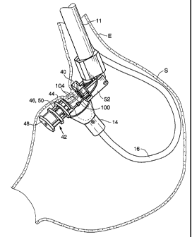
Fig. 2 illustrates one embodiment of a system 10 for tissue plication that is
suitable for endoscopic use, as well as surgical or laparoscopic use if
desired.
Generally speaking, system 10 includes a main shaft 11 having a distal portion
extendable through the esophagus into the stomach. The distal portion of the
main shaft
11 includes a retracting component 12 and a stapling component 13 comprised of
an anvil
40 and a staple head 42. During use of the system 10, the retracting component
12 is used
to engage stomach wall tissue and draw the tissue into a position between the
anvil 40 and
staple head 42, allowing staples to be driven through the stomach wall tissue
to form a
plication. By drawing a "pinch" of the stomach wall inwardly and then stapling
the
tissue, regions of serosal tissue are stapled to one another. Over time, these
serosal tissue
layers will adhere to form relatively strong bonds giving the plications
sufficient
durability to support implants within the stomach.
Retracting component 12 is provided with a vacuum head 14 and a flexible tube
16. Tube 16 preferably includes an insertion configuration in which it extends
approximately longitudinally relative to the main shaft 11 for streamlined
advancement of
the tube 16 through the esophagus. Tube 16 is equipped with pull-wires (not
shown)
and/or alternative means for articulating or retroflexing the vacuum head 14
as needed for
proper positioning within the stomach.
Vacuum head 14 defines a vacuum chamber 18 having an opening that, during
use, is positioned into contact with stomach tissue so as to draw the tissue
into the
chamber 18. Vacuum chamber is preferably formed of a flexible material such as
silicone, urethane or other suitable materials. Tube 16 is fluidly coupled to
a source of
negative pressure such as a syringe or vacuum pump such that application of
suction to
the tube 16 creates a vacuum in the vacuum chamber.
CA 02666594 2009-04-16
WO 2008/033409
PCT/US2007/019833
- 6 -
A hollow needle 20 is advanceable through the tube 16 into the vacuum chamber
18. Hollow needle 20 includes a pointed distal tip sufficiently sharp to
penetrate stomach
wall tissue (Figs. 7A ¨ 7B) when advanced against tissue drawn into the vacuum
chamber. An optional tissue engaging element (not shown in Figs. 2-6) is
positioned
within the hollow needle 20. As shown in Figs. 7C ¨ 7E, the tissue engaging
element is
deployable from the hollow needle 20 and placed in an expanded state after the
needle 20
has been advanced through tissue, and it is then withdrawn proximally using an
attached
tether or catheter following deployment to maintain engagement with the
tissue.
Various types of engaging elements may be used for this purpose. In the
embodiment shown in Fig. 7D, the engaging element 22a may be a balloon 24
mounted
on a small diameter catheter 26 extendable through the needle 20. After the
balloon 24 is
expanded on the serosal side of the stomach wall, tension is applied to the
catheter 26 as
shown in Fig. 7E to hold the retracted tissue between the anvil 40 and staple
head 42 (not
shown in Figs. 7A - 7E but discussed below) for stapling. The vacuum chamber
18 may
be withdrawn from the tissue to move it out of the area between the staple
cartridge and
anvil. Staples are then driven through the tissue in the direction of arrow A.
The balloon
may be removed after stapling or it may be left in place within the body. If
the balloon is
to be left in the body, it may be formed of a biodegradable or bioerodible
material, or a
more permanent biocompatible material.
As mentioned previously, stapling component 13 includes an anvil 40 and a
stapler head 42. Although Figs. 2 ¨6 show the head 42 positioned distally of
the anvil
40, in other embodiments the positions of these features might be reversed.
Likewise,
while in the illustrated embodiments the staple head is advanced to compress
the tissue, in
other embodiments the anvil might instead be advanced.
The stapling component 13 is pivotable relative to the main shaft 11 so that
once
the stapling component 13 is positioned within the stomach, it may be moved
laterally
towards the stomach wall. In the Fig. 2 embodiment, anvil 40 is mounted to an
articulated
base 52 coupled to the main shaft 11 at pivot point 54 (Fig. 5), allowing for
rotation of the
base (and thus the stapling component 13) relative to the main shaft 11. The
base 52 is
moveable to a longitudinal position (not shown) relative to the shaft to
facilitate
streamlined movement of the system 10 through the esophagus. As shown in Fig.
5, a
motor driven worm screw 56 is activated to articulate the base 52.
As best shown in Fig. 5, staple head 42 includes a staple cartridge 44
containing
staples (not visible in the drawing), and a staple driver 46 positioned to
drive staples from
the cartridge 44 when it is advanced proximally into contact with the staples.
Fluid lines
CA 02666594 2009-04-16
WO 2008/033409
PCT/US2007/019833
- 7 -
58a, 58b extend from the cartridge 44 and staple driver 46 respectively and
are coupled to
air, gas or other fluid (any of which will be referred to as "fluid") sources
positioned
external to the body. During use, fluid or gas pressure is directed through
fluid line 58a
and used to advance the staple cartridge 44 into contact with tissue
positioned between
the anvil 40 and cartridge 44 as shown in Fig. 4, thereby compressing the
tissue. In an
alternative embodiment, the fluid line 58a may be replaced with a cable
configured to
drive a lead screw that, when activated, advances the staple cartridge 44 to
compress
tissue disposed within the gap between the anvil 40 and the staple cartridge
44.
Once tissue is compressed between the cartridge 44 and anvil 40, fluid/gas is
then
directed through fluid line 58b to pressurize cylinder 48 sufficiently to
drive the staple
driver 46 into contact with staples positioned in the staple cartridge 44. A
tissue cutting
element 50, which in the illustrated embodiment is a tubular element having a
sharpened
end, is coupled to the staple driver 46 such that it will core through the
tissue during
stapling to form a hole in the plication.
Figs. 23A and 23B show collapsible hydraulic systems that may be used to
advance the cartridge 44 and staple driver 46.
System 110 of Fig. 23A allows for sequential movement of the cartridge and the
driver allowing for compression and then stapling. System 110 includes nested
cylinders
112a, 112b and 112c, fluid inlets 114a, 114b and 114c, and o-ring seals 116a-
d. When
fluid pressure is introduced into the system via inlet 114a, cylinder 112b
advances to the
left. Once seal 116b of cylinder 112b crosses over inlet 114b, fluid pressure
is applied to
cylinder 112a through inlet 114c, causing cylinder 112a to begin moving
towards the left
of the drawing. This arrangement can be used to first compress (e.g. using
staple
cartridge coupled to cylinder 112b), and to then staple (e.g. using a staple
driver coupled
to cylinder 112a) the tissue using a single source of fluid or gas pressure.
System 111 of Fig. 23B is a telescoping fluid power actuator that may be used
when the length of the path of travel needed for a feature (e.g. the stapler
driver, cartridge,
and/or anvil) would require a cylinder that is longer than can be accommodated
by the
environment (e.g the stomach). . System 111 includes cylinders 118a, 118b and
118c,
inlets 120a and 120b, and seals 122. . Pressure applied at inlet 120a will
cause cylinder
118a (which may be coupled to the cartridge) to advance to the left. Once
cylinder 118a
has moved a distance "1", it will engage with a shoulder 124 on cylinder 118b,
causing
cylinder 118b to travel with cylinder 118a as cylinder 118a continues to move.
In this
arrangement, a long stroke for cylinder 118a is gained from a short fluid
power system.
CA 02666594 2009-04-16
WO 2008/033409
PCT/US2007/019833
- 8 -
Fig. 8 shows an alternative embodiment of a modified plication system 10a
which
utilizes rigidizable cables 80 to deflect the stapling component 13 relative
to the main
shaft 11 Cables 80 are preferably formed of a plurality of spine elements 82
strung onto a
pull wire (not shown). Fluid lines 58a, 58b may extend through holes in the
spine
elements, or they may be separate from the cables 80. When tension is applied
to the
cables using actuators positioned outside the body, the spine elements 82
engage one
other to stiffen the cable. The spine elements 82 may be shaped such that the
cable will
assume a predetermined bend when tension is applied to them.
In the Fig. 8 embodiment, a rigidizable cable 86 also carries the vacuum
chamber
18a. Cable 86 includes a pull wire (not shown) extending through spine
elements 88.
The spine elements 88 are shaped to orient the chamber 18a for advancement
between the
anvil 40 and cartridge 44. Cable 86 may extend from a sheath 89 that is
longitudinally
extendable from a lumen in the main shaft 11.
Vacuum chamber 18a may be foldable or compressible for positioning within the
sheath 89. Fig. 10A shows a folded vacuum chamber 18b beginning to expand as
it exits
the distal end of sheath 89, and Fig. 10B shows the vacuum chamber 18b fully
expanded
after exiting the sheath 89. Fig. 10A illustrates that in alternative
embodiments,
endoscopes 98 and/or other instruments may be passed through the vacuum
chamber 18b
to give access and/or visualization to the stomach wall or other organs or
tissues. In the
Fig. 8 embodiment, a collar 96 is positioned on the main shaft 11 for
receiving endoscope
98.
Fig. 9 is an exploded view of the vacuum chamber 18a of the Fig. 8 embodiment,
and illustrates that a pair of gripping jaws 90 is positioned within the
vacuum chamber
18a. The jaws 90 are constructed using a linkage arrangement and are
controlled using a
pull wire extending through a back plate 92 of the vacuum chamber 18a, through
spine
elements 88 (Fig. 8), and to the proximal end of the plicating system outside
the body.
Before vacuum is applied, the jaws are moved to an open position. When vacuum
pressure draws tissue into the vacuum chamber 18a, the jaws 90 are closed to
engage the
tissue.
The Fig. 8/9 vacuum chamber 18a additionally includes stabilizing arms 94
extending into the vacuum chamber 18a. The stabilizing arms are retained in
contact with
the interior walls of the chamber 18a to prevent the chamber from collapsing
when
vacuum is applied. The arms may be moveable between closed and opened
positions to
allow the vacuum chamber to collapse for passage through the esophagus, or
they may
remain fixed in the opened position.
CA 02666594 2009-04-16
WO 2008/033409
PCT/US2007/019833
- 9 -
Plication Reinforcements
Reinforcements of various types may be implanted in or on plications formed
using the plication system. Such reinforcements may function to reinforce the
staple
array, help to more evenly distribute the forces applied to the tissue by the
staples, and/or
facilitate bonding between the opposed serosal layers. Suitable reinforcements
include
ones positionable on or between the serosal tissue layers ("serosal side
reinforcements"),
as well as those delivered on the side of the mucosal tissue ("mucosal side
reinforcements").
For serosal side reinforcements, a reinforcement similar to engaging element
22a
described in connection with Fig. 7D may serve as a permanent or semi-
permanent
implant that will reinforce the staple array applied to the tissue and/or
facilitate serosal
tissue bonding between the layers of stomach wall tissue that are to be
stapled together.
For this purpose, the material may be a synthetic or non-synthetic mesh
(formed of
nitinol, polyester, or other natural or synthetic material), porous or non-
porous material,
slotted material, or any other material through which adhesions will form or
onto which
tissue will grow. Examples include, but are not limited to, polypropylene,
materials sold
under the trade names Goretex or Dacron, or tissue graft material such as the
Surgisis
material sold by Wilson Cook Medical, Inc. The material may be treated with
tissue-
ingrowth promoting substances such as biologics. In an embodiment shown in
Fig. 18A,
the reinforcement 22b is a mesh/braid embedded in or coated with a dissolvable
or
bioabsorbable coating. The reinforcement 22b is preferably positioned in a
manner
similar to that described in connection with the balloon of Fig. 7D.
Specifically, vacuum
chamber 18 is used to engage a region of tissue where a plication is to be
formed. Hollow
needle 20 is advanced from within the chamber 18 through the stomach wall, and
the
reinforcement 22b is advanced from the hollow needle and inflated to a
toroidal shape
between the opposed regions 104 of serosal tissue. The reinforcement 22b may
optionally be used for retraction of the stomach wall via application of
tension on the
tether 33 as shown in Fig. 18D. Staples driven through the tissue pierce and
deflate the
inflated reinforcement as shown in Figs. 18D and 18E, and capture the deflated
reinforcement between the opposed serosal tissue layers. The reinforcement is
left in
place between the two layers of stomach wall tissue after stapling. The
coating on the
deflated balloon dissolves, exposing the interstices of the underlying mesh or
porous
material to serosal growth.
As shown in Fig. 19, the reinforcement 22c may instead be a mesh disk 30
detachably carried (e.g. using sutures 32) on a wire hoop 34 extendable
through needle 20
CA 02666594 2009-04-16
WO 2008/033409
PCT/US2007/019833
- 10 -
in a compressed shape and then self-expandable to the illustrated position to
expand the
mesh on the outside of the stomach. The sutures 32 are severed during
stapling, leaving
the mesh in place between the plicated tissue layers. The hoop 34 is withdrawn
into the
needle 20 and removed from the body.
In another embodiment shown in Figs. 20A and 20B, the reinforcement 22d may
be an elongate nitinol mesh or braid backbone 35 that may be positioned
longitudinally
within the hollow needle 20, but that is shape set to assume a circular or
other suitable
configuration once released from the hollow needed between the serosal tissue
layers. In
modifications to this concept shown in Figs. 21A ¨ 21C, the reinforcement 22e
may be a
wire 37 or ribbon shape set to assume one of a variety of expanded
configurations when it
is pushed out of the hollow needle 20. In the expanded configuration, the wire
may
assume a pattern shaped such that when it is positioned between serosal tissue
layers, it
will be captured by staples advanced through the tissue. As with the other
reinforcements
disclosed above, the pattern may be annular as in Figs. 21A or 21C, or it may
be disk-like
as in Fig. 21B. Moreover, while the patterns are shown to have an
approximately circular
silhouette, other shapes may instead be used.
In another embodiment shown in Figs. 22A and 22C, a reinforcement 22f (which
may be formed of a polyester fabric or other material including those listed
elsewhere in
this application) is carried by a frame 106 having a plurality of outwardly
extending arms
108 that spring to an expanded position when released from hollow tube 20. The
reinforcement 22e is deployed using hollow needle 20 in a manner similar to
those
described above. Specifically, the hollow needle 20 is pierced through engaged
stomach
wall tissue, and the frame is advanced out the distal end of the needle 20 to
allow arms
108 to spread to the expanded position shown in Figs. 22A and 22C, thereby
expanding
the reinforcement 22e between the opposed serosal layers. The element is fixed
between
the layers by the staples driven through the opposed regions of stomach wall,
and the
frame is withdrawn from the needle and out of the body.
Mucosal side reinforcements may take the form of reinforcements that are
positioned on or adjacent to one or both of the mucosal surfaces lining the
"pinch" of
tissue that will form the plication. These reinforcements may be features of
the staples or
staple arrays, or they may be separate components engaged by staples as the
staples are
advanced through the tissue.
Referring to Fig. 21A, conventional stapling procedures will often include two
parallel rows of staples, in which the staples in one row are laterally offset
from the
staples of the other row. According to the disclosed method, it is useful to
employ this
CA 02666594 2009-04-16
WO 2008/033409
PCT/US2007/019833
-11 -
technique to the circular staple pattern delivered using the plicator 10, to
produce two
concentric rings of offset staples, as shown in Fig. 11B. It has been found to
be
additionally beneficial to form mucosal side reinforcements by linking or
interlocking the
staples to provide greater structural reinforcement to the stapled tissue
and/or to more
evenly distribute forces applied to the tissue by the staples. Linked staple
arrays may be
formed by arranging the staples 70 in the cartridge of the plicator 10 in a
single circular
pattern to interlock as shown in Fig. 12A, or in a double circular pattern
with two
concentric rings of interlocked staples. The staples 70a may be curvilinear so
as to form a
locking pattern shown in perspective view of Fig. 12B. A linear arrangement of
staples
70 may also be linked as shown in Fig. 12C.
In alternative embodiments, staples are linked together by reinforcing members
formed of metallic or polymeric materials, such as nitinol, titanium,
stainless steel PEEK,
or other biocompatible materials. According to these embodiments, the
reinforcing
members are positioned on one or both of the mucosal sides of the "pinch" of
tissue
engaged by the plication system such that they are captured by staples being
driven
through the tissue. In a preferred embodiment, the staples capture a cartridge
side
reinforcing ring 72 (Fig. 13A) as they leave the cartridge and/or capture an
anvil side
reinforcing ring 74 (Fig. 13B) as the anvil shapes and bends them. Upon
completion of
the plication, the staples are linked to one another so that they cannot
separate or expand
radially. The use of the reinforcing rings is advantageous compared with prior
art staple
buttressing materials such as sheets formed of bovine pericardium or hydrogel,
both of
which are penetrated (and thus potentially compromised) by staples as they are
driven
through the tissue. The open structure or lattice pattern of the reinforcing
rings provides
openings for the staples to pass through as well as supportive members for the
staples to
wrap around ¨ so that the staples capture but do not penetrate the ring
material. Over
time, tissue may grow into the lattice structure and/or around the supportive
members.
The proportions of the ring, such as the sizes of the openings in the lattice
structure, may
be adjusted to increase or decrease the amount of ingrowth that might occur.
The reinforcing rings are preferably provided separate from the staples
although
they instead may be integral with the staples. In this embodiment, ring 74 is
positioned
against the staple anvil 40 as shown in Figs. 14A and 14B. The anvil may
include
retaining elements to maintaining the ring's position on the anvil. Ring 72 is
seated
within the cartridge 44, with the staples 70 aligned with their prongs 76
extending
through openings 73 in the ring 72. Alternatively, the cartridge may have
retaining
elements to hold the ring in place prior to stapling.
CA 02666594 2009-04-16
WO 2008/033409
PCT/US2007/019833
- 12 -
When staples 70 are driven from the cartridge, they advance further through
openings 73, capturing ring 72 against the adjacent mucosal tissue as shown in
Fig. 15A.
The staple legs/prongs 76 pass through the stomach wall tissue into contact
with the
indentations 78 of the anvil 40. When they contact the anvil 40, the prongs 76
fold
around the staple ring 74 to capture the ring and interlock the staples on the
anvil side of
the plication as shown in Fig. 15B. Rings or other interlocking elements of
this type may
be used with single- or double- staple row configurations.
Rings 72, 74 are shown as generally circular, although alternative
reinforcements
of different shapes and patterns may also be used, including those shaped to
accommodate linear, oval and other staple patterns.
Figs. 16a and 16b show two examples of reinforcements 75a, 75b useful for
linear
staple patterns. As shown, reinforcements are similar to the ring 72 in their
use of a
plurality of interconnecting members that define openings 73a for receiving
staple legs.
The legs of any given staple may pass through two laterally positioned
openings 73a,
longitudinally positioned openings 73a, just a single opening, or any other
combination.
These reinforcements may be used with linear staplers in a manner described
above with
respect to the circular staplers. The reinforcements may include members 77
positioned
to receive implants that might be used within the body (e.g. pH monitors or
other sensors,
stimulation/pacing leads, etc.). Fig. 17 illustrates use of the loops 77 to
support fasteners
79a, 79b in a system for engaging one tissue plication to another tissue
plication. Staples
70 driven through the openings 73a engage the reinforcements and form the
plications.
The fasteners 79a, 79b are then brought into engagement with one another to
draw and
couple the plications together. This may be used to form a restriction and/or
reduce
stomach volume, or for other purposes.
The disclosed reinforcements may be sold as individual components that may be
used together with commercially available staplers to reinforce the
lines/rings of staples
to be delivered by those staplers.
Exemplary Method of Use
One method of using the illustrated system will next be described with
reference
primarily to Figs. 8 and 9.
In preparation for use, the orientation of the staple head 42 and the vacuum
chamber 18a are adjusted using the appropriate pullwires to place them in
their
longitudinal positions.
Next, the assembled plicator 10a is passed into the stomach S via the
esophagus,
preferably through a protective sheath passed through the esophagus. Endoscope
98 is
CA 02666594 2009-04-16
WO 2008/033409
PCT/US2007/019833
- 13 -
also passed into the stomach to provide visualization of the procedure. The
endoscope is
preferably mounted to plicator 10a, or it may be a separate component.
The plicator 10a is advanced towards a target location at which a plication is
to be
formed. The rigidizable cable 86 is manipulated using pull wires to extend
vacuum
chamber 18a between the fluid lines 58a, 58b and against adjacent stomach
tissue.
Suction is applied to the vacuum chamber 18 to draw stomach tissue into the
vacuum
chamber. The gripper arms 90 are closed to pinch the tissue within the
chamber, and the
vacuum chamber 18a is withdrawn from between the fluid lines 58a, 58b,
carrying the
engaged tissue with it (see Fig. 4). Consequently, a pocket 100 forms in the
tissue such
that if the stomach were to be viewed from the outside a depression in the
stomach wall
would be visible. Serosal tissue surfaces 104 line the outside surfaces of the
pocket 100.
If gripper arms are not used, suction maintained to stabilize the tissue
within the vacuum
chamber.
If additional stabilization of the tissue is desired, such as during use of
the Fig. 2
embodiment, hollow needle 20 may be advanced through the engaged tissue as
shown in
Figs. 7A and 7B, and balloon 24 is inflated within the pocket 100 as shown in
Figs. 7C
and 7D. As shown in Fig. 7E, the inflated balloon 24 is withdrawn using
catheter 26, thus
retracting the tissue surrounding the pocket 100. Alternatively, the engaging
element 22b
of Fig. 8A, or element 22d of Figs. 10A and 10B may be deployed and used in
similar
fashion. If element 22c of Fig. 9 is to be used, the hoop 34 is advanced
through the
hollow needle 20 into the pocket 100 where it springs to its opened
configuration to
expand the mesh element 22c.
Referring again to Fig. 8, once the tissue has been drawn between the fluid
lines
58a, 58b, fluid is driven through fluid line 58a to bring the staple cartridge
44 into contact
with the tissue and to compress the tissue between the cartridge 44 and the
anvil 40 (see
Fig. 4). Once the tissue is fully compressed, fluid pressure is via line 58b,
causing the
staple driver 46 to advance into contact with staples in the cartridge 44,
thus driving the
staples through the tissue and simultaneously forming a hole or incision
through the
layers of stomach wall tissue. The sharp ends of the staples fold against the
anvil 40 after
passing through the two layers of stomach wall tissue, thus maintaining the
plication. If
the mucosal reinforcements 72, 74 of Figs. 13A, 13B are used, the staples
engage one or
both mucosal reinforcing rings as during stapling.
The procedure may be repeated to form multiple plications if needed. Following
formation of the plication(s), a medical implant may be coupled to the
hole/incision
formed by the hollow needle 20. Coupling may be carried during the course of
the same
CA 02666594 2014-04-02
- 14 -
procedure or during a later procedure scheduled to permit sufficient formation
of
adhesions between the serosal tissue layers 102 to support the implant.
The system or other components described herein may be packaged with
instructions for use instructing a user to utilize the system according to
methods disclosed
herein.
As is evident from above, the disclosed endoscopic systems function to draw a
tissue into the stomach to form a depression on the exterior surface of the
stomach, and
staple (or suture, or fasten or adhere etc) the opposed stomach wall sections
lining the
depression together another to form a plication. The system may additionally
place
material of a type that will promote strong tissue adhesion within the
depression (on the
exterior of the stomach) and retain the material between the serosal surfaces
to enhance.
Additionally or alternatively, mucosal reinforcements such as structures that
interconnect
the staples may be implanted. While these systems provide convenient
embodiments for
carrying out this function, there are many other widely varying instruments or
systems
may alternatively be used. Moreover, the
disclosed embodiments may be combined with one another in varying ways to
produce
additional embodiments. Thus, the embodiments described herein should be
treated as
representative examples of systems useful for forming endoscopic tissue
plications.