Note: Descriptions are shown in the official language in which they were submitted.
CA 02666600 2009-04-15
WO 2007/046088 PCT/IL2006/001189
AN EECP DEVICE AND AN IMAGE SYSTEM COMPRISING THE SAME
FIELD OF THE INVENTION
The present invention relates to medical apparatus for increasing vascular
blood flow
and tissue perfusion to various organs of a patient and to methods of using
such
apparatus in various imaging modalities in order to improve the anatomical and
physiological visualization.
BACKGROUND OF THE INVENTION
Enhanced external counterpulsation (EECP) is a non-invasive treatment that
uses
timed, sequential inflation of pressure cuffs on the calves, thighs and
buttocks to
augment diastolic pressure, decrease left ventricular afterload, and increase
venous
return. Augmenting diastolic pressure displaces a volume of blood backwards
into the
coronary arteries during diastole when the heart is in a state of relaxation
and the
resistance in the coronary arteries is at a minimum. The resulting increase in
coronary
artery perfusion pressure may enhance coronary collateral development,
increase
cardiac perfusion pressure or increase flow through existing collaterals. In
addition,
when the left ventricle contracts, it faces a reduced aortic pressure to work
against
since the cuffs deflate rapidly rigllt before the systole. EECP has been
primarily
investigated as a treatment for chronic stable angina.
Practically, a regular treatment is exercised in a manner that patients lie
down on a
padded table in a treatment room. T11ree electrodes are applied to the skin of
the chest
and connected to a monitoring system; usually a probe of a blood pressure
sensor and
an electrocardiograph (ECG), which displays the heart's rhythm during
treatment, is
also monitored.
Fig. 1 presents a schematic illustration of EECP counterpulsation mechanism as
defined in the prior art, and a generalized four pulsation steps (A-D). Fig. 1
also
presents a diagram of the same presenting four schematic steps of the
commercially
available EECP equipment:
Inflation initiates retrograde pulse wave (step 1); inflation of lower thigh
cuffs 50 ms
later (step 2); inflation of upper thigh cuffs 50 ms later (step 3) and
lastly, deflation
1
CA 02666600 2009-04-15
WO 2007/046088 PCT/IL2006/001189
facilitates cardiac unloading (step 4). EECP is generally utilized for
treating angina
pectoris and other arterial insufficiency states. Angina is a chronic chest
pain or
discomfort appearing when the heart muscle doesn't get enough blood and oxygen
supply, are insufficient for the work it's doing. External counterpulsation
techniques,
such as the EECP, were shown in the art to improve the balance between the
amount
of oxygen the heart needs and the amount it gets. Both these changes reduce
the pain
of angina, increase level of daily activities & effort and decrease the need
for
medication.
It is well established that timing of the inflation/deflation steps of the
EECP pulsation
is a critical parameter in the procedure. Various approaches were implemented
and
experimented with improved timing means. US patent 6,736,786 presents a
counterpulsation device that operates without the use of compressed air or
pressurized
gas, which includes at least one inflatable cuff that is adapted to be placed
upon a
selected portion of the patient's body. Here, a conduit connects the
inflatable cuff to
an air transfer device so that non-compressed air can be transferred from the
air
transfer device to the cuff through the conduit to inflate the cuff. The
conduit also
connects the cuff to the air transfer device so that air can flow through the
conduit to
deflate the cuff. Another conduit is coupled to the first so that the air in
the system can
be selectively vented into the atmosphere. A series of valves are placed on
the conduit
to selectively control whether air is supplied to or withdrawn from the
inflatable cuff.
The air moving device preferably is a cylinder having a piston that moves
through the
cylinder to move the air from within the cylinder through the conduit and into
or out
of the cuff as desired. The piston moves through the cylinder through the use
of a
linear servo actuator that is controlled by an appropriately programmed
electronic
controller so that the inflation of the cuff is timed with portions of the
patient's EKG
signal and peripheral plethysmographic wave.
Nevertheless, most of the cuffs are elastic sleeve-like members, adapted to
shrink, i.e.,
decrease its diaineter at a given time, to expand and vice versa. Hence, US
patent
4,753,226 discloses an EECP massage apparatus comprising a plurality of air-
filled
balloons. US patent application 2005/043657 provides an exterior
counterpulsation
system that includes a garment for being worn on the exterior of a patient's
body. This
garment is made of electroactive polymer actuators connected thereto. US
patent
application 2002/107461 teaches an EECP device having timing of inflation and
2
CA 02666600 2009-04-15
WO 2007/046088 PCT/IL2006/001189
deflation and reduced temperature of the pressurized gas, such that the gas
flow
temperature of the inflatable devices is near to room temperature, as well as
faster and
more responsive inflation/deflation equipment. The external counter-pulsation
apparatus includes a plurality of inflatable devices received about the lower
extremities of the patient, a source of compressed fluid in communication with
said
plurality of inflatable devices, and a fluid distribution assembly
interconnecting said
source of compressed fluid and said inflatable devices. The fluid distribution
assembly includes a selectively operable inflatioii/deflation valve
interconnected
between each of said inflatable devices and said source of compressed fluid.
The fluid
distribution assembly separately operates each inflation/deflation valve to
sequentially
inflate and deflate each inflatable device.
US patent application 2002/169399 defines a cardiac assist device includes a
sealed
tubular housing for externally applying positive and negative relative
pressure to a
limb in counterpulsation with heart function. The applicator is assembled, in
situ, to
provide customized fit. It includes a fabric or sponge-like iimer layer cut to
size and
situated around the limb. Initially deformable material is sized, sealed
around the
inner fabric layer and then secured by straps or the like to form a relatively
rigid, non-
expandable tubular shell. The shell may include an interior wall composed of a
sheet
of hard plastic or articulated sections of hard plastic or metal. The interior
wall has a
plurality of openings to the sealed shell interior. The exterior shell wall is
positioned
around the interior wall. The shell walls are spaced apart by radially and/or
longitudinally extending spacer elements defining a multi-section air flow
chamber
between the walls. The interior shell wall and spacer elements may be
integral. The
spacer elements include passages so that air pumped into and out of the shell
chamber
is uniformly distributed and moves freely to and from the shell interior.
It is known from the art that medical imaging modalities depict the anatomical
and
physiological status of the human organs. IV contrast materials are used to
visualize
blood flow to organs such as the heart, liver, brain etc, and to demonstrate
the normal
and abnormal blood supply to these organs. Current vascular and cardiac
imaging is
associated with high radiation exposure, high concentration of contrast
material
associated side effects and limited specificity and sensitivity. Imaging tests
are
extensively used in all medical fields, with a constant increase in
utilization for
screening and invasive procedure replacement. Cardiac CT for instance, strives
to
3
CA 02666600 2009-04-15
WO 2007/046088 PCT/IL2006/001189
become a general public screening test for early diagnosis of cardiac
ischemia. The
sensitivity and specificity of the various modalities in demonstrating
vascular and
cardiac pathologies is limited.
While the EECP devices are currently provided useful solely for treating
angina
pectoris and other arterial insufficiency states, their main mechanical
disadvantage is
timing of the cuffs such that an exact and effective operation of the vein or
artery
device inflation/deflation is not provided at an exact and predetermined
timing.
SUMMARY OF THE INVENTION
It is one object of the present invention to disclose a non-invasive rigid-
support
enhanced external counterpulsation device (RS-EECP) providing a precise onset
of a
blood flow characterized by a sharp-wave front. This novel RS-EECP is useful
for
out-patient treatment of arterial insufficiency states, especially angina.
It is comprised of ingredients selected in a non-limiting manner from a timing
means
and a plurality of pressing cuffs. The timing means is adapted to onset the
collapsing
and expanding maneuvers of the cuffs in a sequence of occasions defined along
the
diastolic/systolic cycle, The cuffs are fastened around at least a portion of
the
circumference of at least one organ comprising a vascular bed to
counterpulsate
against an either fixed or maneuverable support. The improvement is that the
support
is at least partially rigid member; so as a quick expansion of said vessel
bed,
following a forceful and effective collapsing of the same is obtained.
It is one aspect of the present invention wherein the RS-EECP comprising two
or
more pressing cuffs, being arranged adjacently along the patient's organ in a
series
such that one cuff or cuffs are located at a retrograde position in respect to
others.
It is another aspect of the present invention wlierein the cuff or cuffs are
located at the
retrograde position are scheduled to collapse prior to others, such as a
unidirectional
blood flow is provided.
4
CA 02666600 2009-04-15
WO 2007/046088 PCT/IL2006/001189
It is another aspect of the present invention to disclose an array of RS-
EECPs, each of
which is defined in a non-limiting manner above, further comprising two or
more RS-
EECPs, being arranged along the patient's body in series and/or in parallel,
such that
at least one RS-EECP is located at a retrograde position in respect to others.
It is another aspect of the present invention wherein one or more RS-EECPs is
located
at the retrograde position, and scheduled to collapse prior to others, such as
a
peristaltic unidirectional blood flow along the treated patient's body portion
is
provided.
It is anotlier aspect of the present invention wherein the aforementioned RS-
EECP is
in cominunication with at least one imaging means (namely 'IRS-EECP'). The
imaging means are adapted to display said blood flow or the flow of markers or
medicaments solubilized therein, or tissue perfusion.
It is another aspect of the present invention wherein the imaging modality is
selected
from cardiac CT, CT-angio, cardiac and vascular MRI, ultrasound Doppler for
the
carotids and renal vessels, isotope based scans, as PET scans or any
combination
thereof.
It is anotller aspect of the present invention wherein the markers,
contrasting agents
and/or image contrasting means are selected from all substances, compositions
or
agents used for enhancing or depicting vascular flow or as a measure of tissue
perfusion or perfusion pressure, in conjunction with the above mentioned
imaging
modalities, for example iodine based materials, isotopes, ferromagnetic
substances,
micro bubbles etc.
It is another aspect of the present invention wherein the medicaments such as
nitroglycerin and dopamine.
It is another aspect of the present invention to disclose a RS-EECP (100)
comprising
in a non-limiting manner a plurality of rotating-cuffs (1) and a mechanism for
rotating
the same (2). The rotating-cuffs (1) comprising inter alia at least one set of
rotating
cuffs being either at least partially flexible or rigid; and at least one
rigid-support. The
said maneuverable cuff has an open configuration (collapsed state) and a
closed
configuration (released state). At said collapsed state, the cuffs are
fastened around at
least a portion of the circumference of at least one organ comprising a
vascular bed to
counterpulsate against an either fixed or maneuverable support.
CA 02666600 2009-04-15
WO 2007/046088 PCT/IL2006/001189
It is another aspect of the present invention to disclose a RS-EECP (100) that
is
further having at least one rotating shaft (3), being either concentric or
eccentric
member. Shaft (3) is connected to a motor, rotating the same; and hence,
either
directly or indirectly, it forcefully compresses the maneuverable cuffs.
It is another aspect of the present invention to disclose a RS-EECP (200)
comprising
inter alia a plurality of pressing-cuffs (1) and a mechanism for pressing the
same (2).
The RS-EECP (200) may further comprise one or more maneuverable cuffs (31)
being connected to a shaft having a linear (e.g., approximately perpendicular)
motion
(32), such as at a given time, said pressing cuffs are fastened towards a
rigid support
(33).
It is another aspect of the present invention to disclose an RS-EECP (100),
further
including an external fixation means adapted to immobilize legs, thighs and
upper
torso during activation of the RS-EECP. The aforesaid means are selected in a
non-
limiting manner from strips, straps, pillory, or any other immobilizing means.
It is another aspect of the present invention to disclose an RS-EECP (100)
further
comprising a portable CPR device, especially adapted to be utilized in trauma
and
pre-hospital medical treatment, e.g., ambulances etc, and in hospitals.
Moreover, the
aforesaid portable CPR is utilizable in domestic, commercial, sport centers,
clinics,
etc.
It is another aspect of the present invention to disclose a portable CPR
utilizable
independently, in conjunction and/or in communication with a defibrillator,
providing
a synergic resuscitating system; said synergic resuscitating system further
comprising
at least one controlling means.
It is a second object of the present invention to present a non-invasive
method for out-
patient treating of arterial insufficiency states, especially angina by
providing a
precise onset of a blood flow characterized by a sharp-wave front.
It is another aspect of the present invention wllerein the aforesaid method
comprising
steps selected in a non-limiting manners from (a) obtaining a timing means and
a
plurality of pressing cuffs; (b) fastening cuffs around at least a portion of
the
circumference of at least one organ comprising a vascular bed to
counterpulsate
against an either fixed or maneuverable support; wherein said support is a
rigid
member; and (c) initiating the collapsing and expanding maneuvers of the cuffs
in a
6
CA 02666600 2009-04-15
WO 2007/046088 PCT/IL2006/001189
sequence of occasions defined along the diastolic/systolic cycle; such as a
quick
expansion of said vessel bed, following a forceful and effective collapsing of
the same
is obtained.
It is another aspect of the present invention wllerein the method comprising a
step of
obtaining two or more pressing cuffs, setting the same adjacently along the
patient's
organ in a series such that one cuff or cuffs are located at a retrograde
position in
respect to others.
It is another aspect of the present invention wherein the aforesaid method
comprising
locating cuff or cuffs at a retrograde position and then collapsing the same
prior to
others, such as a unidirectional blood flow is provided.
It is another aspect of the present invention wherein the method comprising a
step of
obtaining two or more RS-EECPs and arranging the same along the patient's body
in
series and/or in parallel, such that at least one RS-EECP is located at a
retrograde
position in respect to others.
It is another aspect of the present invention wlzerein the metliod comprising
a step of
locating one or more RS-EECPs at the retrograde position and initiating the
same to
collapse prior to others, such as a peristaltic unidirectional blood flow
along the
treated patient's body portion is provided.
It is another aspect of the present invention wherein the method further
comprises an
external fixation means adapted to immobilize legs, thighs and upper torso
during the
activation of the RS-EECP by plural means selected in a non-limiting manner
from
strips, straps, pillory, or any other immobilizing means.
It is another aspect of the present invention wherein the method comprising a
step or
steps of communicating the RS-EECP system with at least one imaging means
(i.e.,
IRS-EECP). The imaging means are preferably adapted for 'displaying said blood
flow
or the flow of markers or medicaments solubilized therein.
It is another aspect of the present invention wherein the imaging is selected
from
providing cardiac CT, CT-angio, cardiac and vascular MRI, ultrasound Doppler
for
the carotids and renal vessels, isotope based scans, as PET scans or any
combination
thereof.
It is another aspect of the present invention wherein the method comprising
utilizing
7
CA 02666600 2009-04-15
WO 2007/046088 PCT/IL2006/001189
medicaments being selected in anon-limiting manner from dopamine,
nitroglycerine
or milrinon (a PDE-III-inhibitor).
It is another aspect of the present invention wherein the method comprises
utilizing a
portable CPR device independently, in conjunction and/or in communication with
a
defibrillator, providing a synergic resuscitating system. The synergic
resuscitating
system further comprising at least one controlling means.
BRIEF DESCRIPTION OF THE FIGURES
In order to understand the invention and to see how it may be implemented in
practice, few preferred embodiments will now be described, by way of non-
limiting
example only, with reference to the accompanying drawing, in which
figure 1 presents a commercially available EECP operating scheme of a
commercially
available device;
figure 2 presents an RS-EECP according to one embodiment of the present
invention;
figure 3 presents an RS-EECP according to yet another embodiment of the
present
invention;
figure 4 presents a commercially available EECP with two flexible cuffs; and,
figure 5 presents an RS-EECP according to yet another embodiment of the
present
invention, comprising one flexible cuff and one rigid support.
DETAILED DESCRIPTION OF THE EMBODIMENTS
The following description is provided, alongside all chapters of the present
invention,
so as to enable any person skilled in the art to make use of said invention
and sets
forth the best modes contemplated by the inventor of carrying out this
invention.
Various modifications, however, will remain apparent to those skilled in the
art, since
the generic principles of the present invention have been defined specifically
to
provide a non-invasive rigid-support enlianced external counterpulsation
device (RS-
EECP) and methods of providing a precise onset of a blood flow characterized
by a
sharp-wave front, useful for out-patient treatment of arterial insufficiency
states,
especially angina.
8
CA 02666600 2009-04-15
WO 2007/046088 PCT/IL2006/001189
The term 'counterpulsation' refers hereinafter to a technique that
synchronizes the
external pumping of blood with the heart's cycle to assist the circulation and
decreasing the work of the heart. The term is usually used wherein
counterpulsation
pumps when the heart is resting to increase blood and oxygen flow to the
heart; and
wherein counterpulsation has a fast decrease in pressure when the heart is
working to
decrease the heart's workload and lessen oxygen demand.
The term 'enhanced external counterpulsation' (EECP) refers hereinafter to a
non-
invasive out-patient treatment for heart disease and, in particular, for
angina. EECP is
designed to relieve angina by improving perfusion in areas of the lieart
deprived of an
adequate blood supply.
The term 'perfusion' a physiological term that refers to the process of
nutritive
delivery of arterial blood to a capillary bed in the biological tissue. The
term
`perfusion pressure' refers to the arterial pressure minus venous pressure.
The term 'diastole' refers hereinafter to the time period when the heart is in
a state of
relaxation and dilatation (expansion). The diastolic pressure is specifically
the
minimuin arterial pressure during relaxation and dilatation of the ventricles
of the
heart. Diastole is the time when the ventricles fill with blood.
The tei7n 'vascular bed' refers hereinafter to the vascular system, or a part
tllereof:
for example, the pulmonary vascular bed describes the blood vessels of the
lungs.
The term'cardiac output' refers hereinafter to the amount of blood that is
pumped by
the heart per unit time, measured in liters per minute (1/min), usually about
4.7
liters/minute. The amount of blood that is put out by the left ventricle of
the heart in
one contraction is called the stroke volume. The stroke volume multiplied by
the heart
rate is the cardiac output.
The term 'arterial insufficiency states' refers hereinafter to heart
pathologies and
vascular bed pathologies, conditions and pains, malfunctions and symptoms,
such as
angina and especially angina pectoris (typical Canadian Cardiovascular Society
Classes I, II and III angina), deep vein or artery thrombosis, edema, lymph
edema, left
ventricular dysfunction, incidence and complications of diabetic and other
cllronic
obstructive coronary disease and arterial insufficiency states.
The term 'cardio-pulmonary resuscitation' (CPR) refers hereinafter to an
emergency first aid protocol for an unconscious person on whom both breathing
and
9
CA 02666600 2009-04-15
WO 2007/046088 PCT/IL2006/001189
pulse cannot be detected. More specifically, the term CPR is used hereinafter
to
define an EECP device especially adapted to portable use.
The imaged RS-EECP (IRS-EECP) according to the present invention increases
blood
flow to the heart, kidneys, liver, etc., increases organ perfusion and
differentially
accentuates flow and perfusion mismatches. Hence, the novel system lowers
radiation
exposure; reduces dose of contrast material; diminishes risk for side effects;
increases
specificity and sensitivity; increases patient compliance and increased span
of
imaging procedures.
Moreover, a main problem characterizing the current imaging techniques is the
exposure to high dose ionizing radiation and its long term effects, and
contrast
material side effects and allergies, leading to serious complications and even
death.
The IRS-EECP according to the present invention reproduces non-invasively the
action of an intra aortic balloon counter pulsation. It increases the blood
flow and
perfusion in various organs such as the heart, kidneys and liver. The system
also
differentially accentuates the flow and perfusion mismatches, between healthy
(normally oxygenated) and non-healthy (relatively ischemic) tissues, improving
the
sensitivity and specificity of medical diagnostic imaging tests.
The aforesaid IRS-EECP is utilized to significantly improve the anatomical and
physiological visualization, in various imaging modalities. This results in
decreased
total doze of radiation, and reduction in the total dose of contrast materials
including
isotopes, resulting in a lower rate of side effects. It also increases patient
compliance,
and reduces costs.
It is in the scope of the present invention wherein the IRS-EECP comprising
imaging
means selected from cardiac CT, CT-angio, cardiac and vascular MRI, ultrasound
Doppler for the carotids and renal vessels, isotope based scans, as PET scans
or any
combination thereof.
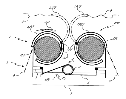
Reference is made now to figure 2, presenting a schematic cross section of a
noninvasive rigid-support EECP device (RS-EECP, 100) adapted for sequential
inflation of a plurality of pressure cuffs against a non-flexible (rigid)-
support on the
calves, thighs and buttocks to augment diastolic pressure, decrease left
ventricular
afterload, and increase venous return. This RS-EECP (100) is a rotating-flaps
model
according to one embodiment of the present invention comprising inter alia two
CA 02666600 2009-04-15
WO 2007/046088 PCT/IL2006/001189
operational modules: a plurality of rotating-flaps provided as the pressure
cuffs (1)
and a flaps rotating mechanism (2). Figure 2 is schematically illustrating the
RS-
EECP device in its two extreme phases: when fully released, i.e., schemes A,
upper
view; and when fully compressed, i.e., scheme B, lower and partial view
presenting
only rotating mechanism (2).
Said rotating-flaps (1) comprising at least one set of rotating flaps, i.e.,
at least one
maneuverable, preferably rotatable flap, being either at least partially
flexible or rigid;
and at least one rigid-support. Said maneuverable flap is having an open
configuration
(1RO, 1LO for example) and a closed configuration (1RC, 1LC for example). In
its
closed configuration, see for example left-hand flap (1LC), flap (1LC) is
cuffing an
elongated tubular bonny organ, e.g., patient's left leg (8L) in the manner
that a rigid-
support (2L) and maneuverable flap (1LC) are embracing leg (8L). A rotating
shaft
(3), being either concentric or eccentric member, is connected in one side to
a motor,
such as an electric rotating motor, and to said rotating shaft (3) in the
other side.
While shaft (3) rotates in direction (4A), it causes tension to straps 6,
rotating-member
7 and straps 9, which forcefully presses maneuverable flap (1LC) along hinge
1H
towards rigid-support (2L) in a direction 4B. The timing inflating the
maneuverable
flaps (i.e., 1LC, 1RC) during diastole, the period when the heart muscle
relaxes and
the chambers fill with blood. The cuffs are being pressed sequentially,
resulting in
increased pressure in the aorta and coronary arteries. Coinpression of the
vascular bed
in the legs further increases the return of venous blood to the heart and
increases
cardiac output.
It is well in the scope of the present invention wherein said pressing of the
rotating
flaps is provided simultaneously, i.e., right and left organs (e.g., legs) are
treated by
the 1 RC and 1 LC flaps, respectively. It is further in the scope of the
present invention
wherein an array of rotating flaps is actuated in a coordinated manner. Such
an array
is selected from rotating flaps located in different locations, e.g., calves,
thighs and
buttocks of the treated patient, and/or a plurality of flaps arranged as an
operating
stack. One possible operating stack is a module comprising two or more
adjacent
flaps, at least one rotating flap is located in a respectively low position,
and at least
one rotating flap is located in a higher position, such that the lower flap or
flaps are
pressed before the higher flap or flaps, and a unidirectional blood flow is
obtained.
11
CA 02666600 2009-04-15
WO 2007/046088 PCT/IL2006/001189
Reference is made now to figure 3, presenting another embodiment of the RS-
EECP
(200) device, namely a pressing-cuffs model, comprising inter alia two
operational
modules: a plurality of pressing-cuffs (1) and a cuffs pressing mechanism (2).
Figure
3 is schematically illustrating RS-EECP device (200) in its two extreme
phases: when
fully released, i.e., schemes A, upper view; and when fully compressed, i.e.,
scheme
B, lower and partial view presenting only one mechanism (2). The maneuverable
cuffs (31) are connected to a shaft having a linear (e.g., approximately
perpendicular)
motion (32). At a given time, said pressing cuffs are fastened towards a solid
support
(33). Said motion can be provided by various means, such as rotating engine
(35) in
connection with a suitable gear (36, 37) such that a rotating clog wlieel is
maneuvering shaft 38, being eccentrically comlected to the same. Said shaft
(38) is
connected in its other end to said vertically actuated shaft 32; and to at
least one
returning spring (39).
It is well in the scope of the present invention wherein said pressing of the
pressing
cuffs is provided simultaneously, i.e., right and left organs (e.g., legs). It
is further in
the scope of the present invention wherein an array pressing cuffs is actuated
in a
coordinated manner. Such an array is selected from pressing cuffs located in
different
locations, e.g., calves, thighs and buttocks of the treated patient, and/or a
plurality of
flaps arranged as an operating stack. One possible operating stack is a module
coinprising two or more adjacent pressing cuffs, at least one pressing cuff is
located in
a respectively low position, and at least one pressing cuff is located in a
higher
position, such that the lower cuff or cuffs are pressed before the higher cuff
or cuffs,
and a unidirectional blood flow is obtained.
EECP mechanisms known in the art are adapted to inflate and deflate a series
of
compressive elastic cuffs that are wrapped around the calves and lower and
upper
thighs. The basic principle involved is that of counterpulsation. The stretchy
cuffs
inflate during diastole, the period when the heart muscle relaxes and the
chambers fill
with blood. The cuffs inflate sequentially from the calves upwards, resulting
in
increased pressure in the aorta and coronary arteries. Compression of the
vascular bed
in the legs also increases the return of venous blood to the heart and
increases cardiac
output.
Reference is made now to figure 4, illustrating in scheme a cross section of a
vein or
artery (41) being enveloped by two elastic cuffs (42, 43) being in its
deflated
12
CA 02666600 2009-04-15
WO 2007/046088 PCT/IL2006/001189
configuration. Scheme B illustrating the same in its inflated state, wherein
the vein or
artery is pressed (44) and narrowed. Scheme C presents an approximated
inflation/deflation curve. At the starting point, the vein or artery internal
diameter is
set to be 100%. By pressing the elastic cuffs described above (40A), the
internal
diameter of the vein or artery is decrease (40B). The timing of setting the
pressure is
defined by the systole / diastole cycle. After on-setting cuffs' deflation
(40C), a lag
period is usually obtained, and then the internal diameter of the vein or
artery or artery
is increase to the initial starting point (40D).
The core of the invention further lies in the novel pressing mechanism wherein
the
vein or artery is pressed against a non-flexible support, such that the
internal diaineter
of the pressed vein or artery is equal or smaller than the art, and the lag
period is
shorten. By that, an accurate timing of the cuffs' inflation/deflation
pulsation is
provided.
Reference is made hence to figure 5, particularly to scheme A, illustrating a
cross
section view of a vein or artery (41) fastened in between a flexible cuff (45)
and a
non-flexible support (46). Scheme B presenting the saine, wherein flexible
cuff (45) is
forcefully pressed towards the solid support, such as the internal diameter of
the vein
or artery is significantly (47) reduced. Scheme B is similar in its principle
to the RS-
EECP device described in figure 2. Scheme C shows two inflation/deflation
curves,
wherein curve 51 presents an effective vein or artery narrowing and a prompt
response provided in the RS-EECP device of the present invention, as compared
with
the art (curve 52, see Fig. 4).
The present further discloses an EECP as defined in any of the above, being in
communication with at least one imaging device, especially CT, MRI, Ultrasound
Nuclear scanning means (isotopes), useful for enhancement blood flow and
perfusion
during imaging test. The EECP as defined above is preferably, yet not
exclusively
being in communication with imaging device especially CT, MRI, Ultrasound,
Nuclear scamiing means (isotopes), useful for enhancement blood flow and
perfusion
during imaging test; wherein said EECP and/or imaging device are in
communication
with a plurality of injectors and possibly with patient's diagnostic devices.
The
method according to claim 16, further comprising stabilizing the patient
during
treatment and external fixing of patient's legs, thighs and upper torso by
immobilizing
means.
13
CA 02666600 2009-04-15
WO 2007/046088 PCT/IL2006/001189
Moreover, the present invention also depicts a non-invasive method as defined
above,
useful for out-patient treatment of arterial insufficiency states, by
providing precise
onset of a blood flow characterized by a sharp-wave front, comprising
providing a
portable CPR device. The said portable CPR means is used independently, in
conjunction and/or in communication witli a defibrillator such that a synergic
resuscitating system is obtained. The synergic resuscitating system as defined
above is
controlled by at least one controlling means.
14