Note: Descriptions are shown in the official language in which they were submitted.
DEMANDES OU BREVETS VOLUMINEUX
LA PRESENTE PARTIE DE CETTE DEMANDE OU CE BREVETS
COMPREND PLUS D'UN TOME.
CECI EST LE TOME DE
NOTE: Pour les tomes additionels, veillez contacter le Bureau Canadien des
Brevets.
JUMBO APPLICATIONS / PATENTS
THIS SECTION OF THE APPLICATION / PATENT CONTAINS MORE
THAN ONE VOLUME.
THIS IS VOLUME 1 OF 2
NOTE: For additional volumes please contact the Canadian Patent Office.
CA 02667364 2009-04-22
WO 2008/089236 PCT/US2008/051168
COMPOSITIONS AND METHODS FOR DIAGNOSING, TREATING, AND
PREVENTING PROSTATE CONDITIONS
CROSS-REFERENCE TO RELATED APPLICATIONS
This application claims benefit of U.S. Provisional Application No.
60/885,142,
filed January 16, 2007, which is hereby incorporated herein by reference in
its entirety.
BACKGROUND
Current anticancer chemotherapies that are based on alkylating agents, anti-
metabolites and natural products are heterogeneous in their mechanism of
action.
Consequently, most of them also act against normal cells resulting in severe
side effects
and toxicity to the patient.
The accumulation of mutations and the loss of cellular control functions cause
progressive phenotypic changes from normal histology to early pre-cancer such
as
intraepithelial neoplasia (IEN) to increasingly severe IEN to superficial
cancer and finally
to invasive disease. Although this process can be relatively aggressive in
some cases, it
generally occurs relatively slowly over years and even decades. Oncogene
addiction is the
physiologic dependence of cancer cells on the continued activation or
overexpression of
single oncogenes for maintaining the malignant phenotype. This dependence
occurs in the
milieu of the other changes that mark neoplastic progression.
Cancer chemoprevention is defined as the prevention of cancer or treatment at
the
pre-cancer state or even earlier. The long period of progression to invasive
cancer is a
major scientific opportunity but also an economic obstacle to showing the
clinical benefit
of candidate chemopreventive drugs. Therefore, an important component of
chemopreventive agent development research in recent years has been to
identify earlier
(pre-cancer) end points or biomarkers that accurately predict an agent's
clinical benefit or
cancer incidence-reducing effect. In many cancers, IEN is an early end point
such as in
prostate.
BRIEF SUMMARY
In accordance with the purpose of this invention, as embodied and broadly
described herein, this invention relates to compositions and methods for
diagnosing,
preventing, and treating prostate cancer and prostate intraepithelial
neoplasia (PIN).
Additional advantages of the disclosed method and compositions will be set
forth
in part in the description which follows, and in part will be understood from
the
-1-
_
CA 02667364 2009-04-22
WO 2008/089236 PCT/US2008/051168
description, or may be learned by practice of the disclosed method and
compositions. The
advantages of the disclosed method and compositions will be realized and
attained by
means of the elements and combinations particularly pointed out in the
appended claims.
It is to be understood that both the foregoing general description and the
following
detailed description are exemplary and explanatory only and are not
restrictive of the
invention as claimed.
BRIEF DESCRIPTION OF THE DRAWINGS
The accompanying drawings, which are incorporated in and constitute a part of
this
specification, illustrate several embodiments of the disclosed method and
compositions
and together with the description, serve to explain the principles of the
disclosed method
and compositions.
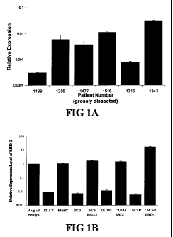
Figure 1 shows quantitative RT-PCR (QRT-PCR) analysis of beta-defensin-1
(DEFB 1) expression. In order to verify induction of DEFB 1 expression, QRT-
PCR was
performed. Figure 1A shows DEFBl relative expression levels compared in
clinical
samples from 6 patients that underwent radical prostatectomies. Figure 1B
shows DEFB1
relative expression levels compared in benign and malignant prostatic clinical
samples,
hPrEC cells and in prostate cancer cell lines before and after DEFB 1
induction. Figure 1 C
shows DEFB1 relative expression levels analyzed in benign tissue, malignant
tissue and
prostate intraepithelial neoplasia (PIN) in a single tissue section. Figure 1D
shows DEFB 1
expression in benign tissue, malignant tissue and PIN in one patient compared
to the
average DEFB 1 expression level found in benign tissue.
Figure 2 shows microscopic analysis of DEFB 1 induced changes in membrane
integrity and cell morphology. Cell morphology of DU145, PC3 and LNCaP was
analyzed by phase contrast microscopy after 48 hours of DEFB 1 induction.
Membrane
ruffling is indicated by black arrows and apoptotic bodies are indicated white
arrows.
Figure 3 shows analysis of DEFB 1 Cytotoxicity in Prostate Cancer Cells. The
prostate cell lines DU145, PC3 and LNCaP were treated with PonA to induce
DEFB1
expression for 1-3 days after which MTT assay was performed to determine cell
viability.
Results represent mean s.d., n=9.
Figure 4 shows induction of cell death in DU145 and PC3 cells by DEFB1.
DEFBl expression was induced in prostate cancer cell lines DU145 (A) and PC3
(B) and
then subjected to annexin V/FITC/propidium iodide staining and flow cytometric
analysis.
Cells positive for propidium iodide and annexin V were considered apoptotic.
Times of
-2-
CA 02667364 2009-04-22
WO 2008/089236 PCT/US2008/051168
induction are shown under each panel. Numbers next to the boxes for each time
point
represent the percentages of propidium iodide (PI)- annexin V+ cells (lower
right
quadrant), and W annexin V+ cells (upper right quadrant). The data are from a
single
experiment that is representative of three separate experiments.
Figure 5 shows pan-caspase analysis following DEFB1 induction. DU145 and
PC3 cells were stained with FAM-VAD-FMK-labeled fluoromethyl ketone to detect
caspase activity. Cells were visible under DIC for each condition. Confocal
microscopic
analysis revealed no caspase staining in control DU145 (B), PC3 cells (F) and
LNCaP (J).
Cells treated with PonA for 24 hours to induce DEFB1 revealed caspase activity
in DU145
(D) and PC3 (H). No caspase activity was detected in LNCaP (L).
Figure 6 shows silencing of paired box homeotic gene 2 (PAX2) protein
expression following PAX2 siRNA Treatment. Figure 6A shows Western blot
analysis of
PC3 and DU145 cells transfected with PAX2 siRNA duplex at day zero (lane 1),
day two
(lane 2), and day four (lane 3). Figure 6B shows Western blot analysis of PC3
and DU145
cells transfected with PAX2 siRNA duplex at day zero (lane 1), day two (lane
2), day four
(lane 3) and day 6 (lane 4). PAX2 protein was undetectable as early as after
four days of
treatment (lane 3) in DU145 cells and after six days of treatment in PC3.
Blots were
stripped and re-probed for 0-actin as an internal control.
Figure 7 shows analysis of prostate cancer cells growth after treatment with
PAX2
siRNA. Phase contrast microscopic analysis of DU145, PC3 and LNCaP at 6 days
in the
presence of normal growth media. Treatment with negative control siRNA had no
effect
on the cells. However, there was a significant reduction in cell number in all
three lines
following treatment with PAX2 siRNA.
Figure 8 shows analysis of cell death following siRNA silencing of PAX2.
Prostate cancer cell lines PC3, DU145, and LNCaP were treated with 0.5 g of a
pool of
four PAX2 siRNA's or four non-specific control siRNA's for 2, 4 or 6 days
after which
MTT assay was done to determine cell viability. Results represent mean s.d.,
n=9.
Figure 9 shows analysis of caspase activity. DU145, PC3 and LNCaP cells were
stained with carboxyfluorescein-labeled fluoromethyl ketone to detected
caspase activity
following treatment with PAX2 siRNA. Confocal microscopic analysis of
untreated and
treated cells show cells were visible with DIC. Analysis under fluorescence
revealed no
caspase staining in control DU145 (B), PC3 cells (F) and LNCaP cells (J).
However, cell
-3-
CA 02667364 2009-04-22
WO 2008/089236 PCT/US2008/051168
treated with PAX2 siRNA induced caspase activity in DU145 (D), PC3 (H) and
LNCaP
(L).
Figure 10 shows analysis of apoptotic factors following PAX2 siRNA treatment.
Changes in expression of pro-apoptotic factors were compared in untreated
control cells
and in cells treated for six days with PAX2 siRNA. Figure 10A shows Bcl-2-
associated X
protein(BAX) expression levels increased in DU145, PC3 and LNCaP. Figure lOB
shows
BH3 interacting domain death agonist (BID) expression increased in DU145 and
LNCaP,
but change in PC3. Figure 10C shows Bcl-2-associated death promoter (BAD)
expression
levels increased in all three cell lines.
Figure 11 shows model of PAX2 binding to DNA recognition sequence. The
PAX2 transcriptional repressor binds to a CCTTG (SEQ ID NO:1) recognition site
immediately adjacent to the DEFB 1 TATA box preventing transcription and DEFB
1
protein expression. Inhibition of PAX2 protein expression allows normal DEFB 1
expression.
Figure 12 illustrates the DEFB 1 reporter construct. The DEFB 1 promoter
consisting of the first 160 bases upstream of the mRNA start site was PCR
amplified from
DU145 cell and ligated into the pGL3 luciferase reporter plasmid.
Figure 13 shows inhibition of PAX2 results in DEFB 1 Expression. DU145, PC3,
LNCaP and HPrEC were treated for 48 hours with PAX2 siRNA. QRT-PCR analysis
before treatment showed no DEFBl expression in DU145, PC3 and LNCaP. However,
DEFB 1 expression was restored following treatment in all lines. There was no
change in
DEFB 1 expression following siRNA treatment of PAX2-null HprEC.
Figure 14 shows inhibition of PAX2 results in increased DEFB1 promoter
activity.
PC3 promoter/pGL3 and DU145 promoter/pGL3 construct were generated and were
transfected into PC3 and DU145 cells, respectively. Promoter activity was
compared
before and after PAX2 inhibition by siRNA treatment. DEFB1 promoter activity
increased 2.65-fold in DU145 and 3.78 fold in PC3 following treatment.
Figure 15 shows ChIP analysis of PAX2 binding to DEFB 1 promoter. ChIP
analysis was performed on DU145 and PC3 cells. Following immunoprecipitation
with an
anti-PAX2 antibody, PCR was performed to detect the DEFB1 promoter region
containing
the GTTCC PAX2 recognition site. This demonstrates that the PAX2
transcriptional
repressor is bound to the DEFB 1 promoter in prostate cancer cell lines.
-4-
CA 02667364 2009-04-22
WO 2008/089236 PCT/US2008/051168
Figure 16 shows predicted structure of the PrdPD and PrdHD with DNA. The
coordinates of the structures of the PrdPD bound to DNA (Xu et al., 1995) and
the PrdHD
bound to DNA (Wilson et al., 1995) were used to construct a model of the two
domains as
they bound to a PHO site. The individual binding sites are abutted next to
each other with
a specific orientation as indicated. The RED domain is oriented based on the
PrdPD
crystal structure.
Figure 17 shows comparison of consensus sequences of different paired domains.
At the top of the Figure is drawn a schematic representation of proteinzEDNA
contacts
described in the crystallographic analysis of the Prd-paired-domain DNA
complex.
Empty boxes indicate a-helices, shaded boxes indicates b-sheets and a thick
line indicate a
b-turn. Contacting amino acids are shown by single-letter code. Only direct
amino
acid base contacts are shown. Empty circles indicate major groove contacts
while red
arrows indicate minor groove contacts. This scheme is aligned to all known
consensus
sequences for paired-domain proteins (top strands only are shown). Vertical
lines between
consensus sequences indicate conserved base-pairs. Numbering of the positions
is shown
at the bottom of the Figure.
Figure 18 shows targeting PAX2 as a chemopreventive strategy. Aberrant PAX2
expression is an early event in the initiation and progression of cancer.
Inhibition of
PAX2 during dysplasia or other precancerous stage can be used for cancer
prevention.
Figure 19 shows effect of angiotensin II (Ang II) on PAX2 expression in DU145
Cells. In order to determine the effect of AngII on PAX2 expression, DEFB1
protein
levels was monitored following treatment. Here PAX2 expression levels
increased as
early as 4 hours and persisted until 48 hours.
Figure 20A shows effect of Losartan (Los) on PAX2 expression in DU145.
DU145 cells were treated with the angiotensin II type 1 receptor (ATR1)
blocker Losartan.
QRT-PCR revealed that PAX2 message levels were decreased by at least half
following
treatment. Figure 20B shows effect of an angiotensin II type 2 receptor (ATR2)
blocker on
PAX2 Expression in DU145. To determine the effect of the ATR2 receptor on PAX2
expression, DU145 cells were treated with the ATR2 receptor blocker PD123319.
Here,
PAX2 expression was increased 7 to 8-fold.
Figure 21 shows Los blocks AngII effect on PAX2 expression in DU145.
Treatment of DU145 cells with 5 M of AngII for 72 hours resulted in a 2-fold
increase in
PAX2 expression. In addition, treatment with 10 M for 72 hours resulted in
more than a
-5-
CA 02667364 2009-04-22
WO 2008/089236 PCT/US2008/051168
3-fold increase in expression. Treatment of cells with 5gM of Losartan
suppressed
proliferation by 50%. In addition, treatment with Losartan for 30min prior to
treatment
with Angll blocked the effect of AngII on proliferation.
Figure 22 shows Angll increases DU145 cell proliferation. Treatment of DU145
cells with 5 M of AngII for 72 hours resulted in a 2-fold increase in
proliferation. In
addition, treatment with 10 M for 72 hours resulted in more than a 3-fold
increase in
proliferation.
Figure 23 shows effect of Los and MAP Kinase inhibitors on PAX2 expression in
DU145 cells. Figure 23A shows treatment of DU145 cells with Losartan
suppresses
phosphor-ERK 1/2 and PAX2 expression; Figure 23B shows MEK kinase inhibitors
and
AICAR suppresses PAX2 protein expression; Figure 23C shows MEK kinase
inhibitors
and Losartan suppresses phospho-STAT3 protein expression.
Figure 24 shows effect of Los and MEK kinase inhibitors on PAX2 activation in
DU145 cells. Figure 24A shows treatment of DU145 cells with inhibitors of AT1R
signaling resulted in a decrease in phosphor-PAX2 protein levels which is the
active form
of PAX2. In addition, treatment with the AMP kinase inducer AICAR resulted in
suppressed PAX2 expression. Figure 24B shows inhibition of AT1R signaling with
Los
decreased phopho-JNK levels. However, Angll increased phosphor-JNK protein
levels.
Figure 25 shows Angll increases PAX2 and decreases DEFB 1 expression in
hPrEC cells. To determine the effect of Angll on PAX2 levels in hPrEC, cells
were
treated for 72 and 96 hours and PAX2 and DEFB 1 expression was examined by QRT-
PCR. Here, Angll treatment resulted in dramatic increases in PAX2 to levels
similar to
PC3 prostate cancer cells. Conversely, DEFB 1 expression was reduced
significantly after
AngII treatment.
Figure 26 shows schematic of AngIl signaling and PAX2 prostate cancer. PAX2
expression in prostate cancer cells is regulated by the AT1R signaling
pathway.
Specifically, the MEK kinase signaling cascade leads to increased PAX2
expression. In
addition, the AT1R and AngII upregulates PAX2 activation via JNK.
Figure 27 shows schematic of blocking PAX2 expression as a therapy for
prostate
cancer. Figure 27A shows PAX2 expression is regulated by the AT1R signaling
pathway.
Inhibition of PAX2 expression results in the re-expression of DEFB 1 and
cancer cell
death. Figure 27B shows compounds which block the AT1R, downstream kinases or
directly suppresses PAX2 offer a novel approach to treating prostate cancer.
-6-
_
CA 02667364 2009-04-22
WO 2008/089236 PCT/US2008/051168
Figure 28 shows comparison of DEFB1 and PAX2 expression with Gleason Score.
DEFB 1 relative expression levels were compared in benign clinical samples
from 6
patients that underwent radical prostatectomies. Here Gleason score inversely
correlated
with DEFB 1 expression levels in adjacent benign prostate tissue. Patients
with relative
DEFB1 expression levels higher than 0.005 had Gleason sores of 6. However,
those with
expression levels less than 0.005 had Gleason scores of 7.
Figure 29 shows PAX2-DEFB 1 ratio as a predictive factor for prostate cancer
development. QRT-PCR was performed on laser capture microdissection (LCM)
prostate tissue sections to determine relative DEFB 1 and PAX2 expression
levels. DEFB 1
expression levels decreased from Normal to PIN to cancer. However, PAX2
expression
increased from normal to PIN to cancer. In addition, patient #1457 with
Gleason score 6
cancer had more DEFBl in normal tissue and PIN compared to patient #1569 with
Gleason score 7 cancer. Conversely, patient #1569 had higher PAX2 levels in
cancerous
regions compared to patient #1457.
Figure 30 shows the Donald Predictive Factor (DPF) is based on the relative
PAX2-DEFB 1 expression ratio. An increase in the DPF of prostate tissue
increases the
chance of developing prostate cancer. Tissue with a PAX2-DEFB1 ratio between 0
and 39
based on the DPF was normal (benign). Tissue with a PAX2-DEFB1 ratio between
40
and 99 represented PIN (pre-cancerous) based on the DPF scale. Finally, tissue
with a
PAX2-DEFB1 ratio between 100 and 500 was malignant (low to high grade cancer).
Figure 31 shows analysis of hBD-1 expression in human prostate tissue. hBD-1
relative expression levels were compared in normal clinical samples from
patients that
underwent radical prostatectomies. The dashed line serves as a point of
reference to
compare values obtained between gross and LCM-derived specimen, and
corresponding
Gleason scores are indicated above each bar. Figure 31A shows hBD-1 expression
levels
compared in tissues obtained by gross dissection. Figure 31B shows hBD-1
expression
levels compared in tissue obtained by Laser Capture Microdissection.
Figure 32 shows analysis of hBD-1 expression in prostate cell lines. Figure
32A
shows hBD-1 expression levels compared relative to hPrEC cells in prostate
cancer cell
lines before and after hBD-1 induction. An asterisk represents statistically
higher
expression levels compared to hPrEC. Double asterisks represent statistically
significant
levels of expression compared to the cell line before hBD-1 induction
(Student's t-test, p
<0.05). Figure 32B shows ectopic hBD-1 expression verified in the prostate
cancer cell
-7-
_.
CA 02667364 2009-04-22
WO 2008/089236 PCT/US2008/051168
line DU145 by immunocytochemistry. hPrEC cells were stained for hBD-1 as
apositive
control (a: DIC and b: fluorescence). DU145 cells were transfected with hBD-1
and
induced for 18 h (c: DIC and d: fluorescence). Sizebar = 20 M.
Figure 33 shows analysis of hBD-1 cytotoxicity in prostate cancer cells. The
prostate cell lines DU145, PC3, PC3/AR+ and LNCaP were treated with Pon A to
induce
hBD-1 expression for 1-3 days after which MTT assay was performed to determine
cell
viability. Each bar represents the mean S.E.M. of three independent
experiments
performed in triplicate.
Figure 34 shows QRT-PCR analysis of hBD-1 and cMYC expression in LCM
human prostate tissue sections of normal, PIN and tumor. Expression for each
gene is
presented as expression ratios compared to 0-actin. Figure 35A shows
comparison of
hBD-1 expression levels in normal, PIN and tumor sections. Figure 35B shows
comparison of cMYC expression level in normal, PIN and tumor sections.
Figure 35 shows QRT-PCR analysis of hBDl expression following PAX2
knockdown with siRNA. hBD-1 expression levels are presented as expression
ratios
compared to fl-actin. An asterisk represents statistically higher expression
levels compared
to the cell line before PAX2 siRNA treatment (Student's t-test, p <0.05).
Figure 36 shows silencing of PAX2 protein expression following PAX2 siRNA
treatment. Figure 37A shows PAX2 expression examined by Western blot analysis
in
HPrEC prostate primary cells (lane 1) and in DU145 (lane 2), PC3 (lane 3) and
LNCaP
(lane 4) prostate cancer cells. Blots were stripped and re-probed for -actin
as an internal
control to ensure equal loading. Figure 37B shows Western blot analysis of
DU145, PC3
and LNCaP all confirmed knockdown of PAX2 expression following transfection
with
PAX2 siRNA duplex. Again, blots were stripped and re-probed for 0-actin as an
internal
control.
Figure 37 shows analysis of prostate cancer cells growth after treatment with
PAX2 siRNA. Phase contrast microscopic analysis of HPrEC (A), LNCaP (C), DU145
(E)
and PC3 (G) at 6 days in the presence of negative control non-specific siRNA.
There was
a significant reduction in cell number in DU145 (D), PC3 (F) and LNCaP (H)
following
treatment with PAX2 siRNA. However, there appeared to be no effect in HPrEC
(B). Bar
= 20 m.
Figure 38 shows analysis of cell death following siRNA silencing of PAX2.
Prostate cancer cell lines PC3, DU145 and LNCaP were treated with PAX2 siRNA
or non-
-8-
__
CA 02667364 2009-04-22
WO 2008/089236 PCT/US2008/051168
specific negative control siRNAs for 2, 4 or 6 days after which MTT assay was
performed.
Knockdown of PAX2 resulted in a decrease in relative cell viability in all
three lines.
Results represent mean zE SD, n=9.
Figure 39 shows analysis of caspase activity. DU145, PC3 and LNCaP cells were
stained with carboxyfluorescein-labeled fluoromethyl ketone to detected
caspase activity
following treatment with PAX2 siRNA. Analysis under fluorescence revealed no
caspase
staining in control DU145 (A), PC3 cells (C) and LNCaP cells (E). However,
cell treated
with PAX2 siRNA induced caspase activity in DU145 (B), PC3 (D) and LNCaP (F).
Bar =
20 m.
Figure 40 shows analysis of apoptotic factors following PAX2 siRNA treatment.
Changes in expression of pro-apoptotic factors were compared in untreated
control cells
and in cells treated for 6 days with PAX2 siRNA. Figure 41A shows BAD
expression
increased in DU145, PC3 and LNCaP following PAX2 knockdown. Figure 41B shows
BID expression levels increased in LNCaP and DU145, but not in PC3 cells.
Figure 41C
shows AKT expression decreased in LNCaP and DU145. However, there was no
change
~
in AKT expression in PC3 cells following PAX2 knockdown. Results represent
mean
SD, n =9. Asterisks represents statistical differences (p < 0.05).
DETAILED DESCRIPTION
The disclosed method and compositions may be understood more readily by
reference to the following detailed description of particular embodiments and
the Example
included therein and to the Figures and their previous and following
description.
Disclosed are materials, compositions, and components that can be used for,
can be
used in conjunction with, can be used in preparation for, or are products of
the disclosed
method and compositions. These and other materials are disclosed herein, and
it is
understood that when combinations, subsets, interactions, groups, etc. of
these materials
are disclosed that while specific reference of each various individual and
collective
combinations and permutation of these compounds may not be explicitly
disclosed, each is
specifically contemplated and described herein. For example, if a peptide is
disclosed and
discussed and a number of modifications that can be made to a number of
molecules
including the peptide are discussed, each and every combination and
permutation of
peptide and the modifications that are possible are specifically contemplated
unless
specifically indicated to the contrary. Thus, if a class of molecules A, B,
and C are
disclosed as well as a class of molecules D, E, and F and an example of a
combination
-9-
CA 02667364 2009-04-22
WO 2008/089236 PCT/US2008/051168
molecule, A-D is disclosed, then even if each is not individually recited,
each is
individually and collectively contemplated. Thus, is this example, each of the
combinations A-E, A-F, B-D, B-E, B-F, C-D, C-E, and C-F are specifically
contemplated
and should be considered disclosed from disclosure of A, B, and C; D, E, and
F; and the
example combination A-D. Likewise, any subset or combination of these is also
specifically contemplated and disclosed. Thus, for example, the sub-group of A-
E, B-F,
and C-E are specifically contemplated and should be considered disclosed from
disclosure
of A, B, and C; D, E, and F; and the example combination A-D. This concept
applies to
all aspects of this application including, but not limited to, steps in
methods of making and
using the disclosed compositions. Thus, if there are a variety of additional
steps that can
be performed it is understood that each of these additional steps can be
performed with
any specific embodiment or combination of embodiments of the disclosed
methods, and
that each such combination is specifically contemplated and should be
considered
disclosed.
Those skilled in the art will recognize, or be able to ascertain using no more
than
routine experimentation, many equivalents to the specific embodiments of the
method and
compositions described herein. Such equivalents are intended to be encompassed
by the
following claims.
It is understood that the disclosed method and compositions are not limited to
the
particular methodology, protocols, and reagents described as these may vary.
It is also to
be understood that the terminology used herein is for the purpose of
describing particular
embodiments only, and is not intended to limit the scope of the present
invention which
will be limited only by the appended claims.
A. Diagnosing, Treating and Preventing Prostate Cancer
Disclosed herein are compositions and methods of diagnosing, preventing, and
treating prostate cancer and prostate intraepithelial neoplasia (PIN).
1. Prostate Cancer
Carcinoma of the prostate has become a significant disease in many countries
and
it is the most commonly diagnosed malignancy in men in the western world, its
occurrence
increasing significantly with age. This increase and the recent deaths of many
public
figures from prostate cancer have served to highlight the need to do something
about this
cancer. It has been suggested that the wider availability of screening may
limit mortality
from prostate cancer.
-10-
CA 02667364 2009-04-22
WO 2008/089236 PCT/US2008/051168
Prostate cancer screening currently consists of a rectal examination and
measurement of prostate specific antigen (PSA) levels. These methods lack
specificity as
digital rectal examination has considerable inter-examiner variability and PSA
levels may
be elevated in benign prostatic hyperplasia (BPH), prostatic inflammation and
other
conditions. The comparative failure of PSA as a diagnostic test was shown in
366 men
who developed prostate cancer while being included in the Physicians Health
Study, a
prospective study of over 22,000 men. PSA levels were measured in serum, which
was
stored at the start of the study, and elevated levels were found in only 47%
of men
developing prostate cancer within the subsequent four years (Gann et al,
1995).
Prostate cancers can be scored using the Gleason system, as well known to
those
skilled in the art (Gleason, et al. 1966). This uses tissue architecture
rather than cytological
features. A grade of 1 to 5 (well to poorly differentiated) is used, and the
combined score
of the most frequent and more severe areas of the lesion are combined. Gleason
scores
provide prognostic information that may be valuable in addition to the
assessment of the
stage of the tumor (staging). Gleason scores of 2 to 4 and 8 to 10 have good
predictive
value, but about three quarters of tumors have intermediate values.
Two principal systems are used for staging prostate cancer: TNM and the Jewett
system (Benson & Olsson, et al. 1989). Staging takes in to account any
metastatic spread
of the tumor and is difficult, because it is difficult to assess either local
lymph node
involvement or local invasion. Tumor size is also difficult to measure as
tumor tissue
cannot be distinguished macroscopically from normal prostate tissue, and
because the
prostate gland lacks a distinct capsule and is surrounded by a layer of
fibrous fatty tissue.
Four categories describe the prostate tumor's (T) stage, ranging from T1 to
T4. For
T1, the cancer is microscopic, unilateral and non palpable. The doctor can't
feel the tumor
or see it with imaging such as transrectal ultrasound. Treatment for BPH may
have
disclosed the disease, or it was confirmed through the use of a needle biopsy
done because
of an elevated PSA. For T2, the doctor can feel the cancer with a DRE. It
appears the
disease is confined to the prostate gland on one or both sides of the gland.
For T3, the
cancer has advanced to tissue immediately outside the gland. For T4, the
cancer has spread
to other parts of the body.
Present screening methods are therefore unsatisfactory; there is no reliable
method
for diagnosing the cancer, or predicting or preventing its possible metastatic
spread, which
is the main cause of death for most patients.
-11-
____
CA 02667364 2009-04-22
WO 2008/089236 PCT/US2008/051168
2. PAX2
Pax genes are a family of nine developmental control genes coding for nuclear
transcription factors. They play an important role in embryogenesis and are
expressed in a
very ordered temporal and spatial pattern. They all contain a "paired box"
region of 384
base pairs encoding a DNA binding domain which is highly conserved throughout
evolution (Stuart, ET, et al. 1994). The influence of Pax genes on
developmental processes
has been demonstrated by the numerous natural mouse and human syndromes that
can be
attributed directly to even a heterozygous insufficiency in a Pax gene. A PAX2
sequence
is given in Dressler, et al. 1990. Examples of cancers in which PAX2
expression has been
detected are listed in Table 1.
Table 1: PAX2-expressing cancers
PAX2 Expressing Estimated New Estimated Estimated New Estimated
Cancers Cases in US Deaths in US Cases Global Deaths Global
Prostate 234,460 27,350 679,023 221,002
Breast 214,600 41,430 1,151,298 410,712
Ovarian 20,180 15,310 204,500 124,860
Renal 38,890 12,840 208,479 101,895
Brain 12,820 18,820 189,485 141,650
Cervical 9,710 3,700 493,243 273,505
Bladder 61,420 13,060 356,556 145,009
Leukemia 35,020 22,280 300,522 222,506
Kaposi Sarcoma Data Not Data Not Data Not Data Not
Available Available Available Available
TOTAL 627,100 154,790 3,583,106 1,641,139
(approx.)
3. DEFBl
Beta-defensins are cationic peptides with broad-spectrum antimicrobial
activity
that are products of epithelia and leukocytes (Ganz and Weiss, 1997). These
two exon,
single gene products are expressed at epithelial surfaces and secreted at
sites including the
skin (Harder et al., 1997), cornea (McNamara et al., 1999), tongue (Mathews et
al., 1999,
Jia et al., 2000), gingiva (Mathews et al., 1999; Krisanaprakornkit et al.,
1998), salivary
glands (Mathews et al., 1999), esophagus (Jia et al., 2000), intestine (O'Neil
et al., 1999),
kidney (Valore et al., 1998; Zucht et al., 1998), urogenital tract (Valore et
al., 1998), and
the respiratory epithelium (Bals et al., 1998; Goldman et al., 1997; McCray
and Bentley.
1997). To date, five beta-defensin genes of epithelial origin have been
identified and
characterized in humans: DEFBl (Bensch et al., 1995), DEFB 2 (Harder et al.,
1997),
DEFB3 (Harder et al., 2001; Jia et al., 2001), DEFB4, and HE2/EP2.
-12-
CA 02667364 2009-04-22
WO 2008/089236 PCT/US2008/051168
The primary structure of each beta-defensin gene product is characterized by
small
size, a six cysteine motif, high cationic charge and exquisite diversity
beyond these
features. The most characteristic feature of defensin proteins is their six-
cysteine motif that
forms a network of three disulfide bonds. The three disulfide bonds in the
beta-defensin
proteins are between C1-C5, C2-C4 and C3-C6. The most common spacing between
adjacent cysteine residues is 6, 4, 9, 6, 0. The spacing between the cysteines
in the beta-
defensin proteins can vary by one or two amino acids except for C5 and C6,
located
nearest the carboxy terminus. In all known vertebrate beta-defensin genes,
these two
cysteine residues are adjacent to each other.
A second feature of the beta-defensin proteins is their small size. Each beta-
defensin gene encodes a preproprotein that ranges in size from 59 to 80 amino
acids with
an average size of 65 amino acids. This gene product is then cleaved by an
unknown
mechanism to create the mature peptide that ranges in size from 36 to 47 amino
acids with
an average size of 45 amino acids. The exceptions to these ranges are the
EP2/HE2 gene
products that contain the beta-defensin motif and are expressed in the
epididymis.
A third feature of beta-defensin proteins is the high concentration of
cationic
residues. The number of positively charged residues (arginine, lysine,
histidine) in the
mature peptide ranges from 6 to 14 with an average of 9.
The final feature of the beta-defensin gene products is their diverse primary
structure but apparent conservation of tertiary structure. Beyond the six
cysteines, no
single amino acid at a given position is conserved in all known members of
this protein
family. However, there are positions that are conserved that appear to be
important for
secondary and tertiary structures and function.
Despite the great diversity of the primary amino acid sequence of the beta-
defensin
proteins, the limited data suggests that the tertiary structure of this
protein family is
conserved. The structural core is a triple-stranded, antiparallel beta-sheet,
as exemplified
for the proteins encoded by BNBD-12 and DEFB2. The three beta-strands are
connected
by a beta-turn, and an alpha-hairpin loop, and the second beta-strand also
contains a beta-
bulge. When these structures are folded into their proper tertiary structure,
the apparently
random sequence of cationic and hydrophobic residues are concentrated into two
faces of
a globular protein. One face is hydrophilic and contains many of the
positively charged
side chains and the other is hydrophobic. In solution, the HBD-2 protein
encoded by the
DEFB2 gene exhibited an alpha-helical segment near the N-terminus not
previously
-13-
_.
CA 02667364 2009-04-22
WO 2008/089236 PCT/US2008/051168
ascribed to solution structures of alpha-defensins or to the beta-defensin
BNBD-12. The
amino acids whose side chains are directed toward the surface of the protein
are less
conserved between beta.defensin proteins while the amino acid residues in the
three beta-
strands of the core beta-sheet are more highly conserved.
Beta-defensin peptides are produced as pre-pro-peptides and then cleaved to
release a C-terminal active peptide fragment, however the pathways for the
intracellular
processing, storage and release of the human beta-defensin peptides in airway
epithelia are
unknown.
4. Diagnosing
A key advantage of the present teaching is that the herein disclosed methods
afford
a more rapid and simplified process to identify from a tissue or bodily fluid
a subject
having or at risk for prostate cancer.
Thus, the herein disclosed methods can comprise the detection, including
measurement, of PAX2 and/or DEFB1 in a tissue of the subject, such as a biopsy
sample
of the prostate. Prostate biopsy is a procedure in which small samples are
removed from a
man's prostate gland to be tested for the presence of cancer. It is typically
performed when
the scores from a PSA blood test rise to a level that is associated with the
possible
presence of prostate cancer.
The herein disclosed methods can comprise the detection, including
measurement,
of PAX2 and/or DEFB1 in a cell of the subject, such as a cell from the
prostate of the
subject.
In addition, the herein disclosed methods can comprise the detection,
including
measurement, of PAX2 and/or DEFB1 in bodily fluid of the subject, such as
blood, urine,
plasma, serum, tears, lymph, bile, cerebrospinal fluid, interstitial fluid,
aqueous or vitreous
humor, colostrum, sputum, amniotic fluid, saliva, anal and vaginal secretions,
perspiration,
semen, transudate, exudate, and synovial fluid. Blood plasma is the liquid
component of
blood, in which the blood cells are suspended. Plasma is the largest single
component of
blood, making up about 55% of total blood volume. Serum refers to blood plasma
in
which clotting factors (such as fibrin) have been removed. Blood plasma
contains many
vital proteins including fibrinogen, globulins and human serum albumin.
Sometimes blood
plasma can contain viral impurities which must be extracted through viral
processing.
Identification of blood protein markers that provide more accurate or earlier
diagnosis of cancer can have a positive impact on cancer treatment and
management. As
-14-
_
CA 02667364 2009-04-22
WO 2008/089236 PCT/US2008/051168
disclosed herein, aberrant PAX2 expression occurs early in the progression of
cancer and
can be an initiating event in tumorigenesis. Therefore, samples from patients
collected to
screen for the presence of PAX2 protein or antigens can be used for the early
detection of
cancer.
Furthermore, the incorporation of PAX2 screening can provide clinicians with a
prognosticator for initiated or pre-cancerous tissue. Candidates for this test
include
patients at high risk (based on age, race) for cancer. As a diagnostic, a
positive PAX2 test
can then be followed by additional screening with biomarker to determine
cancer site. In
addition, these patients can be candidates for PAX2 inhibitors for
chemoprevention for
their cancers. Alternatively, this test can be used on patients as a measure
of the
effectiveness of their cancer therapy or to monitor cancer recurrence.
As another example, patients who present with potential indicators of cancer
such
as the detection of nodules in the prostate during a digital rectal exam by
the clinician, or
those who experience a sudden rise in PSA often are in the "Watchful Waiting"
state. It is
often difficult to ascertain whether these patients have or will develop
cancer. The
detection of PAX2 in samples, such as plasma/serum, from these patients can be
used to
assist the decision to obtain a biopsy in men with suspected prostate cancer,
which can
lead to a reduction in the number of unnecessary prostatic biopsies and
earlier intervention
for their disease.
Also provided herein is a method of diagnosing prostate cancer in a subject,
comprising detecting in cells from the prostate of the subject levels of PAX2
and beta
defensin-1 (DEFB 1), wherein the ratio of PAX2 to DEFB 1 is at least about
100:1.
Also provided herein is a method of diagnosing prostate intraepithelial
neoplasia
(PIN) in a subject, comprising detecting in cells from the prostate of the
subject levels of
PAX2 and beta defensin-1 (DEFBl), wherein the ratio of PAX2 to DEFB1 is at
least
about 40:1 and less than about 100:1.
Also provided herein is a method of identifying a subject as having a normal
prostate, comprising detecting in cells from the prostate of the subject
levels of PAX2 and
beta defensin-1 (DEFB1), wherein the ratio of PAX2 to DEFB1 is less than about
40:1.
Also provided herein is a method of distinguishing among normal, pre-cancerous
and cancerous prostate conditions in a subject, comprising detecting in cells
from the
prostate of the subject levels of PAX2 and beta defensin-1 (DEFBl). In some
aspects,
wherein the ratio of PAX2 to DEFB 1 is less than about 40:1, a normal prostate
condition
-15-
CA 02667364 2009-04-22
WO 2008/089236 PCT/US2008/051168
is detected. In some aspects, wherein the ratio of PAX2 to DEFB 1 is at least
about 40:1
and less than about 100:1, a precancerous condition is detected. In some
aspects, wherein
the ratio of PAX2 to DEFB1 is at least about 100:1, a cancerous prostate is
detected.
5. Diagnosing and Treating
Also provided herein is a method of diagnosing and treating prostate cancer in
a
subject, comprising detecting in cells from the prostate of the subject levels
of PAX2 and
beta defensin-1 (DEFB1), wherein the ratio of PAX2 to DEFB1 is at least about
100:1,
further comprising treating said subject.
Also provided herein is a method of diagnosing and treating prostate
intraepithelial
neoplasia (PIN) in a subject, comprising detecting in cells from the prostate
of the subject
levels of PAX2 and beta defensin-1 (DEFBl), wherein the ratio of PAX2 to DEFBl
is at
least about 40:1 and less than about 100:1, further comprising treating said
subject.
As used in the disclosed methods, treatment for prostate cancer can involve
watchful waiting, surgery, radiation therapy, High Intensity Focused
Ultrasound (HIFU),
chemotherapy, cryosurgery, hormonal therapy, or some combination. Which option
is best
depends on the stage of the disease, the Gleason score, and the PSA level.
Other important
factors are the man's age, his general health, and his feelings about
potential treatments
and their possible side effects.
If the cancer has spread beyond the prostate, treatment options significantly
change, so most doctors who treat prostate cancer use a variety of nomograms
to predict
the probability of spread. Treatment by watchful waiting, HIFU, radiation
therapy,
cryosurgery, and surgery are generally offered to men whose cancer remains
within the
prostate. Hormonal therapy and chemotherapy are often reserved for disease
which has
spread beyond the prostate. However, there are exceptions: radiation therapy
may be used
for some advanced tumors, and hormonal therapy is used for some early stage
tumors.
Cryotherapy, hormonal therapy, and chemotherapy may also be offered if initial
treatment
fails and the cancer progresses.
Watchful waiting, also called "active surveillance," refers to observation and
regular monitoring without invasive treatment. Watchful waiting is often used
when an
early stage, slow-growing prostate cancer is found in an older man. Watchful
waiting may
also be suggested when the risks of surgery, radiation therapy, or hormonal
therapy
outweigh the possible benefits. Other treatments can be started if symptoms
develop, or if
there are signs that the cancer growth is accelerating (e.g., rapidly rising
PSA, increase in
-16-
CA 02667364 2009-04-22
WO 2008/089236 PCT/US2008/051168
Gleason score on repeat biopsy, etc.). Most men who choose watchful waiting
for early
stage tumors eventually have signs of tumor progression, and they may need to
begin
treatment within three years.
Surgical removal of the prostate, or prostatectomy, is a common treatment
either
for early stage prostate cancer, or for cancer which has failed to respond to
radiation
therapy. The most common type is radical retropubic prostatectomy, when the
surgeon
removes the prostate through an abdominal incision. Another type is radical
perineal
prostatectomy, when the surgeon removes the prostate through an incision in
the
perineum, the skin between the scrotum and anus. Radical prostatectomy can
also be
performed laparoscopically, through a series of small (lcm) incisions in the
abdomen,
with or without the assistance of a surgical robot.
Radical prostatectomy is highly effective for tumors which have not spread
beyond
the prostate; cure rates depend on risk factors such as PSA level and Gleason
grade.
However, it may cause nerve damage that significantly alters the quality of
life of the
prostate cancer survivor. Medications such as sildenafil (Viagra), tadalafil
(Cialis), or
vardenafil (Levitra) may be used to restore some degree of potency. For most
men with
organ-confined disease, a more limited "nerve-sparing" technique may help
avoid urinary
incontinence and impotence.
Radical prostatectomy has traditionally been used alone when the cancer is
small.
In the event of positive margins or locally advanced disease found on
pathology, adjuvant
radiation therapy may offer improved survival. Surgery may also be offered
when a cancer
is not responding to radiation therapy. However, because radiation therapy
causes tissue
changes, prostatectomy after radiation has a higher risk of complications.
Transurethral resection of the prostate, commonly called a"TURP," is a
surgical
procedure performed when the tube from the bladder to the penis (urethra) is
blocked by
prostate enlargement. TURP is generally for benign disease and is not meant as
definitive
treatment for prostate cancer. During a TURP, a small tube (cystoscope) is
placed into the
penis and the blocking prostate is cut away.
In metastatic disease, where cancer has spread beyond the prostate, removal of
the
testicles (called orchiectomy) may be done to decrease testosterone levels and
control
cancer growth.
Brachytherapy for prostate cancer is administered using "seeds," small
radioactive
rods implanted directly into the tumor. Radiation therapy, also known as
radiotherapy,
-17-
CA 02667364 2009-04-22
WO 2008/089236 PCT/US2008/051168
uses Gamma-rays to kill prostate cancer cells. Two different kinds of
radiation therapy are
used in prostate cancer treatment: external beam radiation therapy and
brachytherapy.
External beam radiation therapy uses a linear accelerator to produce high-
energy
Gamma-rays which are directed in a beam towards the prostate. A technique
called
Intensity Modulated Radiation Therapy (IMRT) may be used to adjust the
radiation beam
to conform with the shape of the tumor, allowing higher doses to be given to
the prostate
and seminal vesicles with less damage to the bladder and rectum. External beam
radiation
therapy is generally given over several weeks, with daily visits to a
radiation therapy
center.
External beam radiation therapy for prostate cancer is delivered by a linear
accelerator, such as this one.Brachytherapy involves the placement of about
100 small
"seeds" containing radioactive material (such as iodine- 125 or palladium-
103) with a
needle through the skin of the perineum directly into the tumor. These seeds
emit lower-
energy X-rays which are only able to travel a short distance. Brachytherapy
seeds will stay
in the prostate permanently, but men with implanted seeds are not at risk of
exposing
others to radiation.
Radiation therapy is commonly used in prostate cancer treatment. It may be
used
instead of surgery for early cancers, and it may also be used in advanced
stages of prostate
cancer to treat painful bone metastases. Radiation treatments also can be
combined with
hormonal therapy for intermediate risk disease, when radiation therapy alone
is less likely
to cure the cancer. External beam radiation can be combined with brachytherapy
for
intermediate to high risk situations. Also considered is a "triple modality"
combination of
external beam radiation therapy, brachytherapy, and hormonal therapy.
Radiation therapy is often offered to men whose medical problems make surgery
more risky. Radiation therapy appears to cure small tumors that are confined
to the
prostate just about as well as surgery. However, as of 2006 some issues remain
unresolved, such as whether radiation should be given to the rest of the
pelvis, how much
the absorbed dose should be, and whether hormonal therapy should be given at
the same
time.
Cryosurgery is another method of treating prostate cancer. It is less invasive
than
radical prostatectomy, and general anesthesia is less commonly used. Under
ultrasound
guidance, metal rods are inserted through the skin of the perineum into the
prostate. Liquid
nitrogen is used to cool the rods, freezing the surrounding tissue at -196 C
(-320 F). As
-18-
___
CA 02667364 2009-04-22
WO 2008/089236 PCT/US2008/051168
the water within the prostate cells freeze, the cells die. The urethra is
protected from
freezing by a catheter filled with warm liquid. Cryosurgery generally causes
fewer
problems with urinary control than other treatments, but impotence occurs up
to ninety
percent of the time.
Hormonal therapy uses medications or surgery to block prostate cancer cells
from
getting dihydrotestosterone (DHT), a hormone produced in the prostate and
required for
the growth and spread of most prostate cancer cells. Blocking DHT often causes
prostate
cancer to stop growing and even shrink. However, hormonal therapy rarely cures
prostate
cancer because cancers which initially respond to hormonal therapy typically
become
resistant after one to two years. Hormonal therapy is therefore usually used
when cancer
has spread from the prostate. It can also be given to men undergoing radiation
therapy or
surgery to help prevent return of their cancer.
Hormonal therapy for prostate cancer targets the pathways the body uses to
produce DHT. A feedback loop involving the testicles, the hypothalamus, and
the
pituitary, adrenal, and prostate glands controls the blood levels of DHT.
First, low blood
levels of DHT stimulate the hypothalamus to produce gonadotropin releasing
hormone
(GnRH). GnRH then stimulates the pituitary gland to produce luteinizing
hormone (LH),
and LH stimulates the testicles to produce testosterone. Finally, testosterone
from the
testicles and dehydroepiandrosterone from the adrenal glands stimulate the
prostate to
produce more DHT. Hormonal therapy can decrease levels of DHT by interrupting
this
pathway at any point.
Orchiectomy is surgery to remove the testicles. Because the testicles make
most of
the body's testosterone, after orchiectomy testosterone levels drop. Now the
prostate not
only lacks the testosterone stimulus to produce DHT, but also it does not have
enough
testosterone to transform into DHT.
Antiandrogens are medications such as flutamide, bicalutamide, nilutamide, and
cyproterone acetate which directly block the actions of testosterone and DHT
within
prostate cancer cells.
Medications which block the production of adrenal androgens such as DHEA
include ketoconazole and aminoglutethimide. Because the adrenal glands only
make about
5% of the body's androgens, these medications are generally used only in
combination
with other methods that can block the 95% of androgens made by the testicles.
These
-19-
CA 02667364 2009-04-22
WO 2008/089236 PCT/US2008/051168
combined methods are called total androgen blockade (TAB). TAB can also be
achieved
using antiandrogens.
GnRH action can be interrupted in one of two ways. GnRH antagonists suppress
the production of GnRH directly, while GnRH agonists suppress GnRH through the
process of downregulation after an initial stimulation effect. Abarelix is an
example of a
GnRH antagonist, while the GnRH agonists include leuprolide, goserelin,
triptorelin, and
buserelin. Initially, these medications increase the production of LH.
However, because
the constant supply of the medication does not match the body's natural
production
rhythm, production of both LH and GnRH decreases after a few weeks.
6. Treating/ Preventing
Also provided herein is a method of preventing prostate cancer in a subject,
comprising administering to a subject diagnosed with prostate intraepithelial
neoplasia
(PIN) a composition comprising an inhibitor of PAX2 expression or activity.
"Activities"
of a protein include, for example, transcription, translation, intracellular
translocation,
secretion, phosphorylation by kinases, cleavage by proteases, homophilic and
heterophilic
binding to other proteins, ubiquitination. In some aspects, "PAX2 activity"
refers
specifically to the binding of PAX2 to the DEFB-1 promoter. Also provided
herein is a
method of preventing prostate cancer in a subject, comprising diagnosing a
subject with
prostate intraepithelial neoplasia (PIN) and administering to the subject a
composition
comprising an inhibitor of PAX2 expression or activity. The subject can be
diagnosed with
PIN by detecting in cells from the prostate of the subject levels of PAX2 and
beta
defensin-1 (DEFB1), wherein the ratio of PAX2 to DEFB1 is at least about 40:1
and less
than about 100:1.
In some aspects, PAX2 is upregulated at the atrophy stage prior to PIN. Thus,
also
provided is a method of preventing prostate cancer in a subject, comprising
detecting in
cells from the prostate of the subject levels of PAX2 and beta defensin-1
(DEFB1),
wherein the ratio of PAX2 to DEFB1 is at least about 40:1 and less than about
100:1, and
administering to the subject a composition comprising an inhibitor of PAX2
expression or
activity.
Also provided herein is a method of treating prostate intraepithelial
neoplasia
(PIN) in a subject, comprising diagnosing a subject with PIN and administering
to the
subject a composition comprising an inhibitor of PAX2 expression or activity.
The subject
can be diagnosed with PIN by detecting in cells from the prostate of the
subject levels of
-20-
__
CA 02667364 2009-04-22
WO 2008/089236 PCT/US2008/051168
PAX2 and beta defensin-1 (DEFBl), wherein the ratio of PAX2 to DEFB1 is at
least
about 40:1 and less than about 100:1.
Also provided is a method of treating or preventing prostate intraepithelial
neoplasia (PIN) in a subject, comprising detecting in cells from the prostate
of the subject
levels of PAX2 and beta defensin-1 (DEFB1), wherein the ratio of PAX2 to DEFB1
is at
least about 40:1 and less than about 100:1, and administering to the subject a
composition
comprising an inhibitor of PAX2 expression or activity.
Also provided herein is a method of treating prostate cancer in a subject,
comprising diagnosing a subject with prostate cancer and administering to the
subject a
composition comprising an inhibitor of PAX2 expression or activity. The
subject can be
diagnosed with prostate cancer by detecting in cells from the prostate of the
subject levels
of PAX2 and beta defensin-1 (DEFBl), wherein the ratio of PAX2 to DEFB1 is at
least
about 100:1.
The inhibitor of the disclosed methods can be a selective antagonist of
angiotensin
II. The inhibitor of the disclosed methods can be a selective antagonist of
angiotensin-
converting enzyme (ACE). For example, the inhibitor can be enalapril. The
inhibitor can
be can be a selective antagonist of angiotensin II type 1 receptor (AT1R). For
example, the
inhibitor can be valsartan, olmesartan, or telmisartan. The inhibitor can be a
selective
antagonist of MEK. The inhibitor can be a selective antagonist of ERK1,2. The
inhibitor
can be a selective antagonist of STAT3. The inhibitor can be a selective
antagonist of
PAX2. The inhibitor can block the binding of PAX2 to the beta defensin-1 (DEFB
1)
promoter. In some aspects, the disclosed inhibitor of PAX2 expression or
activity is not an
AT 1 R receptor antagonist.
By "selective antagonist" is meant something that directly binds and inhibits
the
activity of the target. "Activities" of a protein include, for example,
transcription,
translation, intracellular translocation, secretion, phosphorylation by
kinases, cleavage by
proteases, homophilic and heterophilic binding to other proteins,
ubiquitination. Thus, for
example, a selective antagonist of a kinase can bind the kinase and inhibit
the
phosphorylation of the target of the kinase. Thus, for example, a selective
antagonist of a
kinase can bind the kinase and prevent the binding of the kinase to its
substrate.
Also provided herein is a method of treating or preventing prostate cancer in
a
subject, comprising administering to said subject a composition comprising a
selective
antagonist of MEK and/or ERK1,2. This can also be a method of inhibiting
expression of
-21-
CA 02667364 2009-04-22
WO 2008/089236 PCT/US2008/051168
PAX2. The subject in this method can first be diagnosed with a pre-cancerous
condition
(e.g., PIN) or with cancer.
The selective antagonist of MEK and/or ERK1,2 can be U0126. U0126 is a
chemically synthesized organic compound that was initially recognized as a
cellular AP-1
antagonist, and found to be a very selective and highly potent inhibitor of
Mitogen-
Activated Protein Kinase (MAPK) cascade by inhibiting its immediate upstream
activators, Mitogen Activated Protein Kinase Kinase land 2 (also known as MEK1
and
MEK2, IC50: 70 and 60 nM respectively). U0126 inhibits both active and
inactive
MEK1,2, unlike PD098059 which only inhibits activation of inactive MEK.
Blockade of
MEK activation would prevent downstream phosphorylation of a number of factors
including p62TCF (Elk-1), an upstream inducer of c-Fos and c-Jun, components
of the
AP-1 complex. Inhibition of MEK/ERK pathway by U0126 also prevents all effects
of
oncogenic H-Ras and K-Ras, inhibits part of the effects triggered by growth
factors and
blocks the production of inflammatory cytokines and matrix metalloproteinases.
The selective antagonist of MEK and/or ERK1,2 can be PD98059. PD98059
(MEK1 Inhibitor) has been shown to act in vivo as a highly selective inhibitor
of MEKl
activation and the MAP kinase cascade. PD98059 binds to the inactive forms of
MEKl
and prevents activation by upstream activators such as c-Raf. PD98059 inhibits
activation
of MEKl and MEK2 with IC50 values of 4 M and 50 M, respectively.
Also provided herein is a method of treating or preventing prostate cancer in
a
subject, comprising administering to said subject a composition comprising a
selective
antagonist of STAT3. This is also a method of inhibiting expression of PAX2.
The subject
in this method can first be diagnosed with a pre-cancerous condition (e.g.,
PIN) or with
cancer.
As shown herein, PAX2 inhibits expression of DEFB 1, and DEFB1 is shown to
have tumor cell killing activity. Thus, provided is a method of treating
cancer in a subject
by inhibiting expression of PAX2. An example of a cancer treated by the
present method
is prostate cancer. The present methods are particularly effective for
treatment of late
stage prostate cancer.
In the cancer treatment methods disclosed, the method of inhibiting expression
of
PAX 2 can be by administration of a nucleic acid encoding a siRNA for PAX 2.
Dharmachon is a commercial source for such siRNAs.
For example, the siRNA for use in the methods can be comprise:
-22-
CA 02667364 2009-04-22
WO 2008/089236 PCT/US2008/051168
AUAGACUCGACUUGACUUCUU (SEQ ID NO:2),
AUCUUCAUCACGUUUCCUCUU (SEQ ID NO:4),
GUAUUCAGCAAUCUUGUCCUU (SEQ ID NO:6),
GAUUUGAUGUGCUCUGAUGUU (SEQ ID NO:8), or combinations thereof, including
fragments of at least 10 nucleic acids and conservative variants thereof.
Further examples of target sequences for molecules that inhibit PAX2 include:
#1 ACCCGACTATGTTCGCCTGG (SEQ ID NO:56),
#2 AAGCTCTGGATCGAGTCTTTG (SEQ ID NO:57),
and #4 ATGTGTCAGGCACACAGACG (SEQ ID NO:58). #4 was shown to inhibit
PAX2 (Davies et al., Hum. Mol. Gen Jan. 15, 13 (2); 235).
Another paper (Muratovska et al., Paired-Box genes are frequently expressed in
cancer and often required for cancer cell survival Oncogene (2003) 22, 7989-
7997)
discloses the following siRNAs: GUCGAGUCUAUCUGCAUCCTT (SEQ ID NO:59)
and GGAUGCAGAUAGACUCGACTT (SEQ ID NO:60).
To down-regulate Pax2 expression, Fonsato et al. transfected tumor-derived
endothelial cells with an anti-sense PAX2 vector. See Fonsato V. et al. Am J
Pathol.
2006;168(2):706-1, which is incorporated herein by referene for its
description of this
molecule. Similarly, Hueber et al. teach that PAX2 antisense cDNA and PAX2-
small
interfering RNA (100 nM) reduce endogenous PAX2 protein. See Hueber et al.
Kidney
Int. 2006, which is incorporated herein for its teaching of PAX2 antisense and
PAX2
siRNA.
Additional inhibitors of PAX2 expression or the binding of PAX2 to the DEFB 1
promoter are provided to increase DEFBl expression in the presently disclosed
methods.
For example, small molecules and antibodies can be designed based on the
present studies
to interfere with or inhibit the binding of PAX2 to the DEFB 1 promoter.
As shown herein, PAX2 inhibits expression of DEFB 1, and DEFB 1 is shown to
have tumor cell killing activity. Thus, a method of treating cancer in a
subject by
administering DEFB1 is also provided. An example of a cancer treated by the
present
method is prostate cancer.
Similarly, provided is a method of treating cancer in a subject by increasing
expression of DEFB1 in the subject. The present methods of administering or
increasing
the expression of DEFB 1 are particularly effective for treatment of late
stage prostate
cancer.
- 23 -
CA 02667364 2009-04-22
WO 2008/089236 PCT/US2008/051168
In one embodiment of the methods of the invention for treating cancer by
administering DEFB 1 or increasing DEFB 1 expression (e.g., by inhibiting
expression or
binding of PAX2), the subject is a subject diagnosed with prostate cancer. In
a further
embodiment of the methods of the invention for treating cancer by
administering DEFB 1
or increasing DEFB 1 expression (e.g., by inhibiting expression or binding of
PAX2), the
subject is a subject diagnosed with advanced (late stage) prostate cancer.
In the method wherein the expression of DEFB 1 is increased, it can be
increased
by blocking the binding of PAX2 to the DEFB 1 promoter. The blocking of
binding of
PAX2 to the DEFB 1 promoter can be by administration of an oligonucleotide
containing
the PAX2 DNA binding site of DEFB1. This oligonucleotide can be complementary
to
the sequence of PAX2 that binds to the DEFB 1 promoter. Alternatively, the
oligonucleotide can interact with the PAX2 in a way that inhibits binding to
DEFB 1. This
interaction can be based on three-dimensional structure rather than primary
nucleotide
sequence.
PAX proteins are a family of transcription factors conserved during evolution
and
able to bind specific DNA sequences through a domains called a "paired domain"
and a
"homeodomain". The paired domain (PD) is a consensus sequence shared by
certain PAX
proteins (e.g., PAX2 and PAX6). The PD directs DNA binding of amino acids
located in
the a3-helix forming a DNA-Protein complex. For PAX2, the amino acids in the
HD
recognize and interact specifically with a CCTTG (SEQ ID NO:1) DNA core
sequence.
Oligonucleotides up to and exceeding 64 bases in length, which include this
sequence or
its complement are expected to be inhibitors.
The DNA-binding specificity of the PAX-8 paired domain was investigated. Site
selection experiments indicate that PAX-8 binds to a consensus sequence
similar to those
bound by PAX-2 and PAX-5. When consensus sequences of various paired domains
are
observed in light of recent structural studies describing paired-domain-DNA
interaction
(Xu, et al. 1995), it appears that base-pairs contacted in the minor groove
are conserved,
while most of the base-pairs contacted in the major groove are not. Therefore
a network of
specific minor groove contacts is a common characteristic of paired-domain-DNA
interactions. The functional importance of such a network can be successfully
tested by
analyzing the effect of consensus-based mutations on the PAX2 binding site of
the DEFB 1
promoter.
The PAX2 DNA binding site of DEFB 1 can comprise SEQ ID NO:1 (CCTTG).
-24-
CA 02667364 2009-04-22
WO 2008/089236 PCT/US2008/051168
The oligonucleotide comprising to the PAX2 DNA binding site of DEFB 1 is
selected from the group consisting of
Xl-CCTTG (SEQ ID NO:l)-X2, wherein Xl is from 1 to 35 contiguous flanking
nucleotides of DEFB1 and X2 is from 1 to 35 nucleotides. The nucleotides can
be
contiguous nucleotides that normally flank the PAX2 DNA binding site of DEFB1.
Alternatively, they can be unrelated to DEFBl, and selected routinely to avoid
interference with the recognition sequence.
For example, the inhibitory oligonucleotides can be selected from the group
consisting of:
CTCCCTTCAGTTCCGTCGAC (SEQ ID NO:9)
CTCCCTTCACCTTGGTCGAC (SEQ ID NO:10)
ACTGTGGCACCTCCCTTCAGTTCCGTCGACGAGGTTGTGC (SEQ ID NO: 12)
ACTGTGGCACCTCCCTTCACCTTGGTCGACGAGGTTGTGC (SEQ ID NO: 13)
The disclosed compositions can be used to treat any disease where uncontrolled
cellular proliferation occurs such as cancers. A non-limiting list of
different types of
cancers is as follows: lymphomas (Hodgkins and non-Hodgkins), leukemias,
carcinomas,
carcinomas of solid tissues, squamous cell carcinomas, adenocarcinomas,
sarcomas,
gliomas, high grade gliomas, blastomas, neuroblastomas, plasmacytomas,
histiocytomas,
melanomas, adenomas, hypoxic tumors, myelomas, AIDS-related lymphomas or
sarcomas, metastatic cancers, or cancers in general.
A representative but non-limiting list of cancers that the disclosed
compositions
can be used to treat is the following: lymphoma, B cell lymphoma, T cell
lymphoma,
mycosis fungoides, Hodgkin's Disease, myeloid leukemia, bladder cancer, brain
cancer,
nervous system cancer, head and neck cancer, squamous cell carcinoma of head
and neck,
kidney cancer, lung cancers such as small cell lung cancer and non-small cell
lung cancer,
neuroblastoma/glioblastoma, ovarian cancer, pancreatic cancer, prostate
cancer, skin
cancer, liver cancer, melanoma, squamous cell carcinomas of the mouth, throat,
larynx,
and lung, colon cancer, cervical cancer, cervical carcinoma, breast cancer,
and epithelial
cancer, renal cancer, genitourinary cancer, pulmonary cancer, esophageal
carcinoma, head
and neck carcinoma, large bowel cancer, hematopoietic cancers; testicular
cancer; colon
and rectal cancers, prostatic cancer, or pancreatic cancer. Compounds
disclosed herein
may also be used for the treatment of precancer conditions such as cervical
and anal
dysplasias, other dysplasias, severe dysplasias, hyperplasias, atypical
hyperplasias, and
- 25 -
CA 02667364 2009-04-22
WO 2008/089236 PCT/US2008/051168
neoplasias. Further, a number of diseases stemming from chronic inflammation,
e.g.,
prostatitis and Benign Prostatic Hypertrophy (BPH), as well as various cancers
of the
prostate, can be impacted by the present methods and compounds.
DEFB1's gene locus (8p23.3) is a hotspot for deletions and has been linked to
patients with poorer prognosis. Thus, DEFB1 (and perhaps PAX2) can be used as
a
biomarker, e.g., in a screening for the early detection of prostate cancer.
Furthermore,
data presented here indicate that its loss may occur as early as PIN (or even
before), and
may be a major contributing factor to the onset of prostate cancer.
B. Compositions
1. Immunoassays
There are numerous methods for detecting analytes, such as proteins, such as
PAX2 and/or DEFB 1, known or newly discovered in the art, which can be used in
the
disclosed methods. For example, PAX2 and/or DEFB 1 can be detected using
standard
immunodetection methods. The steps of various useful immunodetection methods
have
been described in the scientific literature, such as, e.g., Maggio et al.,
Enzyme-
Immunoassay, (1987) and Nakamura, et al., Enzyme Immunoassays: Heterogeneous
and
Homogeneous Systems, Handbook of Experimental Immunology, Vol. 1:
Immunochemistry, 27.1-27.20 (1986), each of which is incorporated herein by
reference in
its entirety and specifically for its teaching regarding immunodetection
methods.
Immunoassays, in their most simple and direct sense, are binding assays
involving binding
between antibodies and antigen. Many types and formats of immunoassays are
known and
all are suitable for detecting the disclosed biomarkers. Examples of
immunoassays are
enzyme linked immunosorbent assays (ELISAs), radioimmunoassays (RIA),
radioimmune
precipitation assays (RIPA), immunobead capture assays, Western blotting, dot
blotting,
gel-shift assays, Flow cytometry, protein arrays, multiplexed bead arrays,
magnetic
capture, in vivo imaging, fluorescence resonance energy transfer (FRET), and
fluorescence
recovery/localization after photobleaching (FRAP/ FLAP).
In general, immunoassays involve contacting a sample suspected of containing a
molecule of interest (such as the disclosed biomarkers) with an antibody to
the molecule
of interest or contacting an antibody to a molecule of interest (such as
antibodies to the
disclosed biomarkers) with a molecule that can be bound by the antibody, as
the case may
be, under conditions effective to allow the formation of immunocomplexes.
Contacting a
sample with the antibody to the molecule of interest or with the molecule that
can be
-26-
CA 02667364 2009-04-22
WO 2008/089236 PCT/US2008/051168
bound by an antibody to the molecule of interest under conditions effective
and for a
period of time sufficient to allow the formation of immune complexes (primary
immune
complexes) is generally a matter of simply bringing into contact the molecule
or antibody
and the sample and incubating the mixture for a period of time long enough for
the
antibodies to form immune complexes with, i.e., to bind to, any molecules
(e.g., antigens)
present to which the antibodies can bind. In many forms of immunoassay, the
sample-
antibody composition, such as a tissue section, ELISA plate, dot blot or
Western blot, can
then be washed to remove any non-specifically bound antibody species, allowing
only
those antibodies specifically bound within the primary immune complexes to be
detected.
Immunoassays can include methods for detecting or quantifying the amount of a
molecule of interest (such as the disclosed biomarkers or their antibodies) in
a sample,
which methods generally involve the detection or quantitation of any immune
complexes
formed during the binding process. In general, the detection of immunocomplex
formation is well known in the art and can be achieved through the application
of
numerous approaches. These methods are generally based upon the detection of a
label or
marker, such as any radioactive, fluorescent, biological or enzymatic tags or
any other
known label. See, for example, U.S. Patents 3,817,837; 3,850,752; 3,939,350;
3,996,345;
4,277,437; 4,275,149 and 4,366,241, each of which is incorporated herein by
reference in
its entirety and specifically for teachings regarding immunodetection methods
and labels.
As used herein, a label can include a fluorescent dye, a member of a binding
pair,
such as biotin/streptavidin, a metal (e.g., gold), or an epitope tag that can
specifically
interact with a molecule that can be detected, such as by producing a colored
substrate or
fluorescence. Substances suitable for detectably labeling proteins include
fluorescent dyes
(also known herein as fluorochromes and fluorophores) and enzymes that react
with
colorometric substrates (e.g., horseradish peroxidase). The use of fluorescent
dyes is
generally preferred in the practice of the invention as they can be detected
at very low
amounts. Furthermore, in the case where multiple antigens are reacted with a
single array,
each antigen can be labeled with a distinct fluorescent compound for
simultaneous
detection. Labeled spots on the array are detected using a fluorimeter, the
presence of a
signal indicating an antigen bound to a specific antibody.
Fluorophores are compounds or molecules that luminesce. Typically fluorophores
absorb electromagnetic energy at one wavelength and emit electromagnetic
energy at a
second wavelength. Representative fluorophores include, but are not limited
to, 1,5
-27-
CA 02667364 2009-04-22
WO 2008/089236 PCT/US2008/051168
IAEDANS; 1,8-ANS; 4- Methylumbelliferone; 5-carboxy-2,7-dichlorofluorescein; 5-
Carboxyfluorescein (5-FAM); 5-Carboxynapthofluorescein; 5-
Carboxytetramethylrhodamine (5-TAMRA); 5-Hydroxy Tryptamine (5-HAT); 5-ROX
(carboxy-X-rhodamine); 6-Carboxyrhodamine 6G; 6-CR 6G; 6-JOE; 7-Amino-4-
methylcoumarin; 7-Aminoactinomycin D (7-AAD); 7-Hydroxy-4- I methylcoumarin; 9-
Amino-6-chloro-2-methoxyacridine (ACMA); ABQ; Acid Fuchsin; Acridine Orange;
Acridine Red; Acridine Yellow; Acriflavin; Acriflavin Feulgen SITSA; Aequorin
(Photoprotein); AFPs - AutoFluorescent Protein - (Quantum Biotechnologies) see
sgGFP,
sgBFP; Alexa Fluor 350TM; Alexa Fluor 430TM; Alexa Fluor 488TM; Alexa Fluor
532TM;
Alexa Fluor 546TM; Alexa Fluor 568TM; Alexa Fluor 594TM; Alexa Fluor 633TM;
Alexa
Fluor 647TM; Alexa Fluor 660TM; Alexa Fluor 680TM; Alizarin Complexon;
Alizarin Red;
Allophycocyanin (APC); AMC, AMCA-S; Aminomethylcoumarin (AMCA); AMCA-X;
Aminoactinomycin D; Aminocoumarin; Anilin Blue; Anthrocyl stearate; APC-Cy7;
APTRA-BTC; APTS; Astrazon Brilliant Red 4G; Astrazon Orange R; Astrazon Red
6B;
Astrazon Yellow 7 GLL; Atabrine; ATTO- TAGTM CBQCA; ATTO-TAGTM FQ;
Auramine; Aurophosphine G; Aurophosphine; BAO 9 (Bisaminophenyloxadiazole);
BCECF (high pH); BCECF (low pH); Berberine Sulphate; Beta Lactamase; BFP blue
shifted GFP (Y66H); Blue Fluorescent Protein; BFP/GFP FRET; Bimane;
Bisbenzemide;
Bisbenzimide (Hoechst); bis- BTC; Blancophor FFG; Blancophor SV; BOBOTM -1;
BOBOTM-3; Bodipy492/515; Bodipy493/503; Bodipy500/510; Bodipy; 505/515; Bodipy
530/550; Bodipy 542/563; Bodipy 558/568; Bodipy 564/570; Bodipy 576/589;
Bodipy
581/591; Bodipy 630/650-X; Bodipy 650/665-X; Bodipy 665/676; Bodipy Fl; Bodipy
FL
ATP; Bodipy Fl-Ceramide; Bodipy R6G SE; Bodipy TMR; Bodipy TMR-X conjugate;
Bodipy TMR-X, SE; Bodipy TR; Bodipy TR ATP; Bodipy TR-X SE; BO-PROTM -1; BO-
PROTM -3; Brilliant Sulphoflavin FF; BTC; BTC-5N; Calcein; Calcein Blue;
Calcium
Crimson - ; Calcium Green; Calcium Green-1 Ca2+ Dye; Calcium Green-2 Ca2+;
Calcium
Green-5N Ca2+; Calcium Green-C 18 Ca2+; Calcium Orange; Calcofluor White;
Carboxy-
X-rhodamine (5-ROX); Cascade B1ueTM; Cascade Yellow; Catecholamine; CCF2
(GeneBlazer); CFDA; CFP (Cyan Fluorescent Protein); CFP/YFP FRET; Chlorophyll;
Chromomycin A; Chromomycin A; CL-NERF; CMFDA; Coelenterazine; Coelenterazine
cp; Coelenterazine f; Coelenterazine fcp; Coelenterazine h; Coelenterazine
hcp;
Coelenterazine ip; Coelenterazine n; Coelenterazine 0; Coumarin Phalloidin; C-
phycocyanine; CPM I Methylcoumarin; CTC; CTC Formazan; Cy2TM; Cy3.1 8;
Cy3.5TM;
-28-
CA 02667364 2009-04-22
WO 2008/089236 PCT/US2008/051168
Cy3TM; Cy5.1 8; Cy5.5TM; Cy5TM; Cy7TM; Cyan GFP; cyclic AMP Fluorosensor
(FiCRhR);
Dabcyl; Dansyl; Dansyl Amine; Dansyl Cadaverine; Dansyl Chloride; Dansyl DHPE;
Dansyl fluoride; DAPI; Dapoxyl; Dapoxyl 2; Dapoxyl 3'DCFDA; DCFH
(Dichlorodihydrofluorescein Diacetate); DDAO; DHR (Dihydorhodamine 123); Di-4-
ANEPPS; Di-8-ANEPPS (non-ratio); DiA (4-Di 16-ASP); Dichlorodihydrofluorescein
Diacetate (DCFH); DiD- Lipophilic Tracer; DiD (Di1C18(5)); DIDS;
Dihydorhodamine
123 (DHR); Dil (Di1C18(3)); I Dinitrophenol; DiO (DiOC18(3)); DiR; DiR
(DilC18(7));
DM-NERF (high pH); DNP; Dopamine; DsRed; DTAF; DY-630-NHS; DY-635-NHS;
EBFP; ECFP; EGFP; ELF 97; Eosin; Erythrosin; Erythrosin ITC; Ethidium Bromide;
Ethidium homodimer-1 (EthD-1); Euchrysin; EukoLight; Europium (111) chloride;
EYFP;
Fast Blue; FDA; Feulgen (Pararosaniline); FIF (Formaldehyd Induced
Fluorescence);
FITC; Flazo Orange; Fluo-3; Fluo-4; Fluorescein (FITC); Fluorescein Diacetate;
Fluoro-
Emerald; Fluoro-Gold (Hydroxystilbamidine); Fluor-Ruby; FluorX; FM 1-43TM; FM
4-46;
Fura RedTM (high pH); Fura RedTM/Fluo-3; Fura-2; Fura-2/BCECF; Genacryl
Brilliant Red
B; Genacryl Brilliant Yellow lOGF; Genacryl Pink 3G; Genacryl Yellow 5GF;
GeneBlazer; (CCF2); GFP (S65T); GFP red shifted (rsGFP); GFP wild type' non-UV
excitation (wtGFP); GFP wild type, UV excitation (wtGFP); GFPuv; Gloxalic
Acid;
Granular blue; Haematoporphyrin; Hoechst 33258; Hoechst 33342; Hoechst 34580;
HPTS; Hydroxycoumarin; Hydroxystilbamidine (FluoroGold); Hydroxytryptamine;
Indo-
1, high calcium; Indo-1 low calcium; Indodicarbocyanine (DiD);
Indotricarbocyanine
(DiR); Intrawhite Cf; JC-1; JO JO-1; JO-PRO-1; LaserPro; Laurodan; LDS 751
(DNA);
LDS 751 (RNA); Leucophor PAF; Leucophor SF; Leucophor WS; Lissamine Rhodamine;
Lissamine Rhodamine B; Calcein/Ethidium homodimer; LOLO-1; LO-PRO-1; ; Lucifer
Yellow; Lyso Tracker Blue; Lyso Tracker Blue-White; Lyso Tracker Green; Lyso
Tracker
Red; Lyso Tracker Yellow; LysoSensor Blue; LysoSensor Green; LysoSensor
Yellow/Blue; Mag Green; Magdala Red (Phloxin B); Mag-Fura Red; Mag-Fura-2; Mag-
Fura-5; Mag-lndo-1; Magnesium Green; Magnesium Orange; Malachite Green; Marina
Blue; I Maxilon Brilliant Flavin 10 GFF; Maxilon Brilliant Flavin 8 GFF;
Merocyanin;
Methoxycoumarin; Mitotracker Green FM; Mitotracker Orange; Mitotracker Red;
Mitramycin; Monobromobimane; Monobromobimane (mBBr-GSH); Monochlorobimane;
MPS (Methyl Green Pyronine Stilbene); NBD; NBD Amine; Nile Red;
Nitrobenzoxedidole; Noradrenaline; Nuclear Fast Red; i Nuclear Yellow; Nylosan
Brilliant lavin E8G; Oregon GreenTM; Oregon GreenTM 488; Oregon GreenTM 500;
Oregon
-29-
CA 02667364 2009-04-22
WO 2008/089236 PCT/US2008/051168
GreenTM 514; Pacific Blue; Pararosaniline (Feulgen); PBFI; PE-Cy5; PE-Cy7;
PerCP;
PerCP-Cy5.5; PE-TexasRed (Red 613); Phloxin B (Magdala Red); Phorwite AR;
Phorwite
BKL; Phorwite Rev; Phorwite RPA; Phosphine 3R; PhotoResist; Phycoerythrin B
[PE];
Phycoerythrin R[PE]; PKH26 (Sigma); PKH67; PMIA; Pontochrome Blue Black; POPO-
1; POPO-3; PO-PRO-1; PO- I PRO-3; Primuline; Procion Yellow; Propidium lodid
(Pl);
PyMPO; Pyrene; Pyronine; Pyronine B; Pyrozal Brilliant Flavin 7GF; QSY 7;
Quinacrine
Mustard; Resorufin; RH 414; Rhod-2; Rhodamine; Rhodamine 110; Rhodamine 123;
Rhodamine 5 GLD; Rhodamine 6G; Rhodamine B; Rhodamine B 200; Rhodamine B
extra; Rhodamine BB; Rhodamine BG; Rhodamine Green; Rhodamine Phallicidine;
Rhodamine: Phalloidine; Rhodamine Red; Rhodamine WT; Rose Bengal; R-
phycocyanine; R-phycoerythrin (PE); rsGFP; S65A; S65C; S65L; S65T; Sapphire
GFP;
SBFI; Serotonin; Sevron Brilliant Red 2B; Sevron Brilliant Red 4G; Sevron I
Brilliant Red
B; Sevron Orange; Sevron Yellow L; sgBFPTM (super glow BFP); sgGFPTM (super
glow
GFP); SITS (Primuline; Stilbene Isothiosulphonic Acid); SNAFL calcein; SNAFL-
1;
SNAFL-2; SNARF calcein; SNARF1; Sodium Green; SpectrumAqua; SpectrumGreen;
SpectrumOrange; Spectrum Red; SPQ (6-methoxy- N-(3 sulfopropyl) quinolinium);
Stilbene; Sulphorhodamine B and C; Sulphorhodamine Extra; SYTO 11; SYTO 12;
SYTO
13; SYTO 14; SYTO 15; SYTO 16; SYTO 17; SYTO 18; SYTO 20; SYTO 21; SYTO 22;
SYTO 23; SYTO 24; SYTO 25; SYTO 40; SYTO 41; SYTO 42; SYTO 43; SYTO 44;
SYTO 45; SYTO 59; SYTO 60; SYTO 61; SYTO 62; SYTO 63; SYTO 64; SYTO 80;
SYTO 81; SYTO 82; SYTO 83; SYTO 84; SYTO 85; SYTOX Blue; SYTOX Green;
SYTOX Orange; Tetracycline; Tetramethylrhodamine (TRITC); Texas RedTM; Texas
Red-
XTM conjugate; Thiadicarbocyanine (DiSC3); Thiazine Red R; Thiazole Orange;
Thioflavin 5; Thioflavin S; Thioflavin TON; Thiolyte; Thiozole Orange; Tinopol
CBS
(Calcofluor White); TIER; TO-PRO-1; TO-PRO-3; TO-PRO-5; TOTO-1; TOTO-3;
TriColor (PE-Cy5); TRITC TetramethylRodaminelsoThioCyanate; True Blue; Tru
Red;
Ultralite; Uranine B; Uvitex SFC; wt GFP; WW 781; X-Rhodamine; XRITC; Xylene
Orange; Y66F; Y66H; Y66W; Yellow GFP; YFP; YO-PRO-1; YO- PRO 3; YOYO-
1;YOYO-3; Sybr Green; Thiazole orange (interchelating dyes); semiconductor
nanoparticles such as quantum dots; or caged fluorophore (which can be
activated with
light or other electromagnetic energy source), or a combination thereof.
Labeling can be either direct or indirect. In direct labeling, the detecting
antibody
(the antibody for the molecule of interest) or detecting molecule (the
molecule that can be
-30-
CA 02667364 2009-04-22
WO 2008/089236 PCT/US2008/051168
bound by an antibody to the molecule of interest) include a label. Detection
of the label
indicates the presence of the detecting antibody or detecting molecule, which
in turn
indicates the presence of the molecule of interest or of an antibody to the
molecule of
interest, respectively. In indirect labeling, an additional molecule or moiety
is brought
into contact with, or generated at the site of, the immunocomplex. For
example, a signal-
generating molecule or moiety such as an enzyme can be attached to or
associated with the
detecting antibody or detecting molecule. The signal-generating molecule can
then
generate a detectable signal at the site of the immunocomplex. For example, an
enzyme,
when supplied with suitable substrate, can produce a visible or detectable
product at the
site of the immunocomplex. ELISAs use this type of indirect labeling.
As another example of indirect labeling, an additional molecule (which can be
referred to as a binding agent) that can bind to either the molecule of
interest or to the
antibody (primary antibody) to the molecule of interest, such as a second
antibody to the
primary antibody, can be contacted with the immunocomplex. The additional
molecule
can have a label or signal-generating molecule or moiety. The additional
molecule can be
an antibody, which can thus be termed a secondary antibody. Binding of a
secondary
antibody to the primary antibody can form a so-called sandwich with the first
(or primary)
antibody and the molecule of interest. The immune complexes can be contacted
with the
labeled, secondary antibody under conditions effective and for a period of
time sufficient
to allow the formation of secondary immune complexes. The secondary immune
complexes can then be generally washed to remove any non-specifically bound
labeled
secondary antibodies, and the remaining label in the secondary immune
complexes can
then be detected. The additional molecule can also be or include one of a pair
of
molecules or moieties that can bind to each other, such as the biotin/avadin
pair. In this
mode, the detecting antibody or detecting molecule should include the other
member of
the pair.
Other modes of indirect labeling include the detection of primary immune
complexes by a two step approach. For example, a molecule (which can be
referred to as a
first binding agent), such as an antibody, that has binding affinity for the
molecule of
interest or corresponding antibody can be used to form secondary immune
complexes, as
described above. After washing, the secondary immune complexes can be
contacted with
another molecule (which can be referred to as a second binding agent) that has
binding
affinity for the first binding agent, again under conditions effective and for
a period of
-31-
CA 02667364 2009-04-22
WO 2008/089236 PCT/US2008/051168
time sufficient to allow the formation of immune complexes (thus forming
tertiary
immune complexes). The second binding agent can be linked to a detectable
label or
signal-genrating molecule or moiety, allowing detection of the tertiary immune
complexes
thus formed. This system can provide for signal amplification.
Immunoassays that involve the detection of as substance, such as a protein or
an
antibody to a specific protein, include label-free assays, protein separation
methods (i.e.,
electrophoresis), solid support capture assays, or in vivo detection. Label-
free assays are
generally diagnostic means of determining the presence or absence of a
specific protein, or
an antibody to a specific protein, in a sample. Protein separation methods are
additionally
useful for evaluating physical properties of the protein, such as size or net
charge. Capture
assays are generally more useful for quantitatively evaluating the
concentration of a
specific protein, or antibody to a specific protein, in a sample. Finally, in
vivo detection is
useful for evaluating the spatial expression patterns of the substance, i.e.,
where the
substance can be found in a subject, tissue or cell.
Provided that the concentrations are sufficient, the molecular complexes ([Ab-
Ag]n) generated by antibody-antigen interaction are visible to the naked eye,
but smaller
amounts may also be detected and measured due to their ability to scatter a
beam of light.
The formation of complexes indicates that both reactants are present, and in
immunoprecipitation assays a constant concentration of a reagent antibody is
used to
measure specific antigen ([Ab-Ag]n), and reagent antigens are used to detect
specific
antibody ([Ab-Ag]n). If the reagent species is previously coated onto cells
(as in
hemagglutination assay) or very small particles (as in latex agglutination
assay),
"clumping" of the coated particles is visible at much lower concentrations. A
variety of
assays based on these elementary principles are in common use, including
Ouchterlony
immunodiffusion assay, rocket immunoelectrophoresis, and immunoturbidometric
and
nephelometric assays. The main limitations of such assays are restricted
sensitivity (lower
detection limits) in comparison to assays employing labels and, in some cases,
the fact that
very high concentrations of analyte can actually inhibit complex formation,
necessitating
safeguards that make the procedures more complex. Some of these Group 1 assays
date
right back to the discovery of antibodies and none of them have an actual
"label" (e.g. Ag-
enz). Other kinds of immunoassays that are label free depend on immunosensors,
and a
variety of instruments that can directly detect antibody-antigen interactions
are now
commercially available. Most depend on generating an evanescent wave on a
sensor
-32-
__
CA 02667364 2009-04-22
WO 2008/089236 PCT/US2008/051168
surface with immobilized ligand, which allows continuous monitoring of binding
to the
ligand. Immunosensors allow the easy investigation of kinetic interactions
and, with the
advent of lower-cost specialized instruments, may in the future find wide
application in
immunoanalysis.
The use of immunoassays to detect a specific protein can involve the
separation of
the proteins by electophoresis. Electrophoresis is the migration of charged
molecules in
solution in response to an electric field. Their rate of migration depends on
the strength of
the field; on the net charge, size and shape of the molecules and also on the
ionic strength,
viscosity and temperature of the medium in which the molecules are moving. As
an
analytical tool, electrophoresis is simple, rapid and highly sensitive. It is
used analytically
to study the properties of a single charged species, and as a separation
technique.
Generally the sample is run in a support matrix such as paper, cellulose
acetate,
starch gel, agarose or polyacrylamide gel. The matrix inhibits convective
mixing caused
by heating and provides a record of the electrophoretic run: at the end of the
run, the
matrix can be stained and used for scanning, autoradiography or storage. In
addition, the
most commonly used support matrices - agarose and polyacrylamide - provide a
means of
separating molecules by size, in that they are porous gels. A porous gel may
act as a sieve
by retarding, or in some cases completely obstructing, the movement of large
macromolecules while allowing smaller molecules to migrate freely. Because
dilute
agarose gels are generally more rigid and easy to handle than polyacrylamide
of the same
concentration, agarose is used to separate larger macromolecules such as
nucleic acids,
large proteins and protein complexes. Polyacrylamide, which is easy to handle
and to
make at higher concentrations, is used to separate most proteins and small
oligonucleotides that require a small gel pore size for retardation.
Proteins are amphoteric compounds; their net charge therefore is determined by
the
pH of the medium in which they are suspended. In a solution with a pH above
its
isoelectric point, a protein has a net negative charge and migrates towards
the anode in an
electrical field. Below its isoelectric point, the protein is positively
charged and migrates
towards the cathode. The net charge carried by a protein is in addition
independent of its
size - i.e., the charge carried per unit mass (or length, given proteins and
nucleic acids are
linear macromolecules) of molecule differs from protein to protein. At a given
pH
therefore, and under non-denaturing conditions, the electrophoretic separation
of proteins
is determined by both size and charge of the molecules.
-33-
__
CA 02667364 2009-04-22
WO 2008/089236 PCT/US2008/051168
Sodium dodecyl sulphate (SDS) is an anionic detergent which denatures proteins
by "wrapping around" the polypeptide backbone - and SDS binds to proteins
fairly
specifically in a mass ratio of 1.4:1. In so doing, SDS confers a negative
charge to the
polypeptide in proportion to its length. Further, it is usually necessary to
reduce disulphide
bridges in proteins (denature) before they adopt the random-coil configuration
necessary
for separation by size; this is done with 2-mercaptoethanol or dithiothreitol
(DTT). In
denaturing SDS-PAGE separations therefore, migration is determined not by
intrinsic
electrical charge of the polypeptide, but by molecular weight.
Determination of molecular weight is done by SDS-PAGE of proteins of known
molecular weight along with the protein to be characterized. A linear
relationship exists
between the logarithm of the molecular weight of an SDS-denatured polypeptide,
or native
nucleic acid, and its Rf. The Rf is calculated as the ratio of the distance
migrated by the
molecule to that migrated by a marker dye-front. A simple way of determining
relative
molecular weight by electrophoresis (Mr) is to plot a standard curve of
distance migrated
vs. log10MW for known samples, and read off the logMr of the sample after
measuring
distance migrated on the same gel.
In two-dimensional electrophoresis, proteins are fractionated first on the
basis of
one physical property, and, in a second step, on the basis of another. For
example,
isoelectric focusing can be used for the first dimension, conveniently carried
out in a tube
gel, and SDS electrophoresis in a slab gel can be used for the second
dimension. One
example of a procedure is that of O'Farrell, P.H., High Resolution Two-
dimensional
Electrophoresis of Proteins, J. Biol. Chem. 250:4007-4021 (1975), herein
incorporated by
reference in its entirety for its teaching regarding two-dimensional
electrophoresis
methods. Other examples include but are not limited to, those found in
Anderson, L and
Anderson, NG, High resolution two-dimensional electrophoresis of human plasma
proteins, Proc. Natl. Acad. Sci. 74:5421-5425 (1977), Ornstein, L., Disc
electrophoresis,
L. Ann. N.Y. Acad. Sci. 121:321349 (1964), each of which is herein
incorporated by
reference in its entirety for teachings regarding electrophoresis methods.
Laemmli, U.K., Cleavage of structural proteins during the assembly of the head
of
bacteriophage T4, Nature 227:680 (1970), which is herein incorporated by
reference in its
entirety for teachings regarding electrophoresis methods, discloses a
discontinuous system
for resolving proteins denatured with SDS. The leading ion in the Laemmli
buffer system
is chloride, and the trailing ion is glycine. Accordingly, the resolving gel
and the stacking
-34-
CA 02667364 2009-04-22
WO 2008/089236 PCT/US2008/051168
gel are made up in Tris-HCl buffers (of different concentration and pH), while
the tank
buffer is Tris-glycine. All buffers contain 0.1 % SDS.
One example of an immunoassay that uses electrophoresis that is contemplated
in
the current methods is Western blot analysis. Western blotting or
immunoblotting allows
the determination of the molecular mass of a protein and the measurement of
relative
amounts of the protein present in different samples. Detection methods include
chemiluminescence and chromagenic detection. Standard methods for Western blot
analysis can be found in, for example, D.M. Bollag et al., Protein Methods (2d
edition
1996) and E. Harlow & D. Lane, Antibodies, a Laboratory Manual (1988), U.S.
Patent
4,452,901, each of which is herein incorporated by reference in their entirety
for teachings
regarding Western blot methods. Generally, proteins are separated by gel
electrophoresis,
usually SDS-PAGE. The proteins are transferred to a sheet of special blotting
paper, e.g.,
nitrocellulose, though other types of paper, or membranes, can be used. The
proteins retain
the same pattern of separation they had on the gel. The blot is incubated with
a generic
protein (such as milk proteins) to bind to any remaining sticky places on the
nitrocellulose.
An antibody is then added to the solution which is able to bind to its
specific protein.
The attachment of specific antibodies to specific immobilized antigens can be
readily visualized by indirect enzyme immunoassay techniques, usually using a
chromogenic substrate (e.g. alkaline phosphatase or horseradish peroxidase) or
chemiluminescent substrates. Other possibilities for probing include the use
of fluorescent
or radioisotope labels (e.g., fluorescein, 125I). Probes for the detection of
antibody binding
can be conjugated anti-immunoglobulins, conjugated staphylococcal Protein A
(binds
IgG), or probes to biotinylated primary antibodies (e.g., conjugated avidin/
streptavidin).
The power of the technique lies in the simultaneous detection of a specific
protein
by means of its antigenicity, and its molecular mass. Proteins are first
separated by mass in
the SDS-PAGE, then specifically detected in the immunoassay step. Thus,
protein
standards (ladders) can be run simultaneously in order to approximate
molecular mass of
the protein of interest in a heterogeneous sample.
The gel shift assay or electrophoretic mobility shift assay (EMSA) can be used
to
detect the interactions between DNA binding proteins and their cognate DNA
recognition
sequences, in both a qualitative and quantitative manner. Exemplary techniques
are
described in Ornstein L., Disc electrophoresis - I: Background and theory,
Ann. NY Acad.
Sci. 121:321-349 (1964), and Matsudiara, PT and DR Burgess, SDS microslab
linear
-35-
CA 02667364 2009-04-22
WO 2008/089236 PCT/US2008/051168
gradient polyacrylamide gel electrophoresis, Anal. Biochem. 87:386-396 (1987),
each of
which is herein incorporated by reference in its entirety for teachings
regarding gel-shift
assays.
In a general gel-shift assay, purified proteins or crude cell extracts can be
incubated with a labeled (e.g., 32P-radiolabeled) DNA or RNA probe, followed
by
separation of the complexes from the free probe through a nondenaturing
polyacrylamide
gel. The complexes migrate more slowly through the gel than unbound probe.
Depending
on the activity of the binding protein, a labeled probe can be either double-
stranded or
single-stranded. For the detection of DNA binding proteins such as
transcription factors,
either purified or partially purified proteins, or nuclear cell extracts can
be used. For
detection of RNA binding proteins, either purified or partially purified
proteins, or nuclear
or cytoplasmic cell extracts can be used. The specificity of the DNA or RNA
binding
protein for the putative binding site is established by competition
experiments using DNA
or RNA fragments or oligonucleotides containing a binding site for the protein
of interest,
or other unrelated sequence. The differences in the nature and intensity of
the complex
fonned in the presence of specific and nonspecific competitor allows
identification of
specific interactions. Refer to Promega, Gel Shift Assay FAQ, available at
<http://www.promega.com/faq/gelshfaq.html> (last visited March 25, 2005),
which is
herein incorporated by reference in its entirety for teachings regarding gel
shift methods.
Gel shift methods can include using, for example, colloidal forms of COOMASSIE
(Imperial Chemicals Industries, Ltd) blue stain to detect proteins in gels
such as
polyacrylamide electrophoresis gels. Such methods are described, for example,
in Neuhoff
et al., Electrophoresis 6:427-448 (1985), and Neuhoff et al., Electrophoresis
9:255-262
(1988), each of which is herein incorporated by reference in its entirety for
teachings
regarding gel shift methods. In addition to the conventional protein assay
methods
referenced above, a combination cleaning and protein staining composition is
described in
U.S. Patent 5,424,000, herein incorporated by reference in its entirety for
its teaching
regarding gel shift methods. The solutions can include phosphoric, sulfuric,
and nitric
acids, and Acid Violet dye.
Radioimmune Precipitation Assay (RIPA) is a sensitive assay using radiolabeled
antigens to detect specific antibodies in serum. The antigens are allowed to
react with the
serum and then precipitated using a special reagent such as, for example,
protein A
sepharose beads. The bound radiolabeled immunoprecipitate is then commonly
analyzed
-36-
CA 02667364 2009-04-22
WO 2008/089236 PCT/US2008/051168
by gel electrophoresis. Radioimmunoprecipitation assay (RIPA) is often used as
a
confirmatory test for diagnosing the presence of HIV antibodies. RIPA is also
referred to
in the art as Farr Assay, Precipitin Assay, Radioimmune Precipitin Assay;
Radioimmunoprecipitation Analysis; Radioimmunoprecipitation Analysis, and
Radioimmunoprecipitation Analysis.
While the above immunoassays that utilize electrophoresis to separate and
detect
the specific proteins of interest allow for evaluation of protein size, they
are not very
sensitive for evaluating protein concentration. However, also contemplated are
immunoassays wherein the protein or antibody specific for the protein is bound
to a solid
support (e.g., tube, well, bead, or cell) to capture the antibody or protein
of interest,
respectively, from a sample, combined with a method of detecting the protein
or antibody
specific for the protein on the support. Examples of such immunoassays include
Radioimmunoassay (RIA), Enzyme-Linked Immunosorbent Assay (ELISA), Flow
cytometry, protein array, multiplexed bead assay, and magnetic capture.
Radioimmunoassay (RIA) is a classic quantitative assay for detection of
antigen-
antibody reactions using a radioactively labeled substance (radioligand),
either directly or
indirectly, to measure the binding of the unlabeled substance to a specific
antibody or
other receptor system. Radioimmunoassay is used, for example, to test hormone
levels in
the blood without the need to use a bioassay. Non-immunogenic substances
(e.g., haptens)
can also be measured if coupled to larger carrier proteins (e.g., bovine gamma-
globulin or
human serum albumin) capable of inducing antibody formation. RIA involves
mixing a
radioactive antigen (because of the ease with which iodine atoms can be
introduced into
tyrosine residues in a protein, the radioactive isotopes1251 or13'I are often
used) with
antibody to that antigen. The antibody is generally linked to a solid support,
such as a tube
or beads. Unlabeled or "cold" antigen is then adding in known quantities and
measuring
the amount of labeled antigen displaced. Initially, the radioactive antigen is
bound to the
antibodies. When cold antigen is added, the two compete for antibody binding
sites - and
at higher concentrations of cold antigen, more binds to the antibody,
displacing the
radioactive variant. The bound antigens are separated from the unbound ones in
solution
and the radioactivity of each used to plot a binding curve. The technique is
both extremely
sensitive, and specific.
Enzyme-Linked Imunosorbent Assay (ELISA), or more generically termed EIA
m
(Enzyme ImmunoAssay), is an immunoassay that can detect an antibody specific
for a
-37-
CA 02667364 2009-04-22
WO 2008/089236 PCT/US2008/051168
protein. In such an assay, a detectable label bound to either an antibody-
binding or
antigen-binding reagent is an enzyme. When exposed to its substrate, this
enzyme reacts in
such a manner as to produce a chemical moiety which can be detected, for
example, by
spectrophotometric, fluorometric or visual means. Enzymes which can be used to
detectably label reagents useful for detection include, but are not limited
to, horseradish
peroxidase, alkaline phosphatase, glucose oxidase, 0-galactosidase,
ribonuclease, urease,
catalase, malate dehydrogenase, staphylococcal nuclease, asparaginase, yeast
alcohol
dehydrogenase, alpha.-glycerophosphate dehydrogenase, triose phosphate
isomerase,
glucose-6-phosphate dehydrogenase, glucoamylase and acetylcholinesterase. For
descriptions of ELISA procedures, see Voller, A. et al., J. Clin. Pathol.
31:507-520 (1978);
Butler, J. E., Meth. Enzymol. 73:482-523 (1981); Maggio, E. (ed.), Enzyme
Immunoassay, CRC Press, Boca Raton, 1980; Butler, J. E., In: Structure of
Antigens, Vol.
1(Van Regenmortel, M., CRC Press, Boca Raton, 1992, pp. 209-259; Butler, J.
E., In: van
Oss, C. J. et al., (eds), Immunochemistry, Marcel Dekker, Inc., New York,
1994, pp. 759-
803; Butler, J. E. (ed.), Immunochemistry of Solid-Phase Immunoassay, CRC
Press, Boca
Raton, 1991); Crowther, "ELISA: Theory and Practice," In: Methods in Molecule
Biology, Vol. 42, Humana Press; New Jersey, 1995;U.S. Patent 4,376,110, each
of which
is incorporated herein by reference in its entirety and specifically for
teachings regarding
ELISA methods.
Variations of ELISA techniques are know to those of skill in the art. In one
variation, antibodies that can bind to proteins can be immobilized onto a
selected surface
exhibiting protein affinity, such as a well in a polystyrene microtiter plate.
Then, a test
composition suspected of containing a marker antigen can be added to the
wells. After
binding and washing to remove non-specifically bound immunocomplexes, the
bound
antigen can be detected. Detection can be achieved by the addition of a second
antibody
specific for the target protein, which is linked to a detectable label. This
type of ELISA is
a simple "sandwich ELISA." Detection also can be achieved by the addition of a
second
antibody, followed by the addition of a third antibody that has binding
affinity for the
second antibody, with the third antibody being linked to a detectable label.
Another variation is a competition ELISA. In competition ELISA's, test samples
compete for binding with known amounts of labeled antigens or antibodies. The
amount of
reactive species in the sample can be determined by mixing the sample with the
known
labeled species before or during incubation with coated wells. The presence of
reactive
-38-
__
CA 02667364 2009-04-22
WO 2008/089236 PCT/US2008/051168
species in the sample acts to reduce the amount of labeled species available
for binding to
the well and thus reduces the ultimate signal.
Regardless of the format employed, ELISAs have certain features in common,
such
as coating, incubating or binding, washing to remove non-specifically bound
species, and
detecting the bound immunecomplexes. Antigen or antibodies can be linked to a
solid
support, such as in the form of plate, beads, dipstick, membrane or colunm
matrix, and the
sample to be analyzed applied to the immobilized antigen or antibody. In
coating a plate
with either antigen or antibody, one will generally incubate the wells of the
plate with a
solution of the antigen or antibody, either overnight or for a specified
period of hours. The
wells of the plate can then be washed to remove incompletely adsorbed
material. Any
remaining available surfaces of the wells can then be "coated" with a
nonspecific protein
that is antigenically neutral with regard to the test antisera. These include
bovine serum
albumin (BSA), casein and solutions of milk powder. The coating allows for
blocking of
nonspecific adsorption sites on the immobilizing surface and thus reduces the
background
caused by nonspecific binding of antisera onto the surface.
In ELISAs, a secondary or tertiary detection means rather than a direct
procedure
can also be used. Thus, after binding of a protein or antibody to the well,
coating with a
non-reactive material to reduce background, and washing to remove unbound
material, the
immobilizing surface is contacted with the control clinical or biological
sample to be
tested under conditions effective to allow immunecomplex (antigen/antibody)
formation.
Detection of the immunecomplex then requires a labeled secondary binding agent
or a
secondary binding agent in conjunction with a labeled third binding agent.
"Under conditions effective to allow immunecomplex (antigen/antibody)
formation" means that the conditions include diluting the antigens and
antibodies with
solutions such as BSA, bovine gamma globulin (BGG) and phosphate buffered
saline
(PBS)/Tween so as to reduce non-specific binding and to promote a reasonable
signal to
noise ratio.
The suitable conditions also mean that the incubation is at a temperature and
for a
period of time sufficient to allow effective binding. Incubation steps can
typically be from
about 1 minute to twelve hours, at temperatures of about 20 to 30 C, or can
be incubated
overnight at about 0 C to about 10 C.
Following all incubation steps in an ELISA, the contacted surface can be
washed
so as to remove non-complexed material. A washing procedure can include
washing with
-39-
CA 02667364 2009-04-22
WO 2008/089236 PCT/US2008/051168
a solution such as PBS/Tween or borate buffer. Following the formation of
specific
immunecomplexes between the test sample and the originally bound material, and
subsequent washing, the occurrence of even minute amounts of immunecomplexes
can be
determined.
To provide a detecting means, the second or third antibody can have an
associated
label to allow detection, as described above. This can be an enzyme that can
generate color
development upon incubating with an appropriate chromogenic substrate. Thus,
for
example, one can contact and incubate the first or second immunecomplex with a
labeled
antibody for a period of time and under conditions that favor the development
of further
immunecomplex formation (e.g., incubation for 2 hours at room temperature in a
PBS-
containing solution such as PBS-Tween).
After incubation with the labeled antibody, and subsequent to washing to
remove
unbound material, the amount of label can be quantified, e.g., by incubation
with a
chromogenic substrate such as urea and bromocresol purple or 2,2'-azido-di-(3-
ethyl-
benzthiazoline-6-sulfonic acid [ABTS] and H202, in the case of peroxidase as
the enzyme
label. Quantitation can then be achieved by measuring the degree of color
generation, e.g.,
using a visible spectra spectrophotometer.
Protein arrays are solid-phase ligand binding assay systems using immobilized
proteins on surfaces which include glass, membranes, microtiter wells, mass
spectrometer
plates, and beads or other particles. The assays are highly parallel
(multiplexed) and often
miniaturized (microarrays, protein chips). Their advantages include being
rapid and
automatable, capable of high sensitivity, economical on reagents, and giving
an abundance
of data for a single experiment. Bioinformatics support is important; the data
handling
demands sophisticated software and data comparison analysis. However, the
software can
be adapted from that used for DNA arrays, as can much of the hardware and
detection
systems.
One of the chief formats is the capture array, in which ligand-binding
reagents,
which are usually antibodies but can also be alternative protein scaffolds,
peptides or
nucleic acid aptamers, are used to detect target molecules in mixtures such as
plasma or
tissue extracts. In diagnostics, capture arrays can be used to carry out
multiple
immunoassays in parallel, both testing for several analytes in individual sera
for example
and testing many serum samples simultaneously. In proteomics, capture arrays
are used to
quantitate and compare the levels of proteins in different samples in health
and disease, i.e.
-40-
CA 02667364 2009-04-22
WO 2008/089236 PCT/US2008/051168
protein expression profiling. Proteins other than specific ligand binders are
used in the
array format for in vitro functional interaction screens such as protein-
protein, protein-
DNA, protein-drug, receptor-ligand, enzyme-substrate, etc. The capture
reagents
themselves are selected and screened against many proteins, which can also be
done in a
multiplex array format against multiple protein targets.
For construction of arrays, sources of proteins include cell-based expression
systems for recombinant proteins, purification from natural sources,
production in vitro by
cell-free translation systems, and synthetic methods for peptides. Many of
these methods
can be automated for high throughput production. For capture arrays and
protein function
analysis, it is important that proteins should be correctly folded and
functional; this is not
always the case, e.g. where recombinant proteins are extracted from bacteria
under
denaturing conditions. Nevertheless, arrays of denatured proteins are useful
in screening
antibodies for cross-reactivity, identifying autoantibodies and selecting
ligand binding
proteins.
Protein arrays have been designed as a miniaturization of familiar immunoassay
methods such as ELISA and dot blotting, often utilizing fluorescent readout,
and
facilitated by robotics and high throughput detection systems to enable
multiple assays to
be carried out in parallel. Commonly used physical supports include glass
slides, silicon,
microwells, nitrocellulose or PVDF membranes, and magnetic and other
microbeads.
While microdrops of protein delivered onto planar surfaces are the most
familiar format,
alternative architectures include CD centrifugation devices based on
developments in
microfluidics (Gyros, Monmouth Junction, NJ) and specialised chip designs,
such as
engineered microchannels in a plate (e.g., The Living ChipTM, Biotrove,
Woburn, MA)
and tiny 3D posts on a silicon surface (Zyomyx, Hayward CA). Particles in
suspension can
also be used as the basis of arrays, providing they are coded for
identification; systems
include colour coding for microbeads (Luminex, Austin, TX; Bio-Rad
Laboratories) and
semiconductor nanocrystals (e.g., QDotsTM, Quantum Dot, Hayward, CA), and
barcoding
for beads (UltraPlexTM, SmartBead Technologies Ltd, Babraham, Cambridge, UK)
and
multimetal microrods (e.g., NanobarcodesTM particles, Nanoplex Technologies,
Mountain
View, CA). Beads can also be assembled into planar arrays on semiconductor
chips
(LEAPS technology, BioArray Solutions, Warren, NJ).
Immobilization of proteins involves both the coupling reagent and the nature
of the
surface being coupled to. A good protein array support surface is chemically
stable before
-41-
._
CA 02667364 2009-04-22
WO 2008/089236 PCT/US2008/051168
and after the coupling procedures, allows good spot morphology, displays
minimal
nonspecific binding, does not contribute a background in detection systems,
and is
compatible with different detection systems. The immobilization method used
are
reproducible, applicable to proteins of different properties (size,
hydrophilic,
hydrophobic), amenable to high throughput and automation, and compatible with
retention
of fully functional protein activity. Orientation of the surface-bound protein
is recognized
as an important factor in presenting it to ligand or substrate in an active
state; for capture
arrays the most efficient binding results are obtained with orientated capture
reagents,
which generally require site-specific labeling of the protein.
Both covalent and noncovalent methods of protein immobilization are used and
have various pros and cons. Passive adsorption to surfaces is methodologically
simple, but
allows little quantitative or orientational control; it may or may not alter
the functional
properties of the protein, and reproducibility and efficiency are variable.
Covalent
coupling methods provide a stable linkage, can be applied to a range of
proteins and have
good reproducibility; however, orientation may be variable, chemical
derivatization may
alter the function of the protein and requires a stable interactive surface.
Biological capture
methods utilizing a tag on the protein provide a stable linkage and bind the
protein
specifically and in reproducible orientation, but the biological reagent must
first be
immobilized adequately and the array may require special handling and have
variable
stability.
Several immobilization chemistries and tags have been described for
fabrication of
protein arrays. Substrates for covalent attachment include glass slides coated
with amino-
or aldehyde-containing silane reagents. In the VersalinxTM system (Prolinx,
Bothell, WA)
reversible covalent coupling is achieved by interaction between the protein
derivatised
with phenyldiboronic acid, and salicylhydroxamic acid immobilized on the
support
surface. This also has low background binding and low intrinsic fluorescence
and allows
the immobilized proteins to retain function. Noncovalent binding of unmodified
protein
occurs within porous structures such as HydroGelTM (PerkinElmer, Wellesley,
MA), based
on a 3-dimensional polyacrylamide gel; this substrate is reported to give a
particularly low
background on glass microarrays, with a high capacity and retention of protein
function.
Widely used biological coupling methods are through biotin/streptavidin or
hexahistidine/Ni interactions, having modified the protein appropriately.
Biotin may be
-42-
CA 02667364 2009-04-22
WO 2008/089236 PCT/US2008/051168
conjugated to a poly-lysine backbone immobilised on a surface such as titanium
dioxide
(Zyomyx) or tantalum pentoxide (Zeptosens, Witterswil, Switzerland).
Array fabrication methods include robotic contact printing, ink-jetting,
piezoelectric spotting and photolithography. A number of commercial arrayers
are
available [e.g. Packard Biosciences] as well as manual equipment [V & P
Scientific].
Bacterial colonies can be robotically gridded onto PVDF membranes for
induction of
protein expression in situ.
At the limit of spot size and density are nanoarrays, with spots on the
nanometer
spatial scale, enabling thousands of reactions to be performed on a single
chip less than
lmm square. BioForce Laboratories have developed nanoarrays with 1521 protein
spots in
85sq microns, equivalent to 25 million spots per sq cm, at the limit for
optical detection;
their readout methods are fluorescence and atomic force microscopy (AFM).
Fluorescence labeling and detection methods are widely used. The same
instrumentation as used for reading DNA microarrays is applicable to protein
arrays. For
differential display, capture (e.g., antibody) arrays can be probed with
fluorescently
labeled proteins from two different cell states, in which cell lysates are
directly conjugated
with different fluorophores (e.g. Cy-3, Cy-5) and mixed, such that the color
acts as a
readout for changes in target abundance. Fluorescent readout sensitivity can
be amplified
10-100 fold by tyramide signal amplification (TSA) (PerkinElmer Lifesciences).
Planar
waveguide technology (Zeptosens) enables ultrasensitive fluorescence
detection, with the
additional advantage of no intervening washing procedures. High sensitivity
can also be
achieved with suspension beads and particles, using phycoerythrin as label
(Luminex) or
the properties of semiconductor nanocrystals (Quantum Dot). A number of novel
alternative readouts have been developed, especially in the commercial biotech
arena.
These include adaptations of surface plasmon resonance (HTS Biosystems,
Intrinsic
Bioprobes, Tempe, AZ), rolling circle DNA amplification (Molecular Staging,
New
Haven CT), mass spectrometry (Intrinsic Bioprobes; Ciphergen, Fremont, CA),
resonance
light scattering (Genicon Sciences, San Diego, CA) and atomic force microscopy
[BioForce Laboratories].
Capture arrays form the basis of diagnostic chips and arrays for expression
profiling. They employ high affinity capture reagents, such as conventional
antibodies,
single domains, engineered scaffolds, peptides or nucleic acid aptamers, to
bind and detect
specific target ligands in high throughput manner.
- 43 -
CA 02667364 2009-04-22
WO 2008/089236 PCT/US2008/051168
Antibody arrays have the required properties of specificity and acceptable
background, and some are available commercially (BD Biosciences, San Jose, CA;
Clontech, Mountain View, CA; BioRad; Sigrna, St. Louis, MO). Antibodies for
capture
arrays are made either by conventional immunization (polyclonal sera and
hybridomas), or
as recombinant fragments, usually expressed in E. coli, after selection from
phage or
ribosome display libraries (Cambridge Antibody Technology, Cambridge, UK;
Biolnvent,
Lund, Sweden; Affitech, Walnut Creek, CA; Biosite, San Diego, CA). In addition
to the
conventional antibodies, Fab and scFv fragments, single V-domains from
camelids or
engineered human equivalents (Domantis, Waltham, MA) may also be useful in
arrays.
The term "scaffold" refers to ligand-binding domains of proteins, which are
engineered into multiple variants capable of binding diverse target molecules
with
antibody-like properties of specificity and affinity. The variants can be
produced in a
genetic library format and selected against individual targets by phage,
bacterial or
ribosome display. Such ligand-binding scaffolds or frameworks include
`Affibodies' based
on Staph. aureus protein A (Affibody, Bromma, Sweden), `Trinectins' based on
fibronectins (Phylos, Lexington, MA) and `Anticalins' based on the lipocalin
structure
(Pieris Proteolab, Freising-Weihenstephan, Germany). These can be used on
capture
arrays in a similar fashion to antibodies and may have advantages of
robustness and ease
of production.
Nonprotein capture molecules, notably the single-stranded nucleic acid
aptamers
which bind protein ligands with high specificity and affinity, are also used
in arrays
(SomaLogic, Boulder, CO). Aptamers are selected from libraries of
oligonucleotides by
the SelexTM procedure and their interaction with protein can be enhanced by
covalent
attachment, through incorporation of brominated deoxyuridine and UV-activated
crosslinking (photoaptamers). Photocrosslinking to ligand reduces the
crossreactivity of
aptamers due to the specific steric requirements. Aptamers have the advantages
of ease of
production by automated oligonucleotide synthesis and the stability and
robustness of
DNA; on photoaptamer arrays, universal fluorescent protein stains can be used
to detect
binding.
Protein analytes binding to antibody arrays may be detected directly or via a
secondary antibody in a sandwich assay. Direct labelling is used for
comparison of
different samples with different colours. Where pairs of antibodies directed
at the same
protein ligand are available, sandwich immunoassays provide high specificity
and
-44-
._
CA 02667364 2009-04-22
WO 2008/089236 PCT/US2008/051168
sensitivity and are therefore the method of choice for low abundance proteins
such as
cytokines; they also give the possibility of detection of protein
modifications. Label- free
detection methods, including mass spectrometry, surface plasmon resonance and
atomic
force microscopy, avoid alteration of ligand. What is required from any method
is optimal
sensitivity and specificity, with low background to give high signal to noise.
Since analyte
concentrations cover a wide range, sensitivity has to be tailored
appropriately; serial
dilution of the sample or use of antibodies of different affinities are
solutions to this
problem. Proteins of interest are frequently those in low concentration in
body fluids and
extracts, requiring detection in the pg range or lower, such as cytokines or
the low
expression products in cells.
An alternative to an array of capture molecules is one made through `molecular
imprinting' technology, in which peptides (e.g., from the C-terminal regions
of proteins)
are used as templates to generate structurally complementary, sequence-
specific cavities in
a polymerizable matrix; the cavities can then specifically capture (denatured)
proteins that
have the appropriate primary amino acid sequence (ProteinPrintTM, Aspira
Biosystems,
Burlingame, CA).
Another methodology which can be used diagnostically and in expression
profiling
is the ProteinChip array (Ciphergen, Fremont, CA), in which solid phase
chromatographic surfaces bind proteins with similar characteristics of charge
or
hydrophobicity from mixtures such as plasma or tumor extracts, and SELDI-TOF
mass
spectrometry is used to detection the retained proteins.
Large-scale functional chips have been constructed by immobilizing large
numbers
of purified proteins and used to assay a wide range of biochemical functions,
such as
protein interactions with other proteins, drug-target interactions, enzyme-
substrates, etc.
Generally they require an expression library, cloned into E. coli, yeast or
similar from
which the expressed proteins are then purified, e.g. via a His tag, and
immobilized. Cell
free protein transcription/translation is a viable alternative for synthesis
of proteins which
do not express well in bacterial or other in vivo systems.
For detecting protein-protein interactions, protein arrays can be in vitro
alternatives
to the cell-based yeast two-hybrid system and may be useful where the latter
is deficient,
such as interactions involving secreted proteins or proteins with disulphide
bridges. High-
throughput analysis of biochemical activities on arrays has been described for
yeast
protein kinases and for various functions (protein-protein and protein-lipid
interactions) of
- 45 -
CA 02667364 2009-04-22
WO 2008/089236 PCT/US2008/051168
the yeast proteome, where a large proportion of all yeast open-reading frames
was
expressed and immobilised on a microarray. Large-scale `proteome chips'
promise to be
very useful in identification of functional interactions, drug screening, etc.
(Proteometrix,
Branford, CT).
As a two-dimensional display of individual elements, a protein array can be
used to
screen phage or ribosome display libraries, in order to select specific
binding partners,
including antibodies, synthetic scaffolds, peptides and aptamers. In this way,
`library
against library' screening can be carried out. Screening of drug candidates in
combinatorial chemical libraries against an array of protein targets
identified from genome
projects is another application of the approach.
A multiplexed bead assay, such as, for example, the BDTM Cytometric Bead
Array,
is a series of spectrally discrete particles that can be used to capture and
quantitate soluble
analytes. The analyte is then measured by detection of a fluorescence-based
emission and
flow cytometric analysis. Multiplexed bead assay generates data that is
comparable to
ELISA based assays, but in a "multiplexed" or simultaneous fashion.
Concentration of
unknowns is calculated for the cytometric bead array as with any sandwich
format assay,
i.e. through the use of known standards and plotting unknowns against a
standard curve.
Further, multiplexed bead assay allows quantification of soluble analytes in
samples never
previously considered due to sample volume limitations. In addition to the
quantitative
data, powerful visual images can be generated revealing unique profiles or
signatures that
provide the user with additional information at a glance.
2. Antibodies
Disclosed herein are antibodies that specifically bind PAX2 or DEFB 1 that can
be
used to detect PAX2 or DEFB 1 in a sample in the herein disclosed diagnostic
methods or
can be used to inhibit the interaction between PAX2 and DEFB 1 in the herein
disclosed
methods of treating or preventing prostate cancer or PIN.
The term "antibodies" is used herein in a broad sense and includes both
polyclonal
and monoclonal antibodies. In addition to intact immunoglobulin molecules,
also included
in the term "antibodies" are fragments or polymers of those immunoglobulin
molecules,
and human or humanized versions of immunoglobulin molecules or fragments
thereof, as
long as they are chosen for their ability to interact with, for example, PAX2
or DEFB1,
such that PAX2 is inhibited from interacting with DEFB 1. Antibodies that bind
the
disclosed regions of PAX2 or DEFB 1 involved in the interaction between PAX2
and
-46-
CA 02667364 2009-04-22
WO 2008/089236 PCT/US2008/051168
DEFB 1 are also disclosed. The antibodies can be tested for their desired
activity using the
in vitro assays described herein, or by analogous methods, after which their
in vivo
therapeutic and/or prophylactic activities are tested according to known
clinical testing
methods.
The term "monoclonal antibody" as used herein refers to an antibody obtained
from a substantially homogeneous population of antibodies, i.e., the
individual antibodies
within the population are identical except for possible naturally occurring
mutations that
may be present in a small subset of the antibody molecules. The monoclonal
antibodies
herein specifically include "chimeric" antibodies in which a portion of the
heavy and/or
light chain is identical with or homologous to corresponding sequences in
antibodies
derived from a particular species or belonging to a particular antibody class
or subclass,
while the remainder of the chain(s) is identical with or homologous to
corresponding
sequences in antibodies derived from another species or belonging to another
antibody
class or subclass, as well as fragments of such antibodies, as long as they
exhibit the
desired antagonistic activity (See, U.S. Pat. No. 4,816,567 and Morrison et
al., Proc. Natl.
Acad. Sci. USA, 81:6851-6855 (1984)).
The disclosed monoclonal antibodies can be made using any procedure which
produces mono clonal antibodies. For example, disclosed monoclonal antibodies
can be
prepared using hybridoma methods, such as those described by Kohler and
Milstein,
Nature, 256:495 (1975). In a hybridoma method, a mouse or other appropriate
host animal
is typically immunized with an immunizing agent to elicit lymphocytes that
produce or are
capable of producing antibodies that will specifically bind to the immunizing
agent.
Alternatively, the lymphocytes may be immunized in vitro, e.g., using the HIV
Env-CD4-
co-receptor complexes described herein.
The monoclonal antibodies may also be made by recombinant DNA methods, such
as those described in U.S. Pat. No. 4,816,567 (Cabilly et al.). DNA encoding
the disclosed
monoclonal antibodies can be readily isolated and sequenced using conventional
procedures (e.g., by using oligonucleotide probes that are capable of binding
specifically
to genes encoding the heavy and light chains of murine antibodies). Libraries
of
antibodies or active antibody fragments can also be generated and screened
using phage
display techniques, e.g., as described in U.S. Patent No. 5,804,440 to Burton
et al. and
U.S. Patent No. 6,096,441 to Barbas et al.
-47-
CA 02667364 2009-04-22
WO 2008/089236 PCT/US2008/051168
In vitro methods are also suitable for preparing monovalent antibodies.
Digestion
of antibodies to produce fragments thereof, particularly, Fab fragments, can
be
accomplished using routine techniques known in the art. For instance,
digestion can be
performed using papain. Examples of papain digestion are described in WO
94/29348
published Dec. 22, 1994 and U.S. Pat. No. 4,342,566. Papain digestion of
antibodies
typically produces two identical antigen binding fragments, called Fab
fragments, each
with a single antigen binding site, and a residual Fc fragment. Pepsin
treatment yields a
fragment that has two antigen combining sites and is still capable of cross-
linking antigen.
The fragments, whether attached to other sequences or not, can also include
insertions, deletions, substitutions, or other selected modifications of
particular regions or
specific amino acids residues, provided the activity of the antibody or
antibody fragment is
not significantly altered or impaired compared to the non-modified antibody or
antibody
fragment. These modifications can provide for some additional property, such
as to
remove/add amino acids capable of disulfide bonding, to increase its bio-
longevity, to alter
its secretory characteristics, etc. In any case, the antibody or antibody
fragment must
possess a bioactive property, such as specific binding to its cognate antigen.
Functional or
active regions of the antibody or antibody fragment may be identified by
mutagenesis of a
specific region of the protein, followed by expression and testing of the
expressed
polypeptide. Such methods are readily apparent to a skilled practitioner in
the art and can
include site-specific mutagenesis of the nucleic acid encoding the antibody or
antibody
fragment. (Zoller, M.J. Curr. Opin. Biotechnol. 3:348-354, 1992).
As used herein, the term "antibody" or "antibodies" can also refer to a human
antibody and/or a humanized antibody. Many non-human antibodies (e.g., those
derived
from mice, rats, or rabbits) are naturally antigenic in humans, and thus can
give rise to
undesirable immune responses when administered to humans. Therefore, the use
of
human or humanized antibodies in the methods serves to lessen the chance that
an
antibody administered to a human will evoke an undesirable immune response.
The disclosed human antibodies can be prepared using any technique. Examples
of techniques for human monoclonal antibody production include those described
by Cole
et al. (Monoclonal Antibodies and Cancer Therapy, Alan R. Liss, p. 77, 1985)
and by
Boemer et al. (J. Immunol., 147(1):86-95, 1991). Human antibodies (and
fragments
thereof) can also be produced using phage display libraries (Hoogenboom et
al., J. Mol.
Biol., 227:381, 1991; Marks et al., J. Mol. Biol., 222:581, 1991).
-48-
CA 02667364 2009-04-22
WO 2008/089236 PCT/US2008/051168
The disclosed human antibodies can also be obtained from transgenic animals.
For
example, transgenic, mutant mice that are capable of producing a full
repertoire of human
antibodies, in response to immunization, have been described (see, e.g.,
Jakobovits et al.,
Proc. Natl. Acad. Sci. USA, 90:2551-255 (1993); Jakobovits et al., Nature,
362:255-258
(1993); Bruggermann et al., Year in Immunol., 7:33 (1993)). Specifically, the
homozygous deletion of the antibody heavy chain joining region (J(H)) gene in
these
chimeric and germ-line mutant mice results in complete inhibition of
endogenous antibody
production, and the successful transfer of the human germ-line antibody gene
array into
such gerin-line mutant mice results in the production of human antibodies upon
antigen
challenge. Antibodies having the desired activity are selected using Env-CD4-
co-receptor
complexes as described herein.
Antibody humanization techniques generally involve the use of recombinant DNA
technology to manipulate the DNA sequence encoding one or more polypeptide
chains of
an antibody molecule. Accordingly, a humanized form of a non-human antibody
(or a
fragment thereof) is a chimeric antibody or antibody chain (or a fragment
thereof, such as
an Fv, Fab, Fab', or other antigen-binding portion of an antibody) which
contains a portion
of an antigen binding site from a non-human (donor) antibody integrated into
the
framework of a human (recipient) antibody.
To generate a humanized antibody, residues from one or more complementarity
determining regions (CDRs) of a recipient (human) antibody molecule are
replaced by
residues from one or more CDRs of a donor (non-human) antibody molecule that
is known
to have desired antigen binding characteristics (e.g., a certain level of
specificity and
affinity for the target antigen). In some instances, Fv framework (FR)
residues of the
human antibody are replaced by corresponding non-human residues. Humanized
antibodies may also contain residues which are found neither in the recipient
antibody nor
in the imported CDR or framework sequences. Generally, a humanized antibody
has one
or more amino acid residues introduced into it from a source which is non-
human. In
practice, humanized antibodies are typically human antibodies in which some
CDR
residues and possibly some FR residues are substituted by residues from
analogous sites in
rodent antibodies. Humanized antibodies generally contain at least a portion
of an
antibody constant region (Fc), typically that of a human antibody (Jones et
al., Nature,
321:522-525 (1986), Reichmann et al., Nature, 332:323-327 (1988), and Presta,
Curr.
Opin. Struct. Biol., 2:593-596 (1992)).
-49-
__
CA 02667364 2009-04-22
WO 2008/089236 PCT/US2008/051168
Methods for humanizing non-human antibodies are well known in the art. For
example, humanized antibodies can be generated according to the methods of
Winter and
co-workers (Jones et al., Nature, 321:522-525 (1986), Riechmann et al.,
Nature,
332:323-327 (1988), Verhoeyen et al., Science, 239:1534-1536 (1988)), by
substituting
rodent CDRs or CDR sequences for the corresponding sequences of a human
antibody.
Methods that can be used to produce humanized antibodies are also described in
U.S.
Patent No. 4,816,567 (Cabilly et al.), U.S. Patent No. 5,565,332 (Hoogenboom
et al.), U.S.
Patent No. 5,721,367 (Kay et al.), U.S. Patent No. 5,837,243 (Deo et al.),
U.S. Patent No.
5, 939,598 (Kucherlapati et al.), U.S. Patent No. 6,130,364 (Jakobovits et
al.), and U.S.
Patent No. 6,180,377 (Morgan et al.).
Administration of the antibodies can be done as disclosed herein. Nucleic acid
approaches for antibody delivery also exist. The broadly neutralizing anti
PAX2 or
DEFB1 antibodies and antibody fragments can also be administered to patients
or subjects
as a nucleic acid preparation (e.g., DNA or RNA) that encodes the antibody or
antibody
fragment, such that the patient's or subject's own cells take up the nucleic
acid and
produce and secrete the encoded antibody or antibody fragment. The delivery of
the
nucleic acid can be by any means, as disclosed herein, for example.
3. Sequence similarities
It is understood that as discussed herein the use of the terms homology and
identity
mean the same thing as similarity. Thus, for example, if the use of the word
homology is
used between two non-natural sequences it is understood that this is not
necessarily
indicating an evolutionary relationship between these two sequences, but
rather is looking
at the similarity or relatedness between their nucleic acid sequences. Many of
the methods
for determining homology between two evolutionarily related molecules are
routinely
applied to any two or more nucleic acids or proteins for the purpose of
measuring
sequence similarity regardless of whether they are evolutionarily related or
not.
In general, it is understood that one way to define any known variants and
derivatives or those that might arise, of the disclosed genes and proteins
herein, is through
defining the variants and derivatives in terms of homology to specific known
sequences.
This identity of particular sequences disclosed herein is also discussed
elsewhere herein.
In general, variants of genes and proteins herein disclosed typically have at
least, about
70, 71, 72, 73, 74, 75, 76, 77, 78, 79, 80, 81, 82, 83, 84, 85, 86, 87, 88,
89, 90, 91, 92, 93,
94, 95, 96, 97, 98, or 99 percent homology to the stated sequence or the
native sequence.
-50-
CA 02667364 2009-04-22
WO 2008/089236 PCT/US2008/051168
Those of skill in the art readily understand how to determine the homology of
two proteins
or nucleic acids, such as genes. For example, the homology can be calculated
after
aligning the two sequences so that the homology is at its highest level.
Another way of calculating homology can be performed by published algorithms.
Optimal alignment of sequences for comparison may be conducted by the local
homology
algorithm of Smith and Watennan Adv. Appl. Math. 2: 482 (1981), by the
homology
alignment algorithm of Needleman and Wunsch, J. MoL Biol. 48: 443 (1970), by
the
search for similarity method of Pearson and Lipman, Proc. Natl. Acad. Sci.
U.S.A. 85:
2444 (1988), by computerized implementations of these algorithms (GAP,
BESTFIT,
FASTA, and TFASTA in the Wisconsin Genetics Software Package, Genetics
Computer
Group, 575 Science Dr., Madison, WI), or by inspection.
The same types of homology can be obtained for nucleic acids by for example
the
algorithms disclosed in Zuker, M. Science 244:48-52, 1989, Jaeger et al. Proc.
Natl. Acad.
Sci. USA 86:7706-7710, 1989, Jaeger et al. Methods Enzymol. 183:281-306, 1989
which
are herein incorporated by reference for at least material related to nucleic
acid alignment.
It is understood that any of the methods typically can be used and that in
certain instances
the results of these various methods may differ, but the skilled artisan
understands if
identity is found with at least one of these methods, the sequences would be
said to have
the stated identity, and be disclosed herein.
For example, as used herein, a sequence recited as having a particular percent
homology to another sequence refers to sequences that have the recited
homology as
calculated by any one or more of the calculation methods described above. For
example, a
first sequence has 80 percent homology, as defined herein, to a second
sequence if the first
sequence is calculated to have 80 percent homology to the second sequence
using the
Zuker calculation method even if the first sequence does not have 80 percent
homology to
the second sequence as calculated by any of the other calculation methods. As
another
example, a first sequence has 80 percent homology, as defined herein, to a
second
sequence if the first sequence is calculated to have 80 percent homology to
the second
sequence using both the Zuker calculation method and the Pearson and Lipman
calculation
method even if the first sequence does not have 80 percent homology to the
second
sequence as calculated by the Smith and Waterman calculation method, the
Needleman
and Wunsch calculation method, the Jaeger calculation methods, or any of the
other
calculation methods. As yet another example, a first sequence has 80 percent
homology,
-51-
CA 02667364 2009-04-22
WO 2008/089236 PCT/US2008/051168
as defined herein, to a second sequence if the first sequence is calculated to
have 80
percent homology to the second sequence using each of calculation methods
(although, in
practice, the different calculation methods will often result in different
calculated
homology percentages).
4. Hybridization/selective hybridization
The term hybridization typically means a sequence driven interaction between
at
least two nucleic acid molecules, such as a primer or a probe and a gene.
Sequence driven
interaction means an interaction that occurs between two nucleotides or
nucleotide analogs
or nucleotide derivatives in a nucleotide specific manner. For example, G
interacting with
C or A interacting with T are sequence driven interactions. Typically sequence
driven
interactions occur on the Watson-Crick face or Hoogsteen face of the
nucleotide. The
hybridization of two nucleic acids is affected by a number of conditions and
parameters
known to those of skill in the art. For example, the salt concentrations, pH,
and
temperature of the reaction all affect whether two nucleic acid molecules will
hybridize.
Parameters for selective hybridization between two nucleic acid molecules are
well
known to those of skill in the art. For example, in some embodiments selective
hybridization conditions can be defined as stringent hybridization conditions.
For
example, stringency of hybridization is controlled by both temperature and
salt
concentration of either or both of the hybridization and washing steps. For
example, the
conditions of hybridization to achieve selective hybridization may involve
hybridization in
high ionic strength solution (6X SSC or 6X SSPE) at a temperature that is
about 12-25 C
below the Tm (the melting temperature at which half of the molecules
dissociate from
their hybridization partners) followed by washing at a combination of
temperature and salt
concentration chosen so that the washing temperature is about 5 C to 20 C
below the Tm.
The temperature and salt conditions are readily determined empirically in
preliminary
experiments in which samples of reference DNA immobilized on filters are
hybridized to a
labeled nucleic acid of interest and then washed under conditions of different
stringencies.
Hybridization temperatures are typically higher for DNA-RNA and RNA-RNA
hybridizations. The conditions can be used as described above to achieve
stringency, or as
is known in the art. (Sambrook et al., Molecular Cloning: A Laboratory Manual,
2nd Ed.,
Cold Spring Harbor Laboratory, Cold Spring Harbor, New York, 1989; Kunkel et
al.
Methods Enzymol. 1987:154:367, 1987 which is herein incorporated by reference
for
material at least related to hybridization of nucleic acids). A preferable
stringent
-52-
_.
CA 02667364 2009-04-22
WO 2008/089236 PCT/US2008/051168
hybridization condition for a DNA:DNA hybridization can be at about 68 C (in
aqueous
solution) in 6X SSC or 6X SSPE followed by washing at 68 C. Stringency of
hybridization and washing, if desired, can be reduced accordingly as the
degree of
complementarity desired is decreased, and further, depending upon the G-C or A-
T
richness of any area wherein variability is searched for. Likewise, stringency
of
hybridization and washing, if desired, can be increased accordingly as
homology desired is
increased, and further, depending upon the G-C or A-T richness of any area
wherein high
homology is desired, all as known in the art.
Another way to define selective hybridization is by looking at the amount
(percentage) of one of the nucleic acids bound to the other nucleic acid. For
example, in
some embodiments selective hybridization conditions would be when at least
about, 60,
65, 70, 71, 72, 73, 74, 75, 76, 77, 78, 79, 80, 81, 82, 83, 84, 85, 86, 87,
88, 89, 90, 91, 92,
93, 94, 95, 96, 97, 98, 99, 100 percent of the limiting nucleic acid is bound
to the non-
limiting nucleic acid. Typically, the non-limiting primer is in for example,
10 or 100 or
1000 fold excess. This type of assay can be performed at under conditions
where both the
limiting and non-limiting primer are for example, 10 fold or 100 fold or 1000
fold below
their kd, or where only one of the nucleic acid molecules is 10 fold or 100
fold or 1000
fold or where one or both nucleic acid molecules are above their kd.
Another way to define selective hybridization is by looking at the percentage
of
primer that gets enzymatically manipulated under conditions where
hybridization is
required to promote the desired enzymatic manipulation. For example, in some
embodiments selective hybridization conditions would be when at least about,
60, 65, 70,
71, 72, 73, 74, 75, 76, 77, 78, 79, 80, 81, 82, 83, 84, 85, 86, 87, 88, 89,
90, 91, 92, 93, 94,
95, 96, 97, 98, 99, 100 percent of the primer is enzymatically manipulated
under
conditions which promote the enzymatic manipulation, for example if the
enzymatic
manipulation is DNA extension, then selective hybridization conditions would
be when at
least about 60, 65, 70, 71, 72, 73, 74, 75, 76, 77, 78, 79, 80, 81, 82, 83,
84, 85, 86, 87, 88,
89, 90, 91, 92, 93, 94, 95, 96, 97, 98, 99, 100 percent of the primer
molecules are
extended. Preferred conditions also include those suggested by the
manufacturer or
indicated in the art as being appropriate for the enzyme performing the
manipulation.
Just as with homology, it is understood that there are a variety of methods
herein
disclosed for determining the level of hybridization between two nucleic acid
molecules.
It is understood that these methods and conditions may provide different
percentages of
-53-
._
CA 02667364 2009-04-22
WO 2008/089236 PCT/US2008/051168
hybridization between two nucleic acid molecules, but unless otherwise
indicated meeting
the parameters of any of the methods would be sufficient. For example if 80%
hybridization was required and as long as hybridization occurs within the
required
parameters in any one of these methods it is considered disclosed herein.
It is understood that those of skill in the art understand that if a
composition or
method meets any one of these criteria for determining hybridization either
collectively or
singly it is a composition or method that is disclosed herein.
5. Nucleic acids
There are a variety of molecules disclosed herein that are nucleic acid based.
The
disclosed nucleic acids are made up of for example, nucleotides, nucleotide
analogs, or
nucleotide substitutes. Non-limiting examples of these and other molecules are
discussed
herein. It is understood that for example, when a vector is expressed in a
cell, that the
expressed xnRNA will typically be made up of A, C, G, and U. Likewise, it is
understood
that if, for example, an antisense molecule is introduced into a cell or cell
environment
through for example exogenous delivery, it is advantagous that the antisense
molecule be
made up of nucleotide analogs that reduce the degradation of the antisense
molecule in the
cellular environment.
i. Nucleotides and related molecules
A nucleotide is a molecule that contains a base moiety, a sugar moiety and a
phosphate moiety. Nucleotides can be linked together through their phosphate
moieties
and sugar moieties creating an internucleoside linkage. The base moiety of a
nucleotide
can be adenin-9-yl (A), cytosin-1-yl (C), guanin-9-yl (G), uracil-l-yl (U),
and thymin-1-yl
(T). The sugar moiety of a nucleotide is a ribose or a deoxyribose. The
phosphate moiety
of a nucleotide is pentavalent phosphate. An non-limiting example of a
nucleotide would
be 3'-AMP (3'-adenosine monophosphate) or 5'-GMP (5'-guanosine monophosphate).
There are many varieties of these types of molecules available in the art and
available
herein.
A nucleotide analog is a nucleotide which contains some type of modification
to
either the base, sugar, or phosphate moieties. Modifications to nucleotides
are well known
in the art and would include for example, 5-methylcytosine (5-me-C), 5-
hydroxymethyl
cytosine, xanthine, hypoxanthine, and 2-aminoadenine as well as modifications
at the
sugar or phosphate moieties. There are many varieties of these types of
molecules
available in the art and available herein.
-54-
___
CA 02667364 2009-04-22
WO 2008/089236 PCT/US2008/051168
Nucleotide substitutes are molecules having similar functional properties to
nucleotides, but which do not contain a phosphate moiety, such as peptide
nucleic acid
(PNA). Nucleotide substitutes are molecules that will recognize nucleic acids
in a
Watson-Crick or Hoogsteen manner, but which are linked together through a
moiety other
than a phosphate moiety. Nucleotide substitutes are able to confonn to a
double helix type
structure when interacting with the appropriate target nucleic acid. There are
many
varieties of these types of molecules available in the art and available
herein.
It is also possible to link other types of molecules (conjugates) to
nucleotides or
nucleotide analogs to enhance for example, cellular uptake. Conjugates can be
chemically
linked to the nucleotide or nucleotide analogs. Such conjugates include but
are not limited
to lipid moieties such as a cholesterol moiety. (Letsinger et al., Proc. Natl.
Acad. Sci.
USA, 1989,86, 6553-6556). There are many varieties of these types of molecules
available in the art and available herein.
A Watson-Crick interaction is at least one interaction with the Watson-Crick
face
of a nucleotide, nucleotide analog, or nucleotide substitute. The Watson-Crick
face of a
nucleotide, nucleotide analog, or nucleotide substitute includes the C2, N1,
and C6
positions of a purine based nucleotide, nucleotide analog, or nucleotide
substitute and the
C2, N3, C4 positions of a pyrimidine based nucleotide, nucleotide analog, or
nucleotide
substitute.
A Hoogsteen interaction is the interaction that takes place on the Hoogsteen
face of
a nucleotide or nucleotide analog, which is exposed in the major groove of
duplex DNA.
The Hoogsteen face includes the N7 position and reactive groups (NH2 or 0) at
the C6
position of purine nucleotides.
H. Sequences
There are a variety of sequences related to the protein molecules involved in
the
signaling pathways disclosed herein, for example PAX2, or any of the nucleic
acids
disclosed herein for making PAX2, all of which are encoded by nucleic acids or
are
nucleic acids. The sequences for the human analogs of these genes, as well as
other
anlogs, and alleles of these genes, and splice variants and other types of
variants, are
available in a variety of protein and gene databases, including Genbank. Those
sequences
available at the time of filing this application at Genbank are herein
incorporated by
reference in their entireties as well as for individual subsequences contained
therein.
Genbank can be accessed at http://www.ncbi.nih.gov/entrez/query.fcgi. Those of
skill in
-55-
CA 02667364 2009-04-22
WO 2008/089236 PCT/US2008/051168
the art understand how to resolve sequence discrepancies and differences and
to adjust the
compositions and methods relating to a particular sequence to other related
sequences.
Primers and/or probes can be designed for any given sequence given the
information
disclosed herein and known in the art.
iii. Functional Nucleic Acids
The PAX2 inhibitor of the provided method can be a functional nucleic acid.
Functional nucleic acids are nucleic acid molecules that have a specific
function, such as
binding a target molecule or catalyzing a specific reaction. Functional
nucleic acid
molecules can be divided into the following categories, which are not meant to
be limiting.
For example, functional nucleic acids include antisense molecules, aptamers,
ribozymes,
triplex forming molecules, RNAi, and external guide sequences. The functional
nucleic
acid molecules can act as affectors, inhibitors, modulators, and stimulators
of a specific
activity possessed by a target molecule, or the functional nucleic acid
molecules can
possess a de novo activity independent of any other molecules.
Functional nucleic acid molecules can interact with any macromolecule, such as
DNA, RNA, polypeptides, or carbohydrate chains. Thus, functional nucleic acids
can
interact with the mRNA of PAX2 or the genomic DNA of PAX2 or they can interact
with
the polypeptide PAX2. Often functional nucleic acids are designed to interact
with other
nucleic acids based on sequence homology between the target molecule and the
functional
nucleic acid molecule. In other situations, the specific recognition between
the functional
nucleic acid molecule and the target molecule is not based on sequence
homology between
the functional nucleic acid molecule and the target molecule, but rather is
based on the
formation of tertiary structure that allows specific recognition to take
place.
Antisense molecules are designed to interact with a target nucleic acid
molecule
through either canonical or non-canonical base pairing. The interaction of the
antisense
molecule and the target molecule is designed to promote the destruction of the
target
molecule through, for example, RNAseH mediated RNA-DNA hybrid degradation.
Alternatively the antisense molecule is designed to interrupt a processing
function that
normally would take place on the target molecule, such as transcription or
replication.
Antisense molecules can be designed based on the sequence of the target
molecule.
Numerous methods for optimization of antisense efficiency by finding the most
accessible
regions of the target molecule exist. Exemplary methods would be in vitro
selection
experiments and DNA modification studies using DMS and DEPC. It is preferred
that
-56-
CA 02667364 2009-04-22
WO 2008/089236 PCT/US2008/051168
antisense molecules bind the target molecule with a dissociation constant
(Kd)less than or
equal to 10-6, 10-8, 10-10, or 10-12. A representative sample of methods and
techniques
which aid in the design and use of antisense molecules can be found in U.S.
Patent Nos.
5,135,917, 5,294,533, 5,627,158, 5,641,754, 5,691,317, 5,780,607, 5,786,138,
5,849,903,
5,856,103, 5,919,772, 5,955,590, 5,990,088, 5,994,320, 5,998,602, 6,005,095,
6,007,995,
6,013,522, 6,017,898, 6,018,042, 6,025,198, 6,033,910, 6,040,296, 6,046,004,
6,046,319,
and 6,057,437.
Aptamers are molecules that interact with a target molecule, preferably in a
specific way. Typically aptamers are small nucleic acids ranging from 15-50
bases in
length that fold into defined secondary and tertiary structures, such as stem-
loops or G-
quartets. Aptamers can bind small molecules, such as ATP (U.S. Patent No.
5,631,146)
and theophiline (U.S. Patent No. 5,580,737), as well as large molecules, such
as reverse
transcriptase (U.S. Patent No. 5,786,462) and thrombin (United States patent
5,543,293).
Aptamers can bind very tightly with Kd's from the target molecule of less than
10-12 M.
It is preferred that the aptamers bind the target molecule with a Kd less than
10-6, 10-8, 10-
10, or 10-12. Aptamers can bind the target molecule with a very high degree of
specificity.
For example, aptamers have been isolated that have greater than a 10,000 fold
difference
in binding affinities between the target molecule and another molecule that
differ at only a
single position on the molecule (U.S. Patent No. 5,543,293). It is preferred
that the
aptamer have a Kd with the target molecule at least 10, 100, 1000, 10,000, or
100,000 fold
lower than the Kd with a background binding molecule. It is preferred when
doing the
comparison for a polypeptide for example, that the background molecule be a
different
polypeptide. Representative examples of how to make and use aptamers to bind a
variety
of different target molecules can be found in U.S. Patent Nos. 5,476,766,
5,503,978,
5,631,146, 5,731,424, 5,780,228, 5,792,613, 5,795,721, 5,846,713, 5,858,660,
5,861,254,
5,864,026, 5,869,641, 5,958,691, 6,001,988, 6,011,020, 6,013,443, 6,020,130,
6,028,186,
6,030,776, and 6,051,698.
Ribozymes are nucleic acid molecules that are capable of catalyzing a chemical
reaction, either intramolecularly or intermolecularly. Ribozymes are thus
catalytic nucleic
acid. It is preferred that the ribozymes catalyze intermolecular reactions.
There are a
number of different types of ribozymes that catalyze nuclease or nucleic acid
polymerase
type reactions which are based on ribozymes found in natural systems, such as
hammerhead ribozymes, (U.S. Patent Nos. 5,334,711, 5,436,330, 5,616,466,
5,633,133,
-57-
.__
CA 02667364 2009-04-22
WO 2008/089236 PCT/US2008/051168
5,646,020, 5,652,094, 5,712,384, 5,770,715, 5,856,463, 5,861,288, 5,891,683,
5,891,684,
5,985,621, 5,989,908, 5,998,193, 5,998,203; International Patent Application
Nos. WO
9858058 by Ludwig and Sproat, WO 9858057 by Ludwig and Sproat, and WO 9718312
by Ludwig and Sproat) hairpin ribozymes (for example, U.S. Patent Nos.
5,631,115,
5,646,031, 5,683,902, 5,712,384, 5,856,188, 5,866,701, 5,869,339, and
6,022,962), and
tetrahymena ribozymes (for example, U.S. Patent Nos. 5,595,873 and 5,652,107).
There
are also a number of ribozymes that are not found in natural systems, but
which have been
engineered to catalyze specific reactions de novo (for example, U.S. Patent
Nos.
5,580,967, 5,688,670, 5,807,718, and 5,910,408). Preferred ribozymes cleave
RNA or
DNA substrates, and more preferably cleave RNA substrates. Ribozymes typically
cleave
nucleic acid substrates through recognition and binding of the target
substrate with
subsequent cleavage. This recognition is often based mostly on canonical or
non-
canonical base pair interactions. This property makes ribozymes particularly
good
candidates for target specific cleavage of nucleic acids because recognition
of the target
substrate is based on the target substrates sequence. Representative examples
of how to
make and use ribozymes to catalyze a variety of different reactions can be
found in U.S.
Patent Nos. 5,646,042, 5,693,535, 5,731,295, 5,811,300, 5,837,855, 5,869,253,
5,877,021,
5,877,022, 5,972,699, 5,972,704, 5,989,906, and 6,017,756.
Triplex forming functional nucleic acid molecules are molecules that can
interact
with either double-stranded or single-stranded nucleic acid. When triplex
molecules
interact with a target region, a structure called a triplex is formed, in
which there are three
strands of DNA forming a complex dependant on both Watson-Crick and Hoogsteen
base-
pairing. Triplex molecules are preferred because they can bind target regions
with high
affinity and specificity. It is preferred that the triplex forming molecules
bind the target
molecule with a Kd less than 10-6, 10-8, 10-10, or 10-12. Representative
examples of how
to make and use triplex forming molecules to bind a variety of different
target molecules
can be found in U.S. Patent Nos. 5,176,996, 5,645,985, 5,650,316, 5,683,874,
5,693,773,
5,834,185, 5,869,246, 5,874,566, and 5,962,426.
External guide sequences (EGSs) are molecules that bind a target nucleic acid
molecule forming a complex, and this complex is recognized by RNase P, which
cleaves
the target molecule. EGSs can be designed to specifically target a RNA
molecule of
choice. RNAse P aids in processing transfer RNA (tRNA) within a cell.
Bacterial RNAse
P can be recruited to cleave virtually any RNA sequence by using an EGS that
causes the
-58-
CA 02667364 2009-04-22
WO 2008/089236 PCT/US2008/051168
target RNA:EGS complex to mimic the natural tRNA substrate. (WO 92/03566 by
Yale,
and Forster and Altman, Science 238:407-409 (1990)).
Similarly, eukaryotic EGS/RNAse P-directed cleavage of RNA can be utilized to
cleave desired targets within eukarotic cells. (Yuan et al., Proc. Natl. Acad.
Sci. USA
89:8006-8010 (1992); WO 93/22434 by Yale; WO 95/24489 by Yale; Yuan and
Altman,
EMBO J 14:159-168 (1995), and Carrara et al., Proc. Natl. Acad. Sci. (USA)
92:2627-
2631 (1995)). Representative examples of how to make and use EGS molecules to
facilitate cleavage of a variety of different target molecules be found in
U.S. Patent Nos.
5,168,053, 5,624,824, 5,683,873, 5,728,521, 5,869,248, and 5,877,162.
Gene expression can also be effectively silenced in a highly specific manner
through RNA interference (RNAi). This silencing was originally observed with
the
addition of double stranded RNA (dsRNA) (Fire,A., et al. (1998) Nature,
391:806-11;
Napoli, C., et al. (1990) Plant Cell 2:279-89; Hannon, G.J. (2002) Nature,
418:244-5 1).
Once dsRNA enters a cell, it is cleaved by an RNase III -like enzyme, Dicer,
into double
stranded small interfering RNAs (siRNA) 21-23 nucleotides in length that
contains 2
nucleotide overhangs on the 3' ends (Elbashir, S.M., et al. (2001) Genes Dev.,
15:188-
200; Bernstein, E., et al. (2001) Nature, 409:363-6; Hammond, S.M., et al.
(2000) Nature,
404:293-6). In an ATP dependent step, the siRNAs become integrated into a
multi-subunit
protein complex, commonly known as the RNAi induced silencing complex (RISC),
which guides the siRNAs to the target RNA sequence (Nykanen, A., et al. (2001)
Cell,
107:309-21). At some point the siRNA duplex unwinds, and it appears that the
antisense
strand remains bound to RISC and directs degradation of the complementary mRNA
sequence by a combination of endo and exonucleases (Martinez, J., et al.
(2002) Cell,
110:563-74). However, the effect of iRNA or siRNA or their use is not limited
to any type
of mechanism.
Short Interfering RNA (siRNA) is a double-stranded RNA that can induce
sequence-specific post-transcriptional gene silencing, thereby decreasing or
even
inhibiting gene expression. In one example, an siRNA triggers the specific
degradation of
homologous RNA molecules, such as mRNAs, within the region of sequence
identity
between both the siRNA and the target RNA. For example, WO 02/44321 discloses
siRNAs capable of sequence-specific degradation of target mRNAs when base-
paired with
3' overhanging ends, herein incorporated by reference for the method of making
these
siRNAs. Sequence specific gene silencing can be achieved in mammalian cells
using
-59-
CA 02667364 2009-04-22
WO 2008/089236 PCT/US2008/051168
synthetic, short double-stranded RNAs that mimic the siRNAs produced by the
enzyme
dicer (Elbashir, S.M., et al. (2001) Nature, 411:494 498) (Ui-Tei, K., et al.
(2000) FEBS
Lett 479:79-82). siRNA can be chemically or in vitro-synthesized or can be the
result of
short double-stranded hairpin-like RNAs (shRNAs) that are processed into
siRNAs inside
the cell. Synthetic siRNAs are generally designed using algorithms and a
conventional
DNA/RNA synthesizer. Suppliers include Ambion (Austin, Texas), ChemGenes
(Ashland,
Massachusetts), Dharmacon (Lafayette, Colorado), Glen Research (Sterling,
Virginia),
MWB Biotech (Esbersberg, Germany), Proligo (Boulder, Colorado), and Qiagen
(Vento,
The Netherlands). siRNA can also be synthesized in vitro using kits such as
Ambion's
SILENCER siRNA Construction Kit. Disclosed herein are any siRNA designed as
described above based on the sequences for PAX2.
The production of siRNA from a vector is more commonly done through the
transcription of a short hairpin RNAs (shRNAs). Kits for the production of
vectors
comprising shRNA are available, such as, for example, Imgenex's
GENESUPPRESSORTM Construction Kits and Invitrogen's BLOCK-ITTM inducible
RNAi plasmid and lentivirus vectors. Disclosed herein are any shRNA designed
as
described above based on the sequences for the herein disclosed inflammatory
mediators.
6. Cell Delivery Systems
There are a number of compositions and methods which can be used to deliver
nucleic acids to cells, either in vitro or in vivo. These methods and
compositions can
largely be broken down into two classes: viral based delivery systems and non-
viral based
delivery systems. For example, the nucleic acids can be delivered through a
number of
direct delivery systems such as, electroporation, lipofection, calcium
phosphate
precipitation, plasmids, viral vectors, viral nucleic acids, phage nucleic
acids, phages,
cosmids, or via transfer of genetic material in cells or carriers such as
cationic liposomes.
Appropriate means for transfection, including viral vectors, chemical
transfectants, or
physico-mechanical methods such as electroporation and direct diffusion of
DNA, are
described by, for example, Wolff, J. A., et al., Science, 247, 1465-1468,
(1990); and
Wolff, J. A. Nature, 352, 815-818, (1991)Such methods are well known in the
art and
readily adaptable for use with the compositions and methods described herein.
In certain
cases, the methods will be modifed to specifically function with large DNA
molecules.
Further, these methods can be used to target certain diseases and cell
populations by using
the targeting characteristics of the carrier.
-60-
_
CA 02667364 2009-04-22
WO 2008/089236 PCT/US2008/051168
i. Nucleic acid based delivery systems
Transfer vectors can be any nucleotide construction used to deliver genes into
cells
(e.g., a plasmid), or as part of a general strategy to deliver genes, e.g., as
part of
recombinant retrovirus or adenovirus (Ram et al. Cancer Res. 53:83-88,
(1993)).
As used herein, plasmid or viral vectors are agents that transport the
disclosed
nucleic acids, such as PAX2 siRNA into the cell without degradation and
include a
promoter yielding expression of the gene in the cells into which it is
delivered. In some
embodiments the vectors are derived from either a virus or a retrovirus. Viral
vectors are,
for example, Adenovirus, Adeno-associated virus, Herpes virus, Vaccinia virus,
Polio
virus, AIDS virus, neuronal trophic virus, Sindbis and other RNA viruses,
including these
viruses with the HIV backbone. Also preferred are any viral families which
share the
properties of these viruses which make them suitable for use as vectors.
Retroviruses
include Murine Maloney Leukemia virus, MMLV, and retroviruses that express the
desirable properties of MMLV as a vector. Retroviral vectors are able to carry
a larger
genetic payload, i.e., a transgene or marker gene, than other viral vectors,
and for this
reason are a commonly used vector. However, they are not as useful in non-
proliferating
cells. Adenovirus vectors are relatively stable and easy to work with, have
high titers, and
can be delivered in aerosol formulation, and can transfect non-dividing cells.
Pox viral
vectors are large and have several sites for inserting genes, they are
thermostable and can
be stored at room temperature. A preferred embodiment is a viral vector which
has been
engineered so as to suppress the immune response of the host organism,
elicited by the
viral antigens. Preferred vectors of this type will carry coding regions for
Interleukin 8 or
10.
Viral vectors can have higher transaction (ability to introduce genes)
abilities than
chemical or physical methods to introduce genes into cells. Typically, viral
vectors
contain, nonstructural early genes, structural late genes, an RNA polymerase
III transcript,
inverted terminal repeats necessary for replication and encapsidation, and
promoters to
control the transcription and replication of the viral genome. When engineered
as vectors,
viruses typically have one or more of the early genes removed and a gene or
gene/promotor cassette is inserted into the viral genome in place of the
removed viral
DNA. Constructs of this type can carry up to about 8 kb of foreign genetic
material. The
necessary functions of the removed early genes are typically supplied by cell
lines which
have been engineered to express the gene products of the early genes in trans.
-61-
__
CA 02667364 2009-04-22
WO 2008/089236 PCT/US2008/051168
a. Retroviral Vectors
A retrovirus is an animal virus belonging to the virus family of Retroviridae,
including any types, subfamilies, genus, or tropisms. Retroviral vectors, in
general, are
described by Verma, I.M., Retroviral vectors for gene transfer. In
Microbiology-1985,
American Society for Microbiology, pp. 229-232, Washington, (1985), which is
incorporated by reference herein. Examples of methods for using retroviral
vectors for
gene therapy are described in U.S. Patent Nos. 4,868,116 and 4,980,286; PCT
applications
WO 90/02806 and WO 89/07136; and Mulligan, (Science 260:926-932 (1993)); the
teachings of which are incorporated herein by reference.
A retrovirus is essentially a package which has packed into it nucleic acid
cargo.
The nucleic acid cargo carries with it a packaging signal, which ensures that
the replicated
daughter molecules will be efficiently packaged within the package coat. In
addition to
the package signal, there are a number of molecules which are needed in cis,
for the
replication, and packaging of the replicated virus. Typically a retroviral
genome, contains
the gag, pol, and env genes which are involved in the making of the protein
coat. It is the
gag, pol, and env genes which are typically replaced by the foreign DNA that
it is to be
transferred to the target cell. Retrovirus vectors typically contain a
packaging signal for
incorporation into the package coat, a sequence which signals the start of the
gag
transcription unit, elements necessary for reverse transcription, including a
primer binding
site to bind the tRNA primer of reverse transcription, terminal repeat
sequences that guide
the switch of RNA strands during DNA synthesis, a purine rich sequence 5' to
the 3' LTR
that serve as the priming site for the synthesis of the second strand of DNA
synthesis, and
specific sequences near the ends of the LTRs that enable the insertion of the
DNA state of
the retrovirus to insert into the host genome. The removal of the gag, pol,
and env genes
allows for about 8 kb of foreign sequence to be inserted into the viral
genome, become
reverse transcribed, and upon replication be packaged into a new retroviral
particle. This
amount of nucleic acid is sufficient for the delivery of a one to many genes
depending on
the size of each transcript. It is preferable to include either positive or
negative selectable
markers along with other genes in the insert.
Since the replication machinery and packaging proteins in most retroviral
vectors
have been removed (gag, pol, and env), the vectors are typically generated by
placing
them into a packaging cell line. A packaging cell line is a cell line which
has been
transfected or transformed with a retrovirus that contains the replication and
packaging
-62-
CA 02667364 2009-04-22
WO 2008/089236 PCT/US2008/051168
machinery, but lacks any packaging signal. When the vector carrying the DNA of
choice
is transfected into these cell lines, the vector containing the gene of
interest is replicated
and packaged into new retroviral particles, by the machinery provided in cis
by the helper
cell. The genomes for the machinery are not packaged because they lack the
necessary
signals.
b. Adenoviral Vectors
The construction of replication-defective adenoviruses has been described
(Berkner et al., J. Virology 61:1213-1220 (1987); Massie et al., Mol. Cell.
Biol. 6:2872-
2883 (1986); Haj-Ahmad et al., J. Virology 57:267-274 (1986); Davidson et al.,
J.
Virology 61:1226-1239 (1987); Zhang "Generation and identification of
recombinant
adenovirus by liposome-mediated transfection and PCR analysis" BioTechniques
15:868-872 (1993)). The benefit of the use of these viruses as vectors is that
they are
limited in the extent to which they can spread to other cell types, since they
can replicate
within an initial infected cell, but are unable to form new infectious viral
particles.
Recombinant adenoviruses have been shown to achieve high efficiency gene
transfer after
direct, in vivo delivery to airway epithelium, hepatocytes, vascular
endothelium, CNS
parenchyma and a number of other tissue sites (Morsy, J. Clin. Invest. 92:1580-
1586
(1993); Kirshenbaum, J. Clin. Invest. 92:381-387 (1993); Roessler, J. Clin.
Invest.
92:1085-1092 (1993); Moullier, Nature Genetics 4:154-159 (1993); La Salle,
Science
259:988-990 (1993); Gomez-Foix, J. Biol. Chem. 267:25129-25134 (1992); Rich,
Human Gene Therapy 4:461-476 (1993); Zabner, Nature Genetics 6:75-83 (1994);
Guzman, Circulation Research 73:1201-1207 (1993); Bout, Human Gene Therapy 5:3-
10
(1994); Zabner, Cell 75:207-216 (1993); Caillaud, Eur. J. Neuroscience 5:1287-
1291
(1993); and Ragot, J. Gen. Virology 74:501-507 (1993)). Recombinant
adenoviruses
achieve gene transduction by binding to specific cell surface receptors, after
which the
virus is internalized by receptor-mediated endocytosis, in the same manner as
wild type or
replication-defective adenovirus (Chardonnet and Dales, Virology 40:462-477
(1970);
Brown and Burlingham, J. Virology 12:386-396 (1973); Svensson and Persson, J.
Virology 55:442-449 (1985); Seth, et al., J. Virol. 51:650-655 (1984); Seth,
et al., Mol.
Cell. Biol. 4:1528-1533 (1984); Varga et al., J. Virology 65:6061-6070 (1991);
Wickham
et al., Cell 73:309-319 (1993)).
A viral vector can be one based on an adenovirus which has had the E1 gene
removed and these virons are generated in a cell line such as the human 293
cell line. In
- 63 -
CA 02667364 2009-04-22
WO 2008/089236 PCT/US2008/051168
another preferred embodiment both the El and E3 genes are removed from the
adenovirus
genome.
c. Adeno-asscociated viral vectors
Another type of viral vector is based on an adeno-associated virus (AAV). This
defective parvovirus is a preferred vector because it can infect many cell
types and is
nonpathogenic to humans. AAV type vectors can transport about 4 to 5 kb and
wild type
AAV is known to stably insert into chromosome 19. Vectors which contain this
site
specific integration property are preferred. An especially preferred
embodiment of this
type of vector is the P4.1 C vector produced by Avigen, San Francisco, CA,
which can
contain the herpes simplex virus thymidine kinase gene, HSV-tk, and/or a
marker gene,
such as the gene encoding the green fluorescent protein, GFP.
In another type of AAV virus, the AAV contains a pair of inverted terminal
repeats
(ITRs) which flank at least one cassette containing a promoter which directs
cell-specific
expression operably linked to a heterologous gene. Heterologous in this
context refers to
any nucleotide sequence or gene which is not native to the AAV or B 19
parvovirus.
Typically the AAV and B 19 coding regions have been deleted, resulting in a
safe,
noncytotoxic vector. The AAV ITRs, or modifications thereof, confer
infectivity and site-
specific integration, but not cytotoxicity, and the promoter directs cell-
specific expression.
United states Patent No. 6,261,834 is herein incorproated by reference for
material related
to the AAV vector.
The disclosed vectors thus provide DNA molecules which are capable of
integration into a mammalian chromosome without substantial toxicity.
The inserted genes in viral and retroviral usually contain promoters, and/or
enhancers to help control the expression of the desired gene product. A
promoter is
generally a sequence or sequences of DNA that function when in a relatively
fixed
location in regard to the transcription start site. A promoter contains core
elements
required for basic interaction of RNA polymerase and transcription factors,
and may
contain upstream elements and response elements.
d. Large payload viral vectors
Molecular genetic experiments with large human herpesviruses have provided a
means whereby large heterologous DNA fragments can be cloned, propagated and
established in cells permissive for infection with herpesviruses (Sun et al.,
Nature genetics
8: 33-41, 1994; Cotter and Robertson,.Curr Opin Mol Ther 5: 633-644, 1999).
These large
-64-
__
CA 02667364 2009-04-22
WO 2008/089236 PCT/US2008/051168
DNA viruses (herpes simplex virus (HSV) and Epstein-Barr virus (EBV), have the
potential to deliver fragments of human heterologous DNA > 150 kb to specific
cells.
EBV recombinants can maintain large pieces of DNA in the infected B-cells as
episomal
DNA. Individual clones carried human genomic inserts up to 330 kb appeared
genetically
stable The maintenance of these episomes requires a specific EBV nuclear
protein,
EBNA1, constitutively expressed during infection with EBV. Additionally, these
vectors
can be used for transfection, where large amounts of protein can be generated
transiently
in vitro. Herpesvirus amplicon systems are also being used to package pieces
of DNA >
220 kb and to infect cells that can stably maintain DNA as episomes.
Other useful systems include, for example, replicating and host-restricted non-
replicating vaccinia virus vectors.
ii. Non-nucleic acid based systems
The disclosed compositions can be delivered to the target cells in a variety
of ways.
For example, the compositions can be delivered through electroporation, or
through
lipofection, or through calcium phosphate precipitation. The delivery
mechanism chosen
will depend in part on the type of cell targeted and whether the delivery is
occurring for
example in vivo or in vitro.
Thus, the compositions can comprise lipids such as liposomes, such as cationic
liposomes (e.g., DOTMA, DOPE, DC-cholesterol) or anionic liposomes. Liposomes
can
further comprise proteins to facilitate targeting a particular cell, if
desired. Administration
of a composition comprising a compound and a cationic liposome can be
administered to
the blood afferent to a target organ or inhaled into the respiratory tract to
target cells of the
respiratory tract. Regarding liposomes, see, e.g., Brigham et al. Am. J. Resp.
Cell. Mol.
Biol. 1:95-100 (1989); Felgner et al. Proc. Natl. Acad. Sci USA 84:7413-7417
(1987);
U.S. Pat. No.4,897,355. Furthermore, the compound can be administered as a
component
of a microcapsule that can be targeted to specific cell types, such as
macrophages, or
where the diffusion of the compound or delivery of the compound from the
microcapsule
is designed for a specific rate or dosage.
In the methods described above which include the administration and uptake of
exogenous DNA into the cells of a subject (i.e., gene transduction or
transfection),
delivery of the compositions to cells can be via a variety of mechanisms. As
one example,
delivery can be via a liposome, using commercially available liposome
preparations such
as LIPOFECTIN, LIPOFECTAMINE (GIBCO-BRL, Inc., Gaithersburg, MD),
-65-
CA 02667364 2009-04-22
WO 2008/089236 PCT/US2008/051168
SUPERFECT (Qiagen, Inc. Hilden, Germany) and TRANSFECTAM (Promega Biotec,
Inc., Madison, WI), as well as other liposomes developed according to
procedures
standard in the art. In addition, the disclosed nucleic acid or vector can be
delivered in
vivo by electroporation, the technology for which is available from
Genetronics, Inc. (San
Diego, CA) as well as by means of a SONOPORATION machine (ImaRx Pharmaceutical
Corp., Tucson, AZ).
The materials may be in solution, suspension (for example, incorporated into
microparticles, liposomes, or cells). These may be targeted to a particular
cell type via
antibodies, receptors, or receptor ligands. The following references are
examples of the
use of this technology to target specific proteins to tumor tissue (Senter, et
al.,
Bioconjugate Chem., 2:447-451, (1991); Bagshawe, K.D., Br. J. Cancer, 60:275-
281,
(1989); Bagshawe, et al., Br. J. Cancer, 58:700-703, (1988); Senter, et al.,
Bioconjugate
Chem., 4:3-9, (1993); Battelli, et al., Cancer Immunol. Immunother., 35:421-
425, (1992);
Pietersz and McKenzie, Immunolog. Reviews, 129:57-80, (1992); and Roffler, et
al.,
Biochem. Pharmacol, 42:2062-2065, (1991)). These techniques can be used for a
variety
of other speciifc cell types. Vehicles such as "stealth" and other antibody
conjugated
liposomes (including lipid mediated drug targeting to colonic carcinoma),
receptor
mediated targeting of DNA through cell specific ligands, lymphocyte directed
tumor
targeting, and highly specific therapeutic retroviral targeting of murine
glioma cells in
vivo. The following references are examples of the use of this technology to
target
specific proteins to tumor tissue (Hughes et al., Cancer Research, 49:6214-
6220, (1989);
and Litzinger and Huang, Biochimica et Biophysica Acta, 1104:179-187, (1992)).
In
general, receptors are involved in pathways of endocytosis, either
constitutive or ligand
induced. These receptors cluster in clathrin-coated pits, enter the cell via
clathrin-coated
vesicles, pass through an acidified endosome in which the receptors are
sorted, and then
either recycle to the cell surface, become stored intracellularly, or are
degraded in
lysosomes. The internalization pathways serve a variety of functions, such as
nutrient
uptake, removal of activated proteins, clearance of macromolecules,
opportunistic entry of
viruses and toxins, dissociation and degradation of ligand, and receptor-level
regulation.
Many receptors follow more than one intracellular pathway, depending on the
cell type,
receptor concentration, type of ligand, ligand valency, and ligand
concentration.
Molecular and cellular mechanisms of receptor-mediated endocytosis has been
reviewed
(Brown and Greene, DNA and Cell Biology 10:6, 399-409 (1991)).
-66-
_
CA 02667364 2009-04-22
WO 2008/089236 PCT/US2008/051168
Nucleic acids that are delivered to cells which are to be integrated into the
host cell
genome, typically contain integration sequences. These sequences are often
viral related
sequences, particularly when viral based systems are used. These viral
intergration
systems can also be incorporated into nucleic acids which are to be delivered
using a non-
nucleic acid based system of deliver, such as a liposome, so that the nucleic
acid contained
in the delivery system can be come integrated into the host genome.
Other general techniques for integration into the host genome include, for
example,
systems designed to promote homologous recombination with the host genome.
These
systems typically rely on sequence flanking the nucleic acid to be expressed
that has
enough homology with a target sequence within the host cell genome that
recombination
between the vector nucleic acid and the target nucleic acid takes place,
causing the
delivered nucleic acid to be integrated into the host genome. These systems
and the
methods necessary to promote homologous recombination are known to those of
skill in
the art.
iii. In vivo/ex vivo
As described above, the compositions can be administered in a pharmaceutically
acceptable carrier and can be delivered to the subject=s cells in vivo and/or
ex vivo by a
variety of mechanisms well known in the art (e.g., uptake of naked DNA,
liposome fusion,
intramuscular injection of DNA via a gene gun, endocytosis and the like).
If ex vivo methods are employed, cells or tissues can be removed and
maintained
outside the body according to standard protocols well known in the art. The
compositions
can be introduced into the cells via any gene transfer mechanism, such as, for
example,
calcium phosphate mediated gene delivery, electroporation, microinjection or
proteoliposomes. The transduced cells can then be infused (e.g., in a
phannaceutically
acceptable carrier) or homotopically transplanted back into the subject per
standard
methods for the cell or tissue type. Standard methods are known for
transplantation or
infusion of various cells into a subject.
7. Kits
The materials described above as well as other materials can be packaged
together
in any suitable combination as a kit useful for performing, or aiding in the
performance of,
the disclosed method. It is useful if the kit components in a given kit are
designed and
adapted for use together in the disclosed method. For example disclosed are
kits for
-67-
___
CA 02667364 2009-04-22
WO 2008/089236 PCT/US2008/051168
detecting, treating, or preventing prostate cancer or PIN, the kit comprising
peptides or
antibodies that specifically bind PAX2 and DEFB 1.
8. Expression systems
The nucleic acids that are delivered to cells typically contain expression
controlling
systems. For example, the inserted genes in viral and retroviral systems
usually contain
promoters, and/or enhancers to help control the expression of the desired gene
product. A
promoter is generally a sequence or sequences of DNA that function when in a
relatively
fixed location in regard to the transcription start site. A promoter contains
core elements
required for basic interaction of RNA polymerase and transcription factors,
and may
contain upstream elements and response elements.
i. Viral Promoters and Enhancers
Preferred promoters controlling transcription from vectors in mammalian host
cells
may be obtained from various sources, for example, the genomes of viruses such
as:
polyoma, Simian Virus 40 (SV40), adenovirus, retroviruses, hepatitis-B virus
and most
preferably cytomegalovirus, or from heterologous mammalian promoters, e.g.
beta actin
promoter. The early and late promoters of the SV40 virus are conveniently
obtained as an
SV40 restriction fragment which also contains the SV40 viral origin of
replication (Fiers
et al., Nature, 273: 113 (1978)). The immediate early promoter of the human
cytomegalovirus is conveniently obtained as a HindIII E restriction fragment
(Greenway,
P.J. et al., Gene 18: 355-360 (1982)). Of course, promoters from the host cell
or related
species also are useful herein.
Enhancer generally refers to a sequence of DNA that functions at no fixed
distance
from the transcription start site and can be either 5' (Laimins, L. et al.,
Proc. Natl. Acad.
Sci. 78: 993 (1981)) or 3' (Lusky, M.L., et al., Mol. Cell Bio. 3: 1108
(1983)) to the
transcription unit. Furthermore, enhancers can be within an intron (Banerji,
J.L. et al.,
Cell 33: 729 (1983)) as well as within the coding sequence itself (Osborne,
T.F., et al.,
Mol. Cell Bio. 4: 1293 (1984)). They are usually between 10 and 300 bp in
length, and
they function in cis. Enhancers f unction to increase transcription from
nearby promoters.
Enhancers also often contain response elements that mediate the regulation of
transcription. Promoters can also contain response elements that mediate the
regulation of
transcription. Enhancers often determine the regulation of expression of a
gene. While
many enhancer sequences are now known from mammalian genes (globin, elastase,
albumin, -fetoprotein and insulin), typically one will use an enhancer from a
eukaryotic
-68-
CA 02667364 2009-04-22
WO 2008/089236 PCT/US2008/051168
cell virus for general expression. Preferred examples are the SV40 enhancer on
the late
side of the replication origin (bp 100-270), the cytomegalovirus early
promoter enhancer,
the polyoma enhancer on the late side of the replication origin, and
adenovirus enhancers.
The promotor and/or enhancer may be specifically activated either by light or
specific chemical events which trigger their function. Systems can be
regulated by
reagents such as tetracycline and dexamethasone. There are also ways to
enhance viral
vector gene expression by exposure to irradiation, such as gamma irradiation,
or alkylating
chemotherapy drugs.
In certain embodiments the promoter and/or enhancer region can act as a
constitutive promoter and/or enhancer to maximize expression of the region of
the
transcription unit to be transcribed. In certain constructs the promoter
and/or enhancer
region be active in all eukaryotic cell types, even if it is only expressed in
a particular type
of cell at a particular time. A preferred promoter of this type is the CMV
promoter (650
bases). Other preferred promoters are SV40 promoters, cytomegalovirus (full
length
promoter), and retroviral vector LTR.
It has been shown that all specific regulatory elements can be cloned and used
to
construct expression vectors that are selectively expressed in specific cell
types such as
melanoma cells. The glial fibrillary acetic protein (GFAP) promoter has been
used to
selectively express genes in cells of glial origin.
Expression vectors used in eukaryotic host cells (yeast, fungi, insect, plant,
animal,
human or nucleated cells) may also contain sequences necessary for the
termination of
transcription which may affect mRNA expression. These regions are transcribed
as
polyadenylated segments in the untranslated portion of the mRNA encoding
tissue factor
protein. The 3' untranslated regions also include transcription termination
sites. It is
preferred that the transcription unit also contain a polyadenylation region.
One benefit of
this region is that it increases the likelihood that the transcribed unit will
be processed and
transported like mRNA. The identification and use of polyadenylation signals
in
expression constructs is well established. It is preferred that homologous
polyadenylation
signals be used in the transgene constructs. In certain transcription units,
the
polyadenylation region is derived from the SV40 early polyadenylation signal
and consists
of about 400 bases. It is also preferred that the transcribed units contain
other standard
sequences alone or in combination with the above sequences improve expression
from, or
stability of, the construct.
-69-
__
CA 02667364 2009-04-22
WO 2008/089236 PCT/US2008/051168
ii. Markers
The viral vectors can include nucleic acid sequence encoding a marker product.
This marker product is used to detennine if the gene has been delivered to the
cell and
once delivered is being expressed. Preferred marker genes are the E. Coli lacZ
gene,
which encodes 13-galactosidase, and green fluorescent protein.
In some embodiments the marker may be a selectable marker. Examples of
suitable selectable markers for mammalian cells are dihydrofolate reductase
(DHFR),
thymidine kinase, neomycin, neomycin analog G418, hydromycin, and puromycin.
When
such selectable markers are successfully transferred into a mammalian host
cell, the
transformed mammalian host cell can survive if placed under selective
pressure. There are
two widely used distinct categories of selective regimes. The first category
is based on a
cell's metabolism and the use of a mutant cell line which lacks the ability to
grow
independent of a supplemented media. Two examples are: CHO DHFR- cells and
mouse
LTK- cells. These cells lack the ability to grow without the addition of such
nutrients as
thymidine or hypoxanthine. Because these cells lack certain genes necessary
for a
complete nucleotide synthesis pathway, they cannot survive unless the missing
nucleotides
are provided in a supplemented media. An alternative to supplementing the
media is to
introduce an intact DHFR or TK gene into cells lacking the respective genes,
thus altering
their growth requirements. Individual cells which were not transformed with
the DHFR or
TK gene will not be capable of survival in non-supplemented media.
The second category is dominant selection which refers to a selection scheme
used
in any cell type and does not require the use of a mutant cell line. These
schemes typically
use a drug to arrest growth of a host cell. Those cells which have a novel
gene would
express a protein conveying drug resistance and would survive the selection.
Examples of
such dominant selection use the drugs neomycin, (Southern P. and Berg, P., J.
Molec.
Appl. Genet. 1: 327 (1982)), mycophenolic acid, (Mulligan, R.C. and Berg, P.
Science
209: 1422 (1980)) or hygromycin, (Sugden, B. et al., Mol. Cell. Biol. 5: 410-
413 (1985)).
The three examples employ bacterial genes under eukaryotic control to convey
resistance
to the appropriate drug G418 or neomycin (geneticin), xgpt (mycophenolic acid)
or
hygromycin, respectively. Others include the neomycin analog G418 and
puramycin.
9. Pharmaceutical carriers
The disclosed compositions can be used therapeutically in combination with a
pharmaceutically acceptable carrier. By "pharmaceutically acceptable" is meant
a
-70-
___
CA 02667364 2009-04-22
WO 2008/089236 PCT/US2008/051168
material that is not biologically or otherwise undesirable, i.e., the material
may be
administered to a subject, along with the nucleic acid or vector, without
causing any
undesirable biological effects or interacting in a deleterious manner with any
of the other
components of the pharmaceutical composition in which it is contained. The
carrier
would naturally be selected to minimize any degradation of the active
ingredient and to
minimize any adverse side effects in the subject, as would be well known to
one of skill in
the art.
Suitable carriers and their formulations are described in Remington: The
Science
and Practice ofPharmacy (19th ed.) ed. A.R. Gennaro, Mack Publishing Company,
Easton, PA 1995. Typically, an appropriate amount of a pharmaceutically-
acceptable salt
is used in the formulation to render the formulation isotonic. Examples of the
pharmaceutically-acceptable carrier include, but are not limited to, saline,
Ringer's
solution and dextrose solution. The pH of the solution is preferably from
about 5 to about
8, and more preferably from about 7 to about 7.5. Further carriers include
sustained
release preparations such as semipermeable matrices of solid hydrophobic
polymers
containing the antibody, which matrices are in the form of shaped articles,
e.g., films,
liposomes or microparticles. It will be apparent to those persons skilled in
the art that
certain carriers may be more preferable depending upon, for instance, the
route of
administration and concentration of composition being administered.
Pharmaceutical carriers are known to those skilled in the art. These most
typically
would be standard carriers for administration of drugs to humans, including
solutions such
as sterile water, saline, and buffered solutions at physiological pH. The
compositions can
be administered intramuscularly or subcutaneously. Other compounds will be
administered according to standard procedures used by those skilled in the
art.
Pharmaceutical compositions may include carriers, thickeners, diluents,
buffers,
preservatives, surface active agents and the like in addition to the molecule
of choice.
Pharmaceutical compositions may also include one or more active ingredients
such as
antimicrobial agents, antiinflammatory agents, anesthetics, and the like.
Preparations for parenteral administration include sterile aqueous or non-
aqueous
solutions, suspensions, and emulsions. Examples of non-aqueous solvents are
propylene
glycol, polyethylene glycol, vegetable oils such as olive oil, and injectable
organic esters
such as ethyl oleate. Aqueous carriers include water, alcoholic/aqueous
solutions,
emulsions or suspensions, including saline and buffered media. Parenteral
vehicles
-71-
CA 02667364 2009-04-22
WO 2008/089236 PCT/US2008/051168
include sodium chloride solution, Ringer's dextrose, dextrose and sodium
chloride,
lactated Ringer's, or fixed oils. Intravenous vehicles include fluid and
nutrient
replenishers, electrolyte replenishers (such as those based on Ringer's
dextrose), and the
like. Preservatives and other additives may also be present such as, for
example,
antimicrobials, anti-oxidants, chelating agents, and inert gases and the like.
Formulations for topical administration may include ointments, lotions,
creams,
gels, drops, suppositories, sprays, liquids and powders. Conventional
pharmaceutical
carriers, aqueous, powder or oily bases, thickeners and the like may be
necessary or
desirable.
Compositions for oral administration include powders or granules, suspensions
or
solutions in water or non-aqueous media, capsules, sachets, or tablets.
Thickeners,
flavorings, diluents, emulsifiers, dispersing aids or binders may be
desirable..
Some of the compositions may potentially be administered as a pharmaceutically
acceptable acid- or base- addition salt, formed by reaction with inorganic
acids such as
hydrochloric acid, hydrobromic acid, perchloric acid, nitric acid, thiocyanic
acid, sulfuric
acid, and phosphoric acid, and organic acids such as formic acid, acetic acid,
propionic
acid, glycolic acid, lactic acid, pyruvic acid, oxalic acid, malonic acid,
succinic acid,
maleic acid, and fumaric acid, or by reaction with an inorganic base such as
sodium
hydroxide, ammonium hydroxide, potassium hydroxide, and organic bases such as
mono-,
di-, trialkyl and aryl amines and substituted ethanolamines.
The materials may be in solution, suspension (for example, incorporated into
microparticles, liposomes, or cells). These may be targeted to a particular
cell type via
antibodies, receptors, or receptor ligands. The following references are
examples of the
use of this technology to target specific proteins to tumor tissue (Senter, et
al.,
Bioconjugate Chem., 2:447-451, (1991); Bagshawe, K.D., Br. J. Cancer, 60:275-
281,
(1989); Bagshawe, et al., Br. J. Cancer, 58:700-703, (1988); Senter, et al.,
Bioconjugate
Chem., 4:3-9, (1993); Battelli, et al., Cancer Immunol. Immunother., 35:421-
425, (1992);
Pietersz and McKenzie, Immunolog. Reviews, 129:57-80, (1992); and Roffler, et
al.,
Biochem. Pharmacol, 42:2062-2065, (1991)). Vehicles such as "stealth" and
other
antibody conjugated liposomes (including lipid mediated drug targeting to
colonic
carcinoma), receptor mediated targeting of DNA through cell specific ligands,
lymphocyte
directed tumor targeting, and highly specific therapeutic retroviral targeting
of murine
glioma cells in vivo. The following references are examples of the use of this
technology
-72-
CA 02667364 2009-04-22
WO 2008/089236 PCT/US2008/051168
to target specific proteins to tumor tissue (Hughes et al., Cancer Research,
49:6214-6220,
(1989); and Litzinger and Huang, Biochimica et Biophysica Acta, 1104:179-187,
(1992)).
In general, receptors are involved in pathways of endocytosis, either
constitutive or ligand
induced. These receptors cluster in clathrin-coated pits, enter the cell via
clathrin-coated
vesicles, pass through an acidified endosome in which the receptors are
sorted, and then
either recycle to the cell surface, become stored intracellularly, or are
degraded in
lysosomes. The internalization pathways serve a variety of functions, such as
nutrient
uptake, removal of activated proteins, clearance of macromolecules,
opportunistic entry of
viruses and toxins, dissociation and degradation of ligand, and receptor-level
regulation.
Many receptors follow more than one intracellular pathway, depending on the
cell type,
receptor concentration, type of ligand, ligand valency, and ligand
concentration.
Molecular and cellular mechanisms of receptor-mediated endocytosis has been
reviewed
(Brown and Greene, DNA and Cell Biology 10:6, 399-409 (1991)).
10. Combinatorial chemistry
The disclosed compositions can be used as targets for any combinatorial
technique
to identify molecules or macromolecular molecules that interact with the
disclosed
compositions in a desired way. Also disclosed are the compositions that are
identified
through combinatorial techniques or screening techniques in which the
compositions
disclosed as the PAX2 sequence or portions thereof (e.g., PAX2 DNA-binding
domain),
are used as the target in a combinatorial or screening protocol.
It is understood that when using the disclosed compositions in combinatorial
techniques or screening methods, molecules, such as macromolecular molecules,
will be
identified that have particular desired properties such as inhibition or
stimulation or the
target molecule's function. The molecules identified and isolated when using
the
disclosed compositions, such as, DEFBl or PAX2, are also disclosed. Thus, the
products
produced using the combinatorial or screening approaches that involve the
disclosed
compositions are also considered herein disclosed.
It is understood that the disclosed methods for identifying molecules that
inhibit
the interactions between, for example, DEFB 1 promoter and PAX2 can be
performed
using high through put means. For example, putative inhibitors can be
identified using
Fluorescence Resonance Energy Transfer (FRET) to quickly identify
interactions. The
underlying theory of the techniques is that when two molecules are close in
space, ie,
interacting at a level beyond background, a signal is produced or a signal can
be quenched.
-73-
.
CA 02667364 2009-04-22
WO 2008/089236 PCT/US2008/051168
Then, a variety of experiments can be performed, including, for example,
adding in a
putative inhibitor. If the inhibitor competes with the interaction between the
two signaling
molecules, the signals will be removed from each other in space, and this will
cause a
decrease or an increase in the signal, depending on the type of signal used.
This decrease
or increasing signal can be correlated to the presence or absence of the
putative inhibitor.
Any signaling means can be used. For example, disclosed are methods of
identifying an
inhibitor of the interaction between any two of the disclosed molecules
comprising,
contacting a first molecule and a second molecule together in the presence of
a putative
inhibitor, wherein the first molecule or second molecule comprises a
fluorescence donor,
wherein the first or second molecule, typically the molecule not comprising
the donor,
comprises a fluorescence acceptor; and measuring Fluorescence Resonance Energy
Transfer (FRET), in the presence of the putative inhibitor and the in absence
of the
putative inhibitor, wherein a decrease in FRET in the presence of the putative
inhibitor as
compared to FRET measurement in its absence indicates the putative inhibitor
inhibits
binding between the two molecules. This type of method can be performed with a
cell
system as well.
Combinatorial chemistry includes but is not limited to all methods for
isolating
small molecules or macromolecules that are capable of binding either a small
molecule or
another macromolecule, typically in an iterative process. Proteins,
oligonucleotides, and
sugars are examples of macromolecules. For example, oligonucleotide molecules
with a
given function, catalytic or ligand-binding, can be isolated from a complex
mixture of
random oligonucleotides in what has been referred to as "in vitro genetics"
(Szostak, TIBS
19:89, 1992). One synthesizes a large pool of molecules bearing random and
defined
sequences and subjects that complex mixture, for example, approximately 1015
individual
sequences in 100 g of a 100 nucleotide RNA, to some selection and enrichment
process.
Through repeated cycles of affinity chromatography and PCR amplification of
the
molecules bound to the ligand on the column, Ellington and Szostak (1990)
estimated that
1 in 1010 RNA molecules folded in such a way as to bind a small molecule dyes.
DNA
molecules with such ligand-binding behavior have been isolated as well
(Ellington and
Szostak, 1992; Bock et al, 1992). Techniques aimed at similar goals exist for
small
organic molecules, proteins, antibodies and other macromolecules known to
those of skill
in the art. Screening sets of molecules for a desired activity whether based
on small
organic libraries, oligonucleotides, or antibodies is broadly referred to as
combinatorial
-74-
_
CA 02667364 2009-04-22
WO 2008/089236 PCT/US2008/051168
chemistry. Combinatorial techniques are particularly suited for defining
binding
interactions between molecules and for isolating molecules that have a
specific binding
activity, often called aptamers when the macromolecules are nucleic acids.
There are a number of methods for isolating proteins which either have de novo
activity or a modified activity. For example, phage display libraries have
been used to
isolate numerous peptides that interact with a specific target. (See for
example, United
States Patent No. 6,031,071; 5,824,520; 5,596,079; and 5,565,332 which are
herein
incorporated by reference at least for their material related to phage display
and methods
relate to combinatorial chemistry)
A preferred method for isolating proteins that have a given function is
described by
Roberts and Szostak (Roberts R.W. and Szostak J.W. Proc. Natl. Acad. Sci. USA,
94(23)12997-302 (1997). This combinatorial chemistry method couples the
functional
power of proteins and the genetic power of nucleic acids. An RNA molecule is
generated
in which a puromycin molecule is covalently attached to the 3'-end of the RNA
molecule.
An in vitro translation of this modified RNA molecule causes the correct
protein, encoded
by the RNA to be translated. In addition, because of the attachment of the
puromycin, a
peptdyl acceptor which cannot be extended, the growing peptide chain is
attached to the
puromycin which is attached to the RNA. Thus, the protein molecule is attached
to the
genetic material that encodes it. Normal in vitro selection procedures can now
be done to
isolate functional peptides. Once the selection procedure for peptide function
is complete
traditional nucleic acid manipulation procedures are performed to amplify the
nucleic acid
that codes for the selected functional peptides. After amplification of the
genetic material,
new RNA is transcribed with puromycin at the 3'-end, new peptide is translated
and
another functional round of selection is performed. Thus, protein selection
can be
performed in an iterative manner just like nucleic acid selection techniques.
The peptide
which is translated is controlled by the sequence of the RNA attached to the
puromycin.
This sequence can be anything from a random sequence engineered for optimum
translation (i.e. no stop codons etc.) or it can be a degenerate sequence of a
known RNA
molecule to look for improved or altered function of a known peptide. The
conditions for
nucleic acid amplification and in vitro translation are well known to those of
ordinary skill
in the art and are preferably performed as in Roberts and Szostak (Roberts
R.W. and
Szostak J.W. Proc. Natl. Acad. Sci. USA, 94(23)12997-302 (1997)).
-75-
CA 02667364 2009-04-22
WO 2008/089236 PCT/US2008/051168
Another preferred method for combinatorial methods designed to isolate
peptides
is described in Cohen et al. (Cohen B.A.,et al., Proc. Natl. Acad. Sci. USA
95(24):14272-7
(1998)). This method utilizes and modifies two-hybrid technology. Yeast two-
hybrid
systems are useful for the detection and analysis of protein:protein
interactions. The
two-hybrid system, initially described in the yeast Saccharomyces cerevisiae,
is a
powerful molecular genetic technique for identifying new regulatory molecules,
specific to
the protein of interest (Fields and Song, Nature 340:245-6 (1989)). Cohen et
al., modified
this technology so that novel interactions between synthetic or engineered
peptide
sequences could be identified which bind a molecule of choice. The benefit of
this type of
technology is that the selection is done in an intracellular environment. The
method
utilizes a library of peptide molecules that attached to an acidic activation
domain.
Using methodology well known to those of skill in the art, in combination with
various combinatorial libraries, one can isolate and characterize those small
molecules or
macromolecules, which bind to or interact with the desired target. The
relative binding
affinity of these compounds can be compared and optimum compounds identified
using
competitive binding studies, which are well known to those of skill in the
art.
Techniques for making combinatorial libraries and screening combinatorial
libraries to isolate molecules which bind a desired target are well known to
those of skill
in the art. Representative techniques and methods can be found in but are not
limited to
United States patents 5,084,824, 5,288,514, 5,449,754, 5,506,337, 5,539,083,
5,545,568,
5,556,762, 5,565,324, 5,565,332, 5,573,905, 5,618,825, 5,619,680, 5,627,210,
5,646,285,
5,663,046, 5,670,326, 5,677,195, 5,683,899, 5,688,696, 5,688,997, 5,698,685,
5,712,146,
5,721,099, 5,723,598, 5,741,713, 5,792,431, 5,807,683, 5,807,754, 5,821,130,
5,831,014,
5,834,195, 5,834,318, 5,834,588, 5,840,500, 5,847,150, 5,856,107, 5,856,496,
5,859,190,
5,864,010, 5,874,443, 5,877,214, 5,880,972, 5,886,126, 5,886,127, 5,891,737,
5,916,899,
5,919,955, 5,925,527, 5,939,268, 5,942,387, 5,945,070, 5,948,696, 5,958,702,
5,958,792,
5,962,337, 5,965,719, 5,972,719, 5,976,894, 5,980,704, 5,985,356, 5,999,086,
6,001,579,
6,004,617, 6,008,321, 6,017,768, 6,025,371, 6,030,917, 6,040,193, 6,045,671,
6,045,755,
6,060,596, and 6,061,636.
Combinatorial libraries can be made from a wide array of molecules using a
number of different synthetic techniques. For example, libraries containing
fused 2,4-
pyrimidinediones (United States patent 6,025,371) dihydrobenzopyrans (United
States
Patent 6,017,768and 5,821,130), amide alcohols (United States Patent
5,976,894),
-76-
._
CA 02667364 2009-04-22
WO 2008/089236 PCT/US2008/051168
hydroxy-amino acid amides (United States Patent 5,972,719) carbohydrates
(United States
patent 5,965,719), 1,4-benzodiazepin-2,5-diones (United States patent
5,962,337), cyclics
(United States patent 5,958,792), biaryl amino acid amides (United States
patent
5,948,696), thiophenes (United States patent 5,942,387), tricyclic
Tetrahydroquinolines
(United States patent 5,925,527), benzofurans (United States patent
5,919,955),
isoquinolines (United States patent 5,916,899), hydantoin and thiohydantoin
(United
States patent 5,859,190), indoles (United States patent 5,856,496), imidazol-
pyrido-indole
and imidazol-pyrido-benzothiophenes (United States patent 5,856,107)
substituted 2-
methylene-2, 3-dihydrothiazoles (United States patent 5,847,150), quinolines
(United
States patent 5,840,500), PNA (United States patent 5,831,014), containing
tags (United
States patent 5,721,099), polyketides (United States patent 5,712,146),
morpholino-
subunits (United States patent 5,698,685 and 5,506,337), sulfamides (United
States patent
5,618,825), and benzodiazepines (United States patent 5,288,514).
Screening molecules similar to the disclosed siRNA molecules for inhibition of
PAX2 suppression of DEFB 1 expresson is a method of isolating desired
compounds.
Molecules isolated which can either be competitive inhibitors or non-
competitive
inhibitors.
In another embodiment the inhibitors are non-competitive inhibitors. One type
of
non-competitive inhibitor will cause allosteric rearrangements.
As used herein combinatorial methods and libraries included traditional
screening
methods and libraries as well as methods and libraries used in iterative
processes.
11. Computer assisted drug design
The disclosed compositions can be used as targets for any molecular modeling
technique to identify either the structure of the disclosed compositions or to
identify
potential or actual molecules, such as small molecules, which interact in a
desired way
with the disclosed compositions. The nucleic acids, peptides, and related
molecules
disclosed herein can be used as targets in any molecular modeling program or
approach.
It is understood that when using the disclosed compositions in modeling
techniques, molecules, such as macromolecular molecules, will be identified
that have
particular desired properties such as inhibition or stimulation of the target
molecule's
function. The molecules identified and isolated when using the disclosed
compositions,
such as, SEQ ID NO:1, are also disclosed. Thus, the products produced using
the
-77-
_
CA 02667364 2009-04-22
WO 2008/089236 PCT/US2008/051168
molecular modeling approaches that involve the disclosed compositions, such
as, SEQ ID
NO: 1, are also considered herein disclosed.
Thus, one way to isolate molecules that bind a molecule of choice is through
rational design. This is achieved through structural information and computer
modeling.
Computer modeling technology allows visualization of the three-dimensional
atomic
structure of a selected molecule and the rational design of new compounds that
will
interact with the molecule. The three-dimensional construct typically depends
on data
from x-ray crystallographic analyses or NMR imaging of the selected molecule.
The
molecular dynamics require force field data. The computer graphics systems
enable
prediction of how a new compound will link to the target molecule and allow
experimental
manipulation of the structures of the compound and target molecule to perfect
binding
specificity. Prediction of what the molecule-compound interaction will be when
small
changes are made in one or both requires molecular mechanics software and
computationally intensive computers, usually coupled with user-friendly, menu-
driven
interfaces between the molecular design program and the user.
Examples of molecular modeling systems are the CHARMm and QUANTA
programs, Polygen Corporation, Waltham, MA. CHARMm performs the energy
minimization and molecular dynamics functions. QUANTA performs the
construction,
graphic modeling and analysis of molecular structure. QUANTA allows
interactive
construction, modification, visualization, and analysis of the behavior of
molecules with
each other.
A number of articles review computer modeling of drugs interactive with
specific
proteins, such as Rotivinen, et al., 1988 Acta Pharmaceutica Fennica 97, 159-
166; Ripka,
New Scientist 54-57 (June 16, 1988); McKinaly and Rossmann, 1989 Annu. Rev.
Pharmacol. Toxiciol. 29, 111-122; Perry and Davies, QSAR: Quantitative
Structure-
Activity Relationships in Drug Design pp. 189-193 (Alan R. Liss, Inc. 1989);
Lewis and
Dean, 1989 Proc. R. Soc. Lond. 236, 125-140 and 141-162; and, with respect to
a model
enzyme for nucleic acid components, Askew, et al., 1989 J. Am. Chem. Soc. 111,
1082-
1090. Other computer programs that screen and graphically depict chemicals are
available
from companies such as BioDesign, Inc., Pasadena, CA., Allelix, Inc,
Mississauga,
Ontario, Canada, and Hypercube, Inc., Cambridge, Ontario. Although these are
primarily
designed for application to drugs specific to particular proteins, they can be
adapted to
-78-
CA 02667364 2009-04-22
WO 2008/089236 PCT/US2008/051168
design of molecules specifically interacting with specific regions of DNA or
RNA, once
that region is identified.
Although described above with reference to design and generation of compounds
which could alter binding, one could also screen libraries of known compounds,
including
natural products or synthetic chemicals, and biologically active materials,
including
proteins, for compounds which alter substrate binding or enzymatic activity.
12. Computer readable mediums
It is understood that the disclosed nucleic acids and proteins can be
represented as
a sequence consisting of the nucleotides of amino acids. There are a variety
of ways to
display these sequences, for example the nucleotide guanosine can be
represented by G or
g. Likewise the amino acid valine can be represented by Val or V. Those of
skill in the
art understand how to display and express any nucleic acid or protein sequence
in any of
the variety of ways that exist, each of which is considered herein disclosed.
Specifically
contemplated herein is the display of these sequences on computer readable
mediums,
such as, commercially available floppy disks, tapes, chips, hard drives,
compact disks, and
video disks, or other computer readable mediums. Also disclosed are the binary
code
representations of the disclosed sequences. Those of skill in the art
understand what
computer readable mediums. Thus, computer readable mediums on which the
nucleic
acids or protein sequences are recorded, stored, or saved.
Disclosed are computer readable mediums comprising the sequences and
information regarding the sequences set forth herein. Also disclosed are
computer readable
mediums comprising the sequences and information regarding the sequences set
forth.
C. Methods
1. Administration
A composition disclosed herein may be administered in a number of ways
depending on whether local or systemic treatment is desired, and on the area
to be treated.
For example, the compositions may be administered orally, parenterally (e.g.,
intravenous,
subcutaneous, intraperitoneal, or intramuscular injection), by inhalation,
extracorporeally,
topically (including transdermally, ophthalmically, vaginally, rectally,
intranasally) or the
like.
As used herein, "topical intranasal administration" means delivery of the
compositions into the nose and nasal passages through one or both of the nares
and can
comprise delivery by a spraying mechanism or droplet mechanism, or through
-79-
.__
CA 02667364 2009-04-22
WO 2008/089236 PCT/US2008/051168
aerosolization of the nucleic acid or vector. Administration of the
compositions by
inhalant can be through the nose or mouth via delivery by a spraying or
droplet
mechanism. Delivery can also be directly to any area of the respiratory system
(e.g.,
lungs) via intubation.
Parenteral administration of the composition, if used, is generally
characterized by
injection. Injectables can be prepared in conventional forms, either as liquid
solutions or
suspensions, solid forms suitable for solution of suspension in liquid prior
to injection, or
as emulsions. A more recently revised approach for parenteral administration
involves use
of a slow release or sustained release system such that a constant dosage is
maintained.
See, e.g., U.S. Patent No. 3,610,795, which is incorporated by reference
herein.
The exact amount of the compositions required will vary from subject to
subject,
depending on the species, age, weight and general condition of the subject,
the severity of
the allergic disorder being treated, the particular nucleic acid or vector
used, its mode of
administration and the like. Thus, it is not possible to specify an exact
amount for every
composition. However, an appropriate amount can be determined by one of
ordinary skill
in the art using only routine experimentation given the teachings herein.
Thus, effective
dosages and schedules for administering the compositions may be determined
empirically,
and making such determinations is within the skill in the art. The dosage
ranges for the
administration of the compositions are those large enough to produce the
desired effect in
which the symptoms disorder are effected. The dosage should not be so large as
to cause
adverse side effects, such as unwanted cross-reactions, anaphylactic
reactions, and the
like. Generally, the dosage will vary with the age, condition, sex and extent
of the disease
in the patient, route of administration, or whether other drugs are included
in the regimen,
and can be determined by one of skill in the art. The dosage can be adjusted
by the
individual physician in the event of any counter indications. Dosage can vary,
and can be
administered in one or more dose administrations daily, for one or several
days. Guidance
can be found in the literature for appropriate dosages for given classes of
pharmaceutical
products.
For example, a typical daily dosage of the disclosed composition used alone
might
range from about 1 g/kg to up to 100 mg/kg of body weight or more per day,
depending
on the factors mentioned above.
Following administration of a disclosed composition for treating, inhibiting,
or
preventing prostate cancer or PIN, the efficacy of the therapeutic can be
assessed in
-80-
CA 02667364 2009-04-22
WO 2008/089236 PCT/US2008/051168
various ways well known to the skilled practitioner. For instance, one of
ordinary skill in
the art will understand that a composition disclosed herein is efficacious in
treating or
inhibiting prostate cancer in a subject by observing that the composition
reduces PSA
antigen or prevents a further increase in size of prostate tumor. PSA antigen
can be
measured by methods that are known in the art, for example, using antibody
assays to
detect the presence of PSA protein in a sample (e.g., but not limited to,
blood) from a
subject or patient, or by measuring the level of circulating PSA levels in the
patient.
The compositions that inhibit the interactions disclosed herein may be
administered prophylactically to patients or subjects who are at risk for
prostate cancer or
who have been newly diagnosed with PIN or prostate cancer.
Other molecules that interact with PSA or DEFBl to inhibit the interactions
which
do not have a specific pharmaceutical function, but which may be used for
tracking
changes within cellular chromosomes or for the delivery of diagnostic tools
for example
can be delivered in ways similar to those described for the pharmaceutical
products.
2. Making
The compositions disclosed herein and the compositions necessary to perform
the
disclosed methods can be made using any method known to those of skill in the
art for that
particular reagent or compound unless otherwise specifically noted.
i. Nucleic acid synthesis
For example, the nucleic acids, such as, the oligonucleotides to be used as
primers
can be made using standard chemical synthesis methods or can be produced using
enzymatic methods or any other known method. Such methods can range from
standard
enzymatic digestion followed by nucleotide fragment isolation (see for
example,
Sambrook et al., Molecular Cloning: A Laboratory Manual, 2nd Edition (Cold
Spring
Harbor Laboratory Press, Cold Spring Harbor, N.Y., 1989) Chapters 5, 6) to
purely
synthetic methods, for example, by the cyanoethyl phosphoramidite method using
a
Milligen or Beckman System 1Plus DNA synthesizer (for example, Model 8700
automated synthesizer of Milligen-Biosearch, Burlington, MA or ABI Mode1380B).
Synthetic methods useful for making oligonucleotides are also described by
Ikuta et al.,
Ann. Rev. Biochem. 53:323-356 (1984), (phosphotriester and phosphite-triester
methods),
and Narang et al., Methods Enzyrnol., 65:610-620 (1980), (phosphotriester
method).
Protein nucleic acid molecules can be made using known methods such as those
described
by Nielsen et al., Bioconjug. Chem. 5:3-7 (1994).
-81-
CA 02667364 2009-04-22
WO 2008/089236 PCT/US2008/051168
ii. Peptide synthesis
One method of producing the disclosed proteins, such as SEQ ID NO:23, is to
link
two or more peptides or polypeptides together by protein chemistry techniques.
For
example, peptides or polypeptides can be chemically synthesized using
currently available
laboratory equipment using either Fmoc (9-fluorenylmethyloxycarbonyl) or Boc
(tert
-butyloxycarbonoyl) chemistry. (Applied Biosystems, Inc., Foster City, CA).
One skilled
in the art can readily appreciate that a peptide or polypeptide corresponding
to the
disclosed proteins, for example, can be synthesized by standard chemical
reactions. For
example, a peptide or polypeptide can be synthesized and not cleaved from its
synthesis
resin whereas the other fragment of a peptide or protein can be synthesized
and
subsequently cleaved from the resin, thereby exposing a terminal group which
is
functionally blocked on the other fragment. By peptide condensation reactions,
these two
fragments can be covalently joined via a peptide bond at their carboxyl and
amino termini,
respectively, to form an antibody, or fragment thereof. (Grant GA (1992)
Synthetic
Peptides: A User Guide. W.H. Freeman and Co., N.Y. (1992); Bodansky M and
Trost B.,
Ed. (1993) Principles of Peptide Synthesis. Springer-Verlag Inc., NY (which is
herein
incorporated by reference at least for material related to peptide synthesis).
Alternatively,
the peptide or polypeptide is independently synthesized in vivo as described
herein. Once
isolated, these independent peptides or polypeptides may be linked to form a
peptide or
fragment thereof via similar peptide condensation reactions.
For example, enzymatic ligation of cloned or synthetic peptide segments allow
relatively short peptide fragments to be joined to produce larger peptide
fragments,
polypeptides or whole protein domains (Abrahmsen L et al., Biochemistry,
30:4151
(1991)). Alternatively, native chemical ligation of synthetic peptides can be
utilized to
synthetically construct large peptides or polypeptides from shorter peptide
fragments.
This method consists of a two step chemical reaction (Dawson et al. Synthesis
of Proteins
by Native Chemical Ligation. Science, 266:776-779 (1994)). The first step is
the
chemoselective reaction of an unprotected synthetic peptide--thioester with
another
unprotected peptide segment containing an amino-terminal Cys residue to give a
thioester-linked intermediate as the initial covalent product. Without a
change in the
reaction conditions, this intermediate undergoes spontaneous, rapid
intramolecular
reaction to form a native peptide bond at the ligation site (Baggiolini M et
al. (1992) FEBS
Lett. 307:97-101; Clark-Lewis I et al., J.Biol.Chem., 269:16075 (1994); Clark-
Lewis I et
-82-
CA 02667364 2009-04-22
WO 2008/089236 PCT/US2008/051168
al., Biochemistry, 30:3128 (1991); Rajarathnam K et al., Biochemistry 33:6623-
30
(1994)).
Alternatively, unprotected peptide segments are chemically linked where the
bond
formed between the peptide segments as a result of the chemical ligation is an
unnatural
(non-peptide) bond (Schnolzer, M et al. Science, 256:221 (1992)). This
technique has
been used to synthesize analogs of protein domains as well as large amounts of
relatively
pure proteins with full biological activity (deLisle Milton RC et al.,
Techniques in Protein
Chemistry IV. Academic Press, New York, pp. 257-267 (1992)).
D. Definitions
Unless defined otherwise, all technical and scientific terms used herein have
the
same meanings as commonly understood by one of skill in the art to which the
disclosed
method and compositions belong. Although any methods and materials similar or
equivalent to those described herein can be used in the practice or testing of
the present
method and compositions, the particularly useful methods, devices, and
materials are as
described. Publications cited herein and the material for which they are cited
are hereby
specifically incorporated by reference. Nothing herein is to be construed as
an admission
that the present invention is not entitled to antedate such disclosure by
virtue of prior
invention. No admission is made that any reference constitutes prior art. The
discussion
of references states what their authors assert, and applicants reserve the
right to challenge
the accuracy and pertinency of the cited documents.
It must be noted that as used herein and in the appended claims, the singular
forms
"a," "an," and "the" include plural reference unless the context clearly
dictates otherwise.
Thus, for example, reference to "a peptide" includes a plurality of such
peptides, reference
to "the peptide" is a reference to one or more peptides and equivalents
thereof known to
those skilled in the art, and so forth.
"Optional" or "optionally" means that the subsequently described event,
circumstance, or material may or may not occur or be present, and that the
description
includes instances where the event, circumstance, or material occurs or is
present and
instances where it does not occur or is not present.
Ranges can be expressed herein as from "about" one particular value, and/or to
"about" another particular value. When such a range is expressed, another
embodiment
includes from the one particular value and/or to the other particular value.
Similarly,
when values are expressed as approximations, by use of the antecedent "about,"
it will be
-83-
_
CA 02667364 2009-04-22
WO 2008/089236 PCT/US2008/051168
understood that the particular value forms another embodiment. It will be
further
understood that the endpoints of each of the ranges are significant both in
relation to the
other endpoint, and independently of the other endpoint. It is also understood
that there
are a number of values disclosed herein, and that each value is also herein
disclosed as
"about" that particular value in addition to the value itself. For example, if
the value "10"
is disclosed, then "about 10" is also disclosed. It is also understood that
when a value is
disclosed that "less than or equal to" the value, "greater than or equal to
the value" and
possible ranges between values are also disclosed, as appropriately understood
by the
skilled artisan. For example, if the value "10" is disclosed the "less than or
equal to 10"as
well as "greater than or equal to 10" is also disclosed. It is also understood
that the
throughout the application, data is provided in a number of different formats,
and that this
data, represents endpoints and starting points, and ranges for any combination
of the data
points. For example, if a particular data point "10" and a particular data
point 15 are
disclosed, it is understood that greater than, greater than or equal to, less
than, less than or
equal to, and equal to 10 and 15 are considered disclosed as well as between
10 and 15. It
is also understood that each unit between two particular units are also
disclosed. For
example, if 10 and 15 are disclosed, then 11, 12, 13, and 14 are also
disclosed.
Throughout the description and claims of this specification, the word
"comprise"
and variations of the word, such as "comprising" and "comprises," means
"including but
not limited to," and is not intended to exclude, for example, other additives,
components,
integers or steps.
Throughout this application, various publications are referenced. The
disclosures
of these publications in their entireties are hereby incorporated by reference
into this
application in order to more fully describe the state of the art to which this
pertains. The
references disclosed are also individually and specifically incorporated by
reference herein
for the material contained in them that is discussed in the sentence in which
the reference
is relied upon.
E. Examples
1. Example 1: Human Beta Defensin-1 is Cytotoxic to Late-Stage Prostate
Cancer and Plays a Role in Prostate Cancer Tumor Immunity
Abstract
DEFB 1 was cloned into an inducible expression system to examine what effect
it
had on normal prostate epithelial cells, as well as androgen receptor positive
(AR+) and
-84-
_
CA 02667364 2009-04-22
WO 2008/089236 PCT/US2008/051168
androgen receptor negative (AR-) prostate cancer cell lines. Induction of
DEFB1
expression resulted in a decrease in cellular growth in AK cells DU145 and
PC3, but had
no effect on the growth of the AR+ prostate cancer cells LNCaP. DEFB 1 also
caused rapid
induction of caspase-mediated apoptosis. Data presented here are the first to
provide
evidence of its role in innate tumor immunity and indicate that its loss
contributes to tumor
progression in prostate cancer.
Materials and Methods
Cell Lines: The cell lines DU145 were cultured in DMEM medium, PC3 were
grown in F12 medium, and LNCaP were grown in RPMI medium (Life Technologies,
Inc., Grand Island, NY). Growth media for all three lines was supplemented
with 10%
(v/v) fetal bovine serum (Life Technologies). The hPrEC cells were cultured in
prostate
epithelium basal media (Cambrex Bio Science, Inc., Walkersville, MD). All cell
lines
were maintained at 37 C and 5% COZ.
Tissue Samples and Laser Capture Microdissection: Prostate tissues obtained
from
consented patients that underwent radical prostatectomy were acquired through
the
Hollings Cancer Center tumor bank in accordance with an Institutional Review
Board-
approved protocol. This included guidelines for the processing, sectioning,
histological
characterization, RNA purification and PCR amplification of samples. Following
pathologic examination of frozen tissue sections, laser capture
microdissection (LCM) was
performed to ensure that the tissue samples assayed consisted of pure
populations of
benign prostate cells. For each tissue section analyzed, LCM was performed at
three
different regions containing benign tissue and the cells collected were then
pooled.
Cloning of DEFB1 Gene: DEFB 1 cDNA was generated from RNA by reverse
transcription-PCR. The PCR primers were designed to contain Clal and KpnI
restriction
sites. DEFB 1 PCR products were restriction digested with Clal and KpnI and
ligated into
a TA cloning vector. The TA/DEFB 1 vector was then transfected into E. coli by
heat
shock and individual clones were selected and expanded. Plasmids were isolated
by Cell
Culture DNA Midiprep (Qiagen, Valencia, CA) and sequence integrity verified by
automated sequencing. The DEFB 1 gene fragment was then ligated into the pTRE2
digested with Clal and Kpnl, which served as an intermediate vector for
orientation
purposes. Then the pTRE2/DEFB 1 construct was digested with Apal and Kpnl to
excise
the DEFB 1 insert, which was ligated into pIND vector of the Ecdysone
Inducible
Expression System (Invitrogen, Carlsbad, CA) also double digested with Apal
and KpnI.
-85-
CA 02667364 2009-04-22
WO 2008/089236 PCT/US2008/051168
The construct was again transfected into E. coli and individual clones were
selected and
expanded. Plasmids were isolated and sequence integrity of pIND/DEFB 1 was
again
verified by automated sequencing.
Transfection: Cells (1 x 10⁶) were seeded onto 100-mm Petri dishes and grown
overnight. Then the cells were co-transfected using Lipofectamine 2000
(Invitrogen,
Carlsbad, CA) with 1 g of pVgRXR plasmid, which expresses the heterodimeric
ecdysone receptor, and 1 g of the pIND/DEFB 1 vector construct or empty pIND
control
vector in Opti-MEM media (Life Technologies, Inc., Grand Island, NY).
RNA Isolation and Quantitative RT-PCR : In order to verify DEFB 1 protein
expression in the cells transfected with DEFB 1 construct, RNA was collected
after a 24
hour induction period with Ponasterone A (Pon A). Briefly, total RNA was
isolated using
the SV Total RNA Isolation System (Promega, Madison, WI) from approximately 1
x 106
cells harvested by trypsinizing. Here, cells were lysed and total RNA was
isolated by
centrifugation through spin columns. For cells collected by LCM, total RNA was
isolated
using the PicoPure RNA Isolation Kit (Arcturus Biosciences, Mt. View, CA)
following the
manufacturer's protocol. Total RNA (0.5 g per reaction) from both sources was
reverse
transcribed into cDNA utilizing random primers (Promega). AMV Reverse
Transcriptase
lI enzyme (500 units per reaction; Promega) was used for first strand
synthesis and Tfl
DNA Polymerase for second strand synthesis (500 units per reaction; Promega)
as per the
manufacturer's protocol. In each case, 50 pg of cDNA was used per ensuing PCR
reaction. Two-step QRT-PCR was performed on cDNA generated using the
MultiScribe
Reverse Transcripatase from the TaqMan Reverse Transcription System and the
SYBR
Green PCR Master Mix (Applied Biosystems).
The primer pair for DEFB 1 (Table 2) was generated from the published DEFB 1
sequence (GenBank Accession No. U50930). Forty cycles of PCR were performed
under
standard conditions using an annealing temperature of 56 C. In addition, (3-
actin (Table 2)
was amplified as a housekeeping gene to normalize the initial content of total
cDNA.
DEFB1 expression was calculated as the relative expression ratio between DEFB1
and fl-
actin and was compared in cells lines induced and uninduced for DEFB 1
expression, as
well as LCM benign prostatic tissue. As a negative control, QRT-PCR reactions
without
cDNA template were also performed. All reactions were run three times in
triplicate.
MTT Cell Viability Assay: To examine the effects of DEFB 1 on cell growth,
metabolic 3-[4,5-dimethylthiazol-2y1]-2,5 diphenyl tetrazolium bromide (MTT)
assays
-86-
CA 02667364 2009-04-22
WO 2008/089236 PCT/US2008/051168
were performed. PC3, DU145 and LNCaP cells co-transfected with pVgRXR plasmid
and
pIND/DEFB 1 construct or empty pIND vector were seeded onto a 96-well plate at
1-5
x103 cells per well. Twenty-four hours after seeding, fresh growth medium was
added
containing 10 M Ponasterone A daily to induce DEFB 1 expression for 24-, 48-
and 72
hours after which the MTT assay was performed according to the manufacturer's
instructions (Promega). Reactions were performed three times in triplicate.
Flow Cytometry: PC3 and DU145 cells co-transfected with the DEFB 1 expression
system were grown in 60-mm dishes and induced for 12, 24, and 48 hours with 10
M
Ponasterone A. Following each incubation period, the medium was collected from
the
plates (to retain any detached cells) and combined with PBS used to wash the
plates. The
remaining attached cells were harvested by trypsinization and combined with
the detached
cells and PBS. The cells were then pelleted at 4 C (500 x g) for 5 min, washed
twice in
PBS, and resuspended in 100u1 of lx Annexin binding buffer (0.1 M Hepes/NaOH
at pH
7.4, 1.4 M NaCI, 25 mM CaC12) containing 5 l of Annexin V-FITC and 5 l of
PI. The
cells were incubated at RT for 15 min in the dark, then diluted with 400 .l
of lx Annexin
binding buffer and analyzed by FACscan (Becton Dickinson, San Jose, CA). All
reactions
were performed three times.
Microscopic Analysis: Cell morphology was analyzed by phase contrast
microscopy. DU145, PC3 and LNCaP cells containing no vector, empty plasmid or
DEFBl plasmid were seeded onto 6 well culture plates (BD Falcon, USA). The
following
day plasmid-containing cells were induced for a period of 48h with media
containing 10
M Ponasterone A, while control cells received fresh media. The cells were then
viewed
under an inverted Zeiss IM 35 microscope (Carl Zeiss, Germany). Phase contrast
pictures
of a field of cells were obtained using the SPOT Insight Mosaic 4.2 camera
(Diagnostic
Instruments, USA). Cells were examined by phase contrast microscopy under 32X
magnification and digital images were stored as uncompressed TIFF files and
exported
into Photoshop CS software (Adobe Systems, San Jose, CA) for image processing
and
hard copy presentation.
Caspase Detection
Detection of caspase activity in the prostate cancer cell lines was performed
using
APO LOGIXTM Carboxyfluorescin Caspase detection kit (Cell Technology, Mountain
View, CA). Active caspases were detected through the use of a FAM-VAD-FMK
inhibitor that irreversibly binds to active caspases. Briefly, DU145 and PC3
cells (1.5-3
-87-
__
CA 02667364 2009-04-22
WO 2008/089236 PCT/US2008/051168
X105) containing the DEFB1 expression system were plated in 35 mm glass bottom
microwell dishes (Matek, Ashland, MA) and treated for 24 hours with media only
or with
media containing PonA as previously described. Next, 10 gl of a 30X working
dilution of
carboxyfluorescein labeled peptide fluoromethyl ketone (FAM-VAD-FMK) was added
to
300 1 of media and added to each 35 mm dish. Cells were then incubated for 1
hour at
37 C under 5% CO2_ Then, the medium was aspirated and the cells were washed
twice
with 2 ml of a 1X Working dilution Wash Buffer. Cells were viewed under
differential
interference contrast (DIC) or under laser excitation at 488nm. The
fluorescent signal was
analyzed using a confocal microscope (Zeiss LSM 5 Pascal) and a 63X DIC oil
lens with a
Vario 2 RGB Laser Scanning Module.
Statistical Analysis
Statistical differences were evaluated using the Student's t-test for unpaired
values.
P values were determined by a two-sided calculation, and a P value of less
than 0.05 was
considered statistically significant.
Results
DEFB1 Expression in Prostate Tissue and Cell Lines: DEFB1 expression levels
were measured by QRT-PCR in benign and malignant prostatic tissue, hPrEC
prostate
epithelial cells and DU145, PC3 and LNCaP prostate cancer cells. DEFB1
expression was
detected in all of the benign clinical samples. The average amount of DEFB 1
relative
expression was 0.0073. In addition, DEFB 1 relative expression in hPrEC cells
was
0.0089. There was no statistical difference in DEFB1 expression detected in
the benign
prostatic tissue samples and hPrEC (Figure lA). Analysis of the relative DEFB
1
expression levels in the prostate cancer cell lines revealed significantly
lower levels in
DU145, PC3 and LNCaP. As a further point of reference, relative DEFB1
expression was
measured in the adjacent malignant section of prostatic tissue from patient
#1215. There
were no significant differences in the level of DEFB 1 expression observed in
the three
prostate cancer lines compared to malignant prostatic tissue from patient
#1215 (Figure
1B). In addition, expression levels in all four samples were close to the no
template
negative controls which confirmed little to no endogenous DEFBl expression
(data not
shown). QRT-PCR was also performed on the prostate cancer cell lines
transfected with
the DEFB 1 expression system. Following a 24 hour induction period, relative
expression
levels were 0.01360 in DU145, 0.01503 in PC3 and 0.138 in LNCaP. Amplification
products were verified by gel electrophoresis.
-88-
CA 02667364 2009-04-22
WO 2008/089236 PCT/US2008/051168
QRT-PCR was performed on LCM tissues regions containing benign, PIN and
cancer. DEFB1 relative expression was 0.0146 in the benign region compared to
0.0009
in the malignant region (Figure 1 C.). This represents a 94% decrease which
again
demonstrates a significant down-regulation of expression. Furthermore,
analysis of PIN
revealed that DEFB 1 expression level was 0.044 which was a 70% decrease.
Comparing
expression in patient #1457 to the average expression level found in benign
regions of six
other patients (Figure 1 A.) revealed a ratio of 1.997 representing almost
twice as much
expression (Figure 1D.). However, the expression ratio was 0.0595 in PIN and
was 0.125
in malignant tissue compared to average expression levels in benign tissue.
DEFB1 Causes Cell Membrane Permeability and Ruffling: Induction of DEFBl in
the prostate cancer cell lines resulted in a significant reduction in cell
number in DU145
and PC3, but had no effect on cell proliferation in LNCaP (Figure 2). As a
negative
control, cell proliferation was monitored in all three lines containing empty
plasmid. There
were no observable changes in cell morphology in DU145, PC3 or LNCaP cells
following
the addition of PonA. In addition, DEFB 1 induction resulted in morphological
changes in
both DU145 and PC3. Here cells appeared more rounded and exhibited membrane
ruffling
indicative of cell death. Apoptotic bodies were also present in both lines.
Expression of DEFB1 Results in Decreased Cell Viability: The MTT assay showed
a reduction in cell viability by DEFB 1 in PC3 and DU145 cells, but no
significant effect
on LNCaP cells (Figure 3). After 24 hours, relative cell viability was 72% in
DU145 and
56% in PC3. Analysis 48 hours after induction revealed 49% cell viability in
DU145 and
37% cell viability in PC3. After 72 hours of DEFB 1 expression resulted in 44%
and 29%
relative cell viability in DU145 and PC3 cells, respectively.
DEFB1 Causes Rapid Caspase-mediated Apoptosis in Late-stage Prostate Cancer
Cells: In order to determine whether the effects of DEFBl on PC3 and DU145
were
cytostatic or cytotoxic, FACS analysis was performed. Under normal growth
conditions,
more than 90% of PC3 and DU145 cultures were viable and non-apoptotic (lower
left
quadrant) and did not stain with annexin V or PI (Figure 4). After inducing
DEFB 1
expression in PC3 cells, the number of apoptotic cells (lower and upper right
quadrants)
totaled 10% at 12 hours, 20% at 24 hours, and 44% at 48 hours. For DU145
cells, the
number of apoptotic cells totaled 12% after 12 hours, 34% at 24 hours, and 59%
after 48
hours of induction. There was no increase in apoptosis observed in cells
containing empty
plasmid following induction with PonA (data not shown).
-89-
___
CA 02667364 2009-04-22
WO 2008/089236 PCT/US2008/051168
Caspase activity was determined by confocal laser microscopic analysis (Figure
5).
DU145 and PC3 cell were induced for DEFB1 expression and activity was
monitored
based on the binding of green fluoresing FAM-VAD-FMK to caspases in cells
actively
undergoing apoptosis. Analysis of cells under DIC showed the presence of
viable control
DU145 (A), PC3 (E) and LNCaP (I) cells at 0 hours. Excitation by the confocal
laser at
488 nm produced no detectable green staining which indicates no caspase
activity in
DU145 (B), PC3 (F) or LNCaP (J). Following induction for 24 hours, DU145 (C),
PC3
(G) and LNCaP (K) cells were again visible under DIC. Confocal analysis under
fluorescence revealed green staining in DU145 (D) and PC3 (H) cell indicating
caspase
activity. However, there was no green staining in LNCaP (L), indicating no
induction of
apoptosis by DEFB 1.
Conclusion
To assess its functional role, the DEFB1 gene was cloned into the ecdysone
inducible expression system and its effect on prostate cancer cells examined.
The present
data demonstrate DEFB 1 cytotoxic activity against late-stage androgen
receptor negative
hormone refractory prostate cancer cells. In conclusion, this study provides
the functional
role of DEFB 1 in prostate cancer. Furthermore, these findings show that DEFB
1 is part of
an innate immune system involved in tumor immunity. Data presented here
demonstrate
that DEFB 1 expressed at physiological levels is cytotoxic to AW hormone
refractory
prostate cancer cells, but not to AR+ hormone sensitive prostate cancer cell
nor to normal
prostate epithelial cells. Given that DEFB 1 is constitutively expressed in
normal prostate
cells without cytotoxicity, it may be that late-stage AR- prostate cancer
cells possess
distinct phenotypic characteristics that render them sensitive to DEFB 1
cytotoxicity. Thus,
DEFB 1 is a viable therapeutic agent for the treatment of late-stage prostate
cancer.
2. Example 2: siRNA Mediated Knockdown of PAX2 Expression Results in
Prostate Cancer Cell Death Independent of p53 Status
Abstract
This example examines the effects of inhibiting PAX2 expression by RNA
interference in prostate cancer cells which differ in p53 gene status. These
results
demonstrate that the inhibition of PAX2 results in cell death irrespective of
p53 status,
indicating that there are additional tumor suppressor genes or cell death
pathways
inhibited by PAX2 in prostate cancer.
-90-
__
CA 02667364 2009-04-22
WO 2008/089236 PCT/US2008/051168
Materials and Methods
Cell Lines: The cell lines PC3, DU145 and LNCaP were obtained from the
American Type Culture Collection (Rockville, MD, USA). PC3 cell were grown in
F-12
media, DU145 in DMEM, and LNCaP in RPMI all supplemented with 10% (v/v) fetal
bovine serum. Cell were maintained at 37 C in 5% C02.
siRNA Silencing of PAX2: In order to achieve efficient gene silencing, a pool
of
four complementary short interfering ribonucleotides (siRNAs) targeting human
PAX2
mRNA (Accession No. NM 003989.1), were synthesized (Dharmacon Research,
Lafayette, CO, USA). A second pool of four siRNAs were used as an internal
control to
test for the specificity of PAX2 siRNAs. Two of the sequences synthesized
target the GL2
luciferase mRNA (Accession No. X65324), and two were non-sequence-specific
(Table
3). For annealing of siRNAs, 35 M of single strands were incubated in
annealing buffer
(100 mM potassium acetate, 30 mM HEPES-KOH at pH 7.4, 2 mM magnesium acetate)
for 1 min at 90 C followed by 1 h incubation at 37 C.
Western Analysis: Briefly, cells were harvested by trypsinization and washed
twice
with PBS. Lysis buffer was prepared according to the manufacturer's
instructions
(Sigma), and was then added to the cells. Following a 15 minute incubation
period at 4 C
on an orbital shaker, cell lysate were then collected and centrifuged for 10
minutes at
12000xg to pellet cellular debris. The protein-containing supernatant were
then collected
and quantitated. Next, 25 g protein extract was loaded onto an 8-16% gradient
SDS-
PAGE (Novex). Following electrophoresis, proteins were transferred to PVDF
membranes, and then blocked with 5% nonfat dry milk in TTBS (0.05% Tween 20
and
100mM Tris-Cl) for 1 hour. Blots were then probed with rabbit anti-PAX2
primary
antibody (Zymed, San Francisco, CA) at a 1:2000 dilution. After washing, the
membranes
were incubated with anti-rabbit antibody conjugated to horseradish peroxidase
(HRP)
(dilution 1:5000; Sigma), and signal detection was visualized using
chemilluminescence
reagents (Pierce) on an Alpha Innotech Fluorchem 8900. As a control, blots
were stripped
and reprobed with mouse anti-(.i-actin primary antibody (1:5000; Sigma-
Aldrich) and
HRP- conjugated anti-mouse secondary antibody (1:5000; Sigma-Aldrich) and
signal
detection was again visualized.
Phase Contrast Microscopy: The effect of PAX2 knock-down on cell growth was
analyzed by phase contrast microscopy. Here, 1-2 x10⁴ cells were seeded onto 6
well
culture plates (BD Falcon, USA). The following day cells were treated with
media only,
-91-
CA 02667364 2009-04-22
WO 2008/089236 PCT/US2008/051168
negative control non-specific siRNA or PAX2 siRNA and allowed to incubate for
six
days. The cells were then viewed under an inverted Zeiss IM 35 microscope
(Carl Zeiss,
Germany) at 32x magnification. Phase contrast pictures of a field of cells
were obtained
using the SPOT Insight Mosaic 4.2 camera (Diagnostic Instruments, USA).
MTT Cytotoxicity Assay: DU145, PC3 and LNCaP cells (lxl05) were transfected
with 0.5gg of the PAX2 siRNA pool or control siRNA pool using Codebreaker
transfection reagent according to the manufacturer's protocol (Promega). Next,
cell
suspensions were diluted and seeded onto a 96-well plate at 1-5 x10³ cells per
well and
allowed to grow for 2-, 4- or 6 days. After culture, cell viability was
determined by
measuring the conversion of 3-[4,5-dimethylthiazol-2y1]-2,5 diphenyl
tetrazolium
bromide, MTT (Promega), to a colored formazan product. Absorbance was read at
540
nm on a scanning multiwell spectrophotometer.
Pan-Caspase Detection: Detection of caspase activity in the prostate cancer
cell
lines was performed using APO LOGIXTM Carboxyfluorescin Caspase detection kit
(Cell
Technology, Mountain View, CA). Active caspases were detected through the use
of a
FAM-VAD-FMK inhibitor that irreversibly binds to active caspases. Briefly,
cells (1-2
X104) onto 35 mm glass bottom microwell dishes (Matek, Ashland, MA) and
treated with
media only or PAX2 siRNA as previously described. Next, 10 l of a 30X working
dilution of carboxyfluorescein labeled peptide fluoromethyl ketone (FAM-VAD-
FMK)
was added to 300 1 of media and added to each 35 mm dish. Cells were then
incubated
for 1 hour at 37 C under 5% CO2. Then, the medium was aspirated and the cells
were
washed twice with 2 ml of a 1X Working dilution Wash Buffer. Cells were viewed
under
differential interference contrast (DIC) or under laser excitation at 488nm.
The
fluorescent signal was analyzed using a confocal microscope (Zeiss LSM 5
Pascal) and a
63X DIC oil lens with a Vario 2 RGB Laser Scanning Module.
Quantitative Real-time RT-PCR: Quantitative real-time RT-PCR was performed in
order to verify gene expression after PAX2 siRNA treatment in PC3, DU145 and
LNCaP
cell lines. Total RNA was isolated using the SV Total RNA Isolation System
(Promega).
Briefly, approximately 1 x 10⁶ cells were harvested by trypsinizing, and
rinsed in PBS.
Cells were then lysed and total RNA was isolated by centrifugation through
spin columns.
Total RNA (0.5 g per reaction) was reverse transcribed into cDNA utilizing
Oligo (dT)
15 primer (Promega) and AMV Reverse Transcriptase II enzyme (500 units per
reaction;
Promega) for first strand synthesis and Tfl DNA Polymerase for for second
strand
-92-
CA 02667364 2009-04-22
WO 2008/089236 PCT/US2008/051168
synthesis (500 units per reaction; Promega) as per the manufacturers'
protocol, with
identical control samples treated without RT enzyme. Typically, 50 pg of each
cDNA was
used per ensuing PCR reactionTwo-step QRT-PCR was performed on cDNA generated
using the MultiScribe Reverse Transcripatase from the TaqMan Reverse
Transcription
System and the SYBR Green PCR Master Mix (PE Biosystems). The primer pairs for
BAX, BID and BAD were generated from the published sequences (Table3).
Reactions
were performed in MicroAmp Optical 96-well Reaction Plate (PE Biosystems).
Forty
cycles of PCR were performed under standard conditions using an annealing
temperature
of 60 C. Quantification was determined by the cycle number where exponential
amplification began (threshold value) and averaged from the values obtained
from the
triplicate repeats. There was an inverse relationship between message level
and threshold
value. In addition, GAPDH was used as a housekeeping gene to normalize the
initial
content of total cDNA. Gene expression was calculated as the relative
expression ratio
between the pro-apoptotic genes and GAPDH. All reactions were carried out in
triplicate.
Results
siRNA Inhibition of PAX2 Protein: In order to confirm that the siRNA effective
targeted the PAX2 mRNA, Western Analysis was performed to monitor PAX2 protein
expression levels over a six day treatment period. Cells were given a single
round of
transfection with the pool of PAX2 siRNA. The results confirmed specific
targeting of
PAX2 mRNA by showing knock-down of PAX2 protein by day four in DU145 (Figure
6a) and by day six in PC3 (Figure 6b).
Knock-down of PAX2 inhibit Prostate Cancer Cell Growth: Cells were analyzed
following a six day treatment period with media only, negative control non-
specific
siRNA or PAX2 siRNA (Figure 7). DU145 (a), PC3 (d) and LNCaP (g) cells all
reached
at least 90% confluency in the culture dishes containing media only. Treatment
of DU145
(b), PC3 (e) and LNCaP (h) with negative control non-specific siRNA had no
effect on
cell growth, and cells again reached confluency after six days. However,
treatment with
PAX2 siRNA resulted in a significant decrease in cell number. DU145 cells were
approximately 15% confluent (c) and PC3 cells were only 10% confluent (f).
LNCaP cell
were 5% confluent following siRNA treatment.
Cytotoxicity Assays: Cell viability was measured after two-, four-, and six-
day
exposure times, and is expressed as a ratio of the 570-630 nm absorbance of
treated cells
divided by that of the untreated control cells (Figure 8). Relative cell
viability following 2
-93-
__
CA 02667364 2009-04-22
WO 2008/089236 PCT/US2008/051168
days of treatment was 77% in LNCaP, 82% in DU145 and 78 % in PC3. After four
days,
relative cell viability was 46% in LNCaP, 53% in DU145 and 63% in PC3. After
six days
of treatment, relative cell viability decreased to 31% in LNCaP, 37% in PC3,
and was 53%
in DU145. As negative controls, cell viability was measured in after a six day
treatment
period with negative control non-specific siRNA or transfection reagent alone.
For both
conditions, there was no statistically significant change in cell viability
compared to
normal growth media.
Pan-Caspase Detection: Caspase activity was detected by confocal laser
microscopic analysis. DU145, PC3 and LNCaP cells were treated with PAX2 siRNA
and
activity was monitored based on the binding of FAM-labeled peptide to caspases
in cells
actively undergoing apoptosis which will fluoresce green. Analysis of cells
with media
only under DIC shows the presence of viable DU145 (A), PC3 (E) and LNCaP (I)
cells at
0 hours (Figure 9). Excitation by the confocal laser at 488 nm produced no
detectable
green staining which indicates no caspase activity in untreated DU145 (B), PC3
(F) or
LNCaP (J). Following four days of treatment with PAX2 siRNA, DU145 (C), PC3
(G) and
LNCaP (K) cells were again visible under DIC. Under fluorescence, the treated
DU145
(D), PC3 (H) and LNCaP (L) cells presented green staining indicating caspase
activity.
Effect of PAX2 Inhibition on Pro-apoptotic Factors
DU145, PC3 and LNCaP cells were treated with siRNA against PAX2 for six days
and expression of pro-apoptotic genes dependent and independent of p53
transcription
regulation were measured to monitor cell death pathways. For BAX, there was a
1.81-fold
increase in LNCaP, a 2.73-fold increase in DU145, and a 1.87-fold increase in
PC3
(Figure l0a). Expression levels of BID increased by 1.38-fold in LNCaP and
1.77-fold in
DU145 (Figure lOb). However, BID expression levels decreased by 1.44-fold in
PC3
following treatment (Figure l Oc). Analysis of BAD revealed a 2.0-fold
increase in
expression in LNCaP, a 1.38-fold increase in DU145, and a 1.58-fold increase
in PC3.
Conclusion
Despite significant advances in cancer therapy there is still little progress
in the
treatment of advanced disease. Successful drug treatment of prostate cancer
requires the
use of therapeutics with specific effects on target cells while maintaining
minimal clinical
effects on the host. The goal of cancer therapy is to trigger tumor-selective
cell death.
Therefore, understanding the mechanisms in such death is critical in
determining the
efficacy of a specific treatment.
-94-
CA 02667364 2009-04-22
WO 2008/089236 PCT/US2008/051168
The dependency of prostate cancer cell survival on PAX2 expression is shown
here. In order to distinguish between death observed in the p53-expressing
cell line
LNCaP, the p53-mutated line DU145, and the p53-null line PC3 downstream events
that
follow p53 activation as a result of PAX2 knock-down were examined. Caspase
activity
was detected in all three lines indicative of the initiation of programmed
cell death. With
this, changes in the expression of pro-apoptotic genes were examined. Here,
BAX
expression was upregulated in all three cell lines independent of p53 status.
The
expression of pro-apoptotic factor BAD was increased in all three lines
following PAX2
inhibition. Following treatment with PAX2 siRNA, BID expression was increased
in
LNCaP and DU145, but actually decreased in PC3. This indicates that cell death
observed
in prostate cancer is influenced by but is not dependent on p53 expression.
The initiation
of apoptosis in prostate cancer cells through different cell death pathways
irrespective of
p53 status indicates that PAX2 inhibits other tumor suppressors
3. Example 3: Inhibition of PAX2 Oncogene Results in DEFB1-Mediated Death
of Prostate Cancer Cells
Abstract
The identification of tumor-specific molecules that serve as targets for the
development of new cancer drugs is considered to be a major goal in cancer
research.
Example I demonstrated that there is a high frequency of DEFB 1 expression
loss in
prostate cancer, and that induction of DEFB 1 expression results in rapid
apoptosis in
androgen receptor negative-stage prostate cancer. These data show that DEFB 1
plays a
role in prostate tumor suppression. In addition, given that it is a naturally
occurring
component of the immune system of normal prostate epithelium, DEFB 1 is
expected to be
a viable therapeutic agent with little to no side effects. Example II
demonstrated that
inhibition of PAX2 expression results in prostate cancer cell death
independent of p53.
These data indicate that there is an addition pro-apoptotic factor or tumor
suppressor that
is inhibited by PAX2. In addition, the data show that the oncogenic factor
PAX2, which is
over-expressed in prostate cancer, is a transcriptional repressor of DEFB1.
The purpose of
this study is to determine if DEFBl loss of expression is due to aberrant
expression of the
PAX2 oncogene, and whether inhibiting PAX2 results in DEFBl-mediated cell
death.
The data show that loss of DEFB1 expression occurs at the transcriptional
level.
Furthermore, computational analysis of the DEFB 1 promoter revealed the
presence of a
GTTCC DNA binding site for the PAX2 transcriptional repressor next to the DEFB
1
-95-
.__.
CA 02667364 2009-04-22
WO 2008/089236 PCT/US2008/051168
TATA box (Figure 1). The results presented here show that PAX2 and DEFB1
exhibit
several attributes of suitable cancer targets, including a role in the
suppression of cell
death. Therefore, DEFB 1 plays a role in tumor immunity and its expression is
modulated
through therapeutic down-regulation of the PAX2 oncogene.
Materials and Methods
RNA Isolation and Quantitative RT-PCR: In order to verify changes in DEFB 1
expression levels RNA was collected after 4 days of PAX2 siRNA treatment.
Briefly,
total RNA was isolated using the SV Total RNA Isolation System (Promega,
Madison,
WI) from approximately 1 X 10⁶ cells harvested by trypsinizing. Here, cells
were lysed
and total RNA was isolated by centrifugation through spin columns. Total RNA
(0.5 gg
per reaction) from both sources was reverse transcribed into cDNA utilizing
random
primers (Promega). AMV Reverse Transcriptase II enzyme (500 units per
reaction;
Promega) was used for first strand synthesis and Tfl DNA Polymerase for second
strand
synthesis (500 units per reaction; Promega) as per the manufacturer's
protocol. In each
case, 50 pg of cDNA was used per ensuing PCR reaction. Two-step QRT-PCR was
performed on cDNA generated using the MultiScribe Reverse Transcripatase from
the
TaqMan Reverse Transcription System and the SYBR Green PCR Master Mix (Applied
Biosystems).
The primer pair for DEFB 1 was generated from the published DEFB 1 sequence
(Accession No. U50930). Forty cycles of PCR were performed under standard
conditions
using an annealing temperature of 56 C. In addition, GAPDH was amplified as a
housekeeping gene to normalize the initial content of total cDNA. DEFB 1
expression
was calculated as the relative expression ratio between DEFB 1 and GAPDH and
was
compared in cells lines before and after siRNA knock-down of PAX2 expression.
All
reactions were run three times in triplicate.
Generation of the DEFB1 Reporter Construct: The pGL3 luciferase reporter
plasmid was used to monitor DEFB 1 reporter activity. Here, a region 160 bases
upstream
of the DEFB 1 transcription initiation site and included the DEFB 1 TATA box.
The region
also included the GTTCC sequence which is necessary for PAX2 binding. The PCR
primers were designed to contain Kpnl and Nhel restriction sites. The DEFB 1
promoter
PCR products were restriction digested Kpn I and NheI and ligated into a
similary
restriction digested pGL3 plasmid (Figure 2). The constructs were transfected
into E. coli
-96-
___.
CA 02667364 2009-04-22
WO 2008/089236 PCT/US2008/051168
and individual clones were selected and expanded. Plasmids were isolated and
sequence
integrity of the DEFB1/pGL3 construct was verified by automated sequencing.
Luciferase Reporter Assay: Here, 1 g of the DEFB 1 reporter construct or the
control pGL3 plasmid was transfected into 1x10⁶ DU145 cells. Next, 0.5x10³
cells were
seeded onto each well of a 96-well plate and allowed to grow overnight. Then
fresh
medium was added containing PAX2 siRNA or media only and the cells were
incubated
for 48 hours. Luciferase was detected by the BrightGlo kit accourding to the
manufacturer's protocol (Promega) and the plates were read on a Veritas
automated 96-
well luminometer. Promoter activity was expressed as relative luminescence.
Analysis ofMembrane Permeability: Acridine orange (AO)/ethidium bromide
(EtBr) dual staining was performed to identify changes in cell membrane
integrity, as well
as apoptotic cells by staining the condensed chromatin. AO stains viable cells
as well as
early apoptotic cells, whereas EtBr stains late stage apoptotic cells that
have lost
membrane permeability. Briefly, cells were seeded into 2 chamber culture
slides (BD
Falcon, USA). Cells transfected with empty pIND plasmid/pvgRXR or pIND
DEFB1/pvgRXR were induced for 24 or 48 h with media containing 10 M
Ponasterone
A. Control cells were provided fresh media at 24 and 48h. In order to
determine the
effect of PAX2 inhibition on membrane integrity, separate culture slides
containing
DU145, PC3 and LNCaP were treated with PAX2 siRNA and incubated for 4 days.
Following this, cells were washed once with PBS and stained with 2 ml of a
mixture (1:1)
of AO (Sigma, USA) and EtBr (Promega, USA) (5ug/ml) solution for 5 min.
Following
staining, the cells were again washed with PBS. Fluorescence was viewed by a
Zeiss LSM
5 Pascal Vario 2 Laser Scanning Confocal Microscope (Carl Zeiss Jena,
Germany). The
excitation color wheel contain BS505-530 (green) and LP560 (red) filter blocks
which
allowed for the separation of emitted green light from AO into the green
channel and red
light from EtBr into the red channel. The laser power output and gain control
settings
within each individual experiment were identical between control and DEFB 1
induced
cells. The excitation was provided by a Kr/Ar mixed gas laser at wavelengths
of 543nm
for AO and 488 nm for EtBr. Slides were analyzed under 40X magnification and
digital
images were stored as uncompressed TIFF files and exported into Photoshop CS
sofl.ware
(Adobe Systems, San Jose, CA) for image processing and hard copy presentation.
ChIP Analysis of PAX2: Chromatin immunoprecipitation (ChIP) allows the
identification of binding sites for DNA-binding proteins based upon in vivo
occupancy of
-97-
_
CA 02667364 2009-04-22
WO 2008/089236 PCT/US2008/051168
a promoter by a transcription factor and enrichment of transcription factor
bound
chromatin by immunoprecipitation. A modification of the protocol described by
the
Farnham laboratory was used; also on line at
http://mcardle.oncology.wisc.edu/farnham/).
The DU145 and PC3 cell lines over-expresses the PAX2 protein, but does not
express
DEFB1. Cells were incubated with PBS containing 1.0% formaldehyde for 10
minutes to
crosslink proteins to DNA. Samples were then sonicated to yield DNA with an
average
length of 600 bp. Sonicated chromatin precleared with Protein A Dynabeads was
incubated with PAX2-specific antibody or "no antibody" control [isotype-
matched control
antibodies]. Washed immunoprecipitates were then collected. After reversal of
the
crosslinks, DNA was analyzed by PCR using promoter-specific primers to
determine
whether DEFB 1 is represented in the PAX2-immunoprecipitated samples. Primers
were
designed to amplify the 160 bp region immediately upstream of the DEFB 1 mRNA
start
site which contained the DEFB 1 TATA box and the functional GTTCC PAX2
recognition
site. For these studies, positive controls included PCR of an aliquot of the
input chromatin
(prior to immunoprecipitation, but crosslinks reversed). All steps were
performed in the
presence of protease inhibitors.
Results
siRNA Inhibition of PAX2 Increases DEFB1 Expression: QRT-PCR analysis of
DEFB 1 expression before siRNA treatment revealed relative expression levels
of 0.00097
in DU145, 0.00001 in PC3, and.00004 LNCaP (Figure 13). Following siRNA knock-
down of PAX2, relative expression was .03294 (338-fold increase) in DU145,
.00020
(22.2-fold increase) in PC3 and 0.00019 (4.92-fold increase) in LNCaP. As a
negative
control, the human prostate epithelial cell line (hPrEC) which is PAX2 null,
revealed
expression levels at 0.00687 before treatment and 0.00661 following siRNA
treatment
confirming no statistical change in DEFB 1 expression.
DEFB1 Causes Cell Membrane Permeability: Membrane integrity was monitored
by confocal analysis (Figure 14). Here, intact cells stain green due to AO
which is
membrane permeable. In addition, cells with compromised plasma membranes would
stain red by EtBr which is membrane impermeable. Here, uninduced DU145 (A) and
PC3
(D) cells stained positively with AO and emitted green color, but did not
stain with EtBr.
However, DEFBl induction in both DU145 (B) and PC3 (E) resulted in the
accumulation
of EtBr in the cytoplasm at 24 hours indicated by the red staining. By 48
hours, DU145
(C) and PC3 (F) possessed condensed nuclei and appeared yellow, which was due
to the
-98-
__
CA 02667364 2009-04-22
WO 2008/089236 PCT/US2008/051168
presence of both green and red staining resulting from the accumulation of AO
and EtBr,
respectively.
Inhibition of PAX2 Results in Membrane Permeability: Cells were treated with
PAX2 siRNA for 4 days and membrane integrity was monitored again by confocal
analysis. Here, both DU145 and PC3 possessed condensed nuclei and appeared
yellow.
However, LNCaP cells' cytoplasm and nuclei remained green following siRNA
treament.
Also red staining at the cell periphery indicates the maintenance of cell
membrane
integrity. These findings indicate that the inhibition of PAX2 results in
specifically
DEFB 1 -mediated cell death in DU1145 and PC3, but not LNCaP cells. Death
observed
in LNCaP is due to the transactivation of the existing wild-type p53 in LNCap
following
PAX2 inhibition.
siRNA Inhibition of PAX2 Increases DEFBI Promoter Activity: Analysis of
DEFB1 promoter activity in DU145 cells containing the DEFB1/pGL3 construct
revealed
a 2.65 fold increase in relative light units following 48 hours of treatment
compared to
untreated cells. In PC3 cells, there was a 3.78-fold increase in relative
light units
compared to untreated cells.
PAX2 Binds to the DEFBl Promoter: ChIP analysis was performed on DU145 and
PC3 cells to determine if the PAX2 transcriptional repressor is bound to the
DEFB 1
promoter (Figure 15). Lane 1 contains a 100 bp molecular weight marker. Lane 2
is a
positive control representing 160 bp region of the DEFBl promoter amplified
from
DU145 before cross-linking and immunoprecipitation. Lane 3 is a negative
control
representing PCR performed without DNA. Lane 4 and 5 are negative controls
representing PCR from immunoprecipitations performed with IgG from cross-
linked
DU145 and PC3, respectively. PCR amplification of 25pg of DNA (lane 6 and 8)
and
50pg of DNA (lane 7 and 9) immunoprecitipated with anti-PAX2 antibody after
crosslinking show 160 bp promoter fragment in DU145 and PC3, respectively.
Conclusion
The present novel data are the first to disclose the role of DEFB 1 in
prostate cancer
tumor immunity. The data also show that the oncogenic factor PAX2 suppresses
DEFB 1
expression. One of the hallmarks of defensin cytotoxicity is the disruption of
membrane
integrity. The present results show that ectopic expression of DEFB 1 in
prostate cancer
cells results in a loss of membrane potential due to compromised cell
membranes. The
same phenomenon is observed after inhibiting PAX2 protein expression. ChIP
analysis
-99-
__
CA 02667364 2009-04-22
WO 2008/089236 PCT/US2008/051168
was also performed and confirmed that PAX2 is bound to the DEFB1 promoter
resulting
in the repression of DEFB 1 expression. Therefore, suppression of PAX2
expression or
function, results in the re-establishment of DEFB 1 expression and
subsequently DEFB 1-
mediated cell death. Also, the present data establish the utility of DEFB 1 as
a directed
therapy for prostate cancer treatment through innate immunity.
4. Example 4: Expression of DEFBl Results in Tumor Shrinkage
The anti-tumoral ability of DEFBI is evaluated by injecting tumor cells that
overexpress DEFB 1 into nude mice. DEFB 1 is cloned into pBI-EGFP vector,
which has a
bidirectional tetracycline responsible promoter. Tet-Off Cell lines are
generated by
transfecting pTet-Off into DU145, PC3 and LNCaP cells and selecting with G418.
The
pBI-EGFP-DEFB 1 plasmid is co-transfected with pTK-Hyg into the Tet-off cell
lines and
selected with hygromycin. Only single-cell suspensions with a viability of
>90% are used.
Each animal receives approximately 500,000 cells administered subcutaneously
into the
right flank of female nude mice. There are two groups, a control group
injected with
vector only clones and a group injected with the DEFBl over-expressing clones.
35 mice
are in each group as determined by a statistician. Animals are weighed twice
weekly,
tumor growth monitored by calipers and tumor volumes determined using the
following
formula: volume = 0.5 x (width)2 x length. All animals are sacrificed by COZ
overdose
when tumor size reaches 2 mm3 or 6 months following implantation; tumors are
excised,
weighed and stored in neutral buffered formalin for pathological examination.
Differences
in tumor growth between the groups are descriptively characterized through
summary
statistics and graphical displays. Statistical significance is evaluated with
either the t-test
or non-parametric equivalent.
5. Example 5: Expression of PAX2 siRNA Results in Up-Regulation of DEFB1
Expression and Tumor Shrinkage In Vivo
Hairpin PAX2 siRNA template oligonucleotides utilized in the in vitro studies
are
utilized to examine the effect of the up-regulation of DEFB 1 expression in
vivo. The
sense and antisense strand (see Table 3) are annealed and cloned into
pSilencer 2.1 U6
hygro siRNA expression vector (Ambion) under the control of the human U6 RNA
pol III
promoter. The cloned plasmid is sequenced, verified and transfected into PC3,
Du145,
and LNCap cell lines. Scrambled shRNA is cloned and used as a negative control
in this
study. Hygromycin resistant colonies are selected, cells are introduced into
the mice
subcutaneously and tumor growth is monitored as described above.
-100 -
CA 02667364 2009-04-22
WO 2008/089236 PCT/US2008/051168
6. Example 6: Small Molecule Inhibitors of PAX2 Binding Results in Up-
Regulation of DEFB1 Expression and Tumor Shrinkage In Vivo
The DNA recognition sequence for PAX2 binding resides in the DEFB 1 promoter
between nucleotides -75 and -71 [+1 refers to the transcriptional start site].
Short
oligonucleotides complementary to the PAX2 DNA-binding domain are provided.
Examples of such oligonucleotides include the 20-mer and 40-mer
oligonucleotides
containing the GTTCC recognition sequence provided below. These lengths were
randomly selected, and other lengths are expected to be effective as blockers
of binding.
As a negative control, oligonicleotides with a scrambled sequence (CTCTG) were
designed to verify specificity. The oligonucleotides are transfected into the
prostate
cancer cells and the HPrEC cells with lipofectamine reagent or Codebreaker
transfection
reagent (Promega, Inc). In order to confirm DNA-protein interactions, double
stranded
oligonucleotides will be labeled with [32P] dCTP and electrophoretic mobility
shift assays
are performed. In addition, DEFBl expression is monitored by QRT-PCR and
Western
analysis following treatment with oligonucleotides. Finally, cell death is
detected by MTT
assay and flow cytometry as previously described.
Recognition Sequence #1: CTCCCTTCAGTTCCGTCGAC (SEQ ID NO:9)
Recognition Sequence #2: CTCCCTTCACCTTGGTCGAC (SEQ ID NO:10)
Scramble Sequence #1: CTCCCTTCACTCTGGTCGAC (SEQ ID NO:11)
Recognition Sequence #3:
ACTGTGGCACCTCCCTTCAGTTCCGTCGACGAGGTTGTGC (SEQ ID NO: 12)
Recognition Sequence #4:
ACTGTGGCACCTCCCTTCACCTTGGTCGACGAGGTTGTGC (SEQ ID NO: 13)
Scramble Sequence #2:
ACTGTGGCACCTCCCTTCACTCTGGTCGACGAGGTTGTGC (SEQ ID NO: 14)
Further examples of oligonucleotides of the invention include:
Recognition Sequence #1: 5'-AGAAGTTCACCCTTGACTGT-3' (SEQ ID NO:24)
Recognition Sequence #2: 5'-AGAAGTTCACGTTCCACTGT-3' (SEQ ID NO:25)
Scramble Sequence #1: 5'-AGAAGTTCACGCTCTACTGT-3' (SEQ ID NO:26)
Recognition Sequence #3:
5'-TTAGCGATTAGAAGTTCACCCTTGACTGTGGCACCTCCC-3' (SEQ ID NO:27)
Recognition Sequence #4:
-101-
_
CA 02667364 2009-04-22
WO 2008/089236 PCT/US2008/051168
5'-GTTAGCGATTAGAAGTTCACGTTCCACTGTGGCACCTCCC-3' (SEQ ID
NO:28)
Scramble Sequence #2:
5'-GTTAGCGATTAGAAGTTCACGCTCTACTGTGGCACCTCCC-3' (SEQ ID
NO:29)
This set of alternative inhibitory oligonucleotides represents the recognition
sequence (along with the CCTTG core sequence) for the PAX2 binding domain and
homeobox. These include actual sequences from the DEFB 1 promoter.
The PAX2 gene is required for the growth and survival of various cancer cells
including prostate. In addition, the inhibition of PAX2 expression results in
cell death
mediated by the innate immunity component DEFB 1. Suppression of DEFB 1
expression
and activity is accomplished by binding of the PAX2 protein to a GTTCC
recognition site
in the DEFB 1 promoter. Therefore, this pathway provides a viable therapeutic
target for
the treatment of prostate cancer. In this method, the sequences bind to the
PAX2 DNA
binding site and block PAX2 binding to the DEFB 1 promoter thus allowing DEFB
1
expression and activity. The oligonucleotide sequences and experiment
described above
are examples of and demonstrate a model for the design of additional PAX2
inhibitor
drugs.
Given that the GTTCC sequence exists in interleukin-3, interleukin-4, the
insulin
receptor and others, PAX2 regulates their expression and activity as well.
Therefore the
PAX2 inhibitors disclosed herein have utility in a number of other diseases
including those
directed related to inflammation including prostatitis and benign prostatic
hypertrophy
(BPH).
7. Example 7: Loss of DEFB1 Expression Results in Increased Tumorigenesis
Generation of Loss of Function Mice: The Cre/loxP system has been useful in
elucidating the molecular mechanisms underlying prostate carcinogenesis. Here
a DEFB 1
Cre conditional KO is used for inducible disruption within the prostate. The
DEFB 1 Cre
conditional KO involves the generation of a targeting vector containing loxP
sites flanking
DEFB 1 coding exons, targeted ES cells with this vector and the generation of
germline
chimeric mice from these targeted ES cells. Heterozygotes are mated to
prostate-specific
Cre transgenics and heterozygous intercross is used to generate prostate-
specific DEFB1
KO mice. Four genotoxic chemical compounds have been found to induce prostate
carcinomas in rodents: N-methyl-N-nitrosourea (MNU), N-nitrosobis 2-oxopropyl.
amine
-102-
CA 02667364 2009-04-22
WO 2008/089236 PCT/US2008/051168
(BOP), 3,2X-dimethyl-4-amino-biphenyl (1VIAB) and 2-amino-l-methyl-6-
phenylimidazow 4,5-bxpyridine (PhIP). DEFBl-transgenic mice are treated with
these
carcinogenic compounds via intra-gastric administration or i.v. injection for
prostate
adenoma and adenocarcinoma induction studies. Prostate samples are studied for
differences in tumor growth and changes gene expression though histological,
immunohistological, niRNA and protein analyses.
Generation of GOF mice: For PAX2 inducible GOF mice, PAX2 GOF (bi-
transgenic) and wild-type (mono-transgenic) littermates are administered
doxycycline
(Dox) from 5 weeks of age to induce prostate-specific PAX2 expression.
Briefly,
PROBASIN-rtTA mono-transgenic mice (prostate cell-specific expression of tet-
dependent rtTA inducer) are crossed to our PAX2 transgenic responder lines.
For
induction, bi-transgenic mice are fed Dox via the drinking water (500 mg/L
freshly
prepared twice a week). Initial experiments verify low background levels, good
inducibility and cell-type specific expression of PAX2 and the EGFP reporter
using
transgenic founder line in bi-transgenic mice. Regarding experimental group
sizes, 5-7
age- and sex-matched individuals in each group (wild-type and GOF) allow for
statistical
significance. For all animals in this study, prostate tissues are collected
initially at
weekly intervals for analysis and comparison, to determine carcinogenic time
parameters.
PCR Genotyping, RT-PCR and qPCR: PROBASIN-rtTA transgenic mice are
genotyped using the following PCR primers and conditions:
PROBASIN5 (forward) 5'-ACTGCCCATTGCCCAAACAC-3' (SEQ ID NO:48);
RTTA3 (reverse) 5'-AAAATCTTGCCAGCTTTCCCC-3' (SEQ ID NO:49);
95 C denaturation for 5 min, followed by 30cycles of 95 C for 30 sec, 57 C for
30 sec,
72 C for 30 sec, followed by a 5 min extension at 72 C, yielding a 600 bp
product. PAX2
inducible transgenic mice are genotyped using the following PCR primers and
conditions:
PAX2For 5'-GTCGGTTACGGAGCGGACCGGAG-3' (SEQ ID NO:50);
Rev5'IRES 5'- TAACATATAGACAAACGCACACCG-3' (SEQ ID NO:51);
95 C denaturation for 5 min, followed by 34cycles of 95 C for 30 sec, 63 C for
30 sec,
72 C for 30 sec, followed by a 5 min extension at 72 C, yielding a 460 bp
product.
Immortomouse hemizygotes are be genotyped using the following PCR primers and
conditions: Immoll, 5'-GCGCTTGTGTC GCCATTGTATTC-3' (SEQ ID NO:52);
Immo12, 5'-GTCACACCACAGAAGTAAGGTTCC-3' (SEQ ID NO:53);
-103-
__.
CA 02667364 2009-04-22
WO 2008/089236 PCT/US2008/051168
94 C 30 sec, 58 C 1 min, 72 C lmin 30 sec, 30 cycles to yield a-lkb transgene
band. For
genotyping PAX2 knockout mice, the following PCR primers and conditions are
used:
PAX2 For 5'-GTCGGTTACGGAGCGGACCGGAG-3' (SEQ ID NO:54);
PAX2Rev 5'-CACAGAGCATTGGCGATCTCGATGC-3' (SEQ ID NO:55);
94 C 1 min, 65 C 1 min, 72 C 30 sec, 36 cycles to yield a 280 bp band.
DEFB1 Peptide Animal Studies: Six-week-old male athymic (nude) mice
purchased from Charles River Laboratories are injected sub-cutaneously over
the scapula
with 106 viable PC3 cells. One week after injection, the animals are randomly
allocated to
one of three groups -group I: control; group II: intraperitoneal injections of
DEFBl, 100
g/day, 5 days a week, for weeks 2-14; group III: intraperitoneal injections of
DEFB1, 100
mg/day, 5 days a week, for weeks 8-14. Animals are maintained in sterile
housing, four
animals to a cage, and observed on a daily basis. At 10-day intervals, the
tumors are
measured by using calipers, and the volumes of the tumors are calculated by
using V = (L
x W2)/2.
Table 2. Sequences of QRT-PCR Primers.
Sense (5'-3')
(3-actin 5'-CCTGGCACCCAGCACAAT-3' SEQ ID NO:30
DEFB1 5'-GTTGCCTGCCAGTCGCCATGAGAACTTCCTAC-3' SEQ ID NO:31
Antisense (5'-3')
(.3-actin 5'-GCCGATCCACACGGAGTACT-3' SEQ ID NO:32
DEFB 1 5'-TGGCCTTCCCTCTGTAACAGGTGCCTTGAATT-3' SEQ ID NO:33
Table 3. PAX2 siRNA Sequences. A pool of four siRNA was utilized to inhibit
PAX2
protein expression.
Sense (5'-3')
Sequence 5'- GAAGUCAAGUCGAGUCUAUUU-3' SEQ ID NO:15
A
Sequence 5'-GAGGAAACGUGAUGAAGAUUU-3' SEQ ID NO:3
B
Sequence 5'-GGACAAGAUUGCUGAAUACUU-3' SEQ ID NO:5
C
Sequence 5'-CAUCAGAGCACAUCAAAUCUU-3' SEQ ID NO:7
D
Antisense (5'-3')
Sequence 5' AUAGACUCGACWGACUUCUU-3' SEQ ID NO:2
A
Sequence 5'-AUCUUCAUCACGUUUCCUCUU-3' SEQ ID NO:4
B
-104-
CA 02667364 2009-04-22
WO 2008/089236 PCT/US2008/051168
Sequence 5'-GUAUUCAGCAAUCUUGUCCUU-3' SEQ ID NO:6
C
Sequence 5'-GAUUUGAUGUGCUCUGAUGUU-3' SEQ ID NO: 8
D
Table 4. Quantitative RT-PCR Primers. Nucleotide sequences of primers used to
amplify
PAX2 and GAPDH.
Sense (5'-3')
GAPDH 5'-CCACCCATGGCAAATTCCATGGCA-3' SEQ ID NO:16
BAD 5'-CTCAGGCCTATGCAAAAAGAGGA-3' SEQ ID NO:17
BID 5'-AACCTACGCACCTACGTGAGGAG-3' SEQ ID NO:18
BAX 5'-GACACCTGAGCTGACCTTGG-3' SEQ ID NO:19
Antisense (5'-3')
GAPDH 5'-TCTAGACGGCAGGTCAGGTCAACC-3' SEQ ID NO:20
BAD 5'-GCCCTCCCTCCAAAGGAGAC-3' SEQ ID NO:21
BID 5'-CGTTCAGTCCATCCCATTTCTG-3' SEQ ID NO:22
BAX 5'-GAGGAAGTCCAGTGTCCAGC-3' SEQ ID NO:23
8. Example 8: Targeting PAX2 Expression for the Chemoprevention of
Intraepithelial Neoplasia and Cancer
Abstract
The accumulation of mutations and the loss of cellular control functions cause
progressive phenotypic changes from normal histology to early pre-cancer such
as
intraepithelial neoplasia (IEN) to increasingly severe IEN to superficial
cancer and finally
to invasive disease. Although this process can be relatively aggressive in
some cases, it
generally occurs relatively slowly over years and even decades. As described
by Weinstein
and others, oncogene addiction is the physiologic dependence of cancer cells
on the
continued activation or overexpression of single oncogenes for maintaining the
malignant
phenotype. This dependence occurs in the milieu of the other changes that mark
neoplastic progression. The addiction and reliance of cancer cells on the PAX2
oncogene
for growth and cell survival is one such example. Conversely, the absence of
tumor
suppressor genes such as DEFB 1 which is transcriptionally repressed by PAX2,
confers a
similar pro-cancer addiction.
Cancer chemoprevention is defined as the prevention of cancer or treatment at
the
pre-cancer state or even earlier. The long period of progression to invasive
cancer is a
major scientific opportunity but also an economic obstacle to showing the
clinical benefit
of candidate chemopreventive drugs. Therefore, an important component of
chemopreventive agent development research in recent years has been to
identify earlier
- 105 -
CA 02667364 2009-04-22
WO 2008/089236 PCT/US2008/051168
(than cancer) end points or biomarkers that accurately predict an agent's
clinical benefit or
cancer incidence-reducing effect. In many cancers, IEN is an early end point
such as in
prostate cancer. Given that the PAX2/DEFB 1 pathway is deregulated during IEN
and
perhaps at even an earlier histopathological state makes it a powerful
predictive biomarker
and an excellent target for chemoprevention of cancer. Shown are a number of
compounds that suppress PAX2 and increases DEFB1 expression that may have
utility as
chemoprevention agents for prostate cancer.
Background
PAX genes are capable of acting as proto-oncogenes through the structural
alterations of transcription factors and genes that regulate cell growth and
apoptosis
resulting in a strong survival signal in prostate cancer. In addition, several
cancers have
been shown to have aberrant PAX2 expression (Figure 18). Angiotensin II
(AngII) is a
major regulator of blood pressure and cardiovascular homeostasis and is
recognized as a
potent mitogen. AngII mediates its biological effects through binding to two
subtypes of
receptors, Angiotensin Type I receptor (ATIR) and Angiotensin Type II receptor
(AT2R)
which belong to the super-family of G-protein-coupled receptors but have
different tissue
distribution and intracellular signaling pathways. In addition to its effects
on blood
pressure, AngII has been shown to play a role in various pathological
situations involving
tissue remodeling, such as wound healing, cardiac hypertrophy and development.
In fact,
recent studies have revealed local expression of several components of the
Renin-
Angiotensin System (RAS) in various cancer cells and tissues including the
prostate.
Upregulation of AT1R provides a considerable advantage to cancer cells that
have learn to
evade apoptosis and growth regulatory elements.
This study demonstrates that the upregulation of the PAX2 oncogene in prostate
cancer is due to deregulated RAS signaling. PAX2 expression is regulated by
the ERK
1/2 signaling pathway which is mediated by the Angiotensin type I receptor. In
addition,
blocking the ATIR with Losartan (Los) suppresses PAX2 expression. In addition,
AICAR which is an AMPK activator has also shown promise as a potential PAX2
inhibitor. Collectively, these studies strongly implicate these classes of
drugs as potential
suppressors of PAX2 expression and may ultimately serve as novels
chemoprevention
agents (Table 5).
-106-
CA 02667364 2009-04-22
WO 2008/089236 PCT/US2008/051168
Table 5. PAX2 Expressing Cancers as Candidates for Chemoprevention Strategies
PAX2 Expressing Estimated New Estimated Estimated New Estimated
Cancers Cases in US 22 Deaths in US22 Cases Global Deaths Global
Prostate 234,460 27,350 679,023 221,002
Breast 214,600 41,430 1,151,298 410,712
Ovarian 20,180 15,310 204,500 124,860
Renal 38,890 12,840 208,479 101,895
Brain 12,820 18,820 189,485 141,650
Cervical 9,710 3,700 493,243 273,505
Bladder 61,420 13,060 356,556 145,009
Leukemia 35,020 22,280 300,522 222,506
Kaposi Sarcoma Data Not Data Not Data Not Data Not
Available Available Available Available
TOTAL (approx.) 627,100 154,790 3,583,106 1,641,139
To date a number of cancers have been shown to aberrantly express PAX2.
Chemoprevention via target PAX2 expression may have a significant impact on
cancer
related deaths.
Materials and Methods
Cell Culture: The cell lines DU145 were cultured in DMEM medium, and PC3
were grown in F12 medium (Life Technologies, Inc., Grand Island, NY). Growth
media
for all three lines was supplemented with 10% (v/v) fetal bovine serum (Life
Technologies). The hPrEC cells were cultured in prostate epithelium basal
media
(Cambrex Bio Science, Inc., Walkersville, MD). All cell lines were maintained
at 37 C
and 5% CO2.
Reagents and Treatments: Cells were treated with 5 or lOuM of AngII, 5uM of
the
ATR1 antagonist Los, 5uM of the ATR2 antagonist PD 123319, 25uM of the MEK
inhibitor U0126, 20uM of the MEK/ERK inhibitor PD98059 or 250 M of the AMP
kinase inducer AICAR.
Western Analysis: Briefly, cells were harvested by trypsinization and washed
twice
with PBS. Lysis buffer was prepared according to the manufacturer's
instructions (Sigma)
and then added to the cells. Following a 15-minute incubation period at 4 C on
an orbital
shaker, cell lysates were collected and centrifuged for 10 minutes at 12000xg
to pellet
cellular debris. The protein-containing supematant were then collected and
quantitated.
Next, 25 ug protein extract was loaded onto an 8-16% gradient SDS-PAGE
(Novex).
Following electrophoresis, proteins were transferred to PVDF membranes, and
then
blocked with 5% nonfat dry milk in TTBS (0.05% Tween 20 and 100mM Tris-Cl) for
1
hour. Blots were then probed with primary antibody (anti-PAX2, -phospho-PAX2, -
JNK,
-phospho-JNK, -ERK1/2, or -phospho-ERK1/2) (Zymed, San Francisco, CA) at
1:1000-
-107 -
CA 02667364 2009-04-22
WO 2008/089236 PCT/US2008/051168
2000 dilutions. After washing, the membranes were incubated with anti-rabbit
antibody
conjugated to horseradish peroxidase (HRP) (dilution 1:5000; Sigma), and
signal detection
was visualized using chemilluminescence reagents (Pierce) on an Alpha Innotech
Fluorchem 8900. As a control, blots were stripped and re-probed with mouse
anti-(3-actin
primary antibody (1:5000; Sigma-Aldrich) and HRP- conjugated anti-mouse
secondary
antibody (1:5000; Sigma-Aldrich), and signal detection was again visualized.
QRT-PCR Analysis: Quantitative real-time RT-PCR was performed to verify
changes in gene expression following PAX2 knockdown in PC3 and DU145 prostate
cancer cell lines and the hPrEC normal prostate epithelial cells.
Approximately 1 x 10⁶
cells were harvested by trypsinizing and the cells were rinsed in PBS. Cells
were then
lysed and total RNA was isolated by centrifugation through spin columns using
the SV
Total RNA Isolation System (Promega). cDNA was generated (0.5 g per reaction)
by
reverse transcription by Oligo (dT) 15 primer (Promega) and AMV Reverse
Transcriptase
II enzyme (500 units per reaction; Promega) for first strand synthesis and Tfl
DNA
Polymerase for second strand synthesis (500 units per reaction; Promega) as
per the
manufacturer's protocol. Typically, 50 pg of each cDNA was used per ensuing
PCR
reaction. Two-step QRT-PCR was performed on cDNA generated using the
MultiScribe
Reverse Transcripatase from the TaqMan Reverse Transcription System and the
SYBR
Green PCR Master Mix (PE Biosystems). Reactions were performed in MicroAmp
Optical 96-well Reaction Plate (PE Biosystems). Forty cycles of PCR were
performed
under standard conditions using an annealing temperature of 60 C.
Quantification was
determined by the cycle number where exponential amplification began
(threshold value)
and averaged from the values obtained from the triplicate repeats. There was
an inverse
relationship between message level and threshold value. In addition, GAPDH was
used as
a housekeeping gene to normalize the initial content of total cDNA. Relative
expression
was calculated as the ratio between each genes and GAPDH. All reactions were
carried
out in triplicate.
Thymidine Incorporation: Proliferation of cells was determined by [3H]
thymidine
ribotide ([3H] TdR) incorporation into DNA. 0.5 x 10⁶ cells/well of suspension
DU145
cells were plated in their appropriate media. Cells were incubated for 72 h
with or without
the presence of AngII at the indicated concentrations. Cells were exposed to
37 kBq/ml
[methyl-3H] thymidine in the same medium for 6 h. The adherent cells were
fixed by 5%
trichloroacetic acid and lysed in SDS/NaOH lysis buffer overnight.
Radioactivity was
-108-
CA 02667364 2009-04-22
WO 2008/089236 PCT/US2008/051168
measured by Beckman LS3801 liquid scintillation counter (Canada). Suspension
cell
culture was harvested by cell harvester (Packard instrument Co., Meriden, CT),
and
radioactivity was measured by 1450 microbeta liquid scintillation counter
(PerkinElmer
Life Sciences).
Statistical Analysis: Statistical differences were evaluated using the
Student's t-test
for unpaired values. P values were determined by a two-sided calculation, and
a P value
of less than 0.05 was considered statistically significant.
Results
To investigate the effect of Angll on PAX2 expression in DU145 prostate cancer
cells, PAX2 expression was examined following treatment with Angll over a 30
min to 48
hour period. As shown in Figure 19, PAX2 expression progressively increased
over time
following AngII treatment. Blocking RAS signaling by treating DU145 with Los
significantly reduced PAX2 expression (Figure 20A). Here, PAX2 expression was
37%
after 48 hours and was 50% after 72 hours of Los treatment compared to
untreated control
DU145 cells (Figure 21). It is known that the AT2R receptor oppose the action
of the
AT1R. Therefore, the effect of blocking the AT2R receptor on PAX2 expression
was
examined. Treatment of DU145 with the AT2R blocker PD123319 resulted in a 7-
fold
increase in PAX2 expression after 48 hours and an 8-fold increase after 96
hours of
treatment (Figure 20B). Collectively, these findings demonstrate that PAX2
expression is
regulated by the ATRl receptor.
It is known that Angll directly affects the proliferation of prostate cancer
cells
through AT1R-mediated activation of MAPK and STAT3 phosphorylation. Treatment
of
DU145 with AngII resulted in a two- to three-fold increase in proliferation
rate (Figure
21). However, treatment with Los decreased proliferated rates by 50%. In
addition,
blocking the AT1R receptor by pre-treating with Los for 30 min suppressed the
effect of
Angll on proliferation.
To further examine the role of the AT1R signaling in the regulation of PAX2
expression and activation, the effect of blocking various components of the
MAP kinase
signaling pathway on PAX2 expression was examined. Here, DU145 cells treated
with
the MEK inhibitor U0126 resulted in a significant reduction of PAX2 expression
(Figure
22). Furthermore, treatment with MEK/ERK inhibitor PD98059 also resulted in
decreased
PAX2. Treatment of DU145 cells with Los had no effect on ERK protein levels,
but
reduced the amount of phospho-ERK (Figure 23A). However, treatment of DU145
with
-109-
CA 02667364 2009-04-22
WO 2008/089236 PCT/US2008/051168
Los resulted in a significant reduction of PAX2 expression. Similar results
were observed
with U0126 and PD98059. It is also known that PAX2 expression is regulated by
STAT3
which is a down-stream target of ERK. Treatment of DU145 with Los, U0126, and
PD98059 reduced phospho-STAT3 protein levels (Figure 23C). These results
demonstrate
that PAX2 is regulated via AT1R in prostate cancer cells.
In addition, the effect of AT1R signaling on PAX2 activation by JNK was
examined. Treatment of DU145 with Los, U0126, and PD98059 all resulted in a
significant decrease or suppression of phospho-PAX2 protein levels (Figure
24A).
However, Los and U0126 did not decrease phospho-JNK protein levels (Figure
24B).
Therefore, the decrease in phospho-PAX2 appears to be due to decreased
PA.X2levels, but
not decreased phosphorylation.
5-Aminoimidazole-4-carboxamide-l-fl-4-ribofuranoside (AICAR) is widely used
as an AMP-kinase activator, which regulates energy homeostasis and response to
metabolic stress. Recent reports have indicated anti-proliferative and pro-
apoptotic action
of activated AMPK using pharmacological agents or AMPK overexpression. AMPK
activation has been shown to induce apoptosis in human gastric cancer cells,
lung cancer
cells, prostate cancer, pancreatic cells, and hepatic carcinoma cells and
enhance oxidative
stress induced apoptosis in mouse neuroblastoma cells, by various mechanisms
that
include inhibition of fatty acid synthase pathway and induction of stress
kinases and
caspase 3. In addition, treatment of PC3 prostate cancer cells increased
expression of p21,
p27, and p53 proteins and inhibition of PI3K-Akt pathway. All of these
pathways are
directly or indirectly regulated by PAX2. Treatment of prostate cancer cells
with AICAR
resulted in the suppression of PAX2 pression expression (Figure 23B) as well
as its
activated form phosphor-PAX2 (Figure 24A). In addition, phospho-STAT3 which
regulated PAX2 expression was also suppressed (Figure 23C).
Finally, it was hypothesized that aberrant RAS signaling which leads to
upregulation and overexpression of PAX2 suppresses the expression of the DEFB
1 tumor
suppressor gene. To investigate this, the normal prostate epithelial primary
culture hPrEC
was treated with AngII and examined both PAX2 and DEFB 1 expression levels. An
inverse relationship between DEFB 1 and PAX2 expression was discovered in
normal
prostate cells versus prostate cancer cells. Untreated hPrEC exhibited 10%
relative PAX2
expression compared to expression in PC3 prostate cancer cells. Conversely,
untreated
PAX2 exhibited only 2% relative DEFB 1 expression compared to expression in
hPrEC.
-110-
CA 02667364 2009-04-22
WO 2008/089236 PCT/US2008/051168
Following 72 hours of treatment with lOuM of AngII, there was a 35% decrease
in
DEFB 1 expression compared to untreated hPrEC, and by 96 hours there was a 50%
decrease in DEFB 1 expression compared to untreated hPrEC cells. However,
there was
66% increase in PAX2 expression at 72 hours, and by 96 hours there was a 79%
increase
in PAX2 expression compared to untreated hPrEC cells. Furthermore, the
increase in
PAX2 expression in hPrEC after 72 hours was 77% of PAX2 levels observed in PC3
prostate cancer cells. After 96 hours of AngIl treatment PAX2 expression was
89% of
PAX2 expression in PC3. These results demonstrate that deregulated RAS
signaling
suppresses DEFB 1 expression via the upregulation of PAX2 expression in
prostate cells.
Discussion
The Renin-Angiotensin system AngII is a major regulator of blood pressure and
cardiovascular homeostasis and is recognized as a potent mitogen. Ang II
mediates its
biological effects through binding to two subtypes of receptors, AT1R and AT2R
which
belong to the superfamily of G-protein-coupled receptors but have different
tissue
distribution and intracellular signaling pathways. Upregulation of AT1
provides a
considerable advantage to cancer cells that have learned to evade apoptosis
and growth
regulatory elements. Furthermore, increased expression of AT1R has been
detected in
prostate cancer tissue compared to expression levels in normal human prostate.
It is now well established that AT1R induces cell proliferation in a variety
of
cellular models, including human cancer cells, by activating various
intracellular cascades
of protein kinases usually associated with growth factor stimulation. Most
notably, AT1R
transactivates the EGFR in prostate cancer cells, leading to extracellular-
regulated kinase
(ERK) activation, phosphorylation of signal transducer and activator of
transcription 3
(STAT3). AT1R-mediated transactivation of EGFR is particularly relevant to
cancer
because EGFR amplification is frequently associated with tumor progression. In
fact,
efficient anticancer strategies are now being developed using monoclonal
antibodies to the
EGFR such as Herceptin (Genentec, Inc.).
Recent interest has focused on the possible role of antihypertensive drugs in
anticancer therapy. For example the use of ACE's in experimental animal models
indicates a protective effect of these drugs against tumor development. Also,
Los and
Candesartan which are both AT1R antagonists, have been shown to reduce tumor
growth
and vascularization in xenograft models of human prostate cancer cells. In
addition,
upregulation of ACE has been detected in benign prostatic hypertrophy. PAX2
was
- 111 -
CA 02667364 2009-04-22
WO 2008/089236 PCT/US2008/051168
upregulated in benign regions of patients with PIN and prostate cancer.
Therefore, it is
plausible that PAX2 is an initiating event in the pathobiology of prostate
cancer and may
be a viable chemoprevention target for the prevention of prostate cancer
development.
Inhibition of apoptosis is a critical pathophysiological factor that
contributes to the
development of prostate cancer. Despite significant advances in cancer
therapeutics, little
progress has been made in the treatment of advanced disease. Given that
carcinogenesis is
a multiyear, multistep, multipath disease of progression, chemoprevention
through the use
of drug or other agents to inhibit, delay, or reverse this process has been
recognized as a
very promising area of cancer research. Successful drug treatment for the
chemoprevention of prostate cancer requires the use of therapeutics with
specific effects
on target cells while maintaining minimal clinical effects on the host with
the overall goal
of suppressing cancer development. Therefore, understanding the mechanisms in
early
stage carcinogenesis is critical in determining the efficacy of a specific
treatment. The
significance of aberrant PAX2 expression and its abrogation of apoptosis, with
subsequent
contribution to tumor formation, suggest that it may be a suitable target for
prostate cancer
treatment. PAX2 was regulated by the AT1R in prostate cancer (Figure 26). In
this,
deregulated RAS signaling resulted in increased PAX2 oncogene expression, and
a
decrease in the expression of DEFB1 tumor suppressor. Therefore, the use of
AT1R
antagonists decreases PAX2 expression and results in increased prostate cancer
cell death
via re-expression of DEFB 1(Figure 27). These results offer a novel finding
that targeting
PAX2 expression via the Renin-Angiotensin signaling pathway, the AMP Kinase
pathway, or other methods involving the inactivation of the PAX2 protein (i.e.
anti-PAX2
antibody vaccination) may be a viable target for cancer prevention (Table 7).
Table 7. Compounds Utilized to Inhibit PAX2 Expression for Chemoprevention
NAME Drug Class
Drug 1 Losartan Angiotensin Type 1 Receptor blocker
Drug 2 PD123319 Angiotensin Type 2 Receptor blocker
Drug 3 U0126 MEK inhibitor
Drug 4 PD98059 MEK/ERK inhibitor
Drug 5 AICAR AMP kinase inducer
Target Drug Function
Drug A Anti-PAX2 Antibody PAX2 Vaccine
Drug B Angiotensinogen Renin-AngII pathway inhibitor
Drug C Angiotensin Converting Enzyme Renin-AngII pathway inhibitor
-112-
CA 02667364 2009-04-22
WO 2008/089236 PCT/US2008/051168
9. Example 9: PAX2-DEFB1 Expression Level as a Grading Tool for Prostate
Tissue and Predictor of Prostate Cancer Development
Materials and Methods
QRT-PCR Analysis: Prostate sections were collected from patients that
underwent
radical prostatectomies. Following pathological examination, laser capture
microdisection
was performed to isolate areas of Normal, Proliferative Intraepithelial
Neoplasia (PIN) and
Cancerous tissue. QRT-PCR was performed as previously described to assess
expression.
DEFB 1 and PAX2 expression in each region and GAPDH was used as an internal
control.
Blood collection and RNA isolation: For QRT-PCR, blood (2.5 ml) from each
individual was colleted into a PAXgeneTM Blood RNA tube (QIAGEN) following the
manufacturer's protocol. Whole blood was thoroughly mixed with PAXgene
stabilization
reagent and stored at room temperature for 6 hours prior to RNA extraction.
Total RNA
was then extracted using the PAXgeneTM Blood RNA kit according to the
manufacturer's
directions (QIAGEN). In order to remove contaminating genomic DNA, total RNA
samples absorbed to the PAXgeneTM Blood RNA System spin column was incubated
with
DNase I (QIAGEN) at 25 C for 20 min to remove genomic DNA. Total RNA was
eluted,
quantitated, and QRT-PCR is performed as previously mentioned to compare PAX2
and
DEFBl expression ratios.
Results
QRT-PCR analysis of LCM normal tissue demonstrated that patients with relative
DEFBl expression levels greater than 0.005 have a lower Gleason Score compared
to
those with expression levels lower than 0.005 (Figure 28A). Thus, there is an
inverse
relationship between DEFB 1 expression and Gleason score. Conversely, there
was a
positive correlation between PAX2 expression and Gleason score in malignant
prostate
tissue and PIN (Figure 28B).
The PAX2 and DEFB 1 expression levels in normal, PIN and cancerous tissues
from separate patients were calculated and compared (Figure 29). Overall, PAX2
expression levels relative to GAPDH internal control ranged between 0 and 0.2
in normal
(benign) tissue, 0.2 and 0.3 in PIN, and between 0.3 and 0.5 in cancerous
(malignant)
tissue (Figure 30). For DEFB1 there was an inverse relationship compared to
PAX2.
Here, DEFBl expression levels relative to GAPDH internal control ranged
between 0.06
and 0.005 in normal (benign) tissue, 0.005 and 0.003 in PIN, and between 0.003
and 0.001
- 113 -
CA 02667364 2009-04-22
WO 2008/089236 PCT/US2008/051168
in cancerous (malignant) tissue. Therefore, disclosed is a predictive scale
(DPF) which
utilizes the PAX2-DEFB 1 expression ratio as a prognosticator of benign,
precancerous
(PIN) and malignant prostate tissue. Tissues with PAX2-DEFB1 ratios between 0
and 39
based on the DPF will represent normal (pathologically benign). Tissue with a
PAX2-
DEFB 1 ratio between 40 and 99 will represent PIN (pre-cancerous) based on the
DPF
scale. Finally, tissue with a PAX2-DEFB1 ratio between 100 and 500 will be
malignant
(low to high grade cancer).
Conclusion
There currently is a critical need for predictive biomarkers for prostate
cancer
development. It is known that the onset of prostate cancer occurs long before
the disease
is detectable by current screening methods such as the PSA test or the digital
rectal exam.
It is thought that a reliable test which could monitor the progression and
early onset of
prostate cancer would greatly reduce the mortality rate through more effective
disease
management. Disclosed herein is a predictive index to allow physicians to know
well in
advance the pathological state of the prostate. The DPF measures the decrease
in the
PAX2-DEFB 1 expression ratio associated with prostate disease progression.
This
powerful measure can not only predict the likelihood of a patient developing
prostate
cancer, but also may pinpoint the early onset of pre-malignant cancer.
Ultimately, this
tool can allow physicians to segregate which patients have more aggressive
disease from
those which do not.
The identification of cancer-specific markers has been utilized to help
identify
circulating tumor cells (CTCs). There is also emerging evidence which
demonstrates that
detection of tumor cells disseminated in peripheral blood can provide
clinically important
data for tumor staging, prognostication, and identification of surrogate
markers for early
assessment of the effectiveness of adjuvant therapy. Furthermore, by comparing
gene
expression profiling of all circulating cells, one can examine the expression
of the DEFB 1
and PAX2 genes which play a role in "immunosurveillance" and "cancer
survival",
respectively as a prognosticator for the early detection of prostate cancer.
10. Example 10: Functional analysis of the host defense peptide Human Beta
Defensin- 1: New insight into its potential role in cancer
Materials and methods
Cell culture: The prostate cancer cell lines were obtained from the American
Type
Culture Collection (Manassas, VA). DU145 cells were cultured in DMEM medium,
PC3
-114-
CA 02667364 2009-04-22
WO 2008/089236 PCT/US2008/051168
and PC3/AR+ were grown in F12 medium, and LNCaP were grown in RPMI medium
(Life Technologies, Inc., Grand Island, NY). Growth media for all three lines
was
supplemented with 10% (v/v) fetal bovine serum (Life Technologies). The hPrEC
primary
culture was obtained from Cambrex Bio Science, Inc. (Walkersville, MD) and
cells were
grown in prostate epithelium basal media. All cells were maintained at 37 C
and 5% C02.
Tissue samples and laser capture microdissection: Prostate tissues were
obtained
from patients who provided informed consent prior to undergoing radical
prostatectomy.
Samples were acquired through the Hollings Cancer Center tumor bank in
accordance
with an Institutional Review Board-approved protocol. This included guidelines
for the
processing, sectioning, histological characterization, RNA purification and
PCR
amplification of samples. Prostate specimens received from the surgeons and
pathologists
were immediately frozenin OCT compound. Each OCT block was cut to produce
serial
sections which were stained and examined. Areas containing benign cells,
prostatic
intraepithelial neoplasia (PIN), and cancer were identified and used to guide
our selection
of regions from unstained slides using the Arcturus PixCell II System
(Sunnyvale, CA).
Caps containing captured material were exposed to 20 l of lysate from the
Arcturus Pico
Pure RNA Isolation Kit and processed immediately. RNA quantity and quality was
evaluated using sets of primers that produce 5' amplicons. The sets include
those for the
ribosomal protein L32 (the 3' amplicon and the 5' amplicon are 298 bases
apart), for the
glucose phosphate isomerase (391 bases apart), and for the glucose phosphate
isomerase
(842 bases apart). Ratios of 0.95 to 0.80 were routinely obtained for these
primer sets
using samples from a variety of prepared tissues. Additional tumor and normal
samples
were grossly dissected by pathologists, snap frozen in liquid nitrogen and
evaluated for
hBD- 1 and cMYC expression.
Cloning of hBD-1 gene: hBD- 1 cDNA was generated from RNA by reverse
transcription-PCR using primers generated from the published hBD-1 sequence
(accession
no. U50930) (Ganz, 2004). The PCR primers were designed to contain Clal and
KpnI
restriction sites. hBD-1 PCR products were restriction digested with ClaI and
KpnI and
ligated into a TA cloning vector. The TA/hBD1 vector was then transfected into
the XL-1
Blue strain of E. coli by heat shock and individual clones were selected and
expanded.
Plasmids were isolated by Cell Culture DNA Midiprep (Qiagen, Valencia, CA) and
sequence integrity verified by automated sequencing. The hBD- 1 gene fragment
was then
ligated into the pTRE2 digested with ClaI and KpnI, which served as an
intermediate
-115-
CA 02667364 2009-04-22
WO 2008/089236 PCT/US2008/051168
vector for orientation purposes. The pTRE2/hBD- 1 construct was digested with
ApaI and
Kpnl to excise the hBD- 1 insert. The insert was ligated into pIND vector of
the Ecdysone
Inducible Expression System (Invitrogen, Carlsbad, CA) also double digested
with Apal
and Kpnl. The construct was transfected into E. coli and individual clones
were selected
and expanded. Plasmids were isolated and sequence integrity of pIND/ hBD- 1
was again
verified by automated sequencing.
Transfection: Cells (1 x 10⁶) were seeded onto 100-mm Petri dishes and grown
overnight. Next, the cells were co-transfected using Lipofectamine 2000
(Invitrogen) with
1 g of pvgRXR plasmid, which expresses the heterodimeric ecdysone receptor,
and 1 g
of the pIND/hBD- 1 vector construct or pIND/(3-galactosidase (fl-gal) control
vector in
Opti-MEM media (Life Technologies, Inc.). Transfection efficiency was
determined by
inducing fl-gal expression with Ponasterone A (PonA) and staining cells with
a(3-
galactosidase detection kit (Invitrogen). Assessment of transfection
efficiency by counting
positive staining (blue) colonies which demonstrated that 60-85% of cells
expressed ~3-
galactosidase for the cell lines.
Immunocytochemistry: In order to verify hBD-1 protein expression, DU145 and
hPrEC cells were seeded onto 2-chamber culture slides (BD Falcon, USA) at 1.5-
2 x 10⁴
cells per chamber. DU145 cells transfected with pvgRXR alone (control) or with
the hBD-
1 plasmid were induced for 18 h with media containing 10 M Pon A, while
untransfected
cells received fresh growth media. Following induction, cells were washed in 1
x PBS and
fixed for 1 h at room temperature with 4% paraformaldehyde. Cells were then
washed six
times with lx PBS and blocked in lx PBS supplemented with 2% BSA, 0.8% normal
goat
serum (Vector Laboratories, Inc., Burlingame, CA) and 0.4% Triton-X 100 for 1
h at room
temperature. Next, cells were incubated overnight in primary rabbit anti-human
BD- 1
polyclonal antibody (PeproTech Inc., Rocky Hill, NJ) diluted 1:1000 in
blocking solution.
Following this, cells were washed six times with blocking solution and
incubated for 1 h at
room temperature in Alexa Fluor 488 goat anti-rabbit IgG (H + L) secondary
antibody at a
dilution of 1:1000 in blocking solution. After washing cells with blocking
solution six
times, coverslips were mounted with Gel Mount (Biomeda, Foster City, CA).
Finally, cells
were viewed under differential interference contrast (DIC) and under laser
excitation at
488 nm. The fluorescent signal was analyzed by confocal microscopy (Zeiss LSM
5
Pascal) using a 63x DIC oil lens with a Vario 2 RGB Laser Scanning Module. The
digital
-116-
CA 02667364 2009-04-22
WO 2008/089236 PCT/US2008/051168
images were exported into Photoshop CS Software (Adobe Systems) for image
processing
and hard copy presentation.
RNA isolation and quantitative RT-PCR: QRT-PCR was performed as previously
described (Gibson et al., 2007). Briefly, total RNA (0.5 g per reaction) from
tissue
sections were reverse transcribed into cDNA utilizing random primers
(Promega). Two-
step QRT-PCR was performed on cDNA generated using the MultiScribe Reverse
Transcriptase from the TaqMan Reverse Transcription System and the SYBR Green
PCR
Master Mix (Applied Biosystems, Foster City, CA). The primer pairs for hBD-1
and c-
MYC were generated from the published sequences (Table 7). Forty cycles of PCR
were
performed under standard conditions using an annealing temperature of 56.4 C
for hBD-1
and c-MYC and 55 C for PAX2. In addition, fl-actin (Table 7) was amplified as
a
housekeeping gene to normalize the initial content of total cDNA. Gene
expression in
benign prostate tissue samples was calculated as the expression ratio compared
to 0-actin.
Levels of hBD- 1 expression in malignant prostate tissue, hPREC prostate
primary culture,
and prostate cancer cell lines before and after induction were calculated
relative to the
average level of hBD- 1 expression in hPrEC cells. As a negative control, QRT-
PCR
reactions without cDNA template were also performed. All reactions were run a
minimum
of three times.
MTT cell viability assay: To examine the effects of hBD- 1 on cell growth,
metabolic 3-[4,5-dimethylthiazol-2y1]-2,5-diphenyl tetrazolium bromide (MTT)
assay was
performed. DU145, LNCaP, PC3 and PC3/AR+ cells co-transfected with pvgRXR
plasmid and pIND/hBD- 1 construct or control pvgRXR plasmid were seeded onto a
96-
well plate at 1-5 x 10³ cells per well. Twenty-four hours after seeding, fresh
growth
medium was added containing 10 M Pon A daily to induce hBD-1 expression for
24, 48
and 72 h after which the MTT assay was performed according to the
manufacturer's
instructions (Promega). Reactions were performed three times in triplicate.
Analysis of membrane integrity: Acridine orange (AO)/ethidium bromide (EtBr)
dual staining was performed to identify changes in cell membrane integrity, as
well as
apoptotic cells by staining the condensed chromatin. AO stains viable cells
and early
apoptotic cells, whereas EtBr stains late stage apoptotic cells that have
compromised
membranes. Briefly, PC3, DU145 and LNCaP cells were seeded into 2-chamber
culture
slides (BD Falcon). Cells transfected with empty plasmid or hBD-1 plasmid were
induced
for 24 or 48 h with media containing 10 M Pon A, while control cells received
fresh
-117-
CA 02667364 2009-04-22
WO 2008/089236 PCT/US2008/051168
growth media at each time point. After induction, cells were washed once with
PBS and
stained with 2 ml of a mixture (1:1) of AO (Sigma, St. Louis, MO) and EtBr
(Promega) (5
g/ml) solution for 5min and were again washed with PBS.
Fluorescence was viewed by a Zeiss LSM 5 Pascal Vario 2 Laser Scanning
Confocal Microscope (Carl Zeiss). The excitation color wheel contains BS505-
530 (green)
and LP560 (red) filter blocks which allowed for the separation of emitted
green light from
AO into the green channel and red light from EtBr into the red channel. The
laser power
output and gain control settings within each individual experiment were
identical between
control and hBD- 1 induced cells. The excitation was provided by a Kr/Ar mixed
gas laser
at wavelengths of 543 nm for AO and 488 nm for EtBr. Slides were analyzed
under 40 x
magnification and digital images were stored as uncompressed TIFF files and
exported
into Photoshop CS software (Adobe Systems) for image processing and hard copy
presentation.
Table 7. Sequences of QRT-PCR primers
Sense (5'-3') Antisense (5'-3')
(3-Actin CCTGGCACCCAGCACAAT GCCGATCCACACGGAGTACT
(SEQ ID NO:30) (SEQ ID NO:32)
hBD-1 TCAGCAGTGGAGGGCAATG CCTCTGTAACAGGTGCCTTGAAT
(SEQ ID NO:60) (SEQ ID NO:61)
cMYC ACAGCAAACCTCCTCACAGCC TGGAGACGTGGCACCTCTTG
(SEQ ID NO:62) (SEQ ID NO:63)
Nucleotide sequences of primers used to amplify hBD-1, cMyc, PAX2, and 0-
actin.
Flow cytometry: PC3 and DU145 cells transfected with the hBD-1 expression
system were grown in 60-mm dishes and induced for 12, 24, and 48 h with 10 M
Pon A.
Following each incubation period, the medium was collected from the plates (to
retain any
detached cells) and combined with PBS used to wash the plates. The remaining
attached
cells were harvested by trypsinization and combined with the detached cells
and PBS. The
cells were then pelleted at 4 C (500 x g) for 5 min, washed twice in PBS, and
resuspended
in 100 l of lx Annexin binding buffer (0.1 M Hepes/NaOH at pH 7.4, 1.4 M
NaCl,
25mM CaCl2) containing 5 l of Annexin V-FITC and 5 l of PI. The cells were
incubated at RT for 15 min in the dark, then diluted with 400 l of lx Annexin
binding
buffer and analyzed by FACscan (Becton Dickinson, San Jose, CA). All reactions
were
performed three times.
- 118 -
CA 02667364 2009-04-22
WO 2008/089236 PCT/US2008/051168
Caspase detection: Detection of caspase activity in the prostate cancer cell
lines
was performed using an APO LOGIXTM Carboxyfluorescin Caspase detection kit
(Cell
Technology, Mountain View, CA). Active caspases were detected through the use
of the
carboxyfluorescein labeled peptide fluoromethyl ketone (FAMVAD-FMK) that
irreversibly binds to active caspases. Briefly, DU145 and LNCaP cells (1.5-3 x
105)
containing the hBD-1 expression system were plated in 35 mm glass bottom
dishes
(Matek, Ashland, MA) and treated for 24 h with media only or with media
containing Pon
A as previously described. Next, 10 1 of a 30 x working dilution of FAM-VAD-
FMK
was added to 300 l of media and added to each 35mm dish. Cells were then
incubated for
1 h at 37 C under 5% C02. The medium was aspirated and the cells were washed
twice
with 2 ml of a 1 x working dilution Wash Buffer. Cells were viewed under
differential
interference contrast (DIC) or under laser excitation at 488 nm. The
fluorescent signal was
analyzed by confocal microscopy as described above.
siRNA silencing of PAX2: SiRNA knock-down and verification was performed as
previously described (Gibson et al., 2007). Briefly, a pool of four
complementary siRNAs
targeting human PAX2 mRNA (accession no. NM 003989.1) were synthesized
(Dharmacon Research, Lafayette, CO, USA). In addition, a second pool of four
non-
specific siRNAs was used as a negative control to test for the specificity of
PAX2 siRNAs.
SiRNA molecules were coated with CodeBreaker transfection reagent (Promega,
Inc.)
according to manufacturer's directions prior to treatment.
Statistical analysis: Statistical analysis was performed by using the
Student's t-test
for unpaired values. P values were determined by a two-sided calculation, and
a P value of
less than 0.05 was consideredstatistically significant. Statistical
differences are indicated
by asterisks.
Results
hBD-1 expression in prostate tissue: 82% of prostate cancer frozen tissue
sections
analyzed exhibited little or no expression of hBD-1 (Donald et al., 2003). To
compare
hBD-1 expression levels, QRTPCR analysis was performed on normal prostate
tissue
obtained by gross dissection or LCM of normal prostate tissue adjacent to
malignant
regions which were randomly chosen. Here, hBD-1 was detected in all of the
gross
dissected normal clinical samples with a range of expression that represents
approximately
a 6.6-fold difference in expression levels (Fig. 31A). LCM captured normal
tissue samples
expressed hBD-1 at levels in a range that represents a 32-fold difference in
expression
-119-
CA 02667364 2009-04-22
WO 2008/089236 PCT/US2008/051168
(Fig. 31B). Matching sample numbers to corresponding patient profiles revealed
that in
most cases, the hBD-1 expression level was higher in patient samples with a
Gleason score
of 6 than in patient samples with a Gleason score of 7. In addition, a
comparison of hBD-
1 expression levels in tissue obtained by gross dissection and LCM from the
same patient,
#1343, demonstrated an 854-fold difference in expression between the two
isolation
techniques. Therefore, these results indicate that LCM provides a more
sensitive technique
to assess hBD-1 expression in prostate tissue.
hBD-1 expression in prostate cell lines: To verify upregulation of hBD-1 in
the
prostate cancer cell lines after transfection with the hBD-1 expression
system, QRTPCR
was performed. In addition, no template negative controls were also performed,
and
amplification products were verified by gel electrophoresis. Here, hBD-1
expression was
significantly lower in the prostate cancer cell lines compared to hPrEC cells.
Following a
24 h induction period, relative expression levels of hBD-1 significantly
increased in
DU145, PC3 and LNCaP as compared to the cell lines prior to hBD-1 induction
(Fig.
32A).
Next, protein expression of hBD-1 in was verified DU145 cells transfected with
the hBD-1 expression system after induction with Pon A by immunocytochemistry.
As a
positive control, hBD-1 expressing hPrEC prostate epithelial cells were also
examined.
Cells were stained with primary antibody against hBD-1 and protein expression
was
monitored based on the green fluorescence of the secondary antibody (Fig.
32B). Analysis
of cells under DIC verify the presence of hPrEC cells and DU145 cells induced
for hBD-1
expression at 18 h. Excitation by the confocal laser at 488 nm produced
revealed green
fluorescence indicating the presence of hBD- 1 protein in hPrEC as a positive
control.
However, there was no detectable green fluorescence in control DU145 cells and
empty
plasmid induced DU145 cells demonstrating no hBD-1 expression. Confocal
analysis of
DU145 cells induced for hBD-1 expression revealed green fluorescence
indicating the
presence of hBD-1 protein following induction with Pon A.
Expression of hBD-1 results in decreased cell viability: MTT assay was
performed
to assess the effect of hBD-1 expression on relative cell viability in DU145,
PC3,
PC3/AR+ and LNCaP prostate cancer cell lines. MTT analysis with empty vector
exhibited no statistical significant change in cell viability. Twenty-four
hours following
hBD-1 induction, relative cell viability was 72% in DU145 and 56% in PC3
cells, and
after 48 h cell viability was reduced to 49% in DU145 and 37% in PC3 cells
(Fig. 33A).
-120-
CA 02667364 2009-04-22
WO 2008/089236 PCT/US2008/051168
Following 72 h of hBD- 1 induction, relative cell viability decreased further
to 44% in
DU145 and 29% PC3 cells. Conversely, there was no significant effect on the
viability of
LNCaP cells. In order to assess whether the resistance to hBD-1 cytotoxicity
observed in
LNCaP was due to the presence of the androgen receptor (AR), the hBD-1
cytotoxicity in
PC3 cells was examined with ectopic AR expression (PC3/AR+). Here, there was
no
difference between PC3/AR+ and PC3 cells. Therefore, the data indicates that
that hBD- 1
is cytotoxic specifically to late-stage prostate cancer cells.
In order to determine whether the effects of hBD-1 on PC3 and DU145 were
cytostatic or cytotoxic, FACS analysis was performed to measure cell death.
Under normal
growth conditions, more than 90% of PC3 and DU145 cultures were viable and non-
apoptotic (lower left quadrant) and did not stain with annexin V or PI (Fig.
4). After
inducing hBD-1 expression in PC3 cells, the number of cells undergoing early
apoptosis
and late apoptosis/necrosis (lower and upper right quadrants, respectively)
totaled 10% at
12 h, 20% at 24 h, and 44% at 48 h. For DU145 cells, the number of cells
undergoing
early apoptosis and late apoptosis/necrosis totaled 12% after 12 h, 34% at 24
h, and 59%
after 48 h of induction. No increase in apoptosis was observed in cells
containing empty
plasmid following induction with Pon A. Annexin V and propidium iodide uptake
studies
have demonstrated that hBD- 1 has cytotoxic activity against DU145 and PC3
prostate
cancer cells and results indicate apoptosis as a mechanism of cell death.
hBD-1 causes alterations in membrane integrity and caspase activation: It was
investigated whether the cell death observed in prostate cancer cells after
hBD- 1
induction is caspase-mediated apoptosis. To better understand the cellular
mechanisms
involved in hBD-1 expression, confocal laser microscopic analysis was
performed (Fig. 5)
on DU145 and LNCaP cells induced for hBD- 1 expression. Pan-caspase activation
was
monitored based on the binding and cleavage of green fluorescing FAM-VAD-FMK
to
caspases in cells actively undergoing apoptosis. Analysis of cells under DIC
showed the
presence of viable control DU145 (Fig. 5A) and LNCaP (Fig. 5E) cells at Oh.
Excitation
by the confocal laser at 488 nm produced no detectable green staining which
indicates no
caspase activity in DU145 (Fig. 5B) or LNCaP (Fig. 5F) control cells.
Following induction
for 24 h, DU145 (Fig. 5C) and LNCaP (Fig. 5G) cells were again visible under
DIC.
Confocal analysis under fluorescence revealed green staining in DU145 (Fig.
5D) cells
indicating pan-caspase activity after the induction of hBD- 1 expression.
However, there
-121-
.
CA 02667364 2009-04-22
WO 2008/089236 PCT/US2008/051168
was no green staining in LNCaP (Fig. 5H) cells induced for hBD- 1 expression.
Therefore,
cell death observed following induction of hBD- 1 is caspase-mediated
apoptosis.
The proposed mechanism of antimicrobial activity of defensin peptides is the
disruption of the microbial membrane due to pore formation (Papo and Shai,
2005). In
order to determine if hBD- 1 expression altered membrane integrity EtBr uptake
was
examined by confocal analysis. Intact cells were stained green due to AO which
is
membrane permeable, while only cells with compromised plasma membranes stained
red
due to incorporation of membrane impermeable EtBr. Control DU145 and PC3 cells
stained positively with AO and emitted green color, but did not stain with
EtBr. However,
hBD-1 induction in both DU145 and PC3 resulted in the accumulation of EtBr in
the
cytoplasm at 24 as indicated by the red staining. By 48 h, DU145 and PC3
possessed
condensed nuclei and appeared yellow due to the colocalization of green and
red staining
from AO and EtBr, respectively. Conversely, there were no observable
alterations to
membrane integrity in LNCaP cells after 48 h of induction as indicated by
positive green
fluorescence with AO, but lack of red EtBr fluorescence. This finding
indicates that
alterations to membrane integrity and permeabiization in response to hBD- 1
expression
differ between early- and late-stage prostate cancer cells.
Comparison of hBD-1 and cMYC expression levels: QRT-PCR analysis was
performed on LCM prostate tissue sections from three patients (Fig. 34). In
patient #1457,
hBD-1 expression exhibited a 2.7-fold decrease from normal to PIN, a 3.5-fold
decrease
from PIN to tumor and a 9.3-fold decrease from normal to tumor (Fig. 34A).
Likewise,
cMYC expression followed a similar expression pattern in patient #1457 where
expression
decreased by 1.7-fold from normal to PIN, 1.7-fold from PIN to tumor and 2.8-
fold from
normal to tumor (Fig. 34B). In addition, there was a statistically significant
decrease in
cMYC expression in the other two patients. Patient #1569 had a 2.3-fold
decrease from
normal to PIN, while in patient #1586 there was a 1.8-fold decrease from
normal to PIN, a
4.3-fold decrease from PIN to tumor and a 7.9-fold decrease from normal to
tumor.
Induction of hBD-1 expression following PAX2 inhibition: To further examine
the
role of PAX2 in regulating hBD- 1 expression, siRNA was utilized to knockdown
PAX2
expression and QRT-PCR performed to monitor hBD-1 expression. Treatment of
hPrEC
cells with PAX2 siRNA exhibited no effect on hBD- 1 expression (Fig. 35).
However,
PAX2 knockdown resulted in a 42-fold increase in LNCaP, a 37-fold increase in
PC3 and
a 1026-fold increase in DU145 expression of hBD-1 compared to untreated cells.
As a
- 122 -
CA 02667364 2009-04-22
WO 2008/089236 PCT/US2008/051168
negative control, cells were treated with non-specific siRNA which had no
significant
effect on hBD-1 expression.
11. Example 11: Inhibition of PAX2 expression results in alternate cell death
pathways in prostate cancer cells differing in p53 status
Materials and methods
Cell lines: The cancer cell lines PC3, DU145 and LNCaP, which all differ in
p53
mutational status, were obtained from the American Type Culture Collection
(Rockville,
MD, USA). PC3 cells were grown in F-12 media, DU145 in DMEM, and LNCaP in RPMI
all supplemented with 10% (v/v) fetal bovine serum. The prostate epithelial
cell line
HPrEC was obtained from Cambrex Bio Science, Inc., (Walkersville, MD) and were
cultured in prostate epithelium basal media. Cells were maintained at 37 C in
5% C02.
siRNA silencing of PAX2: To achieve efficient gene silencing, a pool of four
complementary short interfering ribonucleotides (siRNAs) targeting human PAX2
mRNA
(Accession No. NM 003989.1) were synthesized (Dharmacon Research, Lafayette,
CO,
USA). To ensure specificity, siRNAs were designed to target regions unique to
the PAX2
sequence to prevent subsequent knockdown of other members of the PAX family.
In
addition, a second pool of four siRNAs was used as an internal control to test
for the
specificity of PAX2 siRNAs. Two of the sequences that were synthesized
targeted the
GL2 luciferase mRNA (Accession No. X65324), and the other two targeted a
scrambled
PAX2 mRNA (Table 9).
Western analysis: Briefly, cells were harvested by trypsinization and washed
twice
with PBS. Lysis buffer was prepared according to the manufacturer's
instructions (Sigma)
and then added to the cells. Following a 15-min incubation period at 4 C on an
orbital
shaker, cell lysates were collected and centrifuged for 10 min at 12,000g to
pellet cellular
debris. The protein-containing supernatant were then collected and
quantitated. Next, 25
g protein extract was loaded onto an 8-16% gradient SDS-PAGE (Novex).
Following
electrophoresis, proteins were transferred to PVDF membranes, and then blocked
with 5%
non-fat dry milk in TTBS (0.05% Tween 20 and 100 mM Tris-Cl) for 1 h. Blots
were then
probed with rabbit anti-PAX2 primary antibody (Zymed, San Francisco, CA) at a
1:1000
dilution. After washing, the membranes were incubated with anti-rabbit
antibody
conjugated to horseradish peroxidase (HRP) (dilution 1:5000; Sigma), and
signal detection
was visualized using chemiluminescence reagents (Pierce) on an Alpha Innotech
Fluorchem 8900. As a control, blots were stripped and reprobed with mouse anti-
fl-actin
-123-
CA 02667364 2009-04-22
WO 2008/089236 PCT/US2008/051168
primary antibody (1:5000; Sigma-Aldrich) and HRP-conjugated anti-mouse
secondary
antibody (1:5000; Sigma-Aldrich), and signal detection was again visualized.
Table 8. p53 gene mutation in prostate cancer cell lines
Cell line Nucleotide Amino acid Gene status Reference
change change
DU145 CCT-CTT Pro-Leu Gain/loss-of- Tepper et al. 2005;
function Bodhoven et al. 2003
GTT-TTT Val-Phe
PC3 Deleted a C, Frame-shift No activity Isaacs et al. 1991
GCC-GC
LNCaP No deletion, - Normal Carroll et al. 1993
wild-type function
Table 9. PAX2 siRNA sequences
Sequence Sense (5'-3') Antisense (5'-3')
A GAAGUCAAGUCGAGUCUAUUU AUAGACUCGACUUGACUUCUU
(SEQ ID NO:X) (SEQ ID NO:X)
B GAGGAAACGUGAUGAAGAUUU AUCUUCAUCACGUUUCCUCUU
(SEQ ID NO:X) (SEQ ID NO:X)
C GGACAAGAUUGCUGAAUACUU GUAUUCAGCAAUCUUGUCCUU
(SEQ ID NO:X) (SEQ ID NO:X)
D CAUCAGAGCA-CAUCAAAUCUU GAUUUGAUGUGCUCUGAUGUU
(SEQ ID NO:X) (SEQ ID NO:X)
Phase contrast microscopy: The effect of PAX2 knockdown on cell number was
analyzed by phase contrast microscopy. Here, 1-2 x 10⁴ cells were seeded
overnight onto
six-well culture plates (BD Falcon, USA). Next the cells were treated with
media only,
negative control non-specific siRNA or PAX2 siRNA and allowed to incubate for
6 days.
The cells were then viewed under an inverted Zeiss IM 35 microscope (Carl
Zeiss,
Germany). Phase contrast pictures of a field of cells were obtained using the
SPOT Insight
Mosaic 4.2 camera (Diagnostic Instruments, USA).
MTT cytotoxicity assay: DU145, PC3 and LNCaP cell suspensions were diluted
and seeded onto a 96-well plate at 1-5 x 10³ cells per well. Next the cells
were transfected,
according to the manufacturer's protocol (Promega), with 5 pg/cell of the PAX2
siRNA
pool, control siRNA pool, or the Codebreaker transfection reagent alone. All
cells were
allowed to grow for 2-, 4- or 6 days after treatment. Cell viability was then
determined by
measuring the conversion of 3-[4,5-dimethylthiazol-2y1]-2,5 diphenyl
tetrazolium
-124-
CA 02667364 2009-04-22
WO 2008/089236 PCT/US2008/051168
bromide, MTT (Promega) to a colored formazan product. Absorbance was read at
540 nm
on a scanning multi-well spectrophotometer.
Pan-caspase detection: Detection of caspase activity in the prostate cancer
cell
lines was performed using APO LOGIXTM Carboxyfluorescin Caspase detection kit
(Cell
Technology, Mountain View, CA). Active caspases were detected with a FAMVAD-
FMK
inhibitor which irreversibly binds to active caspases. Briefly, 1-2 x 10⁴
cells were plated
onto 35 mm glass bottom microwell dishes (Matek, Ashland, MA) and treated with
media
only or PAX2 siRNA as previously described. Next, 300 l of carboxyfluorescein
labeled
peptide fluoromethyl ketone (FAM-VAD-FMK) was added to each 35 mm dish and
incubated for 1 h at 37 C under 5% CO2. Finally, the cells were washed twice
with 2 ml of
wash buffer, and viewed under differential interference contrast (DIC) or
under laser
excitation at 488 nm. The fluorescent signal was analyzed using a Zeiss LSM 5
Pascal
confocal microscope with a Vario 2 RGB Laser Scanning Module.
Quantitative real-time RT-PCR: To verify changes in gene expression following
PAX2 knockdown in PC3, DU145 and LNCaP cell lines, quantitative real-time RT-
PCR
was performed. Approximately 1 x 10⁶ cells were harvested by trypsinizing and
the cells
were rinsed in PBS. Cells were then lysed and total RNA was isolated by
centrifugation
through spin columns using the SV Total RNA Isolation System (Pro-mega). cDNA
was
generated (0.5 g per reaction) by reverse transcription by Oligo (dT) 15
primer
(Promega) and AMV Reverse Transcriptase II enzyme (500 U per reaction;
Promega) for
first strand synthesis and Tfl DNA Polymerase for second strand synthesis (500
U per
reaction; Promega) as per the manufacturer's protocol. Typically, 50 pg of
each cDNA
was used per ensuing PCR. Two-step QRT-PCR was performed on cDNA generated
using
the MultiScribe Reverse Transcriptase from the TaqMan Reverse Transcription
System
and the SYBR Green PCR Master Mix (PE Biosystems). The primer pairs for BAX,
BID,
BCL-2, AKT and BAD were generated from the published sequences (Table 10).
Reactions were performed in MicroAmp Optical 96-well Reaction Plate (PE
Biosystems).
Forty cycles of PCR were performed under standard conditions using an
annealing
temperature of 60 C. Quantification was determined by the cycle number where
exponential amplification began (threshold value) and averaged from the values
obtained
from the triplicate repeats. There was an inverse relationship between message
level and
threshold value. In addition, GAPDH was used as a housekeeping gene to
normalize the
-125-
CA 02667364 2009-04-22
WO 2008/089236 PCT/US2008/051168
initial content of total cDNA. Relative expression was calculated as the ratio
between each
genes and GAPDH. All reactions were carried out in triplicate.
Table 10. Quantitative RT-PCR primers
Sense (5'-3') Antisense (5'-3')
GAPDH CCACCCATGGCAAATTCCATGGCA TCTAGACGGCAGGTCAGGTCAACC
(SEQ ID NO: 16) (SEQ ID NO:20)
BAD CTCAGGCCTATGCAAAAAGAGGA GCCCTCCCTCCAAAGGAGAC
(SEQ ID NO:17) (SEQ ID NO:21)
BID AACCTACGCACCTACGTGAGGAG CGTTCAGTCCATCCCATTTCTG
(SEQ ID NO: 18) (SEQ ID NO:22)
BAX GACACCTGAGCTGACCTTGG GAGGAAGTCCAGTGTCCAGC
(SEQ ID NO:19) (SEQ ID NO:23)
BCL-2 TATGATACCCGGGAGATCGTGATC GTGCAGATGCCGGTTCAGGTACTC
(SEQ ID NO:65) (SEQ ID NO:66)
AKT TCAGCCCTGGACTACCTGCA GAGGTCCCGGTACACCACGT
(SEQ ID NO:67) (SEQ ID NO:68)
Membrane permeability assay: Acridine orange (AO)/ethidium bromide (EtBr)
dual staining was performed to identify changes in cell membrane integrity, as
well as
apoptotic cells by staining the condensed chromatin. AO stains viable cells,
as well as
early apoptotic cells, whereas EtBr stains late stage apoptotic cells that
have compromised
membrane integrity. Briefly, PC3 and LNCaP cells were seeded into two-chamber
culture
slides (BD Falcon) and cells were transfected with PAX2 siRNA, non-specific
siRNA or
media only. After treatment, cells were washed once with PBS and stained with
2 ml of a
mixture (1:1) of AO (Sigma, St. Louis, MO) and EtBr (Promega) (5 g/ml)
solution for 5
min. Following staining, the cells were again washed with PBS. Fluorescence
was viewed
by a Zeiss LSM 5 Pascal Vario 2 Laser Scanning Confocal Microscope (Carl
Zeiss). The
excitation color wheel contain BS505-530 (green) and LP560 (red) filter blocks
which
allowed for the separation of emitted green light from AO into the green
channel and red
light from EtBr into the red channel. The laser power output and gain control
settings
within each individual experiment were identical between control and PAX2
siRNA
treated cells. The excitation was provided by a Kr/Ar mixed gas laser at
wavelengths of
543 nm for AO and 488 nm for EtBr. Slides were analyzed under 40X
magnification and
digital images were stored as uncompressed TIFF files and exported into
Photoshop CS
software (Adobe Systems) for image processing and hard copy presentation.
-126-
CA 02667364 2009-04-22
WO 2008/089236 PCT/US2008/051168
Statistical analysis: Statistical differences were evaluated using the
Student's t-test
for unpaired values. P values were determined by a two-sided calculation, and
a P value of
less than 0.05 was considered statistically significant.3.
Results
Analysis of PAX2 protein expression in prostate cells: PAX2 protein expression
was examined by Western analysis in HPrEC prostate primary culture and in
LNCaP,
DU145 and PC3 prostate cancer cell lines. Here, PAX2 protein was detected in
all of the
prostate cancer cell lines (Fig. 36A). However, no PAX2 protein was detectable
in HPrEC.
Blots were stripped and re-probed for 0-actin as internal control to ensure
equal loading.
PAX2 protein expression was also monitored after selective targeting and
inhibition by
PAX2 specific siRNA in DU145, PC3 and LNCaP prostate cancer cell lines. Cells
were
given a single round of transfection with the pool of PAX2 siRNA over a 6-day
treatment
period. PAX2 protein was expressed in control cells treated with media only.
Specific
targeting of PAX2 mRNA was confirmed by observing knockdown of PAX2 protein in
all
three cell lines (Fig. 36B).
Effect of PAX2 knockdown on prostate cancer cell growth: The effect of PAX2
siRNA on cell number and cell viability was analyzed using light microscopy
and MTT
analysis. To examine the effect of PAX2 siRNA on cell number, PC3, DU145 and
LNCaP
cell lines were transfected with media only, non-specific siRNA or PAX2 siRNA
over a
period of 6 days. Each of the cell linesreached a confluency of 80-90% in 60
mm culture
dishes containing media only. Treatment of HPrEC, DU145, PC3 and LNCaP cells
with
non-specific siRNA appeared to have little to no effect on cell growth
compared to cell
treated with media only (Figs. 38A, 38C and 38E, respectively). Treatment of
the PAX2-
null cell line HPrEC with PAX2 siRNA appeared to have no significant effect on
cell
growth (Fig. 37B). However, treatment of the prostate cancer cell lines DU145,
PC3 and
LNCaP with PAX2 siRNA resulted in a significant decrease in cell number (Figs.
38D,
38F and 38H, respectively).
Effect of PAX2 knockdown on prostate cancer cell viability: Cell viability was
measured after 2-, 4-, and 6-day exposure times. Percent viability was
calculated as the
ratio of the 570-630 nm absorbance of cell treated with PAX2 siRNA divided by
untreated control cells. As negative controls, cell viability was measured
after each
treatment period with negative control non-specific siRNA or transfection with
reagent
alone. Relative cell viability was calculated by dividing percent viability
following PAX2
-127-
CA 02667364 2009-04-22
WO 2008/089236 PCT/US2008/051168
siRNA treatment by percent viability following treatment with non-specific
siRNA (Fig.
38). After 2 days of treatment, relative viability was 116% in DU145, 81% in
PC3 and
98% in LNCaP. After 4 days of treatment, relative cell viability decreased to
69% in
DU145, 79% in PC3, and 80% in LNCaP. Finally, by 6 days relative viability was
63% in
DU145, 43% in PC3 and 44% in LNCaP. In addition, cell viability was also
measured
following treatment with transfection reagent alone. Here, each cell line
exhibited no
significant decrease in cell viability.
Detection ofpan-caspase activity: Caspase activity was detected by confocal
laser
microscopic analysis. LNCaP, DU145 and PC3 cells were treated with PAX2 siRNA
and
activity was monitored based on the binding of FAM-labeled peptide to caspases
in cells
actively undergoing apoptosis which will fluoresce green. Analysis of cells
with media
only shows the presence of viable LNCaP, DU145 and PC3 cells, respectively.
Excitation
by the confocal laser at 488 mn produced no detectable green staining which
indicates no
caspase activity in the untreated cells (Figs. 39A, 39C and 39E,
respectively). Following 4
days of treatment with PAX2 siRNA, LNCaP, DU145 and PC3 cells under
fluorescence
presented green staining indicating caspase activity (Figs. 39B, 39D and 39F,
respectively).
Effect of PAX2 inhibition on apoptotic factors: LNCaP, DU145 and PC3 cells
were
treated with siRNA against PAX2 for 4 days and expression of both pro- and
anti-
apoptotic factors were measured by QRTPCR. Following PAX2 knockdown, analysis
of
BAD revealed a 2-fold in LNCaP, 1.58-fold in DU145 and 1.375 in PC3 (Fig.
40A).
Expression levels of BID increased by 1.38-fold in LNCaP and a 1.78-fold
increase in
DU145, but there was no statistically significant difference in BID observed
in PC3 after
suppressing PAX2 expression (Fig. 40B). Analysis of the anti-apoptotic factor
AKT
revealed a 1.25-fold decrease in expression in LNCaP and a 1.28-fold decrease
in DU145
following treatment, but no change was observed in PC3 (Fig. 40C).
Analysis of membrane integrity and necrosis: Membrane integrity was monitored
by confocal analysis in LNCaP, DU145 and PC3 cells. Here, intact cells stained
green due
to AO which is membrane permeable, while cells with compromised plasma
membranes
would stained red due to incorporation of membrane impermeable EtBr into the
cytoplasm, and yellow due to co-localization of AO and EtBr in the nuclei.
Untreated
LNCaP, DU145 and PC3 cells stained positively with AO and emitted green color,
but did
not stain with EtBr. Following PAX2 knockdown, there were no observable
alterations to
-128-
CA 02667364 2009-04-22
WO 2008/089236 PCT/US2008/051168
membrane integrity in LNCaP cells as indicated by positive green fluorescence
with AO
and absence of red EtBr fluorescence. These finding further indicate that
LNCaP cells can
be undergoing apoptotic, but not necrotic cell death following PAX2 knockdown.
Conversely, PAX2 knockdown in DU145 and PC3 resulted in the accumulation of
EtBr in
the cytoplasm as indicated by the red staining. In addition, both DU145 and
PC3
possessed condensed nuclei which appeared yellow due to the co-localization of
green and
red staining from AO and EtBr, respectively. These results indicate that DU145
and PC3
are undergoing an alternate cell death pathway involving necrotic cell death
compared to
LNCaP.
F. References
Ady N, Morat L, Fizazi K, Soria JC, Mathieu MC, Prapotnich D, Sabatier L,
Chauveinc L:
Detection of HER-2/neu-positive circulating epithelial cells in prostate
cancer patients.
Br J Cancer 2004, 90:443-448.
Bals R, Goldman MJ, Wilson JM. Mouse beta-defensin 1 is a salt-sensitive
antimicrobial
peptide present in epithelia of the lung and urogenital tract. Infect Immun.
1998
Mar;66(3):1225-32.
Banchereau, J.; Palucka, A. K.; Dhodapkar, M.; Burkeholder, S.; Taquet, N.;
Rolland, A.;
Taquet, S.; Coquery, S.; Wittkowski, K. M.; Bhardwaj, N.; Pineiro, L.;
Steinman, R.;
Fay, J., Immune and Clinical Responses in Patients with Metastatic Melanoma to
CD34+ Progenitor-derived Dendritic Cell Vaccine. Cancer Res 2001, 61, (17),
6451-
6458.
Bensch KW, Raida M, Magert HJ, Schulz-Knappe P, Forssmann WG. hBD-1: a novel
beta-defensin from human plasma. FEBS Lett. 1995 Jul 17;368(2):331-5.
Benson & Olsson (1989) "The staging and grading of prostatic cancer" in The
Prostate, ed
Fitzpatrick, J. M. and Krane R. J. pp 261-272, Edinburgh, Churchill
Livingstone.
Bockmuhl, U.; Ishwad, C. S.; Ferrell, R. E.; Gollin, S. M., Association of
8p23 deletions
with poor survival in head and neck cancer. Otolaryngol Head Neck Surg 2001,
124,
(4), 451-5.
Bodhoven, A.V. et al. Molecular characterization of Human Prostate Carcinoma
Cell
Lines, Prostate 57 (2003) 205-225.
Boyd, K. E. and Famham, P. J. Coexamination of site-specific transcription
factor binding
and promoter activity in living cells. Mol Cell Biol, 19: 8393-8399., 1999.
Braida, L., Boniotto, M., Pontillo, A., Tovo, P.A., Amoroso, A., Crovella, S.,
2004. A
singlenucleotide polymorphism in the human beta-defensin 1 gene is associated
with
HIV-1 infection in Italian children. Aids 18, 1598-1600. Discenza, M.T., He,
S., Lee,
T.H., Chu, L.L., Bolon, B., Goodyer, P., Eccles,
Buttiglieri, S.; Deregibus, M. C.; Bravo, S.; Cassoni, P.; Chiarle, R.;
Bussolati, B.;
Camussi, G., Role of PAX2 in apoptosis resistance and proinvasive phenotype of
Kaposi's sarcoma cells. J Biol Chem 2004, 279, (6), 4136-43.
Carroll, A.G. et al. p53 oncogene mutations in three human prostate cancer
cell lines,
Prostate 23 (2) (1993) 123-134.
Catalano, M. G.; Pfeffer, U.; Raineri, M.; Ferro, P.; Curto, A.; Capuzzi, P.;
Como, F.;
Berta, L.; Fortunati, N., Altered expression of androgen-receptor isoforms in
human
colon-cancer tissues. Int J Cancer 2000, 86, (3), 325-30.
-129-
__
CA 02667364 2009-04-22
WO 2008/089236 PCT/US2008/051168
Chaib, H.; MacDonald, J. W.; Vessella, R. L.; Washburn, J. G.; Quinn, J. E.;
Odman, A.;
Rubin, M. A.; Macoska, J. A., Haploinsufficiency and reduced expression of
genes
localized to the 8p chromosomal region in human prostate tumors. Genes
Chromosomes Cancer 2003, 37, (3), 306-13.
Coultas, L.; Strasser, A., The role of the Bcl-2 protein family in cancer.
Semin Cancer Biol
2003, 13, (2), 115-23.
Davies et al., Hum. Mol. Gen Jan. 15, 13 (2); 235.
Dearnaley DP, Sloane JP, Ormerod MG, Steele K, Coombes RC, Clink HM, Powles
TJ,
Ford HT, Gazet JC, Neville AM: Increased detection of mammary carcinoma cells
in
marrow smearsusing antisera to epithelial membrane antigen. Br J Cancer 1981,
44:85-
90.
Dhebi, M. et al. The paired-box transcription factor, PAX2, positively
modulates
expression of the Wilms' tumor suppressor gene (WTl), Oncogene 13 (1996) 447-
453.
Discenza, M. T.; He, S.; Lee, T. H.; Chu, L. L.; Bolon, B.; Goodyer, P.;
Eccles, M.;
Pelletier, J., WT1 is a modifier of the PAX2 mutant phenotype: cooperation and
interaction between WT1 and PAX2. Oncogene 2003, 22, (50), 8145-55.
Donald, C. D.; Sun, C. Q.; Lim, S. D.; Macoska, J.; Cohen, C.; Amin, M. B.;
Young, A.
N.; Ganz, T. A.; Marshall, F. F.; Petros, J. A., Cancer-specific loss of beta-
defensin 1
in renal and prostatic carcinomas. Lab Invest 2003, 83, (4), 501-5.
Dorfler, P. et al. C-terminal activating and inhibitory domains determine the
transactivation potcntial of BSAP (PAX-5), PAX-2 and PAX-8, EMBO J. 15 (8)
(1996) 1971-1982.
Dressler et al (1990) Development 109, 787-795
Dressler GR, Douglass EC. Pax-2 is a DNA-binding protein expressed in
embryonic
kidney and Wilms tumor. Proc Natl Acad Sci U S A 1992;89(4):1179-1183.
Dressler GR, Woolf AS. PAX2 in development and renal disease. Int J Dev Biol
1999;43(5):463-468.
Dressler GR. Pax-2, kidney development, and oncogenesis. Med Pediatr Oncol
1996;27(5 ):440-444.
Dunn GP, Bruce AT, Ikeda H, Old LJ, Schreiber RD: Cancer immunoediting: from
immunosurveillance to tumor escape. Nat Immunol 2002, 3:991-998.
Eccles MR, He S, Legge M. Kumar R, Fox J, Zhou C, French M, Tsai RW. PAX genes
in
development and disease: the role of PAX2 in urogenital tract development. hit
J Dev
Bio12002;46(4):535-544.
Eccles MR, Wallis LJ, Fidler AE, Spurr NK, Goodfellow PJ, Reeve AE. Expression
of the
PAX2 gene in human fetal kidney and Wilms' tumor. Cell Growth Differ
1992;3(5):279-289.
Eccles, M.R., HE, S., Legge, M., Kumar, R., Fox, J., Zhou, C., French, M.,
Tsai, R.W.,
2002. PAX genes in development and disease: the role of PAX2 in urogenital
tract
development. Int. J. Dev. Biol. 46 (4), 535-544. Ganz, T., 1999. Defensins and
host
defense. Science 286, 420-421. Ganz, T., 2002. Immunology. Versatile
defensins.
Science 298, 977-979. Ganz, T., 2004. Defensins: antimicrobial peptides of
vertebrates. CR Biol. 327, 539-549.
Eccleset M.R. et al. PAX genes in development and disease: the role of PAX2 in
urogenital tract development, hit. J. Dev. Biol. 46 (4) (2002) 535-544.
Fong, L.; Brockstedt, D.; Benike, C.; Breen, J. K.; Strang, G.; Ruegg, C. L.;
Engleman, E.
G., Dendritic Cell-Based Xenoantigen Vaccination for Prostate Cancer
Immunotherapy. J Immuno12001, 167, (12), 7150-7156.
-130-
CA 02667364 2009-04-22
WO 2008/089236 PCT/US2008/051168
Fonsato V. et al. Expression of Pax2 in human renal tumor-derived endothelial
cells
sustains apoptosis resistance and angiogenesis, Am J Pathol. 2006
Feb;168(2):706-1.
Fromont, G.; Joulin, V.; Chantrel-Groussard, K.; Vallancien, G.; Guillonneau,
B.;
Validire, P.; Latil, A.; Cussenot, 0., Allelic losses in localized prostate
cancer:
association with prognostic factors. J Urol 2003, 170, (4 Pt 1), 1394-7.
Fujii Y, Kageyama Y, Kawakami S, Kihara K, Oshima H: Detection of disseminated
urothelial cancer cells in peripheral venous blood by a cytokeratin 20-
specific nested
reverse transcriptase-polymerase chain reaction. Jpn J Cancer Res 1999, 90:753-
757.
Gann et al (1995) JAMA 273, 289-294
Ganz T, Weiss J. Antimicrobial peptides of phagocytes and epithelia.Semin
Hematol.
1997 Oct;34(4):343-54
Ganz, T., Defensins and host defense. Science 1999, 286, (5439), 420-1.
Ganz, T., Defensins: antimicrobial peptides of vertebrates. C R Bio12004, 327,
(6), 539-
49.
Ganz, T., Immunology. Versatile defensins. Science 2002, 298, (5595), 977-9.
Gerhard M, Juhl H, Kalthoff H, Schreiber HW, Wagener C, Neumaier M: Specific
detection of carcinoembryonic antigen-expressing tumor cells in bone marrow
aspirates by polymerase chain reaction. J Clin Oncol 1994, 12:725-729.
Ghossein RA, Bhattacharya S, Rosai J: Molecular detection of micrometastases
and
circulating tumor cells in solid tumors. Clin Cancer Res 1999, 5:1950-1960.
Ghossein RA, Scher HI, Gerald WL, Kelly WK, Curley T, Amsterdam A, Zhang ZF,
Rosai J: Detection of circulating tumor cells in patients with localized and
metastatic
prostatic carcinoma: clinical implications. J Clin Oncol 1995, 13:1195-1200.
Gibson, W., Green, A., Bullard, R.S., Eaddy, A.R., Donald, C.D., 2007.
Inhibition of
PAX2 expression results in alternate cell death pathways in prostate cancer
cells
differing in p53 status. Cancer Lett. 248 (2), 25 1-261.
Gilbey AM, Burnett D, Coleman RE, Holen I: The detection of circulating breast
cancer
cells in blood. J Clin Patho12004, 57:903-911.
Gleason (1966) "Classification of prostatic carcinomas" Cancer Chemother Rep
50, 125-
128
Gnarra, J. R.; Dressler, G. R., Expression of Pax-2 in human renal cell
carcinoma and
growth inhibition by antisense oligonucleotides. Cancer Res 1995, 55, (18),
4092-8.
Goldman MJ, Anderson GM, Stolzenberg ED, Kari UP, Zasloff M, Wilson JM. Human
beta-defensin-1 is a salt-sensitive antibiotic in lung that is inactivated in
cystic fibrosis.
Cell. 1997 Feb 21;88(4):553-60.
Gropp, R.; Frye, M.; Wagner, T. 0.; Bargon, J., Epithelial defensins impair
adenoviral
infection: implication for adenovirus-mediated gene therapy. Hum Gene Ther
1999,
10, (6), 957-64.
Gunther, M.; Wagner, E.; Ogris, M., Specific targets in tumor tissue for the
delivery of
therapeutic genes. Curr Med Chem Anti-Canc Agents 2005, 5, (2), 157-7 1.
Guseva, N.V. et al. Death receptor-induced cell death in prostate cancer, J.
Cell Biochem.
91(2004) 70-99.
Harder J, Bartels J, Christophers E, Schroder JM. A peptide antibiotic from
human
skin.Nature. 1997 Jun 26;387(6636):861.
Harder J, Bartels J, Christophers E, Schroder JM. Isolation and
characterization of human
beta -defensin-3, a novel human inducible peptide antibiotic. Biol Chem. 2001
Feb
23;276(8):5707-13.
Harder J, Siebert R, Zhang Y, Matthiesen P, Christophers E, Schlegelberger B,
Schroder
JM. Mapping of the gene encoding human beta-defensin-2 (DEFB2) to chromosome
region 8p22-p23.1. Genomics. 1997 Dec 15;46(3):472-5.
-131-
_
CA 02667364 2009-04-22
WO 2008/089236 PCT/US2008/051168
Havik B, Ragnhildstveit E, Lorens JB, Saelemyr K, Fauske 0, Knudsen LK, Fjose
A. A
novel paired domain DNA recognition motif can mediate PAX2 repression of gene
transcription. Biochem Biophys Res Commun 1999;266(2):532-541.
Hildebrandt M, Mapara MY, Korner IJ, Bargou RC, Moldenhauer G, Dorken B:
Reverse
transcriptase-polymerase chain reaction (RT-PCR)-controlled immunomagnetic
purging of breast cancer cells using the magnetic cell separation (MACS)
system: a
sensitive method for monitoring purging efficiency. Exp Hematol 1997, 25:57-
65.
Hoon DS, Sarantou T, Doi F, Chi DD, Kuo C, Conrad AJ, Schmid P, Turner R,
Guiliano
A: Detection of metastatic breast cancer by b-hCG polymerase chain reaction.
Int J
Cancer 1996, 69:369-374.
Hueber et al. PAX2 inactivation enhances cisplatin-induced apoptosis in renal
carcinoma
cells, Kidney Int. 2006 Apr;69(7):1139-45.
Hugel, A.; Wernert, N., Loss of heterozygosity (LOH), malignancy grade and
clonality in
microdissected prostate cancer. Br J Cancer 1999, 79, (3-4), 551-7.
Ino K, Shibata K, Kajiyama H, Yamamoto E, Nagasaka T, Nawa A, Nomura S,
Kikkawa
F. Angiotensin II type 1 receptor expression in ovarian cancer and its
correlation with
tumor angiogenesis and patient survival. Br J Cancer 2006;94(4):552-560.
Isaacs, W.B. et al. Wild-type p53 suppresses growth of human prostate cancer
cells
containing mutant p53 alleles, Cancer Res. 51(1991) 4716-4720.
Jackers, P., Szalai, G., and Watson, D. K. Ets-dependent regulation of target
gene
expression during megakaryopoiesis. in preparation, 2003.
Jemal, A., Siegel, R., Ward, E., Murray, T., Xu, J., Smigal, C., Thun, M.J.,
2006. Cancer
statistics, 2006. CA Cancer J. Clin. 56, 106-130.
Jemal, A.; Tiwari, R. C.; Murray, T.; Ghafoor, A.; Samuels, A.; Ward, E.;
Feuer, E. J.;
Thun, M. J., Cancer statistics, 2004. CA Cancer J Clin 2004, 54, (1), 8-29.
Jia HP, Schutte BC, Schudy A, Linzmeier R, Guthmiller JM, Johnson GK, Tack BF,
Mitros JP, Rosenthal A, Ganz T, McCray PB Jr. Discovery of new human beta-
defensins using a genomics-based approach. Gene. 2001 Jan 24;263(1-2):211-8.
Jia HP, Wowk SA, Schutte BC, Lee SK, Vivado A, Tack BF, Bevins CL, McCray PB
Jr.
A novel murine beta -defensin expressed in tongue, esophagus, and trachea. J
Biol
Chem. 2000 Oct 27;275(43):33314-20.
Johnson PW, Burchill SA, Selby PJ: The molecular detection of circulating
tumor cells. Br
J Cancer 1995, 72:268-276.
Jotsuka T, Okumura Y, Nakano S, Nitta H, Sato T, Miyachi M, Suzumura K,
Yamashita J:
Persistent evidence of circulating tumor cells detected by means of RT-PCR for
CEA
mRNA predicts early relapse: a prospective study in node-negative breast
cancer.
Surgery 2004, 135:419-426.
Juin, P., Hunt, A., Littlewood, T., Griffiths, B., Brown-Swigart, L.,
Korsmeyer, S., Evan,
G., 2002. c-Myc functionally cooperates with Bax to induce apoptosis. Mol.
Cell Biol.
22, 6158-6169.
Jung, J. E., Lee, J., Ha, J., Kim, S. S., Cho, Y. H., Baik, H. H., and Kang,
I. 5-
Aminoimidazole-4-carboxamide-ribonucleoside enhances oxidative stress-induced
apoptosis through activation of nuclear factor-KB in mouse Neuro 2a
neuroblastoma
cells. (2004) Neurosci. Lett. 354, 197-200
Jurevic, R.J., Chrisman, P., Mancl, L., Livingston, R., Dale, B.A., 2002.
Single-nucleotide
polymorphisms and haplotype analysis in beta-defensin genes in different
ethnic
populations. Genet. Test 6, 26 1-269.
Kasahara, K. et al. Detection of genetic alterations in advanced prostate
cancer by
comparitive genomic hybridization, Cancer Genet. Cytogenet. 137 (1) (2002) 59-
63.
-132-
CA 02667364 2009-04-22
WO 2008/089236 PCT/US2008/051168
Kefas, B. A., Cai, Y., Ling, Z., Heimberg, H., Hue, L., Pipeleers, D., and Van
de Casteele,
M. AMP-activated protein kinase can induce apoptosis of insulin-producing MIN6
cells through stimulation of c-Jun-N-terminal kinase. (2003) J. Mol.
Endocrinol. 30,
151-161
Kelloff, G.J., Lippman, S.M., Dannenberg, A.J., et al. Progress in
chemoprevention drug
development: the promise of molecular biomarkers for prevention of
intraepithelial
neoplasia and cancer--a plan to move forward. Clin Cancer Res. 2006 Jun
15;12(12):3661-97.
Khoubehi B, Kessling AM, Adshead JM, Smith GL, Smith RD, Ogden CW. Expression
of
the developmental and oncogenic PAX2 gene in human prostate cancer. J Urol
2001;165(6 Pt 1):2115-2120.
Krisanaprakomkit S, Weinberg A, Perez CN, Dale BA. Expression of the peptide
antibiotic human beta-defensin 1 in cultured gingival epithelial cells and
gingival
tissue.Infect Immun. 1998 Sep;66(9):4222-8.
Lang D, Powell SK, Plummer RS, Young KP, Ruggeri BA. PAX genes: Roles in
development, pathophysiology, and cancer. Biochem Pharmacol 2006.
Lehrer, R.I., Ganz, T., 1996. Endogenous vertebrate antibiotics. Defensins,
protegrins, and
other cysteine-rich antimicrobial peptides. Ann. N.Y. Acad. Sci. 797, 228-239.
Li, J., Jiang, P., Robinson, M., Lawrence, T. S., and Sun, Y. (2003) AMPK-B1
subunit is
a p53-independent stress responsive protein that inhibits tumor cell growth
upon
forced expression Carcinogenesis 24, 827-834
Lin, S.; Ying, S. Y., Differentially expressed genes in activin-induced
apoptotic LNCaP
cells. Biochem Biophys Res Commun 1999, 257, (1), 187-92.
Linzmeier, R.; Ho, C. H.; Hoang, B. V.; Ganz, T., A 450-kb contig of defensin
genes on
human chromosome 8p23. Gene 1999, 233, (1-2), 205-11.
Liu, J., Wilson, T. E., Milbrandt, J., and Johnsen, M. Identifying DNA-binding
sites and
analyzing DNA-binding domains using a yeast selection system. METHODS: A
companion to Methods in Enzymology, 5: 125-137, 1993.
Discenza MT, He S, Lee TH, Chu LL, Bolon B, Goodyer P, Eccles M, Pelletier J.,
2003.
WTl is a modifier of the Pax2 mutant phenotype: cooperation and interaction
between
WT1 and Pax2. Oncogene 22 (50), 8145-8155.
Macoska, J. A.; Paris, P.; Collins, C.; Andaya, A.; Beheshti, B.; Chaib, H.;
Kant, R.;
Begley, L.; MacDonald, J. W.; Squire, J. A., Evolution of 8p loss in
transformed
human prostate epithelial cells. Cancer Genet Cytogenet 2004, 154, (1), 36-43.
Mansouri, A. et al. Pax genes and their roles in cell differentiation and
development, Cur.
Opin. Cell Biol. 8 (1996) 851-857.
Margue, C. M.; Bemasconi, M.; Barr, F. G.; Schafer, B. W., Transcriptional
modulation of
the anti-apoptotic protein BCL-XL by the paired box transcription factors PAX3
and
PAX3/FKHR. Oncogene 2000, 19, (25), 2921-9.
Margue, C.M. et al. Transcriptional modulation of the anti-apoptotic protein
BCL-XL by
the paired box transcription factors PAX3 and PAX3/FKHR, Oncogene 19 (25)
(2003)
2921-2929.
Margure, C.M. et al. Transcriptional modulation of the anti-apoptotic protein
BCL-XL by
the paired box transcription factors PAX3 and PAX3/FKHR, Oncogene 19 (2000)
2921-2929.
Mathews M, Jia HP, Guthmiller JM, Losh G, Graham S, Johnson GK, Tack BF,
McCray
PB Jr. Production of beta-defensin antimicrobial peptides by the oral mucosa
and
salivary glands. Infect Immun. 1999 Jun;67(6):2740-5.
Matsumura M, Niwa Y, Kato N, Komatsu Y, Shiina S, Kawabe T, Kawase T,
Toyoshima
H, Ihori M, Shiratori Y: Detection of a-fetoprotein mRNA, an indicator of
-133-
_
CA 02667364 2009-04-22
WO 2008/089236 PCT/US2008/051168
hernatogenous spreading hepatocellular carcinoma, in the circulation: a
possible
predictor of metastatic hepatocellular carcinoma. Hepatology 1994, 20:1418-
1425.
Maulbecker C C, Gruss P. The oncogenetic potential of Pax genes. Embo J
1993;12(6):2361-7
Mazal, P. R.; Stichenwirth, M.; Koller, A.; Blach, S.; Haitel, A.; Susani, M.,
Expression of
aquaporins and PAX-2 compared to CD10 and cytokeratin 7 in renal neoplasms: a
tissue microarray study. Mod Patho12005, 18, (4), 535-40.
Mazzucchelli, R.; Barbisan, F.; Tarquini, L. M.; Galosi, A. B.; Stramazzotti,
D., Molecular
mechanisms in prostate cancer. A review. Anal Quant Cytol Histol 2004, 26,
(3), 127-
33.
McConnell, M. J.; Cunliffe, H. E.; Chua, L. J.; Ward, T. A.; Eccles, M. R.,
Differential
regulation of the human Wilms tumor suppressor gene (WT1) promoter by two
isoforms of PAX2. Oncogene 1997, 14, (22), 2689-700.
McCray PB Jr, Bentley L. Human airway epithelia express a beta-defensin. Am J
Respir
Cell Mol Biol. 1997 Mar;16(3):343-9.
McNamara NA, Van R, Tuchin OS, Fleiszig SM. lar surface epithelia express mRNA
for
human beta defensin-2Exp Eye Res. 1999 Nov;69(5):483-90.
McNeel, D.G., Malkovsky, M., 2005. Immune-based therapies for prostate cancer.
Immunol. Lett. 96, 3-9.
Meisse, D., Van de Casteele, M., Beauloye, C., Hainault, I., Kefas, B. A.,
Rider, M. H.,
Foufelle, F., and Hue, L. Sustained activation of AMP-activated protein kinase
induces c-Jun N-terminal kinase activation and apoptosis in liver cells.
(2002) FEBS
Lett. 526, 38-42
Michalak, E.; Villunger, A.; Erlacher, M.; Strasser, A., Death squads enlisted
by the tumor
suppressor p53. Biochem Biophys Res Commun 2005, 331, (3), 786-98.
Muratovska A, Zhou C, He S, Goodyer P, Eccles MR. Paired-Box genes are
frequently
expressed in cancer and often required for cancer cell survival. Oncogene
2003;22(39):7989-7997.
Murer, L.; Caridi, G.; Della Vella, M.; Montini, G.; Carasi, C.; Ghiggeri, G.;
Zacchello,
G., Expression of nuclear transcription factor PAX2 in renal biopsies of
juvenile
nephronophthisis. Nephron 2002, 91, (4), 588-93.
Nakamura, Y. Isolation of p53-target genes and their functional analysis,
Cancer Sci. 95
(1) (2004) 7-11.
Nelson, W.G., De Marzo, A.M., DeWeese, T.L., Isaacs, W.B., 2004. The role of
inflammation in the pathogenesis of prostate cancer. J. Urol. 172, S6-S 11
(discussion
S 11-S 12).
Nigro, J. M., Sikorski, R., Reed, S. I., and Vogelstein, B. Human p53 and
CDC2Hs genes
combine to inhibit the proliferation of Saccharomyces cerevisiae. Mol Cell
Biol, 12:
1357-1365., 1992.
Nishimura, M.; Abiko, Y.; Kurashige, Y.; Takeshima, M.; Yamazaki, M.; Kusano,
K.;
Saitoh, M.; Nakashima, K.; Inoue, T.; Kaku, T., Effect of defensin peptides on
eukaryotic cells: primary epithelial cells, fibroblasts and squamous cell
carcinoma cell
lines. Journal of Dermatological Science 2004, 36, (2), 87.
Noguchi S, Aihara T, Motomura K, Inaji H, Imaoka S, Koyama H: Detection of
breast
cancer micrometastases in axillary lymph nodes by means of reverse
transcriptase-
polymerase chain reaction. Comparison between MUC 1 mRNA and keratin 19 mRNA
amplification. Am J Pathol 1996, 148:649-656.
O'Hara SM, Moreno JG, Zweitzig DR, Gross S, Gomella LG, Terstappen LW:
Multigene
reverse transcription-PCR profiling of circulating tumor cells in hormone-
refractory
prostate cancer. Clin Chem 2004, 50:826-835.
-134-
CA 02667364 2009-04-22
WO 2008/089236 PCT/US2008/051168
Ogata, T.; Muroya, K.; Sasagawa, I.; Kosho, T.; Wakui, K.; Sakazume, S.; Ito,
K.;
Matsuo, N.; Ohashi, H.; Nagai, T., Genetic evidence for a novel gene(s)
involved in
urogenital development on 10q26. Kidney Int 2000, 58, (6), 2281-90.
O'Neil DA, Porter EM, Elewaut D, Anderson GM, Eckmann L, Ganz T, Kagnoff MF.
Expression and regulation of the human beta-defensins hBD-1 and hBD-2 in
intestinal
epithelium. J Immunol. 1999 Dec 15;163(12):6718-24.
Orlando, V. Mapping chromosomal proteins in vivo by formaldehyde-crosslinked-
chromatin immunoprecipitation. Trends Biochem Sci, 25: 99-104., 2000.
Ostrom, L.; Tang, M. J.; Gruss, P.; Dressler, G. R., Reduced PAX2 gene dosage
increases
apoptosis and slows the progression of renal cystic disease. Dev Biol 2000,
219, (2),
250-8.
Palapattu, G.S., Sutciffe, S., Bastian, P.J., Platz, E.A., De Marzo, A.M.,
Isaacs, W.B.,
Nelson, W.G., 2005. Prostate carcinogenesis andinflammation: emerging
insights.
Carcinogenesis 26, 1170-1181.
Pantel K, Riethmuller G: Micrometastasis detection and treatment with
monoclonal
antibodies. Curr Top Microbiol Immunol 1996, 213:1-18.
Papo, N., Shai, Y., 2005. Host defense peptides as new weapons in cancer
treatment. Cell
Mol. Life Sci. 62,784-790.
Perfettini, J. L.; Kroemer, R. T.; Kroemer, G., Fatal liaisons of p53 with Bax
and Bak. Nat
Cell Biol 2004, 6, (5), 386-8.
Perfettini, J. L.; Roumier, T.; Kroemer, G., Mitochondrial fusion and fission
in the control
of apoptosis. Trends Cell Biol 2005, 15, (4), 179-83.
Perfettini, J.L. et al. Fatal liaisons of p53 with Bax and Bak, Nat. Cell
Biol. 6 (5) (2004)
386-388.
Perfettini, J.L. et al. Mitochondrial fusion and fission in the control of
apoptosis, Trends
Cell Biol. 15 (4) (2005) 179-183.
Prasad, M.A., Trybus, T.M., Wojno, K.J., Macoska, J.A., 1998. Homozygous and
frequent
deletion of proximal 8p sequences in human prostate cancers: identification of
a
potential tumor suppressor gene site. Genes Chromosomes Cancer 23, 255-262.
Raj GV, Moreno JG, Gomella LG: Utilization of polymerase chain reaction
technology in
the detection of solid tumors. Cancer 1998, 82:1419-1442.
Reiger, K.M. et al. Human bladder carcinoma lines as indicators of oncogenic
change
relevant to urothelial neoplastic progression, Br. J. Cancer 72 (3) (1995) 683-
690.
Robson, E.J. et al. PANorama of PAX genes in cancer and development, Nat. Rev.
Cancer
6 (1) (2006) 52-62.
Saitoh, M., Nagai, K., Nakagawa, K., Yamamura, T., Yamamoto, S., and
Nishizaki, T.
Adenosine induces apoptosis in the human gastric cancer cells via an intrinsic
pathway
relevant to activation of AMP-activated protein kinase. (2004) Biochem.
Pharmacol.
67, 2005-2011
Sanyanusin P et al (1996) Genomic structure of the PAX2 gene Genomics 35(1),
258-261
Schmidt B, Anastasiadis AG, Seifert HH, Franke KH, Oya M, Ackermann R:
Detection of
circulating prostate cells during radical prostatectomy by standardized PSMA
RT-
PCR: association with positive lymph nodes and high malignant grade.
Anticancer Res
2003, 23:3991-3999.
Seiden MV, Kantoff PW, Krithivas K, Propert K, Bryant M, Haltom E, aynes L,
Kaplan I,
Bubley G, DeWolf W, Sklar J: Detection of circulating tumor cells in men with
localized prostate cancer. J Clin Oncol 1994, 12:2634-2639.
Shariat SF, Kattan MW, Song W, Bernard D, Gottenger E, Wheeler TM, Slawin KM:
Early postoperative peripheral blood reverse transcription PCR assay for
prostate-
-135-
.__
CA 02667364 2009-04-22
WO 2008/089236 PCT/US2008/051168
specific antigen is associated with prostate cancer progression in patients
undergoing
radical prostatectomy. Cancer Res 2003, 63:5874-5878.
Sherman, H., Chapnik, N., Froy, 0., 2006. Albumin and amino acids upregulate
the
expression of human beta-defensin 1. Mol. Immunol. 43, 1617-1623.
Sikorski, R. S. and Hieter, P. A system of shuttle vectors and yeast host
strains designed
for efficient manipulation of DNA in Saccharomyces cerevisiae. Genetics, 122:
19-27.,
1989.
Soeth E, Vogel I, Roder C, Juhl H, Marxsen J, Kruger U, Henne-Bruns D, Kremer
B,
Kalthoff H: Comparative analysis of bone marrow and venous blood isolates from
gastrointestinal cancer patients for the detection of disseminated tumor cells
using
reverse transcription PCR. Cancer Res 1997, 57:3106-3110.
Stambolic, V. et al. Negative regulation of PKB/Aktdependent cell survival by
the tumor
suppressor PTEN, Cell Oct 2 95 (1) (1998) 29-39.
Strasser, A. The role of BH3-only proteins in the immune system, Nat. Rev.
Immunol. 5
(3) (2005) 189-200.
Stuart E T et al Mammalian Pax genes. Annual Review of Genetics
1994;28(219):219-36
Stuart E T et al. PAX and HOX in neoplasia. Advances in Genetics
1995;33(255):255-74.
Stuart ET, Haffner R, Oren M, Gruss P. Loss of p53 function through PAX-
mediated
transcriptional repression. Embo J 1995;14(22):5638-5645.
Tagge, E. P.; Hanson, P.; Re, G. G.; Othersen, H. B., Jr.; Smith, C. D.;
Garvin, A. J.,
Paired box gene expression in Wilms' tumor. J Pediatr Surg 1994, 29, (2), 134-
41.
Takeuchi, S.; lida, M.; Kobayashi, S.; Jin, K.; Matsuda, T.; Kojima, H.,
Differential
effects of phthalate esters on transcriptional activities via human estrogen
receptors
alpha and beta, and androgen receptor. Toxicology 2005, 210, (2-3), 223-33.
Teixeira, M. R.; Ribeiro, F. R.; Eknaes, M.; Waehre, H.; Stenwig, A. E.;
Giercksky, K. E.;
Heim, S.; Lothe, R. A., Genomic analysis of prostate carcinoma specimens
obtained
via ultrasound-guided needle biopsy may be of use in preoperative decision-
making.
Cancer 2004, 101, (8), 1786-93.
Tepper, C.G. et al. Profiling of gene expression changes caused by p53 gain-
offunction
mutant alleles in prostate cancer cells, Prostate 65 (4) (2005) 375-389.
Tien, A.H., Xu, L., Helgason, C.D., 2005. Altered immunity accompanies disease
progression in a mouse model of prostate dysplasia. Cancer Res. 65, 2947-2955.
Tokino, T.; Nakamura, Y., The role of p53-target genes in human cancer. Crit
Rev Oncol
Hematol 2000, 33, (1), 1-6.
Torres, M. et al. PAX-2 controls multiple steps of urogenital development,
Development
121 (1995) 4057-4065.
Uemura H, Hasumi H, Ishiguro H, Teranishi J, Miyoshi Y, Kubota Y. Renin-
angiotensin
system is an important factor in hormone refractory prostate cancer. Prostate
2006;66(8):822-830.
Uemura H, Ishiguro H, Kubota Y. Angiotensin II receptor blocker: possibility
of antitumor
agent for prostate cancer. Mini Rev Med Chem 2006;6(7):835-844.
Valore EV, Park CH, Quayle AJ, Wiles KR, McCray PB Jr, Ganz T. Human beta-
defensin-1: an antimicrobial peptide of urogenital tissues. J Clin Invest.
1998 Apr
15;101(8):1633-42.
Vecchione, A.; Ishii, H.; Baldassarre, G.; Bassi, P.; Trapasso, F.; Alder, H.;
Pagano, F.;
Gomella, L. G.; Croce, C. M.; Baffa, R., FEZ1/LZTS1 is down-regulated in high-
grade
bladder cancer, and its restoration suppresses tumorigenicity in transitional
cell
carcinoma cells. Am J Pathol 2002, 160, (4), 1345-52.
Vogelstein, B. et al. The multi-step nature of cancer, Trends Genet. 9 (4)
(1993) 138-141.
-136-
CA 02667364 2009-04-22
WO 2008/089236 PCT/US2008/051168
Wallin, J.J. et al. Dependence of BSAP repressor and activator functions on
BSAP
concentration, Science 279 (1998) 1961-1964.
Wang ZP, Eisenberger MA, Carducci MA, Partin AW, Scher HI, Ts'o PO:
Identification
and characterization of circulating prostate carcinoma cells. Cancer 2000,
88:2787-
2795.
Wang, Z.; Lai, F. M., [Analysis of loss of heterozygosity on chromosome 8 in
human
prostate carcinoma and high grade prostatic intraepithelial neoplasia].
Zhonghua Nan
Ke Xue 2004, 10, (1), 26-8, 31.
Wells, J. and Farnham, P. J. Characterizing transcription factor binding sites
using
formaldehyde crosslinking and immunoprecipitation. Methods, 26: 48-56., 2002.
Wilson, T. E., Fahrner, T. J., Johnston, M., and Milbrandt, J. Identification
of the DNA
binding site for NGFI-B by genetic selection in yeast. Science, 252: 1296-
1300., 1991.
Xiang, X., Saha, A. K., Wen, R., Ruderman, N. B., and Luo, Z. AMP-activated
protein
kinase activators can inhibit the growth of prostate cancer cells by multiple
mechanisms. (2004) Biochem. Biophys. Res. Commun. 321, 161-167
Xu, Rould, Jun, Desplan and Pabo (1995) Cell 80, 639-650.
Yang, D.; Biragyn, A.; Hoover, D. M.; Lubkowski, J.; Oppenheim, J. J.,
Multiple Roles of
Antimicrobial Defensins, Cathelicidins, and Eosinophil-Derived Neurotoxin in
Host
Defense. Annual Review of Immunology 2004, 22, (1), 181-215.
Ylikoski A, Pettersson K, Nurmi J, Irjala K, Karp M, Lilja H, Lovgren T, Nunni
M:
Simultaneous quantification of prostate-specific antigen and human glandular
kallikrein 2 mRNA in bloodsamples from patients with prostate cancer and
benign
disease. Clin Chem 2002, 48:1265-1271.
Yuan SS, Yeh YT, Lee EY. Pax-2 interacts with RB and reverses its repression
on the
promoter of Rig-1, a Robo member. Biochem Biophys Res Commun
2002;296(4):1019-1025.
Zucht HD, Grabowsky J, Schrader M, Liepke C, Jurgens M, Schulz-Knappe P,
Forssmann
WG. Human beta-defensin- 1: A urinary peptide present in variant molecular
forms and
its putative functional implication. Eur J Med Res. 1998 Jul 20;3(7):315-23.
DEMANDES OU BREVETS VOLUMINEUX
LA PRESENTE PARTIE I)E CETTE DEMANDE OU CE BREVETS
COMPREND PLUS D'UN TOME.
CECI EST LE TOME DE _2
NOTE: Pour les tomes additionels, veillez contacter le Bureau Canadien des
Brevets.
JUMBO APPLICATIONS / PATENTS
THIS SECTION OF THE APPLICATION / PATENT CONTAINS MORE
THAN ONE VOLUME.
THIS IS VOLUME OF
NOTE: For additional volumes please contact the Canadian Patent Office.