Note: Descriptions are shown in the official language in which they were submitted.
CA 02667582 2009-04-24
WO 2008/049226 PCT/CA2007/001902
INHIBITION OF SOX9 FUNCTION IN THE TREATMENT OF PROTEOGLYCAN-ASSOCIATED
PATHOPHYSIOLOGICAL CONDITIONS
FIELD OF THE INVENTION
[0001] The present invention relates to a method of treating conditions
associated with the production of proteoglycans (PGs). In particular, the
present
invention relates to methods of treatment in which PG production is regulated
by
inhibition of SOX9.
BACKGROUND OF THE INVENTION
[0002] Altered proteoglycan metabolism has been implicated in a number of
conditions including cardiac fibrosis, kidney disease, Pseudoxanthoma
elasticum (PXE)
and regenerative failure and poor recovery in the injured or diseased nervous
system.
PXE is a systemic degenerative disorder of connective tissue characterised by
progressive mineralisation and fragmentation of elastic fibres and increased
deposition
of proteoglycans. These alterations in the extracellular matrix lead to a loss
of elasticity
in the skin, the eyes, and the cardiovascular system. PXE severity is
associated with
certain variations of XT-II, and it has been shown that overall
xylosyltransferase
activity is elevated in patients with certain variations of XT-I.
[0003] Cardiac fibrosis is a process that is characterized by a massive
remodeling of the myocardial extracellular matrix (ECM) and the subsequent
substitution of the functional tissue by inelastic fibrotic tissue. These
alterations lead to
an impaired organ function and finally to chronic heart failure. Up-regulation
of
proteoglycan expression is a main characteristic for the progression of this
myocardial
failure. During the fibrotic remodeling of the ventricular tissue, increased
levels of the
proteoglycans decorin and biglycan were found, confirming the importance of
these
matrix components in this process
[0004] The absence of axonal regeneration after spinal cord injury (SCI) has
been attributed in part to the nonpermissive environment of the glial scar
(Fawcett and
Asher 1999). Although macrophages, microglia oligodendrocytes, invading
Schwann
cells and meningeal fibroblasts contribute to the glial scar, astrocytes
predominate
CA 02667582 2009-04-24
WO 2008/049226 PCT/CA2007/001902
(Fawcett and Asher 1999). Reactive astrocytes in the injured CNS are
heterogeneous
with respect to their production of scar proteins (Fitch and Silver 1997).
Whereas in
the majority of cases the extracellualr matrix molecules (ECM) produced by
astrocytes
have been shown to inhibit axonal regeneration (Bahr et al. 1995; Davies et
al. 1999;
McKeon et al. 1991; Reier and Houle 1988), astrocytes also have been shown to
secrete
ECM molecules that promote axonal growth (McKeon et al. 1991). Thus,
astrocytes
may promote or inhibit regeneration after SCI depending upon the balance of
growth-
inhibiting and growth-promoting ECM molecules that they produce.
[0005] Chondroitin sulfate proteoglycans (CSPGs) are probably the most
important of the inhibitory molecules produced by reactive astrocytes
(Eddleston and
Mucke 1993; Fawcett and Asher 1999). In vivo and in vitro studies have shown
that
regenerating axons cease to extend their axons into areas rich in CSPGs
(Davies et al.
1997; Davies et al. 1999; McKeon et al. 1991 ; Zuo et al. 1998). CSPGs share a
common structure comprising a central core protein with a number of
chondroitin
sulfate side chains (Morgenstern et al. 2002). Chondroitin sulfate side chain
synthesis
is initiated by the addition of xylose onto a serine moiety of the core
protein. This
function is carried out by the enzyme xylosyltransferase (XT) that exists in
two
isoforms encoded by two different genes XT-I and XT-II (Gotting et al. 2000).
These
side chains are subsequently sulfated by either chondroitin 4-sulfotransferase
(C4ST)
(Yamauchi et al. 2000) or chondroitin 6-sulfotransferase (Fukuta et al. 1995)
although
in astrocytes C4ST predominates (Gallo and Bertolotto 1990).
[0006] Astrocytes can also produce an array of growth promoting molecules
including laminin (Liesi and Silver 1988), N-cadherin (Tomaselli et al. 1988),
Neural
cell adhesion molecule (NCAM) (Neugebauer et al. 1988) and fibronectin
(Matthiessen
et al. 1989). Using in vitro models of axon growth, laminin and fibronectin
have been
shown to be good substrates for neurite extension (Costa et al. 2002; Fok-
Seang et al.
1995; Hammarback et al. 1988; McKeon et al. 1991; Rogers et al. 1983; Rogers
et al.
1987). In vivo models demonstrate that sensory axon regeneration is dependent
on
astrocyte-associated fibronectin (Davies et al. 1997; Davies et al. 1999; Tom
et al.
2004) and that intrathecal administration of laminin-'yl promotes regeneration
in a rat
model of SCI (Wiksten et al. 2004).
2
CA 02667582 2009-04-24
WO 2008/049226 PCT/CA2007/001902
[0007] It would be desirable to identify pathways and factors that
differentially
regulate the expression of growth-inhibiting molecules such as proteoglycans
and
growth-promoting molecules such as laminin and fibronectin in order to develop
therapies for diseases and other conditions associated with the up-regulation
of growth-
inhibiting molecules and/or down-regulation of growth-promoting molecules.
SUMMARY OF THE INVENTION
[0008] It has now been shown that down-regulation of SOX9 results in
decreased production of growth-inhibiting factors such as proteoglycans and
increased
production of growth-promoting factors such as laminin and fibronectin. It has
also
been shown that proteoglycans are associated with a multitude of conditions,
including
pathological conditions, that may be regulated by inhibiting SOX9.
[0009] Thus, in one aspect of the present invention, a method of treating a
condition associated with the production of at least one proteoglycan in a
mammal is
provided. The method comprises the step of inhibiting SOX9 activity in the
mammal.
[0010] In another aspect of the present invention, a method of promoting
neuron growth or regeneration in a mammal is provided comprising the step of
inhibiting SOX9 activity in the mammal.
[0011] In another aspect, a method of treating in a mammal a condition
associated with proteoglycan production in a mammal is provided comprising
administering to the mammal a therapeutically effective amount of a compound
that
inhibits SOX9 expression.
[0012] In a further aspect of the present invention, a composition for
treating in
a mammal a condition associated with the production of at least one
proteoglycan is
provided. The composition comprises an inhibitor of SOX9.
[0013] In another aspect of the present invention, a use of a SOX9 inhibitor
for
the manufacture of a medicament for treating a condition in a mammal that is
associated with the production of at least one proteoglycan.
3
CA 02667582 2009-04-24
WO 2008/049226 PCT/CA2007/001902
[0014] In a further aspect, a method of screening candidate compounds for
inhibition of SOX9 is provided. The method comprises the steps of:
a) incubating a candidate compound with a SOX9-expressing cell line
comprising a SOX9 reporter construct, said construct comprising a SOX9 binding
region linked to a control region that regulates the expression of a reporter
gene;
b) measuring the output of the reporter gene,
wherein a reduced output of the reporter gene in comparison to a control
output
obtained in the absence of incubation with the candidate indicates that the
candidate
compound is a SOX9 inhibitor.
[0015] These and other aspects of the present invention are described in the
detailed description by reference to the drawings in which:
BRIEF DESCRIPTION OF THE DRAWINGS
[0016] Figure 1 illustrates by bar graph gene expression profiling of XT-I, XT-
II, C4ST, laminin and fibronectin after spinal chord injury (SCI);
[0017] Figure 2 illustrates by bar graph the Quantitative PCR (Q-PCR)
confirmation of the expression profiles of (A) TGF02 and (B) IL-6 in the
spinal lesion;
[0018] Figure 3 is a bar graph illustrating SOX9 mRNA levels in the spinal
cord following injury as determined using Q-PCR;
[0019] Figure 4 illustrates the expression levels of XT-I (A), XT-II (B) and
GFAP (C) in primary astrocyte cultures;
[0020] Figure 5 illustrates the effect of TGF(32, IL-6 and PDGF on XT-I (B),
XT-II (A), C4ST (C), CS56 protein (D), fibronectin (E) and laminin (F) gene
expression in primary astrocytes in comparison to the effect of TNFa or bFGF
(H);
[0021] Figure 6 illustrates the Q-PCR indication that SOX9 expression up-
regulates XT-I, XT-II and C4ST but not laminin or fibronectin gene expression
(A) and
that TGF[i2, IL-6 and PDGF increase the expression of SOX9;
4
CA 02667582 2009-04-24
WO 2008/049226 PCT/CA2007/001902
[0022] Figure 7 illustrates Q-PCR results indicating that SOX9 expression is
necessary for basal and TGF(32-driven expression of XT-I (B), XT-I1 (C) and
C4ST
(D), and that laminin and fibronectin gene expression is increased in the
absence of
SOX9;
[0023] Figure 8 graphically shows the Q-PCR results indicating that anti-
CD11d mAb treatment reduces TGF(32 (A), SOX9 (B), XT-I (C), XT-II (D) and C4ST
(E) expression while increasing laminin (F) and fibronectin (G) gene
expression at
acute time points after SCI;
[0024] Figure 9 illustrates SOX9 protein sequences (A, B); and
[0025] Figure 10 graphically illustrates increased luciferase activity in a
SOX9
luciferase reporter construct following treatment with TGF-02.
DETAILED DESCRIPTION OF THE INVENTION
[0026] A method of treating a condition associated with the production of a
proteoglycan in a mammal is provided. The method comprises the step of
inhibiting
SOX9 activity in the mammal.
[0027] SOX9 is a transcription factor required for chondrocyte differentiation
and cartilage formation. In humans, SOX9 is a 56 Kda protein having 509 amino
acids.
SOX9 protein and nucleic acid sequences, including human and other mammalian
SOX9 sequences (SEQ ID Nos. 1-3), are exemplified in Figure 9. For the
purposes of
the present invention, the term "SOX9" encompasses any functional mammalian
SOX9
protein including functional variants thereof. The term "functional" refers to
a SOX9
protein that retains the activity of a native, naturally occurring SOX9
protein, for
example, regulation of a xylosyltransferase such as XT-1 or a sulfotransferase
such as
C4ST.
[0028] The term "proteoglycan" refers to a family of glycoproteins comprising
a core protein and one or more covalently linked glycosaminoglycan chains
which are
CA 02667582 2009-04-24
WO 2008/049226 PCT/CA2007/001902
formed, at least in part, by the action of a xylosyltransferase and
sulfotransferase.
Examples of such proteoglycans include chondroitin sulfate proteoglycans
(CSPGs)
with core proteins such as phosphan, NG2 and brevican; dermatan sulfate
proteoglycans (DSPGs) with core proteins such as decorin; heparin sulfate
proteoglycans (HSPGs) with core proteins such as syndecans, glypicans,
perlecan,
agrin and collagen XVII; and keratin sulfate proteoglycans (KSPGs) with core
proteins
such as Lumican, Keratocan, Mimecan, Fibromodulin, PRELP, Osteoadherin and
Aggrecan. Xylosyltransferases for example, XT-I or XT-II catalyze the first
and rate
limiting step in the addition of glycosaminoglycan chains to the proteoglycan
core
protein by the addition of xylose.
[0029] The term "production" as it relates to proteoglycans, and conditions
associated therewith, refers to the transcriptional regulation of a molecule
that modifies
or regulates proteoglycan activity wherein the molecule includes, but is not
limited to,
the core proteoglycan protein, the glycosaminoglycan chains and proteoglycan-
synthesizing enzymes such as XT-I, XT-II and C4ST.
[0030] The term "mammal" as used herein refers to both human and non-
human mammals.
[0031] The term "conditions associated with proteoglycan production" is used
herein to encompass undesirable conditions and pathologies to which
proteoglycan
production contributes and in which reduction of at least one proteoglycan
ameliorates
the condition or pathology. For example, proteoglycan production, such as CSPG
is
known to contribute to conditions in which normal neuronal growth or neuronal
plasticity, including neuronal regeneration, is blocked or otherwise impeded.
Examples
of such conditions include, but are not limited to, primary conditions of the
nervous
system that include but are not limited to, spinal cord injury, traumatic
brain injury,
neurodegenerative diseases, such as Friedreich's ataxia, spinocerebellar
ataxia,
Alzheimer's disease, Parkinson's Disease, Lou Gehrig's Disease (ALS),
demyelinative
diseases, such as multiple sclerosis, transverse myelitis, resulting from
spinal cord
injury, inflammation, and diseases associated with retinal neuronal
degeneration such
as age-related amblyopia, maculopathies and retinopathies such as viral,
toxic, diabetic
6
CA 02667582 2009-04-24
WO 2008/049226 PCT/CA2007/001902
and ischemic, inherited retinal degeneration such as Kjellin and Barnard-
Scholz
syndromes, degenerative myopia, acute retinal necrosis and age-related
pathologies
such as loss of cognitive function. Examples also include conditions that
cause
cerebrovascular injury including, but not limited to, stroke, vascular
malformations,
such as arteriovenous malformation (AVM), dural arteriovenous fistula (AVF),
spinal
hemangioma, cavernous angioma and aneurysm, ischemia resulting from occlusion
of
spinal blood vessels, including dissecting aortic aneurisms, emboli,
arteriosclerosis and
developmental disorders, such as spina bifida, meningomyolcoele, or other
causes.
Proteoglycans are also known to contribute to fibrosis-related pathologies or
undesirable conditions and, thus, such pathologies/conditions are encompassed
by the
term "conditions associated with proteoglycan production". Examples of
fibrosis-
related pathologies and/or conditions include cystic fibrosis of the pancreas
and lungs,
heart disease such as cardiomyopathies, cardiac fibrosis including
endomyocardial
fibrosis and idiopathic myocardiopathy, atherosclerosis, cirrhosis of the
liver, idiopathic
pulmonary fibrosis of the lung, diffuse parenchymal lung disease, mediastinal
fibrosis,
myelofibrosis, post-vasectomy pain syndrome, retroperitoneal fibrosis,
progressive
massive fibrosis, proliferative fibrosis, neoplastic fibrosis, tuberculosis
(TB), fibrosis of
the spleen from sickle-cell anemia, rheumatoid arthritis, atherosclerosis,
nephropathy
such as diabetic nephropathy, conditions of the sclera and cornea including
corneal
scarring and primary disorders of fibrosis such as pseudoxanthoma elasticum
(PXE).
[0032] In one aspect of the present invention, the method of treating
conditions
associated with proteoglycan production comprises inhibiting SOX9. As one of
skill in
the art will appreciate, expression of SOX9 can be inhibited at the nucleic
acid level
while SOX9 protein activity can be inhibited at the protein level. In either
case, the
result of inhibiting, or at least reducing, SOX9 activity is achieved. The
term "inhibit"
as it used herein with respect to SOX9 activity is meant to refer to any
reduction of
SOX9 activity including both complete as well as partial inhibition of SOX9
activity.
[0033] SOX9 activity may be inhibited by inhibiting SOX9 gene expression
using well-established methodologies such as anti-sense, snp or siRNA
technologies.
SOX9-encoding nucleic acid molecules may be used to prepare antisense
oligonucleotides effective to bind to SOX9 nucleic and inhibit the expression
thereof.
7
CA 02667582 2009-04-24
WO 2008/049226 PCT/CA2007/001902
The term "antisense oligonucleotide" as used herein means a nucleotide
sequence that
is complementary to at least a portion of a target SOX9 nucleic acid sequence.
The
term "oligonucleotide" refers to an oligomer or polymer of nucleotide or
nucleoside
monomers consisting of naturally occurring bases, sugars, and intersugar
(backbone)
linkages. The term also includes modified or substituted oligomers comprising
non-
naturally occurring monomers or portions thereof, which function similarly.
Such
modified or substituted oligonucleotides may be preferred over naturally
occurring
forms because of properties such as enhanced cellular uptake, or increased
stability in
the presence of nucleases. The term also includes chimeric oligonucleotides
which
contain two or more chemically distinct regions. For example, chimeric
oligonucleiotides may contain at least one region of modified nucleotides that
confer
beneficial properties (e.g. increased nuclease resistance, increased uptake
into cells) as
well as the antisense binding region. In addition, two or more antisense
oligonucleotides may be linked to form a chimeric oligonucleotide.
[0034] The antisense oligonucleotides of the present invention may be
ribonucleic or deoxyribonucleic acids and may contain naturally occurring
bases
including adenine, guanine, cytosine, thymidine and uracil. The
oligonucleotides may
also contain modified bases such as xanthine, hypoxanthine, 2-aminoadenine, 6-
methyl,
2-propyl and other alkyl adenines, 5-halo uracil, 5-halo cytosine, 6-aza
thymine, pseudo
uracil, 4-thiouracil, 8-halo adenine, 8-aminoadenine, 8-thiol adenine, 8-
thiolalkyl
adenines, 8-hydroxyl adenine and other 8-substituted adenines, 8-halo
guanines, 8-
amino guanine, 8-thiol guanine, 8-thiolalkyl guanines, 8-hydrodyl guanine and
other 8-
substituted guanines, other aza and deaza uracils, thymidines, cytosines,
adenines, or
guanines, 5-tri-fluoromethyl uracil and 5-trifluoro cytosine.
[0035] Other antisense oligonucleotides of the invention may contain modified
phosphorous, oxygen heteroatoms in the phosphate backbone, short chain alkyl
or
cycloalkyl intersugar linkages or short chain heteroatomic or heterocyclic
intersugar
linkages. For example, the antisense oligonucleotides may contain
phosphorothioates,
phosphotriesters, methyl phosphonates and phosphorodithioates. In addition,
the
antisense oligonucleotides may contain a combination of linkages, for example,
8
CA 02667582 2009-04-24
WO 2008/049226 PCT/CA2007/001902
phosphorothioate bonds may link only the four to six 3'-terminal bases, may
link all the
nucleotides or may link only 1 pair of bases.
[0036] The antisense oligonucleotides of the invention may also comprise
nucleotide analogs that may be better suited as therapeutic or experimental
reagents.
An example of an oligonucleotide analogue is a peptide nucleic acid (PNA) in
which
the deoxribose (or ribose) phosphate backbone in the DNA (or RNA), is replaced
with
a polymide backbone which is similar to that found in peptides (P.E. Nielson,
et al
Science 1991, 254, 1497). PNA analogues have been shown to be resistant to
degradation by enzymes and to have extended lives in vivo and in vitro. PNAs
also
form stronger bonds with a complementary DNA sequence due to the lack of
charge
repulsion between the PNA strand and the DNA strand. Other oligonucleotide
analogues may contain nucleotides having polymer backbones, cyclic backbones,
or
acyclic backbones. For example, the nucleotides may have morpholino backbone
structures (U.S. Pat. No. 5,034,506). Oligonucleotide analogues may also
contain
groups such as reporter groups, protective groups and groups for improving the
pharmacokinetic properties of the oligonucleotide. Antisense oligonucleotides
may
also incorporate sugar mimetics as will be appreciated by one of skill in the
art.
[0037] Antisense nucleic acid molecules may be constructed using chemical
synthesis and enzymatic ligation reactions using procedures known in the art
based on a
given SOX9 nucleic acid sequence such as that provided herein. The antisense
nucleic
acid molecules of the invention, or fragments thereof, may be chemically
synthesized
using naturally occurring nucleotides or variously modified nucleotides
designed to
increase the biological stability of the molecules or to increase the physical
stability of
the duplex formed with mRNA or the native gene, e.g. phosphorothioate
derivatives
and acridine substituted nucleotides. The antisense sequences may also be
produced
biologically. In this case, an antisense encoding nucleic acid is incorporated
within an
expression vector that is then introduced into cells in the form of a
recombinant
plasmid, phagemid or attenuated virus in which antisense sequences are
produced under
the control of a high efficiency regulatory region, the activity of which may
be
determined by the cell type into which the vector is introduced.
9
CA 02667582 2009-04-24
WO 2008/049226 PCT/CA2007/001902
[0038] In another embodiment, siRNA technology can be applied to inhibit
expression of SOX9. Application of nucleic acid fragments such as siRNA
fragments
that correspond with regions in a SOX9 gene and which selectively target a
SOX9 gene
may be used to block SOX9 expression. Such blocking occurs when the siRNA
fragments bind to the SOX9 gene thereby preventing translation of the gene to
yield
functional SOX9.
[0039] SiRNA, small interfering RNA molecules, corresponding to SOX9 are
made using well-established methods of nucleic acid syntheses as outlined
above with
respect to antisense oligonucleotides. Since the structure of target SOX9
genes is
known, fragments of RNA that correspond therewith can readily be made. The
effectiveness of selected siRNA to block SOX9 activity can be confirmed using
a
SOX9-expressing cell line. Briefly, selected siRNA may be incubated with a
SOX9-
expressing cell line under appropriate growth conditions. Following a
sufficient
reaction time, i.e. for the siRNA to bind with SOX9 mRNA to result in
decreased levels
of the SOX9 mRNA, the reaction mixture is tested to determine if such a
decrease has
occurred. Suitable siRNA will prevent processing of the SOX9 gene to yield
functional
SOX9 protein. This can be detected by assaying for SOX9 activity in a cell-
based
assay, for example, to identify expression of a reporter gene that is
regulated by SOX9
binding, as described in more detail herein.
[0040] It will be appreciated by one of skill in the art that siRNA fragments
useful in the present method may be derived from specific regions of SOX9-
encoding
nucleic acid which may provide more effective inhibition of gene expression,
for
example, the 5' end of the gene. In addition, as one of skill in the art will
appreciate,
useful siRNA fragments may not correspond exactly with a SOX9 target gene, but
may
incorporate sequence modifications, for example, addition, deletion or
substitution of
one or more of the nucleotide bases therein, provided that the modified siRNA
retains it
ability to bind to the target SOX9 gene. Selected siRNA fragments may
additionally be
modified in order to yield fragments that are more desirable for use. For
example,
siRNA fragments may be modified to attain increased stability in a manner
similar to
that described for antisense oligonucleotides.
CA 02667582 2009-04-24
WO 2008/049226 PCT/CA2007/001902
[0041] Once prepared, oligonucleotides determined to be useful to inhibit
SOX9 gene expression, such as antisense oligonucleotides and siRNA, may be
used in
a therapeutic method to treat a mammal having a condition associated with
neuronal
injury or degeneration. A suitable oligonucleotide may be introduced into
tissues or
cells of the mammal using techniques in the art including vectors (retroviral
vectors,
adenoviral vectors and DNA virus vectors) or by using physical techniques such
as
microinjection.
[0042] SOX9 activity may also be inhibited at the protein level, for example,
using inhibitors designed to block SOX9 either directly or indirectly. SOX9
inhibitors
may include biological compounds, and synthetic small molecules or peptide
mimetics,
for example, based on such biological compounds.
[0043] Biological SOX9 inhibitors also include immunological inhibitors such
as monoclonal antibodies prepared using the well-established hybridoma
technology
developed by Kohler and Milstein (Nature 256, 495-497(1975)). Hybridoma cells
can
be screened immunochemically for production of antibodies specifically
reactive with a
selected SOX9 region and the monoclonal antibodies can be isolated. The term
"antibody" as used herein is intended to include fragments thereof which also
specifically react with a SOX9 protein according to the invention, as well as
chimeric
antibody derivatives, i.e., antibody molecules resulting from the combination
of a
variable non-human animal peptide region and a constant human peptide region.
[0044] Candidate SOX9 inhibitors such as synthetic small molecules or peptide
mimetics may also be prepared, for example, based on known biological
inhibitors, but
which incorporate desirable features such as protease resistance. Generally,
such
peptide mimetics are designed based on techniques well-established in the art,
including computer modelling.
[0045] Candidate inhibitors may be screened for inhibitory activity by
assaying
for SOX9 activity in a cell-based system. Suitable assays utilize primary or
established
SOX9-expressing cell lines, such astrocyte, cardiac fibroblast, kidney
mesangial, or
corneal cell lines. SOX9 activity may be monitored in such cell lines by
measuring the
11
CA 02667582 2009-04-24
WO 2008/049226 PCT/CA2007/001902
level of one or more markers of SOX9 inhibition including, but not limited to,
mRNA
or protein levels of SOX9, a proteoglycan such as CSPG, HSPG or KSPG, a
xylotransferase such as XT-I, XT-II, a sulfotransferase such as C4ST, protein
levels of
laminin or fibronectin and other outputs such as protein activity, protein
modifications,
cell function, cell activities, and the like. In the presence of a compound
which inhibits
SOX9, proteoglycan, enzyme and SOX9 levels are each reduced in comparison to
control levels determined in a SOX9-expressing cell line which is incubated in
the
absence of the candidate compound, while levels of laminin and fibronectin
increase in
comparison to a control. Proteoglycan levels can be readily detected
immunologically,
using labelled antibodies directed to selected proteoglycans, such as CS56
(Sigma)
directed to CSPG, or by staining, for example, using safranin-O. As will be
appreciated
by one of skill in the art, the levels of markers of SOX9 inhibition can also
be
determined using one or more of a number of standard techniques such as slot
blots or
western blots (for protein quantitation) or Q-PCR (for mRNA quantitation) in
primary
astrocyte cultures or in another suitable cell culture following incubation
with the
candidate inhibitor for a suitable period of time, for example 24-48 hours.
[0046] In another SOX9 screening assay, a SOX9-expressing cell line
comprising a SOX9 reporter construct may be used. The construct incorporates a
SOX9 binding region linked to control region, e.g. a promoter, that regulates
the
expression of a reporter gene. The Sox9 binding region may be, for example,
repeats of
the SOX9 binding site, or a SOX9 binding region from the promoter region of
the XT-1
gene or from the C4ST gene as exemplified herein in the specific examples that
follow.
The reporter gene may be any gene whose output, e.g. expression, protein
levels,
protein activity, protein modifications, cell function, cell activities, and
the like, is
readily detectable, for example, the luciferase gene, the green fluorescent
protein gene
and the [3-galactosidase gene. In the presence of SOX9, the control region is
turned on
and the reporter gene is expressed. In the presence of a SOX9 inhibitor, the
control
region is not turned on, and expression of the reporter gene is reduced or
prevented.
[0047] In another embodiment, a method of screening for Sox9 inhibitors may
comprise a combination of determinations as set out above, for example, a
determination of the level of one or more markers of SOX9 inhibition as
described
12
CA 02667582 2009-04-24
WO 2008/049226 PCT/CA2007/001902
above, as well as measuring the output of a reporter construct. The measure of
markers
of SOX9 inhibition may be accomplished using techniques established in the art
including, for example, immunological techniques, staining, and quantitative
PCR.
This combination method serves to confirm that any noted inhibition of SOX9 is
regulating proteoglycan production.
[0048] A therapeutic inhibitor of SOX9 may be administered to a mammal in
need of treatment of a condition associated with proteoglycan production as
previously
described. Inhibitors of SOX9 expression and inhibitors of SOX9 activity,
including
both nucleic acid based inhibitors and other inhibitors, may be administered
in
combination with a suitable pharmaceutically acceptable carrier. The
expression
"pharmaceutically acceptable" means acceptable for use in the pharmaceutical
and
veterinary arts, i.e. not being unacceptably toxic or otherwise unsuitable.
Examples of
pharmaceutically acceptable carriers include diluents, excipients and the
like.
Reference may be made to "Remington's: The Science and Practice of Pharmacy",
21st
Ed., Lippincott Williams & Wilkins, 2005, for guidance on drug formulations
generally. The selection of adjuvant depends on the type of inhibitor and the
intended
mode of administration of the composition. In one embodiment of the invention,
the
compounds are formulated for administration by infusion, or by injection
either
subcutaneously, intravenously, intrathecally, intraspinally or as part of an
artificial
matrix, and are accordingly utilized as aqueous solutions in sterile and
pyrogen-free
form and optionally buffered or made isotonic. Thus, the compounds may be
administered in distilled water or, more desirably, in saline, phosphate-
buffered saline
or 5% dextrose solution. Compositions for oral administration via tablet,
capsule or
suspension are prepared using adjuvants including sugars, such as lactose,
glucose and
sucrose; starches such as corn starch and potato starch; cellulose and
derivatives
thereof, including sodium carboxymethylcellulose, ethylcellulose and cellulose
acetates; powdered tragancanth; malt; gelatin; talc; stearic acids; magnesium
stearate;
calcium sulfate; vegetable oils, such as peanut oils, cotton seed oil, sesame
oil, olive oil
and corn oil; polyols such as propylene glycol, glycerine, sorbital, mannitol
and
polyethylene glycol; agar; alginic acids; water; isotonic saline and phosphate
buffer
solutions. Wetting agents, lubricants such as sodium lauryl sulfate,
stabilizers, tableting
agents, anti-oxidants, preservatives, colouring agents and flavouring agents
may also be
13
CA 02667582 2009-04-24
WO 2008/049226 PCT/CA2007/001902
present. Aerosol formulations, for example, for nasal delivery, may also be
prepared in
which suitable propellant adjuvants are used. Other adjuvants may also be
added to the
composition regardless of how it is to be administered, for example, anti-
microbial
agents may be added to the composition to prevent microbial growth over
prolonged
storage periods.
[0049] The inhibitor may be administered in combination with other therapeutic
agents to enhance the treatment protocol.
[0050] The present invention advantageously provides a means of inhibiting the
activity of proteoglycans, such as CSPGs, KSPGs and HSPGs, when the activity
thereof is associated with undesirable conditions, for example, the formation
of scar
tissue (e.g. glial scar formation in connection with neurons), and excess
connective
tissue (e.g. in fibrosis-related conditions) which inhibit normal growth,
regeneration or
activity of cells or connective tissue within an affected region. In
accordance with the
present invention, inhibition of SOX9 advantageously down-regulates the
activity or
production of proteoglycans associated with such conditions by down-regulating
enzymes involved in the synthesis thereof, e.g. XT-1, XT-11 and C4ST. In
addition,
the inhibition of SOX9 results in an increased production of growth-promoting
molecules such as fibronectin and laminin. Thus, the present invention
provides a
means by which a mammal afflicted with an undesirable condition associated
with
proteoglycan production can be treated to inhibit factors generally involved
in growth-
inhibition as well as promote growth.
[0051] Embodiments of the invention are described in the following specific
examples which are not to be construed as limiting.
Example 1 - SOX9 effect on axonal izrowth reiulators
Materials and Methods
Animals and Surgeries
[0052] All protocols for these experiments were approved by the University of
Western Ontario Animal Care Committee in accordance with the policies
established in
14
CA 02667582 2009-04-24
WO 2008/049226 PCT/CA2007/001902
the Guide to Care and Use of Experimental Animals prepared by the Canadian
Council
on Animal Care. Thirty-two female Wistar Rats (Charles River) weighing 250-300
g
were premedicated with diazepam (3.5 mg/kg, i.p.) and atropine (0.05 mg/kg
s.c.).
Anesthesia was induced with 4% halothane and maintained with 1- 1.5%
halothane. A
laminectomy was performed to expose the T4 spinal segment and a modified
aneurysm
clip calibrated to a 50 g weight, was passed extradurally around the cord.
Severe spinal
cord compression was achieved by releasing the clip and allowing it to remain
closed
for one minute (Fehlings and Tator 1995). The surgical wounds were closed and
the
rats were given 5mg/kg of Baytril (Bayer Inc.), 5 mL of 0.9% saline twice
daily for
three days and buprenophine (0.01mg/kg s.c.) as needed. Bladders were manually
emptied twice daily. After 12 hours or 3, 7, 21 or 42 days, the rats were
anesthetized
with 1:2 ratio of ketamine: xylazine (0.13 ml/ 100g) the injured segment of
the spinal
cord was removed, immediately homogenized in ice cold Trizol solution
(Invitrogen)
and the RNA extracted as described (Carmel et al. 2001).
Primary cell culture
[0053] Primary astrocyte cultures were prepared from newborn rats at postnatal
day 1(P1) (Wilson and Dixon 1989). The upper portion of the skull was removed
and
the meninges carefully dissected away to avoid contamination of the culture
with
fibroblasts. The neocortices were removed, placed into serum-free advanced D-
MEM
(Dulbecco's Modified Eagle Medium, Invitrogen), homogenized by pipeting and
gravity-filtered through 70 m cell strainer (Falcon). The cells were plated
onto 6-well
dishes (Falcon). The percentage of GFAP-expressing cells in these cultures was
found
to be > 95%. Cytokine treatments of primary astrocytes with PDGF, IL-6, TNFa
and
TGF(32 (R and D systems) and bFGF2 (Invitrogen) were carried out for 12 hours.
RNA
was extracted using the Qiagen (Germany) RNA-easy kit following manufacturer's
specifications. The transfections of primary astrocytes with SiRNAs and the
CMV-
SOX9 expression construct were conducted using Lipofectamine 2000 (Invitrogen)
according to manufacturer's specifications in 6-well dishes (Falcon). The
siRNA
(AAAGUUGUCGCUCCCACUGAAGUUU) (SEQ ID No. 4) was used at a
concentration of 150 pM. The universal negative control scrambled SiRNA was
used
CA 02667582 2009-04-24
WO 2008/049226 PCT/CA2007/001902
according to manufacturer's (Invitrogen) specifications. Transfection
efficiencies with
a fluorescently-tagged control SiRNA was 35% - 40%. The CMV-SOX9 construct has
previously been described (Foster et al. 1994; Lefebvre et al. 1997) and
plasmid
transfection efficiency estimated by cotransfection with a CMV-GFP construct
was
approximately 10%.
RNA in situ Hybridization
[0054] Rats were perfused with 4% paraformaldehyde 21 or 42 days after SCI,
their spinal cords removed and cryostat-sectioned horizontally at 16 m. RNA
in situ
hybridization for XT-I expression was carried out using standard procedures
(Schaeren-
Wiemers and Gerfin-Moser 1993). A 491 bp fragment from nucleotides 226 - 717
(NCBI accession number XM 341912.1, incorporated herein by reference) of the
rattus
XT-I gene was amplified by reverse transcription PCR, and subcloned into pGEM-
T
Easy (Promega). An antisense riboprobe was generated using the T7 RNA
polymerase
and digoxigenin-labeled UTP. The riboprobe signal was detected using an anti-
digoxigenin alkaline phosphatase-conjugated antibody (1:500; Roche) and 4-
nitro blue
tetrazolium chloride with 5-bromo-4-chloro-3-indolyl-phosphate (NBT-BCIP;
Roche).
Sense riboprobes were used as negative controls.
Immunohistochemistry
[0055] Rats were perfused with 4% paraformaldehyde 21 or 42 days after SCI,
their spinal cords removed and cryostat-sectioned horizontally at 16 m.
Slides were
processed for immunohistochemistry using anti-GFAP antibodies (BD Pharmigen)
at a
1:200 dilution to identify reactive astrocytes, anti-CDI lb antibodies (Sigma)
at a 1:200
dilution to identify macrophages or with an antibody, CS56 (Sigma), that
recognizes
the terminal portions of chondroitin sulfate-4 or -6 side chains and thus
detects a variety
of CSPGs (Avnur and Geiger 1984; Fawcett and Asher 1999) at a 1:50 dilution.
16
CA 02667582 2009-04-24
WO 2008/049226 PCT/CA2007/001902
Slot Blot Analysis
[0056] Tissue and cell samples were lysed in RIPA buffer [20mM, Tris-HCl
(pH 7.6), 150 mM NaCI, 0.5% sodium deoxycholate, 1% Triton X-100, 0.1% SDS].
Then the proteins (3 g/well) were transferred to polyvinylidene difluoride
membranes
(Millipore, Mississauga, ON) using Bio-Dot slot blot apparatus (BioRad,
Mississauga,
ON). The membranes were first blocked in 10% non-fat powdered milk and then
incubated with primary antibody at 1:200 dilution overnight for CS-56 (Sigma,
Missouri, USA). Following the incubation with HRP-conjugated donkey anti-mouse
antibody (1:10,000), membranes were incubated in ECL plus Western blotting
detection reagents (Amersham, Buckinghamshire, UK) according to the
manufacturer's
specifications. Immunoreactive bands were scanned by an imaging densitometer
(BioRad GF-700 Imaging Densitometer, Mississauga, ON); and results were
quantified
using Multi-Analist software (BioRad, Mississauga, ON). All values were
normalized
by dividing the densitometric values for expression by the values for
expression of (3-
actin (anti-(3-actin antibody from Sigma 1:10,000 dilution).
In silico Analysis of Putative Promoter Regions
[0057] The putative promoter regions of CBGs were identified using
ELDORADO software. Transcription start sites were automatically assigned to
the
genes using databases integrated in to the promoter identification program
ELDORADO (Cohen et al. 2006). Promoter nucleotide sequences were analyzed
using
DIALIGN software tool (Genomatix Software, GmbH).
Microarray Hybridization and Data Analysis
[0058] All GeneChips were processed at the London Regional Genomics
Centre (Robarts Research Institute, London, Ontario, Canada). RNA quality was
assessed using the Agilent 2100 Bioanalyzer (Agilent Technologies Inc., Palo
Alto,
CA) and the RNA 6000 Nano kit (Caliper Life Sciences, Mountain View, CA). RNA
was extracted from a 2 mm portion of spinal cord centered on T4 (at the
epicenter of
17
CA 02667582 2009-04-24
WO 2008/049226 PCT/CA2007/001902
the lesion in the spinal cord injured rats). Biotinylated complimentary RNA
(cRNA)
was prepared from 10 g of total RNA as per the Affymetrix GeneChip Technical
Analysis Manual (Affymetrix, Santa Clara, CA). Double-stranded cDNA was
synthesized using SuperScriptll (Invitrogen, Carlsbad, CA) and oligo (dT)24
primers.
Biotin-labeled cRNA was prepared by in vitro transcription using the BioArray
High-
Yield RNA Transcript Labeling kit (Enzo Biochem, New York) incorporating
biotinylated UTP and CTP. 10 g of labeled cRNA was hybridized to RAE230A
GeneChips for 16 hours at 45 C. GeneChips were scanned with the Affymetrix
GeneChip Scanner 3000 (Affymetrix, Santa Clara, CA). Probe signal intensities
were
generated with GCOS 1.3 (Affymetrix Inc., Santa Clara, CA) using default
values for
the Statistical Expression algorithm parameters and a Target Signal of 150 for
all probe
sets and a Normalization Value of 1. Gene expression level data was generated
using
the RMA preprocessor in GeneSpring GX 7.0 (Agilent Technologies Inc., Palo
Alto,
CA). Data were then transformed, (measurements less than 0.01 set to 0.01) and
normalized per chip to the 50th percentile, and per gene to median.
18
CA 02667582 2009-04-24
WO 2008/049226 PCT/CA2007/001902
Quantitative Polymerase Chain Reaction (Q-PCR).
[0059] First strand cDNA was synthesized from 1 g RNA per condition (cell
culture or animal tissue) using the High Capacity cDNA Archive Kit according
to the
manufacturer protocol (Applied Biosystems Foster City CA). The primer probe
sets,
optical adhesive covers, and PCR plates were purchased from Applied Biosystems
(Foster City, CA). The probes were labeled with 5' FAM and with 3'TAMRA as
quencher with the exception of the ribosomal probe , which was labeled with 5'
VIC.
For Taq Man assays the thermal cycler conditions were 10 minutes at 95 C
followed by
40 cycles of 30 seconds at 95 C followed by 30 seconds at 60 C. A standard
curve of
cycle thresholds using cDNA serial dilutions was established and used to
calculate
abundance of each target mRNA. Technical triplicates and at least biological
triplicates
were run on all conditions tested. Values were normalized to the amounts of
18S
mRNA as determined by Q-PCR. The data were analyzed by a two way ANOVA
following by a Bonferroni test with Dunn's correction for multiple comparisons
or
Dunnet's procedure when comparisons were made with a single variable
(control).
Student's t-test was used when only two groups were compared.
Primer-probe sets for TaqMan gene expression assays:
Target Gene Probe and Primer catalog number (from Applied
Biosystems)
18S 4308329
XT-I 1391062A
XT-II Mm00517563 ml
C4ST Mm00517563 ml
Laminin-yl Mm00711808_m1
SOX9 Mm 0048840 m 1
TG(32 Rn00579674_m 1
IL-6 Rn00561420 m 1
Fibronectin-I Rn00569575 ml
GFAP Rn00566603 ml
19
CA 02667582 2009-04-24
WO 2008/049226 PCT/CA2007/001902
RESULTS
Expression Profiles of XT-I, XT-II and C4ST After SCI
[0060] XT-I, XT-II and C4ST all showed similar patterns of gene expression
after SCI as detected by Q-PCR (Fig. 1 A-C). The mRNA levels of these genes
peak
from 12 hours to 3 days following SCI, then return to baseline levels by 7
days post-
injury. XT-I, XT-II and C4ST increase their expression levels at later time
points so
that by 42 days post-injury the increase in mRNA levels of XT-I, XT-II and
C4ST is 2,
and 7-fold respectively relative to controls. For ease of writing XT-I, XT-II
and C4ST
will hereon in be referred to as CBGs with the understanding that they
represent only a
subset of the enzymes necessary for the generation of chondroitin sulfate side
chains.
Increases in the expression of XT-1, XT-II and C4ST after SCI are accompanied
by
increased CSPG levels as measured by slot blot analysis using protein extracts
from the
spinal lesion and an antibody, CS-56. The expression profiles as revealed by Q-
PCR
demonstrates that like CBGs, laminin and fibronectin mRNA levels are elevated
early
after SCI but unlike CBGs, they are not elevated at later time points (21 and
42 days)
post-injury (Fig. 1 D,E).
Identification of the Cellular Source of CBG mRNA by in situ Hybridization
[0061] To determine the cellular source of CBG mRNA after SCI, RNA in situ
hybridization analysis on sections of rat injured spinal cords were conducted
with a
XT-I anti-sense riboprobe. Immunohistochemistry on these same sections using
an anti-
CDl lb mAb to detect macrophages and an anti-GFAP antibody to detect
astrocytes
indicated that 6 weeks after injury both these cell types express XT-I in the
lesion .
IL-6, PDGF, and TGF(32 Are Putative Regulators of XT-I, XT-II and C4ST
[0062] The first strategy used to identify potential positive regulators of XT-
I,
XT-II and C4ST gene transcription was based on the premise that a subset of
molecules
that are able to up-regulate expression will show expression patterns similar
to the Q-
PCR-delineated expression patterns of XT-I, XT-II and C4ST as described above.
For
CA 02667582 2009-04-24
WO 2008/049226 PCT/CA2007/001902
example, an inducer of XT-I, XT-II and C4ST expression will itself show
elevated
levels of expression when XT-I, XT-II and C4ST expression levels are high and
low
levels of expression when XT-I, XT-II and C4ST expression levels are low.
Thus, gene
expression profiles were analyzed in the rat injured spinal cord using an
Affymetrix
platform (the Affymetrix Rat 230A gene chip) after a clip compression SCI in
the rat.
Since inflammation is known to be a key regulator of fibrosis and scarring,
that analysis
was restricted to known cytokine mediators of inflammation. From this group 3
cytokines, IL-6, TGF02 and PDGF with expression profiles most similar to the
expression profiles of XT-I, XT-II and C4ST were identified. The microarray-
delineated expression profile for two of these cytokines, TGF^2 and IL-6, was
verified
by Q-PCR on mRNA isolated from lesion sites at 12 hours, 3, 7, 21 and 42 days
after
SCI (Fig. 2 A,B). In agreement with the microarray data, these cytokines
demonstrated
rapid increases in mRNA levels 12 hours after SCI followed by a decrease to
baseline
levels by 7 days after SCI and a subsequent increase inTGF(32 but not IL-6
mRNA at
21 and 42 days after SCI. Thus these cytokines and XT-I, XT-II and C4ST have
similar patterns of expression after SCI.
In silico Analysis of Promoter Regions of XT-I, XT-II and C4ST
[0063] The second strategy used to identify regulators of XT-I, XT-II and C4ST
was to identify transcription factor binding sites in common to the promoters
of all
three genes. This strategy was based on the premise that, if XT-I, XT-II and
C4ST
constitute part of a gene battery, then they would be controlled by an
overlapping set of
transcription factors. The putative promoter regions of XT-I, XT-II and C4ST
were
defined using Genomatix suite software (Genomatix Software GmbH). To reduce
the
possibility of incorrectly identifying putative transcription factor binding
sites, the
promoter sequences of human, rat and mouse XT-I, XT-II and C4ST genes were
compared and only the transcription factors with predicted binding sites in
all three
genes in all three species were accepted as candidate regulators of XT-I, XT-
II and
C4ST. Using Genomatix software, SOX9 was identified as a transcription factor
that
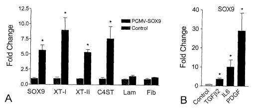
regulates XT-I, XT-II and C4ST expression.
21
CA 02667582 2009-04-24
WO 2008/049226 PCT/CA2007/001902
[0064] The Genomatix-predicted SOX9 binding site in the XT-1 gene's
promoter is highlighted in the following nucleotide sequence upstream of the
XT-1
genes transcriptional start site:
From the upstream region of XT-I gene a sequence including:
I GGCTTATCTG GCTCAAGACT GTTCTCAATC TGAAATGCCA TCCCTGGCTT AGCATTTCCT
61 CTCTATCCTA ACCCCCAAGT AACTCCACTA ACCCCCAAAT AACTCCACTG TACCTCCCCA
121 AATAACTCCA CTAACCCCCA AATAGCTCCA TTATAACTCC CCAAATAGCT TCCACTATCT
181 CTTGTTCTGC AAACTTATGT TCCAACAGGG CTGAGTCTTT TGTCTGCTGC TCAGCATCTA
241 GAATGACATT TGCGTAGAGA TGAACAGGGC ACTACACAGT AGCAGTTACA GGTGAGAACT
301 GCTTACAGGG GCTGGCTCTG GCAGTAATCA CACTGTAAAT CAACTAAGGG AGATGGTATT
361 TCCATTTTAA ACATGGGGAA ACTGAGGCTT CATGATGTTA GAAAGTACTT GCCCGAGACT
421 AATTACAATA CTGAATTTGA ATTCAGGTTT AACTGAACTT CAGTAAGCAT GACATCGCAG
481 GAGCGGCCCT CCCTCTAAAG ATGCGGAGCC TGCCTCTGTT CTTCTTCTCA GTGTGCTCCT
541 TCACTGGGCG AGAGTGCAAG GCCATCTGGC TGCAGGTGAC AGGAGTGTTC GTCATGCTGA
601 C (SEQ ID No. 5)
[0065] The bioinformatics analyses predicted SOX9 binding sequences in two
separate regions upstream of the C4ST gene's transcriptional start site.
Sequences from
these two areas are shown below. The Genomatix-predicted SOX9 binding sites
are
highlighted.
Area 1:
I GCAGGCTGAG AGGAGAAGCT ACTGAGTCTT AAAGGCATAT GGCCCCAGCA TCCCGGGGCC
61 TGAAAGCTGT GACAATATTG AGGGTCAAGA GTACTAAAGC CTGGAGACTA GAGCTGTCAT
121 TTCT TGTCCTA GGGGTTAAAG CCTAGGTCAC TGAATGTTCA
181 GACCACTGGG AGTCCAGGGC TTTTTCTCCA AGGACCTCAG CTGCAGCCTC TGACTCTGCC
241 AGTAAGGCAC TTGGGTCGGA GCACCTGTAC CTGAGAGGTT TTCTGCTACT AATATCCATC
301 TATGTAGAGT AGAGAACTCC AGCCTGATAA CTAGTAACTG GGATAGACAC TGCTTTTCCT
361 TGTCCTGGGT TTACAGCTTT ACCCATTAAG ACAGTCAGGC ACGTCTATCT CCAGCCTAGA
421 GCACAGGACA ATGCTTTTGG GCGGGCCCTA AACTAAGGGC AGGACTGGGC GTGTCCTGGA
481 CCTCCTCCGC ACAGTGGGAG GACGCACCGG ATGACCGTCG CCTGCCACGC GCCAGGCACA
541 GCATGGGAAG GCGCTCCTGT TGCCGGCGGC CCTTGCCGGT GGTGGCAAGT CTGGGTGCTG
601 CACTTCTGTT CCTGTGCGCC GCGCCGCGCG CCCTGCGTCC CGGT (SEQ ID No. 6)
Area 2:
1 TTTGCACCTG GTTTCCAATC TTTCTGGTGG CCTCCATGGA TGCTCATCTC TAGGGACAAC
61 AGTGGGCTGA GTTATTCTCA ATTTAGTCAC CAGGTGGCAG CCTAGAAGGC GAAAACTTAC
121 TGATGATTGG AAGACTGGAC TAGGTTCTGG TCTGAGAAAC CCTGTGAGTT TGGGTGAGAT
181 TTGGGGCAGA TAGGTATCTG GGTTCTGGGC TGGGCTCAAA GGAAGCAGAC ATTCCCCGAG
241 GATGAGGCAT CCTGAGAAGG ACGTGGTTTT AGTGTGAGCT GGGTTCCCAC CCAAAACAGG
301 AGTTAGAACC ATCGTTGCTA TTTGAAGCTA AATGTATAAA ATGTAATTTG TTTCATAGTC
361 TGCTATAGAT CATTGTCATA ACAGGAACCA ATTAGGTTTG
421 TTGAATACTA ATATCAAGTC CTTACAGGGC ACGTATCCAA CCTGAGGCTA CTCAAATAGC
481 TCTGCTTCTC ATTGAACACA ATGAGGTTTA ATATTACCGC CATTGTACAG GGAAATGGAG
541 TACAGATGGC AGGTAAGACA CTAGTGTTGG TGCAGCACCT CATCCCATAC ACTCAAGGCT
601 A (SEQ ID No.7)
[0066] To investigate the likelihood that SOX9 may regulate CBG expression,
SOX9 expression was analyzed after SCI in the rat by Q-PCR. SOX9 showed a
rapid
22
CA 02667582 2009-04-24
WO 2008/049226 PCT/CA2007/001902
12-fold increase in expression levels at 12 hours post-injury. By the third
day after SCI
the mRNA levels of SOX9 were not different from the control but they increased
again
I I-fold relative to control one week after SCI and remained elevated through
to 42 days
post-injury (Fig. 3). Immunohistochemistry using an antibody that recognizes
the
phosphorylated active form of SOX9 and an anti-GFAP antibody clearly shows co-
expression of SOX9 and GFAP in the spinal cord lesion at 42 days post-injury.
Characterization of CBG Expression in Primary Astrocyte Cultures
[0067] To test putative regulators of XT-I, XT-II and C4ST, a cell culture
system was developed to reflect the cellular make-up of the injured spinal
cord. Since
astrocytes are a major source of CSPGs at the glial scar (Alonso and Privat
1993;
Fawcett and Asher 1999; Reier and Houle 1988) and express CBGs and SOX9,
primary
astrocyte cultures were used to investigate the transcriptional regulation of
XT-I, XT-II
and C4ST. To provide baseline values on the expression of these genes in
primary
(3after 3 and 6 weeks in culture. The levels of XT-I and XT-II mRNA in primary
astrocytes was approximately 2-fold greater in 3-week than in 6-week cultures
(Fig. 4
A, B). The higher levels of XT-I and -II mRNA in 3 week-old primary astrocyte
cultures reflected that these cultures were in an activated state induced by
the isolation
procedure. This was supported by the observation that the expression of GFAP,
a gene
expressed by reactive astrocytes following CNS injury (Janeczko 1988; Vijayan
et al.
1990) was also elevated in 3 week-old compared to 6 week-old astrocyte
cultures (Fig.
4 C).
TGF2, IL-6 and PDGF Increase the Expression Levels of XT-I, XT-II and C4ST
[0068] To evaluate TGF(32, PDGF and IL-6 as candidate XT-I, XT-II and C4ST
transcriptional regulators, rat primary astrocyte cultures were exposed to 1,
10 or 100
ng/ml of each cytokine. After a 12 hour cytokine exposure, XT-1, XT-II and
C4ST
mRNA levels were measured relative to untreated cultures by Q-PCR. Six week-
old
cultures were used as, by this time point, the astrocytes are quiescent (as
evidenced by
reduced GFAP expression) and baseline levels of XT-I, XT-II and C4ST genes are
low.
Treatment of 6 week-old primary astrocyte cultures with TGF ^ 2, IL-6 and PDGF
23
CA 02667582 2009-04-24
WO 2008/049226 PCT/CA2007/001902
resulted in a strong, concentration-dependent up-regulation of XT-II mRNA
above
control values (Fig. 6 A). Similar increases in expression were observed for
XT-I and
C4ST (Fig. 6 B, C). The increased expression of CBGs in these cultures was
matched
by an increased expression of CSPG protein as assessed by slot blot analysis
(Fig. 6 D).
Fibronectin and laminin expression were similarly increased following these
cytokine
treatments (Fig. 6 F, G). In support of this experimental approach, cytokines
such as
TNF02 and bFGF that have expression profiles different from the expression
profiles of
XT-I, XT-II and C4ST were found to have no effect on XT-II mRNA levels in
primary
astrocytes (Fig. 6 H).
SOX9 Regulates CBG but not Laminin mRNA Levels
[0069] To test whether SOX9 regulates expression of XT-I, XT-II and C4ST in
vitro, primary astrocytes were transfected with a SOX9 expression construct
and
assessed for CBG mRNA levels by Q-PCR 48 hours later. CMV-driven SOX9
expression resulted in significant increases in XT-I, XT-II and C4ST mRNA
(Fig. 6 A).
The levels of fibronectin and laminin mRNA in these same cultures were
unaffected by
SOX9 over-expression. To determine whether IL-6, PDGF and TGF^2 might increase
XT-I, XT-II and C4ST gene expression by up-regulating SOX9 expression, the
expression levels of SOX9 mRNA were assayed after these cytokine treatments in
primary astrocyte cultures. The cytokine treatments (TGF(32, IL-6 and PDGF)
that up-
regulated the expression of XT-I, XT-II and C4ST caused a significant increase
in
SOX9 mRNA levels (Fig. 6 B).
[0070] To test the effect of SOX9 knock-down on the expression of XT-I, XT-
II and C4ST, a control (scrambled) small interfering RNA (SiRNA) or an anti-
SOX9
SiRNA was transfected into primary astrocytes and mRNA levels of SOX9, XT-I,
XT-
II and C4ST were assayed by Q-PCR 12 hours later. Transfection of primary
astrocytes
with an anti-SOX9 siRNA resulted in a 75 12% reduction in SOX9 mRNA levels and
a 71 5.5% reduction in XT-I mRNA (Fig 7 A, B). Transfection of TGF'~ ~,treated
primary astrocytes with the anti-SOX9 SiRNA resulted in a 87 13% reduction in
SOX9 mRNA levels and a 68 6.4% reduction in XT-I mRNA, while TGFC] [I
treatment alone resulted in increased SOX9 and XT-1 mRNA levels. Similar
24
CA 02667582 2009-04-24
WO 2008/049226 PCT/CA2007/001902
reductions were observed also in XT-II and C4ST expression in the presence of
anti-
SOX9 SiRNA in both TGF(32-treated and untreated cultures (Fig. 7 C, D). SOX9
knock-down did not decrease laminin or fibronectin gene expression. (Fig 7 E,
F).
[0071] The results clearly show that SOX9 expression is both necessary and
sufficient for CBG expression in primary astrocytes and that cytokine up-
regulation of
CBG expression is SOX9 dependent. The anti-SOX9 SiRNA transfections show that
laminin and fibronectin expression are negatively regulated by SOX9.
Example 2- Effect of anti-CD11d mAb-treatment on SCI
EXPERIMENTAL METHODS
Animals and Surgeries
[0072] As described in Example 1.
Microarray Analysis
[0073] All GeneChips were processed at the London Regional Genomics
Centre (Robarts Research Institute, London, ON). The quality of each RNA
sample was
assessed using the Agilent 2100 Bioanalyzer (Agilent Technologies Inc.,
California,
USA) and the RNA 6000 Nano kit (Caliper Life Sciences, California, USA).
Biotinylated complementary RNA (cRNA) was prepared from 10 g of total RNA as
per the Affymetrix GeneChip Technical Analysis Manual (Affymetrix, California,
USA). Double-stranded cDNA was synthesized using SuperScriptll (Invitrogen,
California, USA) and oligo (dT)24 primers. Biotin-labeled cRNA was prepared by
cDNA in vitro transcription using the BioArray High-Yield RNA Transcript
Labeling
kit (Enzo Biochem, New York, USA) incorporating biotinylated UTP and CTP. The
biotin-labeled cRNA (10 g) was hybridized to RAE230A GeneChips for 16 h at 45
C
as described in the Affymetrix Technical Analysis Manual (Affymetrix,
California,
USA). RNA samples from each animal (3 anti-CDlld-treated and 3 untreated
animals
at each time point) were hybridized to separate GeneChips. Three RNA samples
from
3 different uninjured animals were likewise hybridized to 3 separate GeneChips
to
provide control levels of gene expression. The GeneChips were stained with
CA 02667582 2009-04-24
WO 2008/049226 PCT/CA2007/001902
Streptavidin-Phycoerythrin, followed by an antibody solution, and a second
Streptavidin-Phycoerythrin solution; a GeneChip Fluidics Station 400 performed
all
liquid handling. GeneChips were scanned with the Affymetrix GeneChip Scanner
3000
(Affymetrix, California, USA). Probe signal intensities were generated using
GCOS1.3
(Affymetrix Inc., California, USA) with default values for the statistical
expression
algorithm parameters and a target signal of 150 for all probe sets and a
normalization
value of 1. Gene level data were generated using the RMA preprocessor in
GeneSpring
GX 7.3 (Agilent Technologies Inc., California, USA). The data were transformed
(measurements less than 0.01 were set to 0.01) and normalized per chip to the
50`"
percentile, and per gene to median. Statistically significant changes in mRNA
levels
that correlated to treatment and/or time post-injury was compiled using a two-
way
ANOVA (p<0.05). The Benjamini and Hochberg false discovery test that corrects
for
multiple testing was used to determine differences between mean values. All
data
analysis and mining were performed using GeneSpring GX 7.3 (Agilent
Technologies
Inc., California, USA).
Quantitative Polymerase Chain Reaction (Q-PCR)
[0074] In this study, l^g RNA per condition (cell culture or animal tissue)
was
used to synthesize first strand cDNA, using High Capacity cDNA Archive Kit
according to the manufacturer's specification (Applied Biosystems, California,
USA).
The primer probe sets, optical adhesive covers, and PCR plates were purchased
from
Applied Biosystems California, USA. These probes were labeled at the 5' end
with
FAM (Applied Biosystems) and at the 3' end with TAMRA (Applied Biosystems) as
quencher with the exception of the ribosomal probe, which was labeled with 5'
VIC
(Applied Biosystems). For the Taq Man assays the thermal cycler conditions
were 10
minutes at 95 C, followed by 40 cycles of 30 seconds at 95 C to denature the
DNA and
30 seconds at 60 C to anneal and extend the template. A standard curve of
cycle
thresholds using cDNA serial dilutions was established and used to calculate
abundance
of a target gene. The values were normalized to the amounts of 18S mRNA. The
data
were analyzed using one way ANOVA followed by a Bonferroni test for multiple
comparisons.
26
CA 02667582 2009-04-24
WO 2008/049226 PCT/CA2007/001902
TaqMan gene expression primer-probe sets:
Target Gene Probe and Primer Catalog Number
(Applied Biosystems)
CD80 Rn00580581-m 1
CD4 Rn00562286_m 1
XT-I 1391062A
XT-II Mm00517563 m1
C4ST Mm00517563-m 1
Laminin Mm00711808 ml
SOX-9 Mm 0048840 ml
TG(3-2 Rn00579674-m 1
IL-6 Rn00561420 m1
Fibronectin Rn00569575 m1
BMP-7 Rn 0158889 ml
Tissue processing
[0075] At 3, 7 or 21 days post-SCI, control and anti-CDI ld treated rats (N =
5
for each group at each time point) were given an intraperitoneal overdose of
26%
Ketamine (100 mg/ml, Vetalar, Bioniche, Belleville, ON) and 0.06% Xylazine (20
mg/ml, Rompun, Bayer, Toronto, ON) in a 2:1 mixture. Each rat was
intracardially
perfused with 250 ml of oxygenated tissue culture medium (pH 7.4, Dulbecco's
modified Eagle medium, Gibco Invitrogen Corp, Burlington, ON) followed by 500
ml
of 4% formaldehyde fixative in 0.1 M phosphate buffer solution (PBS, pH 7.4),
both at
room temperature. A section of spinal cord centered around the lesion was
removed
such that it was 0.5 cm rostral and caudal to the lesion site (T3-T4). All
cords were
processed as previously described (Saville et al., 2004). Eight sets of slides
containing
serial 16 ^ m thick sections from each animal were collected and used in the
immunohistochemical analyses.
27
CA 02667582 2009-04-24
WO 2008/049226 PCT/CA2007/001902
RESULTS
Wound Healing and Scar Genes
[0076] Altered expression of immune response genes could have profound
effects on the expression of genes associated with wound healing. CSPGs,
laminin and
fibronectin are key components of the glial scar that may determine the degree
of
neurological recovery possible in spinal cord-injured animals (Bradbury et
al., 2002;
Grimpe and Silver, 2004). Thus, the possibility that improved recovery in the
anti-
CDl ld mAb-treated rats is due in part to changes in the expression of these
genes
involved in scar formation was investigated. Q-PCR confirmed that IL-6 and TGF
LJ2
mRNA are down-regulated acutely in treated rats (Fig. 8 A). Reduced cytokine
expression is matched by acute reductions in SOX9, XT-I, XT-II and C4ST mRNA
levels in anti-CDl ld mAb-treated rats (Fig. 8 C-E). In keeping with the
finding that
SOX9 inhibits the expression of laminin and fibronectin in primary astrocyte
cultures,
the decrease in SOX9 expression in anti-CD 11 d mAb treated rats was
accompanied by
increases in laminin and fibronectin mRNA levels (Fig. 8 F, 8G). Slot blot
analyses
using an anti-laminin antibody and the CS56 antibody that recognizes a variety
of
CSPGs (Avnur and Geiger, 1984) indicates that these differences in mRNA levels
correlate with significant decrease in the ratio of CSPG:laminin protein in
the lesions of
anti-CDl ld mAb-treated rats (Fig. 8 H). Immunohistochemistry on sections from
the
lesion epicenters demonstrates the changed nature of the glial scar. Using
alternating
tissue sections taken from the same SCI rat lesion areas used in the CD8a
analysis,
double labeling with anti-laminin and CS56 antibodies, anti-neurofilament and
anti-
CS56 antibodies and anti-laminin and anti-neurofilament antibodies shows
increased
amounts of laminin relative to CSPGs in the anti-CDlld-treated spinal cord-
injured
rats and an increase in neurofilament stained axons in the lesion epicenters
that is most
prominent in laminin-rich areas. Thus, increased axon sprouting or sparing is
associated
with increased laminin and decreased CSPG production in rats treated with the
anti-
CDl ld mAb.
Example 3 - Expression of CSPGs in Various Neuropathological Samples
28
CA 02667582 2009-04-24
WO 2008/049226 PCT/CA2007/001902
[0077] To elucidate the role of CSPGs in human SCI, traumatic brain injury
(TBI), hemorrhagic stroke, ischemic stroke, and Alzheimers Disease (AD),
immunohistochemistry was carried out on sections from subjects with these
conditions.
Histological sections were obtained from the Pathology department at London's
University Hospital (Dr. David Ramsay). Sections were stained with CS56 (an
antibody recognizing many CSPGs) in combination with antibodies raised against
either GFAP (to stain for reactive astrocytes), SM132 (neurons), or CD68
(microglia/macrophages).
METHODS
Tissue Processing
[0078] Human sections for all neuropathological conditions were obtained from
Dr. David Ramsay (Department of Pathology, University of Western Ontario).
Mice
were cardiac perfused with 4% paraformaldehyde to fix tissue. Brains were
dissected
out and embedded in paraffin. l0um sections were cut using a microtome, and
mounted on slides.
Immunohistochemistry
[0079] All sections were processed for depraffinization using a series of
xylene
and ethanol washes, followed by incubation in 10% hydrogen peroxide in
methanol.
After deparaffinization, antigen retrieval was carried out by boiling sections
in citric
acid (pH 6.0) for 15 minutes. Sections were then washed for 10 minutes in PBS,
and
blocked in 10% Goat Serum and 0.5% Triton-X in PBS for 1 hour.
[0080] Human sections were double stained with a combination of primary
antibodies against CS56 (1/100, Sigma, St.Louis, MO), SOX9 (1/100, Chemicon,
Temecula, CA), GFAP (1/100, Molecular Probes, Carlsbad, CA), SM132 (1/100,
Covance, Princeton, NJ), and CD68 (1/100, Dako, Carpintera, CA). Mouse MCAO
sections were double stained with a combination of primary antibodies against
SOX9,
GFAP, and TUJ1 (1/100 Chemicon, Temecula, CA). Secondary antibodies for
different combinations were used as shown in Tables 2 and 3.
29
CA 02667582 2009-04-24
WO 2008/049226 PCT/CA2007/001902
Table 2 Secondary antibody combinations and concentrations for different
combinations of primary antibodies in human fluorescent double staining
experiments.
Primary Antibody Secondary 1 Secondary 2
Combination
CS56/GFAP Fluorescein IgM Rhodamine IgGI
CS56/SM132 Fluorescein IgM Rhodamine IgG2b
CS56/CD68 Fluorescein IgM) Rhodamine IgGI
CS56/SOX9 Fluorescein IgM Alexafluor 594 Goat Anti-
rabbit IgG
SOX9/GFAP Alexafluor 594 Goat Anti- Alexafluor 488 Goat Anti-
rabbit IgG mouse IgG
SOX9/SM132 Alexafluor 594 Goat Anti- Alexafluor 488 Goat Anti-
rabbit IgG mouse IgG
SOX9/CD68 Alexafluor 594 Goat Anti- Alexafluor 488 Goat Anti-
rabbit IgG mouse IgG
Table 3. Secondary antibody combinations and concentrations for different
combinations of primary antibodies in mouse MCAO fluorescent double staining
experiments.
Primary Antibody Secondary 1 Secondary 2
Combination
SOX9/GFAP Alexafluor 594 Goat Anti- Alexafluor 488 Goat Anti-
rabbit IgG mouse IgG
SOX9/Tuj 1 Alexafluor 594 Goat Anti- Alexafluor 488 Goat Anti-
rabbit IgG mouse IgG
All Alexafluor-conjugated secondary antibodies were obtained from Molecular
Probes
(Carlsbad, CA). All other secondary antibodies were obtained from Jackson
ImmunoResearch. Sox9-positive cells were counted manually in 3 different
sections
from each patient.
Real-time Quantitative PCR
Mice were anesthetised with ketamine:xylazine (2:1) and perfused with saline.
Brains
were dissected out and the cortices removed. The cortices were then placed in
Trizol
and homogenized with a tissue homogenizer. RNA was extracted using ,,, and
stored at
CA 02667582 2009-04-24
WO 2008/049226 PCT/CA2007/001902
-80 C. CDNA was then synthesized. Primer probe sets for SOX9 and 18S
(identified
previously) were used to quantify gene expression using quantitative PCR.
RESULTS
[0081] The immunohistochemistry demonstrated CSPG expression in all 5
neuropathological conditions studied. In control sections immunohistochemistry
demonstrated that CSPGs are not expressed outside of perineuronal nets (PNNs)
in
uninjured healthy brains. Double labelling with anti-GFAP and CS56 antibodies
showed that reactive astrocytes are present in the region of CS56
immunoreactivity. In
human TBI, hemorrhagic stroke, and ischemic stroke, the reactive astrocytes
can be
seen around the outer edge of the areas rich in CSPGs. In SCI and AD, the
reactive
astrocytes are present throughout the CSPG-rich region. Neurons associated
with
CSPG-rich areas were only observed in sections from ischemic stroke. CD68-
positive
microglia and macrophages were also observed in areas immunoreactive for
CSPGs.
SOX9 Expression in Human Neuropathological Sections
[0082] Since previous studies in our laboratory have shown that the
transcription factor SOX9 is necessary and sufficient for the expression of
enzymes
involved in chondroitin sulphate side chain synthesis, the expression of SOX9
was
examined in CSPG-rich regions. SOX9 positive nuclei were observed in all areas
of
CS56-immunoreactivity. To elucidate cellular localization of SOX9, double
staining
was carried out with an anti-SOX9 antibody, in combination with one of anti-
GFAP,
anti-SM132, or anti-CD68 antibodies, to detect reactive astrocytes, neurons,
and
macrophages, respectively. SOX9 was found in the nuclei of reactive astrocytes
in all
neuropathological conditions studied, but not in the uninjured brain. In
addition,
SOX9 was found in the nuclei and cytoplasm of neurons in healthy and injured
or
diseased brains and in the nuclei of CD68 positive cells. The expression in
healthy
uninjured neurons probably reflects the involvement of SOX9 in the expression
of
CSPGs that constitute part of the PNNs.
[0083] Concurrent with the human studies, a mouse model of stroke (MCAO)
was studied to confirm the expression and cellular localization of SOX9 in
cerebrovasular injury. SOX9 was found in the nuclei of reactive astrocytes in
MCAO-
31
CA 02667582 2009-04-24
WO 2008/049226 PCT/CA2007/001902
injured brains, but not in uninjured brains. SOX9 was also found in the nuclei
of
neurons in both healthy and injured brains. Through quantitative PCR, it was
shown
that SOX9 mRNA expression is elevated in the injured cortex of MCAO mice as
compared to the uninjured control.
Example 4 - Assay to screen for SOX9 inhibitors
[0084] Proteoglycans, and in particular CSPGs, produced by reactive astrocytes
in the injured or diseased central nervous system (CNS) are inhibitory to
regeneration.
Using both gain-of-function and loss-of-function experiments, the
transcription factor
SOX9 has been found to be both necessary and sufficient to up-regulate the
expression
of XT-I, XT-II and C4ST in primary astrocyte cultures. It has also been
demonstrated
that, whereas SOX9 up-regulates the production of CSPGs, it down-regulates the
expression of laminin and fibronectin.
[0085] An assay has been developed to screen for SOX9 inhibitors.
[0086] Astrocytes, such as wither primary astrocytes (rodent or human) or an
established astrocyte cell line designated as Neu7 (Fok-Seang, Smith-Thomas et
al.
1995), were transfected with a SOX9 reporter construct under standard
conditions. The
SOX9 reporter construct (a gift from Dr. Michael Underhill, University of
British
Columbia) has 4 repeats of the SOX9 binding site coupled to the mouse Col2al
minimal promoter (-89 to +6) cloned upstream of a luciferase gene in the
plasmid pGL4
(Promega) (Weston, Chandraratna et al. 2002). Changes in luciferase levels in
transfected cultures is used as a read-out of SOX9 activity. The anti-SOX9
SiRNA
previously used (or astrocytes from the SOX9 conditional knock out) is used as
a
positive control and the scrambled siRNA is used as a negative control for
this screen.
The screen is used to identify compounds that reduce the levels of luciferase
activity
relative to control wells. Such compounds will be SOX9 inhibiting and will be
considered as positive "hits". In a secondary screen false positives that
cause a
reduction in luciferase activity due to effects on cell viability, will be
eliminated by
assaying cell death in treated cultures by propidium iodide uptake. When using
primary astrocyte cultures the transfection will be normalized to control
plasmid co-
transfected with the SOX9 reporter construct.
32
CA 02667582 2009-04-24
WO 2008/049226 PCT/CA2007/001902
[0087] Validation of this screen has been obtained from experiments that have
shown that primary astrocytes transfected with the SOX9 luciferase reporter
construct
demonstrate approximately a 2-fold increase in luciferase activity after
treatment with
TGF-(32 as TGF[i-2 increases SOX9 expression and activity in primary
astrocytes (Fig.
10). Specifically, primary astrocytes obtained from newborn rats were cultured
in 6-
well dishes for 6 weeks in Advanced-DMEM with 10% FBS. The culture medium was
replaced with 2 ml of serum-free Advanced-DMEM with 10 ul of Lipofectamine
2000
(Invitrogen) and 10 ug SOX9-reporter plasmid DNA per well. Transfection
efficiency
was calculated by co-transfection with a control plasmid. After 24 hours the
cells were
treated with TGF-02 (final concentration l OnM). Twenty-four hours after the
TGF- 02
application the cells were lysed and luciferase activity was measured using
the
Luciferase Assay System (PROMEGA) (according to the manufacturer's protocol)
and
a luminometer.
33
CA 02667582 2009-04-24
WO 2008/049226 PCT/CA2007/001902
FULL CITATIONS FOR REFERENCES REFERRED TO IN THE
SPECIFICATION
1. Alonso G, Privat A. 1993. Reactive astrocytes involved in the formation of
lesional scars differ in the mediobasal hypothalamus and in other forebrain
regions. J Neurosci Res 34(5):523-38.
2. Asher RA, Morgenstem DA, Fidler PS, Adcock KH, Oohira A, Braistead JE,
Levine JM, Margolis RU, Rogers JH, Fawcett JW. 2000. Neurocan is
upregulated in injured brain and in cytokine-treated astrocytes. J Neurosci
20(7):2427-38.
3. Avnur Z, Geiger B. 1984. Immunocytochemical localization of native
chondroitin-sulfate in tissues and cultured cells using specific monoclonal
antibody. Cell 38(3):811-22.
4. Bahr M, Przyrembel C, Bastmeyer M. 1995. Astrocytes from adult rat optic
nerves are nonpermissive for regenerating retinal ganglion cell axons. Exp
Neurol 131(2):211-20.
5. Blatti SP, Foster DN, Ranganathan G, Moses HL, Getz MJ. 1988. Induction of
fibronectin gene transcription and mRNA is a primary response to growth-factor
stimulation of AKR-2B cells. Proc Natl Acad Sci U S A 85(4):1119-23.
6. Bradbury EJ, Moon LD, Popat RJ, King VR, Bennett GS, Patel PN, Fawcett
JW, McMahon SB. 2002. Chondroitinase ABC promotes functional recovery
after spinal cord injury. Nature 416(6881):636-40.
7. Carmel JB, Galante A, Soteropoulos P, Tolias P, Recce M, Young W, Hart RP.
2001. Gene expression profiling of acute spinal cord injury reveals spreading
inflammatory signals and neuron loss. Physiol Genomics 7(2):201-13.
8. Christner PJ, Jimenez SA. 2004. Animal models of systemic sclerosis:
insights
into systemic sclerosis pathogenesis and potential therapeutic approaches.
Curr
Opin Rheumatol 16(6):746-52.
9. Cohen CD, Klingenhoff A, Boucherot A, Nitsche A, Henger A, Brunner B,
Schmid H, Merkle M, Saleem MA, Koller KP and others. 2006. Comparative
promoter analysis allows de novo identification of specialized cell junction-
associated proteins. Proc Natl Acad Sci U S A 103(15):5682-7.
10. Costa S, Planchenault T, Charriere-Bertrand C, Mouchel Y, Fages C, Juliano
S,
Lefrancois T, Barlovatz-Meimon G, Tardy M. 2002. Astroglial permissivity for
neuritic outgrowth in neuron-astrocyte cocultures depends on regulation of
laminin bioavailability. Glia 37(2):105-13.
34
CA 02667582 2009-04-24
WO 2008/049226 PCT/CA2007/001902
11. Davies JE, Tang X, Denning JW, Archibald SJ, Davies SJ. 2004. Decorin
suppresses neurocan, brevican, phosphacan and NG2 expression and promotes
axon growth across adult rat spinal cord injuries. Eur J Neurosci 19(5):1226-
42.
12. Davies SJ, Fitch MT, Memberg SP, Hall AK, Raisman G, Silver J. 1997.
Regeneration of adult axons in white matter tracts of the central nervous
system.
Nature 390(6661):680-3.
13. Davies SJ, Goucher DR, Doller C, Silver J. 1999. Robust regeneration of
adult
sensory axons in degenerating white matter of the adult rat spinal cord. J
Neurosci 19(14):5810-22.
14. Eddleston M, Mucke L. 1993. Molecular profile of reactive astrocytes--
implications for their role in neurologic disease. Neuroscience 54(1):15-36.
15. Eddy AA. 1996. Molecular insights into renal interstitial fibrosis. J Am
Soc
Nephrol 7(12):2495-508.
16. Fawcett JW, Asher RA. 1999. The glial scar and central nervous system
repair.
Brain Res Bull 49(6):377-91.
17. Fehlings MG, Tator CH. 1995. The relationships among the severity of
spinal
cord injury, residual neurological function, axon counts, and counts of
retrogradely labeled neurons after experimental spinal cord injury. Exp Neurol
132(2):220-8.
18. Fitch MT, Silver J. 1997. Activated macrophages and the blood-brain
barrier:
inflammation after CNS injury leads to increases in putative inhibitory
molecules. Exp Neurol 148(2):587-603.
19. Fok-Seang J, Smith-Thomas LC, Meiners S, Muir E, Du JS, Housden E,
Johnson AR, Faissner A, Geller HM, Keynes RJ and others. 1995. An analysis
of astrocytic cell lines with different abilities to promote axon growth.
Brain
Res 689(2):207-23.
20. Foster JW, Dominguez-Steglich MA, Guioli S, Kowk G, Weller PA, Stevanovic
M, Weissenbach J, Mansour S, Young ID, Goodfellow PN and others. 1994.
Campomelic dysplasia and autosomal sex reversal caused by mutations in an
SRY-related gene. Nature 372(6506):525-30.
21. Fukuta M, Uchimura K, Nakashima K, Kato M, Kimata K, Shinomura T,
Habuchi O. 1995. Molecular cloning and expression of chick chondrocyte
chondroitin 6-sulfotransferase. J Biol Chem 270(31):18575-80.
22. Furumatsu T, Tsuda M, Taniguchi N, Tajima Y, Asahara H. 2005. Smad3
induces chondrogenesis through the activation of SOX9 via CREB-binding
protein/p300 recruitment. J Biol Chem 280(9):8343-50.
CA 02667582 2009-04-24
WO 2008/049226 PCT/CA2007/001902
23. Gallo V, Bertolotto A. 1990. Extracellular matrix of cultured glial cells:
selective expression of chondroitin 4-sulfate by type-2 astrocytes and their
progenitors. Exp Cell Res 187(2):211-23.
24. Gotting C, Kuhn J, Zahn R, Brinkmann T, Kleesiek K. 2000. Molecular
cloning
and expression of human UDP-d-Xylose:proteoglycan core protein beta-d-
xylosyltransferase and its first isoform XT-II. J Mol Biol 304(4):517-28.
25. Grimpe B, Silver J. 2004. A novel DNA enzyme reduces glycosaminoglycan
chains in the glial scar and allows microtransplanted dorsal root ganglia
axons
to regenerate beyond lesions in the spinal cord. J Neurosci 24(6):1393-7.
26. Gris P, Murphy S, Jacob JE, Atkinson I, Brown A. 2003. Differential gene
expression profiles in embryonic, adult-injured and adult-uninjured rat spinal
cords. Mol Cell Neurosci 24(3):555-67.
27. Hammarback JA, McCarthy JB, Palm SL, Furcht LT, Letourneau PC. 1988.
Growth cone guidance by substrate-bound laminin pathways is correlated with
neuron-to-pathway adhesivity. Dev Biol 126(1):29-39.
28. Hartsough MT, Mulder KM. 1997. Transforming growth factor-beta signaling
in epithelial cells. Pharmacol Ther 75(1):21-41.
29. Heldin CH, Westermark B. 1999. Mechanism of action and in vivo role of
platelet-derived growth factor. Physiol Rev 79(4):1283-316.
30. Janeczko K. 1988. The proliferative response of astrocytes to injury in
neonatal
rat brain. A combined immunocytochemical and autoradiographic study. Brain
Res 456(2):280-5.
31. Jones LL, Margolis RU, Tuszynski MH. 2003. The chondroitin sulfate
proteoglycans neurocan, brevican, phosphacan, and versican are differentially
regulated following spinal cord injury. Exp Neurol 182(2):399-411.
32. Kawakami Y, Tsuda M, Takahashi S, Taniguchi N, Esteban CR, Zemmyo M,
Furumatsu T, Lotz M, Belmonte JC, Asahara H. 2005. Transcriptional
coactivator PGC-lalpha regulates chondrogenesis via association with Sox9.
Proc Natl Acad Sci U S A 102(7):2414-9
33. Kolb M, Margetts PJ, Galt T, Sime PJ, Xing Z, Schmidt M, Gauldie J. 2001.
Transient transgene expression of decorin in the lung reduces the fibrotic
response to bleomycin. Am J Respir Crit Care Med 163(3 Pt 1):770-7.
34. Kordes U, Cheng YC, Scotting PJ. 2005. Sox group E gene expression
distinguishes different types and maturational stages of glial cells in
developing
chick and mouse. Brain Res Dev Brain Res 157(2):209-13.
35. Lagord C, Berry M, Logan A. 2002. Expression of TGFbeta2 but not TGFbetal
correlates with the deposition of scar tissue in the lesioned spinal cord. Mol
Cell
Neurosci 20(1):69-92.
36
CA 02667582 2009-04-24
WO 2008/049226 PCT/CA2007/001902
36. Lefebvre V, Huang W, Harley VR, Goodfellow PN, de Crombrugghe B. 1997.
SOX9 is a potent activator of the chondrocyte-specific enhancer of the pro
alphal(II) collagen gene. Mol Cell Biol 17(4):2336-46.
37. Liesi P, Silver J. 1988. Is astrocyte laminin involved in axon guidance in
the
mammalian CNS? Dev Biol 130(2):774-85.
38. Logan A, Baird A, Berry M. 1999a. Decorin attenuates gliotic scar
formation in
the rat cerebral hemisphere. Exp Neurol 159(2):504-10.
39. Logan A, Green J, Hunter A, Jackson R, Berry M. 1999b. Inhibition of glial
scarring in the injured rat brain by a recombinant human monoclonal antibody
to transforming growth factor-beta2. Eur J Neurosci 11(7):2367-74.
40. Matthiessen HP, Schmalenbach C, Muller HW. 1989. Astroglia-released
neurite
growth-inducing activity for embryonic hippocampal neurons is associated with
laminin bound in a sulfated complex and free fibronectin. Glia 2(3):177-88.
41. McKeon RJ, Jurynec MJ, Buck CR. 1999. The chondroitin sulfate
proteoglycans neurocan and phosphacan are expressed by reactive astrocytes in
the chronic CNS glial scar. J Neurosci 19(24):10778-88.
42. McKeon RJ, Schreiber RC, Rudge JS, Silver J. 1991. Reduction of neurite
outgrowth in a model of glial scarring following CNS injury is correlated with
the expression of inhibitory molecules on reactive astrocytes. J Neurosci
11(11):3398-411.
43. Morgenstern DA, Asher RA, Fawcett JW. 2002. Chondroitin sulphate
proteoglycans in the CNS injury response. Prog Brain Res 137:313-32.
44. Nakamura M, Okada S, Toyama Y, Okano H. 2005. Role of IL-6 in spinal cord
injury in a mouse model. Clin Rev Allergy Immunol 28(3):197-204.
45. Nelander S, Larsson E, Kristiansson E, Mansson R, Nerman 0, Sigvardsson M,
Mostad P, Lindahl P. 2005. Predictive screening for regulators of conserved
functional gene modules (gene batteries) in mammals. BMC Genomics 6(1):68.
46. Neugebauer KM, Tomaselli KJ, Lilien J, Reichardt LF. 1988. N-cadherin,
NCAM, and integrins promote retinal neurite outgrowth on astrocytes in vitro.
J
Cell Biol 107(3):1177-87.
47. Okada S, Nakamura M, Mikami Y, Shimazaki T, Mihara M, Ohsugi Y,
Iwamoto Y, Yoshizaki K, Kishimoto T, Toyama Y and others. 2004. Blockade
of interleukin-6 receptor suppresses reactive astrogliosis and ameliorates
functional recovery in experimental spinal cord injury. J Neurosci Res
76(2):265-76.
48. Pasinetti GM, Nichols NR, Tocco G, Morgan T, Laping N, Finch CE. 1993.
Transforming growth factor beta 1 and fibronectin messenger RNA in rat brain:
responses to injury and cell-type localization. Neuroscience 54(4):893-907.
37
CA 02667582 2009-04-24
WO 2008/049226 PCT/CA2007/001902
49. Reier PJ, Houle JD. 1988. The glial scar: its bearing on axonal elongation
and
transplantation approaches to CNS repair. Adv Neurol 47:87-138.
50. Rogers SL, Letourneau PC, Palm SL, McCarthy J, Furcht LT. 1983. Neurite
extension by peripheral and central nervous system neurons in response to
substratum-bound fibronectin and laminin. Dev Bio198(1):212-20.
51. Rogers SL, Letourneau PC, Peterson BA, Furcht LT, McCarthy JB. 1987.
Selective interaction of peripheral and central nervous system cells with two
distinct cell-binding domains of fibronectin. J Cell Biol 105(3):1435-42.
52. Schaeren-Wiemers N, Gerfin-Moser A. 1993. A single protocol to detect
transcripts of various types and expression levels in neural tissue and
cultured
cells: in situ hybridization using digoxigeni-labeled cRNA probes.
Histochemistry 100:431-440.
53. Schonherr E, Jarvelainen HT, Sandell LJ, Wight TN. 1991. Effects of
platelet-
derived growth factor and transforming growth factor-beta 1 on the synthesis
of
a large versican-like chondroitin sulfate proteoglycan by arterial smooth
muscle
cells. J Biol Chem 266(26):17640-7.
54. Stolt CC, Lommes P, Sock E, Chaboissier MC, Schedl A, Wegner M. 2003. The
Sox9 transcription factor determines glial fate choice in the developing
spinal
cord. Genes Dev 17(13):1677-89.
55. Tom VJ, Doller CM, Malouf AT, Silver J. 2004. Astrocyte-associated
fibronectin is critical for axonal regeneration in adult white matter. J
Neurosci
24(42):9282-90.
56. Tomaselli KJ, Neugebauer KM, Bixby JL, Lilien J, Reichardt LF. 1988. N-
cadherin and integrins: two receptor systems that mediate neuronal process
outgrowth on astrocyte surfaces. Neuron 1(1):33-43.
57. Vijayan VK, Lee YL, Eng LF. 1990. Increase in glial fibrillary acidic
protein
following neural trauma. Mol Chem Neuropathol 13(1-2):107-18.
58. Wehrli BM, Huang W, De Crombrugghe B, Ayala AG, Czerniak B. 2003.
Sox9, a master regulator of chondrogenesis, distinguishes mesenchymal
chondrosarcoma from other small blue round cell tumors. Hum Pathol
34(3):263-9.
59. Wiksten M, Vaananen AJ, Liebkind R, Liesi P. 2004. Regeneration of adult
rat
spinal cord is promoted by the soluble KDI domain of gammal laminin. J
Neurosci Res 78(3):403-10.
60. Wilson JX, Dixon SJ. 1989. Ascorbic acid transport in mouse and rat
astrocytes
is reversibly inhibited by furosemide, SITS, and DIDS. Neurochem Res
14(12):1169-75.
38
CA 02667582 2009-04-24
WO 2008/049226 PCT/CA2007/001902
61. Yamauchi S, Mita S, Matsubara T, Fukuta M, Habuchi H, Kimata K, Habuchi
0. 2000. Molecular cloning and expression of chondroitin 4-sulfotransferase. J
Biol Chem 275(12):8975-81.
62. Zuo J, Neubauer D, Dyess K, Ferguson TA, Muir D. 1998. Degradation of
chondroitin sulfate proteoglycan enhances the neurite- promoting potential of
spinal cord tissue. Exp Neurol 154(2):654-62.
63. Fok-Seang, J., L. C. Smith-Thomas, et al. (1995). "An analysis of
astrocytic cell
lines with different abilities to promote axon growth." Brain Res 689(2): 207-
23.
64. Gris, P., A. Tighe, et al. (2007). "Transcriptional regulation of scar
gene
expression in primary astrocytes." Glia 55(11): 1145-55.
65. Weston, A. D., R. A. S. Chandraratna, et al. (2002). "Requirement for RAR-
mediated gene repression in skeletal progenitor differentiation." J. Cell
Biol.
158(1): 39-51.
All references are incorporated herein by reference.
39