Note: Descriptions are shown in the official language in which they were submitted.
CA 02667673 2009-04-27
WO 2008/049164 PCT/AU2007/001622
1
RETINAL REGENERATION
This invention relates to a method of improving the function of the
retina of the human eye by improving the transport properties of Bruch's
membrane. This invention may be beneficially used in the treatment of eye
diseases, such as early Age-related Macular Degeneration (AMD) and
Diabetic Macular Edema (DME) in which the function of Bruch's membrane
has become impaired as part of a disease pathogenesis, or the treatment of
degradation related to aging. The transport properties of Bruch's membrane
are improved by a treatment which triggers Retinal Pigmented Epithelial
(RPE) cell changes, including migration and division.
BACKGROUND TO THE INVENTION
The light sensing and signaling processes of the human retina require
a high level of support in terms of energy supply and waste removal to
ensure optimal functionality. A monolayer of epithelial cells, known as the
retinal pigmented epithelium (RPE) separates the light sensing and signaling
processes from the blood supply of the choroid and it controls many bi-
directional support functions. The RPE cells are attached to a basement
membrane, known as Bruch's membrane, which is a thin extra-cellular matrix
of collagen layers which acts as a semi-permeable barrier between the RPE
cells and blood vessels of the choroid. The work of Marshall, Hussain, et. al.
over many years has shown that degradation of the transport functions of
Bruch's membrane is a major contributor to loss or decline in visual function
with normal aging or a more rapid decline due to diseases such as age-
related macular degeneration (AMD) and is well described in the following
references:
Starita C., Hussain A.A., Marshall J. (1995). Decreasing hydraulic
conductivity of Bruch's membrane: relevance to photoreceptor survival and
lipofuscinoses. American Journal of Medical Genetics. 57(2):235-7.
CA 02667673 2009-04-27
WO 2008/049164 PCT/AU2007/001622
2
Moore D.J., Hussain A.A., Marshall J. (1995). Age-related variation in the
hydraulic conductivity of Bruch's membrane. Investigative Ophthalmology &
Visual Science. 36(7):1290-7.
Starita C., Hussain A.A., Pagliarini S., Marshall J. (1996) Hydrodynamics of
ageing Bruch's membrane: implications for macular disease. Experimental
Eye Research. 62(5):565-72.
Starita C., Hussain A.A., Patmore A., Marshall J. (1997) Localisation of the
site of major resistance to fluid transport in Bruch's membrane. Invest.
Ophthalmol.Vis Sci. 38: 762-767.
Marshall J., Hussain A.A., Starita C., Moore D.J., Patmore A.L. (1998).
Ageing and Bruch's membrane. In: Marmor MF ed. Retinal Pigment
Epithelium: Function and disease. New York, Oxford University Press; pp.
669-692.
Hussain AA., Rowe L., Marshall J. (2002) Age-related alterations in the
diffusional transport of amino acids across the human Bruch's-choroid
complex. Journal of the Optical Society of America, A, Optics, Image
Science, & Vision. 19(1): 166-72.
Hussain AA., Starita C., and Marshall J. (2004) Chapter IV. Transport
characteristics of ageing human Bruch's membrane: Implications forAMD. In
Focus on Macular Degeneration Research, (Editor O. R. loseliani). Pages
59-113. Nova Science Publishers, Inc. New York.
Guo L., Hussain AA., Limb GA., Marshall J (1999). Age-dependent variation
in metalloproteinase activity of isolated human Bruch's membrane and
choroid. Investigative Ophthalmol. Vis Sci. 40(11): 2676-82.
Although these transport functions begin to degrade from birth,
serious vision loss may not occur until later in life when the RPE/Bruch's
membrane/choroid complex degrades to a point at which it can no longer
sustain the neuro-retina, resulting in atrophy of the neuro-retina or stress
induced responses such as choroidal new vessel (CNV) growth.
Although changes in diet and environment have been recommended
to reduce the rate of age related loss of visual acuity, no direct treatment
exists, and almost all current treatments forAMD are focused on treating late
stage complications such as CNV's. Current treatments for CNV's include
photo-dynamic therapy (PDT) (as described in United States patent number
5756541 assigned to QLT Phototherapeutics Inc) where a photosensitive
drug is administered intravenously and then activated by a light source which
CA 02667673 2009-04-27
WO 2008/049164 PCT/AU2007/001622
3
is directed at the CNV, and intra-vitreal injections of drugs which inhibit
the
growth factors which promote new blood vessel growth (anti-VEGF).
In Diabetic Macular Edema (DME) fluid leakage from retinal blood
vessels can pool within retinal spaces or between the RPE/photoreceptor
interface. If the RPE is unable to remove this fluid due to compromised
transport through Bruch's membrane vision loss can occur. Large clinical
trials have shown that early laser treatment can reduce the risk of severe
vision loss from DME, although the collateral damage caused by current
laser treatment makes it unsuitable for treatment near the center of vision
(fovea). Intra-vitreal anti-VEGF drugs have recently been used to stop or
reduce the leakage however they do not improve the ability to remove
existing fluid accumulation.
Lasers have been used for many years to treat retinal disorders,
predominately using their ability to coagulate tissue. The degree of laser
energy absorption in retinal layers and structures is highly dependant on the
wavelength used and one of the major absorbing chromophores within the
retina is the melanin which pigments the RPE cells. Although the current
retinal lasers use wavelengths that are strongly absorbed by the melanin of
the RPE cells, the duration of the laser pulses which are currently used
allows time for thermal diffusion from the RPE cells to adjacent structures
and is particularly damaging to the neuro-retina resulting in permanent loss
of visual function at the treatment site.
Anderson and Parrish introduced the idea of Selective
Photothermolysis in April 1983 in the journal Science, Vol 220 in which they
taught that suitably brief pulses of selectively absorbed optical radiation
can
cause selective damage to pigmented structures, cells, and organelles in
vivo. A laser device to perform selective photothermolysis was then
described in US5066293 filed in March 1989 which included a method of
treating vascular lesions. This concept of confining damage by the use of
short laser pulses was then applied to retinal treatment by Roider and
Birngruber in a paper titled "Spatial confinement of photo-coagulation effects
using high repetition rate laser pulses" which was presented at the
CA 02667673 2009-04-27
WO 2008/049164 PCT/AU2007/001622
4
Conference on Lasers and Electro-Optics in May 1990 and then expanded
on by Roider, Norman, Flotte, and Birngruber in a paper titled "Response of
the Retinal Pigment Epithelium to Selective Photocoagulation", Archives of
Ophthalmology, Vol 110, December 1992, accepted for publication April
1992 and presented at the annual meeting of the Association for Research in
Vision and Ophthalmology in April 1991. In this latter paper an animal
experiment was able to demonstrate selective damage to the RPE while
largely sparing the overlying photoreceptors. This technique has become
known as selective retinal therapy (SRT) and has since been applied to a
number of late stage retinal diseases with the aim of producing a therapeutic
benefit by forcing RPE cells to migrate and divide, but with limited success.
The technique is well described by Lin in United States patent application
20040039378. Roider, Brinkmann, Wirbelauer, Laqua and Birngruber
(Subthreshold photocoagulation in macular diseases: a pilot study, Br J
Ophthalmol. 2000 Jan;84(1):40-7) have carried out small clinical trials to
demonstrate that short duration laser pulses can be used to contain the
energy within the RPE cells and prevent neuro-retinal damage.
In United States patent application 20050048044, Schwartz describes
the need to improve the function of Bruch's membrane, but the method
described is similar to PDT in that a drug is administered that can be
activated on the target membrane. Once activated the drug has a tissue
degrading action on the membrane with the aim of improving it's transport
properties.
OBJECT OF THE INVENTION
It is the object of this invention to provide a method of improving the
function of the retina of the human eye by improving the transport properties
of Bruch's membrane. Further objects will be evident from the following
description.
CA 02667673 2009-04-27
WO 2008/049164 PCT/AU2007/001622
DISCLOSURE OF THE INVENTION
In one form, although it need not be the only or indeed the broadest
form, the invention resides in a method of retinal regeneration by irradiation
5 through the cornea of the eye to the retinal pigmented epithelium by a laser
pulse or sequence of laser pulses having a pulse duration in the range of
10ps to 20 s.
The laser pulse or pulses preferably have a wavelength in the range
500nm to 900nm. A wavelength of 532nm is appropriate.
The radiant exposure of the laser pulses is sufficient to cause effect in
the retinal pigmented epithelial.
In a further form the invention resides in a method of improving retinal
function predominantly by partial reversal of the degradation of the transport
properties of Bruch's membrane, comprising;
selecting a retinal area for treatment which does not display signs of
severe neuro-retinal or RPE damage or hemorrhage; and
performing an intervention involving the application of electromagnetic
radiation through the cornea to the back of the eye, wherein the
radiation is applied as a pulse or pulses with a duration in the range of
about 10ps to 20ps and at a wavelength in the range of about 520nm
to 900nm, which will allow containment of absorbed energy within
chromophores contained within the retinal pigmented epithelium; and
wherein a radiant exposure is applied which results in the damaging
or altering of the said retinal pigmented epithelium cells in such a
manner as to trigger cellular responses which improve the hydraulic
conductivity of Bruch's membrane without causing irreversible
damage to adjacent retinal structures and layers.
The radiant exposure used during the procedure will preferentially be
within the range 10mJ/cm2 to 400mJ/cmZ per pulse, which induces
substantial retinal pigmented epithelium cell death with minimal retinal
pigmented epithelium cell membrane rupture.
CA 02667673 2009-04-27
WO 2008/049164 PCT/AU2007/001622
6
BRIEF DETAILS OF THE DRAWINGS
To assist in understanding the invention preferred embodiments will now be
described with reference to the following figures in which:
FIG. 1 is a cross-sectional diagram of a normal human retina;
FIG. 2 is a graph which shows the typical degradation of Bruch's membrane
transport due to aging and disease;
FIG. 3 is a graph which shows the effect of partial reversal of Bruch's
membrane degradation of transport function;
FIG. 4 is a sequential flow diagram showing the basic steps involved in the
process of retinal regeneration and a detailed breakdown of
the healing responses following treatment;
FIG. 5 is a cross-sectional diagram of a human retina showing neuro-
retinal damage from thermal diffusion;
FIG. 6 is a cross-sectional diagram of a human retina showing thermal
confinement within the RPE and
FIG. 7 is a graph showing the measured hydraulic conductivity of Human
donor Bruch's membrane.
DETAILED DESCRIPTION OF THE DRAWINGS
An image of the human retina is shown in FIG 1. Bruch's membrane 1
is located between the RPE 2 and the choroid 3. As described above,
Bruch's membrane is a semi-permeable barrier between the blood supply
delivered by the choroid and the RPE, which underlies the photosensitive
neuro-retina 4. The neuro-retina 4 comprises photoreceptors 5, bipolar cells
6 and Ganglion cells 7.
FIG. 2 shows a typical representation of the decline in the transport
properties of Bruch's membrane. Accelerated degradation 21 compared to
normal are-related degradation 22 can occur due to defective genes,
environmental factors or disease which may lead to serious vision loss if the
transport drops below a critical level 23 which is the minimum requirement
for sustaining the neuro-retina. When this critical level is reached the
CA 02667673 2009-04-27
WO 2008/049164 PCT/AU2007/001622
7
overlying neuro-retina will begin to die in the macular region, resulting in a
condition known as geographic atrophy, which will spread as the degradation
continues, however as the transport is degraded down close to this point of
system failure other complication can occur, such as CNV growth, which can
further accelerate vision loss through blood leakage into the neuro-retina.
Current treatments such as PDT or anti-VEGF drugs can be applied to slow
or stop CNV growth and leakage however there is no current treatment
available to alleviate the macular degeneration. Prior to any vision loss from
geographic atrophy or CNV leakage other signs of degradation can be
observed. One sign is the appearance of drusen between the RPE and
neuro-retina, which is an accumulation of waste products, while another is an
increase in the time required for the retina to adapt from light to dark
conditions, which is caused by restricted energy supply to the
photoreceptors. The level of a fluorescent waste product of the vision
process, known as lipofuscin, within RPE cells can also provide a means of
evaluating the degradation of the RPE/Bruch's membrane complex and can
be viewed using fundus autofluorescence imaging. While it has been known
for some time that these signs are precursors of the more serious and sight
threatening problems of neuro-retinal atrophy and CNV growth, they are
rarely used in clinical situations because no early intervention treatment
exists.
FIG. 3 demonstrates the potential benefit of using the method of this
invention to provide a partial reversal of the degradation of Bruch's
membrane transport in delaying the decline and loss of visual function due to
aging or disease. In this example Retinal Regeneration laser Therapy (2RT)
has been applied at 60 years of age at point 24 which has achieved a partial
reversal of Bruch's membrane degradation resulting in a delayed decline
from aging or disease at point 25. Note that the rate of degradation from
disease 21 is unchanged but the age at which line 21 crosses the critical
level 23, where serious vision loss may occur, has now been considerably
increased 26. It is an important feature of this method that the treatment is
CA 02667673 2009-04-27
WO 2008/049164 PCT/AU2007/001622
8
intended to be applied to areas of the retina which have suffered degradation
but are still functional.
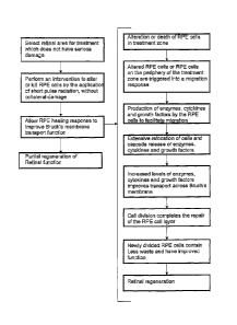
FIG. 4 is a sequential flow diagram which describes the method of
retinal regeneration and a detailed breakdown of the healing responses
following treatment. The initial assessment of impaired Bruch's membrane
function can be performed using the indicators mentioned previously but it is
intended that the method of retinal regeneration therapy is preferably
performed before geographic atrophy or CNV growth occurs. The central
area of the retina, known as the macular, has the greatest density of
photoreceptors and correspondingly the highest demand on the RPE/Bruch's
membranelchoriocappilaris complex and the highest rate of degradation, so
for this reason the general macular region is primary target for regeneration.
Because the improvement in Bruch's membrane transport extends beyond
the irradiated area a pattern of separated treatment spots may be applied to
treat a broad macular area. Areas in which the neuro-retina and RPE have
already died from geographic atrophy or CNV's have developed or any areas
of structural damage would not be selected for treatment.
RPE cells are pigmented with melanin contained within organelles
known as melanosomes 8 (see FIG 1) which perform the function of
absorbing light which has passed through the neuro-retina in order to prevent
back reflected light from degrading vision. Melanin absorbs light over a wide
wavelength range however for treatment purposes the wavelength range
from about 500nm to 900nm is preferred. The blue end of the spectrum is
usually avoided due to it's photo-toxicity and at wavelengths beyond the
infra-red end of the spectrum the amount of absorption reduces which allows
a greater amount of radiation to pass though the RPE and into the choroid.
Laser radiation is preferably used to deliver specific wavelengths and
a wavelength of 532nm would be useful to perform the method of this
invention, which can be obtained by frequency doubling the 1064nm laser
radiation from an Nd:YAG laser cavity.
A critical aspect of this method is the application of radiation which
can kill or alter RPE cells but cause no irreversible damage to the neuro-
CA 02667673 2009-04-27
WO 2008/049164 PCT/AU2007/001622
9
retina or other retinal layers or structures. To achieve this it is necessary
to
contain the effects of the energy absorption by the melanosomes within the
RPE cells. This is only possible if radiation energy is deposited into the
melanosomes in less than about 20ps, to prevent thermal diffusion beyond
the RPE cell membrane from occurring, however current retinal lasers
typically use 10 - 200ms pulse durations resulting in collateral damage as
shown in FIG. 5, causing irreversible damage to the neuro-retina. ln FIG 5
the laser beam 50 impinges on the RPE 2 and energy is absorbed in the
irradiated zone 51. However, thermal damage extends to a wider zone 52.
FIG. 6 shows the effect of shorter laser pulse durations of <20ps in
which thermal effects are contained within the RPE cells, allowing them to be
altered or killed without damage to the photoreceptors or other layers or
structures. Pulse durations less than 10ps are unlikely to be useful due to
mechanically disruptive effects caused by stress confinement within the
beam path. Pulse durations in the range 1 ns to 5ns are readily achievable
and most suitable. In FIG 6 the laser beam 60 impinges on the RPE 2 and
energy is absorbed to alter the RPE cells 61 without adjacent thermal
damage.
A laser system capable of this type of treatment has been described
in our co-pending patent application W02006021040 however other devices
which meet the described criteria could also be used. In particular it would
be
possible to use a flashlamp pumped, passively Q-switched Nd:YAG laser
cavity which is extra-cavity frequency doubled to produce 532nm pulses of
approximately 3ns in duration, similar to that described in our co-pending
patent application W02004027487. At this pulse duration energy absorption
by the melanosomes in granules within the RPE cells can readily produce
micro-bubbles which can be effective in killing or altering the RPE cells.
In laboratory experimentation it has been established that RPE cells
can be killed by intra-cellular micro-bubbles over a wide energy range without
rupturing cell membranes. In human explant samples in-vitro this range was
found to be from approximately 35 to 160mJ/cm2 when using 3ns pulses and
a wavelength of 532nm. Typically a sequence of three pulses is found to be
CA 02667673 2009-04-27
WO 2008/049164 PCT/AU2007/001622
appropriate although a single pulse or possibly 5 or more pulses may also be
suitable. A sequence of up to 5 pulses may be required to ensure that all
areas of the laser spot have received adequate irradiation, however a
cumulative thermal effect on the melanosomes is not required or desirable
5 so a low repetition rate is preferred.
The radiant exposure level required to kill or alter RPE cells, without
rupturing the cell membranes, will produce no visible effect when these short
pulse durations are used and in addition, the level of absorption will be
dependant on the melanin content of the RPE cells which varies from patient
10 to patient and with the region of the retina that is being treated. For
these
reasons it is useful to have a method of individual dose determination. This
can be simply achieved by using visual effect scaling in which the exposure
level required to produce a visual effect, such as bubbles or a lesion, can be
determined by applying higher energy radiation in the periphery of the retina
and then scaling down this level to an appropriate level for the regeneration
therapy. This process is known as visible effect scaling. A typical radiant
exposure which is at the threshold of producing a visible effect in the
periphery of the retina may be 160mJ/cm2 which could be produced using an
energy of 200pJ and a 400pm treatment spot. The energy may then be
scaled back to one third of that value, for example, and an energy setting of
67pJ used to deliver a radiant exposure of 53mJ/cm2 for performing the
retinal regeneration therapy.
Laboratory experimentation has shown that when 3ns pulses are used
the first visible effect is from the formation of a macro-bubble, which
results
from intra-cellular micro-bubbles bursting the RPE cell membranes and
coalescing into a visible macro-bubble. At this threshold level only minor non-
permanent damage occurs to photoreceptors making it an ideal energy level
marker to enable individual dose determination. Radiant exposure levels well
above the visible effect threshold are to be avoided to reduce the risk of
damaging photoreceptors. The optimum dose will use radiant exposure
levels which are able to internally damage the RPE cells and trigger acute, or
chronic, cell death without rupturing the cell membrane. Typically this may
CA 02667673 2009-04-27
WO 2008/049164 PCT/AU2007/001622
11
require a radiant exposure of 10mJ/cm2 to 400mJ/cm2 per pulse although a
range of nominally 30mJ/cm2 to 250mJ/cmZ.per pulse will generally be
appropriate
FIG. 4 also shows the sequence of cellular responses following the
retinal regeneration treatment which result in improved Bruch's membrane
transport and can be summarized as follows:
1. The alteration or death of RPE cells within the treated areas triggers
altered or undamaged RPE cells on the periphery of the laser treatment
zone to migrate, or initiate a migratory response, in order to restore the
continuity of the RPE monolayer. However, before the cells can migrate
they must degrade their attachment to Bruch's membrane and do so by
increasing their production and expression of enzymes such as active
matrix metalloproteinase (MMP), cytokines and growth factors.
Laboratory experimentation has shown the up-regulation of active MMP-9
following laser insult and the paper by Ahir A., Guo L., Hussain AA.,
Marshall J. (2002) Expression of metalloproteinases from human retinal
pigment epithelial cells and their effects on the hydraulic conductivity of
Bruch's membrane, Investigative Ophthalmology and Visual Science,
43(2): 458-65 has shown MMP up-regulation during cell migration.
2. The migration of cells then results in an extensive relocation of cells in
the surrounding areas and an accompanying cascade release of
enzymes, cytokines and growth factors. This causes an improvement in
the transport properties of Bruch's membrane in and around the treated
area. The paper mentioned above in 1. also shows the improvement in
the transport functions of Bruch's membrane following the application of
active MMP's and the proliferation of Human RPE cells.
3. Cell division completes the healing process of the RPE cell layer, leaving
no lasting damage to the target area or surrounding areas and the newly
CA 02667673 2009-04-27
WO 2008/049164 PCT/AU2007/001622
12
divided cells contain reduced waste products and are better able to
perform their functions, such as fluid transport.
The measured hydraulic conductivity of Human donor Bruch's
membrane is shown graphically in FIG 7, which demonstrates that the
theoretical improvement shown in FIG 3 can be obtained by initiating the
migration and division of RPE cells. The original hydraulic conductivities are
shown as the dashed line and circles at the data points. After measuring
conductivity, these samples of Bruch's membrane were plated with ARPE-1 9
cells and incubated for 24 hours. The RPE cells were then removed and
conductivities re-assessed (solid line with dots at the data points).
Proliferating ARPE-19 cells resulted in considerable improvement in the
hydraulic transport properties of ageing human Bruch's membrane.
In the figure, the dashed horizontal line refers to the minimum
hydraulic conductivity required to cope with fluid output from the RPE. These
ARPE-1 9 experiments show that elevation of ageing curves is possible in
order to avoid the early insults that can progress to macular disease.
This invention may be used to provide Retinal Regeneration Therapy
(2RT), in order to treat early age-related macular degeneration, diabetic
macular edema, or other diseases where the function of the neuro-retina is
compromised due to impaired function of the RPE/Bruch's
membrane/choriocapillaris complex. This procedure will be most effective in
the earliest stages of these diseases before permanent damage has
occurred to the neuro-retina or to delay retinal degradation through aging.