Note: Descriptions are shown in the official language in which they were submitted.
CA 02668296 2014-08-11
DEVICES AND METHODS TO SIMULATE AN OCULAR ENVIRONMENT
FIELD OF THE INVENTION
This invention related to devices and methods simulate an ocular
environment to enable the testing of ophthalmic lens.
BACKGROUND
Most diseases of the eye are treated with topical ophthalmic solutions
containing pharmaceutical agents. It has been postulated that delivery and
efficacy of these agents would be greatly increased if the agents were
incorporated in ophthalmic lenses and those lenses were used as drug delivery
devices. These agents may be added to the ophthalmic lenses by a variety of
methods including soaking the agent into a formed lens, adding the agent to
the formulation of the lens prior to its formation and the like. Others have
postulated methods of testing the uptake and discharge rates of such
pharmaceutical agents to and from the ophthalmic lenses. These methods
include placing ophthalmic lenses in solutions and monitoring the
concentration
of the pharmaceutical agent over time. Even though these methods work, due
to the volume of solution used in the test, the conditions do not mimic the
conditions that an ophthalmic lens is exposed to when inserted into an ocular
environment.
In an ocular environment, very small volumes of tear fluid pass over the
lens during its use. Therefore it would be beneficial if one could mimic those
conditions to test the performance of ophthalmic lenses. This need is met by
the following invention
BRIEF DESCRIPTION OF THE DRAWINGS
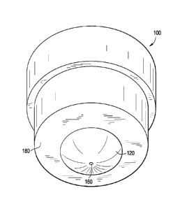
Fig. 1 illustrates a perspective drawing of a male mold.
Fig. 2 illustrates a perspective drawing of a female mold.
Fig. 3 illustrates a close up cross-sectional drawing of the mated apparatus.
Fig. 4 illustrates a perspective drawing of a male mold.
Fig. 5 illustrates a perspective drawing of a female mold.
CA 02668296 2009-04-30
WO 2008/055047 PCT/US2007/082584
Fig. 6 illustrates a cross-sectional drawing of the mated apparatus.
Fig. 7 illustrates a close up cross-sectional drawing of the mated apparatus.
DETAILED DESCRIPTION OF THE INVENTION
This invention includes an apparatus for testing an ophthalmic lens
comprising a male mold and a female mold,
wherein said male mold comprises a convex testing surface, an outer male
surface, male seating ridge extending from the perimeter of the convex
testing surface, and an aperture extending from said outer male surface to
said convex testing surface,
wherein said female mold comprises an outer female surface a concave
testing surface, female seating ridge extending from the perimeter of the
concave testing surface, and an aperture extending from said concave
testing surface to said outer female surface,
wherein when the male mold and the female mold are mated, the male
seating ridge sits on the female seating ridge and creates a testing area
between the male convex testing surface and the female concave testing
surface.
An embodiment of the invention is illustrated in the following figures.
Fig. 1, a perspective drawing of a male mold 100, contains convex
testing surface 120, aperture 160, and, male seating ridge 180. Fig. 2, a
perspective drawing of a female mold 200, contains concave testing surface
220, aperture 260, and female seating ridge 280. Fig. 3, a close up cross
section of mated apparatus, where the mated convex testing surface 120 and
concave testing surface 220, define the testing area 300 between those
surfaces. Preferably the convex and concave testing surfaces are of a size and
shape to mimic the shape of the eye and an eyelid. Testing area 300 is large
enough to hold an ophthalmic lens (not shown) and a volume of solution. It is
preferred that the testing area be sized to house an ophthalmic lens and about
50 pL to about 500 pL of solution, more preferably, about 100 pL to about
400 pL of solution, most preferably about 200 pL of solution.
2
CA 02668296 2009-04-30
WO 2008/055047 PCT/US2007/082584
The convex or concave testing surfaces of the apparatus may contain
grooves that provide pathways for the small volumes of solutions to pass over
the surfaces of ophthalmic lenses contained in the testing area. These grooves
may be in any number or orientation, but preferably a convex or concave
testing surface contains at least one latitudinal groove and one radial
groove. It
is preferred that such grooves intersect at a point on the convex or concave
testing surface. In an embodiment of the invention is illustrated in the
following
figures both the concave and the convex surfaces contain radial and
latitudinal
grooves.
Fig. 4, a perspective drawing of a male mold 10, contains convex testing
surface 12, four radial grooves 14, aperture 16, male seating ridge 18, and
six
concentric latitudinal grooves 19. Fig. 5, a perspective drawing of a female
mold 20, contains concave testing surface 22, four radial grooves, 24,
aperture
26, female seating ridge 28, and nine concentric latitudinal grooves 29. The
radial grooves on the convex testing surface intersect with the latitudinal
grooves so as to allow solutions that flow through the apertures, to more
easily
flow to the entire convex testing surface. This structural arrangement exists
on
the concave testing surface as well. Each testing surface may contain the
same or different numbers of radial grooves. It is preferred that each testing
surface contain contains at least two radial grooves, more preferably three
radial grooves, most preferably four radial grooves. With respect to
latitudinal
grooves, the number of these grooves on each testing surface may be the
same or different. It is preferred that each testing surface contain contains
at
least four latitudinal grooves, more preferably at least five latitudinal
grooves,
most preferably at least eight latitudinal grooves. The outer male and female
surfaces may be the same or different shapes. In one embodiment of the
invention (not illustrated) the outer male surface is concave and the outer
female surface is convex. Preferably, the convex and concave testing surfaces
are of a size and shape to mimics the shape of an eye and an eyelid.
Fig. 6, a cross section of the mated apparatus, with male mold 10,
female mold 20, male outer surface 13, female outer surface 23, male seating
ridge 18, and female seating ridge 28. As illustrated in Fig. 6, aperture 16
extends from male outer surface 13 to the convex testing surface 12 to the
3
CA 02668296 2009-04-30
WO 2008/055047
PCT/US2007/082584
testing area not shown. Further, aperture 26 extends from concave testing
surface 22 to outer female surface 23. Fig. 7, a close up cross section of
mated apparatus, where the mated convex testing surface 12 and concave
testing surface 22, define the testing area 30 those surfaces. Testing area 30
is large enough to hold an ophthalmic lens (not shown) and a volume of
solution. It is preferred that the testing area be sized to house an
ophthalmic
lens and about 50 pL to about 500 pL of solution, more preferably, about
100 pL to about 400 pL of solution, most preferably about 200 pL of solution.
The apparatus of the invention may be prepared from durable
thermoplastic materials, such as thermoplastic resins, polyolefins, and
thermoplastic polyesters. Examples of such materials include but are not
limited to low medium, medium and high density polypropylene, polyethylene
and co-polymers thereof, poly-4-methylpentene, fluorinated ethylene propylene
copolymers, ethylene fluoroethylene copolymers, polyacetal resins,
polacrylether, polyarylether sulfones, nylons, and the like. The apparatus may
be prepared by injection molding thermoforming and the like.
Further the invention includes a method of testing the diffusion rate of an
ophthalmic device comprising a pharmaceutical agent, wherein the method
comprises the steps of
(a) placing an ophthalmic lens comprising a pharmaceutical agent in
the testing area of an apparatus comprising a male mold and a
female mold,
wherein said male mold comprises a convex testing surface an
outer male surface, male seating ridge extending from the
perimeter of the convex testing surface, and an aperture
extending from said outer male surface to said convex testing
surface,
wherein said female mold comprises an outer female surface a
concave testing surface, female seating ridge extending from the
perimeter of the concave testing surface, and an aperture
4
CA 02668296 2009-04-30
WO 2008/055047 PCT/US2007/082584
extending from said concave testing surface to said outer female
surface,
wherein when the male mold and the female mold are mated, the
male seating ridge sits on the female seating ridge and creates a
testing area between the male convex testing surface and the
female concave testing surface.
(b) adding a solution to the aperture of the male mold from the outer
male surface, and
(c) monitoring the solution that emerges from the aperture of the
outer female surface to determine the presence or absence of the
pharmaceutical agent.
As used herein the term male mold, female mold, radial groove, latitudinal
groove, testing area convex testing surface, and concave testing surface are
as
described above.
As used herein, "pharmaceutical agents refers to pharmaceutical or
nutraceutical compounds used to treat conditions of the eye, and such
compound degrade in the presence of oxygen and certain transition metals.
Examples of pharmaceutical compounds include antihistamines, antibiotics,
antibacterial agents, antiviral agents, antifungal agents, analgesics,
anesthetics, antiallergeneic agents, mast cell stabilizers, steroidal and non-
steroidal anti-inflammatory agents, angiogenesis inhibitors; antimetabolites,
fibrinolytics, neuroprotective drugs, angiostatic steroids,
mydriatics,cyclopegic
mydriatics; miotics; vasoconstrictors; vasodilators, anticlotting agents;
anticancer agents, antisense agents, immunomodulatory agents, carbonic
anhydrase inhibitors, integrin antabonistsl; cyclooxygenase inhibitors, VEGF
antagonists; immunosuppressant agents and the like. Particularly, examples of
pharmaceutical compounds include but are not limited to acrivastine,
antazoline, astemizole, azatadine, azelastine, buclizine, bupivacaine,
cetirizine,
clemastine, cyclizine, cyproheptadine, ebastine, emedastine, ephedrine,
eucatropine, fexofenadine, homatropine, hydroxyzine, ketotifen, levocabastine,
levoceterizine, lomefloxacin, meclizine, mepivacaine, mequitazine,
5
CA 02668296 2009-04-30
WO 2008/055047 PCT/US2007/082584
methdilazine, methapyrilene, mianserin, naphazoline norastemizole,
norebastine, ofloxacin, oxymetazoline, pheniramine, phenylephrine,
physostigmine, picumast, promethazine, scopolamine, terfenadine,
tetrahydozoline, thiethylperazine, timolol, trimeprazine, triprolidine,
pharmaceutically acceptable salts and mixtures thereof. Preferred
pharmaceutical compounds include acrivatine, antazoline, astemizole,
azatadine, azelastine, clemastine, cyproheptadine, ebastine, emedastine,
eucatropine, fexofenadine, homatropine, hydroxyzine, ketotife, levocabastine,
levoceterizine, meclizine, mequitazine, methdialazine, methapyrilene,
norastemizole, norebastine, oxymetazoline, physootigmine, picumast,
promethazine, scopolamine, terfenadine, tetrahyerozoline, fimilol,
trimeprazine,
triprolidine, and pharmaceutically acceptable salts thereof. Particularly
preferred pharmaceutical compounds include phenarimine, ketotifen, ketotifen
fumarate nor ketotifen fumarate, 11-dihydro-11-(1-methyl-4-piperidinylidene)-
5H-imidazo[2,1-b][3]benzazepine-3-carboxaldehyde (CAS# 147084-10-4),
olapatadine and mixtures thereof. More particularly preferred pharmaceutical
compounds include ketotifen fumarate, 11-dihydro-11-(1-methyl-4-
piperidinylidene)-5H-imidazo[2,1-b][3]benzazepine-3-carboxaldehyde (CAS#
147084-10-4) and mixtures thereof.
Examples of nutraceutical compounds include vitamins and supplements
such as vitamins A, D, E, lutein, zeaxanthin, lipoic acid, flavonoids,
ophthalmicially compatible fatty acids, such as omega 3 and omega 6 fatty
acids, combinations thereof, combinations with pharmaceutical compounds and
the like. The methods of the invention may be used to detect the discharge
rate (or uptake rate) of ophthalmic lenses containing about 8 pg or more of
pharmaceutical agent. Preferably, the discharge rate for ophthalmic lenses
that
contain about 8 pg to about 90 pg, more preferably about 10 pg to about 40 pg,
more preferably about 10 pg to about 25 pg may be determined by the
methods of this invention.
As used herein, "ophthalmic lens" refers to a device that resides in or on
the eye. These devices can provide optical correction or may be cosmetic.
Ophthalmic lenses include but are not limited to soft contact lenses,
intraocular
lenses, overlay lenses, ocular inserts, and optical inserts. The preferred
lenses
6
CA 02668296 2014-08-11
of the invention are soft contact lenses made from silicone elastomers or
hydrogels, which include but are not limited to silicone hydrogels, and
fluorohydrogels. Soft contact lens formulations are disclosed in US Patent No.
5,710,302, WO 9421698, EP 406161, JP 2000016905, U.S. Pat. No.
5,998,498, U.S. Patent No. 6,087,415, U.S. Pat. No. 5,760,100, U.S. Pat.
No.5,776, 999, U.S. Pat. No. 5,789,461, U.S. Pat. No. 5,849,811, and U.S. Pat.
No. 5,965,631. The particularly preferred ophthalmic lenses of the inventions
are known by the United States Approved Names of acofilcon A, alofilcon A,
alphafilcon A, amifilcon A, astifilcon A, atalafilcon A, balafilcon A,
bisfilcon A,
bufilcon A, comfilcon, crofilcon A, cyclofilcon A, darfilcon A, deltafilcon A,
deltafilcon B, dimefilcon A, drooxifilcon A, epsifilcon A, esterifilcon A,
etafilcon
A, focofilcon A, genfilcon A, govafilcon A, hefilcon A, hefilcon B, hefilcon
D,
hilafilcon A, hilafilcon B, hioxifilcon B, hioxifilcon C, hixoifilcon A,
hydrofilcon A,
lenefilcon A, licryfilcon A, licryfilcon B, lidofilcon A, lidofilcon B,
lotrafilcon A,
lotrafilcon B, mafilcon A, mesifilcon A, methafilcon B, mipafilcon A,
nelfilcon A,
netrafilcon A, ocufilcon A, ocufilcon B, ocufilcon C, ocufilcon D, ocufilcon
E,
ofilcon A, omafilcon A, oxyfilcon A, pentafilcon A, perfilcon A, pevafilcon A,
phemfilcon A, polymacon, silafilcon A, siloxyfilcon A, tefilcon A, tetrafilcon
A,
trifilcon A, vifilcon, and xylofilcon A. More particularly preferred
ophthalmic
lenses of the invention are genfilcon A, lenefilcon A, comfilcon, lotrafilcon
A,
lotraifilcon B, and balafilcon A. The most preferred lenses include etafilcon
A,
nelfilcon A, hilafilcon, vifilcon, and polymacon.
The "solutions" that are used in methods of this invention may be water-
based solutions. Solutions that mimic natural tear film are preferred. Typical
solutions include, without limitation, saline solutions, other buffered
solutions,
and deionized water. The preferred aqueous solution is deioinized water or
saline solution containing salts including, without limitation, sodium
chloride,
sodium borate, sodium phosphate, sodium hydrogenphosphate, sodium
dihydrogenphosphate, or the corresponding potassium salts of the same.
These ingredients are generally combined to form buffered solutions that
include an acid and its conjugate base, so that addition of acids and bases
cause only a relatively small change in pH. The buffered solutions may
7
CA 02668296 2009-04-30
WO 2008/055047 PCT/US2007/082584
additionally include 2-(N-morpholino)ethanesulfonic acid (MES), sodium
hydroxide, 2,2-bis(hydroxymethyl)-2,2',2"-nitrilotriethanol,
n-tris(hydroxymethyl)methy1-2-aminoethanesulfonic acid, citric acid, sodium
citrate, sodium carbonate, sodium bicarbonate, acetic acid, sodium acetate,
ethylenediamine tetraacetic acid and the like and combinations thereof.
As used herein "monitoring" refers to methods of analyzing the solution
to determine the concentration of pharmaceutical agent in the solution.
Examples of such detecting methods include but are not limited to H PLC, UV
Spectormeters and the like.
Still further the invention includes, a method of measuring the uptake
rate of a pharmaceutical agent to an ophthalmic lens, wherein the method
comprises the steps of
(a) placing an ophthalmic lens in the testing area of an apparatus
comprising a male mold and a female mold,
wherein said male mold comprises a convex testing surface an
outer male surface, male seating ridge extending from the
perimeter of the convex testing surface, and an aperture
extending from said outer male surface to said convex testing
surface,
wherein said female mold comprises an outer female surface a
concave testing surface, female seating ridge extending from the
perimeter of the concave testing surface, and an aperture
extending from said concave testing surface to said outer female
surface,
wherein when the male mold and the female mold are mated, the
male seating ridge sits on the female seating ridge and creates a
testing area between the male convex testing surface and the
female concave testing surface.
(b) adding a solution comprising a pharmaceutical agent to the
aperture of the male mold from the outer male surface, and
8
CA 02668296 2009-04-30
WO 2008/055047
PCT/US2007/082584
(c) monitoring the solution that emerges from the aperture of the
outer female surface to determine the presence or absence of
the pharmaceutical agent.
As used herein the term male mold, female mold, radial groove, latitudinal
groove, testing area convex testing surface, concave testing surface,
pharmaceutical agent, ophthalmic lens, solution and monitoring are as
described above.
There are other circumstances when one would desire to test the
performance of an ophthalmic lens in an ocular environment, other than when
said ophthalmic lens contains a pharmaceutical agent. For example if one
wanted to determine whether surfactants, excipients, preservatives, wetting
agents or other components of solutions ("eyecare solution components") were
absorbed by the lens, it would be useful to have a test that mimics the
performance of the lens in an ocular environment. In light of this need this
invention includes a method of measuring the uptake rate of an eyecare
solution component to an ophthalmic lens, wherein the method comprises the
steps of
(a) placing an ophthalmic lens in the testing area of an apparatus
comprising a male mold and a female mold,
wherein said male mold comprises a convex testing surface an
outer male surface, male seating ridge extending from the
perimeter of the convex testing surface, and an aperture
extending from said outer male surface to said convex testing
surface,
wherein said female mold comprises an outer female surface a
concave testing surface, female seating ridge extending from the
perimeter of the concave testing surface, and an aperture
extending from said concave testing surface to said outer female
surface,
wherein when the male mold and the female mold are mated, the
male seating ridge sits on the female seating ridge and creates a
9
CA 02668296 2009-04-30
WO 2008/055047
PCT/US2007/082584
testing area between the male convex testing surface and the
female concave testing surface.
(b) adding a solution comprising eyecare solution components to the
aperture of the male mold from the outer male surface, and
(c) monitoring the solution that emerges from the aperture of the
outer female surface to determine the presence or absence of the
eyecare solution component.
As used herein the term male mold, female mold, radial groove, latitudinal
groove, testing area convex testing surface, concave testing surface, eyecare
solution component, ophthalmic lens, solution and monitoring are as described
above.
The specific embodiments of the apparatuses and methods of the
invention illustrate, but do not limit the invention. They are meant only to
suggest a method of practicing the invention. Those knowledgeable in contact
lenses as well as other specialties may find other methods of practicing the
invention. However, those methods are deemed to be within the scope of this
invention.