Note: Descriptions are shown in the official language in which they were submitted.
CA 02668341 2009-04-30
Catheter
Description
The present invention relates to catheters and introducing
the same in general, and to venous catheters in embodiments
and central venous catheters in particular, such as in the
form of multi-lumen catheters, for example.
A venous catheter, or also a central venous catheter,
designates a flexible tube with one or more channels and/or
lumens therein, which is introduced into the venous system
until its distal end is short of the heart, and so that it
is possible to take measurements through the lumen openings,
such as of the central venous pressure, and/or to supply
liquid, such as electrolyte and nutrient solutions or
medicine. A single-lumen catheter only possesses one lumen,
the opening of which is at the distal end of the catheter,
for example. In multi-lumen catheters the lumen openings may
be arranged in a spaced manner along the length of the
catheter so as to prevent the reagents administered via the
lumens from interfering with each other. The introduction or
catheterization of the venous catheter is mostly done via a
central vein by way of the Seldinger technique, wherein at
first a guidewire is placed at the target position via a
needle. Via the guidewire, the catheter is advanced and
guided up to a desired position so as to end there. The
navigation and/or the positional control of the wire may,
for example, take place via an electrocardiographic lead
(ECG lead) and by utilizing the electrical impulses
triggering the heartbeats, and particularly utilizing the
polarization reversal in the case of the guidewire gliding
past the sinus node. Once the guidewire has reached the
- 1 -
CA 02668341 2010-09-09
desired position, the venous catheter is advanced via the
guidewire until its proximal end corresponds to a
corresponding mark on the guidewire, whereupon the guidewire
is removed again. As an alternative to or in addition to the
ECG positional control of the venous catheter, the
positional control may take place by means of X-ray, to
which end the material of the flexible venous catheter is
constituted of barium sulfate, for example, to a great
extent.
A relatively great effort regarding navigation or positional
control of the distal end of the venous catheter is a
disadvantage of the previously outlined procedure, because
the ECG lead does not always provide reliable results or may
only be employed to a limited extent, and the X-ray control
is connected with high costs and much time involved.
Hence, it is the object of the present invention to provide
a catheter, a catheter system as well as a method of
preparing a catheter for introducing the same, so that more
reliable and/or more effortless introduction of a catheter
is possible.
This object is achieved by a catheter according to one
aspect of the present invention, a catheter system according
to another aspect of the present invention, and a method of
preparing a catheter for introducing the same according to
yet another aspect.
According to an aspect of the present invention, there is
provided a catheter with an ultrasound marking means, which
is recognizable by means of ultrasound in the body of a
living being, at a distal end thereof, wherein the catheter
- 2 -
CA 02668341 2010-09-09
comprises a lumen, and the ultrasound marking means is
arranged in the lumen near the distal end such that it is
removable via a proximal end of the catheter, wherein the
ultrasound marking means is a tube closed at its distal end,
which is introduced into the lumen.
According to another aspect of the invention, there is
provided Catheter system, comprising a catheter with a
lumen, and an ultrasound marking means insertable into the
lumen of the catheter, which is recognizable in the body of
a living being by means of ultrasound and is elongated so as
to be able to extend from near a distal end of the catheter
through the lumen beyond a proximal end of the catheter to a
connection of the catheter so as to project from the
connection, wherein the ultrasound marking means is a tube
closed at its distal end, which is insertable into the
lumen.
According to a further aspect of the invention, there is
provided Method of preparing a catheter for introducing the
same, comprising, inserting an ultrasound marking means,
which is recognizable in the body of a living being by means
of ultrasound and is elongated so as to be able to extend
from near a distal end of the catheter through the lumen
beyond a proximal end of the catheter to a connection of the
catheter so as to project from the connection, into the
lumen to the proximity of the distal end, wherein the
ultrasound marking means is a tube closed at its distal end.
It is a central idea of the present invention that, with
relatively little effort, a catheter can easily be made
visible in ultrasound images, and hence positional control
-2a-
CA 02668341 2010-09-09
of a catheter can be enabled with relatively little effort,
by providing an ultrasound marking means, which is
-2b-
CA 02668341 2009-04-30
recognizable in the body of a living being by means of
ultrasound, at the distal end of a catheter, for example
temporarily, so as to be removed again after having
successfully guided the distal end of the catheter to a
target position of the ultrasound marking means.
According to one embodiment, it is provided to introduce a
passive or active elongated probe into a lumen of a catheter
for temporary use during the catheter navigation or
positional control and the ensuing removal and to package
the same in this form as a catheter system, for example, and
to sterilize the same.
Preferred embodiments of the present invention will be
explained in greater detail in the following with reference
to the accompanying drawings, in which:
Fig. 1 is a schematic drawing of a two-lumen catheter;
and
Fig. 2 is a schematic sectional view of the distal end of
the two-lumen catheter of Fig. 1, according to a
comparative example;
Fig. 3 is a schematic drawing of a sectional view of the
distal end of the two-lumen catheter of Fig. 1,
according to a further comparative example;
Fig. 4 is a sectional view of the two-lumen catheter of
Fig. 2 and/or Fig. 3 along the sectional line A-A;
Fig. 5 is a schematic sectional view of a distal end of a
single-lumen catheter with a passive probe of a
- 3 -
CA 02668341 2010-09-14
catheter system introduced, according to an
embodiment of the present invention; and
Fig. 6 is a schematic drawing of the proximal end of the
arrangement of Fig. 5.
Before explaining embodiments of the present invention in
greater detail in the following with reference to the
drawings, it is pointed out that like elements are provided
with like reference numerals in these figures, and that
repeated description of these elements will be omitted.
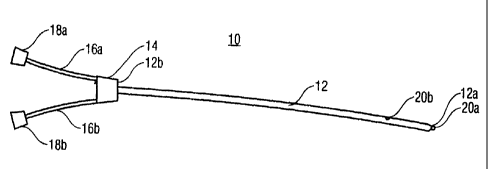
Fig. 1 exemplarily shows a two-lumen catheter which may be
employed as a venous catheter, for example. The catheter of
Fig. 1, generally shown at 10, includes a flexile catheter
tube 12 with a distal end 12a and a proximal end 12b, a
distributor or manifold 14, two connecting tubes 16a and
16b, and two plug connections 18a and 18b, which may be
formed as screwed connectors and are also sometimes referred
to as Luer-Lock connections in the following. The catheter
tube 12 comprises, in its interior (not visible in Fig. 1),
two channels or lumens, one of which opens at a lumen
opening 20a arranged at the distal tip, and the other lumen
opening at a lateral opening 20b arranged at a distance from
the distal tip or more proximally, but still near the distal
end 12a. The distributor 14 connects one of the lumens to
the connecting tube 16a and the other lumen to the
connecting tube 16b, which in turn are connected to one of
the Luer-Lock connections 18a, 18b each, at the proximal end
12b, so that further tubes can be connected thereto for the
supply of liquid or taking blood or other fluidic coupling
to the lumen openings 20a, 20b.
- 4 -
CA 02668341 2009-04-30
Fig. 2 shows the distal end 12a of the tube 12 in greater
detail, for a comparative example of the present invention.
What can be seen are the two lumens 22a and 22b of the
catheter tube 12a, which extend therealong and, as shown in
Fig. 4, have different cross-sections. Alternatively, they
may, of course, also have the same cross-section. As can be
seen, the lumen 22b opens at the lateral lumen opening 20b,
which is arranged slightly distant from the distal tip 24 of
the catheter, whereas the lumen 22a opens at the lumen
opening 20a at the distal tip 24. However, the lumen 22b
extends slightly beyond the lumen opening 20b in the
direction of the distal tip 24, so as to end at the distal
tip 24 at a part 24a of the tip 24. The lumen 22b thus
forms, from the lumen opening 22b in the direction of the
distal tip 24, a sack opening, which may optionally be
filled partially or completely with material 26 at its
distal end, as will still be described in the following in
connection with the description of manufacture. However,
this sack opening further forms a cavity 28 closed by a
closing element 30 at the open end of the sack opening
and/or near the lumen opening 20b. The closing element 30
closes the cavity 28 in a fluid-tight manner in the part of
the lumen 22b that is distal with respect to the lumen
opening 20b, and for example consists of the same material
as the material 32 forming the lumens 22a and 22b. For
example, the cavity 28 is filled with air, but may also be
filled with a vacuum or a foamed or porous material.
The two-lumen catheter according to Figs. 2 and 4 offers
advantages when used as a venous catheter, for example. So
as to illustrate this, introducing the catheter will be
described in the following. At first, the multi-lumen
catheter is introduced into the patient, such as into a vein
- 5 -
CA 02668341 2009-04-30
of the neck. After the distal end 12a has been introduced
into the vein, the catheter 10 is advanced further along the
vein in the direction of the heart, wherein this may be done
with or without using a guidewire. In the patient's body,
the two-lumen catheter 10 is observed by means of
ultrasound. Although the material 32 of the catheter cannot
be visualized very well in the ultrasound images due to the
low acoustic impedance difference to the surrounding blood,
the cavity 28 located near the distal tip 24 certainly can,
which cavity reflects the ultrasound waves of an ultrasound
transducer well because of its interface from the
acoustically thick material 32 or material 32 of high
acoustic impedance to the interior of the cavity 28, which
is acoustically thin and/or has lower impedance, thus
leading to high contrast in the images. Depending on the
observation by means of ultrasound and/or the location of
the cavity 28 in the ultrasound images, the catheter may
then be navigated and/or advanced to the desired position,
for example such that the tip 24 is positioned immediately
before the heart. Advantageously, ultrasound devices mean
less effort, bulk and cost and are less complex in their
handling than X-ray devices, so that the positional control,
when introducing the catheter according to the Figs. 2 and
4, is simplified as compared with an X-ray solution.
Before a further example of a two-lumen catheter is
described with reference to Fig. 3, one possible
manufacturing method for the catheter of Fig. 2 will be
described in the following. At first, a catheter tube with
the lumens 22a and 22b is produced from the material 32,
such as by extruding. The result is a tube with a
continuously constant cross-section, such as with that of
Fig. 4. Then, the lumen 22 is closed at the distal end 12a,
- 6 -
CA 02668341 2009-04-30
so that the lumen 22a still has its lumen opening 20a at the
tip of the catheter tube at the distal end 12a, however. To
this end, for example, the lumen 22b is closed with a
material 26 at the distal sectional edge of the extruded
tube, and a tip 24 with the closing part 24a is attached to
the sectional area thus prepared. The material 26 and the
material of the tip 24 may each be equal to or different
from the material 32 of the catheter tube. The material 32
is, for example, polyurethane with or without a barium
sulfate proportion. Closing the lumen 22b may also be
performed via thermal closing and a thermal and/or
mechanical shaping of the tip 24 to the shape shown in Fig.
2. Prior to or following the formation of the tip 24 and/or
distal-side closure of the lumen 22b, the lateral lumen
opening 20b is formed, such as by a cutting process taking
place in a transverse direction. Then, the lumen 22b is
closed by means of the closing element 30, wherein glue,
which is brought to the desired position through the opening
20b, is used as the closing material 30, for example.
Alternatively, the same material as the material 32 or
material 26 may be used for the element 30, which is
suitably connected to the material 32, such as by means of a
solvent or an adhesive. The atmosphere present when closing
the cavity with the element 30, such as air, then is in the
cavity 28.
As an alternative, it is, of course, also possible to
introduce the closing element 30 from the distal side
through the lumen 22b and/or the cavity 28, which still is
open at the distal end at this time, into the lumen 22b
prior to closing the lumen 22b and/or the formation of the
distal tip 24, and then finally close the cavity 28 only
later when forming the tip 24. The formation of the lumen
- 7 -
CA 02668341 2009-04-30
opening 20b could also take place in another order with
respect to the remaining steps. Furthermore, prior to
closing the cavity 28 by the closing element 30 and/or the
part 24a of the tip 24, it could be filled with a foamed or
porous material 34. Due to its air and/or gas inclusions,
this material also leads to a good contrast in ultrasound
images. This foamed material would then be separated from
the surrounding serum, such as blood, by the closing element
30 and/or the material 32, so that its possibly rugged
surface does not offer any point of attack for biological
growth or fouling and/or the attachment of germs, and the
surface of the catheter may still be continuously smooth to
prevent attachments of such germs. As an alternative, the
closing material may be omitted, and the porous material 34
may be produced with a smooth interface, an example of which
will be explained in the following with reference to Fig. 3.
Fig. 3 shows an example alternative to Fig. 2 of a distal
end 12a' of the two-lumen catheter of Fig. 1, but with the
cross-section A-A from Fig. 4 also applying to this example.
The comparative example of Fig. 3 differs from that of Fig.
2 in that the portion of the lumen 22b with the lateral
lumen opening 20b, which extends from the opening 20b to the
distal end of this lumen 22b, is not closed in a cavity, but
is only filled with foamed material 34 having inclusions,
which again have the above-outlined advantages in the
ultrasound imaging. For example, polyurethane with or
without suitable additives may be used as the foamed
material 34. Similarly, as described previously with respect
to the closing element 30, the foamed material 34 may be
introduced through the lateral lumen opening 20b when
producing the catheter of Fig. 3 sometime after forming the
opening 20, and preferably also the attachment of the tip,
- 8 -
CA 02668341 2009-04-30
or through a distal opening of the lumen 22b at a time at
which the tip 24 with the closing part 24a has not yet been
attached and/or formed. With respect to the advantages of
the catheter of Fig. 3 when introducing same, reference is
made to the statements regarding the catheter of Fig. 2,
which also apply to the comparative example in this respect.
Regarding the manufacture of the catheter of Fig. 3, the
above statements with respect to Fig. 2 apply, unless
described in a deviating manner with respect to Fig. 3.
Although part of the rough surface of the foamed material
(34) is exposed to the surrounding serum, such as blood, in
the catheter of Fig. 3, the risk of germ contamination or
attachments is very low, since the zone is very limited
locally. After all, the material 34 may also be produced
with a smooth interface 34a.
Finally, it is to be mentioned that the above comparative
examples have dealt with a two-lumen catheter only
exemplarily, and that they are also applicable to multi-
lumen catheters with more lumens. In general, in an n-lumen
catheter with n-1 lateral lumen openings or lateral eyes,
the portion of each of these n-1 lumens arranged between the
respective lateral eye and the end of the lumen directed to
the distal end of the multi-lumen catheter may be used to
form a closed cavity 28 and/or accommodate foamed material
34. Furthermore, it is pointed out that the cross-sections
visible in Fig. 4 also only are of exemplary nature. Thus,
the outer perimeter of the cross-section of the catheter
tube 12 may also be shaped in a different way than circular,
as shown in Fig. 4. As previously mentioned, the material 26
further may be missing at the end of the lumen 22b.
9 -
CA 02668341 2009-04-30
In the foregoing, it has been described that the material 34
possesses gas bubbles. Other materials are also possible,
however, namely ones mixed with ultrasound-visible
substances. This applies both to the material 34 in Fig. 3
and to the "filling" of the cavity 28 in Fig. 2. In the
present application, for example, something is to be
considered recognizable or visible in the body of a living
being by means of ultrasound if it actively sends out
ultrasound or reflects ultrasound impinging through the
inside of the body with a sufficient reflection coefficient,
such as by using a material with an acoustic impedance of,
for example, less than 10% of the acoustic impedance of
water or less than 10% of the acoustic impedance of the
catheter material, in the interior of the catheter.
Moreover, the above comparative examples may be modified in
that, instead of providing a "passive" ultrasound marking in
the form of the cavity 28 and/or the material 34, an active
ultrasound marking means is provided at the distal end of
the multi-lumen catheter, or at least near the lumen
openings. For example, in the part of the lumen 22b between
the lumen opening 20b and the proximal end thereof, an
active "probe" in the form of an ultrasound-emitting
transmitter may be inserted. Preferably, this active probe
has its own energy supply in the form of a battery, for
example. The active probe could merely be put into the lumen
22b, preferably only up to the proximity of the lateral eye
20b, or even all the way to the sack opening of the lumen
22b.
Instead of an active probe generating or emitting ultrasound
signals, however, also a "passive" probe in the form of a
gas-filled ball or a gas-filled tube, which is closed at the
- 10 -
CA 02668341 2009-04-30
front and the rear, could be inserted and/or mounted in the
lumen 22b, such as up to the opening 22b or up to the cavity
28 and/or the sack opening from Fig. 2 and from Fig. 3,
respectively.
The insertion of the passive or active probe, and/or the
manufacture of a multi-lumen catheter with such a probe, may
take place in the prescribed way by inserting the probe into
the cavity 28 in the manufacture according to Fig. 2, for
example, before the same is closed by the material 30 or the
distal tip 24, or by replacing the material introduction
step for the material 34 in the manufacture according to
Fig. 3 by a probe introduction step. The advantages
resulting by way of the modified comparative examples just
described with an active and passive probe in connection
with introducing the catheter are easily obvious from the
description of the examples of Figs. 2 and 3, which also
applies to the modifications in this respect, even though,
when using an active probe, the advantage does not lie in
the utilization of the high reflectivity at the interface
from acoustically thick to acoustically thin material, but
in the utilization of the ultrasound signal directly emitted
by the active probe. A suitable active probe may comprise
means for activating and maybe deactivating the ultrasound
signal generation, depending on a triggering event, such as
depending on an electromagnetic trigger signal, so that the
ultrasound signal generation can be limited to the time
period of the employment of the catheter.
The preceding comparative examples dealt with catheters in
which the ultrasound marking means in form of the cavity 28,
the material 34 and/or the passive/active probe has been
provided to permanently remain in the multi-lumen catheter,
- 11 -
CA 02668341 2009-04-30
and particularly in the otherwise unused part of a lumen
with a lateral eye, even though, in the case of the merely
inserted passive/active probe, the same basically could be
removed via the lumen 22b. In the following embodiment, the
introduction of a catheter accurately to a target position,
such as short of the heart, is facilitated by the fact that
a lumen of a catheter, which may also be a single-lumen
catheter in this embodiment, is temporarily provided with a
passive or active probe that may be removed again through
the lumen. Such an embodiment will be explained in greater
detail with reference to Figs. 5 and 6, which show the
distal end 50 and/or the proximal end 52 of a catheter 54,
at the proximal end 52 of which a connection 56 is arranged,
such as a Luer-Lock connection. According to the embodiment
of Figs. 5 and 6, the catheter 54 is a single-lumen catheter
with a lumen 58 and forms, together with a passive probe 60,
which is also shown, a catheter system facilitating the
introduction of the catheter 54, as described in the
following. To this end, the passive probe 60 here
exemplarily is formed as a flexible tube, with the outer
perimeter of its cross-section being small enough for the
tube 60 to be introduced into the lumen 58 via the
connection 56 or the proximal end 52. In particular, the
tube 60 is long enough so as to extend from the distal end
50 through the lumen 58 beyond the connection 56 to the
outside. More specifically, the tube 60 is long enough so as
to extend from a proximity of a lumen opening 62 of the
lumen 60 at a lumen tip 64 of the distal end 50 through the
lumen 58 beyond the connection 56, wherein a marking 66 is
provided visibly on the exterior of the tube 60 for an
operator, so as to be at a predetermined distance to the
connection 56, such as immediately in front of it, at a
position of the tube 60 as inserted up to the proximity of
- 12 -
CA 02668341 2009-04-30
the tip 62. The tube 60 is hollow on the inside and further
is closed at its distal end 50a and also at its proximal end
60b, for example, so as to form a gas-filled space 68 on the
inside, for example.
After the components of the catheter system of Figs. 5 and 6
have been described in the foregoing, its use when
introducing the catheter 54 will be described in the
following. Prior to the use and/or the introduction of the
catheter 54, or during the use and/or the introduction, the
catheter 54 and the flexible tube 60 are, for example, in
the state shown in Figs. 5 and 6, namely in the state of the
tube 60 being introduced with its distal end 60a up to the
proximity of the distal tip 62 of the catheter 54 into the
lumen 58, and packaged in a sterile way, for example. Once
the catheter 54 has been introduced into a vein or
catheterized, for example, it is advanced along the vein to
the proximity of a target position, such as before the
heart. Since the tube 60 is in the interior of the lumen 58
as a passive probe and extends up to the proximity of the
tip of the lumen 62, the position of the catheter tip 62
and/or the entire catheter 54 relative to the target
position can be controlled by means of ultrasound, as
described previously, and thus the navigation of the
catheter 54 with its tip 62 can be guided to the target
position, since good contrast of the passive probe 60
results from the interface of the tube material to the tube
interior 68 in the ultrasound images. On the basis of the
marking 66, the physician here can ensure that the distal
end 60a is in the desired proximity to the catheter tip 62.
The remaining offset, as can be seen in Fig. 5, for example
is known to the operator in advance, so that he or she may
take same into account in the positional control during the
- 13 -
CA 02668341 2009-04-30
observation of the ultrasound images. After having guided
the catheter tip 62 to the target position in this manner,
as desired, the passive probe 60 can be removed therefrom
through the lumen 58, to which end the tube 60 can be drawn
at its projecting proximal end. The passive probe 60 here is
provided as a disposable article, for example.
For preparing the introduction of the catheter 54, the tube
60 thus has been introduced into the lumen 58 up to the
distal end 50, so as to then be packaged and sterilized
together with the catheter 54, for example, such as by a
packaging permeable for sterilization gas.
With reference to the previous embodiment of Figs. 5 and 6,
it is pointed out that the same is not limited to a single-
lumen catheter. Rather, a passive probe 60 can, of course,
also be employed in a multi-lumen catheter, wherein the
introduction may take place into each of the lumens, such
as, but not exclusively the lumen with its lumen opening at
the catheter tip. Furthermore, it pointed out that, instead
of the passive probe described, also an elongated active
probe could be used, which, for example, has a similar
exterior as the previously described flexible tube 60, but
has an ultrasound transmitter at its distal end. Finally,
also a solid flexible rod may be used as a passive probe, as
long as it enables ultrasound visibility, or a flexible rod
of foamed material or the like.
Although it has previously been described that the passive
probe 60 is packaged together with the catheter 54 in an
introduced state, for example, it may also be provided for
both components of the catheter system, namely the catheter
54 and the passive probe 60, to be packaged separately. When
- 14 -
CA 02668341 2009-04-30
introducing the catheter 54, it may at first be introduced
into a vein, for example, and advanced a little, so as to
then introduce the passive probe 60 into the lumen 58.
Either the passive probe 60 may be advanced therethrough
through the vein to the target position, in order to let the
catheter 54 follow only when the distal end 60a of the
passive probe reaches the target position as controlled by
ultrasound, and then remove the passive probe 60 from the
lumen 58 like in the foregoing, or the catheter is navigated
to its target position via ultrasound control with the aid
of the probe.
Finally, it is to be pointed out that it is possible to
integrate, into the existing software of an ultrasound
device, a software routine acting as a means for displaying
a signal for catheter navigation, and capable of recognizing
the ultrasound marking means, such as the closed cavity
and/or the foamed material filling, or the passive/active
probe in the ultrasound images of the ultrasound image
sequence generated while one of the previously described
catheters is pushed along the vein in the direction of the
heart, and also recognize the location thereof, and display
a signal for the physician, for example, on the basis of
this recognized location, serving for navigation of the
multi-lumen catheter to the desired position before the
heart. For example, an acoustic sound is given off at the
moment at which the catheter tip glides past the desired
location, wherein the software routine to this end also
takes into account the longitudinal offset between the
ultrasound marking means, such as the cavity (28) and/or the
foamed material (34) or the active/passive probe observed in
the ultrasound images, to the tip (24), for example. Of
course, the navigation signal display routine just described
- 15 -
CA 02668341 2009-04-30
may also be implemented in firmware or in hardware.
Moreover, the routine also may recognize the target position
automatically, for example by the user only indicating to
the routine that "the distal end" is to be arranged "before
the venous entry of the heart" and/or a "central venous
catheter" is desired, whereupon the routine automatically
recognizes this location from certain features in the
ultrasound images.
Finally, it is still to be pointed out that the present
invention is not limited to venous catheters. Rather, the
present invention may also be applied to other catheters and
to other catheter applications in serums other than blood.
- 16 -