Note: Descriptions are shown in the official language in which they were submitted.
CA 02668356 2009-05-01
WO 2008/052795 PCT/EP2007/009528
1
Thrombinoscope B.V.
07/181 PCT
Method for measuring the concentration of transient
proteolytic activity in composite biological media containing cells
Field of the Invention
The present invention is in the field of diagnostics and relates more
particularly
to a method for monitoring, in real time, the course of the concentration of
biologically
active enzymes that are transiently present in blood or other body fluids, and
to a kit for
use in this method.
Background of the Invention
In body fluids, there exist several physiologically important biochemical
systems that act through activation and subsequent inactivation of proteolytic
enzymes,
such as, in blood, the coagulation system, the fibrinolytic system and the
complement
system, and in gastro-intestinal juices the digestive enzymes. For the
assessment of
biological function of these systems it is important to be able to follow the
course of such
proteolytic activity in time. Such function assessment is of paramount
diagnostic
importance because disturbances of such systems can lead to fatal diseases
like coronary
infarction, stroke or fatal bleeding (blood coagulation and fibrinolysis),
generalised
infections and autoimmune diseases (complement system) or disturbed adsorption
of food
(gastrointestinal juices).
In methods of determining the thrombin generation in plasma or platelet-rich
plasma according to the state of the art, the plasma turns into a gel after
several minutes.
This is due to the conversion of fibrinogen into insoluble fibrin. This fibrin
polymerizes, so
that a fibrin-network is formed which, in fact, is the actual clot. This clot
is similar to a
sponge that contains fluid (i.e. plasma or serum) in which thrombin is
generated. During
this generation of thrombin the signal substrate is converted into a signal-
producing
leaving group. Since the gel is relatively transparent it is possible to
record the fluorescent
signal in time in clotting plasma. However, in case of measurement in whole
blood the
amount of signal is considerably reduced (typically less than 5% of the amount
measured
in clear plasma) and any movement of the erythrocytes greatly influences the
signal. The
fibrin network (the gel) that is formed captures the red blood cells and after
a while "clot
retraction" occurs that results in inhomogeneity of the gel thereby separating
fluid captured
inside the gel from fluid that is present outside the fibrin network.
Following the picture of
the "spongy network", clot retraction results in a situation that is similar
to part of the
CA 02668356 2009-05-01
WO 2008/052795 PCT/EP2007/009528
2
sponge being squeezed out. This has such an influence on the signal that
measurement
would be almost impossible.
WO 03/093831 describes a suitable technique to measure the activity of
thrombin in real time in plasma or platelet-rich plasma. The technique
includes the addition
of a signal substrate to said biological medium. The proteolytic enzyme is
able to convert
the substrate, and the leaving group of the substrate can be measured with an
appropriate
technique. This can be the measurement of fluorescence, optical density, NMR,
and the
like, the choice of which mainly depends on the nature of the signal. When
fluorescence is
used, then it is in principle possible to measure in turbid solutions such as
platelet-rich
plasma or plasma that contains fibrin. Also measurement in whole blood, i.e.,
plasma
containing platelets, white blood cells and erythrocytes, would be possible.
However, the
presence of red blood cells has a great disturbing impact on the signal.
Therefore, there is still a need for a method for measuring the thrombin
generation in real time in whole blood in a reliable and simple manner. The
present
invention provides such a method.
Summary of the invention
We have now surprisingly found that when the measurement of proteolytic
activity, in particular thrombin activity, substantially as described in WO
03/093831 is
carried out in clotting blood or plasma under frequent mixing conditions, the
clot will not be
formed as a gel but as a much denser clot thereby leaving substantially all
fluid outside the
fibrin network.
Accordingly, in one aspect of the present invention a method is provided for
determining in real time the course of proteolytic activity, in particular
thrombin activity, in a
sample of clotting blood or plasma or other body fluid as it appears in and
disappears from
the sample, which comprises the following steps:
a) adding a signal substrate to said sample, said signal substrate causing a
detectable signal related to the amount of conversion product formed upon
reaction by the
generated proteolytic activity,
b1) monitoring the signal development in time in said sample to provide a
curve, and
c1) mixing said sample frequently so that clot formation occurs in a dense
manner such that the majority of the sample remains fluid and that cell
precipitation is
inhibited,
wherein said steps b1) and c1) are repeated and performed in an alternate
way.
CA 02668356 2009-05-01
WO 2008/052795 PCT/EP2007/009528
3
In a particular and preferred embodiment said method comprises the following
additional and alternative steps:
d) adding a means with a constant known stable proteolytic activity on the
signal substrate as defined in step a) but otherwise inert, to a second
parallel sample in
which no proteolytic activity is triggered,
e) adding the same signal substrate as defined in step a) to step d), said
signal
substrate causing a detectable signal upon reaction by the means with known
stable
proteolytic activity,
b2) determining the time course of signal development in said first sample and
said second parallel sample to provide a curve from each of them,
f) comparing said curves to derive the course of proteolytic activity in time
in
the first sample, and
c2) mixing the first and the second sample frequently so that clot formation
occurs in said first sample in a dense manner such that the majority of said
first sample
remains fluid and that cell precipitation in each sample is inhibited,
wherein said steps b2) and c2) are repeated and performed in an alternate
way.
In another aspect of the invention a kit is provided for carrying out the
method
of the invention.
These and other aspects will be described in more detail below.
Brief description of the drawings
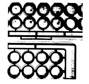
Figure 1 is a picture taken from a selection of a 96-well plate that was
frequently mixed during the formation of the clot (top) and another 96-well
plate that was
not mixed. In the top picture it is seen that a dense clot is formed in each
well. In the
bottom picture the clear plasma has turned into a turbid gel. Mixing was done
by an orbital
shake of 5 seconds that was repeated every 20 seconds. The picture shows an
example of
what can be assumed will also happen when whole blood is used: under shaking
conditions the clot is more dense.
Figure 2 is a typical example of a measurement of thrombin generation in
whole blood. The reaction mixture contained whole blood from a healthy
volunteer, as well
as 0.5 pM Tissue Factor, fluorogenic substrate (400 pM ZGGR-AMC) and calcium
chloride. Fluorescence was measured in a 96-well plate fluorometer and
thrombin in time
was calculated from the signal.
Figure 3 shows the relationship between initial rate (fluorescent units per
minute) of a thrombin calibrator curve and the hematocrit. The thrombin
calibrator is a
CA 02668356 2009-05-01
WO 2008/052795 PCT/EP2007/009528
4
complex of thrombin with alpha2-macroglobulin (a2-M), which is capable of
converting the
fluorogenic substrate (ZGGR-AMC), but is not inhibited by plasmatic inhibitors
of thrombin
(such as anti-thrombin), and it does not participate in the coagulation
process. The
thrombin calibrator converts the substrate, and the fluorophore that is
measured produces
a fluorescent signal that is measured in a fluorometer. Although the amount of
activity is
equal in all samples, the initial rate differs due to the haematocrit (ratio
of cell volume and
total volume of whole blood). Since the haematocrit differs considerably from
donor to
donor, the initial rate of the thrombin calibrator can be used to correct for
these differences.
Detailed description of the invention
By "frequent mixing conditions", as used herein, we mean moving the
container in which the reaction takes place or the fluid within the container
such that the
fluid and non-fluid substances in the fluid, such as cells, become more
uniformly
dispersed. Evidently, a substantially uniform dispersion would be the
preferred condition.
For example, by performing an orbital shake of the container in which thrombin
generation
takes place, the clot will not be formed as a gel but rather as a much denser
clot thereby
leaving substantially all fluid outside the network. This can also be achieved
by a rotating
magnetic stirrer inside the fluid or by another type of stirrer which is known
to a person
skilled in the art. As a result, the devastating influence of clot retraction
on the signal no
longer occurs.
The container in which thrombin generation occurs, usually a cuvette, is
normally transparent and positioned inside a temperature-controlled apparatus
which is
able to record the signal produced by the conversion of the substrate. When
measurement
of fluorescence takes place from the top or the bottom of the container, or
cuvette, then
precipitation of red blood cells will have an additional disturbing effect on
the signal.
Measurement from the side of the container instead of from the bottom or top
will reduce
the influence of precipitation. Alternatively, precipitation can be prevented
by mixing
repeatedly so that precipitating cells move up again inside the fluid after or
during
formation of the clot. For instance, a repeated measurement of fluorescent
signal within a
second each followed by an orbital shake of several seconds is sufficient to
carry out a
determination of thrombin in whole blood. The drawback of a substantially
reduced amount
of signal can partly be overcome by choosing optimal wavelengths of the
fluorescent
probes in combination with favourable positioning of the optical system that
receives the
fluorescent signal.
The monitoring of the signal development in time in the sample to be measured
and the mixing of the sample are repeated and performed in an alternate way
until the
CA 02668356 2009-05-01
WO 2008/052795 PCT/EP2007/009528
determination is completed. The whole measurement usually will take from about
10 to 90
minutes. The frequency of the alternate operation of mixing and determining
usually
amounts from about 3 to 8 times per minute. The mixing usually will take about
3 to 5
seconds each time. The determination usually is in the range of 10-80
milliseconds each
5 time. The method will be fully clear to a person skilled in the art after
reading the present
description. Further details may be derived in particular from WO 03/093831,
which is
incorporated herein by reference.
Although the method according to the present invention is particularly
suitable
for determining in real time the course of proteolytic activity, in particular
thrombin activity,
in clotting blood or plasma inclusive platelet-rich, platelet-poor or platelet-
free plasma, it
may also be used in other body fluids which may show clotting activity, for
example saliva,
serum, urine, cerebrospinal fluid, sperm, and faeces.
The present invention further provides a kit for carrying out the method as
set
out above. Such a kit conveniently comprises the following components in
suitable
containers or other conventional packaging means:
- a known concentration of a2M-thrombin complex;
- a known concentration of thrombin;
- a solution containing the leaving group of the signal substrate that is
used, the
leaving group being capable of producing a signal that can be measured by
known
techniques, such as fluorescence, optical density, NMR, and the like;
- a trigger reagent containing thromboplastin, tissue factor, elagic acid or
kaolin, to
start the clotting reaction;
- an additive facilitating interpretation of the course of thrombin
concentration, in
particular when specific abnormalities of the hemostatic-thrombotic system are
encountered or expected. Suitable additives are, for example, thrombomodulin
or
activated protein C, which are useful inter alia for the diagnosis of factor V
Leiden,
or specific antithrombotic or antiplatelet drugs;
- a reagent containing a signal substrate;
- a software program directly loadable into the memory of a computer for
calculatin
the thrombin activity curve as determined by the method as defined above, when
said program is run on a computer;
- an instruction manual.
The kit may suitably comprise freeze-dried reagents.
The invention is further illustrated by the following examples which, however,
are not to be construed as limiting the scope of the invention in any respect.
CA 02668356 2009-05-01
WO 2008/052795 PCT/EP2007/009528
6
Experimental
a. Typical setup of an experiment without internal calibration
Typically, an experiment is performed as follows. Trigger reagent (20 NL) is
added to the well of a 96-well plate. The trigger reagent contains a low
concentration of
Tissue Factor (usually between 0.5 and 20 pM final concentration). Citrated
whole blood
usually between 50 and 150 pL, in the present example 80 pL, is added and
finally the
reaction starts after addition of a mixture of fluorogenic substrate (for
instance Z-GGR-
AMC) and calcium chloride (usually between 200 and 600 pM, and between 14 and
17
mM final concentration, respectively, in the present case 400 pM and 16.7 mM,
respectively). After several minutes (lag time) the production of thrombin is
started, the
concentration goes up until its peak is reached and then comes down again. For
a typical
curve, see Figure 2. The first derivative of the fluorescence (measured for
instance at 355
nm excitation and 460 nm emission wavelengths) that is followed in time is a
reasonable
measure of the amount of thrombin in time, although it is recommended to
perform
additional corrections of the signal in order to properly calculate the
concentration of
thrombin in time. The nature of these corrections are well known to a person
skilled in the
art and therefore the correction methods need not to be detailed here.
b. Typical setup of an experiment with internal calibration
The sample of whole blood is divided into two samples, sample A and sample
B. Sample A is added to a well of a 96-well plate that contains trigger
(diluted Tissue
Factor) and sample B is added to a well that contains a calibrated amount of
alpha2-
macroglobulin-thrombin complex. This complex has amidolytic activity but
cannot be
inhibited by plasmatic inhibitors. To both wells fluorogenic substrate is
added, sample A
will produce thrombin and sample B will not (thrombin production is either
inhibited or the
citrated sample is not recalcified). From the fluorescence produced in sample
B by the
conversion of substrate by the calibrated amount of alpha2-macroglobulin-
thrombin, the
concentration of thrombin in time in sample A can be calculated.
