Note: Descriptions are shown in the official language in which they were submitted.
CA 02668529 2009-05-04
WO 2008/057481 PCT/US2007/023251
VENTRICULAR ASSIST DEVICE CAPABLE OF IMPLANTATION OF STEM
CELLS
STATEMENT OF RIGHTS TO INVENTIONS MADE UNDER FEDERALLY
SPONSORED RESEARCH
[0001] No federal funds were used in the development of the present invention.
BACKGROUND
[0002] This application claims priority to U.S. Provisional Patent Application
Serial Number 60/856,562, filed on November 3, 2006, entitled VENTRICULAR
ASSIST
DEVICE CAPABLE OF IMPLANTATION OF STEM CELLS, the entire content of which is
hereby incorporated by reference.
[0003] This invention pertains to ventricular assist devices, and particularly
to a
ventricular assist device that can support a heart either through culture
and/or therapeutic
external administration of stem cells.
[0004] Aging of the population and prolongation of the lives of cardiac
patients by
modern therapeutic innovations has led to an increasing prevalence of heart
failure ("HF").
Despite improvements in both medical and surgical therapy, the mortality rate
in patients with
HF has remained unacceptably high.
[0005] The surgical management of patients with end-stage heart failure is
slowly
evolving. Heart transplantation remains the ultimate treatment for heart
failure, but the
persistent shortage of donor hearts continues to limit the annual growth of
this approach.
Thus, heart transplantation is not an available option for most patients with
HF and continues
to be performed only at large, highly specialized medical centers. Ventricular
assist devices
("VADs") are currently most commonly used as a bridge to transplantation, but
are now being
designed as destination therapy for many HF patients.
[0006] Studies have been completed showing the beneficial effect of gene
therapy
for myocardial neovascularization. Animal studies show evidence of cardiac
progenitor cells,
or cardiac stem cells, existing in the atria and ventricles. These cells have
been harvested
-1-
CA 02668529 2009-05-04
WO 2008/057481 PCT/US2007/023251
from myocardium of several different vertebrate species and subsequently grown
in vitro.
These same types of cells exist in human myocardium.
[0007] The current state of the art therapy is in evolution. VADs are coming
to the
forefront of therapy with cardiac transplantation. However, at the present
time, VADs can
only support a patient in cardiogenic shock until a donor heart becomes
available for
transplant. The current generation of VADs do nothing to help regenerate the
heart to restore
it to a normal level of functioning. Indeed, research demonstrates that the
longer a ventricular
assist device is in place, the more likely it is that the heart muscle will be
replaced by scar
tissue, resulting in an atrophic, non-functioning heart, incapable of
functioning without the
VAD in place. In addition, a few centers have gained FDA approval to begin
Phase I human
clinical trials with bone marrow mononuclear cells as therapy for myocardial
ischemia. While
bone marrow mononuclear cell therapy holds significant promise for therapy,
early long-term
data indicates that the bone marrow-derived cells do not differentiate into
mature myocytes or
blood vessels.
[0008] What is needed, therefore, is a therapy for HF patients that can
support a
patient in end-stage heart failure, such as a VAD, and that can utilize other
biologic and/or
pharmacologic therapies to regenerate the heart and help restore it to a
nonmal level of
functioning.
-2-
CA 02668529 2009-05-04
WO 2008/057481 PCT/US2007/023251
SUMMARY
[0009] The present invention relates generally to the field of ventricular
assist
devices. In particular, this invention relates to a biologic ventricular
assist device that also has
the capability to capture, grow, and administer stem cells, other therapeutic
biologic agents
and other therapeutic pharmacologic agents, either alone or in combination, to
regenerate and
restore damaged myocardium in the heart.
[0010] Native progenitor or stem cells which are capable of repairing and
regenerating the organs of the human body offer a novel means to address the
problem of
myocardial atrophy with the VAD in place. These progenitor or stem cells are
routinely
present both within solid organs and circulating in the blood stream. The
advantage of the
cardiac progenitor cells is that they are already resident within the
myocardium and have
demonstrated in other animal studies to differentiate only into cardiac
myocytes, coronary
arterioles, and capillary structures, and are already believed to do so within
the myocardium
during ischemic periods. Unfortunately, their numbers at any given time are so
small that
these cells have not been thought to be a practical means of externally
directing large scale
tissue regeneration or repair. The current biologic ventricular assist device
allows for the
capture, growth, and administration of therapeutic biologic or pharmacologic
entities which
include but are not limited to: cells, stem cells, genes, genetically modified
and/or cultured
stem cells, drugs, and components of the extracellular matrix either alone or
in combination,
to allow them to be applied in a truly therapeutic fashion to regenerate and
restore damaged
myocardium.
100111 The biologic ventricular assist device, BIOVADTM (any ventricular
assist
device that allows for the capture, growth, and administration of therapeutic
biologic or
pharmacologic entities including but not limited to: cells, stem cells, genes,
genetically
modified and/or cultured stem cells, drugs, and components of the
extracellular matrix either
alone or in combination), offers a novel means to regenerate and restore the
native heart while
the VAD is in place, with the ultimate goal of allowing the removal of the VAD
and obviating
the need for a heart transplant. The biologic ventricular assist device does
this by using the
native cardiac progenitor cells isolated at the time that the VAD is placed,
growing them to
confluence either within or outside the body, then re-administering them to
the patient via an
-3-
CA 02668529 2009-05-04
WO 2008/057481 PCT/US2007/023251
electro-mechanical and/or ultrasound/echocardiographic imaging delivery system
which
allows electro-mechanical echocardiographic imaging of the heart, such as a
NOGA catheter
system. This electro-mechanical and/or ultrasound/echocardiographic imaging
and delivery
system will pass through an external sleeve system placed along the drive line
and along the
course of the device back into the internal cardiac chambers to allow the
delivery of the
appropriate dose of cardiac progenitor or stem cells. This external to
internal sleeve system
allows for repeated delivery of therapeutic biologic or pharmacologic entities
including but
not limited to: cells, stem cells, genes, genetically modified and/or cultured
stem cells, drugs,
and components of the extracellular matrix, either alone or in combination, to
the native
myocardium over time, allowing the heart to repair itself in a graded, step-
wise, physiologic
fashion.
[0012] The tissue obtained during VAD placement is usually discarded following
surgery. During cannulation of the atrium prior to going on cardiopulmonary
bypass, a small
and inconsequential piece of atrium can be obtained from the cannulation site.
Further, the
ventricular apex is cored out for placement of the device. This section of the
wall of the apex
of the left ventricle and/or the resected portion of the atrium are used as
the source of cardiac
progenitor and stem cells resident in the myocardium. These cells can be
isolated and grown
externally to supply the cardiac stem cells for later re-administration.
[0013] The biologic ventricular assist device also utilizes an in-line chamber
to
capture circulating stem cells which are normally in small numbers in
circulation, grow them
up to a critical mass or density at which they become therapeutic using a "bio-
reactor" within
the chamber, and then return them to the damaged heart either in an internal
automated or
external selectively determined fashion to repair and restore the damaged
heart. This
ultimately allows for the removal of the ventricular assist device. The
principle of the
biologic ventricular assist device is that a chamber is placed in-line with
the circuit through
which blood flows. This in-line chamber contains a series of polymeric filters
embedded with
chemokines and cytokines which serve to attract and capture stem cells which
are circulating
in very low numbers in the blood stream. The chamber also contains nutrient
elements which
allow the stem cells to proliferate in a contained "bio-reactor." Once the
cells reach
confluence, they can be removed to allow genetic modification prior to re-
administration or
they can be returned directly back to the heart to help regenerate viable
heart tissue and
-4-
CA 02668529 2009-05-04
WO 2008/057481 PCT/US2007/023251
ultimately restore a heart which is capable of supporting its own function
sufficiently to allow
the VAD to be removed.
[0014] The chamber to capture circulating stem cells and allow their
proliferation
within a bioreactor is adaptable to any in-line blood circuit with any
connection to the
bloodstream. The most obvious related extensions of this technology would be
to patients
undergoing dialysis, plasmapheresis, or any clinical setting in which a
chamber system can be
placed in-line to capture circulating cellular elements within the blood.
-5-
CA 02668529 2009-05-04
WO 2008/057481 PCT/US2007/023251
BRIEF DESCRIPTION OF THE DRAWINGS
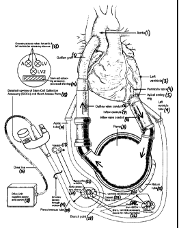
[0015] Figure 1 shows a schematic of one embodiment of the biologic
ventricular
assist device.
[0016] Figure 2 shows an schematic of one embodiment of the stem cell
collection
accessory.
-6-
CA 02668529 2009-05-04
WO 2008/057481 PCT/US2007/023251
DETAILED DESCRIPTION OF PREFERRED EMBODIMENTS
[0017] One aspect of the present invention pertains to a biologic ventricular
assist
device (Figure 1) that is capable of capturing, culturing, and delivering stem
cells within a
heart to which the device is attached. The ventricular assist device includes
an inflow path
(7,8), a pump (11), and an outflow path (6,2). The inflow path (7,8) of the
ventricular assist
device is attached to the left ventricle of the heart (3), and blood flows
into the inflow path
(7,8) from the left ventricle (3). The inflow path (7,8) then passes into the
pump (11), which
directs the blood into the outflow path (6,2). The outflow path directs the
blood back into the
ascending aorta of the heart (1). A drive line (16) typically connects the
pump to a drive unit
(18) that is external to the body. In the current invention, an external path
is also attached at
various points to the inflow path (7,8), the pump (11), and the outflow path
(6,2) of the
ventricular assist device (Figure 1). Blood also flows through the external
path. The external
path leads to a stem cell collection accessory ("SCCA", Figure 2) which
captures circulating
stem cells in the blood or from the heart.
100181 Another aspect of the present invention is the stem cell collection
accessory
("SCCA", Figure 2), which is a path or chamber through which blood flows after
it is directed
there by the external path. The path or chamber has walls of selective
permeability and one or
more layers of gels or polymers (36, 35, 33, 31, 34) having a chemical
gradient sufficient to
cause migration of the stem cells from the blood through the walls and into
the surrounding
gel.
[0019] In the present invention, once the captured stem cells are grown to
confluence, the stem cells are re-suspended and delivered back to the internal
chambers of the
heart using a delivery system that also passes through the external path. This
external path
leading back to the heart allows for repeated delivery of not only the
captured and cultured
stem cells but also therapeutic biologic or pharmacologic entities including
but not limited to:
cells, stem cells, genes, genetically modified and/or cultured stem cells and
drugs, either alone
or in combination, over time.
[0020] In a preferred embodiment, an inflow cannula (7) and inflow valve
conduit
(8) passing out of the left ventricle of the heart (3) make up the inflow path
of the biologic
ventricular assist device (Figure 1). The inflow path (7,8) directs the blood
into the pump
-7-
CA 02668529 2009-05-04
WO 2008/057481 PCT/US2007/023251
(11), which then directs the blood into the outflow path (6,2). In a preferred
embodiment, an
outflow valve conduit (6) and outflow graft (2) make up the outflow path. In
an additional
preferred embodiment, the external path of the biologic ventricular assist
device is made up of
a left ventricle tube (9), an aortic tube (10), a percutaneous tube (20), and
a return tube (12).
[00211 Figure 1 shows one preferred embodiment of the biologic ventricular
assist
device. The heart is illustrated, including the left ventricle (3) and aorta
(1). Positioned at the
ventricular apex (4) is an apical sewing ring (5) that allows attachment of
the inflow cannula
(7) of the device. The inflow cannula (7) passes into the inflow valve conduit
(8), allowing
blood exiting the left ventricle (3) to flow into the pump (11). Also exiting
the inflow valve
conduit (8) is a left ventricle tube (9) through which blood can bypass the
pump (11) and
proceed in a direction toward the stem cell collection accessory ("SCCA",
Figure 2). The left
ventricle tube (9) can also contain one or more accessory sleeves (23) or
lines for
instrumentation (26). In a preferred embodiment, these accessory sleeves can
be called left
ventricle accessory sleeves (23). The tube may be attached at the inflow valve
conduit (8)
with one-way valves for the accessory sleeves (23) and open ports for the
lines involved with
blood flow.
[0022] At a branch point (27), the left ventricle tube (9) merges into a
percutaneous tube (20) leading out of the body, past the incision and out of
the skin, with a
drive line (16) leading eventually back to the drive unit (18). Also at this
branch point (27),
an aortic tube (10) enters the percutaneous tube (20) from a point at the
outflow valve conduit
(6). The aortic tube (10) may contain one or more accessory sleeves (22) or
lines allowing for
the bypass flow of blood directly out of the outflow valve conduit and the
aorta (21) toward
the stem cell collection accessory ("SCCA"). In a preferred embodiment, these
may be called
an aortic accessory sleeve (22) and a bypass flow line (21). The aortic tube
(10) may be
attached at the outflow valve conduit (6) with one-way valves for the
accessory sleeves and
open ports for the lines involved with bypass blood flow. The percutaneous
tube (20) also
contains the accessory sleeves and lines allowing for ex-vivo delivery of
therapeutic biologic
or pharmacologic entities including but not limited to: cells, stem cells,
genes, genetically
modified and/or cultured stem cells, drugs, and components of the
extracellular matrix, either
alone or in combination, within the other tubes, as well as the coaxial drive
line that runs
between the drive unit (18) and the pump (11). Blood exiting the pump (11)
that is not
-8-
CA 02668529 2009-05-04
WO 2008/057481 PCT/US2007/023251
involved in bypass flow passes through the outflow valve conduit (6) and
through the outflow
graft back (2) into the ascending aorta (1).
[0023] Also entering the percutaneous tube (20) at the branch point (27) is a
return
tube (12) that can return blood to the pump (11) after it passes out of the
stem cell collection
accessory ("SCCA", Figure 2) and through the percutaneous tube (20). The
return tube (12)
can also contain one or more accessory sleeves (23, 26)or lines allowing for
ex-vivo delivery
of therapeutic biologic or pharmacologic entities including but not limited
to: cells, stem cells,
genes, genetically modified and/or cultured stem cells, drugs, and components
of the
extracellular matrix, either alone or in combination, as well as accessory
sleeves or lines
allowing for instrumentation. Where the return tube (12) meets the pump (11),
a drive line
(16) may also enter the tube for passage back to the drive unit (18). These
accessory sleeves
or lines may be called in a preferred embodiment left ventricle accessory
sleeves (23), and a
return flow line (25).
[0024] The percutaneous tube (20) passes outside of the body at the skin line
and
enters an adapter containing a vent filter, a stem cell collection accessory
("SCCA", Figure 2),
and one-way access valves for access to the aortic accessory sleeve, the left
ventricle
accessory sleeve, and the second left ventricle accessory sleeve (13). The
drive line (16) can
continue past the adapter to the drive unit (18).
100251 Figure 2 shows an illustration of a preferred embodiment of a stem cell
collecting accessory. Blood enters the stem cell collecting accessory from the
bypass flow
line (37). The bypass flow line (37) contains blood that passed through the
pump (11), exited
at the outflow valve conduit (6), passed through the aortic tube (10), passed
into the
percutaneous tube (20) at the branch point (27), and entered the adapter. This
blood flow
comes from the high pressure side of the device. The stem cell collecting
accessory (Figure
2) is generally surrounded by a biocompatible polymer (36). Within the stem
cell collecting
accessory itself is a chamber (39) through which blood passes. 0-rings (42)
may be located at
either end of the chamber. The first layer surrounding the chamber is a cell-
permeable
membrane (35). The 0-rings (42) also serve as formation aids for this cell-
permeable
membrane. Outside of the cell-permeable membrane (35) is an inner enzyme-
degradable
thermoreversible hydrogel (34) which contains a gradient of cytokines
diffusing toward the
-9-
CA 02668529 2009-05-04
WO 2008/057481 PCT/US2007/023251
flow of blood. The gradient in this hydrogel serves to capture circulating
progenitor or stem
cells as they migrate through the cell-permeable membrane. Chemoinvasive cells
(30)
migrate into this hydrogel from the blood. Outside the hydrogel is a cytokine-
permeable
membrane(33) through which the stem cells do not easily pass. Outside of the
cytokine-
permeable membrane (33) is an outer enzyme-degradable thermoreversible
hydrogel (31) that
is doped with cytokines in sufficient concentration to sustain an
approximately unchanging
gradient over the exposure lifetime. This outer hydrogel is moderately
diffusion-inhibiting.
The outermost layer is a rigid outer wall (32).
[0026] The blood that flows through the stem cell collecting accessory (Figure
2)
then enters the return flow line (25). The return flow line (25) passes
through the
percutaneous tube (20), passes into the return tube (12) or any line allowing
for the return
flow of blood at the branch point (27), and re-enters the pump (11). This
blood flow is
directed to the low pressure side of the device.
[0027] Once they have grown to confluence within the stem cell collection
assembly, the cardiac or circulating progenitor or stem cells are removed from
the stem cell
collecting accessory, re-suspended in solution and then re-administered via
the electro-
mechanical and/or ultrasound/echocardiographic imaging and delivery system
directed
through the external sleeve system within the percutaneous tube and other
tubes placed along
the drive line and along the course of the device back into the internal
cardiac chambers to
allow the delivery of the appropriate dose of cardiac progenitor or stem
cells.
EXAMPLE 1. CARDIAC PROGENITOR CELL ISOLATION
[0028] Isolation and Characterization of Cardiac Stem Cells. Tissue samples
are obtained from patients receiving a left ventricular assist device (LVAD).
The 1-2 cm3
samples are excised from the left ventricular apex to allow for placement of
the device.
Typically, this "core" is discarded upon excision. However, this is a viable
source of tissue,
regardless of the pathological background, to isolate resident cardiac stem
cells.
[0029] Processing of Human Cardiac Stem Cells from Clinical Samples. The
cardiac tissue "core" is minced with a scalpel into 2-3 mm3 pieces, and 20
pieces (generally,
500mg) are placed into 2 ml of 0.13 mg/ml Liberase Blendzyme 4 (Roche
Diagnostics Corp.,
-10-
CA 02668529 2009-05-04
WO 2008/057481 PCT/US2007/023251
San Diego, CA) re-suspended in serum free Hams F12 media. The tissues are
incubated for
30 minutes with a brief vortexing every 10 minutes. The larger tissues are
collected by
centrifugation at 500R PM for 2 minutes and the supernatant collected and
strained through a
30uM nylon mesh. The remaining tissue is re-suspended in 2ml of 0.13mg/ml
Liberase
Blendzyme 4 and the procedure is repeated for a total of three times. Each
time the
supernatant is collected, the sample is strained through the nylon mesh and
the cells spun at
800 RPM for 10 minutes to pellet the cells. These cells are re-suspended in
calcium and
magnesium free PBS supplemented with 0.1 % BSA (Sigma, St. Louis, MO) and 2 mM
EDTA
and placed in the incubator until all sample digestions have been completed.
Following
digestion, the cells will be pooled and counted on a hemacytometer. Viability
will be
measured by trypan blue exclusion.
[0030] Magnetic isolation of CD117P S/PgpP S stem cells. CD117P S/PgpP S (P-
glycoprotein) cells are labeled with I g of biotinylated mouse anti-human
CD117
(eBioscience, San Diego, CA) and biotinylated mouse anti-Pgp (Chemicon,
Temecula, CA)
per 1 x 10⁷ cells for 30 minutes at 4 C. Following incubation, the cells are
washed in PBS
plus 0.1 %BSA and 2mM EDTA and re-suspended in 2x 10⁷ cells/ml of wash buffer.
A total
of 25 l of Streptavidin coated CELLection Dynabeads (Dynal , Invitrogen,
Carlsbad, CA)
is added to the cells and incubated with gentle tilting and rotation for 30
minutes. The cells
are placed into the Dynal MPC-L magnet for 2 minutes. The supernatant is
removed and
the bead bound cells are washed 3 times in wash buffer. The supernatant is
collected and
stored separately. The bead bound cells are re-suspended in 200 l of RPMI
1640 plus 1%
FBS and 4 l of 10,000U/ml DNasel is added for 15 minutes with gentle tilting
and rotation.
The sample is then vortexed vigorously and placed into the magnet for 2
minutes. The
supernatant is then collected and the tube washed once in RPMI 1640 plus 1%
FBS. The
supernatant is pelleted at 800 RPM for 10 minutes and re-suspended in growth
media, Ham's
F12 supplemented with 5% FBS and 10 ng/ml each of LIF (Chemicon) and bFGF
(Chemicon). The cells are counted on a hemacytometer and plated in a 6-well
plate (Nunc,
Rochester, NY) at 2 x 10⁴ cells/cm2. The supernatant is replaced after one
week and the plate
washed with PBS and maintenance media is added, Ham's F12 supplemented with 5%
FBS,
ng/ml LIF and bFGF and 20 ng/ml of EGF (Chemicon). Media is changed every 3-4
days
until 50% confluency. Upon 50% confluency, the plate is passaged into a 75 cm2
flask
-11-
CA 02668529 2009-05-04
WO 2008/057481 PCT/US2007/023251
(Nunc). The negatively sorted cells are plated at a density of 5 x
lO5cells/cm2 on 75 cmZ
flasks in growth media (described in D.1.2.). These cells are treated
similarly to positive
selected cells in regards to media and passaging. Yield, morphology,
homogeneity, and cell
growth characteristics are documented for each sample and their isolates.
[0031] Adherent isolation of cardiac stem cells. Cells processed from clinical
cardiac samples are plated at a density of 5 x 10⁵ cells/cm2 on 75cmz flasks
in growth media.
Adherent and non-adherent fractions are collected based on the following time
points: 1 hour,
2 days, 5 days and 7 days. The adherent fractions then have the media replaced
with
maintenance media. The supernatant containing the non-adherent cells, is
pelleted and re-
suspended in maintenance media and plated on 75cm 2 flasks. Once 50%
confluence is
reached, the cells are passaged to 175 cm2 flasks in maintenance media. Yield,
morphology,
homogeneity, and cell growth characteristics are documented for each sample
and their
isolates.
[0032] Flow cytometry characterization. Cells are characterized through flow
cytometry for phenotypic surface markers to determine the efficacy and
homogeneity of the
isolation techniques. Cells are stained, with mouse anti-human antibodies
(Pharmingen, BD
Biosciences, Mississauga, Canada) for stem cell markers CD105, CD117, CD133,
CD166, the
drug resistance marker, P-glycoprotein (Pgp), as well as lineage markers, CD4,
CD8, CD20,
CD34, CD45, CD45RO and the endothelial marker CD31 and adhesion marker CD44.
Cells
are trypsinized with TrypLE (Invitrogen), pelleted, and re-suspended in PBS
plus 5% BSA at
a density of 1 x 10⁶ cells/ml. 200 l of the cells are aliquoted into 12 x 75
mm tubes and 0.5
g of appropriate antibodies are added to each tube. Four different antibodies
are added per
tube that possess particular fluorescent characteristics so that there is
little fluorescent
emission overlap. The antibodies are incubated at 4 C for 30 minutes, washed
in PBS plus
5% BSA and re-suspended in 1 ml PBS. Cells are analyzed using the Becton
Dickinson
FACScan Analyzer and Ce1lQuest software (Becton-Dickinson, BD Biosciences).
[0033] Differentiation capacity of cardiac stem cells. Cells are trypsinized
and
placed onto a Nunc eight-well LabTekTM chamber slides (Sigma) at 1 x 10³
cells/cmZ and
grown under normal or differentiative conditions. Media are changed every 3-4
days. The
number of positive cells are counted using a fluorescent microscope and
representative
-12-
CA 02668529 2009-05-04
WO 2008/057481 PCT/US2007/023251
micrographs are taken with the Olympus BX50WI (Center Valley, PA) two photon
confocal
microscope available. Background staining consists of Prolong GoldTM anti-fade
plus DAPI
(Molecular Probes, Invitrogen).
[0034] Cardiomyogenic differentiation of stem cells following co-culture with
neonatal cardiomyocytes. To induce cardiomyogenic differentiation, human
cardiac stem
cells ("hCSC's") are co-cultured with neonatal human ventricular myocytes
("NRVMs").
CSCs are labeled with PKH-26 (Sigma) prior to addition to the NRVMs cultures
at a 1:4 ratio
and cultured for up to 2 weeks with media changes every 3-4 days. PKH-26
labeled cells
retain both biological and proliferative activity, and are ideal for cell
tracking studies. The
linkers are physiologically stable (lasting up to 100 days) and show little to
no toxic side
effects. PKH-26 has an excitation and emission of 551/567 nm that is
compatible with
rhodamine or phycoerythrin detection systems. However, it may also be excited
by the
488nm emission of an argon-ion laser. Briefly, cells are trypsinized from the
plate, pelleted
and washed twice in serum-free media. After the final wash, the cells are
suspended at 4 x
105 in 50 l diluent. 50 l of 2X PKH-26 dye is added and the cells are
incubated at room
temperature for approximately 5 minutes. This time may change as each cell
type exhibits
different properties in lipid uptake. To ensure homogenous staining, cells are
incubated for
different times and analyzed by confocal microscopy. The reaction is stopped
by adding an
equal amount of growth media with FBS and the cells are washed 3-5 times to
remove any
unbound dye. Cells are stained for rabbit anti-human cardiac troponin I
(Abcam,Cambridge,
UK), biotinylated goat anti-human GATA-4 and mouse anti-human Nkx2.5
(R&DSystems,
Minneapolis, MN).
[0035] Endothelial differentiation of stem cells. To induce differentiation
into
endothelial cells, hCSCs are plated at 5 x 10⁴/cm2 in DMEM or EBM-2 (Cambrex)
with 2%
FBS, supplemented with 10-8 M dexamethasone and 10 ng/ml VEGF, in chamber
slides
coated with either 0.1 % gelatin or fibronectin for 14 days with media changes
every 3-4 days.
Tube-like structures may form after five days, but after 14 days they exhibit
endothelial
specific markers. Cells are stained for rabbit anti-human von Willebrands
Factor ("vWF"),
and mouse anti-human CD3 1.
-13-
CA 02668529 2009-05-04
WO 2008/057481 PCT/US2007/023251
[0036] Smooth muscle differentiation. Cardiac stem cells and MSCs are induced
to differentiate into smooth muscle cells by placing 5 x 10⁴/cm2 stem cells on
fibronectin
coated glass chamber slides in 2% DMEM or EBM-2 (Cambrex) supplemented with
50ng/ml
PDGF-BB for 14 days. The SMC marker, mouse anti-human alpha-smooth muscle
actin
(Abcam), is used.
-14-