Note: Descriptions are shown in the official language in which they were submitted.
* CA 02668548 2013-07-16
1
DEVICE AND METHOD FOR IMPROVING FUNCTION OF HEART VALVE
Field of the Invention
The present invention pertains in general to the
field of repair of heart valves having various
malformations and dysfunctions. More specifically, the
invention relates to heart valve repair techniques and
procedures involving annuloplasty devices.
Background of the Invention
Diseased mitral and tricuspid valves frequently need
replacement or repair. The mitral and tricuspid valve
leaflets or supporting chordae may degenerate and weaken or
the annulus may dilate leading to valve leak, i.e. an
insufficiency of valve function. The leaflets and chords
may become calcified and thickened rendering them stenotic,
which implies obstructing a forward flow through the valve.
Finally, the valve relies on insertion of the chordae
inside the ventricle. If the ventricle changes in shape,
the valve support may become non-functional and the valve
may leak.
Mitral and tricuspid valve replacement and repair are
traditionally performed with a suture technique.
During valve replacement, sutures are spaced around
the annulus, i.e. the point where the valve leaflet
attaches to the heart, and then the sutures are attached to
a prosthetic valve. The prosthetic valve is lowered into
position and when the sutures are tied, the prosthetic
valve is fastened to the anriulus. The surgeon may remove
all or part of the valve leaflets before inserting the
prosthetic valve.
In valve repair, a diseased valve is left in situ and
surgical procedures are performed to restore its function.
Frequently an annuloplasty ring is used to reduce the size
of the annulus. The annuloplasty ring serves to reduce the
diameter of the annulus and allows the leaflets to oppose
CA 02668548 2013-07-16
2
each other normally, thus restoring valve function. Sutures
are used to attach the prosthetic annuloplasty ring to the
annulus and to assist in placating the annulus.
In general, the annuloplasty rings and replacement
valves must be sutured to the valve annulus and this is
time consuming and tedious. Moreover, if the annuloplasty
ring is severely malpositioned, then the stitches must be
removed by the surgeon, and the ring repositioned relative
to the valve annulus during restitching. In other cases, a
less than optimum annuloplasty may be tolerated by the
surgeon rather than lengthening the time of the surgery to
restitch the ring.
During heart surgery, a premium is placed on reducing
the amount of time used to replace and repair valves as the
heart is frequently arrested and without perfusion. It
would therefore be very useful to have an improved method,
procedure, and/or device providing an efficient attachment
of a prosthesis into the mitral or tricuspid valve
position.
For instance in US 2002/0173841 and in US 6,419,696,
which are assigned to the same applicant as the present
application, an annuloplasty device is disclosed. The
annuloplasty device comprises a first and a second support
ring, which are connected to each other to form a coiled
configuration. The first and second support rings are
arranged to abut opposite sides of a valve annulus to trap
valve tissue between them. This annuloplasty device may be
easily applied to the valve by rotating the device into
position on opposite sides of the valve annulus. To ensure
a proper and lasting fixation to the valve annulus such
device can be fixated by barbs, retaining members,
interlocking portions, fasteners or locking elements, all
being integrated in the device. Fixation can also be made
by means of suturing.
In WO 2006/091163, which are assigned to the same
applicant as the present application, a device for
CA 02668548 2013-07-16
3
improving the function of a heart valve is disclosed that
comprises a first loop- shaped support, which is configured
to abut a first side of the heart valve, and a second loop-
shaped support, which is configured to abut a second side
of the heart valve opposite to said first side, whereby a
portion of the valve tissue is trapped between the first
and second supports. An outer boundary of the second
support is greater than an outer boundary of the first
support.
In US 4042979 an adjustable valvuloplasty ring is
disclosed that comprises a C-shaped frame that is sized and
shaped to extend about the circumference of the left
atrioventricular orifice along the base of the anterior
cusp of the mitral valve; an expandable sleeve connected to
the frame that together therewith forms a closed annulus,
the sleeve being adapted to extend about the remainder of
the circumference of the orifice; and a drawstring running
through the sleeve by which the sleeve may be contracted to
constrict and remodel the orifice and secured in place to
maintain such constriction.
However, the prosthetic devices disclosed in the
above mentioned documents might be further improved for a
more convenient, faster pos'itionable, and/or even more
reliable device and method of valve repair and valve
replacement. It is a specific object of the invention to
provide a device, which allows for an easy and durable
fixation to the valve annulus.
Furthermore, an improvement that is desired to be
provided by improved devices and methods comprises allowing
a prevention or minimization of backflow of blood, e.g.
passing by or underneath the prosthetic devices of the
prior art.
Hence, an improved annuloplasty device and medical
procedure would be advantageous and in particular allowing
for increased flexibility, cost-effectiveness, convenience
CA 02668548 2013-07-16
4
and speed of positioning, increased reliability and/or
patient safety would be advantageous.
Summary of the Invention
An object of the invention is to provide an improved
medical device and method of valve repair and valve
replacement. Another object of the invention may be to
provide an annuloplasty device, which allows for an easy
and durable fixation to the valve annulus.
Accordingly, embodiments of the present invention
preferably seek to mitigate, alleviate or eliminate one or
more deficiencies, disadvantages or issues in the art, such
as the above-identified, singly or in any combination by
providing a medical device and a method according to the
invention.
According to a first aspect of the invention, there
is provided a medical device for improving the function of
a heart valve comprised of valve tissue including an
annulus and a plurality of leaflets, the device comprising:
a first loop-shaped support, which is configured to abut a
first side of the heart valve, and a first flange unit (or
flange element or flange means) being connected to said
first loop-shaped support, and which is configured to be
arranged against said annulus when said first loop-shaped
support is abutting said heart valve.
This device may be used to perform annuloplasty, that
is to reshape the valve annulus, in order to improve the
function of the valve. The flange unit provides a well
defined surface to be used when fixating the device against
the annulus no matter if the device in use is positioned
abutting the atrial or the ventricle side of annulus.
This implies that the device may easily be fixated to
the annulus in a speedy manner. This is of importance since
during heart surgery, a premium is placed on reducing the
amount of time used to replace and repair valves as the
heart is frequently arrested and without perfusion.
CA 02668548 2013-07-16
Also, the flange unit may provides for a sealing
surface against said annulus allowing prevention of
backflow of blood from the ventricle side to the atrial
side.
5 Further, the provision of a flange unit implies that
a smooth transition section may be formed between the outer
periphery of the device and annulus.
Further, a well defined surface for attachment of
fixating means such as sutures or clips may be provided. A
smooth transition section as well as a well defined
attachment surface are two parameters of importance for a
smooth formation and growth of endothelia.
In addition, the flange unit may be used for carrying
or fixation of a prosthetic valve.
The device may further comprise a second loop-shaped
support, which is configured to abut a second side of the
heart valve opposite to said first side, whereby a portion
of the valve tissue is trapped between the first and second
supports. The trapping of valve tissue between the first
and second loop-shaped supports implies that the desired
shape of the valve, both natural or prosthetic, may be
fixated. Further, the trapping implies that the device may
temporarily be kept in correct position while fixating the
device permanently to an annulus by means of e.g. sutures
or clips.
The first loop-shaped support may be formed
continuously with the second loop-shaped support to form a
substantially coil-shaped body. This implies that the
device and its coil-shape may be applied at a commissure
between the leaflets of the heart valve and be rotated
approximately 360 such that one loop-shaped support is
inserted through the commissure to extend along one side of
the valve and the other loop-shaped support is arranged
along the opposite side of the valve. Thus, valve tissue
will be trapped between the supports to fixate a desired
shape of the valve. Depending on the extension of the
= CA 02668548 2013-07-16
6
flange means, the latter may provide an attachment surface
on one of or on both sides of the annulus for fixation of
the device.
The first flange unit may extend from the first loop-
shaped support to the second loop-shaped support, whereby
the flange unit may be configured to be arranged against
the annulus on opposite sides of the valve tissue being
trapped between the first and second supports. This implies
that the flange unit may form a flange surface on both
sides of the annulus or heart valve, which surface may
provide for fixation, not only of the device but also of a
prosthetic valve. Further, the flange unit may form a
sealing surface that, depending on the position of the
device, allows reduction or prevention of possible backflow
of blood from the ventricle side to the atrial side.
The second loop-shaped support may comprise a second
flange unit being connected thereto, which flange unit may
be configured to be arranged against the annulus on a side
thereof being opposite the first loop-shaped support when
the second loop-shaped support is abutting the heart valve.
This allows prevention of paravalvular leakage.
At least one of the first and second flange unit may
be adapted to form a connection of at least one of the
loop-shaped supports and a prosthetic valve against the
annulus. This implies a rapid fixation, which is of
importance since during heart surgery a premium is placed
on reducing the time required.
At least one of the first and second flange unit may
have an intermittent or continuous extension along the
periphery of its corresponding loop-shaped support. By way
of example, in case of an intermittent extension the flange
unit may be formed by two local sections diametrically
opposing each other, whereby the two sections, when the
device is positioned in the heart valve, are abutting the
commissures forming a sealing surface thereto.
CA 02668548 2013-07-16
=
7
At least one of the first and second flange units may
be made of a fabric material. The fabric material may be a
woven material. A fabric has the advantage that it presents
a rough surface enhancing ingrowth or anchoring of
endothelia. Further, a fabric is easily penetrated by
sutures or clips. Also, a fabric allows the flange unit to
be easily conformed to the annulus.
The fabric material may be impregnated with or
integrate a pharmaceutical agent further improving
embodiments of the devices and method. The pharmaceutical
agent may for instance be an anti inflammatory, stenos
preventing, or endotheliazation promoting agent.
Further, at least one of the first and second flange
unit may comprise a reinforcing element. The reinforcing
element provides an indication and definition of an area in
which clips or sutures are to be put when fixating the
medical device to the annulus. Further, the reinforcing
element contributes to reducing the risk of pockets being
formed along the circumferential surface. Also, the element
prevents unthreading of the fabric in the flange.
At least one of the first and second flange unit may
protrude or extend out from and form an angle a (see e.g.
Fig. 5) of approximately 30-60 , such as e.g approximately
40-50 below a diametric plane formed by one of the loop-
shaped supports. By the flange unit initially extending
below the diametric plane, the visibility during insertion
is enhanced. In some embodiments, during insertion, the
flange unit due to inherent flexibility may be fold, e.g.
upwards in Fig. 5, even fold back over its point of
fixation relative the diametric plane, or above the
diametric plane with an outer edge of the flange unit. The
point of fixation of at least one of the flange unit may be
fixed in relation to the diametric plane, radially outward
from at least one of the loop-shaped supports.
The flange unit may protrude with other angles, even
in a fold back, i.e. more than 90 . This may be during or
=
CA 02668548 2013-07-16
8
prior to a time of use or implantation thereof. The angle
may be variable over time, e.g. to the herein described
shape memory effect of some embodiments of the flange unit.
The flange unit may in some embodiments be arranged
to change shape during insertion, e.g. by a resilient
arrangement thereof. The flange unit may also be made of a
shape memory material that returns to a pre-defined shape
of form during insertion of the medical device, e.g. by a
temperature triggered effect as known in the art of shape
memory materials.
At least one of the first and second flange unit
extends radially inwards or outwards from its corresponding
loop-shaped support. A radially inward extension provides a
support for the valve leaflets, whereas a radially outward
extension provides a support against the annulus. The first
side of the heart valve is the atrial side and the second
side is the ventricle side.
According to a second aspect of the invention there
is provided a method for repairing a heart valve comprised
of valve tissue including an annulus and a plurality of
leaflets for allowing and preventing blood flow, the method
comprising: inserting a device comprising at least one
loop-shaped support and at least one flange unit being
connected to the loop-shaped support to a heart valve,
positioning the loop-shaped support such that it abuts a
first side of the heart valve, positioning the flange unit
such that it abuts the annulus, and fixating the device by
attaching the flange unit to the annulus.
The advantages provided by a device having a flange
unit have previously been discussed above. The inventive
method for repairing a heart valve uses a corresponding
device, whereby at least the same benefits are achieved.
The flange unit may be attached to the annulus by
using sutures or clips, which allows for a quick and easy
fixation using well established means. Alternatively, or in
=
=
CA 02668548 2013-07-16
9
addition, barb elements or tissue adhesives may be used for
the attachment to the annulus.
The provision of a flange unit implies that a smooth
transition section may be formed between the outer
periphery of the device and annulus. Further, the flange
unit presents a well defined and easy detectable surface
for attachment of the clips or sutures. A smooth transition
section as well as a well defined attachment surface allows
for a smooth formation and growth of endothelia. Endothelia
formation may further be improved by an endotheliazation
agent.
The flange unit may be conformed to the annulus
before fixating the device. By conforming the flange unit,
the transition section may be additionally smoothened,
further enhancing growth of endothelia.
The device may be inserted to the heart valve by
using a catheter, whereupon the catheter is withdrawn
leaving the device.
In the method the first side of the heart valve may
be the atrial side.
Further, in another asp.ect, the invention provides a
kit comprising a device for improving the function of a
heart valve comprised of valve tissue including an annulus
and a plurality of leaflets, the device comprising: a first
loop-shaped support, which is configured to abut a first
side of the heart valve, and a first flange unit being
connected to the first loop-shaped support, and which is
configured to be arranged against the annulus when the
first loop-shaped support is abutting the heart valve, and
an artificial valve.
This device may be used in a medical procedure to
perform annuloplasty, that is to reshape the valve annulus,
in order to improve the function of the valve. The flange
unit provides a well defined surface to be used when
fixating the device against the annulus. This implies that
the device may be fixated to the annulus very easily and in
CA 02668548 2013-07-16
a speedy manner. The latter is of importance since during
heart surgery, a premium is placed on reducing the amount
of time used to replace and repair valves as the heart is
frequently arrested and without perfusion. Also, the flange
5 unit provides a sealing surface against the annulus
allowing prevention of backflow of blood from the ventricle
side to the atrial side. By the device carrying an
arificial prosthetic valve, the steps and time involved
when performing the surgery may be reduced. Further, the
10 positioning of such prosthetic valve in relation to the
annulus is facilitated.
The device may further comprise a second loop-shaped
support, which is configured to abut a second side of the
heart valve opposite to the first side, whereby a portion
of the valve tissue is trapped between the first and second
supports. The trapping of valve tissue between the first
and second loop-shaped supports implies that the desired
shape of the valve may be fixated. Further, the trapping
implies that the device may temporarily be kept in correct
position while substantially fixating the device
permanently to an annulus by means of e.g. sutures or
clips.
The first loop-shaped support may be continuous with
the second loop-shaped support to form a coil-shaped body.
This implies that the device and its coil-shape may be
applied at a commissure between the leaflets of the heart
valve and be rotated 360' such that one loop-shaped support
is inserted through the commissure to extend along one side
of the valve and the other loop-shaped support being
arranged along the opposite side of the valve. Thus, valve
tissue will be trapped between the supports to fixate a
desired shape of the valve. Depending on the extension of
the flange means, the latter may provide an attachment
surface on one of or on both .sides of annulus for fixation
of the device.
= CA 02668548 2013-07-16
11
The first flange unit may extend from the first loop-
shaped support to the second loop-shaped support, whereby
the flange unit may be configured to be arranged against
the annulus on opposite sides of the valve tissue being
trapped between the first and second supports. This implies
that the flange unit may form a surface on both sides of
the heart valve, which surface may be used for fixation,
not only of the device but also of a prosthetic valve.
Further, the flange unit may form a sealing surface that,
depending on the position of the device, allows prevention
of possible backflow of blood from the ventricle side to
the atrial side.
The second loop-shaped support may comprise a second
flange unit being connected thereto, which flange unit may
be configured to be arranged -against the annulus on a side
thereof being opposite the first loop-shaped support when
the second loop-shaped support is abutting the heart valve.
This allows prevention of paravalvular leakage.
At least one of the first and second flange unit may
have an intermittent or continuous extension along the
periphery of its corresponding loop-shaped support. By way
of example, in case of an intermittent extension the flange
unit may be formed by two local sections diametrically
opposing each other, whereby the two sections, when the
device is positioned in the heart valve, are abutting the
commissures forming a sealing surface thereto.
At least one of the first and second flange unit may
be made of a fabric material. A fabric has the advantage
that it presents a rough surface enhancing growth of
endothelia. Further, a fabric is easily penetrated by
sutures or clips. Also, a fabric allows the flange unit to
be easily conformed to the annulus.
Further, at least one of the first and second flange
unit may comprise a reinforcing element. The element
provides an indication and definition of the area in which
clips or sutures are to be put when fixating the device to
CA 02668548 2013-07-16
12
the annulus. Further, the element reduces the risk of
pockets being formed along the circumferential surface.
Also, the element prevents unthreading of the fabric in the
flange.
At least one of the first and second flange unit may
extend out from and form an angle of 30-600, such as 40-50
below a diametric plane formed by one of the loop-shaped
supports. By the flange unit initially extending below the
diametric plane, the visibility during insertion is
enhanced.
At least one of the first and second flange unit may
extend radially inwards or outwards from its corresponding
loop-shaped support.
The artificial prosthetic valve may be arranged on
one of the loop-shaped supports. In the case the device is
intended to be inserted to the heart from the atrial side,
the artificial valve is preferably arranged on the support
intended to be positioned on the atrial side of annulus and
vice verse.
Further, in another aspect, the invention may relate
to a method for replacing a heart valve comprised of valve
tissue including an annulus and a plurality of leaflets for
allowing and preventing blood flow, the method comprising:
inserting a device comprising an artificial valve, at least
a loop-shaped support and at least one flange unit being
connected to the loop-shaped support to a heart valve,
positioning the loop-shaped support such that it abuts a
first side of the heart valve, positioning the flange unit
such that it abuts the annulus, and fixating the device by
attaching the flange unit to the annulus.
The advantages provided by a device having a flange
unit and an artificial valve have previously been discussed
above. The inventive method for replacing a heart valve
uses a corresponding device, whereby the same benefits are
achieved.
=
CA 02668548 2013-07-16
13
The flange unit may be attached to the annulus by
using suitable fixation units, e.g. sutures or clips, which
allows for a quick fixation using well established means.
The flange unit may be conformed to the annulus
before fixating the device. By conforming the flange to the
annulos the surface to be covered by endothelia is reduced,
allowing the growth to be enhanced and accelerated.
The device may be inserted to the heart valve by
using a catheter, whereupon the catheter is withdrawn
leaving the device.
In the method, the first side of the heart valve is
preferably the atrial side.
The artificial valve may be arranged on one of said
loop-shaped supports.
Some embodiments of the invention provide for a
reduced amount of time used to repair and/or replace
cardiac valves.
Some embodiments of the invention also provide for a
reduced or prevented backflow of blood, e.g. by a smooth
transition section may be formed between the outer
periphery of the device and annulus.
Some embodiments of the invention provide for a more
convenient repair, e.g. by means of a well defined surface
for attachment of fixating means such as sutures or clips.
Some embodiments of the invention provide for a
smooth formation and growth of endothelia.
It should be emphasized that the term
"comprises/comprising" when used in this specification is
taken to specify the presence of stated features, integers,
steps or components but does not preclude the presence or
addition of one or more other features, integers, steps,
components or groups thereof.
Brief Description of the Drawings
These and other aspects, features and advantages of
which embodiments of the invention are capable of will be
=
=
CA 02668548 2013-07-16
14
apparent and elucidated from the following description of
embodiments of the present invention, reference being made
to the accompanying drawings, in which
Fig.1 schematically illustrates a patient with a
heart shown in cross-section and a device of an embodiment
of the present invention schematically illustrated as
supporting the mitral valve;
Fig. 1A is a cross-sectional view of the left
ventricle showing the mitral valve in perspective;
Fig. 2 is a perspective view of a body of a device
according to a first embodiment of the invention;
Fig, 3 is a cross-sectional view of the body in Fig.
2;
Fig. 4 is a perspective view of the first embodiment
of the device comprising the body shown in Fig. 2;
Fig. 5 is a cross-sectional view of the device in
Fig. 4;
Fig. 6 is a perspective view of a second embodiment
of the device;
Fig. 7 is a perspective view of a third embodiment of
the device;
Fig. 8 is a perspective view of a fourth embodiment
of the device;
Fig. 9a, 9b are perspective views that illustrate
insertion of an embodiment of the device;
Fig. 10 is a cross-sectional view showing an
embodiment of the device inserted in a heart valve;
Fig. 11 and 12 are schematic illustrations that show
a heart valve before and after remodelling by using the
device;
Fig. 13 is a cross sectional view that shows the
device fixed to the annulus;
Fig. 14a is a cross sectional view that shows a first
embodiment of the device comprising an artificial
prosthetic heart valve;
=
=
CA 02668548 2013-07-16
Fig. 14b is a cross sectional view that shows a
second embodiment of the device comprising an artificial
valve;
Fig. 15 is a cross-sectional view of an alternative
5 device having one loop-shaped support carrying the flange
unit;
Fig. 16a, 16b are cross sectional views of
embodiments involving a shape change; and
Fig. 17 is a cross sectional view schematically
10 illustrating a flange unit having barb elements for
affixing to tissue.
Description of embodiments
Specific embodiments of the invention will now be
15 described with reference to the accompanying drawings.
This invention may, however, be embodied in many different
forms and should not be construed as limited to the
embodiments set forth herein; rather, these embodiments are
provided so that this disclosure will be thorough and
complete, and will fully convey the scope of the invention
to those skilled in the art. The terminology used in the
detailed description of the embodiments illustrated in the
accompanying drawings is not intended to be limiting of the
invention. In the
drawings, like numbers refer to like
elements.
Fig. 1 illustrates a patient 10 having a heart 12
shown in cross-section including a left ventricle 14 and a
right ventricle 16. The concepts of the present invention
are suitable to be applied, for example, to a mitral valve
18 which supplies blood into left ventricle 14. Mitral
valve 18, as better shown in Fig. 1A, includes an annulus
20 and a pair of leaflets 22, 24 which selectively allow
and prevent blood flow into left ventricle 14. It will be
appreciated that the term valve tissue is used extensively
throughout this disclosure in reference to the drawings.
The inventive principles are equally applicable when
=
CA 02668548 2013-07-16
16
referring to any valve tissue such as annulus tissue,
leaflet tissue or other attached vessel tissue. Leaflets
22, 24 are supported for coaptation by chordae tendinae or
chords 26, 28 extending upwardly from respective papillary
muscles 30, 32. Blood enters left ventricle 14 through
mitral valve 18 and is expelled during subsequent
contraction of heart 12 through aortic valve 34. It will be
appreciated that the present invention is applicable to
tricuspidal heart valves as well.
A body 41 comprised in a device 40 according to a
first embodiment of the present invention is shown in Figs.
2 and 3. The body 41 comprises a first and a second loop-
shaped support 42, 44.
As used herein, the term "loop-shaped" should be
construed as a curved shape that may be closed, as at least
a part of a ring with e.g. a circular, elliptic, or D-
shaped form or any other closed form which may fit the
shape of the valve annulus. The term "loop-shaped" also
includes a curved shape that is open forming an arcuate
shape, such as a C-shape or U-shape, which includes an
angular turn of at least 180 such that the support may
abut valve tissue along a major part of the annular valve
shape. The term "loop-shaped" also includes a curved shape
overlapping itself to form a portion of a coil.
The term "loop-shaped" also includes three
dimensional curves as mentioned in the previous paragraph.
The loop shape of at least a part of at least one of
the supports 42, 44 may also in some embodiments be patient
configured. The shape may be designed specifically to an
anatomy of a patient. The patient specific loop shape may
be virtually derived from 3D patient data, e.g. acquired by
image modalities, such as Magnetic Resonance (MR) or
Computer Tomography (CT) Imaging.
In US 6,419,696, US 6,730,121, US 6,964,684, and WO
2006/091163, which are assigned to the same applicant as
the present invention, devices are disclosed for repairing
CA 02668548 2013-07-16
17
and replacing a heart valve in various embodiments. The
devices include at least first and second support rings
connected together in loop-shaped configurations to abut
opposite sides of a valve annulus. A replacement valve may
be secured to the loop-shaped devices.
The first support 42 may be continuous and/or
integral with the second support 44 such that the supports
42, 44 assume a coiled configuration in the form of a
spiral or keyring-type configuration with two loops.
The second support 44 may have an outer boundary or
extent which is greater in relation to the outer boundary
of the first support 42. The supports 42, 44 may in an
embodiment have corresponding shapes with the second
support 44 being in larger scale than the first support 42.
This is advantageous in creating a pinch of the valve
tissue between the first 42 and second supports 44.
An end 45 of the second support 44, which will lead
the coil during insertion of the device at the valve, may
in an embodiment have a greater pitch than the rest of the
coil. This implies that the leading end 45 of the coil
during rotation into position in the valve will project
from immediate contact with the valve tissue and,
therefore, the risk that the coil is caught by the chords
is diminished.
The body 41 is shown in cross-section in Fig. 3. The
body 41 has in an embodiment at least partly a round cross-
sectional shape. In other embodiments, the cross section of
the body 41 may be substantially flat, oval, flattened
and/or have flattened edges.
In embodiments, the opposed surfaces 46 thus provide
a pinch to trap valve tissue there between. A round cross-
section is also advantageous in creating a pinch of the
valve tissue which will not harm the leaflets in their
movement during normal heart action.
The second loop-shaped support 44 is slightly
displaced radially with respect to the first loop-shaped
CA 02668548 2013-07-16
18
support 42. This implies that the first and second loop-
shaped supports 42, 44 are not arranged directly on top of
each other in some embodiments. The pinch between the first
42 and second supports 44 is therefore not sharply defined
in a radial direction of the valve. This implies that a
pinching force between the supports is not focussed to a
specific radial position of the valve. As a result, the
pinching force does not affect the movement of the leaflets
during normal heart action and there is a diminished risk
of rupture in the leaflets at the pinch.
The supports may in some embodiments be interrelated
in such manner that the outer boundary of the first support
42 has a diameter corresponding to a line through the
centre of the second support 44. Thus, the supports 42, 44
may overlap somewhat such that tissue is not allowed to
move through the pinch and the shape of the valve is
maintained advantageously.
Further, the cross-section of the supports 42, 44 is
substantially round, which also gives a soft contact
between the supports and the valve tissue to further
diminish the risk of rupture in the leaflets.
The body 41 may be formed from a core of a rigid
material, such as a metal, e.g, titanium, or plastic. Any
suitable medical grade material(s) may be used.
The rigid material may provide a passive spring
function such that the loops of the coil may be forced a
small distance away from each other but will flex back
towards each other when the force is released. The core of
the body 41 may be coated by a softer layer, such as a
textile.
The body 41 may alternatively be formed from a shape
memory material. The body 41 will then assume a desired,
programmed shape, when e.g. heated to a specific
temperature. This allows the body 41 to be compressed or
straightened of the form better suited for delivering
during insertion and to assume a spiral shape when inserted
CA 02668548 2013-07-16
19
at the heart valve. Also, the flange unit may be made of
such a shape memory material, e.g. to provide a first,
delivery shape and a second, delivered shape thereof.
Now turning to Figs. 4 and 5, a first embodiment of
the medical device 40 is disclosed. The device 40 comprises
a body 41 in accordance with that described above with
reference to Figs. 2 and 3, whereby the body 41 as such is
not further discussed.
The device 40 comprises a flange unit 50 being
connected to the body 41 and more precisely to the first
loop-shaped support 42. The flange unit 50 has in an
embodiment a continuous extension along the periphery of
the first loop-shaped support 42.
In some embodiments, the flange unit 50 may be
integral with at least a portion of the body 41, as e.g.
shown in Fig. 16a.
In some embodiments the flange unit 50 is made of a
tube shaped flexible material 52 being passed onto the
first loop-shaped support 42, whereby a loose substantially
co-axial connection between the loop-shaped support and the
flange unit is achieved. The connection may also be fixed
or rigid. The flexible material may by way of example be a
fabric or woven structure made of Polyethylene (PE) or
polytetrafluoroethylene (PTFE). A fabric has the advantage
that it presents a rough, holed or porous surface enhancing
growth of and overgrowth of endothelia. Further, a fabric
is easily penetrated by sutures or clips. In addition, the
flexible material admits the flange unit 50 to be conformed
to the annulus.
The flange unit 50 does in the disclosed embodiment
form a flange surface 54 extending downwards out from the
body. More precisely the flange unit 50 forms in some
embodiments and angle a to a horizontal, diametric plane
formed by the first loop-shaped support. The angle a is
approximately between 30-600, such as 40-50 to the
diametric plane. Such angle improves the visibility during
=
CA 02668548 2013-07-16
insertion of the device. In some embodiments, improved
visibility may be provided during insertion of the device,
whereupon the flange unit 50 changes shape to a position
facilitating fixation thereof to surrounding tissue. Thus,
5 medical procedures for heart valve repair and/or
replacement may be speeded up considerably.
In a practical embodiment the flange surface 54 has a
width in the range of approximately 2-4 mm such as 2.5-3.5
mm. The width of the flange radially outwards allows an
10 indication for the surgeon of the area in which sutures or
clips should be positioned when fixating the device to the
annulus. This is further discussed below with reference to
Fig. 13.
Initially, before inserted into the heart valve, the
15 flange surface 54 extends downwardly. When positioned in
the atrial side of the heart valve, the device will be
arranged abutting the annulus whereby the flange unit will
be conformed to the annulus, changing its angle from
extending downwardly to extending upwardly. This ability to
20 conform is a combination of the flexibility of the (fabric)
material and the width of the flange means.
On its outer periphery, the flange unit 50 may
comprise a reinforcing element 65, which is schematically
illustrated in Fig. 4. Such reinforcing element may by way
of example have the form of a thread or a bead.
Now turning to Fig. 6, a second embodiment of the
device 40 is disclosed. The device differs from that
disclosed in Figs. 4 and 5 in that the flange unit 50
extends from the first loop-shaped support 42 to the second
loop-shaped support 44. The flange unit 50 may be formed in
one piece or be separated into a first and a second piece,
wherein the first piece is connected to the first loop-
shaped support and the second piece is connected to the
second loop-shaped support. The connection may be a rigid
connection or a loose connection. The latter may be
CA 02668548 2013-07-16
21
achieved by the flange unit being passed onto the loop-
shaped support(s).
The flange unit may be continuous or intermittent
along its extension.
The second embodiment is suitable no matter if the
device is to be used for repairing or replacing a valve.
Now turning to Fig. 7, a third embodiment of the
device 40 is disclosed. The device 40 differs from that
disclosed in Figs. 4 and 5 in that the flange unit 50
extends along the second loop-shaped support 44. When
positioned in the heart valve, the second loop-shaped
support 44 is intended to abut the ventricle side of the
heart valve, whereas the first loop-shaped support 42 is
intended to abut the atrial side. The flange unit 50 may be
continuous or intermittent along its extension. The third
embodiment may be suitable when used in valve replacement.
An artificial, i.e prosthetic valve may be carried by
either the body or the flange means.
Now turning to Fig. 8; a fourth embodiment of the
device 40 is disclosed. The device 40 differs from that
disclosed in Figs. 4 and 5 in that the flange unit 50
extends along the second loop-shaped support 44 and forms
two flange surfaces 54, both being connected to the second
loop-shaped support 44. The flange surfaces 54 are so
arranged on the loop-shaped support 44 that they overlap
the commissures when the device is arranged in the heart
valve abutting the annulus. Thereby the two flange surfaces
form a sealing preventing possible leakage of blood from
the ventricle side to the atrial side.
In the above discussed embodiments of the device, the
flange unit has been disclosed as being either continuous
or intermittent along its extension. The flange unit may
further have a non-uniform width varying along its
extension. By way of example the width may be larger in a
region corresponding to a: position overlapping the
CA 02668548 2013-07-16
22
=
commissure when the device is arranged in the heart valve
abutting the annulus.
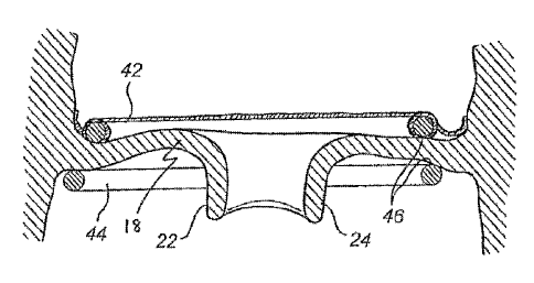
Referring now to Figs. 9-11, a method for repairing a
heart valve by means of the device according to the first
embodiment will be described.
First, access to the heart valve is achieved by
conventional techniques, including arresting the heart and
opening the chest. Alternatively, an intraluminal catheter
based delivery technique may be applied. In Fig. 9a, the
device 40 is shown when being inserted to the mitral valve
18 from the atrial side. The device 40 is being carried on
a carrier or tool (not shown), which is connected to a stem
for remote control of the positioning of the carrier. An
end 56 of the second loop-shaped support 44 is brought to
the opening of the mitral valve 18 at a commissure 60
between the leaflets 22, 24, .as shown in Fig. 9b. The end
56 is led through the opening and the carrier is turned 360
degrees. Thus, the second support 44 will be rotated into
place on one side of the valve 18, whereas the first
support 42 and the flange unit is placed on the opposite
side of the valve 18. During this rotational movement the
flange unit 50 is deflected from its original direction
forming an angle of 30-60 downwards from the diametric
plane formed by the support 42 to a direction extending in
an angle upwards from the diametric plane corresponding to
the wall formed by the annulus 20. The deflection allowed
by the flexibility of the flange unit 50 results in a close
abutment between the flange unit 50 and the atrial side of
the annulus 20. If necessary, the flange unit 50 may be
additionally conformed to the annulus 20. In this way, the
device 40 is arranged in engagement with the valve 18, as
shown in Fig. 10.
Further, the supports 42, 44 are placed on opposite
sides of the valve 18 pinching valve tissue between them to
maintain a shape of the valve 18. The leaflets 22, 24 may
now be drawn towards each other through the pinch of the
CA 02668548 2013-07-16
23
support rings 42, 44 so as to remodel the shape of the
valve 18. The leaflets may be drawn through the pinch by
means of a forceps instrument. The supports 42, 44 may flex
away from each other to allow drawing leaflets 22, 24
through the pinch and towards each other for preventing the
leaflets 22, 24 to slip back. The valve annulus 20 may in
this way be remodelled and the new shape is maintained by
the supports 42, 44, see Figs. 11 and 12 showing before and
after remodelling. In Fig. 11 a defective closure region
400 of the valve leaflets 22, 24 is shown. The supports 42,
44 may have roughened, opposed surfaces 46 to better keep
the leaflets 22, 24 from slipping through the pinch and to
hold the valve annulus 20 in its reshaped form.
The device 40 may now be secured to the valve 18 for
strengthening the fixation of the relative position between
the supports 42, 44 and the valve tissue, see Fig. 13. The
fixation may be made by clips or sutures 62 which are
arranged through the flange unit 50 and its circumferential
flange surface 54. By the latter being made of fabric it is
easily penetrated. The clips or sutures 62 are preferably
oriented and positioned in the circumferential direction of
the flange unit 50. The number of fixation points is
arbitrary for the provision of a durable fixation.
The flange unit 50 provides in some embodiments a
better seat and prevents sliding of the device 40. Thus,
the device 40 is positioned more stable in the procedure,
which is advantageous, especially for long-term performance
of the device after insertion.
As illustrated in Fig. .10, the second support 44 is
slightly displaced radially with respect to the first
support 42. This implies that the first and second supports
42, 44 are not arranged directly on top of each other. The
pinch between the first and second supports is therefore
not sharply defined in a radial direction of the valve.
This implies that a pinching force between the supports is
not focussed to a specific radial position of the valve. As
CA 02668548 2013-07-16
24
a result, the pinching force does not affect the movement
of the leaflets during normal heart action and there is a
diminished risk of rupture in the leaflets at the pinch.
The supports are interrelated in such manner that the outer
boundary of the first support 42 has a diameter
corresponding to a line through the centre of the second
support 44. Thus, the supports 42, 44 overlap somewhat such
that tissue is not allowed to move through the pinch and
the shape of the valve is maintained. Further, the cross-
section of the supports 42, 44 is round, which also gives a
soft contact between the supports and the valve tissue to
further diminish the risk of rupture in the leaflets.
The method described above is applicable no matter
the shape, position or extension of the flange means.
Further, the method is applicable no matter if the device
is inserted from the atrial side or the ventricle side.
A device having a flange unit on the first, upper
loop-shaped support is suitable when the device is to be
positioned on the atrial side, providing a fixation surface
to the atrial side of the annulus. Such device is also
suitable when carrying an artificial valve. Further, a
device having a flange unit on the second loop-shaped
support is suitable when the second loop-shaped support is
to be positioned on the ventricle side of the heart valve.
A device having a flange unit extending from the
first to the second loop-shaped support is suitable no
matter if the device is positioned on the atrial side or
the ventricle side of the heart valve.
With reference to Fig. 14a and Fig. 14b, it is to be
understood that the device may be used for replacement of
heart valves as well. For that purpose the device 40
comprises in addition to a body 41 and a flange unit 50 an
artificial valve 64. The flange unit 50 may be carried by
the first loop shaped support 42 as is shown in Fig. 14a.
Alternatively, as is shown in Fig. 14b, the flange unit 50
may extend from the first 42 to the second 44 support.
CA 02668548 2013-07-16
Although not shown, it is to be understood that each
support 42, 44 may carry its own flange unit 50, or that
the flange unit may be carried by the second support 44
only.
5 The method of inserting, positioning and fixation of
the device is generally the same as that used when
repairing a heart valve, whereby the method as such is not
further discussed.
By way of example, the device 40 and its body 41 has
10 been disclosed as having a first 42 and a second 44 loop-
shaped support. The device 40 is applicable with only one
loop-shaped support carrying the flange unit -50. One such
embodiment is disclosed in Fig. 15.
Further, the access to the heart valve may be
15 achieved endoscopically, or transluminally, catheter based.
In such case, the device 40 needs to be inserted through a
narrow tube (endoscope or catheter). This implies that the
device 40 will need to be compressed during insertion in
order to pass through the endoscope or catheter. The device
20 40 needs to assume its proper shape after having been
passed through the endoscope. Therefore, using an
endoscopic or catheter based approach, the body may
advantageously be formed from a shape memory material. This
allows the device 40 to be Compressed and also to have a
25 stable shape when being applied to the heart valve. In the
alternative, where the access to the heart valve may be
achieved through a catheter, which is passed through the
vascular system to the heart. In this case, the supports
may be formed from a shape-memory material, which during
insertion extends along the catheter in a flexible state
and, when pushed out of the catheter at the heart valve,
assumes a pre-stressed coil-shape in order to abut the
heart valve on opposite sides.
The first and second loop-shaped supports may be
connected to each other by means of a connecting part so as
to form a coil-shape. The coil-shape of the device is
CA 02668548 2013-07-16
26
advantageous during insertion, since the device may then be
rotated into position, as described above. However, the
connecting part is detachable from at least one of the
supports. Thus, when the device has been inserted, the
connecting part may be detached and removed from the
opening of the valve.
The loop-shaped support(s) and the flange unit may be
provided as separate parts.
Further, it is to be understood that the flange
means, or at least a wing part thereof, may form an
arbitrary angle to its corresponding loop-shaped support.
. Fig. 16a, 16b are cross sectional views of
embodiments involving a shape change.
In Fig. 16a the change of shape of a flange unit 50
is illustrated, e.g. for being out of a line of sight for a
surgeon during insertion (dotted line) and, when in contact
with body tissue, turning to a second shape (continuous
line) for attaching to the tissue.
In Fig. 16a the change of shape of a flange unit 50
is illustrated in two steps or directions. Firstly the
flange unit may shrink in a first direction, in order to
eliminate any wrinkles or folds therein. Subsequently or
concurrently, the flange unit 50 may change shape in a
second direction, e.g. as described with reference to Fig.
16a.
Fig. 17 is a cross sectional view schematically
illustrating a flange unit 50 having barb elements 500 for
affixing the device 40 to tissue. The flange unit 50 may
thus be a carrier for fixation elements. The flange unit
50, may thus be inserted into the body more effectively.
In some embodiments, different materials may be used
for parts of the device 40. For instance, the inner rings
42, 44 may be made of a stiffer more stable than a more
flexible outer part, e.g. the flange unit 50.
While several embodiments of the present invention
have been described and illustrated herein, those of
CA 02668548 2013-07-16
27
ordinary skill in the art will readily envision a variety
of other means and/or structures for performing the
functions and/or obtaining the results and/or one or more
of the advantages described herein, and each of such
variations and/or modifications is deemed to be within the
scope of the present invention. More generally, those
skilled in the art will readily appreciate that all
parameters, dimensions, materials, and configurations
described herein are meant to be exemplary and that the
actual parameters, dimensions, materials, and/or
configurations will depend upon the specific application or
applications for which the teachings of the present
invention is/are used.
Those skilled in the art will recognize, or be able
to ascertain using no more than routine experimentation,
many equivalents to the specific embodiments of the
invention described herein. It is, therefore, to be
understood that the foregoing embodiments are presented by
way of example only, and that the scope of the claims
should not be limited by the preferred embodiments set
forth, in the examples, but should be given the broadest
interpretation consistent with the description as a whole.
The present invention is directed to each individual
feature, system, article, material, kit, and/or method
described herein. In addition, any combination of two or
more such features, systems, articles, materials, kits,
and/or methods, if such features, systems, articles,
materials, kits, and/or methods are not mutually
inconsistent, is included within the scope of the present
invention as limited by the appended patent claims.