Note: Descriptions are shown in the official language in which they were submitted.
CA 02669088 2009-05-08
WO 2008/055313 PCT/AU2007/001722
- 1 -
DETECTION SYSTEM AND USES THEREFOR
Field of the Invention
The present invention relates to a system for detecting molecular
associations.
The present invention has particular application to detecting the association
of
different molecules, and thus detecting the formation of hetero-dimers and/or -
oligomers.
Background Art
The background to the invention will be discussed in the context of the
association
of proteins. However, the scope of the invention should not be understood to
be
limited thereto.
Proteins do not act in isolation in a cell, but in stable or transitory
complexes, with
protein-protein interactions being key determinants of protein function
(Auerbach
et al., (2002), Proteomics, 2, 611-623). Furthermore, proteins and protein
complexes interact with other cellular components like DNA, RNA and small
molecules. Understanding both the individual proteins involved in these
interactions and their interactions are important for a better understanding
of
biological processes.
Several tools exist for demonstrating protein-protein interactions either in
vitro,
coimmunoprecipitation with the potential for cross-linking at the cell
surface, or in
vivo, including for example the resonance energy transfer (RET) technologies
of
fluorescence RET (FRET) and bioluminescence RET (BRET).
The interaction of G-protein coupled receptors (GPCRs) represents an excellent
example of the physiological and potential pharmacological relevance of the
association of proteins, and in particular the relevance of hetero-dimers
and/or ¨
oligomers. As Milligan (Milligan, (2006), Drug Discovery Today, 11, 541-549)
observes, homo-dimerisation and ¨oligomerisation have limited implications for
the drug discovery industry, while "differential pharmacology, function and
regulation of GCPR hetero-dimers and ¨oligomers suggest means to selectively
target GPCRs in different tissues and hint that the mechanism of function of
several pharmacological agents might be different in vivo than anticipated
from
simple ligand screening programmes that rely on heterologous expression of a
CA 02669088 2009-05-08
WO 2008/055313 PCT/AU2007/001722
- 2 -
single GPCR".
Coimmunoprecipitation has been used to identify GPCR heterodimers (Jordan BA
& Devi LA (1999) G-protein-coupled receptor heterodimerization modulates
receptor function Nature 399, 697-700). However, coimmunoprecipitation does
not enable the distinction between constitutive and random associations. In
particular, there is concern that artefactual aggregation occurs following
cell lysis
and solubilization (Kroeger KM et a/. (2003) G-protein coupled receptor
oligomerization in neuroendocrine pathways. Front. Neuroendocrinol. 24, 254-
278). Further, coimmunoprecipitation is not amenable to automation or high
throughput screening.
Fluorescence resonance energy transfer (FRET) is capable of detecting in vivo
protein-protein interactions (Forster, (1948), Ann. Phys. 2, 57-75). This
technique
became particularly attractive and applicable to assays in living cells when
the
green fluorescent protein (GFP) and its mutant variants with different
spectral
characteristics were cloned. This allowed the genetic attachment of GFP and
its
variants to any target protein by fusing the encoding DNA sequences (Heim et
al.,
(1994), PNAS. USA. 91, 12501-12504). FRET is able to monitor interactions that
occur anywhere inside the cell. FRET can be determined in any cell type
(mammalian, yeast, bacterial etc.) or cell-free system. It can be detected by
fluorescence spectroscopy, fluorescence microscopy and fluorescence activated
cell sorting (FACS).
Bioluminescence resonance energy transfer (BRET) is another technique that has
been developed to study in vivo protein-protein interactions (Xu et al.,
(1999),
PNAS. USA, 96, 151-156; Eidne et al., (2002), Trends Endocrin. Metabol. 13,
415-421). Like FRET, BRET allows detection within living cells or cell-free
systems and is not restricted to a particular cellular compartment.
It is difficult to differentiate background signal from signals resulting from
constitutive interactions using FRET or BRET to assess direct interactions
between labelled proteins. Furthermore, interaction affinity does not relate
directly
to signal intensity as RET is dependent upon the relative orientation of and
distance between the energy donor and acceptor.
CA 02669088 2009-05-08
WO 2008/055313 PCT/AU2007/001722
- 3 -
'Saturation' BRET [Mercier JF, Salahpour A, Angers S, Breit A & Bouvier M
(2002) Quantitative assessment of beta 1- and beta 2-adrenergic receptor homo-
and heterodimerization by bioluminescence resonance energy transfer. J Biol
Chem 277, 44925-44931.] has been suggested as a method to differentiate
background and constitutive signals, however, this requires expression of
increasing concentrations of acceptor-labelled protein relative to donor-
labelled
protein in order to generate saturation curves and is by no means high
throughput.
The preceding discussion is intended only to facilitate an understanding of
the
invention. It should not be construed as in any way limiting the scope or
application of the following description of the invention, nor should it be
construed
as an admission that any of the information discussed was within the common
general knowledge of the person skilled in the appropriate art at the priority
date.
Disclosure of the Invention
The inventors have developed a detection system capable of overcoming or at
least alleviating some of the problems identified in analysis of protein
associations.
Most importantly, the system of the invention is capable of identifying
potentially
constitutive associations arising between different molecules (i.e.,
heterodimers)
as well as those arising from multiple copies of the same molecule (i.e.,
oligomers), utilising a signal that is dependent upon external modulation,
thereby
enabling the ability to distinguish between constitutive associations and
random
associations.
In a first aspect of the invention, there is provided a system for the
detection of the
molecular associations, the system comprising:
i). a first agent, comprising a first interacting group coupled to a first
reporter component;
ii). a second agent, comprising a second interacting group coupled to a
second reporter component;
iii). a third agent, comprising a third interacting group;
iv). a modulator; and
v). a detector;
wherein proximity of the first and second reporter components generates a
signal
CA 02669088 2009-05-08
WO 2008/055313 PCT/AU2007/001722
- 4 -
capable of detection by the detector; and wherein the modulator modulates the
association of the second interacting group with the third interacting group;
such
that monitoring the signal generated by proximity of the first and second
reporter
components by the detector constitutes monitoring the association of the first
and
third agents.
The system may further comprise a reporter component initiator, wherein
proximity of the first and second reporter components generates a signal
capable
of detection by the detector only in the presence of the reporter component
initiator.
In a second aspect of the invention, there is provided a method for the
detection
of the molecular associations, the method comprising the steps of:
i). Providing a first agent, comprising a first interacting group
coupled to
a first reporter component;
ìi). Providing a second agent, comprising a second interacting
group
coupled to a second reporter component;
iii). Providing a third agent, comprising a third interacting group;
iv). Providing a modulator;
wherein; proximity of the first and second reporter components
generates a signal; and wherein the modulator modulates the
association of the second interacting group with the third interacting
group; then
v). Detecting and/or monitoring any signal generated by proximity of the
first and second reporter components.
such that monitoring the signal generated by proximity of the first and second
reporter components by the detector constitutes monitoring the association of
the
first and third agents.
Before the step of detecting and/or monitoring any signal generated by
proximity
of the first and second reporter components, the method may further comprise
the
step of providing a reporter component initiator, wherein proximity of the
first and
second reporter components generates a signal capable of detection by the
detector only in the presence of the reporter component initiator.
In one form of the invention, the steps of providing a reporter component
initiator;
and providing a modulator; occur after the steps of providing the first,
second and
third agents.
CA 02669088 2009-05-08
WO 2008/055313 PCT/AU2007/001722
- 5 -
In a particularly advantageous form of the invention, the first, second and
third
agents are- provided by way of co-expression in a cell, to which the modulator
is
introduced.
In a third aspect, the present invention provides a method for determining
whether
and/or the extent to which a test compound interacts with a second protein
when
the second protein is associated with a first protein, the method comprising
the
steps of:
a). contacting said test compound with a system comprising:
i). a first agent, comprising the first protein coupled to a first reporter
component;
ii). a second agent, comprising an interacting group coupled to a
second reporter component;
iii). a third agent, comprising the second protein;
wherein proximity of the first and second reporter components generates a
signal; and wherein the modulator modulates the association of the
interacting group with the second protein;
b). determining the signal as a determination of whether and/or the
extent to
which said test compound interacts with a second protein when the second
protein is associated with a first protein.
In one embodiment, the second protein is a receptor, and the third aspect of
the
invention comprises a method for determining whether a test compound is an
agonist of the second protein when the second protein is associated with the
first
protein, and the step of determining the signal as a determination of whether
and/or the extent to which said test compound interacts with a second protein
when the second protein is associated with a first protein more specifically
comprises the step of detecting an increase in the signal as a determination
of
whether and/or the extent to which the test compound is an agonist of the
second
protein when the second protein is associated with the first protein.
The first protein may also be a receptor.
In one embodiment, the second protein is a receptor, and the third aspect of
the
invention comprises a method for determining whether and/or the extent to
which
a test compound is an antagonist or partial agonist of the second protein when
the
CA 02669088 2009-05-08
WO 2008/055313 PCT/AU2007/001722
- 6 -
second protein is associated with the first protein, the method comprising the
steps of:
a). contacting said test compound with a system comprising:
i). a first agent, comprising the first protein coupled to a first reporter
component;
ii). a second agent, comprising an interacting group coupled to a
second reporter component;
iii). a third agent, comprising the second protein;
iv). an agonist of the second protein;
wherein proximity of the first and second reporter components generates a
signal; and wherein the modulator modulates the association of the
interacting group with the second protein;
b). detecting a decrease in the signal as a determination of whether
and/or the
extent to which the test compound is an antagonist or partial agonist of the
second protein when the second protein is associated with the first protein.
The first protein may also be a receptor.
In one embodiment, the second protein is a constitutively active receptor, and
the
third aspect of the invention comprises a method for determining whether
and/or
the extent to which a test compound is an inverse agonist of the second
protein
when the second protein is associated with the first protein, and the step of
determining the signal as a determination of whether and/or the extent to
which
said test compound interacts with a second protein when the second protein is
associated with a first protein more specifically comprises the step of
detecting a
decrease in the signal as a determination of whether the test compound is an
inverse agonist of the second protein when the second protein is associated
with
the first protein.
In one embodiment, where the first and second proteins are both receptors,
present invention comprises a method for screening a test compound for first
protein / second protein hetero-dimer / -oligomer selective activity, the
method
comprising the steps of:
a) determining whether, and/or the extent to which, the test compound
interacts with the second protein while the second protein is associated
with the first protein; and
b) if the test compound interacts with the second protein while the second
protein is associated with the first protein, determining whether, or the
CA 02669088 2009-05-08
WO 2008/055313 PCT/AU2007/001722
- 7 -
extent to which the test compound interacts with the second protein in the
absence of the first protein;
such that a test compound that exhibits greater affinity and/or potency and/or
efficacy when interacting with the second protein while the second protein is
associated with the first protein is selective for the first protein / second
protein
hetero-dimer/-oligomer.
In one embodiment, where the first and second proteins are both receptors,
present invention comprises a method for screening a test compound for first
protein / second protein hetero-dimer / -oligomer selective antagonism or
partial
agonism, the method comprising the steps of:
a) determining whether, and/or the extent to which, the test compound is an
antagonist or partial agonist of the first protein / second protein hetero-
dimer /
-oligomer, by contacting said test compound with a system comprising:
i). a first
agent, comprising the first protein coupled to a first reporter
component;
ii). a second agent, comprising an interacting group coupled to a
second reporter component;
iii). a third agent, comprising the second protein;
iv). an agonist
of the first protein, the second protein and/or the first
protein / second protein hetero-dimer / -oligomer;
wherein proximity of the first and second reporter components generates a
signal; and wherein the modulator modulates the association of the
interacting group with the second protein;
b). detecting a decrease in the signal as a determination of whether and/or
the
extent to which the test compound is an antagonist or partial agonist of the
first protein / second protein hetero-dimer / -oligomer;
c) if the test compound is an antagonist or partial agonist of the first
protein /
second protein hetero-dimer / -oligomer, determining whether, or the extent to
which the test compound interacts with the second protein in the absence of
=the first protein and the first protein in the absence of the second protein;
such
that a test compound that exhibits greater agonistic or partial agonistic
properties when interacting with the first protein / second protein hetero-
dimer
/ -oligomer is selective for the first protein / second protein hetero-dimer/-
CA 02669088 2009-05-08
WO 2008/055313 PCT/AU2007/001722
- 8 -
oligomer.
In one embodiment, where the first and second proteins are both receptors,
there
is provided a method for screening a test compound for first protein / second
protein hetero-dimer/-oligomer selective antagonism or partial agonism, the
method comprising the steps of:
a) determining whether, and/or the extent to which, the test compound
is an antagonist or partial agonist of the first protein / second protein
hetero-dimer/-oligomer, by contacting said test compound with a
system comprising:
i). a first agent, comprising the second protein coupled to a first
reporter component;
ii). a second agent, comprising an interacting group coupled to a
second reporter component;
iii). a third agent, comprising the first protein;
iv). an agonist of the second protein, the first protein and/or the first
protein / second protein hetero-dimer/-oligomer;
wherein proximity of the first and second reporter components
generates a signal; and wherein the modulator modulates the
association of the interacting group with the first protein;
b). detecting a decrease in the signal as a determination of whether and/or
the extent to which the test compound is an antagonist or partial
agonist of the first protein / second protein hetero-dimer/-oligomer;
c) if the test compound is an antagonist or partial agonist of the first
protein / second protein hetero-dimer/-oligomer, determining whether,
or the extent to which the test compound is an antagonist or partial
agonist of the first protein in the absence of the second protein and the
second protein in the absence of the first protein; such that a test
compound that exhibits greater antagonistic or partial agonistic
properties when interacting with the first protein / second protein
hetero-dimer/-oligomer is selective for the first protein / second protein
hetero-dimer/-oligomer.
CA 02669088 2009-05-08
WO 2008/055313 PCT/AU2007/001722
- 9 -
In one embodiment, where the first and second proteins are both receptors,
there
is provided a method for screening a test compound for first protein / second
protein hetero-dimer/-oligomer selective inverse agonim, the method comprising
the steps of:
a) determining whether, and/or the extent to which, the test compound is
an inverse agonist of the first protein / second protein hetero-dimer/-
oligomer, by contacting said test compound with a system comprising:
1). a first agent, comprising the first protein coupled to a first
reporter component;
ii). a second agent, comprising an interacting group coupled to a
second reporter component;
iii). a third agent, comprising a constitutively active second protein;
wherein proximity of the first and second reporter components
generates a signal; and wherein the modulator modulates the
association of the interacting group with the second protein;
b) detecting a decrease in the signal as a determination of whether and/or
the extent to which the test compound is an inverse agonist of the first
protein / second protein hetero-dimer/-oligomer;
c) if the test compound is an inverse agonist of the first protein / second
protein hetero-dimer/-oligomer, determining whether, or the extent to
which the test compound is an inverse agonist of the first protein in the
absence of the second protein and the second protein in the absence
of the first protein; such that a test compound that exhibits greater
inverse agonistic properties when interacting with the irst protein /
second protein hetero-dimer/-oligomer is selective for the first
protein/second protein hetero-dimer/-oligomer.
In one embodiment, where the first and second proteins are both receptors,
there
is provided a method for screening a test compound for first protein / second
protein hetero-dimer/-oligomer selective inverse agonism, the method
comprising
the steps of:
a) determining whether, and/or the extent to which, the test compound is
an inverse agonist of the first protein / second protein hetero-dimer/-
CA 02669088 2009-05-08
WO 2008/055313 PCT/AU2007/001722
- 10 -
oligomer, by contacting said test compound with a system comprising:
i). a first agent, comprising the second protein coupled to a first
reporter component;
ii). a second agent, comprising an interacting group coupled to a
second reporter component;
Ýii). a third agent, comprising a constitutively first protein;
wherein proximity of the first and second reporter components
generates a signal; and wherein the modulator modulates the
association of the interacting group with the first protein;
b) detecting a decrease in the signal as a determination of whether and/or
the extent to which the test compound is an inverse agonist of the first
protein / second protein hetero-dimer/-oligomer;
c) if the test compound is an inverse agonist of the first protein / second
protein hetero-dimer/-oligomer, determining whether, or the extent to
which the test compound is an inverse agonist of the second protein in
the absence of the first protein and the first protein in the absence of
the second protein; such that a test compound that exhibits greater
inverse agonistic properties when interacting with the first protein /
second protein hetero-dimer/-oligomer is selective for the first protein /
second protein hetero-dimer/-oligomer.
In a fourth aspect of the invention, there is provided a kit for the detection
of the
molecular associations, the kit comprising:
i). a first agent, comprising a first interacting group coupled to a first
reporter component;
ii). a second agent, comprising a second interacting group coupled to a
second reporter component;
iii). a third agent, comprising a third interacting group;
iv). a modulator; and
wherein proximity of the first and second reporter components generates a
signal
capable of detection; and wherein the modulator modulates the association of
the
second interacting group with the third interacting group.
In one form of the invention, the kit further comprises a reporter component
initiator, and proximity of the first and second reporter components generates
a
CA 02669088 2009-05-08
WO 2008/055313 PCT/AU2007/001722
- 11 -
signal capable of detection only in the presence of the reporter component
initiator.
In one form of the invention, the modulator increases the propensity of the
second
interacting group to associate with the third interacting group, such that
detection
of the signal generated by proximity of the first and second reporter
components
by the detector constitutes detection of the association of the first and
third
agents.
It should be understood that the modulator may interact with either the first,
second or third interacting groups, or simultaneously with both first and
third
interacting groups, or simultaneously with second and third interacting
groups, to
modulate the association of the second interacting group with the third
interacting
group.
In an alternate form of the invention, the modulator decreases the propensity
of
the second interacting group to associate with the third interacting group,
such
that detection of a reduction of the signal generated by proximity of the
first and
second reporter components by the detector constitutes detection of the
dissociation of the first and third agents.
In a highly advantageous form of the invention, the first interacting group
differs
from the third interacting group. Thus, in this form, detection of the signal
generated by the proximity of the first and second reporter components
constitutes detection of the hetero-dimerisation and/or ¨oligomerisation of
the first
and third interacting groups.
In a preferred form of the invention, the third agent does not comprise a
component capable of generating a signal that substantially interferes with
and/or
contributes to the signal generated by the proximity of the first and second
reporter components. Thus, in this form, detection of the signal generated by
the
proximity of the first and second reporter components is facilitated.
In some systems, such as where the first and third interacting groups are
expressed at low levels, and/or where the selected combination of the first
and
second reporter components generates a weak signal, facilitation of the
detection
of the signal in accordance with the preferred form of the invention described
above may render an assay viable or, even more advantageously, amenable to
CA 02669088 2009-05-08
WO 2008/055313 PCT/AU2007/001722
- 12 -
high throughput screening.
In a preferred form of the invention, the third agent is selected from the
group:
receptors, ion channels, enzymes, carriers, transporters, integral membrane
proteins, cytoskeletal proteins, adhesion molecules, signalling proteins,
scaffolding proteins, accessory proteins, trafficking proteins, transcription
factors,
nuclear co-factors and nucleic acid molecules, as defined below.
In a preferred form of the invention, the modulator is a ligand or an enzyme.
In a preferred form of the invention, the third agent is produced by
expression in a
cell. Where the third agent is produced by expression in a cell, the method of
the
invention may be carried out in whole cells, cellular fractions or in a cell-
free
system.
In a particularly advantageous embodiment of the invention, the first and
third
interacting groups are provided in the form of receptors, and the modulator is
provided in the form of a ligand that modulates the receptor of the third
interacting
group, such that modulation of the signal generated by proximity of the first
and
second reporter components by the modulator is indicative of association of
the
receptors of the first and third interacting groups.
In a fifth aspect of the invention, there is provided a method for screening a
test
compound for selective activity against a heterodimer of a first protein and a
second protein, the method comprising the steps of:
a) contacting said test compound at increasing concentrations with a
system
comprising:
i).
a first agent, comprising the first protein coupled to a first reporter
component;
ii). a second
agent, comprising an interacting group coupled to a
second reporter component;
iii). a third agent, comprising the second protein;
wherein proximity of the first and second reporter components generates a
signal; and wherein the modulator modulates the association of the
interacting group with the second protein;
b). determining signal as a determination of whether said test compound
modulates said association of the interacting group with the second protein
at each concentration to produce a dose-response curve;
CA 02669088 2009-05-08
WO 2008/055313 PCT/AU2007/001722
- 13 -
c). determining the Hill slope of the dose-response curve, wherein, a Hill
slope in
excess of 1 indicates interaction of the test compound with the hetero-dimer.
In a sixth aspect of the invention, there is provided a first protein-second
protein
hetero-dimer/-oligomer identified by any one of the systems, methods or kits
of
the invention.
In a seventh aspect of the invention, there is provided a hetero-dimeric or
hetero-
oligomeric receptor, comprising at least one first receptor subunit associated
with
at least one second receptor subunit, identified by any one of the systems,
methods or kits of the invention.
In an eighth aspect of the invention, there is provided a method for the
treatment
of a patient suffering from a first receptor-related ailment by administering
a
therapeutically effective amount of a second receptor agonist, inverse agonist
or
antagonist, wherein the first and second receptors form a hetero-dimer/-
oligomer
identified by any one of the systems, methods or kits of the invention.
In one embodiment, the second receptor agonist, inverse agonist or antagonist
is
co-administered with a first receptor agonist, inverse agonist or antagonist.
In a ninth aspect of the invention, there is provided a method for the
treatment of a
patient suffering from a second receptor-related ailment by administering a
therapeutically effective amount of a first receptor agonist, inverse agonist
or
antagonist, wherein the first and second receptors form a hetero-dimer/-
oligomer
identified by any one of the systems, methods or kits of the invention.
In one embodiment, the first receptor agonist, inverse agonist or antagonist
is co-
administered with a second receptor agonist, inverse agonist or antagonist.
In a tenth aspect of the invention, there is provided a method for the
manufacture
of a medicament for the treatment of a patient suffering from an first
receptor-
related ailment comprising use of a therapeutically effective amount of a
second
receptor agonist, inverse agonist or antagonist, wherein the first and second
receptors form a hetero-dimer/-oligomer identified by any one of the systems,
methods or kits of the invention.
The medicament may also contain a first receptor agonist, inverse agonist or
CA 02669088 2009-05-08
WO 2008/055313 PCT/AU2007/001722
- 14 -
antagonist.
In an eleventh aspect of the invention, there is provided a method for the
manufacture of a medicament for the treatment of a patient suffering from a
second receptor-related ailment comprising use of a therapeutically effective
amount of an first receptor agonist, inverse agonist or antagonist, wherein
the first
and second receptors form a hetero-dimer/-oligomer identified by any one of
the
systems, methods or kits of the invention.
The medicament may also contain a second receptor agonist, inverse agonist or
antagonist.
In a twelfth aspect of the invention, there is provided a method for the
treatment of
a patient suffering from a first receptor-related ailment by administering a
therapeutically effective amount of a selective second receptor agonist,
antagonist
or inverse agonist binding agent, or fragment thereof, wherein the first and
second
receptors form a hetero-dimer/-oligomer identified by any one of the systems,
methods or kits of the preceding claims.
The selective second receptor agonist, antagonist or inverse agonist binding
agent may be an antibody, including a humanised antibody, a polyclonal
antibody,
a monoclonal antibody, a chimeric antibody, a CDR-grafted antibody and/or an
anti-idiotypic antibody.
In a thirteenth aspect of the invention, there is provided a method for the
treatment of a patient suffering from a second receptor-related ailment by
administering a therapeutically effective amount of a selective first receptor
agonist, antagonist or inverse agonist binding agent, or fragment thereof,
wherein
the first and second receptors form a hetero-dimer/-oligomer identified by any
one
of the systems, methods or kits of the preceding claims.
The selective first receptor agonist, antagonist or inverse agonist binding
agent
may be an antibody, including a humanised antibody, a polyclonal antibody, a
monoclonal antibody, a chimeric antibody, a CDR-grafted antibody and/or an
anti-
idiotypic antibody.
In a fourteenth aspect of the invention, there is provided a method for the
treatment of a patient suffering from a second receptor-related ailment by
CA 02669088 2009-05-08
WO 2008/055313 PCT/AU2007/001722
- 15 -
administering a therapeutically effective amount of a selective first receptor
agonist, antagonist or inverse agonist binding agent, or fragment thereof,
wherein
the first and second receptors form a hetero-dimer/-oligomer identified by any
one
of the systems, methods or kits of the preceding claims.
The selective first receptor agonist, antagonist or inverse agonist binding
agent
may be an antibody, including a humanised antibody, a polyclonal antibody, a
monoclonal antibody, a chimeric antibody, a CDR-grafted antibody and/or an
anti-
id iotypic antibody.
In a fifteenth aspect of the invention, there is provided an agonist of the
second
protein, when the second protein is associated with the first protein,
identified by
the methods of the invention.
In a sixteenth aspect of the invention, there is provided an antagonist or
partial
agonist of the second protein, when the second protein is associated with the
first
protein, identified by the methods of the invention.
In a seventeenth aspect of the invention, there is provided an inverse agonist
of
the second protein, when the second protein is associated with the first
protein,
identified by the methods of the invention.
In a eighteenth aspect of the invention, there are provided selective agonists
and/or antagonists and/or inverse agonists of the first protein-second protein
hetero-dimer/-oligomer identified by the methods of the invention.
Brief Description of the Drawings
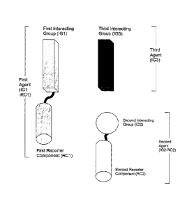
Figure 1 shows the composition of the agents forming the basis of the system
for
detecting molecular associations: A first agent comprises a first interacting
group
coupled to a first reporter component; a second agent comprises a second
interacting group coupled to a second reporter component; and a third agent
comprises a third interacting group.
CA 02669088 2009-05-08
WO 2008/055313 PCT/AU2007/001722
- 16 -
Figure 2 shows how the administration of the modulator modulates the
association of the second interacting group with the third interacting group,
preferably by interacting with the third interacting group, either alone, or
simultaneously with the first interacting group.
Figure 3 shows that if the first and third interacting groups are associated,
modulation of the association of the second and third interacting groups
consequently modulates the proximity of the first and second reporter
components
thereby modulating the signal that is able to be detected by the detector.
Therefore monitoring the signal generated by proximity of the first and second
reporter components by the detector constitutes monitoring the association of
the
first and third agents. If the first and third interacting groups are not
associated,
the first and second reporter components will remain spatially separated and
generation of a detectable signal is unlikely.
Figure 4 shows the thyrotropin releasing hormone receptor (TRHR) as IG1, Rluc
as RC1, beta-arrestin 2 (barr2) as IG2, Venus as RC2 and a range of different
GPCRs as IG3.
eBRET measurements at 37C were carried out on HEK293 cells transiently co-
expressing TRHR/Rluc and barr2/Venus with either pcDNA3, orexin receptor 2
(OxR2), CXC chemokine receptor 2 (CXCR2), hemagglutin epitope-tagged
melanocortin receptor 3 or 4 (HA-MC3R or HA-MC4R), or dopamine D2 receptor
long form (D2LR) or short form (D2SR) following the treatment of each with
their
respective ligands. The different ligand treatment (10-6M) for each receptor
was
thyrotropin releasing hormone (TRH) for TRHR/Rluc (with pcDNA3); orexin A
(OxA) for OXR2; interleukin-8 (IL-8) for CXCR2; alpha-melanocyte-stimulating
hormone (a-MSH) for HA-MC3R, HA-MC4R; and bromocriptine (BROM) for D2LR
and D2SR.
Figure 5 shows the thyrotropin releasing hormone receptor (TRHR) as IG1, Rluc
as RC1, either beta-arrestin 1 (barr1) or beta-arrestin 2 (barr2) as IG2, EGFP
as
CA 02669088 2009-05-08
WO 2008/055313 PCT/AU2007/001722
- 17 -
RC2 and OxR2 as IG3. eBRET measurements at 370 were carried out on
HEK293 cells transiently co-expressing TRHR/Rluc and EGFP/barr1 or
EGFP/barr2 with either pcDNA3 or OXR2. Ligand treatments were either OxA or
TRH only or both OxA and TRH combined. Phosphate-buffered saline (PBS) was
used as a vehicle control.
Figure 6 shows the thyrotropin releasing hormone receptor (TRHR) as IG1, Rluc
as RC1, beta-arrestin 2 (barr2) as IG2, Venus as RC2 and OxR1 or OxR2 as IG3.
eBRET measurements were carried out at 37C on HEK293 cells transiently co-
expressing TRHR/Rluc and barr2/Venus with either pcDNA3, OxR1 or OxR2
following pretreatment with 10-6M OxR1-selective antagonist, SB-334867-A, for
approximately 40 minutes and then 10-6M OxA (IG3 ligand; modulator) or 10-6M
TRH (IG1 ligand), or both, was added. Where antagonist was not preincubated,
cells were treated with PBS instead for the same amount of time.
Figure 7 shows the thyrotropin releasing hormone receptor (TRHR) as 11, Rluc
as RC1, beta-arrestin 1 (bard ) or beta-arrestin 2 (barr2) as IG2, EGFP as RC2
and hemagglutin epitope-tagged OxR2 (HA-OxR2) as IG3. eBRET measurements
at 37C were carried out on HEK293 cells transiently co-expressing TRHR/Rluc
and EGFP/barr1 or EGFP/barr2 with either pcDNA3 or HA-OxR2. Ligand
treatments were either OxA or TRH only. Phosphate-buffered saline (PBS) was
used as a vehicle control.
Figure 8 shows the thyrotropin releasing hormone receptor (TRHR) as IG1, Rluc
as RC1, beta-arrestin 1 (barr1) or beta-arrestin 1 phosphorylation-independent
mutant R169E (barr1R169E) as IG2, EGFP as RC2 and OxR2 as IG3. eBRET
measurements at 37C were carried out on HEK293 cells transiently co-expressing
TRHR/Rluc and EGFP/barr1 or EGFP/barr1R169E with either pcDNA3 or OxR2.
Ligand treatments were either OxA or TRH only. Phosphate-buffered saline (PBS)
was used as a vehicle control.
Figure 9 shows the thyrotropin releasing hormone receptor truncated at amino
acid 335 (TRHR335) as IG1, Rluc as RC1, beta-arrestin 1 (barr1) as IG2, EGFP
CA 02669088 2009-05-08
WO 2008/055313 PCT/AU2007/001722
- 18 -
as RC2 and OxR2 or TRHR as 103. eBRET measurements at 37C were carried
out on HEK293 cells transiently co-expressing TRHR335/Rluc and EGFP/barr1
with either OxR2 or TRHR. Ligand treatments were either OxA or TRH only.
Figure 10 shows a dose-response curve for the thyrotropin releasing hormone
receptor (TRHR) as 101, Rluc as RC1, beta-arrestin 2 (barr2) as 102, Venus as
RC2 and in the absence of 103. BRET measurements at 37C were carried out on
HEK293 cells transiently co-expressing TRHR/Rluc, barr2Nenus and pcDNA3
with increasing doses of TRH. Sigmoidal dose response curves were plotted
using Prism (GraphPad), either assuming a Hill slope of 1 or allowing for
variable
slope. The EC50 and Hill slope values for the variable slope curve are
included in
a table in the graph.
Figure 11 shows a dose-response curve for OxR2 as 101 , Rluc as RC1, barr2 as
102, Venus as RC2 and in the absence of 103. BRET measurements at 37C were
carried out on HEK293 cells transiently co-expressing OxR2/Rluc, barr2/Venus
and pcDNA3 with increasing doses of OxA. Sigmoidal dose response curves were
plotted using Prism (GraphPad), either assuming a Hill slope of 1 or allowing
for
variable slope. The EC50 and Hill slope values for the variable slope curve
are
included in a table in the graph.
Figure 12 shows dose-response curves for the thyrotropin releasing hormone
receptor (TRHR) as 101, Rluc as RC1, beta-arrestin 2 (barr2) as 102, Venus as
RC2 and OxR2 as 103. BRET measurements at 37C were carried out on HEK293
cells transiently co-expressing TRHR/Rluc, barr2Nenus and OxR2 with increasing
doses of OxA. Sigmoidal dose response curves were plotted using Prism
(GraphPad), either assuming a Hill slope of 1 or allowing for variable slope.
The
EC50 and Hill slope values for the variable slope curves are included in a
table in
the graph. Curves generated using coelenterazine h and EnduRen as two forms
of Rluc substrate (reporter component initiator) are shown.
Figure 13 shows dose-response curves for TRHR as 101, Rluc as RC1, barr1 as
102, EGFP as RC2 in the presence or absence of OxR2 as 103. BRET
CA 02669088 2009-05-08
WO 2008/055313 PCT/AU2007/001722
- 19 -
measurements at 37C were carried out on HEK293 cells transiently co-expressing
TRHR/Rluc and EGFP/barr1 in the absence of OxR2 with increasing doses of
TRH, as well as HEK293 cells transiently co-expressing TRHR/Rluc and
EGFP/barr1 with OxR2 with increasing doses of OxA with and without 10-6M TRH.
A curve mathematically generated by addition of the ligand-induced signal
generated with 10-6M TRH (from the TRH: TRHR/Rluc + EGFP/barr1 curve) to
each of the points generated for the OxA: TRHR/Rluc + EGFP/barr1 + OxR2
curve is also plotted (TRHR/Rluc + EGFP/barr1 + OxR2: TRH (10-6M) + OxA:
Data calculated).
Figure 14 shows dose-response curves for TRHR as IG1, Rluc as RC1, barr1 as
EGFP as RC2 in the presence or absence of OxR2 as IG3. BRET
measurements at 37C were carried out on HEK293 cells transiently co-expressing
TRHR/Rluc and EGFP/barr1 in the absence of OxR2 with increasing doses of
TRH, as well as HEK293 cells transiently co-expressing TRHR/Rluc and
EGFP/barr1 with OxR2 with increasing doses of OxA, or increasing doses of TRH
with 10-6M OxA. A curve mathematically generated by addition of the ligand-
induced signal generated with 10-6M OxA (from the OxA: TRHR/Rluc +
EGFP/barr1 + OxR2 curve) to each of the points generated for the TRH:
TRHR/Rluc + EGFP/barr1 curve is also plotted (TRHR/Rluc + EGFP/barr1 +
OxR2: TRH + OxA (10-6M): Data calculated).
Figure 15 shows dose response curves for TRHR335 as !GI, Rluc as RC1, barr2
as IG2, Venus as RC2 and OxR2 as IG3. BRET measurements at 37C were
carried out on HEK293 cells transiently co-expressing TRHR335/Rluc,
barr2Nenus and OxR2 with increasing doses of TRH and OxA alone or in
combination.
Figure 16 shows cumulative eBRET reads over time for each combination of
receptors (IG1 and IG3; data captured over 83mins). TRHR is IG1, Rluc is RC1,
barr1 is IG2, EGFP is RC2 and OxR2 is IG3. The same amount of EGFP/barr1
(1G2-RC2) is transfected for each experiment. TRHR/Rluc (IG1-RC1) is
transfected at a constant amount (0.1pg DNA/well) while OxR2 (IG3) is
CA 02669088 2009-05-08
WO 2008/055313 PCT/AU2007/001722
- 20 -
transfected at varying amounts of DNA (0, 0.01, 0.05, 0.1, 0.5, 0.7pg DNA
/well).
eBRET measurements at 37C were carried out on HEK293 cells following
addition of 10-6M OxA (modulator) to each well. The signal is only detected
when
OxR2 (103) is expressed (no signal was recorded at Opg OxR2).
Figure 17 shows dose response curves for TRHR as 101, Rluc as RC1, barr2 as
102, Venus as RC2 and OxR2 as 103. BRET measurements at 37C were carried
out on HEK293 cells transiently co-expressing TRHR/Rluc, barr2Nenus and
OxR2 with increasing doses of OxA in either 96-well or 384-well microplates.
Figure 18 shows OxR2 as 101, Rluc8 as RC1, beta-arrestin 2 (barr2) as 102,
Venus as RC2 and hemagglutin epitope-tagged TRHR (HA-TRHR) as 103.
eBRET measurements at 37C were carried out on HEK293 cells transiently co-
expressing OxR2/Rluc8 and barr2Nenus with either pcDNA3 or HA-TRHR.
Ligand treatments were either OxA or TRH only. Phosphate-buffered saline (PBS)
was used as a vehicle control. Data presented as ligand-induced BRET ratios.
Figure 19 shows the thyrotropin releasing hormone receptor (TRHR) as 101,
Rluc8 as RC1, beta-arrestin 2 (barr2) as 102, Venus as RC2 and hemagglutin
epitope-tagged OxR2 (HA-OxR2) as 103. eBRET measurements at 37C were
carried out on HEK293 cells transiently co-expressing TRHR/Rluc8 and
barr2Nenus with HA-OxR2 aliquoted into all wells of a 96-well plate. Phosphate-
buffered saline (PBS) was added to the first two rows and the last two rows of
the
96-well plate (48 wells in total) as a vehicle control. Data presented as
fluorescence/luminescence.
Figure 20 shows the thyrotropin releasing hormone receptor (TRHR) as 101,
Rluc8 as RC1, beta-arrestin 2 (barr2) as 102, Venus as RC2 and hemagglutin
epitope-tagged OxR2 (HA-OxR2) as 103. eBRET measurements at 37C were
carried out on HEK293 cells transiently co-expressing TRHR/Rluc8 and
barr2Nenus with HA-OxR2 aliquoted into all wells of a 96-well plate. OxA was
added to the middle four rows of the 96-well plate (48 wells in total). Data
presented as fluorescence/luminescence.
CA 02669088 2009-05-08
WO 2008/055313 PCT/AU2007/001722
-21 -
Figure 21 shows z-factor data for the thyrotropin releasing hormone receptor
(TRHR) as 101, Rluc8 as RC1, beta-arrestin 2 (barr2) as 102, Venus as RC2 and
hemagglutin epitope-tagged OxR2 (HA-OxR2) as 103. As shown in figures 19 and
20, eBRET measurements at 37C were carried out on HEK293 cells transiently
co-expressing TRHR/Rluc8 and barr2Nenus with HA-OxR2 aliquoted into all
wells of a 96-well plate. Phosphate-buffered saline (PBS) was added to the
first
two rows and the last two rows of the 96-well plate (48 wells in total) as a
vehicle
control. OxA was added to the middle four rows of the 96-well plate (48 wells
in
total). Data presented as fluorescence/luminescence.
Figure 22 shows CC chemokine receptor 5 (CCR5) as 101, Topaz (TYFP) as
RC1, beta-arrestin 2 (barr2) as 102, Rluc as RC2 and CC chemokine receptor 2
(CCR2) as 103. eBRET measurements at 37C were carried out on HEK293 cells
transiently co-expressing CCR5(5)TYFP (contains a 5 amino acid linker between
CCR5 and TYFP) and barr2/Rluc either with CCR2 or pcDNA3. Ligand treatments
were either monocyte chemoattractant protein 1 (MCP1; CCR2 selective ligand),
macrophage inflammatory protein lb (MIP1b; CCR5 selective ligand), or both
MCP1 and MIP1 b combined. Phosphate-buffered saline (PBS) was used as a
vehicle control.
Figure 23 shows a dose-response curve for the CC chemokine receptor 5 (CCR5)
as 101, Topaz (TYFP) as RC1, beta-arrestin 2 (barr2) as 102, Rluc as RC2 and
CC chemokine receptor 2 (CCR2) as 103. BRET measurements at 37C were
carried out on HEK293 cells transiently co-expressing CCR5(5)TYFP (contains a
5 amino acid linker between CCR5 and TYFP), barr2/Rluc and CCR2 treated with
increasing doses of monocyte chemoattractant protein 1 (MCP1, CCR2 selective
ligand). A sigmoidal dose response curve was plotted using Prism (GraphPad)
allowing for variable slope.
Figure 24 shows bradykinin B2 receptor (B2R) as 101, Rluc8 as RC1, beta-
arrestin 2 (barr2) as 102, Venus as RC2 and hemagglutin epitope-tagged
angiotensin 11 receptor type 1 (HA-AT1R) as 103. eBRET measurements at 37C
were carried out on HEK293 cells transiently co-expressing B2R/Rluc8,
barr2Nenus and HA-AT1R treated with Angiotensin 11 (Ang11). Phosphate-buffered
saline (PBS) was used as a vehicle control. Data presented as ligand-induced
BRET ratio.
CA 02669088 2009-05-08
WO 2008/055313 PCT/AU2007/001722
- 22 -
Figure 25 shows a dose-response curve for the bradykinin B2 receptor (B2R) as
1G1, Rluc8 as RC1, beta-arrestin 2 (barr2) as 1G2, Venus as RC2 and
hemagglutin epitope-tagged angiotensin 11 receptor type 1 (HA-AT1R) as 1G3.
BRET measurements at 37C were carried out on HEK293 cells transiently co-
expressing B2R/Rluc8, barr2Nenus and HA-AT1R treated with increasing doses
of Angiotensin 11 (Ang11). A signnoidal dose response curve was plotted using
Prism (GraphPad) allowing for variable slope.
ABBREVIATIONS
ACE angiotensin-converting enzyme.
a-MSH alpha-melanocyte-stimulating hormone.
Angll angiotensin 11.
AT1R angiotensin 11 receptor type 1.
B2R bradykinin B2 receptor.
barr beta-arrestin.
BK bradykinin.
BRET Bioluminescence resonance energy transfer.
BROM Bromocriptine.
CB Cannabinoid receptor.
CCR CC chemokine receptor.
CCR5(5)TYFP CCR5 linked to TYFP via a 5 amino acid
linker region.
CSF Cerebrospinal fluid.
CXCR CXC chemokine receptor.
D2LR Dopamine D2 receptor (long-form).
D2SR Dopamine D2 receptor (short-form).
DOP Delta opioid.
eBRET extended BRET: BRET monitored over extended time
periods.
ECFP Enhanced Cyan Fluorescent Protein, which is a variant of
the
Aequorea victoria green
fluorescent protein gene (GFP).
EGFP Enhanced Green Fluorescent Protein is a red-shifted variant
of wild-type GFP.
EYFP Enhanced Yellow Fluorescent Protein.
FRET Fluorescence resonance energy transfer.
CA 02669088 2009-05-08
WO 2008/055313 PCT/AU2007/001722
- 23 -
GPCRs G-protein coupled receptors.
HA Hemagglutin epitope-tag.
hES cells human embryonic stem cells.
His(6) Histidine tag consisting of 6 consecutive
histidine residues.
IG Interacting group.
IL-8 Interleukin-8.
KOP Kappa opioid.
MCP1 Monocyte chemoattractant protein 1 (CCR2 selective
ligand).
MCR Melanocortin receptor.
MIPlb Macrophage inflammatory protein lb (CCR5 selective
ligand).
mRFP1 Monomeric red fluorescent protein.
OR Opioid receptor.
OxA Orexin A.
OxB Orexin B.
OxR Orexin receptor.
PBS Phosphate-buffered saline.
pcDNA3 Eukaryotic expression vector.
RC Reporter component.
REM Rapid eye movement.
RET Resonance energy transfer.
Rluc Renilla luciferase.
Rluc8 An improved Renilla luciferase.
SWS Slow wave sleep.
TRH Thyrotropin releasing hormone.
TRHR Thyrotropin releasing hormone receptor.
TYFP Topaz Yellow Fluorescent Protein.
Venus An improved Yellow Fluorescent Protein.
wt Wild type.
Best Mode(s) for Carrying Out the Invention
General
Before describing the present invention in detail, it is to be understood that
this
invention is not limited to particularly exemplified bioluminescent or
fluorescent
CA 02669088 2014-09-09
- 24 -
proteins, analytes, or methods disclosed herein. It is also to be understood
that
the terminology used herein is for the purpose of describing particular
embodiments of the invention only, and is not intended to be limiting.
Publications mentioned herein are cited for the purpose of describing and
disclosing the protocols, reagents and vectors that are reported in the
publications
and which may be used in connection with the invention. Nothing herein is to
be
construed as an admission that the invention is not entitled to antedate such
disclosure by virtue of prior invention.
Furthermore, the practice of the present invention employs, unless otherwise
indicated, conventional molecular biology, chemistry and fluorescence
techniques,
within the skill of the art. Such techniques are well known to the skilled
worker,
and are explained fully in the literature. See, eg., Coligan, Dunn, Ploegh,
Speicher and Wingfield "Current protocols in Protein Science" (1999) Volume I
and II (John Wiley & Sons Inc.); and Bailey, J.E. and 0111s, D.F., Biochemical
Engineering Fundamentals, McGraw-Hill Book Company, NY, 1986; Lakowicz, J.
R. Principles of Fluorescence Spectroscopy, New York: Plenum Press (1983) for
fluorescence techniques.
As used herein and in the appended claims, the singular forms "a," "an," and
"the"
include the plural unless the context clearly dictates otherwise. Thus, for
example, a reference to "a protein" includes a plurality of such proteins, and
a
reference to "an analyte" is a reference to one or more analytes, and so
forth.
Unless defined otherwise, all technical and scientific terms used herein have
the
same meanings as commonly understood by one of ordinary skill in the art to
which this invention belongs. Although any materials and methods similar or
equivalent to those described herein can be used to practice or test the
present
invention, the preferred materials and methods are now described.
The invention described herein may include one or more ranges of values (e.g.
size, concentration etc). A range of values will be understood to include all
values
within the range, including the values defining the range, and values adjacent
to
the range that lead to the same or substantially the same outcome as the
values
immediately adjacent to that value which defines the boundary to the range.
CA 02669088 2009-05-08
WO 2008/055313 PCT/AU2007/001722
- 25 -
Throughout this specification, unless the context requires otherwise, the word
"comprise" or variations, such as "comprises" or "comprising" will be
understood to
imply the inclusion of a stated integer, or group of integers, but not the
exclusion
of any other integers or group of integers.
Specific
As is apparent from the preceding summary of the invention, the invention
relates
to systems, methods and kits for detecting the association of two agents. The
term "association", as used herein, refers to a combination of interacting
groups
associated via any known direct or indirect stabilising atomic or molecular
level
interaction or any combination thereof, where the interactions include,
without
limitation, bonding interactions such as covalent bonding, ionic bonding,
hydrogen
bonding, co-ordinate bonding, or any other molecular bonding interaction,
electrostatic interactions, polar or hydrophobic interactions, or any other
classical
or quantum mechanical stabilising atomic or molecular interaction.
The term "association" also encompasses any interaction or conformational
change involving interacting groups that brings the reporter components into
sufficient proximity to generate the signal. In a preferred embodiment of the
invention, the distance between the RCS is preferably in the range of between
1
and 10 nm. Direct physical contact between either the IG or the RC of the
agents
is not required and may be mediated by one or more linkage molecule(s).
As is also apparent from the preceding summary of the invention, the
association
of primary interest is the association of the first and third agents, detected
by way
of the association of the second and third agents. More specifically, the
association of interest is the association of the interacting groups of the
first and
third agents, detected by way of the association of the interacting groups of
the
second and third agents, which is itself detected by the signal produced by
proximity of the first and second reporter components. The association of the
second and third agents is of interest to the extent that it is modulated by
the
modulator and thus indicative of the potentially constitutive nature of the
association of the first and third agents.
The first, second and third agents
CA 02669088 2009-05-08
WO 2008/055313 PCT/AU2007/001722
- 26 -
The first and second agents each comprise an interacting group (IG), wherein
the
IG is coupled directly or indirectly to a reporter component (RC). The third
agent
comprises an IG.
Agents may advantageously be engineered or modified to contain chemical
groups, peptide sequences, proteins or nucleic acid molecules that may (i)
facilitate their purification and/or (ii) target them to a subcellular
compartment of a
eukaryotic host cell and/or (iii) enable them to penetrate the cell membrane
of a
eukaryotic cell when added to the medium surrounding the cell and/or (iv)
enable
their expression levels to be assessed by the use of antibodies or otherwise.
Interacting groups
The term "interacting group" or "IG" as used herein refers to a molecule or
complex of molecules that interacts directly or indirectly with another IG.
Thus,
the interacting group or IG of the first, second and third agents may be a
compound, a protein, a protein domain, a protein loop, a protein-terminus, a
peptide, a hormone, a protein-lipid complex, a lipid, a carbohydrate, a
carbohydrate-containing compound, a nucleic acid, an oligonucleotide, a
pharmaceutical agent, a pharmaceutical drug target, an antibody, an antigenic
substance, a virus, a bacterium, and a cell or any complex thereof.
Further or alternately, each interacting group may be a receptor of any type,
an
ion channel, an enzyme, a carrier, a transporter, an integral membrane
protein, a
cytoskeletal protein, an adhesion molecule, a signalling protein, a
scaffolding
protein, an accessory protein, a trafficking protein, a transcription factor,
a nuclear
co-factor or a nucleic acid molecule, as defined below.
When any or each of the first, second and third interacting groups is a
nucleic acid
molecule then any form of nucleic acid molecule may be used. For example, the
nucleic acid molecule might include genomic deoxynucleic acid (DNA),
recombinant DNA, complimentary DNA (cDNA), peptide nucleic acid (PNA),
ribonucleic acid (RNA), RNA including hetero-nuclear RNA (hnRNA), transfer
RNA (tRNA), small interfering RNA (siRNA), messenger RNA (mRNA), or
ribosomal RNA (rRNA) and hybrid molecules thereof.
Essentially, the interacting group is an entity capable of forming a complex
with
one or more other entities. For example, an antibody in context with the
present
CA 02669088 2009-05-08
WO 2008/055313 PCT/AU2007/001722
- 27 -
invention would be a first IG in that it is capable of forming a complex with
an
antigen, wherein the antigen would be the second IG (see infra). Another
example
of an IG of the present invention would be a ligand, which is capable of
forming a
complex with a receptor. A further example is the interaction of an enzyme
with its
substrate.
Additionally, the IGs may be part of the same molecule. Accordingly, for
example,
the third intracellular loop of a G-protein coupled receptor (GPCR) could be a
first
IG and the C-terminus of the same receptor could be a second IG which would
associate when the receptor is activated or inactivated.
Modulator
The modulator modulates the association of the second interacting group with
the
third interacting group either directly (such as a ligand) or indirectly (such
as by
changing pH or temperature).
The modulator may modulate the association of the second interacting group
with
the third interacting group by interacting with one or more of the first,
second or
third interacting groups. However, in preferred forms of the invention, the
modulator may modulate the association of the second interacting group with
the
third interacting group by interacting with the third interacting group,
either alone,
or simultaneously with the first interacting group.
In another advantageous form of the invention, more than one modulator is
added
in combination. This may include adding a modulator that modulates the
association of the second interacting group with the third interacting group
by
interacting with the third interacting group, in combination with a modulator
that
modulates the association of the second interacting group with the third
interacting group by interacting with the first interacting group.
Reporter components: coupling to IGs
As is apparent from the summary of the invention, the first and second agents
comprise reporter components coupled to interacting groups. The terms
"coupled", "coupled directly" and "coupled indirectly" as used herein means
that
the reporter component is attached to or associated with the IG to form an
agent
that is capable of being analysed or detected. The preferred method of
coupling
CA 02669088 2009-05-08
WO 2008/055313 PCT/AU2007/001722
- 28 -
is determined by the nature of the IGs and RCS.
The first and second reporter components can be any known compound, organic
or inorganic, proteinaceous or non-proteinaceous or complex thereof, capable
of
emitting a detectable signal. In some embodiments, the reporter component is
selected from the group consisting of an enzyme, a luminescent molecule or
part
thereof, a fluorescent molecule or part thereof and a transcription factor or
other
molecule coupled to the interacting group.
The direct or indirect coupling of the first and second reporter components to
the
first and second interacting groups, respectively, may be by any known
covalent
or non -covalent means of coupling two molecules, including chemical cross-
linking, chemical modification of proteins, chemical modification of amino
acids,
chemical modification of nucleic acids, chemical modification of
carbohydrates,
chemical modification of lipids, chemical modification of any other organic or
inorganic molecule, biotin-avidin interactions, antigen-antibody interaction
and
nucleic acid hybridisation.
In one form of the invention, the first and/or second reporter component is
coupled
indirectly to the first and/or second interacting group respectively by a
linker. In
some embodiments, the linker comprises an enzyme cleavage site.
An example of a direct method of coupling a proteinaceous IG and a
proteinaceous RC is genetic fusion, wherein the genes encoding the IG and the
bioluminescent or fluorescent protein are fused to produce a single
polypeptide
chain.
Another example of a direct coupling method is conjugation, wherein the
coupling
of the IG with the fluorophore uses enzymes such as ligases, hydrolases,
particularly phosphatases, esterases and glycosidases, or oxidoreductases,
particularly peroxidases.
In a particularly preferred embodiment of the invention, the first and/or
second
interacting group(s) and the first and/or second reporter component(s),
respectively, each respectively form part of single polypeptide chains.
Additional
functionality may form part of the same polypeptide chain. For example, the
first
and/or second interacting group(s) and the first and/or second reporter
component(s) respectively form part of a single polypeptide chain additionally
CA 02669088 2009-05-08
WO 2008/055313 PCT/AU2007/001722
- 29 -
comprising:
(i) a sequence coding for a peptide sequence used for affinity
purification of a fusion construct; and/or
(ii) a sequence coding for a peptide sequence which directs the fusion
construct to a subcellular compartment of a eukaryotic cell; and/or
(iii) a sequence coding for a peptide sequence that facilitates the
penetration of a eukaryotic cell membrane; and/or
(iv) a sequence enabling expression levels to be assessed by the use
of antibodies or otherwise;
to produce a fusion protein of the interacting group, the reporter component
and
said additional peptide.
Suitable reporter components
Proximity of the first and second reporter components generates a signal
capable
of detection by the detector. The first and second RCS constitute a
complementary pair, in the sense that the first RC may be interchanged with
the
second RC (i.e. the first RC coupled to the second IG, and the second RC
coupled to the first IG) without appreciably affecting the functioning of the
invention.
Thus, reporter components can include enzymes, luminescent or bioluminescent
molecules, fluorescent molecules, and transcription factors or other molecules
coupled to the interacting group by linkers incorporating enzyme cleavage
sites.
In short any known molecule, organic or inorganic, proteinaceous or non-
proteinaceous or complexes thereof, capable of emitting a detectable signal as
a
result of their spatial proximity.
In a preferred form of the invention, the third agent does not comprise a
reporter
component capable of generating a signal that substantially interferes with
and/or
contributes to the signal generated by the proximity of the first and second
reporter components. Thus, in this form, detection of the signal generated by
the
proximity of the first and second reporter components is facilitated.
Preferably, signal generated by the proximity of the first and second reporter
components in the presence of the reporter component initiator is selected
from
the group consisting of: luminescence, fluorescence and colorimetric change.
CA 02669088 2009-05-08
WO 2008/055313 PCT/AU2007/001722
- 30 -
In some embodiments, the luminescence is produced by a bioluminescent protein
selected from the group consisting of luciferase, galactosidase, lactamase,
peroxidase, or any protein capable of luminescence in the presence of a
suitable
substrate.
Preferable combinations of first and second reporter components include a
luminescent reporter component with a fluorescent reporter component, a
luminescent reporter component with a non-fluorescent quencher, a fluorescent
reporter component with a non-fluorescent quencher, first and second
fluorescent
reporter components capable of resonance energy transfer.
However, useful combinations of first and second reporter components are by no
means limited to such.
Alternate combinations of first and second reporter components that may be
utilised by the present invention include those exemplified in US6893827
(Applera
Corporation); US6800445 (Applera Corporation); US7049076 (Sentigen
Biosciences, Inc., and The Trustees of Columbia University of the City of New
York); US6110693 (Duke University); US5891646 (Duke University); and
WO/2005/031309 (ODYSSEY THERA INC.).
In some aspects of the present invention, the detection system involves
combinations of pairs of RCS, capable of being a donor and/or acceptor
molecule.
Accordingly, the RCS that can be used according to the present invention can
be
selected based on the physical properties thereof, as is known in the art of
resonance energy transfer (RET), the two being selected so that they together
comprise the donor and acceptor molecules of a RET pair. If one of the RCS
within a RET pair is a bioluminescent protein, the RET is known as
bioluminescence RET (BRET). If both RCs forming a RET pair are fluorophores
the resulting RET is known as fluorescence RET (FRET). Examples of known
suitable donor and acceptor pairs include:
Renilla luciferase and yellow fluorescent protein;
Renilla luciferase and green fluorescent protein;
Cyan fluorescent protein and yellow fluorescent protein;
fluorescein and tetramethylrhodamine;
5-(2'-aminoethyl) aminonaphthalene-1-sulfonic acid (EDANS) and
fluorescein;
CA 02669088 2009-05-08
WO 2008/055313 PCT/AU2007/001722
-31 -
See generally R. Haugland, Handbook of Fluorescent Probes and Research
Chemicals (Sixth Ed. 1995). One or both of the fluorophores can be a
fluorescent
protein such as green fluorescent protein, and it is particularly advantageous
to
employ a fluorescent protein as the fluorophore when the IG is a protein or
peptide by preparing a fusion protein of the IG and a fluorescent protein.
The complementary first and second reporter components may be provided in the
form of complementary portions of a single protein, such as an enzyme, or a
fluorophore, whose function is restored when the complementary portions are
brought into proximity. For example, Split-Rluc complementation (Paulmurugan,
R. & Gambhir, S.S. Monitoring protein-protein interactions using split
synthetic
Renilla luciferase protein-fragment-assisted complementation. Anal. Chem. 75,
1584-1589 (2003)) and split GFP complementation (Hu, C.D. & Kerrpola, T.K.
Simultaneous visualisation of multiple protein interactions in living cells
using
multicolour fluorescence complementation analysis Nat. Biotechnol. 21, 539-545
(2003)).
In one embodiment, one of the first and/or second reporter components is a non-
fluorescent quencher. The non-fluorescent quencher can be any known non-
fluorescent chromophore with the ability to absorb light and to quench
fluorescence and/or luminescence. The non-fluorescent quencher can therefore
be any known molecule, whether proteinaceous or non-proteinaceous.
Preferably, the non-fluorescent quencher is selected from the group consisting
of
dabcy; non-fluorescent pocilloporins, QSY-7, QSY-9, QSY-21, QSY-35, BHq-1,
BHQ-2 and BHQ-3.
The term "luminescent molecule" as used herein refers to any molecule capable
of generating luminescence. Bioluminescent proteins include luciferases, which
have been found in bacteria, fungi, insects and marine creatures. They
catalyse
the oxidation of a specific substrate (generally known as luciferins) under
light
emission (Hastings (1996) Gene 173, 5-11). The most widely known substrate is
coelenterazine which occurs in cnidarians, copepods, chaetgnaths, ctenophores,
decapod shrimps, mysid shrimps, radiolarians and some fish taxa (Greer &
Szalay, (2002), Luminescence, 17, 43-74). Two of the most widely used
luciferases are:
(i) Renilla luciferase (from R. reniformis), a 35 kDa protein,
which uses
coelenterazine as a substrate and emits light at 480 nm (Lorenz et
al., (1991), PNAS. USA, 88, 4438-4442); and
CA 02669088 2009-05-08
WO 2008/055313 PCT/AU2007/001722
- 32 -
(ii) Firefly luciferase (from Photinus pyralis), a 61 kDa protein,
which
uses luciferin as a substrate and emits light at 560 nm (de Wet et al.,
(1987), MoL Cell. Biol., 2987, 725-737).
Mutant forms of Renilla luciferase have been developed to improve performance
in assays involving bioluminescence. Humanised codon usage has been used to
render the luciferase DNA "less foreign" to mammalian transcriptional
machinery
and ensure maximal gene expression. Examples of point-mutated codon-
humanized Renilla luciferases are Rluc2 (C124A/M185V) and Rluc8
(A55T/C124A/S130A/K136R/A143M/M185V/M253L/S287L), which exhibit
significantly improved properties compared to non-mutated luciferases,
including
increased light output when used with coelenterazine analogues and better
stability in serum at 37 C (Loening et al. (2006) Protein Eng Des Sel 19, 391-
400;
Loening et al. (2007) Nat Methods 4, 641-643; De et al. (2007) Cancer Res 67,
7175-7183).
Gaussia luciferase (from Gaussia princeps) has also been used in biochemical
assays (Verhaegen et al., (2002), Anal. Chem., 74: 4378-4385). Gaussia
luciferase is a 20 kDa protein that oxidises coelenterazine in a rapid
reaction
resulting in a bright light emission at 470 nm.
Desirably, the bioluminescent proteins used with the present invention exhibit
an
intense and constant light emission as long as the substrate is present.
As the bioluminescent proteins are coupled to IGs, it is preferable to use
bioluminescent proteins with a small molecular weight to reduce the
possibility of
inhibition of the interaction between the IGs due to steric hindrance.
Additionally, the bioluminescent proteins preferably consist of a single
polypeptide
chain to facilitate an easy production of the first and second agents.
Additionally, the bioluminescent proteins preferably do not form oligomers or
aggregates, which could otherwise inhibit the function of the IG coupled
thereto.
The bioluminescent proteins Renilla luciferase, Gaussia luciferase and Firefly
luciferase meet all or most of these criteria.
CA 02669088 2009-05-08
WO 2008/055313 PCT/AU2007/001722
- 33 -
Fluorescent RCS
The term "fluorescent molecule" as used herein refers to any molecule capable
of
fluorescence or phosphorescence, including proteins. There are a number of
fluorescent proteins that can be employed in this invention. For example, the
most widely used fluorescent protein in molecular and cell biology are the
green
fluorescent protein (GFP) from the jellyfish Aequorea victoria (Tsien, (1998),
Annu. Rev. Biochem., 67, 509-544) and the variants derived from its sequence.
'Enhanced' fluorescent proteins (e.g. EGFP) were developed by point mutations
that increase the solubility and fluorescence and accelerate protein folding
(Zernicka-Goetz et al., (1997), Development, 124, 1133-1137). A Phe to Leu
point
mutation at position 64 has increased stability of the protein at 370C and a
Ser to
Thr mutation at position 65 resulting in an increased fluorescence (Okabe et
al.,
(1997), FEBS Letters, 407, 313-319; Clontech Palo Alto, Calif.). The EGFP
which
is commercially available from Clontech incorporates a humanised codon usage
rendering it "less foreign" to mammalian transcriptional machinery and
ensuring
maximal gene expression. Additionally, the spectral properties of the green
fluorescent protein can be altered by site-directed mutagenesis of specific
amino
acids, for example blue (EBFP), cyan (ECFP) and yellow (EYFP) mutants of
EGFP have been produced (Zhang et al., (2002), Nat. Rev. Mol. Cell Biol., 3,
906-
918). Another important class of fluorescent proteins is the red fluorescent
proteins (RFP) from the coral species Discosoma (DsRed) (Matz et al., (1999),
Nat. Biotechnol. 17, 969-973) and Heteractis crispa (HcRed) (Gurskaya et al.,
(2001), FEBS Lett. 507, 16-20).
In some embodiments, fluorescent proteins with a high fluorescence quantum
yield are used with the present invention.
In some embodiments, the molecular weight of fluorescent proteins used with
the
present invention will be small enough to avoid steric hindrance between the
IGs.
Preferably, monomeric proteins are used to avoid aggregation and interference
with the function of a coupled IG. GFP forms a weak dimer but its tendency to
dimerise can be minimised by the mutation of hydrophobic amino acids in the
dimerisation interface (Zacharias et al., (2002), Science, 296, 913-916). The
red
fluorescent protein DsRed is an obligate tetrameric protein. Recently, 17
point
mutations of the DsRed sequence have been described that render DsRed a
dimeric protein (dimer2). The subunits of the dimer can be connected via a
CA 02669088 2009-05-08
WO 2008/055313 PCT/AU2007/001722
- 34 -
peptide linker to form a tethered dimer (t-dimer2(12)) that physically acts as
a
monomer. Additional 16 point mutations convert the dimer2 into a monomeric
variant (mRFP1) (Campbell et aL, (2002), PNAS. USA, 99, 7877-7882). The red
fluorescent protein HcRed is a dimeric protein and is not fluorescent as a
monomer. However, the two subunits can be fused by a short peptide linker
connecting the C-terminus of the first subunit with the N-terminus of the
second.
This fusion protein (t-HcRed) acts effectively as a monomeric unit, similar to
t-
dimer2(12) (Fradkov et aL, (2002), Biochem. J., 368, 17-21).
Preferably, fluorescent proteins used with the present invention exhibit short
maturation times for the formation of their fluorophores. The fluorophore in
these
molecules is formed by specific re-arrangements of the polypeptide chain. This
process can take from less than 1 h to more than 24 h (Zhang et aL, (2002),
Nat.
Rev. MoL Cell BioL, 3, 906-918). As a slow maturation process limits the
availability and concentration of functional RC, the use of rapidly maturing
proteins is preferred. Rapidly maturing fluorescent proteins are for example
the
green fluorescent protein EGFP and its colour variants and the red fluorescent
proteins t-dimer2 and mRFP1. Slow maturing proteins are for example DsRed
and HcRed.
In some embodiments, the fluorescence is produced by a fluorescent protein
selected from the group consisting of green fluorescent protein (GFP) or
variants
thereof, blue fluorescent variant 'of GFP (BFP), cyan fluorescent variant of
GFP
(CFP), yellow fluorescent variant of GFP (YFP), enhanced GFP (EGFP),
enhanced CFP (ECFP), enhanced YFP (EYFP), GFPS65T, Emerald, Topaz
(TYFP), Venus, Citrine, mCitrine, GFPuv, destabilised EGFP (dEGFP),
destabilised ECFP (dECFP), destabilised EYFP (dEYFP), mCFPm, Cerulean, T-
Sapphire, CyPet, YPet, mKO, HcRed, t-HcRed, DsRed, DsRed2, DsRed-
monomer, J-Red, dimer2, t-dimer2(12), mRFP1, pocilloporin, Renilla GFP,
Monster GFP, paGFP, Kaede protein and kindling protein, Phycobiliproteins and
Phycobiliprotein conjugates including B-Phycoerythrin, R-Phycoerythrin and
Allophycocyanin. Other examples of fluorescent proteins include mHoneydew,
mBanana, mOrange, dTomato, tdTomato, mTangerine, mStrawberry, mCherry,
mGrape1, mRaspberry, mGrape2, mPlum (Shaner et al. (2005) Nat. Methods, 2,
905-909) and any other proteins named after fruits generated by Tsien R et
al.,
and any fluorescent molecules expressed in corals or derivatives thereof.
CA 02669088 2009-05-08
WO 2008/055313 PCT/AU2007/001722
- 35 -
A particularly advantageous fluorescent protein useful as a complementary
reporter component to a luciferase is Venus (Nagai, T et al., A variant of
yellow
fluorescent protein with efficient maturation for cell-biological
applications, Nat.
Biotechnol. (2002) 20, 87-90; and Hamden, F. et al. (2005) High-Throughput
Screening of G Protein¨Coupled Receptor Antagonists Using a Bioluminescence
Resonance Energy Transfer 1¨Based Beta-Arrestin2 Recruitment Assay, Journal
of Biomolecular Screening 10, 463-475).
The terms "fluorescent moiety" or "fluorescent moieties" are used herein
interchangeably and refer to non-proteinaceous molecules that are capable of
generating fluorescence. Non-proteinaceous fluorescent molecules are usually
small molecules that can be attached to other molecules.
Each non-
proteinaceous fluorescent molecule has specific spectral characteristics.
There
are a number of different fluorescent moieties that can be employed in this
invention. Non-limiting examples include rhodannine, rhodamine derivatives,
dansyl, umbelliferone, fluorescein, fluorescein derivatives, Oregon green,
Texas
Red, Alexa Fluor dyes and Cy dyes. A very attractive class of fluorescent
moiety
with regards to this invention are fluorescent nanocrystals (Bruchez et al.,
(1998),
Science, 281, 2013-2016). Fluorescent nanocrystals exhibit a strong
fluorescence
and their fluorescence emission can be adjusted by the crystal size over a
wavelength range of more than 1000 nm. The excitation of all nanocrystals
occurs at the same wavelength independent of their fluorescence emission.
Therefore, various nanocrystals can be excited by the same light source or via
RET from the same bioluminescent protein or fluorescent molecule.
Preferably fluorescent moieties with high fluorescence quantum yields are
used.
In some embodiments, the fluorescence is produced by a fluorescent moiety
selected from the group consisting of Alexa Fluor dyes and derivatives, Bodipy
dyes and derivatives, Cy dyes and derivatives, fluorescein and derivatives,
dansyl, umbelliferone, fluorescent and luminescent microspheres, fluorescent
nanocrystals, Marina Blue, Cascade Blue, Cascade Yellow, Pacific Blue, Oregon
Green and derivatives, Tetramethylrhodamine and derivatives, Rhodamine and
derivatives, Texas Red and derivatives, rare earth element chelates or any
combination or derivative thereof or any other molecule with fluorescent
properties.
A new type of fluorescent moiety was reported recently and involves both
CA 02669088 2009-05-08
WO 2008/055313 PCT/AU2007/001722
- 36 -
proteinaceous and non-proteinaceous components (Griffin et al., (1998),
Science,
281, 269-272; Adams et al., (2002), J. Am. Chem. Soc., 124, 6063-6076). The
biarsenical-tetracysteine system fuses a short tetracysteine containing
peptide to
a target protein. This peptide forms a stable, fluorescent complex with a cell-
permeable, non-fluorigenic biarsenical dye. Depending on the molecular
structure
of the dye, different fluorophores are obtained.
Reporter component initiator
In some embodiments, at least one reporter component initiator will be
utilised to
generate a signal from the reporter component. The term "reporter component
initiator" as used herein refers to a molecule, a condition and/or source of
energy
that enables the combination of the first and second reporter components, if
in
close proximity, to produce a detectable signal.
In some embodiments, the reporter component initiator acts directly, while in
other
embodiments the action is indirect.
Preferably, the reporter component initiator is a reagent including any known
compound, organic or inorganic, proteinaceous or non-proteinaceous, substrate,
ligand, antibody, enzyme, nucleic acid, carbohydrate, lipid, drug compound,
agonist, antagonist, inverse agonist or compound or complex thereof or a
change
of conditions including temperature, ionic strength or pH or an energy source
including light. The reporter component initiator may be a reporter gene.
In some embodiments, the reporter component initiator is any molecule that can
be used in conjunction with an enzyme, transcription factor, fluorescent or
bioluminescent molecule to generate a signal.
Examples of reporter component initiators include substrates for
bioluminescent
reactions, excitation light for fluorophores, reporter genes to respond to
reporter
components incorporating transcription factors, buffers, appropriate media,
conditions required for the uncaging of caged molecules such as UV radiation
or
live cells to utilise endogenous enzyme activity, suitable temperature, ionic
strength and pH to enable the suitable functioning of proteins including
enzymes
and fluorophores, and suitable conditions to maintain preparation viability
including the use of buffers and/or CO2 perfusion.
CA 02669088 2009-05-08
WO 2008/055313 PCT/AU2007/001722
- 37 -
In the context of bioluminescence, the reporter component initiator will be a
substrate. With respect to bioluminescence, the choice of the substrate can
impact on the wavelength, intensity and duration of the light generated by the
bioluminescent protein. For Renilla luciferase for example, coelenterazine
analogues are available that result in light emission between 418 and 512 nm
(Inouye et al., (1997), Biochem. J., 233, 349-353). A coelenterazine analogue
(400A, 'DeepBlueC') has been described emitting light at 400 nm with Renilla
luciferase (PCT application W001/46691).
Desirably, the half-life of the substrate is such as to enable light emission
for a
sufficient period within which to measure such. Particularly desirable is a
substrate that provides a sufficient half-life to enable a steady state to be
established prior to the addition of the modulator. EnduRen (see Pfleger et
al.,
Extended bioluminescence resonance energy transfer (eBRET) for monitoring
prolonged protein¨protein interactions in live cells (2006) Cellular
Signalling, 18,
1664-1670) has been found to be a highly preferable alternative, particularly
from
the perspective of high throughput screening.
Recently generated protected derivatives of bisdeoxycoelenterazine (DeepBlueC)
may be preferable to DeepBlueC in situations where the spectral properties of
this
substrate are advantageous but the decay kinetics limit effective use (Levi et
al.
(2007) J Am Chem Soc 129, 11900-11901).
Substrates used with this invention are preferably cell-permeable and are able
to
pass through the cellular membrane to become available to intracellular
molecules. Coelenterazine and most of its derivatives are highly cell
permeable
(Shimomura et al., (1997), Biochem. J., 326: 297-298), whereas luciferin does
not
efficiently cross the membrane of mammalian cells. However, a caged luciferin
compound has been developed that passes through the cell membrane and is
released by cellular enzymes or UV light once inside the cytoplasm (Yang et
al.,
(1993), Biotechniques, 15, 848-850.
The term "energy source" as used herein refers to any energy source capable of
activating a specific fluorophore. In some embodiments, the energy source is
light. Non-limiting examples of light sources include lasers, Hg-lamps or Xe-
lamps. The light source further has a means of limiting the emitted light to a
specific wavelength or a specific range of wavelengths. This can be, for
example,
a suitable filter mounted to a filter wheel or a filter slide, a
monochromator, a
CA 02669088 2009-05-08
WO 2008/055313 PCT/AU2007/001722
- 38 -
dichroic mirror or lasers that only produce light of a single wavelength.
As discussed above, interacting groups may be coupled to reporter components
in the first and second agents either directly or indirectly. Where the
reporter
components are bioluminescent or fluorescent proteins, the bioluminescent or
fluorescent proteins may be coupled (e.g., covalently bonded) to a suitable IG
either directly or indirectly (e.g., via a linker group). Means of coupling
bioluminescent or fluorescent protein to an agent are well known in the art.
An
example of a direct method of coupling a proteinaceous IG and a proteinaceous
RC is genetic fusion, wherein the genes encoding the IG and the bioluminescent
or fluorescent protein are fused to produce a single polypeptide chain.
Another example of a direct coupling method is conjugation, wherein the
coupling
of the IG with the fluorophore uses enzymes such as ligases, hydrolases,
particularly phosphatases, esterases and glycosidases, or oxidoreductases,
particularly peroxidases.
Fluorescent moieties and non-proteinaceous, non-fluorescent quenchers have the
disadvantage that their attachment to proteinaceous IGs is more difficult and
often
cannot occur inside live cells, in contrast to proteinaceous fluorescent
moieties
that can be genetically fused to proteinaceous IGs. An example of direct
coupling
of non-proteinaceous fluorescent moieties and non-fluorescent quenchers to IGs
involves moieties covalently linked to reactive groups, which are able to form
a
covalent bond with specific chemical groups of the IG.
Examples are
iodoacetamides and maleimides reacting with SH-groups of cysteine residues,
and succinimidyl esters, carboxylic acids and sulfonyl chlorides reacting with
NH3-groups of lysine residues (Ishii et al., (1986), Biophys. J. 50, 75-89;
Staros
et al., (1986), AnaL Biochem. 156, 220-222; Lefevre et aL, (1996), Bioconjug.
Chem. 7, 482-489).
Another known way to attach a fluorescent moiety to an IG typically involves
grafting a fluorescent moiety onto the IG or by incorporating the fluorescent
moiety into the IG during its synthesis. It is important that the labelled IG
retains
the critical properties of the unlabelled IG such as selective binding to a
receptor
or nucleic acid, activation or inhibition of a particular enzyme, or ability
to
incorporate into a biological membrane. There are a wide variety of
fluorescent
moieties available, including for example, dipyrrometheneboron difluoride
dyes,
rhodamine, rhodamine derivatives, Texas Red, dansyl, umbelliferone, etc. For a
CA 02669088 2009-05-08
WO 2008/055313 PCT/AU2007/001722
- 39 -
review of various labelling or signal producing systems that may be used, see
U.S. Pat. No. 4,391,904.
One example of an indirect method of coupling a fluorescent moiety to an IG
such
as a protein or nucleic acid, involves the covalent bonding of the fluorescent
moiety to a protein such as avidin, which is capable of binding biotin,
wherein the
biotin is covalently bound to the IG such that the IG and the fluorescent
moiety are
coupled indirectly together via the interaction between biotin and avidin.
Another example of an indirect method of coupling the IG and bioluminescent or
fluorescent protein is via a linker group. A linker group can function as a
spacer to
distance the bioluminescent or fluorescent protein from the agent in order to
avoid
interference with binding capabilities. A linker group can also serve to
increase the
chemical reactivity of a substituent on an agent, and thus increase the
coupling
efficiency. An increase in chemical reactivity may also facilitate the use of
agents,
or functional groups on agents, which otherwise would not be possible.
It will be evident to those skilled in the art that a variety of bifunctional
or
polyfunctional reagents, both honio- and hetero-functional (such as those
described in the catalogue of the Pierce Chemical Co., Rockford, Ill.), may be
employed as the linker group. Coupling may be effected, for example, through
amino groups, carboxyl groups, sulfhydryl groups or oxidised carbohydrate
residues. There are numerous references describing such methodology, e.g.,
U.S.
Pat. No. 4,671,958.
In some embodiments, a proteinaceous RC or a proteinaceous IG is produced
recombinantly by inserting a DNA sequence that encodes a RC or IG into an
expression vector by standard molecular biology techniques well known to those
skilled in the art. The DNA sequences are operably linked to suitable
transcriptional or translational regulatory elements. The regulatory elements
responsible for expression of DNA are located only 5' to the DNA sequence
encoding the first polypeptides. Similarly, stop codons required to end
translation
and transcription termination signals are only present 3' to the DNA sequence
encoding the second polypeptide. The polypeptide of the fused RC and IG is
expressed in an appropriate host.
Any of a variety of expression vectors known to those of ordinary skill in the
art
may be employed to express recombinant polypeptides of this invention.
CA 02669088 2009-05-08
WO 2008/055313 PCT/AU2007/001722
- 40 -
Expression may be achieved in any appropriate host cell that has been
transformed or transfected with an expression vector containing a DNA molecule
that encodes a recombinant polypeptide. Suitable host cells include
prokaryotes,
yeast and higher eukaryotic cells. Preferably, the host cells employed are E.
coli,
yeast or a mammalian cell line, such as CHO, HEK293 or COS-7 cells.
In another embodiment a proteinaceous IG-RC agent is produced recombinantly
as a fusion construct. A DNA sequence encoding a fusion protein of the present
invention is constructed using known recombinant DNA techniques to assemble
separate DNA sequences encoding the proteinaceous RC polypeptide and the IG
polypeptide into an appropriate expression vector. The 3' end of the first DNA
sequence is ligated, with or without a peptide linker, to the 5' end of the
second
DNA sequence so that the reading frames of both sequences are in frame to
permit mRNA translation of the two DNA sequences into a single fusion protein
that retains the biological activity of both the RC and IG. The orientation of
RC
and the IG within the fusion construct may be swapped to increase its
functionality
or expression.
A peptide linker sequence may be employed to separate the bioluminescent
protein and IG polypeptide by a distance sufficient to ensure that each
polypeptide
folds into its secondary and tertiary structures. Such a peptide linker
sequence is
incorporated into the fusion protein using standard techniques well known in
the
art. Suitable peptide linker sequences may be chosen based on the following
factors: (1) their ability to adopt a flexible extended conformation; (2)
their inability
to adopt a secondary structure that could interact with functional epitopes on
the
bioluminescent protein or IG; and (3) the lack of hydrophobic or charged
residues
that might react with the polypeptide functional epitopes or decrease the
solubility
of the fusion protein. Preferred peptide linker sequences contain Gly, Asn and
Ser residues. Other near neutral amino acids, such as Thr and Ala may also be
used in the linker sequence. Amino acid sequences which may be usefully
employed as linkers include those disclosed in Maratea et al., (1985), Gene,
40,
39-46; Murphy et al., (1986), PNAS. USA, 83, 8258-8262; U.S. Pat. Nos.
4,935,233 and 4,751,180. The linker sequence may be from 1 to about 50 amino
acids in length. Peptide sequences are not required when the bioluminescent
protein or IG have non-essential N-terminal amino acid regions that can be
used
to separate the functional domains and prevent steric interference.
The ligated DNA sequences are operably linked to suitable transcriptional or
CA 02669088 2009-05-08
WO 2008/055313 PCT/AU2007/001722
-41 -
translational regulatory elements. The regulatory elements responsible for
expression of DNA are located only 5' to the DNA sequence encoding the first
polypeptides. Similarly, stop codons that are required to end translation and
transcription termination signals are only present 3' to the DNA sequence
encoding the second polypeptide.
In some embodiments, the sequence encoding the recombinant polypeptide is
further genetically fused to a sequence encoding a peptide that facilitates
the
purification of the fusion construct via affinity chromatography. Examples
include
histidine tags, maltose-binding protein tags, cellulose-binding protein tags,
intein
tags, S-tags and GST tags.
In another embodiment, the sequence encoding the recombinant polypeptide is
genetically fused to a sequence encoding a peptide that facilitates the
targeting of
the fusion construct to a specific subcellular compartment of a eukaryotic
host cell
or for secretion into the surrounding medium. Examples include nuclear
localisation signals, mitochondrial import sequences, KDEL sequences to target
the endoplasmic reticulum and export signals.
In yet another embodiment the sequence encoding the recombinant polypeptide is
genetically fused to a sequence encoding a peptide that facilitates the
penetration
of eukaryotic cell membranes and thus the uptake of the fusion construct into
the
cell (Schwartz et al., (2000), Curr. Opin. MoL Thar., 2, 162-167). Examples
include peptide sequences derived from the HIV Tat protein, Herpes simplex
virus
VP22 and Kaposi FGF-4.
As an alternative to recombinant methods, polypeptides and oligopeptides can
be
chemically synthesised. Such methods typically include solid-state approaches,
but can also utilise solution based chemistries and combinations or
combinations
of solid-state and solution approaches. Examples of solid-state methodologies
for
synthesising proteins are described by Merrifield, (1964), J. Am. Chem. Soc.,
85,
2149; and Houghton, (1985), PNAS. USA., 82, 5132.
Once the interacting groups and reporter components have been formed as
described above they can be utilised in the methods of the present invention.
In some embodiments, each of the first, second and third agents is
proteinaceous,
with the first and second agents being coupled by genetic fusion to express IG-
RC
CA 02669088 2009-05-08
WO 2008/055313 PCT/AU2007/001722
- 42 -
fusion constructs, together with the IG third agent in a suitable host cell.
The
activation and detection of the RCS as well as an association of the IGs
occurs
inside the living host cell, inside cellular organelles, inside its cell
membrane or at
its surface.
In another embodiment a subset of IG-RC agents is proteinaceous and coupled
by genetic fusion to express IG-RC fusion constructs in a suitable host cell.
Another subset of IG-RC agents, proteinaceous, non-proteinaceous or
combinations thereof, is added to the host cell with the optional ability of
penetrating the host cell membrane. The activation and detection of the RCS as
well as an association of the IGs occurs inside the living host cell, inside
cellular
organelles, inside its cell membrane or at its surface.
In yet another embodiment the IG-RC agents, regardless of their nature and of
the
method of preparations, are provided in solutions that may also contain
suitable
buffer substances. The IG-RC agents may be part of a cell extract, a cell
fraction
or a synthesis mixture, or may be at least about 90% pure, more preferably at
least about 95% pure and most preferably at least about 99% pure. Purification
occurs according to standard procedures of the art, including ammonium
sulphate
precipitation, affinity columns, ion exchange and/or size exclusion and/or
hydrophobic interaction chromatography, HPLC, FPLC, gel electrophoresis,
capillary electrophoresis and the like (see, generally, Scopes, (1982),
Protein
Purification, Springer-Overflag, N.Y., Deutsche, Methods in Enzymology Vol.
182:
Guide to Protein Purification., Academic Press, Inc. N.Y. (1990)).
Signal
The term "signal" as used herein includes luminescence, fluorescence and
colorimetric change measured as a change in absorbance. This may result from
the enzymatic activity of reporter components and/or up- or down-regulation of
reporter genes that are modulated by transcription factors acting as reporter
components. The signal may also be dependent upon energy transfer between
reporter components.
In some embodiments, the signal will be "emitted light", wherein the step of
detecting the signal will be the detection of photons of specific wavelengths
of
light by photo detector. Example photo detectors include photomultiplier tubes
or
CCD cameras. The detector further comprises a means of restricting the
detected
CA 02669088 2009-05-08
WO 2008/055313 PCT/AU2007/001722
- 43 -
light to a specific wavelength or a specific range of wavelengths. This can be
for
example suitable filters mounted to a filter wheel or a filter slide or a
monochromator or a dichroic mirror.
In some embodiments the first RC is activated by excitation light specific for
this
RC and the light emitted by this RC is absorbed by the second RC. Resultant
fluorescence from the second RC is then detected, or if the second RC is a
quencher, a reduction in fluorescence from the first RC is then detected.
In some embodiments, the first RC is activated by addition of a substrate
specific
for this RC and the light emitted by this RC is absorbed by the second RC.
Resultant fluorescence from the second RC is then detected, or if the second
RC
is a quencher, a reduction in luminescence from the first RC is then detected.
It should be apparent that the application of the present invention is not
limited to
the interaction of receptors modulated by ligands. In one embodiment of the
invention, the second interacting group is coupled to the third interacting
group by
way of an enzyme cleavage site, and the modulator is an enzyme adapted to act
on the enzyme cleavage site, such that detection of a reduction in the signal
generated by proximity of the first and second reporter components by the
detector constitutes detection of the dissociation of the second and third
agents
and consequently constitutes monitoring the association of the first and third
agents.
EXAMPLES OF SYSTEMS AMENABLE TO ANALYSIS BY THE PRESENT
INVENTION
An example of an application for the invention is the analysis of cytokine
receptor
signalling. Cytokine receptors form hetero-dimers of membrane-bound subunits
when activated by binding of their ligand. One subunit is usually specific for
the
ligand whereas the other one is responsible for signal transduction and is
shared
by other ligand-specific subunits.
The activated receptors interact with
intracellular proteins like signal transducer and activator of transcription
(STAT)
proteins (Ishihara et al. (2002), Biochim. Biophys. Acta, 1592, 281-296). Thus
cytokine receptor signalling involves a network of signal transducing
molecules
and receptor molecules with many overlapping and redundant functions. It is
often
difficult to attribute a particular effect to the actions of specific
molecules or
receptors. A first agent may be derived from the signal transduction receptor
CA 02669088 2009-05-08
WO 2008/055313 PCT/AU2007/001722
- 44 -
subunit (RC1-IG1), a second agent from the signal transducing protein (RC2-
1G2)
forming a suitable RET pair (RC1-1G1:IG2-RC2) and a third agent from a ligand-
specific receptor subunit.
When the ligand interacts with the third agent in the form of the ligand-
specific
receptor subunit, the second interacting group, in the form of the signal
transducing protein associates with the third agent. If the signal
transduction
receptor subunit is associated with the ligand-specific receptor subunit, the
first
and second reporter components will be proximate and generate a detectable
signal.
Another example is the analysis of G-protein coupled receptors (GPCRs) that
form homo or hetero-dimers. Recent studies have shown that GPCRs may not
only act as monomers but also as homo- and hetero-dimers which causes altered
ligand binding, signalling and endocytosis (Rios et al. (2000) Pharmacol.
Ther. 92,
71-87). The effect of drugs acting as agonists or antagonists of a specific
receptor may therefore depend on the binding partners of this receptor. It may
be
desirable to limit the effect of a drug to a cellular response mediated by a
specific
receptor dimer. The system provided by this invention is capable of monitoring
the activity of a specific GPCR hetero-dimer.
The GPCRs themselves act as IGs. One GPCR is attached to an RC (IG1-RC1,
IG3). A second IG (1G2-RC2) is derived from a molecule that interacts with
GPCRs upon ligand binding (e.g. beta-arrestin, or a mutant thereof). The
detection system not only detects the formation of the receptor heterodimer
but
can distinguish whether a ligand or drug acts as an agonist, partial agonist,
antagonist, inverse agonist or partial inverse agonist at the receptor hetero-
dimer.
Another example is the transcriptional regulation of gene expression.
Transcription factors act in multiprotein-DNA complexes and the composition of
these complexes determines their specificity and activity (Wolberger et a/.
(1999)
Ann. Rev. Biophys. Biomol. Struct. 28, 29-56). For example the transcription
factor Fos is only active as a hetero-dimer with a member of the Jun
transcription
factor family (Chinenov et al. (2001) Oncogene 20, 2438-2452). The Fos/Jun
dimer can activate or repress the transcription of numerous genes. The
specificity
and activity of the complex is regulated by additional proteins interacting
with the
dimer, like ETS transcription factors, NF-AT or Smad proteins (Wang et al.
(1994)
Mol. Cell Biol. 14, 1153-1159; Stranick et al. (1994) J. Biol. Chem. 272,
16453-
CA 02669088 2009-05-08
WO 2008/055313 PCT/AU2007/001722
- 45 -
16465; Zhang et al. (1998) Nature 394, 909-913). IGs can be derived from Fos
and Jun proteins with one being attached to a reporter component. A further IG
is
derived from a transcriptional regulator interacting with the Fos/Jun complex.
This
IG is attached to a second RC that emits or quenches light transferred from
the
RC attached to the Fos or Jun protein. This signal is specific for the
activity of the
trimeric complex involving a particular combination of Fos/Jun proteins.
Activation
of Fos/Jun by interaction with other regulators or activation of different
Fos/Jun
complexes with the same regulator will result in different signals depending
on
whether they occur due to first and second IG association, or due to first and
second RC proximity resulting from second and third IG association, or due to
a
combination of both.
Another example is the development of novel antiviral drugs. A major problem
of
therapies for HIV and other viruses is the adaptability of the virus by point
mutations of viral proteins to gradually become resistant to all drugs being
developed so far. Therapies that target multiple events in the viral life
cycle are
therefore more successful, and mixtures of different drugs, so-called
combination
therapies have found wide clinical use. Promising, novel anti-retroviral drugs
are
virus entry inhibitors (Starr-Spires et al. (2002), Clin. Lab. Med. 22, 681-
701). The
entry of HIV virions is mediated via two cellular receptors: CD4 and CXCR4 or
CCR5, depending on the virus strain. Antibodies or drugs only blocking the
virus-
CD4 interaction rapidly loose their efficiency as the viral surface changes.
The
system provided by this invention allows the simultaneous detection of the
viral
binding to both receptors. One of the two receptors can be labelled with an
RC,
as is the viral surface protein, yielding a specific signal when the trimeric
complex
is formed. Thus, compounds can be identified that efficiently block both
interactions or inhibit required conformational changes of the viral protein
to bind
to both receptors. As two vital interactions are targeted simultaneously the
emergence of resistant viruses is less likely.
In another example, the invention is used to analyse the composition,
conformation, assembly or dissociation of a large, stable molecular complex.
The
presence or absence of a RET signal indicates the assembly and functionality
of
the complex or of conformational changes/movements within the complex or
components of the complex. Examples of complexes include transcription factor
complexes, ribosomes, proteasomes, chaperones, oligomeric receptors, ion
channels etc.
CA 02669088 2009-05-08
WO 2008/055313 PCT/AU2007/001722
- 46 -
It is likely that repertoires of hetero-dimers in different tissues are unique
and that
they represent 'novel drug targets. For example, 6'guanidinoaltrindole, an
analogue of a well-known KOP receptor ligand, has been identified as a DOP-
KOP hetero-dimer selective agonist, with efficacy as a spinally selective
analgesic, leading to the conclusion that DOP-KOP heterodirners are expressed
in the spinal cord, but not in the brain (Waldhoer, M. et al. (2005) A hetero-
dimer
selective agonist shows in vivo relevance of G-protein coupled receptor
dimers.
Proc. Natl. Acad. Sci. USA 102, 9050-9055).Thus, the present invention enables
the identification of novel drug targets.
In many cases unexplained observations in the literature may be unmasked by
the hetero-dimer pairs revealed by the present invention. For example, the
following variants of the opioid receptor are known: 2 mu OR, 2 delta OR and 3
kappa OR. However, there is only one gene for each. It has been suggested that
hetero-dimer formation within and outside the family may explain these
results.
Other examples where the pharmacology suggests more targets than genes
include the Beta-AR4 receptor, calcitonin-gene related peptide 1 and 2, C5A
receptor subtypes, ETB receptor subtypes, galanin receptor subtypes,
neuropeptide Y3 subtype and platelet activating factor receptor subtypes. In
many cases the additional receptor/s could be explained by the presence of a
hetero-dimer pair, the identification of which is enabled by the present
invention.
Turning specifically to GPCRs, top selling GPCR-targeted medications include
Claritin (Histamine Receptor / Allergy indication); Cozarr, Teveten
(Angiotensin
Receptor/ Hypertension indication) and Clozapine (Dopamine /Schizoprenia) ¨
and others highlighted in the following table (GPCR Drugs).
Trade Name Entity Indication Receptor Target
Coreg Carvedilol Congestive heart failure
Alpha-1A Adrenergic
Receptor/Beta 1 adrenergic
receptor
Toprol-XL Metoprolol succinate Hypertension and angina
Beta-1 adrenergic receptor
Zoladex Goserelin acetate Breast cancer Luteinizing
Hormone
Releasing Hormone (LHRH)
Receptor
Cozaar Losartan potassium High blood pressure Type-1
angiotensin II
Receptor (AT1)
Claritin Loratadine Allergic rhinitis
Clarinex Histamine H1 Receptor
Buspar Buspirone Anxiety 5-HT-1A Receptor / D(2)
Dopamine Receptor
CA 02669088 2009-05-08
WO 2008/055313 PCT/AU2007/001722
- 47 -
Clozaril Clozapine Schizophrenia D(4)
Dopamine
Receptor/Histamine H1
Receptor/D(2) Dopamine
Receptor/D 1
dopamine
receptor-interacting protein
calcyon/Histamine H4
Receptor/5-HT-2A Receptor
Allegra Fexofenidine Allergic rhinitis
Telfast Histamine H1 Receptor
Seroquel Quetiapine Bipolar disease 5-hydroxytryptamine
2A
receptor 5-
hydroxytryptamine 2C
receptor / D(2) dopamine
receptor 5-
hydroxytryptamine 2B
receptor
Zyprexa Olanzapine Schizophrenia & Bipolar Muscarinic
acetylcholine
disease receptor M5 / D(1 A)
dopamine receptor
/
Muscarinic
acetylcholine
receptor M1 / Muscarinic
acetylcholine receptor M3 /
5-HT-2A Receptor / D(4)
dopamine receptor
/
Histamine H1 receptor /
Muscarinic
acetylcholine
receptor M4 / Muscarinic
acetylcholine receptor M2 /
D(2) Dopamine Receptor / 5-
hydroxytryptamine 2C
receptor /
Risperdal Risperidone Schizophrenia Histamine H1 receptor /
D(2)
Dopamine Receptor / Alpha-
IA adrenergic receptor /
Beta-1 adrenergic receptor /
5-HT-2A Receptor
Zyrtec Cetirizine Allergic rhinitis Histamine H1 Receptor
Singulair Asthma & Allergies Cysteinyl
leukotriene
Montelukast Receptor 1
Diovan Valsartan Hypertension Type-1
angiotensin II
Receptor (AT1 )
Duragesic Fentanyl Pain Opioid mu Receptor (0P3)
Blopress Candesartan Hypertension Type-1
angiotensin II
Receptor (AT1 )
Zantac Ulcers
Ranitidine Histamine H2 receptor
Tagamet Ulcers
Cimetidine Histamine H2 Receptor
Teveten Eprosartan Hypertension Type-1
angiotensin II
Receptor (AT1 )
Neurontin Gabapentin Seizures [Voltage-gated
sodium
channel] / Adenosine A1
receptor / [NMDA receptor] /
alpha2-adrenergic receptor
Plavix Clopidogrel Thrombotic events P2Y1 2 platelet
ADP
Receptor
Many of these drugs interact with multiple GPCRs, with some of the effects
likely
to include hetero-dimer targets. The present invention allows a systematic
approach to confirm effects elicited through hetero-dimers and allows
deduction of
explanation for actions, and/or side effects, which in turn allow development
of
strategies to optimise a pharmaceutical compounds' therapeutic benefit.
CA 02669088 2009-05-08
WO 2008/055313 PCT/AU2007/001722
- 48 -
As is the case with 6'guanidinoaltrindole, known ligands may exhibit differing
abilities to trigger a hetero-dimeric receptor, which may uncover new
applications
for pre-existing molecules:
- Hilairet et al. 2003 (J. Biol. Chem. 278, 23731-23737) have recently
shown that CBI antagonists suppress appetite by acting through a
CB1/0xR1 hetero-dimer pair.
- It has been shown that somatostatin SSTR5 receptor will hetero-
dimerise with a dopamine D2 receptor (Rocheville et al. (2000) Science
288, 154-157).
An angiotensin AT1 receptor (AT1R)/bradykinin B2 receptor (B2R) hetero-dimer
is
believed to be responsible for pre-eclampsia in pregnant women. Evidence
suggests that the hetero-dimer is more sensitive to Angiotensin 11 (Angll;
AbdAlla
et al. (2001) Nat. Med. 7, 1003-1009). Angiotensin 11 and bradykinin (BK) play
counter-regulatory roles, with Angll acting as the primary vasoconstrictor in
the
cardiovascular system and BK antagonising these effects by eliciting
vasodilation.
Using transiently transfected HEK293 cells it was shown that
heterodimerisation
of B2R and AT1R occurred and was dependent on the relative amount of
receptors present (AbdAlla et al. (2000) Nature 407, 94-98). In addition, the
degree of Gi- and Gq-mediated signalling was augmented and internalisation
profiles of the receptors were altered in conditions where the receptors were
coupled. The enhanced signalling efficacy was attributed to increased
activation
of AT1R, as it predominantly couples to Gi and Gq proteins. Sensitisation of
AT1R
when associated with B2R led the authors to carry out a successive study to
investigate the potential effects this may have in vivo. As previous data had
suggested that increased expression of AT1R was not correlated with
preeclampsia (Masse et al. (1998) Clin Biochem 31,251-255; Pouliot et a/.
(1998)
Obstet Gynecol 91, 591-595) and that circulating levels of angiotensin 11 were
not
significantly different compared to control subjects (de Jong et al. (1991)
Clin
Perinatol 8, 683-711), Abdalla et al. (2001, Nat. Med. 7, 1003-1009)
hypothesised
that hypersensitivity to Angll seen in preeclamptic women (Abdul-Karim &
Assalin
(1961) Am J Obstet Gynecol 13, 421-424; Oney & Kaulhausen (1982) Am J
Obstet Gynecol 142, 17-20) may somehow be related to heterodimerisation
between AT1R and B2R. Indeed, the study was able to illustrate that
hypersensitive preeclamptic symptoms were strongly correlated with an
increased
level of B2R expression on platelets, which in turn resulted in increased
heterodimerisation with AT1R and elevated sensitivity to circulating levels of
Angll
CA 02669088 2009-05-08
WO 2008/055313 PCT/AU2007/001722
- 49 -
(AbdAlla et al. (2001) Nat. Med. 7, 1003-1009). Furthermore, they were able to
show that heterodimerisation rendered AT1R resistant to free-radical
inactivation
that exacerbated Angll sensitivity, establishing the first disease model
associated
with irregular GPCR heterodinnerisation (for recent reviews see Quitterer et
al.
(2004) Semin Nephrol 24, 115-119; Shah (2005) Am J Physiol Renal Physiol 288,
F614-625).
Data supporting the co-expression of AT1R and B2R in the same tissue type is
not limited to work by Abdalla and colleagues. Human embryonic stem cells
(hES)
were treated with specific growth factors in order to commit these cells to an
epithelial lineage, then subsequently assessed for expression of GPCRs by RT-
PCR (Huang et al. (2007) J Cell Physiol 211, 816-825). Intriguingly,
differentiated
hES and undifferentiated hES (transfected with AT1R and B2R by lentivirus)
both
expressed these GPCRs, however only the differentiated cells were capable of
activating G-protein-mediated signalling pathways in the face of agonist
challenge.
A study attempted to determine the mechanisms contributing to the ability of
angiotensin-converting enzyme (ACE) inhibitors to enhance liver regeneration
in
rats following partial hepatectomy (Yayama et al. (2007) Biol Pharm Bull 30,
591-
594). ACE catalyses the formation of Angll, thus inhibition of this enzyme
effectively blocks the synthesis of AT1R agonist. Liver regeneration was
significantly greater in animals treated with an ACE inhibitor compared to
controls,
and in combination with AT1R antagonists further improved the extent of liver
regeneration. This effect was partially inhibited by a B2R-specific
antagonist,
indicating B2R activation and AT1R inhibition by ACE inhibitors may underlie
the
regenerative properties of such compounds.
One peculiarity of the opioid system is the inability of the receptors to
recycle and
this results in desensitisation of the opioid effect. This is due to the
receptors
failing to internalise upon ligand stimulation. The present invention may
enable the
identification of a receptor that will hetero-dimerize with an opioid
receptor, in
which case it would be possible that co-administration of a ligand for this
partner
receptor could trigger internalisation of the receptor hetero-dimer, and
thereby
avoid opioid receptor de-sensitisation.
Orphan receptors have unknown functions and no information available about
their respective ligands. Many GPCRs are orphan receptors. The present
CA 02669088 2009-05-08
WO 2008/055313 PCT/AU2007/001722
- 50 -
invention may be utilised to test whether any orphan receptors can form hetero-
dimer pairs with a panel of potential suitor receptors, including GPCRs. The
ability to trigger the orphan receptor with known agonist and antagonists can
then
be tested by co-expression studies. A recent example of this is with the MrgE
orphan receptor, which was found to hetero-dimerise with the MrgD receptor and
to enhance the potency of the MrgD agonist B-alanine. These receptors are
potentially involved in pain control.
Generally, for high-throughput screening and drug discovery, this type of
assay
can be used to find compounds inhibiting or activating the function of a
molecule
in its environment within a specific multi-component molecular associate. The
function of the same molecule within another associate may not be affected.
It will be clear to those skilled in the art that the aspects of molecular
interaction
as described above play an important role in numerous cellular functions and
are
not limited to those described in the examples.
Thyrotropin releasing hormone receptor/orexin receptor hetero-dimer/-oligomer
As will be apparent from the following examples, the inventors herein have
applied the system of the invention to identify and characterize the molecular
association of the thyrotropin releasing hormone receptor with the orexin
receptor.
The phrase "thyrotropin releasing hormone receptor" or "TRHR" is to be
understood to at least include the G protein-coupled receptor analogous to
that
activated by the thyrotropin releasing hormone (TRH) in the thyrotrope cells
of the
anterior pituitary gland, as well as a number of structures in the central
nervous
system (Riehl et al. (2000) Neuropsychopharmacology 23, 34-45), that has,
among other roles, a major regulatory role in stimulating the synthesis and
secretion of thyrotropin (thyroid-stimulating hormone; TSH) and is synonymous
with thyrotropin releasing hormone receptor 1 (TRHR1) (Gershengorn (2003)
Thyrotropin-releasing hormone receptor signaling, in Encyclopedia of hormones.
Eds Henry HL and Norman AW. Academic Press. Vol 3; 502-510). The phrase
"thyrotropin releasing hormone receptor" or "TRHR" is also to be understood to
mean thyrotropin releasing hormone receptor 2 or TRHR2, a second subtype of
thyrotropin releasing hormone receptor known to be expressed at least in the
rat
and mouse and whose function is yet to be clearly elucidated (Gershengom
(2003) Thyrotropin-releasing hormone receptor signaling, in Encyclopedia of
CA 02669088 2009-05-08
WO 2008/055313 PCT/AU2007/001722
- 51 -
hormones. Eds Henry HL and Norman AW. Academic Press. Vol 3; 502-510). The
phrase "thyrotropin releasing hormone receptor" or "TRHR" is to be further
understood to include newly discovered TRHR family members. Throughout the
examples, thyrotropin releasing hormone receptor and the acronym TRHR refers
to TRHR1.
The phrase "orexin receptor or "OxR" is to be understood to mean either orexin
receptor 1 (OxR1; OXR1; Xi R; hypocretin-1-receptor; hcrtr 1) or orexin
receptor
2 (OxR2; OXR2; OX2R; hypocretin-2-receptor; hctr 2) being G protein-coupled
receptors analogous to those described by Sakurai et al. to be activated by
orexin
A (OxA; hypocretin-1; Hcrt-1) and orexin B (OxB; hypocretin-2; Hcrt-2)
(Sakurai et
al. (1998) Cell 92, 573-585). "Orexin receptor" or "OxR" is to be further
understood to include newly discovered orexin receptor family members.
It will be apparent to a person skilled in the art that association of the
thyrotropin
releasing hormone receptor with orexin receptor enables the use of ligands of
one
receptor (be they agonists, inverse agonists or antagonists) in the treatment
of
ailments related to the other receptor.
Thus, the present invention encompasses a method for the treatment of a
patient
suffering from an orexin-related ailment by administering a therapeutically
effective amount of a thyrotropin-releasing hormone receptor agonist, inverse
agonist or antagonist.
The thyrotropin-releasing hormone receptor agonist, inverse agonist or
antagonist
may be co-administered with an orexin receptor agonist, inverse agonist or
antagonist.
The present invention further encompasses a method for the treatment of a
patient suffering from a thyrotropin-releasing hormone -related ailment by
administering a therapeutically effective amount of an orexin receptor
agonist,
inverse agonist or antagonist.
The present invention further encompasses a method for the manufacture of a
medicament for the treatment of a patient suffering from an orexin-related
ailment
by administering a therapeutically effective amount of a thyrotropin releasing
hormone receptor agonist, inverse agonist or antagonist.
CA 02669088 2009-05-08
WO 2008/055313 PCT/AU2007/001722
- 52 -
The medicament may further contain an orexin receptor agonist, inverse agonist
or antagonist.
The present invention further encompasses a method for the manufacture of a
medicament for the treatment of a patient suffering from a thyrotropin-
releasing
hormone -related ailment by administering a therapeutically effective amount
of an
orexin receptor agonist, inverse agonist or antagonist.
The medicament may further contain a thyrotropin-releasing hormone receptor
agonist, inverse agonist or antagonist.
Thus, the present invention encompasses a method for the treatment of a
patient
suffering from an orexin-related ailment by administering a therapeutically
effective amount of a thyrotropin-selective binding agent, or fragment
thereof.
The thyrotropin-selective binding agent may be an antibody, including a
humanised antibody, a polyclonal antibody, a monoclonal antibody, a chimeric
antibody, a CDR-grafted antibody and/or an anti-idiotypic antibody.
The present invention further encompasses a method for the treatment of a
patient suffering from a thyrotropin-releasing hormone -related ailment by
administering a therapeutically effective amount of an orexin-selective
binding
agent, or fragment thereof.
The orexin-selective binding agent may be an antibody, including a humanised
antibody, a polyclonal antibody, a monoclonal antibody, a chimeric antibody, a
CDR-grafted antibody and/or an anti-idiotypic antibody.
The functions of thyrotropin-releasing hormone (TRH) in the central nervous
system (CNS) are reported by Gary (Gary, Keith A., et al., The Thyrotropin-
Releasing Hormone (TRH) Hypothesis of Homeostatic Regulation: Implications for
TRH-Based Therapeutics, JPET 305:410-416, 2003) as four anatomically distinct
components that together comprise a general TRH homeostatic system, being 1)
the hypothalamic-hypophysiotropic neuroendocrine system, 2) the
brainstem/nnidbrain/spinal cord system, 3) the limbic/cortical system, and 4)
the
chronobiological system.
CA 02669088 2009-05-08
WO 2008/055313 PCT/AU2007/001722
- 53 -
Gary further notes that "an appreciation of the global function of TRH to
modulate
and normalize CNS activity, along with an appreciation of the inherent
limitations
of TRH itself as a therapeutic agent, leads to rational expectations of
therapeutic
benefit from metabolically stable TRH-mimetic drugs in a remarkably broad
spectrum of clinical situations, both as monotherapy and as an adjunct to
other
therapeutic agents".
Thyrotropin releasing hormone -related ailments include aliments that are
related
to increased or decreased production of thyrotropin releasing hormone, and/or
increased or decreased responsiveness of cells to thyrotropin releasing
hormone.
The following list (Gary, Keith A., et al., The Thyrotropin-Releasing Hormone
(TRH) Hypothesis of Homeostatic Regulation: Implications for TRH-Based
Therapeutics, JPET 305:410-416, 2003) provides some examples of TRH-related
ailments:
Depression, especially accompanied by hypersomnolence;
Chronic fatigue syndromes;
Excessive daytime sleepiness (including narcolepsy), neurasthenia, and
lethargy;
Sedation secondary to drugs, chemotherapy, or radiation therapy;
Sedative intoxication/respiratory distress (ER setting);
Recovery from general anesthesia;
Attention deficit/hyperactive disorder;
Disturbances of circadian rhythm (e.g. jet lag);
Bipolar affective disorder as a mood stabilizer;
Anxiety disorders*:
Alzheimer's disease and other dementias with cognition deficits*;
Seizure disorders*; and
Motor neuron disorders*.
* May be particularly effective as adjunctive therapy
However, it should be understood that the phrase thyrotropin releasing hormone-
related ailment is not limited thereto.
Orexin-related ailments include aliments that are related to increased or
decreased production of orexin, and/or increased or decreased responsiveness
of
cells to orexin. A major example of an orexin-related ailment is narcolepsy
with
cataplexy. This is associated with low or undetectable levels of cerebrospinal
fluid
(CSF) orexin A levels in about 90% of patients (Baumann and Bassett' (2005)
CA 02669088 2009-05-08
WO 2008/055313 PCT/AU2007/001722
- 54 -
Sleep Medicine Reviews 9, 253-268). Mutations of the orexin receptor 2 gene
lead to familial canine narcolepsy and a loss of orexin neurons and low CSF
orexin A were observed with sporadic canine narcolepsy. Neurological disorders
arising from acute traumatic brain injury, Guillain-Barre syndrome and
advanced
Parkinson's syndrome may also be linked with low or undetectable levels of CSF
orexin A levels in some instances. Sakurai has postulated a role for the
orexin
system in feeding and energy homeostasis as the activity of orexin neurons is
inhibited by glucose and leptin, and stimulated by ghrelin, a stomach-derived
peptide which promotes feeding. This may have implications for the treatment
of
obesity (Sakurai (2005) Sleep Medicine Reviews 9, 231-241).
However, it should be understood that the phrase orexin-related ailment is not
limited thereto.
Known orexin receptor modulators include orexin A (OxA; hypocretin-1; Hcrt-1),
orexin B (0x13; hypocretin-2; Hcrt-2) and fragments thereof (Lang et al.
(2004) J
Med Chem 47, 1153-1160).
Known antagonists for both OxR1 and OxR2 include 6,7-dimethoxy-1,2,3,4-
tetrahydroisoquinoline analogues (Hirose M et al. (2003) Bioorg. Med. Chem.
Lett.
13, 4497-4499), Almorexant
((2R)-2-{(1S)-6,7-dimethoxy-142-(4-
trifluoromethylpheny1)-ethy1]-3,4-dihydro-1H-isoquinolin-2-y1}-N-methy1-2-
phenyl-
acetamide; ACT-078573; Actelion Pharmaceuticals Ltd., Allschwil, Switzerland;
Brisbare-Roch et al. (2007) Nature Medicine 13, 150-155).
Known OxR1 antagonists include SB-334867-A (1-(2-methylbenzoxazol-6-y1)-3-
[1 ,5]naphthyridin-4-y1 urea hydrochloride), SB-674042 (1-(5-(2-f(uoro-pheny1)-
2-
methyl-thiazol-4-y1)-1-((S)-2-(5-phenyl-(1,3,4)oxadiazol-2ylmethyl)-pyrrolidin-
1-y1)-
methanone), SB-408124
(1-(6 ,8-difluo ro-2-methyl-quinolin-4-y1)-3-(4-
dimethylamino-phenyl)-urea) and SB-410220 (1-(5,8-difluoro-quinolin-4-y1)-3-(4-
dimethylamino-pheny1)-urea) (Haynes et a/. (2000) Regulatory Peptides 96, 45-
51; Langmead et al. (2004) British Journal of Pharmacology 141, 340-346).
Known OxR2 antagonists include N-Arylmethyl tert-leucyl 6,7-dimethoxy-1,2,3,4-
tetrahydroiso-quinoline analogues and N-acyl
6, 7-d imethoxy-1,2,3,4-
CA 02669088 2009-05-08
WO 2008/055313 PCT/AU2007/001722
- 55 -
tetrahydroisoquinoline analogues (Hirose M et al. (2003) Bioorg. Med. Chem.
Lett.
13, 4497-4499), and substituted 4-pheny141,3]dioxanes, particularly 1-(2,4-
dibromo-pheny1)-3-((4S,5S)-2,2-dimethy1-4-phenyl-[1,3]dioxan-5-y1)-urea
(McAtee
LC et a/. (2004) Bioorg. Med. Chem. Lett. 14, 4225-4229).
Known modulators of the thyrotropin releasing hormone receptor include
thyrotropin releasing hormone (TRH; thyroliberin; TRF; pG1u-His-Pro-N HO,
[G1u2]TRH, [G1u4TRH with the amino-terminal pyroglutamyl residue replaced with
a pyridinium moiety (Prokai-Tatrai et al. (2005) Med. Chem. 1, 141-152),
methyl-
TRH, (3-methyl-His2)TRH, montirelin ((3R,6R)-6-methyl-5-oxo-3-thiomorpholinyl
carbonyl-L-histidyl-L-prolinamide tetrahydrate; CG-3703; Grunenthal GmbH,
Aachen, Germany), CNK-602A (N-[(6-methyl-5-oxo-3-thiomorpholinyl) carbon*
L-histidyl-L-prolinamide; Renming et al. (1992) Eur. J. Pharmacol. 223, 185-
192),
taltirelin ((-)-N-[(S)-hexahydro-1-methy1-2,6-dioxo-4-pyrimidinylcarbonyll-L-
histidyl-
L-prolinamide tetrahydrate; Ceredist; TA-0910; Tanabe Seiyaku Co., Ltd.,
Osaka,
Japan), JTP-2942 (NalPha-R1 S,2R)-2-methy1-4-oxocyclopentylcarbonyll-L-
histidyl-
L-prolinamide monohydrate; Japan Tobacco, Inc., Tokyo, Japan), azetirelin (YM-
14673; Yamanouchi Pharmaceutical Co., Ltd, Tokyo, Japan), DN-1417 (Gamma-
butyrolactone-gamma-carbonyl-histidyl-prolinamide citrate; Miyamoto M et al.
(1981) Life ScL 28, 861-869), RX-77368 (pG1u-His-(3,3'-dimethyl)-Pro-NH2,
Ferring Pharmaceuticals, Feltham, Middlesex, UK), CG-3509 (Grunenthal GmBH,
Stolberg, Germany), MK-771 (1-pyro-2-aminoadipyl-L-histidyl-L-thiazolidine-4-
carboxamide; Merck, Rahway, NJ), posatirelin (RGH 2202; L-6-ketopiperidine-2-
carbonyl-L-leucyl-L-proline amide; Gedeon Richter Pharmaceuticals, Budapest,
Hungary), Ro 24-9975 (1S,3R,5(2S),5S)-5-[(5-oxo-1-phenylmethyl)-2-
pyrrolidinyl]-
methyl]-5-[(1H-imidazol-5-y1)methyl]-cyclohexaneacetamide; Hoffman-La Roche,
Basel, Switzerland), protirelin (5-oxo-L-prolyl-L-histidyl-L-proline amide;
Thyrel
TRH; Ferring Pharmaceuticals, Tarrytown, NY), midazolam, diazepam and
chlordiazepoxide (inverse agonists; Jinsi-Parimoo A and Gershengorn MC (1997)
Endocrinology 138, 1471-1475).
A strong association between the orexin system and narcolepsy with cataplexy
has been established (Sakurai (2005) Sleep Medicine Reviews 9, 231-241).
Furthermore, Nishino et al. suggest that TRH analogs may be useful for the
CA 02669088 2009-05-08
WO 2008/055313 PCT/AU2007/001722
- 56 -
treatment of excessive daytime sleepiness in narcolepsy (Nishino et al. (1997)
The Journal of Neuroscience 17, 6401-6408). The TRH analogs CG-3703 and
TA-0910 significantly reduced slow wave sleep (SWS) and rapid eye movement
(REM) sleep in a dose- and time-dependent manner. Furthermore, the TRH
analogs completely suppressed cataplexy in most of the animals studied. Serum
T3 and T4 did not change significantly "suggesting that the anticataplectic
and
alerting effects of TRH and analogs of TRH are mediated by neuromodulatory
CNS properties and not by indirect effects on the thyroid axis." (Nishino et
al.
(1997) The journal of neuroscience 17, 6401-6408). These observations were
supported by a further study in 2000 (Riehl et al. (2000)
Neuropsychopharmacology 23, 34-45). The mode of action of TRH and orexins
(and analogs thereof) in the pathophysiology of narcolepsy remains to be
elucidated, however, the hetero-dimer/-oligomer interaction identified in this
invention contributes to the integration of these receptor systems. Riehl et
al.
comment, "The mechanism underlying the involvement of the hypocretin system
in the pathophysiology of narcolepsy remains unclear. It is interesting to
note,
however, that hypocretin [orexin]-containing neurons are exclusively localized
in
the lateral hypothalamus (Sakurai et al. 1998 [Cell, 92, 573-5851; Peyron et
al.
1998 [J. Neurosc. 18, 9996-10015]), an area that is rich in TRH neurons
(Kreider
et al. 1985 [Peptides 6, 997-1000]). In addition, both hypocretin [orexin] and
TRH
receptors are G-protein coupled receptors for neuropeptides, and that the TRH
receptor exhibits the second highest (25%) homology (with the Y2 neuropeptide
Y
receptor having the highest homology) to the hypocretin [orexin] receptors
(Sakurai et al. 1998 [Cell, 92, 573-585]), suggesting that TRH may play an
important role in the pathophysiology of narcolepsy through an unknown
specific
interaction with the hypocretin [orexin] system." (Riehl et al. (2000)
Neuropsychopharmacology 23, 34-45). The authors have identified the likelihood
of TRH and orexin system integration without identifying that such integration
could occur as a result of the receptor hetero-dimerization/-oligomerization
identified in this invention.
In addition to narcolepsy, the TRH and orexin receptor systems may integrate
with
regard to the control of feeding and metabolic homeostasis. Thyroid hormone
CA 02669088 2009-05-08
WO 2008/055313 PCT/AU2007/001722
- 57 -
secretion is suppressed during starvation, whereas preprohypocretin (the
precursor of orexin peptides) mRNA is upregulated in the lateral hypothalamus.
Such observations led Kok et al. to investigate the integration of the TRH and
orexin systems as, "although the topography of hypocretin- [orexin-] and
thyrotrope neural circuits suggests that TRH neuronal activity is governed by
hypocretin [orexin] input, the nature of the signal (i.e. excitatory or
inhibitory)
remains unclear" (Kok et al. (2005) AJP ¨ Endocrinology and Metabolism 288,
892-899). This study demonstrated significantly lower average plasma TSH
concentrations in orexin-deficient narcoleptic humans compared to controls. It
is
important to note that, as well as feedforward signalling, complex feedback
pathways involving autocrine and paracrine feedback via receptors expressed on
or in the locality of hormone-/neurotransmitter-secreting neurons are likely
to be
common in such systems and may play a physiological or pathophysiological role
in system integration where these receptors form hetero-dimers/-oligomers.
The present invention comprises a method for screening a test compound for
thyrotropin releasing hormone receptor / orexin receptor hetero-dimer/-
oligomer
selective activity, the method comprising the steps of:
a) determining whether, and/or the extent to which, the test compound
interacts with the orexin receptor while the orexin receptor is associated
with the thyrotropin releasing hormone receptor; and
b) if the test compound interacts with the orexin receptor while the orexin
receptor is associated with the thyrotropin releasing hormone receptor,
determining whether, or the extent to which the test compound interacts
with the orexin receptor in the absence of the thyrotropin releasing
hormone receptor;
such that a test compound that exhibits greater affinity and/or potency and/or
efficacy when interacting with the orexin receptor while the orexin receptor
is
associated with the thyrotropin releasing hormone receptor is selective for
the
thyrotropin releasing hormone receptor / orexin receptor hetero-dimer/-
oligomer.
The present invention comprises a method for screening a test compound for
thyrotropin releasing hormone receptor / orexin receptor hetero-dimer/-
oligomer
selective activity, the method comprising the steps of:
a) determining whether, and/or the extent to which, the test compound
interacts with the thyrotropin releasing hormone receptor while the
CA 02669088 2009-05-08
WO 2008/055313 PCT/AU2007/001722
- 58 -
thyrotropin releasing hormone receptor is associated with the orexin
receptor; and
b)
if the test compound interacts with the thyrotropin releasing hormone
receptor while the thyrotropin releasing hormone receptor is associated
with the orexin receptor, determining whether, or the extent to which the
test compound interacts with( the thyrotropin releasing hormone receptor
in the absence of the orexin receptor;
such that a test compound that exhibits greater affinity and/or potency and/or
efficacy when interacting with the thyrotropin releasing hormone receptor
while
the thyrotropin releasing hormone receptor is associated with the orexin
receptor
is selective for the thyrotropin releasing hormone receptor / orexin receptor
hetero-dimer/-oligomer.
In a preferred embodiment of the invention, the step of determining whether,
and/or the extent to which, the test compound interacts with the thyrotropin
releasing hormone receptor while the thyrotropin releasing hormone receptor is
associated with the orexin receptor; and/or the step of determining whether,
and/or the extent to which, the test compound interacts with the orexin
receptor
while the orexin receptor is associated with the thyrotropin releasing hormone
receptor are performed by way of the methods of the present invention.
The present invention further encompasses a method for the treatment of a
patient suffering from a thyrotropin-releasing hormone-related ailment or an
orexin-related ailment by administering a therapeutically effective amount of
a
thyrotropin releasing hormone receptor / orexin receptor hetero-dimer/-
oligomer
selective agonist, inverse agonist or antagonist.
The present invention further encompasses the use of a therapeutically
effective
amount of a thyrotropin releasing hormone receptor / orexin receptor hetero-
dimer/-oligomer selective agonist, inverse agonist or antagonist for the
manufacture of a medicament for the treatment of a patient suffering from a
thyrotropin-releasing hormone-related ailment or an orexin-related ailment.
The present invention includes selective agonists and/or antagonists and/or
inverse agonists of the thyrotropin releasing hormone receptor/orexin receptor
hetero-dimer/-oligomer.
CA 02669088 2009-05-08
WO 2008/055313 PCT/AU2007/001722
- 59 -
As used herein the term "patient" refers to any animal that may be suffering
from
one or more of orexin- or thyrotropin releasing hormone-related ailments. Most
preferably the animal is a mammal. The term will be understood to include for
example human, farm animals (i.e., cattle, horses, goats, sheep and pigs),
household pets (i.e., cats and dogs) and the like.
The phrase "therapeutically effective amount" as used herein refers to an
amount
sufficient to modulate a biological activity associated with the interaction
of orexin
receptor agonist, inverse agonist or antagonist with the orexin receptor or
thyrotropin releasing hormone receptor agonist, inverse agonst or antagonist
with
the thyrotropin-releasing hormone receptor or of orexin receptor/thyrotropin-
releasing hormone receptor hetero-dimer/oligomer-specific agonist, inverse
agonist
or antagonist with an orexin receptor/thyrotropin-releasing hormone receptor
hetero-
dimer/oligomer . In the context of aspects of the invention where both a
thyrotropin-releasing hormone receptor agonist, inverse agonist or antagonist
and
a orexin receptor agonist, inverse agonist or antagonist are administered in
combination, a therapeutically effective amount of a thyrotropin-releasing
hormone receptor agonist, inverse agonist or antagonist or a therapeutically
effective amount of an orexin receptor agonist, inverse agonist or antagonist
in
combination may be lower than therapeutically effective amounts of thyrotropin-
releasing hormone receptor agonist, inverse agonist or antagonist or orexin
receptor agonist, inverse agonist or antagonist when administered alone. That
is,
the administration of a thyrotropin-releasing hormone receptor agonist,
inverse
agonist or antagonist and a orexin receptor agonist, inverse agonist or
antagonist
in combination may generate a therapeutic effect at what would otherwise be
sub-
therapeutic doses of either.
Medicaments of the invention may be administered by injection, or prepared for
oral,
pulmonary, nasal or for any other form of administration.
Preferably the
medicaments are administered, for example, intravenously, subcutaneously,
intramuscularly, intraorbitally, ophthalmically, intraventricularly,
intracranially,
intracapsularly, intraspinally, intracisternally, intraperitoneally, buccal,
rectally,
vaginally, intranasally or by aerosol administration.
The mode of administration must, however, be at least suitable for the form in
which the medicament has been prepared. The mode of administration for the
most effective response may need to be determined empirically and the means of
CA 02669088 2009-05-08
WO 2008/055313 PCT/AU2007/001722
- 60 -
administration described below are given as examples, and do not limit the
method of delivery of the composition of the present invention in any way. All
the
above formulations are commonly used in the pharmaceutical industry and are
commonly known to suitably qualified practitioners.
In addition to the agonist(s) and/or inverse agonist(s) and/or antagonist(s),
the
medicaments of the invention may include pharmaceutically acceptable nontoxic
excipients and carriers and administered by any parenteral techniques such as
subcutaneous, intravenous and intraperitoneal injections.
In addition the
formulations may optionally contain one or more adjuvants.
The pharmaceutical forms suitable for injectable use include sterile aqueous
solutions (where water-soluble) or dispersions and sterile powders for the
extemporaneous preparation of sterile injectable solutions or dispersion.
Alternatively, the compounds of the invention may be encapsulated in liposomes
and delivered in injectable solutions to assist their transport across cell
membrane.
Alternatively or in addition such preparations may contain constituents of
self-
assembling pore structures to facilitate transport across the cellular
membrane.
The carrier may be a solvent or dispersion medium containing, for example,
water,
ethanol, polyol (for example, glycerol, propylene glycol and liquid
polyethylene
glycol, and the like), suitable mixtures thereof, and vegetable oils. Proper
fluidity
may be maintained, for example, by the use of a coating such as lecithin, by
the
maintenance of the required particle size in the case of dispersion and by the
use
of surfactants. Prolonged absorption of the injectable compositions can be
brought
about by the use in the compositions of agents delaying absorption, for
example,
aluminum monostearate and gelatin.
Sterile injectable solutions may be prepared by incorporating the active
compounds in the required amount in an appropriate solvent with various of the
other ingredients enumerated above, as required, followed by filtered
sterilisation.
Generally, dispersions are prepared by incorporating the various sterilised
active
ingredient into a sterile vehicle that contains the basic dispersion medium
and the
required other ingredients from those enumerated above. In the case of sterile
powders for the preparation of sterile injectable solutions, the preferred
methods of
preparation are vacuum drying and freeze-drying techniques that yield a powder
of
the active ingredient plus any additional desired ingredient from previously
sterile-
filtered solution thereof.
CA 02669088 2014-09-09
-61 -
Contemplated for use herein are oral solid dosage forms, which are described
generally in Martin, Remington's Pharmaceutical Sciences, 18th Ed. (1990 Mack
Publishing Co. Easton PA 18042) at Chapter 89.
Solid dosage forms include tablets, capsules, pills, troches or lozenges,
cachets or pellets. Also, liposomal or proteinoid encapsulation may be used to
formulate the present compositions (as, for example, proteinoid microspheres
reported in U.S. Patent No. 4,925,673). Liposomal encapsulation may be used
and
the liposomes may be derivatised with various polymers (Eg., U.S. Patent No.
5,013,556). A description of possible solid dosage forms for the therapeutic
is given
by Marshall, in Modern Pharmaceutics, Chapter 10, Banker and Rhodes ed.,
(1979);
In general, the formulation will include the
compounds described as part of the invention (or a chemically modified form
thereof), and inert ingredients which allow for protection against the stomach
environment, and release of the biologically active material in the intestine.
For the agonists, antagonists and inverse agonists of the invention the
location of
release may be the stomach, the small intestine (the duodenum, the jejunem, or
the
ileum), or the large intestine. One skilled in the art has available
formulations that
will not dissolve in the stomach, yet will release the material in the
duodenum or
elsewhere in the intestine. Preferably, the release will avoid the deleterious
effects
of the stomach environment, either by protection of the composition or by
release of
the compounds beyond the stomach environment, such as in the intestine.
To ensure full gastric resistance, a coating impermeable to at least pH 5.0 is
essential. Examples of the more common inert ingredients that are used as
enteric
coatings are cellulose acetate trimellitate (CAT),
hydroxypropylmethylcellulose
phthalate (HPMCP), HPMCP 50, HPMCP 55, polyvinyl acetate phthalate (PVAP),
Eudragit L30D, Aquateric, cellulose acetate phthalate (CAP), Eudragit L,
Eudragit S,
and Shellac. These coatings may be used as mixed films.
A coating or mixture of coatings can also be used on tablets, which are not
intended
for protection against the stomach. This can include sugar coatings, or
coatings that
make the tablet easier to swallow. Capsules may consist of a hard shell (such
as
gelatin) for delivery of dry therapeutic i.e. powder; for liquid forms, a soft
gelatin shell
may be used. The shell material of cachets could be thick starch or other
edible
paper. For pills, lozenges, moulded tablets or tablet triturates, moist
massing
techniques can be used.
CA 02669088 2009-05-08
WO 2008/055313 PCT/AU2007/001722
- 62 -
The therapeutic can be included in the formulation as fine multiparticulates
in the
form of granules or pellets of particle size about 1mm. The formulation of the
material for capsule administration could also be as a powder, lightly
compressed
plugs or even as tablets. The therapeutic could be prepared by compression.
Colourants and flavouring agents may all be included. For example, compounds
may be formulated (such as by liposome or microsphere encapsulation) and then
further contained within an edible product, such as a refrigerated beverage
containing colorants and flavouring agents.
One may dilute or increase the volume of the therapeutic with an inert
material.
These diluents could include carbohydrates, especially mannitol, alpha-
lactose,
anhydrous lactose, cellulose, sucrose, modified dextrans and starch. Certain
inorganic salts may be also be used as fillers including calcium triphosphate,
magnesium carbonate and sodium chloride. Some commercially available diluents
are Fast-Flo, Emdex, STA-Rx 1500, Emcompress and Avicell.
Disintegrants may be included in the formulation of the therapeutic into a
solid
dosage form. Materials used as disintegrants include but are not limited to
starch
including the commercial disintegrant based on starch, Explotab. Sodium starch
glycolate, Amberlite, sodium carboxymethylcellulose, ultramylopectin, sodium
alginate, gelatin, orange peel, acid carboxymethyl cellulose, natural sponge
and
bentonite may all be used. Another form of the disintegrants are the insoluble
cationic exchange resins. Powdered gums may be used as disintegrants and as
binders and these can include powdered gums such as agar, Karaya or
tragacanth.
Alginic acid and its sodium salt are also useful as disintegrants.
Binders may be used to hold the therapeutic compounds together to form a hard
tablet and include materials from natural products such as acacia, tragacanth,
starch
and gelatin. Others include methylcellulose (MC), ethyl cellulose (EC) and
carboxymethyl cellulose (CMC).
Polyvinyl pyrrolidone (PVP) and
hydroxypropylmethyl cellulose (HPMC) could both be used in alcoholic solutions
to
granulate the therapeutic.
An antifrictional agent may be included in the formulation of the therapeutic
to
prevent sticking during the formulation process. Lubricants may be used as a
layer
between the therapeutic and the die wall, and these can include but are not
limited
to: stearic acid including its magnesium and calcium salts,
polytetrafluoroethylene
CA 02669088 2009-05-08
WO 2008/055313 PCT/AU2007/001722
- 63 -
(PTFE), liquid paraffin, vegetable oils and waxes. Soluble lubricants may also
be
used such as sodium lauryl sulfate, magnesium lauryl sulfate, polyethylene
glycol of
various molecular weights, and Carbowax 4000 and 6000.
Glidants that might improve the flow properties of the compound during
formulation
and to aid rearrangement during compression might be added. The glidants may
include starch, talc, pyrogenic silica and hydrated silicoaluminate.
To aid dissolution of the therapeutic into the aqueous environment, a
surfactant
might be added as a wetting agent. Surfactants may include anionic detergents
such as sodium lauryl sulfate, dioctyl sodium sulfosuccinate and dioctyl
sodium
sulfonate. Cationic detergents might be used and could include benzalkonium
chloride or benzethomium chloride. The list of potential nonionic detergents
that
could be included in the formulation as surfactants are lauromacrogol 400,
polyoxyl
40 stearate, polyoxyethylene hydrogenated castor oil 10, 50 and 60, glycerol
monostearate, polysorbate 40, 60, 65 and 80, sucrose fatty acid ester, methyl
cellulose and carboxymethyl cellulose. These surfactants could be present in
the
formulation of the compounds either alone or as a mixture in different ratios.
Additives which potentially enhance uptake of the compounds are for instance
the
fatty acids oleic acid, linoleic acid and linolenic acid.
Controlled release formulation may be desirable. The compounds could be
incorporated into an inert matrix that permits release by either diffusion or
leaching
mechanisms Le., gums. Slowly degenerating matrices may also be incorporated
into the formulation. Another form of a controlled release of this therapeutic
is by a
method based on the Oros therapeutic system (Alza Corp.), i.e. the drug is
enclosed
in a semipermeable membrane which allows water to enter and push drug out
through a single small opening due to osmotic effects. Some enteric coatings
also
have a delayed release effect.
A mix of materials might be used to provide the optimum film coating. Film
coating
may be carried out in a pan coater or in a fluidized bed or by compression
coating.
Also contemplated herein is pulmonary delivery of the compounds. The compounds
may be delivered to the lungs of a mammal while inhaling and traverses across
the
lung epithelial lining to the blood stream.
CA 02669088 2009-05-08
WO 2008/055313 PCT/AU2007/001722
- 64 -
Contemplated for use in the practice of this invention are a wide range of
mechanical devices designed for pulmonary delivery of therapeutic products,
including but not limited to nebulizers, metered-dose inhalers, and powder
inhalers,
all of which are familiar to those skilled in the art.
Some specific examples of commercially available devices suitable for the
practice
of this invention are the Ultravent nebulizer, manufactured by Mallinckrodt,
Inc., St.
Louis, Missouri; the Acorn II nebulizer, manufactured by Marquest Medical
Products,
Englewood, Colorado; the Ventolin metered dose inhaler, manufactured by Glaxo
Inc., Research Triangle Park, North Carolina; and the Spinhaler
powder inhaler, manufactured by Fisons Corp., Bedford,
Massachusetts.
All such devices require the use of formulations suitable for the dispensing
of the
compounds. Typically, each formulation is specific to the type of device
employed
and may involve the use of an appropriate propellant material, in addition to
the
usual diluents, adjuvants and/or carriers useful in therapy. Also, the use of
liposomes, microcapsules or microspheres, inclusion complexes, or other types
of
carriers is contemplated.
Formulations suitable for use with a nebulizer, either jet or ultrasonic, will
typically
comprise the compounds suspended in water. The formulation may also include a
buffer and a simple sugar (e.g., for protein stabilization and regulation of
osmotic
pressure). The nebulizer formulation may also contain a surfactant, to reduce
or
prevent surface induced aggregation of the compounds caused by atomization of
the solution in forming the aerosol.
Formulations for use with a metered-dose inhaler device will generally
comprise a
finely divided powder containing the compounds suspended in a propellant with
the
aid of a surfactant. The propellant may be any conventional material employed
for
this purpose, such as a chlorofluorocarbon, a hydrochlorofiuorocarbon, a
hydrofiuorocarbon, or a hydrocarbon, including trichlorofluoromethane,
dichlorodifiuoromethane, dichlorotetrafluoroethanol, and 1,1,1,2-
tetrafluoroethane, or
combinations thereof. Suitable surfactants include sorbitan trioleate and soya
lecithin. Oleic acid may also be useful as a surfactant.
Formulations for dispensing from a powder inhaler device will comprise a
finely
divided dry powder containing the compound and may also include a bulking
agent,
such as lactose, sorbitol, sucrose, or mannitol in amounts which facilitate
dispersal
CA 02669088 2009-05-08
WO 2008/055313 PCT/AU2007/001722
- 65 -
of the powder from the device, e.g., 50 to 90% by weight of the formulation.
The
compounds (or derivative) should most advantageously be prepared in
particulate
form with an average particle size of less than 10 microns, most preferably
0.5 to 5
microns, for most effective delivery to the distal lung.
Nasal delivery of the compounds is also contemplated. Nasal delivery allows
the
passage of the protein to the blood stream directly after administering the
therapeutic product to the nose, without the necessity for deposition of the
product in
the lung. Formulations for nasal delivery include those with dextran or
cyclodextran.
It will be appreciated that the medicaments of the invention may be given as a
single dose schedule, or preferably, in a multiple dose schedule. A multiple
dose
schedule is one in which a primary course of delivery may be with 1 to 10
separate doses, followed by other doses given at subsequent time intervals
required to maintain or reinforce the treatment. The dosage regimen will also,
at
least in part, be determined by the need of the individual and the judgement
of the
practitioner.
The invention will now be further described by way of reference only to the
following non-limiting examples. It should be understood, however, that the
examples following are illustrative only, and should not be taken in any way
as a
restriction on the generality of the invention described above. In particular,
while
the invention is described in detail in relation to the use of specific
interacting
groups, it will be clearly understood that the findings herein are not limited
to
these interacting groups.
EXAMPLES
General methods
Cells were seeded in 6-well plates at a density of approximately 630,000
cells/well
(HEK293FT) or approximately 150,000 cells/well (COS-7) and maintained at 37
C, 5% CO2 in Complete Media (DMEM containing 0.3 mg/ml glutamine, 100
IU/m1 penicillin and 100 jig/ml streptomycin (Gibco)) supplemented with 10%
fetal
calf serum (FCS; Gibco). Transient transfections were carried out 24 h after
seeding using GeneJuice (Novagen) or Metafectene (Biontex) according to
manufacturer instructions. 24 h post-transfection, cells were washed with PBS,
detached using 0.05% trypsin/0.53 mM EDTA, resuspended in HEPES-buffered
CA 02669088 2009-05-08
WO 2008/055313 PCT/AU2007/001722
- 66 -
phenol red free Complete Media containing 5% FCS and added to a poly-L-lysine-
coated white microplate (Nunc). 48 h post-transfection, eBRET assays were
carried out following pre-incubation of cells with EnduRenTM (Promega) at a
final
concentration of 30-40 M, at 37 C, 5% CO2 for 2 h. For original BRET
studies,
the HEPES-buffered phenol red free Complete Media was replaced with PBS and
coelenterazine h (Molecular Probes) added to a final concentration of 5 'LIM
immediately prior to commencing the assay. BRET measurements were taken at
37 C using the Victor Light plate reader with Wallac 1420 software (Perkin-
Elmer). Filtered light emissions were sequentially measured for 3-5 s in each
of
the 'donor wavelength window' (400-475 nm) and 'acceptor wavelength window'
(>500 nm for EGFP or 520-540 nm for EYFP, Topaz (TYFP) or Venus). The
BRET signal observed between interacting proteins is normalized by subtracting
the background BRET ratio. This can be done in one of two ways (see Pfleger et
al. (2006) Cell Signal 18, 1664-1670; Pfleger et al. (2006) Nat Protoc 1, 336-
344):
1) the ratio of light emission through the 'acceptor wavelength window' over
the
400-475 nm emission for a cell sample containing only the donor construct is
subtracted from the same ratio for a sample containing the interacting
acceptor
and donor fusion proteins; 2) the ratio of light emission through the
'acceptor
wavelength window' over the 400-475 nm emission for a cell sample treated with
vehicle is subtracted from the same ratio for a second aliquot of the same
cell
sample treated with ligand. In the following examples, the first calculation
will be
used, unless the signal is described as the ligand-induced BRET ratio'.
Alternatively, and particularly when illustrating z-factor data (Zhang et al.
(1999) J
Biomol Screen 4, 67-73), the BRET signal observed between interacting proteins
can be shown in conjunction with (as oppose to being subtracted by) the
background BRET ratio to evaluate error associated with the BRET signal
observed between interacting proteins and the error associated with the
background BRET ratio independently. In this case, data are shown as
'fluorescence/luminescence' being the ratio of light emission through the
'acceptor
wavelength window' over the 400-475 nm emission for a particular cell sample.
Unless otherwise stated, BRET signals were measured in 96-well microplates.
EXAMPLE 1 MEASUREMENT OF A DETECTABLE SIGNAL INDICATIVE OF
THE MOLECULAR ASSOCIATION OF THE THYROTROPIN RELEASING
HORMONE RECEPTOR WITH THE OREXIN RECEPTOR
Referring now to Figure 4, eBRET signals were measured from cells transiently
co-expressing TRHR/Rluc and barr2/Venus with either pcDNA3, OxR2, CXCR2,
CA 02669088 2009-05-08
WO 2008/055313 PCT/AU2007/001722
- 67 -
HA-MC3R, HA-MC4R, D2LR or D2SR following the treatment of each with their
respective ligands.
Prior to ligand treatment (added at 0 minutes), a baseline eBRET signal was
recorded for each of the receptor combinations. Within the first minute, TRH
treatment of cells co-expressing TRHR/Rluc and barr2Nenus with pcDNA3,
resulted in the eBRET signal rapidly reaching a peak of greater than 0.17 and
this
signal remained high for the entire recording period (nearly 2 hours). A
signal was
also observed following OxA treatment of cells co-expressing TRHR/Rluc,
barr2/Venus and OxR2. This signal however took up to 30 minutes to reach
approximately 0.07-0.08. No ligand-induced eBRET signals were observed for
any of the other receptor combinations.
This example demonstrates that a signal resulting from the proximity of RC1
and
RC2 is detected specifically for the combination where the thyrotropin
releasing
hormone receptor (TRHR) is IG1, Rluc is RC1, beta-arrestin 2 (barr2) is 1G2,
Venus is RC2 and OxR2 is 1G3, and when the modulator, in this case OxA,
modulates the association of 1G2 and IG3 as a result of interacting
specifically
with 1G3. A signal is not detected when IG3 is CXCR2, HA-MC3R, HA-MC4R,
D2LR or D2SR and agonists specific for these 1G3s modulate the association of
IG2 and IG3, demonstrating the specificity of the signal for the combination
with
OxR2 as IG3.
More generally, this example demonstrates the ability of the method of the
invention to identify and/or monitor specific molecular associations between
specific interacting groups, and that the inventors have identified the
molecular
association of the thyrotropin releasing hormone receptor with the orexin
receptor
using the method described in this invention.
This example further demonstrates that the kinetic profile observed for the
signal
resulting from RC1 and RC2 proximity due to modulation of the association of
IG2
and IG3 is distinct from the kinetic profile observed for an eBRET signal
resulting
from RC1 and RC2 proximity due to association of IG1 and IG2 when this IG1-1G2
association is modulated by ligand, in this case TRH, interacting specifically
with
CA 02669088 2009-05-08
WO 2008/055313 PCT/AU2007/001722
- 68 -
101. The former profile is substantially delayed compared to the latter
profile.
EXAMPLE 2 MEASUREMENT OF ADDITIVE DETECTABLE SIGNALS
INDICATIVE OF THE MOLECULAR ASSOCIATION OF THE THYROTROPIN
RELEASING HORMONE RECEPTOR WITH THE OREXIN RECEPTOR
Referring now to Figure 5, eBRET signals were measured from cells transiently
co-expressing TRHR/Rluc and EGFP/barr1 or EGFP/barr2 with either pcDNA3 or
OXR2. Ligand treatments were either OxA or TRH only or both OxA and TRH
combined.
Prior to ligand treatment (added at 0 minutes), baseline eBRET signals were
recorded for each of the receptor combinations. PBS treated cells expressing
each of the combinations exhibited only baseline eBRET signals for the entire
recording period (70 minutes). Following treatment with OxA, cells expressing
OxR2 and either EGFP/barr1 (crosses) or EGFP/barr2 (grey inverted triangles)
exhibited an eBRET signal reaching a plateau after more than 10 minutes. In
cells expressing TRHR/Rluc only (no OxR2) with either of the barrs, TRH
treatment resulted in a rapid stimulation of the eBRET signal. The signal with
barr2 (black circles) was greater than that for barr1 (grey triangles) however
there
was no difference for either arrestin if OxA was present (barr2, black
triangles;
barr1, grey circles). In cells expressing both TRHR/Rluc and OxR2 (barr1, grey
squares; barr2, black squares), the addition of both ligands showed an
increased
eBRET signal over and above that seen following addition of OxA or TRH alone.
This example demonstrates that a signal resulting from the proximity of RC1
and
RC2 is detected for the combination where the thyrotropin releasing hormone
receptor (TRHR) is 101, Rluc is RC1, either beta-arrestin 1 (bard) or beta-
arrestin
2 (barr2) is 102, EGFP is RC2 and OxR2 is 103 when the modulator, OxA,
modulates the association of 102 and 103 as a result of interacting
specifically
with 103.
CA 02669088 2009-05-08
WO 2008/055313 PCT/AU2007/001722
- 69 -
Therefore, this example demonstrates signal detection using an alternative
combination from that shown in example 1, including use of a different 102 and
RC2.
As in example 1, this example demonstrates the delayed kinetic profile
observed
for the signal resulting from RCI and RC2 proximity due to modulation of the
association of 102 and IG3, in this case by OxA, as distinct from the more
rapid
kinetic profile observed for an eBRET signal resulting from RCI and RC2
proximity due to association of 101 and 102 when this 101-102 association is
modulated by ligand, in this case TRH, interacting specifically with 101.
However,
in addition to that demonstrated in example 1, this example demonstrates the
additive effect of combined treatment with 101 ligand (TRH) and 103 ligand
(OxA;
modulator).
Therefore, this example provides further and distinct demonstration of the
ability
of the method described in this invention to identify and/or monitor specific
molecular associations between specific interacting groups, as well as further
and
distinct evidence for the molecular association of the thyrotropin releasing
hormone receptor with the orexin receptor using the method described in this
invention, as this additive effect is indicative of RCI and RC2 proximity as a
result
of IG1-1G2 association in addition to 1G2-103-101 association. This provides
evidence against signals originating from non-specific !GI-102 association in
the
absence of an 1G1-specific ligand. Without wishing to be bound by theory, this
additive effect may also be partly due to IG1 ligand acting as a modulator to
modulate the association of 102 and 103 via allosteric effects on 103.
Furthermore, this additive effect may also be partly due to an active IG
conformation (one that is bound to agonist) being more favourable for signal
generation, perhaps enabling increased proximity of RCI and RC2, or more
favourable relative orientation of RCI and RC2.
EXAMPLE 3 MEASUREMENT OF THE EFFECT ON SIGNAL GENERATION OF
AN ANTAGONIST THAT COMPETES FOR MODULATOR BINDING
CA 02669088 2009-05-08
WO 2008/055313 PCT/AU2007/001722
- 70 -
Referring now to Figure 6, eBRET signals were measured from cells transiently
co-expressing TRHR/Rluc and barr2/Venus with either pcDNA3, OxR1 or OxR2
following pretreatment with 10-6M OxR1-selective antagonist, SB-334867-A, for
approximately 40 minutes prior to addition of 10-6M OxA ((03 ligand;
modulator)
or 10-6M TRH (IG1 ligand), or both. Cells not pretreated with antagonist were
pretreated with PBS instead for the same amount of time.
Prior to agonist treatment (added at 0 minutes), baseline eBRET signal was
recorded for each of the receptor combinations. A small eBRET signal was
observed for OxA-treated TRHR/Rluc and barr2Nenus and OxR1 (grey
diamonds). This signal was reduced in the presence of antagonist (open
squares). The addition of both TRH and OxA to the OxR1-expressing cells
resulted in a much larger signal (white triangles) and the size of this signal
was
reduced in the presence of the antagonist (grey circles). An eBRET signal was
observed following OxA treatment of cells co-expressing TRHR/Rluc, barr2Nenus
and OxR2 (b(ack diamonds). This signal was not affected by the pre-treatment
of
antagonist (white squares). The addition of both TRH and OxA to the OxR2-
expressing cells resulted in a signal that did not differ in either the
presence (black
circles) or the absence (grey triangles) of antagonist.
This example shows a signal resulting from the proximity of RC1 and RC2
detected for the combination where the thyrotropin releasing hormone receptor
(TRHR) is 101, Rluc is RC1, beta-arrestin 2 (barr2) is 102, Venus is RC2 and
OxR1 or OxR2 is 103 when the modulator, OxA, modulates the association of 102
and 103 as a result of interacting specifically with 103.
This example demonstrates that specific antagonism of modulator binding, in
this
case the specific antagonism of OxA acting on OxR1 by the OxR1-selective
antagonist SB-334867-A, can be detected as a result of its effect on the
signal
due to the proximity of RC1 and RC2 modulated by the .modulator, in this case
OxA.
EXAMPLE 4 USE OF A TAG ON IG3 THAT DOES NOT CONSTITUTE A
CA 02669088 2009-05-08
WO 2008/055313 PCT/AU2007/001722
- 71 -
REPORTER COMPONENT
Referring now to Figure 7, eBRET signals were measured from cells transiently
co-expressing TRHR/Rluc and EGFP/barr1 or EGFP/barr2 with either pcDNA3 or
HA-OxR2. Ligand treatments were either OxA or TRH only.
Prior to ligand treatment (added at 0 minutes), baseline eBRET signals were
recorded for each of the receptor combinations. PBS treated cells expressing
each of the combinations exhibited only baseline eBRET signals for the entire
recording period (80 minutes) (data not shown). Following treatment with OxA,
cells expressing HA-OxR2 and either of the EGFP/barrs exhibited an eBRET
signal reaching a plateau after more than 10 minutes (EGFP/barrl , black
diamonds and EGFP/barr2, black circles). In cells expressing TRHR/Rluc only
(no HA-OxR2), TRH stimulated a rapid increase in eBRET signal reaching a peak
in the first few minutes, the signal then drifted down slightly over the
remainder of
the recording period (grey squares). No increase in eBRET signal above
baseline
was observed following OxA addition to cells lacking HA-OxR2 (grey triangles).
This example shows a signal resulting from the proximity of RC1 and RC2
detected for the combination where the thyrotropin releasing hormone receptor
(TRHR) is IG1, Rluc is RC1, beta-arrestin 1 (bard ) or beta-arrestin 2 (barr2)
is
IG2, EGFP is RC2 and hemagglutin (HA) epitope-tagged OxR2 (HA-OxR2) is IG3
when the modulator, OxA, modulates the association of IG2 and IG3 as a result
of
interacting specifically with IG3.
This example demonstrates that IG3 can be tagged, such as by the addition of a
hemagglutin (HA) epitope-tag, however, this tag does not constitute a reporter
component and does not interfere with and/or contribute to the signal
generated
by the proximity of RC1 and RC2. Such tagging enables additional information
to
be ascertained, such as the relative expression level of IG3.
CA 02669088 2009-05-08
WO 2008/055313 PCT/AU2007/001722
- 72 -
EXAMPLE 5 USE OF A MUTANT BETA-ARRESTIN AS AN INTERACTING
GROUP
Referring now to Figure 8, eBRET signals were measured from cells transiently
co-expressing TRHR/Rluc and EGFP/barrl or EGFP/barr1 phosphorylation-
independent mutant R169E (EGFP/barr1R169E) with either pcDNA3 or OxR2.
Ligand treatments were either OxA or TRH only.
Prior to ligand treatment (added at 0 minutes), baseline eBRET signals were
recorded for each of the receptor combinations. PBS treated cells expressing
each of the combinations exhibited only baseline eBRET signals for the entire
recording period (100 minutes) (white squares, white diamonds and black
diamonds). Following treatment with OxA, cells expressing OxR2 and either
EGFP/barr1 (black circles) or EGFP/barr1R169E (black triangles) exhibited an
eBRET signal with the EGFP/barr1 reaching a plateau after more than 10 minutes
while the EGFP/barr1R169E showed a lower signal which reached a plateau by
minutes. In cells expressing TRHR/Rluc only (no OxR2) with either of the
barrs, TRH stimulated a rapid increase in eBRET signal reaching a peak in the
20 first few minutes, the signal then drifted down slightly over the
remainder of the
recording period. The signal for the EGFP/barr1R169E (white triangles) was
lower than that for EGFP/barr1 (white circles), which may reflect lower
expression
levels of this protein.
This example shows a signal resulting from the proximity of RC1 and RC2
detected for the combination where the thyrotropin releasing hormone receptor
(TRHR) is IG1, Rluc is RC1, barrl or barr1R169E is IG2, EGFP is RC2 and OxR2
is IG3.
This example demonstrates that a detectable signal can be generated when using
a mutant beta-arrestin, such as the beta-arrestin 1 phosphorylation-
independent
mutant R169E, as one of the interacting groups.
CA 02669088 2009-05-08
WO 2008/055313 PCT/AU2007/001722
- 73 -
EXAMPLE 6 MEASUREMENT OF A DETECTABLE SIGNAL INDICATIVE OF
THE MOLECULAR ASSOCIATION OF THE C-TERMINALLY TRUNCATED
THYROTROPIN RELEASING HORMONE RECEPTOR WITH THE OREXIN
RECEPTOR
Referring now to Figure 9, eBRET signals were measured from cells transiently
co-expressing TRHR335/Rluc and EGFP/barr1 with either OxR2 or TRHR. Ligand
treatments were either OxA or TRH only.
Prior to ligand treatment (added at 0 minutes), baseline eBRET signals were
recorded for each of the receptor combinations. Following treatment with OxA,
cells expressing OxR2 (black circles) exhibited an eBRET signal reaching a
plateau after about 20 minutes. In contrast, no eBRET signal above baseline
was
observed from cells expressing TRHR when treated with OxA (white triangles),
or
from cells expressing OxR2 when treated with TRH (black squares).
This example shows a signal resulting from the proximity of RC1 and RC2
detected for the combination where the thyrotropin releasing hormone receptor
truncated at amino acid 335 (TRHR335) is IG1, Rluc is RC1, beta-arrestin 1
(barrl) is IG2, EGFP is RC2 and OxR2 or TRHR is IG3.
This example demonstrates that a detectable signal can be generated when 1G1
does not interact with IG2, in this case, a truncated TRHR that does not
interact
with barrl (Heding et al. (2000) Endocrinology 141, 299-306). The lack of
signal
observed in Figure 9 upon treatment of TRHR335/Rluc + EGFP/barri + OxR2
with TRH confirms that the signal observed upon OxA treatment of this agent
combination is not due to IG1-1G2 association.
Therefore, this example provides further and distinct evidence for the
molecular
association of the thyrotropin releasing hormone receptor with the orexin
receptor,
as the inability of IG1 to interact with IG2 is indicative of RC1 and RC2
proximity
as a result of IG2-1G3-1G1 association and not IG1-1G2 association. This
provides
further evidence against signals originating from non-specific IG1-1G2
association
CA 02669088 2009-05-08
WO 2008/055313 PCT/AU2007/001722
- 74 -
in the absence of an IG1-specific ligand.
Furthermore, this example demonstrates that the signal results from 102-103-
101
association as opposed to 103 activation causing transactivation of 101, which
then associates with 102, thereby bringing RC1 and RC2 into close proximity
without 102 and 103 associating.
EXAMPLE 7 MEASUREMENT OF A DETECTABLE SIGNAL IN A
CHARACTERISTIC DOSE-DEPENDENT MANNER INDICATIVE OF THE
MOLECULAR ASSOCIATION OF TRHR WITH OXR2
Referring now to Figures 10, 11 and 12, BRET signals were measured from cells
transiently co-expressing: TRHR/Rluc and barr2Nenus with pcDNA3 (treated with
increasing doses of TRH; Figure 10); OxR2/Rluc and barr2Nenus with pcDNA3
(treated with increasing doses of OxA; Figure 11); and TRHR/Rluc and
barr2Nenus with OxR2 (treated with increasing doses of OxA; Figure 12).
This example shows; a TRH dose-response curve for TRHR as 101, Rluc as RC1,
barr2 as 102, Venus as RC2 and in the absence of 103 (Figure 10); an OxA dose-
response curve for OxR2 as 101, Rluc as RC1, barr2 as 102, Venus as RC2 and
in the absence of 103 (Figure 11); and OxA dose-response curves for the TRHR
as 101, Rluc as RC1, barr2 as 102, Venus as RC2 and OxR2 as 103 (Figure 12).
This example demonstrates that signals can be detected in a dose-dependent
manner. Furthermore, the ECK, values for signals resulting from the modulator
(OxA) acting on 103 (OxR2) and consequent proximity of 101-RC1 (TRHR/Rluc)
and 102-RC2 (barr2Nenus; Figure 12) are comparable to those from OxA
activation of 101 (OxR2) resulting in proximity of 101-RC1 (OxR2/Rluc) and 102-
RC2 (barr2Nenus, Figure 11), and distinct from those from TRH activation of
101
(TRHR) resulting in proximity of 101-RC1 (TRHR/Rluc) and 102-RC2
(barr2Nenus; Figure 10).
Therefore, this example further demonstrates that the signal results from 102-
103-
CA 02669088 2009-05-08
WO 2008/055313 PCT/AU2007/001722
- 75 -
IG1 association as opposed to IG1-1G2 association.
The dose-response Hill slopes for OxA activation of IG1 (OxR2) resulting in
proximity of IG1 -RC1 (OxR2/Rluc) and 1G2-RC2 (barr2Nenus; Figure 11); and
TRH activation of IG1 (TRHR) resulting in proximity of IG1-RC1 (TRHR/Rluc) and
1G2-RC2 (barr2Nenus; Figure 10) are both approximately 1. In contrast, the
dose-
response Hill slopes for modulator (OxA) acting on IG3 (OxR2) resulting in
proximity of IG1-RC1 (TRHR/Rluc) and 1G2-RC2 (barr2Nenus; Figure 12) are
substantially greater than 1.
Therefore, this example demonstrates the potential for identifying and
monitoring
specific molecular associations using the Hill slope as an indicator.
This example further demonstrates that different forms of Rluc substrate
(reporter
component initiator), in this case coelenterazine h and EnduRen, can be used
to
generate data with similar EC50 values (Figure 12).
EXAMPLE 8 MEASUREMENT OF ADDITIVE DETECTABLE SIGNALS IN A
DOSE-DEPENDENT MANNER INDICATIVE OF THE MOLECULAR
ASSOCIATION OF TRHR WITH OXR2
Referring now to Figures 13 and 14, BRET signals were measured from cells
transiently co-expressing TRHR/Rluc and EGFP/barr1 in the absence of OxR2
with increasing doses of TRH, as well as cells transiently co-expressing
TRHR/Rluc and EGFP/barrl with OxR2 with increasing doses of OxA with and
without 10-6M TRH, or increasing doses of TRH with 10-6M OxA.
This example shows a curve mathematically generated by addition of the ligand-
induced signal generated with 10-6M TRH (from the TRH: TRHR/Rluc +
EGFP/barr1 curve) to each of the points generated for the OxA: TRHR/Rluc +
EGFP/barr1 + OxR2 curve (TRHR/Rluc + EGFP/barr1 + OxR2: TRH (10-6M) +
OxA: Data calculated) overlain on a curve generated from data observed for the
TRHR/Rluc + EGFP/barr1 + OxR2: TRH (10-6M) + OxA combination (Figure 13).
CA 02669088 2009-05-08
WO 2008/055313 PCT/AU2007/001722
- 76 -
Furthermore, this example shows a curve mathematically generated by addition
of
the ligand-induced signal generated with 10-6M OxA (from the OxA: TRHR/Rluc +
EGFP/barr1 + OxR2 curve) to each of the points generated for the TRH:
TRHR/Rluc + EGFP/barr1 curve (TRHR/Rluc + EGFP/barr1 + OxR2: TRH + OxA
(10-6M): Data calculated) overlain on a curve generated from data observed for
the TRHR/Rluc + EGFP/barr1 + OxR2: TRH + OxA (10-6M) combination (Figure
14).
Therefore, this example clearly demonstrates the additive effect of combined
treatment with IG1 ligand (TRH) and 1G3 ligand (OxA; modulator) in a dose
dependent manner.
Therefore, this example provides further evidence for the molecular
association of
the thyrotropin releasing hormone receptor with the orexin receptor, as this
additive effect is indicative of RC1 and RC2 proximity as a result of 1G1-1G2
association in addition to 1G2-1G3-1G1 association. This provides further
evidence
against signals originating from non-specific IG1-1G2 association in the
absence
of an IGI -specific ligand. Without wishing to be bound by theory, this
additive
effect may also be partly due to 1G1 ligand acting as a modulator to modulate
the
association of IG2 and 1G3 via allosteric effects on IG3. Furthermore, this
additive
effect may also be partly due to an active IG conformation (one that is bound
to
agonist) being more favourable for signal generation, perhaps enabling
increased
proximity of RC1 and RC2, or more favourable relative orientation of RC1 and
RC2.
EXAMPLE 9 MEASUREMENT OF ADDITIVE DETECTABLE SIGNALS IN A
DOSE-DEPENDENT MANNER INDICATIVE OF THE MOLECULAR
ASSOCIATION OF TRHR335 WITH OXR2
Referring now to Figure 15, BRET signals were measured from cells transiently
co-expressing TRHR335/Rluc, barr2Nenus and OxR2 with increasing doses of
TRH and OxA alone or in combination.
CA 02669088 2009-05-08
WO 2008/055313 PCT/AU2007/001722
- 77 -
This example demonstrates, using dose response curves, that TRH addition does
not result in a BRET signal due to RC1 (Rluc) and RC2 (Venus) proximity as a
result of interacting with 101 (TRHR335) when IG1 (TRHR335) is not able to
interact with 102 (barr2). However, a BRET signal due to RC1 (Rluc) and RC2
(Venus) proximity as a result of interacting with 103 (OxR2) is observed,
indicating
an association of 101 (TRHR335) and 103 (OxR2). This confirms the data in
example 6.
This example further shows that, despite the lack of BRET signal resulting
from
TRH addition, an increased signal above that observed with OxA addition alone
is
observed upon addition of both TRH and OxA.
This demonstrates that activation of 101 (TRHR335) does influence signal
generation, despite not being able to contribute to 101-102 (TRHR335-barr2)
association. Without wishing to be bound by theory, this may imply that 101 is
influencing 103 by an allosteric mechanism. This may also imply that an active
IG
conformation (one that is bound to agonist) is more favourable for signal
generation, perhaps enabling increased proximity of RC1 and RC2, or more
favourable relative orientation of RC1 and RC2.
Therefore, this example further demonstrates that co-treatment of 101 and 103
can result in additional signal generation and/or information compared to
treatment of 103 alone and that such co-treatment is encompassed by the
present
invention.
EXAMPLE 10 MEASUREMENT OF A DETECTABLE SIGNAL INDICATIVE OF
THE MOLECULAR ASSOCIATION OF TRHR WITH OXR2 AT VARIOUS
EXPRESSION LEVELS
Referring now to Figure 16, eBRET signals were measured from cells transiently
co-expressing TRHR/Rluc, EGFP/barr1 and OxR2 following addition of 10-6M
OxA.
CA 02669088 2009-05-08
WO 2008/055313 PCT/AU2007/001722
- 78 -
This example shows cumulative eBRET reads over time for each combination of
receptors (101 and 103; data captured over 83mins). The same amount of
EGFP/barrl (102-RC2) is transfected for each experiment. TRHR/Rluc (101-RC1)
is transfected at a constant amount (0.1pg DNA/well) while OxR2 (103) is
transfected at varying amounts of DNA.(0, 0.01, 0.05, 0.1, 0.5, 0.7pg DNA
/well).
The signal is only detected when OxR2 (103) is expressed (no signal was
recorded at Opg OxR2).
This example demonstrates that signal can be detected when DNA concentrations
of OxR2 are as low as 0.01pg DNA/well.
Furthermore, this example demonstrates that increasing the amounts of OxR2
DNA in each transfection results in increases in the detectable signal. The
largest
detectable signal is observed at a 1:1 ratio of DNA concentration (0.1:0.1 pg
DNA/well). Further increases in the OxR2 DNA concentration (0.5 or 0.7 pg
DNA/well) with levels higher than the amount of TRHR/Rluc DNA results in a
lower signal being detected.
This example implies that increasing the number of 103 molecules (OxR2) leads
to a point being reached beyond which the number of 101 molecules (TRHR)
becomes limiting for the formation of hetero-dimers/-oligomers. Consequently,
there would be increasing propensity for 103 molecules (OxR2) not associated
with 101 molecules (TRHR) to associate with IG2-RC2 (EGFP/barr1) upon
interacting with the modulator (OxA) without a signal being generated.
Therefore,
signal generation would be inhibited due to the competition for 102-RC2
(EGFP/barr1) association.
Therefore, this example provides further and distinct evidence for the
molecular
association of the thyrotropin releasing hormone receptor with the orexin
receptor
using the method described in this invention, as such decreases in signal with
increases in 103 concentration beyond that of 101 concentration would not be
expected to occur if the signal was not dependent upon specific molecular
CA 02669088 2009-05-08
WO 2008/055313 PCT/AU2007/001722
- 79 -
association of IG1 and IG3.
EXAMPLE 11 MEASUREMENT OF A DETECTABLE SIGNAL INDICATIVE OF
THE MOLECULAR ASSOCIATION OF TRHR WITH OXR2 IN 384-WELL
PLATES
Referring now to Figure 17, BRET signals were measured from cells transiently
co-expressing TRHR/Rluc, barr2/Venus and OxR2 with increasing doses of OxA
in 96-well and 384-well microplates.
BRET measurements were carried out using the same concentration of cells
expressing the same concentration of agents, the same concentration of Rluc
substrate (reporter component initiator) and the same concentration of ligand
(modulator). The total volume added to each well of the 384-well plate was
approximately half that added to each well of the 96-well plate.
This example demonstrates measurement of a detectable signal indicative of the
molecular association of TRHR with OxR2 in a dose-dependent manner in 384-
well plates in addition to 96-well plates.
Therefore, this example demonstrates that the method described in the
invention
is able to be scaled down, thereby making it amenable to high-throughput
screening applications.
EXAMPLE 12 MEASUREMENT OF A DETECTABLE SIGNAL INDICATIVE OF
THE MOLECULAR ASSOCIATION OF THE THYROTROPIN RELEASING
HORMONE RECEPTOR AS IG3 WITH THE OREXIN RECEPTOR AS IG1
Referring now to Figure 18, eBRET signals were measured from cells transiently
co-expressing OxR2/Rluc8 and barr2/Venus either with HA-TRHR or pcDNA3.
Ligand treatments were either OxA or TRH.
Prior to ligand or vehicle treatment (added at 0 minutes), a baseline eBRET
signal
was recorded for each of the receptor combinations. Within the first 5
minutes,
CA 02669088 2009-05-08
WO 2008/055313 PCT/AU2007/001722
- 80 -
OxA treatment of cells co-expressing OxR2/Rluc8 and barr2Nenus with HA-
TRHR, resulted in the eBRET signal rapidly reaching a peak of 0.1 and this
signal
remained high for the entire recording period (over an hour). A signal was
also
observed following TRH treatment of cells co-expressing OxR2/Rluc8,
barr2Nenus and HA-TRHR. This signal however gradually increased over time to
reach 0.05. No ligand-induced eBRET signal was observed following TRH
treatment of cells co-expressing OxR2/Rluc8 and barr2Nenus with pcDNA3.
This example demonstrates that a signal resulting from the proximity of RC1
and
RC2 is detected specifically for the combination where OxR2 is IG1, Rluc8 is
RC1, beta-arrestin 2 (barr2) is 1G2, Venus is RC2 and HA-TRHR is 1G3, and when
the modulator, in this case TRH, modulates the association of IG2 and 103 as a
result of interacting specifically with 1G3.
This example demonstrates that the molecular association of the thyrotropin
releasing hormone receptor with the orexin receptor is detected with the
thyrotropin releasing hormone receptor as IG3 and the orexin receptor as IG1.
This demonstrates detection of the molecular association of these receptors
using
an alternative arrangement of IG's compared to previous examples.
This example also demonstrates the use of a second type of luciferase, Rluc8,
which in this case is used as RC1 with Venus as RC2.
This example further demonstrates that the alternative method of calculating
the
eBRET signal described in Pfleger et al., 2006 (Cell Signal 18, 1664-1670) and
Pfleger et al., 2006 (Nat Protoc 1, 336-344) can be used in the measurement of
a
detectable signal indicative of the molecular association of the thyrotropin-
releasing hormone receptor and the orexin receptor.
As in example 4, this example demonstrates that IG3 can be tagged, such as by
the addition of a hemagglutin (HA) epitope-tag, however, this tag does not
constitute a reporter component and does not interfere with and/or contribute
to
the signal generated by the proximity of RC1 and RC2. Such tagging enables
additional information to be ascertained, such as the relative expression
level of
IG3, using the method described in this invention.
CA 02669088 2009-05-08
WO 2008/055313 PCT/AU2007/001722
-81 -
EXAMPLE 13 MEASUREMENT OF A DETECTABLE SIGNAL INDICATIVE OF
THE MOLECULAR ASSOCIATION OF THE THYROTROPIN RELEASING
HORMONE RECEPTOR WITH THE OREXIN RECEPTOR WITH A Z-FACTOR
IN EXCESS OF 0.6
Referring now to Figures 19, 20 and 21, eBRET signals were measured from cells
transiently co-expressing TRHR/Rluc8 and barr2Nenus with HA-OxR2 aliquoted
into all wells of a 96-well plate. Phosphate-buffered saline (PBS) was added
to the
first two rows and the last two rows of the 96-well plate (48 wells in total)
as a
vehicle control. OxA was added to the middle four rows of the 96-well plate
(48
wells in total). Data are presented as fluorescence/luminescence.
Prior to ligand or vehicle treatment (added at 0 minutes), baseline readings
were
recorded. OxA treatment of cells co-expressing TRHR/Rluc8 and barr2/Venus
with HA-OxR2 resulted in an increase in the fluorescence/luminescence ratio
(Figure 20) that was not observed following treatment with phosphate-buffered
saline (PBS) vehicle control (Figure 19). Analysis of the
fluorescence/luminescence ratios comparing 48-wells treated with OxA (defined
as 'signal' with respect to z-factor calculation) and 48-wells treated with
PBS
(defined as 'background' with respect to z-factor calculation) results in a z-
factor
of 0.67 using the calculation described by Zhang et aL, 1999 (J Biomol Screen
4,
67-73). Means are shown as solid lines and 3 standard deviations from the mean
are shown as dotted lines.
This example demonstrates that a signal resulting from the proximity of RC1
and
RC2 is detected specifically for the combination where TRHR is IG1, Rluc8 is
RC1, beta-arrestin 2 (barr2) is IG2, Venus is RC2 and HA-OxR2 is IG3, and when
the modulator, in this case OxA, modulates the association of IG2 and IG3 as a
result of interacting specifically with IG3.
This example demonstrates that the molecular association of the thyrotropin
releasing hormone receptor with the orexin receptor is detected in a manner
that
results in a z-factor in excess of 0.6 and is therefore amenable to high-
throughput
CA 02669088 2009-05-08
WO 2008/055313 PCT/AU2007/001722
- 82 -
screening.
This example further demonstrates a third method of representing BRET data
that
can be used in representing a detectable signal indicative of the molecular
association of the thyrotropin-releasing hormone receptor and the orexin
receptor.
As in examples 4 and 12, this example demonstrates that 103 can be tagged,
such as by the addition of a hemagglutin (HA) epitope-tag, however, this tag
does
not constitute a reporter component and does not interfere with and/or
contribute
to the signal generated by the proximity of RC1 and RC2. Such tagging enables
additional information to be ascertained, such as the relative expression
level of
103, using the method described in this invention.
EXAMPLE 14 MEASUREMENT OF A DETECTABLE SIGNAL INDICATIVE OF
THE KNOWN MOLECULAR ASSOCIATION OF CCR2 AND CCR5
Referring now to Figures 22 and 23: eBRET signals were measured from cells
transiently co-expressing CCR5(5)TYFP and barr2/Rluc either with CCR2 or
pcDNA3 (ligand treatments were either MCP1, MIP1b, or both MCP1 and M1P1 b
combined; Figure 22); ligand-induced BRET signals were measured from cells
transiently co-expressing CCR5(5)TYFP, barr2/Rluc and CCR2 treated with
increasing doses of monocyte chemoattractant protein 1 (MCP1; CCR2 selective
ligand) and expressed as BRET ratio (% maximum; Figure 23).
Prior to ligand treatment (added at 0 minutes), baseline eBRET signals were
recorded for each of the receptor combinations (Figure 22). The CCR2 selective
ligand, MCP1, elicited no eBRET signal in cells expressing CCR5(5)TYFP with
barr2/Rluc and pcDNA3 (open circles), whereas the CCR5 selective ligand M1131b
produced a robust signal (black squares). Addition of MCP1 made no difference
to
this signal (open squares). MCP1 was able to produce a signal between
barr2/Rluc and CCR5(5)TYFP in the presence of CCR2 (black circles). MIP1b
treatment of these cells also produced a signal (grey circles). This was lower
than
the signal produced in the absence of CCR2, which may be due to lower receptor
expression levels when co-expressing three proteins and/or may be a
CA 02669088 2009-05-08
WO 2008/055313 PCT/AU2007/001722
- 83 -
consequence of receptor conformational changes upon heterodimerization.
Treatment with both ligands in cells expressing both receptors produced the
highest eBRET signal (inverted black triangles). PBS treated cells expressing
each of the combinations (open triangles and grey squares) exhibited only
baseline eBRET signals for the entire recording period (95 minutes).
This example further shows an MCP1 dose-response curve for the CC chemokine
receptor 5 (CCR5) as 101, Topaz (TYFP) as RC1, barr2 as 102, Rluc as RC2 and
CC chemokine receptor 2 (CCR2) as 1G3 (Figure 23).
This example demonstrates that a signal resulting from the proximity of RC1
and
RC2 is detected for the combination where CCR5 is 101, Topaz (TYFP) is RC1,
beta-arrestin 2 (barr2) is 102, Rluc is RC2 and CCR2 is 103 when the
modulator,
MCP1 (CCR2 selective ligand), modulates the association of 102 and 103 as a
result of interacting specifically with 103.
Therefore, this example demonstrates signal detection using 101 and 103 as
GPCRs published in the literature as forming a functional hetero-dimer/-
oligomer
(Mellado et al. (2001) EMBO Journal 20, 2497-2507), as distinct from the novel
hetero-dimer/-oligomer demonstrated in example 1, thereby providing
independent validation of the methods of the invention.
For the barr2/Rluc + CCR(5)TYFP + CCR2 combination, co-treatment with MCP1
and MIP1b resulted in a signal greater than the addition of the signals
generated
with MCP1 and MIP1b alone (Figure 22).
Therefore, this example demonstrates that enhanced signals with co-treatment
with ligands for both 101 and 103 may not be entirely explained by the
additive
effect of 101-102 association and 102-103-101 association. Therefore, without
wishing to be bound by theory, this example demonstrates that an 101 ligand
may
also act as a modulator to modulate the association of 102 and 103 via
allosteric
effects on 103. Furthermore, an active IG conformation (one that is bound to
agonist) may be more favourable for signal generation, perhaps enabling
CA 02669088 2009-05-08
WO 2008/055313 PCT/AU2007/001722
- 84 -
increased proximity of RC1 and RC2, or more favourable relative orientation of
RC1 and RC2.
Therefore, this example demonstrates that co-treatment of IG1 and 1G3 can
result
in additional signal generation and/or information compared to treatment of
1G3
alone and that such co-treatment is encompassed by the present invention.
This example also demonstrates the use of a third type of fluorophore, Topaz
(TYFP), which in this case is used as RC1 with Rluc as RC2.
EXAMPLE 15 MEASUREMENT OF A DETECTABLE SIGNAL INDICATIVE OF
THE KNOWN MOLECULAR ASSOCIATION OF B2R AND AT1R
Referring now to Figures 24 and 25: eBRET signals (Figure 24) and a dose-
response curve (Figure 25) were measured from cells transiently co-expressing
B2R/Rluc8, barr2Nenus and IA-AT1R treated with Angiotensin 11 (Angll).
Prior to ligand treatment (added at 0 minutes), baseline eBRET signals were
recorded (Figure 24). Angll treatment produced a robust increase in the eBRET
signal.
This example further shows an Angll dose-response curve for the bradykinin B2
receptor (B2R) as IG1, Rluc8 as RC1, barr2 as IG2, Venus as RC2 and
hemagglutin epitope-tagged angiotensin II receptor type 1 (HA-AT1R) as IG3
(Figure 23).
This example demonstrates that a signal resulting from the proximity of RC1
and
RC2 is detected for the combination where B2R is 1G1, Rluc8 is RC1, barr2 is
1G2, Venus is RC2 and HA-AT1R is IG3 when the modulator, Angll, modulates
the association of IG2 and IG3 as a result of interacting specifically with
1G3.
Therefore, this example demonstrates signal detection using IG1 and IG3 as a
second combination of GPCRs published in the literature as forming a
functional
hetero-dimer/-oligomer (AbdAlla et aL (2000) Nature 407, 94-98; AbdAlla et al.
CA 02669088 2009-05-08
WO 2008/055313 PCT/AU2007/001722
- 85 -
(2001) Nat. Med. 7, 1003-1009), as distinct from the novel hetero-dimer/-
oligomer
demonstrated in example 1, thereby providing further independent validation of
the methods of the invention.