Note: Descriptions are shown in the official language in which they were submitted.
CA 02669388 2009-05-12
WO 2008/070588 PCT/US2007/086196
DEVICES AND METHODS FOR ACCESSING THE EPIDURAL SPACE
CROSS REFERENCE TO RELATED APPLICATIONS
[0001] This application claims priority to United ctates Provisional Patent
Application
Ser. No. 60/872,317 filed December 1, 2006 titled "A Device to Access the
Epidural Space,"
the entirety of which is incorporated herein by reference.
INCORPORATION BY REFERENCE
[0002] All publications and patent applications mentioned in this
specification are herein
incorporated by reference to the same extent as if each individual publication
or patent
application was specifically and individually indicated to be incorporated by
reference.
FIELD OF THE INVENTION
[0003] This invention relates generally to the field of anesthesia and
epidural anesthesia
devices to provide access to the epidural space.
BACKGROUND OF THE INVENTION
[0004] Epidural anesthesia blocks pain sensation at nerve roots that branch
directly from
the spinal cord by bathing them with local anesthetic agents delivered to the
epidural space, a
small space adjacent to the outer protective covering of the spinal cord. This
route of
anesthetic delivery provides an effective method for pain control during
childbirth, major
surgery, and chronic back pain. However, accessing the epidural space to
administer
anesthetic remains challenging due to its small size and proximity to the
spinal cord. The
currently accepted method of blindly accessing the epidural space with a
straight needle is
often a time consuming process of trial and error that carries a complication
rate of 2-20%.
The excessive time demands of placement and threat of complications result in
hesitation and
underutilization of epidural anesthesia. Less than half of the 7 million
obstetric and surgical
patients eligible for epidural anesthesia receive it.
1
CA 02669388 2009-05-12
WO 2008/070588 PCT/US2007/086196
[0005] Epidural anesthesia is a block on pain sensation at the location of the
nerve roots
which exit bilaterally from the spinal cord at each vertebral level. Once the
needle or a small
catheter is positioned appropriately, local anesthetic such as lidocaine is
injected into the
epidural space to bathe the spinal nerve roots, resulting in loss of pain
sensation. Epidural
anesthesia has been demonstrated to reduce stress response to surgery, to
decrease
intraoperative blood loss, to lower postoperative incidence of thromboembolic
events, and to
decrease morbidity and mortality in high-risk surgical patients. (See Bernards
CM "Epidural
and Spinal Anesthesia". Clinical Anesthesia, 5th Edition. Ed. Barash PG,
Cullen BF,
Stoelting RK. Philadelphia: Lippincott Williams & Wilkins, 2006). In addition,
a catheter
can be left in the epidural space for up to 5 days to provide continuous pain
management in
the postoperative setting, where epidural anesthesia has been demonstrated to
be more
effective in enabling rapid patient mobilization and earlier return of
digestive function than
other pathways for administering pain medications. (See Chandraskhar S and
Pian-Smith
MC. "Spinal, Epidural, and Caudal Anesthesia" Clinical Anesthesia Procedures
of the
Massachusetts General Hospital, 6th Edition. Ed: Hurford WE, Bailin MT,
Davison JK,
Haspel KL, Rosow C, Vassallo SA; Deparhnent of Anesthesia and Critical Care,
Massachusetts General Hospital. Philadelpha: Lippincott Williams & Wilkins,
2002).
[0006] Accessing the epidural space can be extremely challenging. The epidural
space is
a potential space that is generally collapsed and enlarges when the tissues
that bound it are
separated. FIG. 1B illustrates the tissues that define the epidural space 19
including the dura
mater (or dura 5) which is a protective covering that sheaths the spinal cord
4, the
ligamentum flavum 6 which is a ligament adjacent to the dura 5 that runs
longitudinally along
the spinal column, and the bony sides of the vertebral canal. Other anatomical
structures near
the epidural space 19 illustrated in FIG. lA include the pedicle 11, vertebral
body 1,
intervertebral disc 2, transverse process 10, spinous process 9 and a spinal
nerve root 3. To
2
CA 02669388 2009-05-12
WO 2008/070588 PCT/US2007/086196
access the epidural space 19, the patient is positioned either seated or on
their side and
instructed to flex their back outward to maximize spacing between the outer
vertebral
components. The spinous processes are palpated, and the interlaminar space is
estimated. A
needle trajectory is then chosen by the anesthesiologist and a Tuohy needle is
inserted in the
midline. This needle has both (1) a cutting tip which is offset in order to
reduce inadvertent
injury to adjacent structures and (2) a hollow lumen to allow for placement of
a small catheter
through which pain medication can be administered. As the needle is advanced,
it passes
through (in order from the skin 8): soft tissue 14, intersninous ligament 7,
and ligamentum
flavum 6 then ideally stops in the epidural space 19.
[0007] FIGs. lA and 1B illustrate conventional placement of the needle 15 into
the
epidural space 19. FIG. 1A is a perspective view of the needle 15 in position
with the
surrounding anatomy. FIG. 1B is a section view the epidural space 19 showing a
properly
placed needle 15 and the creation of the epidural space by injecting a fluid
12 from the
syringe 20 that is rigidly connected to the needle 15.
[0008] Prior to encountering the ligamentum flavum, a specially designed glass
or plastic
low-resistance syringe 20 filled with air or saline 12 is attached to the
Tuohy needle 15. The
needle 15 then is advanced slowly and gentle pressure is maintained on the
syringe plunger
18 to assess the resistance to flow at the tip 16 of the needle 15. A loss of
resistance to flow,
as assessed through subjective feel when the air or fluid 12 is ejected from
the syringe 20,
indicates that the needle 15 has passed through the ligamentum flavum 6 into
the epidural
space 19. The needle 15 is held in position carefully to allow placement of
the epidural
catheter 25 then withdrawn from the epidural space over the catheter 25.
Conditions such as
degeiierative jaint disease of the spine and morbid obesity add to the
difficulty of epidural
access.
3
CA 02669388 2009-05-12
WO 2008/070588 PCT/US2007/086196
[0009] The challenges of accessing the epidural space can lead to
complications in 2-20%
of patients. The most commonly reported complications in the literature are
headaches due to
puncture of the dura, failure of pain blockade, backache, and epidural vein
puncture. FIG. 1 C
illustrates a section view of the epidural space 19 with a puncture 22 in the
dura 5 produced
by the needle distal tip 16. FIG. 1 D illustrates a section view of the
epidural space 19 with an
epidural vein 24 ruptured by the needle distal tip 16. FIG. 1E is a section
view of the
epidural space 19 with a catheter 25 improperly deployed outside the epidural
space 6.
[00010] Postdural puncture headache (PDPH) is estimated to occur in 1-5% of
all epidural
procedures. The headache results from leakage of cerebrospinal fluid (CSF) 13
through an
accidental dural puncture by the epidural needle. Initial treatment is bed
rest requiring
hospitalization, and in a significant number of patients with PDPH, an
injection of blood into
the epidural space, known as a blood patch, is required to close the
inadvertent puncture site.
Failure of effective pain control occurs in 5-20% of patients with 10-15% of
these failures
attributed to incorrect epidural catheter placement, which then results in
epidural replacement
or reliance on less effective means of pain control. Postoperative backache
occurs in up to
30% of patients and can lead to temporary disability. Inadvertent puncture of
a vein adjacent
to the dura occurs in 1-11 % of epidural procedures. If recognized, this is a
minor
complication requiring a new puncture; however, if unrecognized, catheter
placement in an
epidural vein can result in toxic systemic administrati3n of anesthetic.
Additional
complications including significant nerve damage, meningitis, paraplegia, and
death are rare
(1 in 10,000 to 1 in 100,000).
[00011] The current technique of epidural access involves advancement of a
Tuohy needle
into the epidural space. This method relies heavily on a steady hand and the
ability to
immediately halt needle advancement once loss of resistance is detected to
avoid damaging
critical structures including the dura. Despite proven patient benefits, many
practitioners are
4
CA 02669388 2009-05-12
WO 2008/070588 PCT/US2007/086196
reluctant to use epidural anesthesia because of the challenges and risks
described above. A
survey of local practitioners revealed that excess time and fear of
complications are factors
that significantly limit the utilization of epidural anesthesia. What is
needed are improved
devices and methods for accessing the epidural space.
SUMMARY OF THE INVENTION
[00012] In one embodiment of the present invention, there is provided an
apparatus for
accessing the epidural space in a mammal having a cutting sheath having an
open proximal
end and a distal end adapted to transition from a closed cutting configuration
to an open
configuration. There is a hollow portion within the sh. ath extending from the
open proximal
end along the longitudinal axis of the sheath. There is a tissue engagement
device disposed
within the hollow portion of the cutting sheath, the tissue engagement device
having an
elongate body with a proximal end and a blunt distal end. There is an
engagement feature on
the surface of the elongate body, an aperture formed in the distal end of the
elongate body;
and a conduit within the elongate body in communication with the aperture and
the elongate
body proximal end.
[00013] In one aspect, the engagement feature on the surface of the elongate
body is a
screw thread. In another aspect, the screw thread comprises an asymmetric
thread form or,
alternatively, a reverse buttress thread form. In one alternative embodiment,
the engagement
feature on the surface of the elongate body comprises a plurality of ridges.
In one aspect,
there is also a balloon positioned within the distal end of the tissue
engagement device.
[00014] In another embodiment, the cutting sheath distal end transitions from
a closed
cutting configuration to an open configuration by moving the elongate body
within the
hollow portion of the cutting sheath. In one aspect of this embodiment, moving
the elongate
body within the hollow portion of the cutting sheath comprises sliding the
elongate body
within the hollow portion of the cutting sheath. In another aspect of this
embodiment,
CA 02669388 2009-05-12
WO 2008/070588 PCT/US2007/086196
moving the elongate body within the hollow portion of the cutting sheath
comprises rotating
the elongate body within the hollow portion of the cutting sheath.
[00015] In one altexnative embodiment, the cutting sheath includes at least
one predefined
movable section. In one aspect of this embodiment, the cutting sheath has a
hinge that joins
the at least one predefined movable section to the distal end of the cutting
sheath. In another
aspect, the at least one predefined movable section is defined by a scoring
pattern in a
sidewall of the cutting sheath. In still another aspect, the at least one
predefined movable
section is cut into the distal end of the sheath. In one alternative
embodiment, the aperture is
formed in the sidewall of the elongate body proximal to the blunt distal end.
In another
alternative embodiment, the aperture is formed in the blunt distal end.
[00016] Another embodiment of the present invention provides a method of
accessing an
epidural space in a mammal. One step of the method is forming an opening in
the mammal
to a position at or near the ligamentum flavum using a cutting sheath having
an open
proximal end and a distal end adapted to transition from a closed cutting
configuration to an
open configuration and a hollow portion within the sheath extending from the
open proximal
end. Another step of the method is positioning within the hollow portion of
the cutting
sheath a tissue engagement device having an elongate body with a proximal end,
a blunt
distal end and an engagement feature on the exterior surface of the elongate
body. Another
step of the method is transitioning the distal end of the cutting sheath from
the closed cutting
configuration to the open configuration. Another step of the method is
manipulating the
engagement feature on the exterior surface of the elongate body to
controllably advance the
blunt distal end of the elongate body at least partially through the
ligamentum flavum.
[00017] In one aspect, the transitioning step includes moving the tissue
engagement device
relative to the cutting sheath. In one alternative aspect, moving the tissue
engagement device
relative to the cutting sheath comprises pulling the cutting sheath proximally
relative to the
6
CA 02669388 2009-05-12
WO 2008/070588 PCT/US2007/086196
tissue engagement device. In another alternative aspect, moving the tissue
engagement
device relative to the cutting sheath comprises rotating the tissue engagement
device within
the hollow central portion of the cutting sheath. In another alternative
embodiment, the
transitioning step includes moving a portion of the distal end of the cutting
sheath about a
hinge. In one aspect, at least one of the transitioning step or the
manipulating step is
performed by rotating the tissue engagement device. In another aspect, at
least one of the
transitioning step or the manipulating step is performed by longitudinal
movement between
the tissue engagement device and the cutting sheath. In still another aspect,
there is a step of
ceasing the manipulating step when a pressure drop within the tissue
engagement device is
detected. In yet another aspect, at least one section oil. Lhe distal end of
the cutting sheath
moves in a predetermined manner during the opening step. In another
alternative, the method
also includes a step of advancing a substance, therapeutic instrument or
diagnostic instrument
completely through a conduit within the tissue engagement device. In another
alternative, the
manipulating step includes the step of inflating a balloon within the distal
end of the elongate
body to at least partially dissect the ligamentum flavum. In another aspect,
the transitioning
step exposes the blunt distal end of the tissue engagement device to the
ligamentum flavum.
In an alternative embodiment, the transitioning step includes removing a
stylet from within
the hollow portion prior to the positioning step.
[00018] In still another alternative embodiment, there is provided apparatus
for accessing
the epidural space in a mammal having a cutting sheath that has an open
proximal end and a
distal end adapted to transition from a closed cutting configuration to an
open configuration.
There is a hollow portion within the sheath extending from the open proximal
end to the
distal end. When the cutting sheath is in the closed cutting configuration,
there is a stylet
placed within the hollow portion. When the cutting sheath is in the open
configuration, there
is a tissue engagement device disposed within the hollow portion. The tissue
engagement
7
CA 02669388 2009-05-12
WO 2008/070588 PCT/US2007/086196
device has an elongate body with a proximal end and a blunt distal end, an
engagement
feature on the surface of the elongate body, an aperture formed in the distal
end of the
elongate body and a conduit within the elongate body in communication with the
aperture
and the elongate body proximal end.
BRIEF DESCRIPTION OF THE DRAWINGS
[00019] The novel features of the invention are set forth with particularity
in the claims
that follow. A better understanding of the features and advantages of the
present invention
will be obtained by reference to the following detailed description that sets
forth illustrative
embodiments, in which the principles of the invention are utilized, and the
accompanying
drawings of which:
[00020] FIG. 1 A is a perspective view of the epidural space and surrounding
anatomical
structures with a needle inserted into the epidural space;
[00021] FIG. 1B is an enlarged view the epidural space of FIG. 1A showing the
position of
a properly placed needle tip within the epidural space;
[00022] FIG. 1 C is an enlarged view the epidural space of FIG. 1 B showing a
needle
puncturing the dura;
[00023] FIG. 1D is an enlarged view the epidural space of FIG. 1B showing a
needle
puncturing an epidural vein;
[00024] FIG. 1 E is an enlarged view the epidural space of FIG. 1 B showing a
catheter
improperly placed outside of the epidural space;
[00025] FIG. 2 is a flow chart 100 describing a method of accessing the
epidural space;
[00026] FIG. 3A is a perspective view of the ligamentum flavum and surrounding
anatomical structures with a cutting sheath in a closed cutting configuration
positioned at the
ligamentum flavum;
8
CA 02669388 2009-05-12
WO 2008/070588 PCT/US2007/086196
[00027] FIG. 3B is an enlarged view of FIG. 3A illustrating the position of
the cutting
sheath in relation to the ligamentum flavum and the surrounding anatomy;
[00028] FIG. 4A is a perspective view of the cutting sheath as positioned in
FIG. 3A
including a tissue engagement device within the cutting sheath and a flexible
line connecting
a syringe to the tissue engagement device;
[00029] FIG. 4B is an enlarged view of the cutting sheath and tissue
engagement device
illustrated in FIG. 4A;
[00030] FIG. 4C is an enlarged view of the cutting sheath in FIG. 3B in a
closed cutting
configuration in position relative to the ligamentum flavum;
[00031] FIG. 4D is a perspective view of the cutting sheath and tissue
engaging device of
FIG. 4A indicating the rotation of the tissue engagement device and the
transition of the
cutting sheath from a closed cutting configuration to an open configuration;
[00032] FIG. 4E is an enlarged view of the cutting sheath and tissue
engagement device as
positioned in FIG. 4D and the blunt end of the tissue engagement device within
the
ligamentum flavum;
[00033] FIG. 4F is an enlarged view of FIG. 4E showing the tissue engagement
device
within and cutting sheath against the ligamentum flavum;
[00034] FIG. 4G is an enlarged view of the cutting sheath and tissue
engagement device
when the blunt end and aperture of the tissue engagement device pass through
the
ligamentum flavum and enter the epidural space;
[00035] FIG. 4H is an enlarged view of FIG. 4G w'.:2n the blunt end and
aperture of the
tissue engagement device pass through the ligamentum flavum and enter the
epidural space;
[00036] FIG. 5A is a perspective view of the cutting sheath and tissue
engaging device
positioned as in FIGs. 4G and 4H indicating the loss of resistance and the
fluid passing into
and enlarging the epidural space;
9
CA 02669388 2009-05-12
WO 2008/070588 PCT/US2007/086196
[00037] FIG. 5B is an enlarged view of the cutting sheath, tissue engagement
device and
epidural space of FIG. 5A;
[00038] FIG. 6A illustrates a catheter being inserted into the epidural space
using the
tissue engagement device positioned as shown in FIGs. 5A and 5B;
[00039] FIG. 6B is an enlarged view of FIG. 6A illustrating the catheter
inserted into the
epiduralspace;
[00040] FIGs. 7A and 7B are, respectively, perspective and enlarged views of a
catheter
remaining in the epidural space after the removal of the cutting sheath and
the tissue
engagement device;
[00041] FIGs. 8A and 8B illustrate, respectively, a section view of an
apparatus for
accessing the epidural space and the tissue engageme~i't device of FIG. 8A;
[00042] FIG. 8C is an enlarged view of an engagement feature in FIGs. 8A and
8B;
[00043] FIGs. 9A and 9B illustrate an apparatus for accessing the epidural
space. FIG. 9A
illustrates a perspective view of a cutting sheath having a closed cutting
configuration that is
completely closed shown in position against the ligamentum flavum. FIG. 9B
illustrates the
cutting sheath transitioned into an open configuration against the ligamentum
flavum and the
blunt tip and aperture of the tissue engagement device within the epidural
space;
[00044] FIGs. l0A and l OB illustrate an apparatus for accessing the epidural
space. FIG.
10A illustrates a perspective view of a cutting sheath having a closed cutting
configuration
that is completely closed shown in position against the ligamentum flavum.
FIG. l OB
illustrates the cutting sheath transitioned into and open configuration
against the ligamentum
flavum and the blunt tip and aperture of the tissue engagement device within
the epidural
space;
[00045] FIGs. 11 A and 11 B illustrate an apparatus for accessing the epidural
space. FIG.
11A illustrates a perspective view of a cutting sheath having a hinge used to
transition the
CA 02669388 2009-05-12
WO 2008/070588 PCT/US2007/086196
distal end from a closed cutting configuration to an open configuration. The
sheath is closed
and shown in position against the ligamentum flavum. FIG. 11B illustrates the
cutting sheath
where the sheath has transitioned into an open configuration against the
ligamentum flavum
and the blunt tip and aperture of the tissue engagement device within the
epidural space;
[00046] FIGs. 12A and 12B illustrate a section view of a hinged tissue
engagement device.
FIG. 12A illustrates the tissue engagement device in a closed configuration.
FIG. 12B
illustrates the tissue engagement device in an open configuration;
[00047] FIG. 13 is a flow chart of a method of using the tissue engagement
device of FIG.
12A and 12B; and
[00048] FIG. 14 is a section view of an apparatus for accessing the epidural
space that has
a cutting sheath and a tissue engagement device within the cutting sheath.
DETAILED DESCRIPTION OF THE INVENTION
[00049] Embodiments of the present invention relate to devices that provide
access to the
epidural space and use the familiar "loss-of-resistance" technique to detect
entry into the
space. However, embodiments of the inventive devices have one or more
advantages over
existing, conventional epidural access devices and techniques. These
advantages include but
are not limited to (1) controlled dissection through the ligamentum flavum to
safely enter the
epidural space, (2) protection of the critical structures adjacent to the
epidural space including
the dura, spinal cord, and epidural veins, and (3) needle advancement and
epidural space
detection with a handpiece via a flexible cable to minimize the torque
encountered with the
current rigid one-piece system. These and other advantages are provided while
maintaining
the familiar and reliable loss-of-resistance method to detect entry into the
epidural space.
[00050] Moreover, embodiments of the present invention are directed to methods
and
devices for providing controlled access to the epidural space. The
characteristics,
11
CA 02669388 2009-05-12
WO 2008/070588 PCT/US2007/086196
advantages, and techniques of controllably engaging and dissecting the
ligamentum flavum
during epidural space access are more fully explained in the description that
follows.
[00051] One embodiment of a method for accessing the epidural space of the
mammal will
be described through reference to the flow chart 100 in FIG. 2. An exemplary
method of
epidural access will be described using an apparatus 200 for accessing the
epidural space.
During the discussion of an exemplary epidural access method, reference will
be made to
FIGs. 3A-7B that illustrate the apparatus 200 during the various steps of
epidural access. The
exemplary apparatus 200 includes an embodiment of a cutting sheath 210 and a
tissue
engagement device 220. As shown in FIGs. 3A, 3B, 4A-4H, the cutting sheath 210
has an
open proximal end 212 and a distal end 216 adapted to transition from a closed
cutting
configuration (FIGs. 3A, 3B, 4A, 4B, 4C) to an open configuration (FIG. 4D-4H
and 5A-5B).
FIGs. 4E and 4F illustrate a hollow portion 214 within the sheath 210 that
extends from the
open proximal end 212 along the longitudinal axis of the sheath. In the
illustrated
embodiments, the cutting sheath 210 terminates distally in a sharp conical
apex. The shape
of the cutting sheath distal end may be conical as dep:: ' ed or have other
shapes. An
alternative shape is a rounded or bullet-shaped configuration. Embodiments of
the cutting
sheath of the present invention may be used to pierce, dissect, transect,
and/or displace skin,
soft tissue, ligaments and other structures to provide access to the
ligamentum flavum.
[00052] FIGs. 4A, 4B, 4D, 4E, 4F, 4G, 4H, 5A and 5B illustrate an exemplary
tissue
engagement device 220 disposed within the hollow portion 214 of the cutting
sheath 210. As
shown in FIG. 4F, the tissue engagement device 220 has an elongate body 222
with a
proximal end 224 and a blunt distal end 226. There is an engagement feature
228 on the
surface of the elongate body 222. An aperture 230 is formed in the distal end
of the elongate
body. A conduit 232 within the elongate body 222 is in communication with the
aperture 230
and the elongate body proximal end 224.
12
CA 02669388 2009-05-12
WO 2008/070588 PCT/US2007/086196
[00053] Returning to FIG. 2. First, there is the step of forming an opening to
a position at
or near the ligamentum flavum using a cutting sheath (step 110). FIG 3A is a
generally
posterior view of a cutting sheath 210 inserted through the back, passing
through skin 8 and
underlying soft tissues 14 up to the ligamentum flavum 6. The sharp distal end
216 of the
sheath 210 is configured to easily penetrate through skin 8 and soft tissues
14 above the
ligamentum flavum 6 similar to a conventional epidural needle 15.
[00054] FIG 3B depicts a cross-sectional view of the penetration of the sheath
210 to the
ligamentum flavum 6. FIG. 3B also illustrates the cutting sheath 210 in
position with respect
to the surrounding anatomical structures including the skin 8, underlying soft
tissue 14,
interspinous ligament 7, ligamentum flavum 6, epidural space 19, the dura 5,
the
cerebrospinal fluid 13, the spinal cord 4 and the spinous process 9. The
cutting sheath 210
may or may not partially penetrate the outer surface of the ligamentum flavum
6. Positioning
the cutting sheath 210 up to or partially into the ligamentum flavum 6 is
achieved by stopping
advancement once an increased resistance to penetration is detected when
pushing to advance
the cutting sheath 210 through the tissue. This increased resistance is common
and relates
typically to the stiffer nature of the ligamentum flavum 6 relative to
surrounding soft tissues
14. At this point, the step of forming an opening to a position at or near the
ligamentum
flavum using a cutting sheath is completed.
[00055] Returning to FIG. 2. The next step is posi ;.,)ning a blunt-tipped
tissue
engagement device within a hollow portion of the cutting sheath (step 120).
During the
forming step, the cutting sheath 210 may be used with or without the tissue
engagement
device inside the hollow portion 214. As such, the step of positioning the
tissue engagement
device may be performed before or after the step of forming an opening to the
ligamentum
flavum with the cutting sheath. In one aspect, the tissue engagement device is
disposed
within the hollow portion 214 of the cutting sheath 210 while the cutting
sheath is being
13
CA 02669388 2009-05-12
WO 2008/070588 PCT/US2007/086196
advanced towards and into contact with the ligamentum flavum 6. Referring to
FIG 4B, the
sheath 210 is configured with a hollow interior 214 and open proximal end 212
to receive a
tissue engagement device 220. FIGs. 4A-4B depict the tissue engagement device
220 within
the cutting sheath in a position just outside the exterior surface of the
ligamentum flavum 6.
Alternatively, the cutting sheath tip 216 may be partially inserted into the
ligamentum flavum
6.
[00056] Once the tissue engagement device is in position, the next step is
transitioning the
distal end of the cutting sheath from a closed cutting configuration to an
open configuration
(step 125). This step relates to conversion of the cutting sheath 210 from a
closed cutting
configuration to an open configuration. Once in the open configuration, the
tissue
engagement device inside the cutting sheath is exposed to surrounding tissue.
FIGs. 4C-4E
show in detail the sheath 210 after transitioning from a closed cutting
configuration to an
open configuration to allow controlled advancement of the tissue engagement
device 220
through the ligamentum flavum 6.
[00057] As depicted in FIG 4C, the cutting sheath distal end 216 consists of
two flaps 217,
219 separated by a small gap or separation area 207. FIG. 4E illustrates the
transition of the
sheath 210 from a closed cutting configuration to an open configuration when
the tissue
engagement device distal end 226 is advanced. As the tissue engagement device
distal end
226 advances, the flaps 217, 219 deflect and separate along the predefined
separation line
207. In one aspect, relative movement between the tissue engagement device and
the cutting
sheath is facilitated by retraction of the sheath 210 relative to the tissue
engagement device
220. The sheath distal end 216 may transition between closed and open states
via a number
of mechanisms. One mechanism includes built in preferential separation planes
similar to
those formed using separation area 207. Flaps on these preferential separation
planes are
biased closed and remained closed without external forces acting on them. The
area 213 near
14
CA 02669388 2009-05-12
WO 2008/070588 PCT/US2007/086196
the proximal end of the separation area 207 may also be modified to aid in
predefined
movement of the distal tip. The area 213 may contain different materials,
structures or be
formed differently from the remainder of the sheath 210 in order to facilitate
the movement
of the flaps 217, 219. The area 213 may be modified so as to behave as a hinge
between the
flaps 217, 219 and the body of the sheath. Transition of the cutting sheath
from a closed
cutting configuration to an open configuration may be facilitated in a number
of ways. The
transition may be facilitated by retracting the sheath relative to the tissue
engagement device,
distal advancement of the tissue engagement device relative to the sheath
distal end via
rotation or direct linear translation of the tissue engagement device.
[000581 Returning to FIG 2, the next step is manipulating an engagement
feature on the
tissue engagement device to controllably advance into the ligamentum flavum
(step 130).
After exposure of the tissue engagement device to the surrounding tissue, the
tissue
engagement device is manipulated to engage the ligamentum flavum. One
embodiment of an
engagement feature includes the use of threads 228 on the outer aspect of the
distal end of the
tissue engagement device to engage the ligamentum flavum and facilitate
controlled
advancement of the blunt tip via rotational action as depicted in FIGs. 4D,
4E. Manipulation
may include any form of relative movement between the engagement features on
the tissue
engagement device and the ligamentum flavum. Manipulation includes, for
example,
rotational movement such as with a threaded form of engagement feature 228 in
the tissue
engagement device 220 (FIGs. 4D, 4E and 4F). The sheath is configured such
that the tissue
engagement device is able to rotate and/or translate within the sheath to
advance through the
ligamentum flavum without appreciably advancing the sheath into the ligamentum
flavum.
Rotation of the tissue engagement device when engaged with the ligamentum
flavum
facilitates translation of the threaded device through the tissue. In contrast
to the purely push-
to-advance method of the prior art using cutting-tipped Tuohy needles to
access the epidural
CA 02669388 2009-05-12
WO 2008/070588 PCT/US2007/086196
space, this step illustrates the transition from a push-to-advance mode to a
controlled
advancement mode. In this case, the advancement is controlled by controlling
the rotation of
the tissue engagement device. The use of one or more engagement features
enables the tissue
engagement device to controllably advance the blunt tip 226 through the
ligamentum flavum
6. Converting the action required to advance the tissue engagement device from
a push-to-
advance to a threaded rotate-to-advance mode reduces the possibility of
inadvertent
uncontrolled forward advancement of the device that can lead to damage of
critical structures
(FIGs. 1 C-1 E). The external threads 228 secure the tissue engagement device
220 within the
ligamentum flavum to provide additional protection against uncontrolled motion
by
increasing the linear force required to push the tissue engagement device 220
through the
ligamentum flavum 6. Thread characteristics including shape and spacing may be
varied to
enable desired engagement and advancement characteristics. For example, a
variable pitch
thread may be configured to enable more coarse and rapid then finer and slower
advancement
of the tissue engagement device through the ligamentum flavum.
[00059] Returning to FIG. 2, the next step is to employ positive pressure on a
fluid
reservoir in fluid communication with an aperture on the distal end of the
tissue engagement
device (step 135). During controlled advancement of the tissue engagement
device, the
aperture 230 is within the ligamentum flavum 6 as shown in FIGs. 4D, 4E and
4F. As shown
in FIG. 4D, an operator uses the well known technique of applying positive
pressure to a low
resistance syringe (indicated by the arrow directed to plunger 18) connected
to the proximal
end of the tissue engagement apparatus. As long as the aperture 230 remains in
the
ligamentum flavum, the operator detects a high resistance to the flow of fluid
or gas 12.
Embodiments of the tissue engagement devices of the present invention are
adapted to allow
passage of substances and devices from their proximal to distal ends using the
conduit 232
and the aperture 230.
16
CA 02669388 2009-05-12
WO 2008/070588 PCT/US2007/086196
[00060] One embodiment enabling this passage involves an open proximal end of
the
tissue engagement device, an internal lumen enabling the air-tight passage of
fluids, gases,
and devices between the proximal and distal ends of the entire device and an
aperture in
proximity to the tip as seen in FIGs. 4A-4H. The connPction between the tissue
engagement
device 220 and the low resistance syringe 20 may be a conventional rigid
connection (FIG.
1 A). Alternatively, the proximal end of the tissue engagement device
terminates in a hub that
allows connection to and relative rotation with a flexible tubular member 295.
The hub may
be a connector 296 that provides a fluid tight seal between the flexible
tubing 295 and the
conduit 232. The connector 296 is modified accordingly to maintain a seal
depending upon
the technique used to manipulate the tissue engagement device. In some
embodiments, the
connector 296 will be adapted to maintain a seal during longitudinal
manipulation (FIGs. 12A
and 12B) and in other embodiments, the connector 296 will be adapted to
maintain a seal
during rotational manipulation (FIGs. 4A, 4D). The hub or connector 296 may be
any of a
variety of conventional fluid connectors that provide the desired fluid seal.
Advantageously,
use of the connector 296 allows manipulation of the tissue engagement device
to occur
independent of the flexible tubing 295 and the syringe 20. The flexible
tubular member 295
enables the operator to reduce forces that could dislodge the tissue
engagement device tip,
forces that would otherwise be transmitted to the tissue engagement device via
a rigid
connection with the syringe (FIG. lA).
[00061] A low resistance syringe may also be incorporated into a handheld
device that
actuates the sheath and tissue engagement device via a flexible housing. The
flexible tubular
member 295 allows a operator to lie down or otherwise move the syringe or
handpiece
relative to the tissue engagement device without compromising the internal
position of the
sheath and/or the tissue engagement device. During advancement of the tissue
engagement
device, the remote handpiece (depicted as a syringe 2u in FIG. 4D) may include
a mechanism
17
CA 02669388 2009-05-12
WO 2008/070588 PCT/US2007/086196
to transmit force to the tissue engagement device for controlled advancement
of the distal end
of the tissue engagement device. A conventional low resistance syringe 20 or
other similar
device in the handpiece would ideally transmit fluid through the flexible
tubing member 295
to the aperture 230 thereby providing the needed pressure feedback to confirm
entry into the
epidural space.
[00062] Returning to FIG. 2. The pressure sensing method is used to determine
if the
engagement device is within the epidural space. The operator will assess
whether a pressure
drop on the reservoir indicates fluid communication with the epidural space
(step 145). If the
operator determines that the epidural space has not been accessed, then the
answer at step 145
is "NO." In this case, the operator will continue to employ positive pressure
on the fluid
reservoir (step 150). The operator will proceed to step 140 and continue to
manipulate and
controllably advance the tissue engagement device.
[00063] Once the tissue engagement device bridges the ligamentum flavum and
the
aperture is at least partially within the epidural space, the operator detects
a significant
decrease in the resistance to the flow of fluid or gas 12 as the syringe 20
empties its contents
into the epidural space 19 and pushes away the dura 5 from the ligamentum
flavum 6 In this
case, the answer at step 145 is "YES." The operator stops advancement of the
tissue
engagement device (step 160).
[00064] A conduit is now in place between the proximal end of the tissue
engagement
device external to the body and the aperture 230 positioned in the epidural
space 19. The
blunt tip 226 is adapted to protect critical structures in and around the
epidural space from
being transected including the dura and epidural veins. FIGs. 4G, 4H
illustrate how the blunt
tip 226 better protects structures when compared to sharp-tipped needles
conventionally used
to penetrate the ligamentum flavum. As illustrated, the blunt tip 226 presses
against the dura
without penetrating or piercing the structure. In one embodiment illustrated
in FIG. 4H, the
18
CA 02669388 2009-05-12
WO 2008/070588 PCT/US2007/086196
tissue engagement device blunt tip 226 is a rounded, conical apex configured
to be
sufficiently rounded to push away the dura 5 instead of transecting the
tissue.
[00065] FIGs. 5A, 5B illustrate access to the epidural space and the resultant
loss of
resistance indication. As compared to FIG. 4D, FIG. 5A shows the plunger 18
depressed and
that the fluid 12 has moved from the syringe 20 into the epidural space 19.
FIGs. 5A, 5B,
and 4H illustrate the result of advancing the device distal end 226 and
aperture 230 beyond
the ligamentum flavum 6. The release of gas and/or fluid 12 in the syringe 20
into the
epidural space 19 results in enlargement of the epidural space 19. As seen in
FIGs. 4H and
5B, the rounded distal end 226 protects against dural puncture and epidural
vein puncture,
and the adjacent threads 228 enable the tissue engagement device to be secured
within the
ligamentum flavum 6.
[00066] Once access to the epidural space has been established, an operator
may use the
conduit and the aperture of the tissue engagement to deliver devices or
therapy to the epidural
space. For example, an operator may thread a catheter through the tissue
engagement device
and into the epidural space (step 170). FIGs. 6A and 6B illustrate the
advancement of a
catheter 25 through the tissue engagement device 220 and into the epidural
space 19.
Devices, such as electrode leads or other devices that may have an effect on
structures
accessible via the epidural space, may be delivered via the tissue engagement
device and left
in the epidural space. Generally, the diameter of the conduit 232 between the
proximal end
224 to the aperture 230 in the tissue engagement device is determined by the
medication or
device to be delivered with typical values in the vicinity of 1 mm when
conventional epidural
catheters are delivered.
[00067] Thereafter, the cutting sheath and tissue erigagement device can then
be removed,
leaving the catheter in place within the epidural space (180). Mechanisms
suited for the
withdrawal of the sheath and/or the tissue engagement device include, without
limitation, a
19
CA 02669388 2009-05-12
WO 2008/070588 PCT/US2007/086196
rotate-to-retract mode or a pull-to-retract mode. FIGs. 7A and 7B illustrate
the final
condition after removal of the sheath and tissue engagement device. The result
is a catheter
25 safely inserted into the epidural space 19 using embodiments of the
inventive devices and
methods described herein.
[00068] Finally, if an epidural catheter 25 or other device is left in place,
therapy such as
continuous administration of local anesthetic can commence (step 190).
[00069] Standard helical threads are illustrated and described as the
engagement feature
228 on the tissue engagement device 220 described above. However, the
illustrated
engagement features of the present invention are not intended to be limiting.
Other thread
shapes, sizes, varieties, and configurations may be used. Moreover, it is
believed that certain
thread configurations may provide greater control during insertion and
controlled
advancement while remaining easy to withdraw from the engaged tissue. For
example,
variations in thread form tip sharpness allow engagement of the surrounding
tissue by cutting
into the tissue to varying degrees. The spacing of the threads further
dictates how the device
engages with surrounding tissue. A small spacing may not allow the tissue to
sufficiently
deform between the threads leading to poor engagement. However, if the spacing
is too
great, there will not be a sufficient number of threads in the tissue to drive
the blunt tip
through the ligamentum flavum.
[00070] For satisfactory operation thread spacing or pitch must also be
tempered by
desired advancement characteristics in which the device can be advanced
efficiently with few
rotations to allow for sufficiently rapid epidural space access while
maintaining appropriately
fine control to not damage critical structures in the device vicinity such as
epidural veins and
the dura. The thread pitch ideally allows device advancement through the
ligamentum
flavum with minimal rotations while enabling fine control once in the epidural
space. The
thread may be configured with a variable pitch to enable larger and smaller
increments of
CA 02669388 2009-05-12
WO 2008/070588 PCT/US2007/086196
advancement with rotation. For example, the thread form toward the tissue
engagement
device tip may have a larger pitch than that more distal from the tip allowing
for rapid
insertion then more fine control with rotation once farther into the
ligamentum flavum.
[00071] Thread height and/or overall device diameter can be similarly varied
along the
length of the distal end to enable larger engagement force of surrounding
tissue with larger
device diameter. For a given thread height, the thread pitch can be specified
by adjusting the
thread form angles or by adjusting spacing of a fixed thread shape and
incorporating a
spacing between the threads. The threads may also be right-handed or left-
handed depending
on desired operation characteristics.
[00072] The overall thread form further plays a significant role in the
operation
characteristics of the apparatus. The shape of the overall thread in addition
to the thread tip
affects the operational characteristics of the device. When engaging tissue,
the threads
generally resist linear push-to-advance motion of the tissue engagement
device. The degree
to which the threads resist such motion can be dictated by their shape. One
thread form has
been described with regard to engagement features 228 above. Another exemplary
thread
form is depicted in FIG. 8A, 8B, and 8C. As best seen in FIG. 8C, a single
section view of a
thread shape 328 is illustrated. This view presents a thread tip 309, leading
face 308, and a
trailing face 306. The orientation of the leading face 308 relative to the
longitudinal axis of
the engagement device is defined by the leading face angle P. Similarly, the
orientation of
the trailing face 306 relative to the longitudinal axis of the engagement
device is defined by
the trailing face angle a.
[00073] Gently sloping thread forms are defined by larger angles a and P. In
general,
gently sloping thread forms result in lower forces required for linear
advancement. In the
case of symmetric thread forms, the force required for linear advancement of a
tissue
engagement device is similar to the force needed for linear retraction of a
tissue engagement
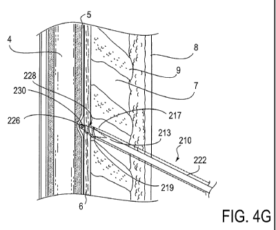
21
CA 02669388 2009-05-12
WO 2008/070588 PCT/US2007/086196
member. For example, a symmetric thread form may be designed to resist both
forward and
reverse linear motion of the engagement device in the tissue. The threads may
be designed to
resist inadvertent motion in the ligamentum flavum due to typical linear
forces encountered
during device advancement by the operator. The threuls additionally may be
configured to
reduce such motion when forces beyond the typical range are encountered. Such
a design
allows the ligamentum flavum to tend to be pulled away from the dura during
device
advancement with rotation and application of a pulling force, providing
additional protection
against damage to the dura during entry into the epidural space. Asymmetric
thread forms
can skew this symmetry in linear advancement or retraction force and enable
greater
resistance to forward linear advancement and lower resistance to linear
retraction of the tissue
engagement device. Departures from symmetry involve variations in the shapes
of each
thread face including different curvatures and angles, for example. For
straight-edged thread
forms trailing and leading angles a and (3, respectively, with different
values can result in a
tissue engagement device having linear advancement characteristics different
from its linear
retraction characteristics. For example, the threads could be configured such
that the force
required to advance the tissue engagement device with linear force is
significantly greater
than that required to linearly retract the device, providing an added measure
of safety for
structures in front of the engagement device and ease of simple pull-to-
retract device
withdrawal. In such a case, the trailing edge angle, a, would be specified
greater than the
leading edge angle, (3, to enable lower forces during pull-to-retract
withdrawal of the tissue
engagement device compared to forces that would be required to push-to-advance
the device.
The more gently trailing edge can also reduce tissue trauma by allowing the
engaged tissue
to deform more gradually as the device is linearly retracted as depicted in
FIG. 8C.
[00074] One embodiment of a threaded engagement mechanism with an asymmetric
thread form is the reverse buttress thread form depicted in FIGs. 8A, 8B and
8C. The cross-
22
CA 02669388 2009-05-12
WO 2008/070588 PCT/US2007/086196
sectional views of the engagement features 228 depict a reverse buttress
thread form with a
standard leading face angle (3 of 90 and a trailing face angle a of 150 . The
asymmetric
thread shape of the reverse buttress design resists push-to-advance forces
when advancing the
engagement device toward the epidural space. At the same time the sloped
geometry of the
trailing face facilitates lower tissue resistance to pull-to-retract forces.
The main advantage
of this configuration is that it allows the apparatus to be directly pulled
out of the ligamentum
flavum, similar to conventional methods, rather than requiring rotation of the
device to
disengage the ligamentum flavum.
[00075] Symmetric thread forms with heights between 0.1 - 0.27 mm have been
demonstrated to provide good tissue engagement properties in the ligamentum
flavum.
Spacing between thread forms in the range of 0.1 - 0.2 mm have demonstrated
desirable
tissue engagement characteristics. It is believed that leading face angles,
(3, less than 120
would provide further increased resistance to linear translation. It is also
believed that
trailing face angles, a, greater than 120 would provide reduced resistance to
linear
retraction.
[00076] FIGs. 9A and 9B illustrate an apparatus 400 for accessing the epidural
space. The
apparatus 400 includes a cutting sheath 410 and a tissue engagement device 420
within the
sheath 410. The cutting sheath 410 includes a distal end 416 divided into
multiple sections
three of which are visible in FIG. 9A. The sections 417, 418 and 419 are
separated by score
lines 417a and 418a. As with previous embodiments of epidural access devices,
a tissue
engagement device is within the cutting sheath. FIG. 9B illustrates the
cutting sheath 410 in
an open configuration with the blunt tip 426 of the tissue engagement device
420 in the
epidural space 19 and in non-penetrating contract with the dura 5. The tissue
engagement
device 420 includes threaded engagement elements 428, an aperture 230 and
other features as
described with previous tissue engagement device embodiments.
23
CA 02669388 2009-05-12
WO 2008/070588 PCT/US2007/086196
[00077] In contrast to the cutting sheath in FIG. 4, FIGs. 9A and 9B depict an
alternative
sheath distal end design with a closed cutting configuration that is
completely closed. In FIG.
4C the flaps on the distal end of the sheath are separated by a small slot cut
into the device,
and the closed cutting configuration is adapted to preclude the tissue
engagement device from
moving beyond the distal end of the sheath. However, depending on the
configuration the
small spacing between the flaps may allow a small amount of tissue to enter
the sheath during
penetration. Alternatively, the flaps 217 and 219 may also be biased closed in
a manner that
prevents tissue from entering the sheath such as by overlapping the flap
edges. Returning to
FIG. 9, as before, the open configuration (FIG. 9B) is adapted to allow the
tissue engagement
device 420 to pass beyond the distal end of the sheath 416 exposing the tissue
engagement
device 420 to the ligamentum flavum 6. The blunt end 426 deflects but does not
penetrate
the dura 5 as shown in FIG. 9B. FIG. 9A illustrates a Closed configuration
adapted to prevent
passage of tissue into the inner hollow compartment of the sheath. The sheath
is then
configured to open in a predetermined manner using a number of smaller flaps
(i.e., flaps
417, 418 and 419) that are preferentially separate along predefined score
lines 417a, 418a
which may be on the exterior of the sheath as depicted in FIG. 9A.
Alternatively, the score
lines may be located on the interior surface of the sheath so that a smooth
exterior surface is
presented to the tissue during penetration. Optionally, score lines may be
present on both or
either of the sheath interior and exterior surfaces. As the flaps 417, 418,
and 419 open, they
may bend and deflect open as shown or rotate about naturally occurring hinges
that arise due
to the mechanical deformation of the sheath. Living hinges may also be
incorporated into the
sheath distal end to define locations about which the distal flaps will
rotate. Such movements
may be providing using the properties described above for the modified area
213 in sheath
210.
24
CA 02669388 2009-05-12
WO 2008/070588 PCT/US2007/086196
[00078] FIGs. 10A and l OB illustrate an apparatus 500 for accessing the
epidural space.
The apparatus 500 includes a cutting sheath 510 and a tissue engagement device
420 within
the sheath 510. The cutting sheath 510 includes a distal end 516 having at
least one movable
flap. In the embodiment illustrated in FIG. 10A, the movable flap is flap 517.
The movable
flap 517 is configured to move relative to flap 519 and the distal end 516. In
other
embodiments, both flaps 517, 519 may be configured to move. As with previous
embodiments of epidural access devices, a tissue engagement device is within
the cutting
sheath 510. FIG. l OB illustrates the cutting sheath 510 in an open
configuration where the
movable flap 519 has moved to transition the distal end of the sheath 510 to
an open
configuration. Also shown in FIG. 10B, the blunt tip 426 of the tissue
engagement device
420 is in the epidural space 19 and in non-penetrating contract with the dura
5. The tissue
engagement device 420 includes threaded engagement elements 428, an aperture
230 and
other features as described with previous tissue engagement device
embodiments.
[00079] FIGs. 10A and 10B depict still another alternative sheath distal end
designed to
predictably open with tissue engagement device advancement. The distal end
preferentially
remains closed during insertion due to the configurati.;ni of the flap 517.
The flap 517 may be
biased closed or preferentially separate along defined lines with advancement
of the tissue
engagement device through the distal end of the sheath similar to the modes
utilizing score
lines described above with regard to FIGs. 9A, 9B. The asymmetrical shapes of
flaps 517,
519 can further facilitate steering of the device distal end 516 by rotating
the body of the
sheath 510 in a technique similarly performed with conventional Tuohy needles.
During
tissue engagement device advancement, the flap 517 opens by deflection as
illustrated. The
flap 517 rotates about a naturally forming hinge due to the mechanical
properties of the
sheath material, or, alternatively, a built-in living hinge.
CA 02669388 2009-05-12
WO 2008/070588 PCT/US2007/086196
[00080] FIGs. 11A and 1 lB illustrate an apparatus 600 for accessing the
epidural space.
The apparatus 600 includes a cutting sheath 610 and a tissue engagement device
220 within
the sheath 610. The cutting sheath 610 includes a distal end 616 having at
least one
predefined movable section that may rotate about a hinge. In the embodiment
illustrated in
FIG. 11 A, the distal end 616 is divided into sections 612, 614. A hinge 605
is positioned
proximal to the distal end 616. Depending upon the configuration of the
sections 612, 614
and the hinge 605, one or both of the sections 612, 614 may be a movable
section. As with
previous embodiments of epidural access devices, a tissue engagement device is
within the
cutting sheath 610. FIG. 11 B illustrates the cutting sheath 610 in an open
configuration
where the movable sections 612, 614 have moved to transition the distal end of
the sheath
610 to an open configuration. Also shown in FIG. 1113, the blunt tip 226 of
the tissue
engagement device 220 is in the epidural space 19 and in non-penetrating
contract with the
dura 5. The tissue engagement device 220 is described above.
[00081] FIGs. 11A and 11B depict another manner of allowing the sheath distal
end to
transition from a sharp cutting configuration to an open configuration wherein
the distal end
of the sheath opens a least one predefined movable section about a hinge
mechanism. The
device described herein alters its configuration to facilitate a different
mode of advancing
through tissue. In this embodiment the apparatus transitions from a cutting or
penetrating
push-to-advance mode to a controlled advancement mode via opening of the
sheath tip
comprising movable components about a hinge at the distal end of the sheath.
The hinge may
be a mechanical hinge as depicted in FIG. 1 lA. In this embodiment the hinge
605 is
incorporated into the sheath distal end and may be actuated externally or by
manipulation of
the tissue engagement device to cause the hinge to open as illustrated in FIG.
11 B. The
initial configuration of the hinge as shown in FIG. 11 A shows two movable
sections
separated by a small space. The movable components at the sheath distal end
616 may also
26
CA 02669388 2009-05-12
WO 2008/070588 PCT/US2007/086196
be closed in such a manner without such small openings, preventing tissue from
entering the
sheath. Alternatively, the movable components may start in a closed cutting
configuration
that is completely closed. The movable components may be defined by score
lines on the
internal or external surface of the sheath or a combination of the two. The
separation of the
movable components would then occur with actuation of the hinge 605 or passive
separation
of the movable components by advancing the internal tissue engagement device.
[00082] The step of manipulating an engagement feature on the tissue
engagement device
to controllably advance through the ligamentum flavum may take on any number
of different
forms. Threading and rotational manipulation have been described but other
forms of
manipulation are possible. Another form of manipulation includes longitudinal
relative
movement between the engagement features on the tissue engagement device and
the
ligamentum flavum. The longitudinal aspect of the manipulation refers to
movement of the
tissue engagement device relative to the longitudinal access of the device.
[000831 FIG. 12A illustrates a tissue engagement device 720 with the hinged
distal end in
a closed condition. The tissue engagement device 720 is one example of a
device that utilizes
longitudinal movement for controlled advancement through the ligamentum
flavum. The
tissue engagement device 720 includes an elongated body 722, a blunt distal
end at 726 and a
conduit 732. While other embodiments of the tissue engagement device 720 may
be
provided with a single movable section and hinge, an embodiment having two
movable
sections 712, 714 each with a hinge 710 is illustrated in FIGs. 12A and 12B. A
hinge 710
allows a movable section to move in such a fashion that the engagement
features 728 engage
the ligamentum flavum. In the illustrated embodiment, the two moving sections
712, 714 are
attached to the elongated body 722 by hinges 710.
[00084] A plurality of tissue engagement features "i28 are positioned along
the moving
sections 712, 714. In the illustrated embodiment, the engagement features 728
are ridges or
27
CA 02669388 2009-05-12
WO 2008/070588 PCT/US2007/086196
teeth. The shape, size and orientation of the engagement features 728 is
selected to engage
with the tissue of the ligamentum flavum when the distal end is expanded as
shown in FIG.
12B.
[00085] An expansion element is disposed within the conduit 732 for the
purpose of
moving the movable sections 712, 714 from a closed position (FIG. 12A) to an
open position
(FIG. 12B). The at least one hinge proximal to the ridges 728 allows sections
712, 714 at the
tip of the device to move in such a fashion that the ridgPs 728 engage in the
ligamentum
flavum to grasp and dissect apart the ligamentum flavum at the distal end of
the device as
well as form an opening ahead of the tip to create a pathway through the
ligamentum flavum.
In one embodiment, the expansion element lies at least partially distal to the
hinge 710 in
order to control the lateral extension of the one or more movable sections
712, 714. After
each dissecting action, the sections 712, 714 flaps can then be repositioned
in a closed
manner as seen in FIG 12A, the device advanced into the newly created opening
in front of
the device tip, and actuated again to open and dissect additional tissue as
shown in FIG 12B.
[00086] In the embodiment illustrated in FIGs. 12A, 12B, the expansion element
is a
balloon 760. The balloon 760 is attached to an interior surface of the moving
sections 712,
714 in such a way that when the balloon 760 is inflated the movable sections
712, 714 rotate
about their respective hinges 710. The balloon 760 facilitates actuation of
the movable flaps
by inflating and deflating to change between open and closed modes. The
balloon 760 also
includes a lumen or aperture 730 that allows communication between the conduit
732 and the
distal end of the device. A second conduit 740 connects the balloon 760 and an
inflation
source (not shown). During use, the operator would change balloon 760
inflation to control
the amount of movement of the movable sections 712, 714. FIG. 12B illustrates
the tissue
engagement device 720 in an open condition where balloon 760 is inflated and
the movable
28
CA 02669388 2009-05-12
WO 2008/070588 PCT/US2007/086196
sections 712, 714 are separated. Deflating the balloon 760 returns the tissue
engagement
device 722 to the condition shown in FIG. 12A.
[00087] The aperture in the device enables use of the familiar epidural space
access
detection technique of applying pressure to a fluid or gas filled syringe
during advancement
with the operator detecting a significant decrease in the resistance to the
flow of fluid or gas
as the syringe empties its contents into the epidural space. Once in the
epidural space, a
device can be placed through the device similar to the tissue engagement
device
embodiments described earlier. The lumen 730 in the balloon allows fluid or
gas passage for
epidural space access detection as well as delivery of desired medications,
catheters, or other
devices into the epidural space. For added protection of critical structures
such as the dura
and epidural veins, the distal tips of the engagement d vice present blunted
surfaces to
contacted tissue structures during operation as depicted in FIGs. 12A and 12B.
[00088] FIG. 13 is a flow chart 770 that describes an alternate technique for
the
manipulation of the tissue engagement device 720 for controlled dissection of
and
advancement through the ligamentum flavum. First, at step 775, longitudinally
advance the
tissue engagement device 720 into the ligamentum flavum. This step is
performed while the
tissue engagement device 720 is in the closed condition as illustrated in FIG.
12A. Next, at
step 780, expand the distal end of the of the tissue engagement device to
controllably dissect
the ligamentum flavum. This step is performed by inflating the balloon 760 to
move the
movable sections 712, 714 to the open condition illustrated in FIG. 12B. Next,
at step 785,
collapse the distal end of the tissue engagement device. This step is
performed by deflating
the balloon 760. As the balloon 760 deflates, the moving sections 712, 714 are
brought back
together to the closed condition illustrated in FIG. 12A. Next, advance the
tissue engagement
device (step 790). Finally, at step 795, steps 775, 780, 785 and 790 repeat
until reaching the
epidural space. The operator will alternately inflate and deflate the balloon
760 in
29
CA 02669388 2009-05-12
WO 2008/070588 PCT/US2007/086196
conjunction with incremental advancement of the tissue engagement device 720
until
reaching the epidural space. Detection of the epidural space using the loss-of-
resistance
technique and the placement of devices or therapy into the epidural space
would then follow
as described above.
[00089] As described in the embodiments above, tlie closed cutting
configuration of a
cutting sheath may be provided in a number of ways. Still another way of
providing a closed
cutting configuration is to occlude a normally open cutting sheath. Occlusion
of the normally
open cutting sheath may take many forms. For example, a stylet or a tissue
engagement
device placed into the hollow portion of the sheath could provide the
occlusion. Accordingly,
there is another alternative apparatus for accessing the epidural space in a
mammal by
providing a cutting sheath having an open proximal end and a distal end
adapted to transition
from a closed cutting configuration to an open configuration. The cutting
sheath has a hollow
portion extending from the open proximal end to the distal end. In one aspect,
a stylet is
placed within the hollow portion when the cutting sheath is in the closed
cutting
configuration, and a tissue engagement device disposed within the hollow
portion when the
cutting sheath is in the open configuration. In another aspect, a tissue
engagement device is
disposed within the hollow portion of the sheath to provide the closed
configuration and then,
when advanced from the sheath, provides for the transition to an open
configuration.
[00090] One such apparatus that demonstrates the use of occlusion to provide a
closed
cutting configuration is the apparatus 800 illustrated in FIG. 14. The
apparatus 800 includes
a cutting sheath 810 having a distal end 816, an open proximal end 812 and a
hollow portion
814 extending there between. FIG. 14 illustrates the case where a tissue
engagement device
220 is positioned within the hollow portion 814 to place the cutting sheath
810 in a closed
cutting condition. In this embodiment the closed cutting configuration
involves locating the
tip of the tissue engagement device just proximal to an open distal end of the
cutting sheath.
CA 02669388 2009-05-12
WO 2008/070588 PCT/US2007/086196
Positioning a tissue engagement device (or stylet) in this manner reduces
penetration of tissue
into the inner hollow compartment 814 of the sheath 810. The transition to
open
configuration in this embodiment occurs when the tissue engagement device 220
is advanced
relative to the distal open end of the sheath 810 to engage the ligamentum
flavum.
[00091] Alternatively, the closed configuration may be provided using a
conventional
stylet to occlude an open-ended cutting sheath. The stylet remains within the
cutting sheath
during the advancement of the cutting sheath up to or slightly penetrating
into the
ligamentum flavum. The stylet is sized to fit within and occlude the hollow
portion and open
distal end of the cutting sheath. In one embodiment, the cutting sheath 810 is
a needle with a
hollow portion 814 and a sharp tip 816. In one aspect, this embodiment
converts to an open
configuration by removing the stylet once the ligamentum flavum is detected by
the operator.
After removal of the stylet, the hollow portion of the sheath is open and a
tissue engagement
device is allowed to pass through the sheath as shown and described above with
regard to
FIG. 14. Thereafter, the tissue engagement device is manipulated to engage the
ligamentum
flavum. Controlled advancement through the ligamentum flavum proceeds in a
manner
dependent upon the controlled advancement mode utilized by the particular
tissue
engagement device.
[00092] The cutting sheath and the tissue engagement device used in the
apparatus for
accessing the epidural space may be made from any of the materials typically
used in medical
devices including medical grade plastics, stainless steel, and other
materials. Other
exemplary materials include, but are not limited to, plastics and
thermoplastics including
acrylic, polyetheretherketone, polyvinylchloride, polycarbonate, polyethelene,
polyetherimide, polytetrafluoroethylene, polysulfone, acrylonitrile-butadine-
styrene or the
like, or metals including, but not limited to, aluminum, steel, titanium, or a
shape-memory
alloy such as nitinol, or ceramics including, but not limited to, alumina,
zirconia, carbides and
31
CA 02669388 2009-05-12
WO 2008/070588 PCT/US2007/086196
the like. Additionally, the components of the inventive apparatus may include
one or a
combination of materials to endow a particular device with desired properties.
The device
surfaces may be further treated to endow them with desired properties such as
smoothness or
roughness, hydrophobicity or hydrophilicity, as well as other features. The
sheath and tissue
engagement device may be rigid or flexible or be comprised of both rigid and
flexible
portions. For example, the sheath distal penetrating end may be rigidly
configured while the
remaining shaft of the sheath is flexible. The components may be opaque,
clear, radiopaque,
radiolucent, echogenic, nonechogenic or contain combinations of such
characteristics at
different portions along its length. For example, the distal end of the tissue
engagement
device may be radiopaque while the main body is radiolucent.
[00093] In an embodiment with a flexible cable 295 connecting the tissue
engagement
device to a syringe and remote actuation mechanism, the flexible cable
connection could be
composed of plastic, rubber, metal, ceramic as listed above for the cutting
sheath and tissue
engagement device or any flexible and formable material or possibly a series
of linkages of
rigid pieces.
[00094] The tissue engagement device of the invention is preferentially
fabricated from a
rigid material designed to accommodate the required push, pull, and rotational
forces applied
during normal operation. Threaded engagement features and mechanisms may be
machined
or otherwise introduced such as via a molding process onto a tubular body to
form the desired
design and pattern.
[00095] The dimensions of the device are so configured to be comparable to
devices
currently used by those familiar with the art. The external diameter of the
device can be
configured to be in the range of 1-2 mm. The engagement components are
situated along the
region between the distal end of the tissue engagement device and nominally 6-
10 mm behind
the end to enable sufficient engagement of the ligamentum flavum through its
thickness
32
CA 02669388 2009-05-12
WO 2008/070588 PCT/US2007/086196
which is typically in the range of 2-4 mm. These dimensions serve to provide
guidelines in
the spirit of the invention and should not be construed as anything but.
[00096] The description of the above is meant to be illustrative of the scope
and spirit of
the invention. It will be obvious to one skilled in the art that many
modifications can be
made to the present invention without departing from the nature and scope
thereof. The sizes
and distances presented are for illustration purposes only and should not be
construed to limit
the scope of the invention.
33