Note: Descriptions are shown in the official language in which they were submitted.
CA 02669393 2009-05-12
WO 2008/063966 PCT/US2007/084448
RESPIRATORY THERAPY DEVICE AND METHOD
Background of the Invention
[Ol] The present disclosure relates to respiratory therapy devices and methods
for
administering breathing-relating treatments (e.g., oscillatory, continuous,
etc.) to a patient.
More particularly, it relates to respiratory therapy devices capable of
creating oscillatory
respiratory pressure pulses in response to the patient's expiratory airflow
alone, or when
connected to a source of positive pressure fluid (e.g., air, oxygen, etc.), or
both. One or more
additional therapies (e.g., continuous positive airway pressure, continuous
positive expiratory
pressure, delivery of aerosolized medication, etc.) are optionally available
in some
embodiments.
[02] A wide variety of respiratory therapy devices are currently available for
assisting,
treating, or improving a patient's respiratory health. For example, positive
airway pressure
(PAP) has long been recognized to be an effective tool in promoting bronchial
hygiene by
facilitating improved oxygenation, increased lung volumes, and reduced venous
return in
patients with congestive heart failure. More recently, positive airway
pressure has been
recognized as useful in promoting mobilization and clearance of secretions
(e.g., mucous)
from a patient's lungs. In this regard, expiratory positive airway pressure
(EPAP) in the form
of high frequency oscillation (BFO) of the patient's air column is a
recognized technique that
facilitates secretion removal. In general terms, HFO reduces the viscosity of
sputum in vitro,
which in turn has a positive effect on clearance induced by an in vitro
simulated cough. In
this regard, HFO can be delivered or created via a force applied to the
patient's chest wall
(i.e., chest physical therapy (CPT), such as an electrically driven pad that
vibrates against the
patient's chest), or by applying forces directly to the patient's airway
(i.e., breathing
treatment, such as high frequency airway oscillation). Many patients and
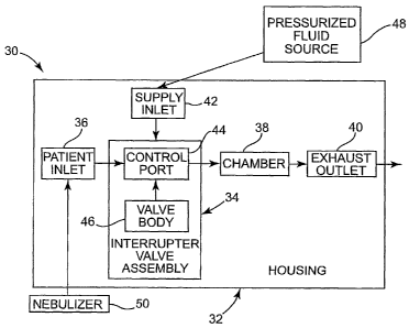
caregivers prefer
the breathing treatment approach as it is less obtrusive and can more easily
be administered.
To this end, PAP bronchial hygiene techniques have emerged as an effective
alternative to
CPT for expanding the lungs and mobilizing secretions.
[03] In the context of high frequency oscillatory breathing treatments,
various devices are
available. In general terms, respiratory therapy devices typically include one
or more tubular
-1-
CA 02669393 2009-05-12
WO 2008/063966 PCT/US2007/084448
bodies through which a patient breaths, with the tubular body or bodies
creating or defining a
patient breathing circuit. With this in mind, the oscillatory airflow effect
can be created by
periodically generating a pressure or positive airflow in the patient
breathing circuit during
one or both of an inspiratory phase or expiratory phase of the patient's
breathing cycle. For
example, a positive expiratory pressure (PEP) can work "against" the patient's
breath during
the expiratory phase of breathing. The pressure can be generated by creating a
periodic (or in
some instances continuous) resistance or restriction in the patient breathing
circuit to
expiratory airflow from the patient, or by introducing a forced fluid flow
(from a positive
pressure gas source) into the patient's breathing circuit in a direction
opposite of the patient's
exhaled air. With the airflow resistance approach, a separate, positive
pressure gas source is
not required. More particularly, many oscillatory positive expiratory pressure
("oscillatory
PEP") therapy devices utilize the patient's breath alone to drive an
oscillatory fluid flow
restriction, and thus can be referred to as "passive" devices (in contrast to
an "active"
respiratory therapy device that relies on a separate source of positive
pressure gas as
described below). Passive oscillatory PEP devices are self-administering and
portable.
[04] The Flutter mucus clearance device (available from Axcan Scandipharm
Inc., of
Birmingham, AL), is one example of an available passive, oscillatory PEP
therapy device. In
general terms, the Flutter device is pipe-shaped, with a steel ball in a
"bowl" portion of a
housing that is loosely covered by a perforated cap. The ball is situated
within an airway path
defined by the device's housing; when the patient exhales into the housing,
then, the ball
temporarily obstructs airflow, thus creating an expiratory positive airway
pressure. The bowl
within which the ball is located allows the ball to repeatedly move (e.g.,
roll and/or bounce)
or flutter to create an oscillatory or vibrational resistance to the exhaled
airflow. While
relatively inexpensive and viable, theFlutter device is fairly sensitive,
requiring the patient to
maintain the device at a particular angle to achieve a consistent PEP effect.
Other passive
oscillatory positive expiratory pressure devices, such as the Acapella
vibratory PEP therapy
system (available from Smiths Medical of London, England) and the Quake@
secretion
clearance therapy device (available from Thayer Medical Corp., of Tucson, AZ)
are known
alternatives to the Flutter device, and purport to be less sensitive to the
position in which the
patient holds the device during use. While these and other portable
oscillatory PEP therapy
-2-
CA 02669393 2009-05-12
WO 2008/063966 PCTIUS2007/084448
devices are viable, opportunities for improvement remain, and patients
continue to desire
more uniform oscillatory PEP results.
[05] As an alternative to the passive oscillatory PEP devices described above,
continuous
high frequency oscillatory (CHFO) treatment systems are also available. In
general terms,
the CHFO system includes a hand-held device establishing a patient breathing
circuit to
which a source of positive pressure gas (e.g., air, oxygen, etc.), is fluidly
connected. The
pressure source and/or the device further include appropriate mechanisms
(e.g., control
valves provided as part of a driver unit apart from the hand-held device) that
effectuate
intermittent flow of gas into the patient breathing circuit, and thus
percussive ventilation of
the patient's lungs. With this approach, the patient breathes through a
mouthpiece that
delivers high-flow, "mini-bursts" of gas. During these percussive bursts, a
continuous
airway pressure above ambient is maintained while the pulsatile percussive
airflow
periodically increases airway pressure. Each percussive cycle can be
programmed by the
patient or caregiver with certain systems, and can be used throughout both
inspiratory and
expiratory phases of the breathing cycle.
[06] Examples of CHFO devices include the IPV ventilator device (from
PercussionAire
Corp., of Sandpoint, ID) and a PercussiveNebTM system (from Vortran Medical
Technology
1, Inc., of Sacramento, CA). These and other similar "active" systems are
readily capable of
providing not only CHFO treatments, but also other positive airflow modes of
operation (e.g.,
continuous positive airway pressure (CPAP)). However, a positive pressure
source is
required, such that available active respiratory therapy systems are not
readily portable, and
are relatively expensive (especially as compared to the passive oscillatory
PEP devices
described above). Oftentimes, then, active respiratory treatment systems are
only available at
the caregiver's facility, and the patient is unable to continue the
respiratory therapy at home.
Instead, a separate device, such as a portable, passive oscillatory PEP device
as described
above must also be provided. Further, the hand-held portion of some
conventional active
respiratory therapy systems must be connected to an appropriate driver unit
that in turn is
programmed to effectuate the desired fluid flow to the patient (e.g., CHFO,
CPAP, etc.).
That is to say, the hand-held portion of some active systems is not self-
operating, but instead
relies on the driver unit for applications. Any efforts to address these and
other Iimitations of
-3-
CA 02669393 2009-05-12
WO 2008/063966 PCT/US2007/084448
available active respiratory therapy devices would be well-received. This
limitation
represents a significant drawback.
[07] In light of the above, a need exists for respiratory devices capable of
providing
oscillatory PEP therapy utilizing the patient's breath alone, as well as CHFO
therapy (and
optionally other therapies such as CPAP) when connected to a positive pressure
source. In
addition, improved passive oscillatory PEP or active respiratory therapy
devices are also
needed.
Summary of the Invention
[08] Some aspects in accordance with principles of the present disclosure
relate to a device
for providing respiratory therapy to a patient during at least a portion of a
patient breathing
cycle otherwise including an inspiratory phase and an expiratory phase. The
device includes
a housing and an interrupter valve assembly. The housing includes a patient
inlet, an exhaust
outlet, a chamber, and a pressurized fluid supply inlet. The chamber is
fluidly disposed
between the patient inlet and the exhaust outlet. The interrupter valve
assembly is associated
with the housing and includes a control port fluidly connecting the patient
inlet and the
chamber. Further, the interrupter valve assembly includes a valve body adapted
to selectively
olistruct fluid flow through the control port. With this in mind, the device
is adapted to
operate in a first, passive mode and a second, active mode. In the passive
mode, positive
airflow to the supply inlet does not occur. The interrupter valve assembly
interacts with
exhaled air from the patient to create an oscillatory positive expiratory
pressure effect during
at least the expiratory phase. Conversely, in the active mode, positive fluid
flow to the fluid
supply inlet occurs and the interrupter valve assembly interacts with this
fluid flow to create a
continuous high frequency oscillation effect. With this configuration, then,
the respiratory
device can serve as a passive, oscillatory PEP device for use by a patient at
virtually any
location. In addition, when connected to a positive pressure gas source, the
respiratory
therapy device provides active therapy. In some embodiments, the interrupter
valve assembly
includes a drive mechanism akin to a reverse roots blower, utilizing forced
air (e.g., either the
patient's exhaled airflow or airflow from a separate positive gas source) to
cause rotation of
the roots blower lobes, that in turn cause the valve body to periodically open
and close the
-4-
CA 02669393 2009-05-12
WO 2008/063966 PCT/US2007/084448
control port. In other embodiments, the device can provide or facilitate one
or more
additional therapies such as continuous PEP, CPAP, delivery of aerosolized
medication, etc.
[09] Other aspects in accordance with the present disclosure relate to a
method of
providing respiratory therapy to a patient during at least a portion of a
patient breathing cycle
including an inspiratory phase and an expiratory phase. The method includes
providing a
respiratory therapy device including a housing and an interrupter valve
assembly. The
housing includes a patient inlet, an exhaust outlet, and a pressurized fluid
supply inlet. The
interrupter valve assembly is adapted to selectively interrupt fluid flow to
or from the patient
inlet. A source of pressurized fluid is fluidly coupled to the fluid supply
inlet. Continuous
high frequency oscillation treatment is administered to the patient via the
therapy device, with
the therapy device operating in an active mode. Fluid flow from the source of
pressurized
fluid to the fluid supply inlet is discontinued. The patient is then prompted
to repeatedly
perform a patient breathing cycle using the therapy device. In this regard,
the therapy device
administers an oscillatory positive expiratory pressure treatment to the
patient while
operating in a passive mode. In some embodiments, the passive mode of
operation is
characterized by the level of oscillatory positive expiratory pressure
treatment being a
function of a breathing effort of the patient, whereas the active mode of
operation is
characterized by a level of continuous high frequency oscillation treatment
being independent
of the patient's breathing effort. In yet other embodiments, the method
further includes
administering one or more additional therapies to the patient via the device,
such as CPAP,
continuous PEP, delivery of aerosolized medication, etc.
Brief Description of the Drawinp_s
[10] FIG. 1 is a block diagram illustrating a respiratory therapy device in
accordance with
principles of the present disclosure;
[11] FIG. 2 is an exploded, perspective view of a respiratory therapy device
in accordance
with principles of the present disclosure;
[12] FIG. 3A is a perspective view of a housing portion of the device of FIG.
2;
[13] FIG. 3B is a bottom view of the housing of FIG. 3A;
-5-
CA 02669393 2009-05-12
WO 2008/063966 PCT/US2007/084448
[14] FIG. 4A is a longitudinal, cross-sectional view of the housing of FIG. 3A
taken along
a patient supply inlet;
[15] FIG. 4B is a rear, perspective view of a leading portion of the housing
of FIG. 3A;
[16] FIG. 4C is a longitudinal, cross-sectional view of the housing of FIG. 3A
taken along
a drive supply inlet;
[17] FIG. 5A is an exploded, perspective view of a drive mechanism portion of
the device
of FIG. 2;
[18] FIG. 5B is a perspective view of the drive mechanism of FIG. 5A upon
final
assembly;
[19] FIG. 6A is a perspective view illustrating partial assembly of the device
of FIG. 2;
[20] FIG. 6B is a longitudinal, cross-sectional view of the device of FIG. 2
upon final
assembly, taken along a patient supply inlet;
[21] FIGS. 7A and 7B illustrate use of the device of FIG. 2 in a passive mode;
[22] FIGS. 8A-8C illustrate use of the device of FIG. 2 in an active mode;
[23] FIG. 9 is an exploded, perspective view of an alternative respiratory
therapy device in
accordance with principles of the present invention;
[24] FIG. 10 is a front, plan view of a trailing housing portion of the device
of FIG. 9;
[25] FIG. 11 is a perspective, cutaway view of a portion of the device of FIG.
9 upon final
assembly;
[26] FIG. 12 is a exploded, perspective view illustrating assembly of the
device of FIG. 9;
[27] FIG. 13A is a perspective view of the device of FIG. 9;
[28] FIG. 13B is a longitudinal, perspective view of the device of FIG. 9;
-6-
~. , CA 02669393 2009-05-12
WO 2008/063966 PCT/US2007/084448
[29] FIGS. 14A and 14B illustrate use of the device of FIG. 9 in which airflow
passes from
a patient inlet to a chamber;
[30] FIGS. 15A and 15B illustrate use of the device of FIG. 9 in which airflow
is
obstructed from a patient inlet to a chamber;
[31] FIG. 16 is a simplified, side sectional view of an alternative
respiratory therapy device
in accordance with principles of the present disclosure;
[32] FIG. 17 is an exploded, perspective view of another embodiment
respiratory therapy
device in accordance with principles of the present disclosure;
[33] FIG. 18A is a longitudinal, cross-sectional view of the device of FIG.
17;
[34] FIG. 18B is an enlarged view of a portion of FIG. 18A;
[35] FIGS. 19A and 19B illustrate use of the device of FIG. 17;
[36] FIG. 20 is a schematic illustration of an interrupter valve assembly
useful with the
device of FIG. 17;
[37] FIGS. 21A and 21B are simplified, schematic illustrations of an
alternative interrupter
valve assembly useful with the device of FIG. 17;
[38] FIG. 22 is a longitudinal, cross-sectional view of another embodiment
respiratory
therapy device in accordance with principles of the present disclosure;
[39] FIG. 23A is an exploded, perspective view of another embodiment
respiratory therapy
device in accordance with principles of the present disclosure;
[40] FIG. 23B is a perspective, cutaway view of the device of FIG. 23A upon
fmal
assembly;
[41] FIG. 24 is an enlarged, perspective view of an orifice assembly portion
of the device
of FIG. 23A;
-7-
CA 02669393 2009-05-12
WO 2008/063966 PCT/US2007/084448
[42] FIG. 25is a schematic, electrical diagram of control circuitry useful
with the device of
FIG. 23A;
[43] FIGS. 26A and 26B illustrate the device of FIG. 23A upon final assembly;
[44] FIGS. 27A and 27B illustrate use of the device of FIG. 23A; and
[45] FIG. 28 is a longitudinal, cross-sectional view of another embodiment
respiratory
therapy device in accordance with principles of the present disclosure;
Detailed Description of the Invention
[46] In general terms, aspects of the present disclosure relate to respiratory
therapy devices
and related methods of use that are: 1) capable of operating in either of an
active mode (e.g.,
CHFO) or a passive mode (e.g., oscillatory PEP); or 2) improved passive-only
oscillatory
PEP devices; or 3) improved active-only devices (CHFO and/or CPAP). As used
throughout
this specification, an "active" therapy device is in reference to a device
that requires a
separate source of positive pressure fluid to effectuate a designated
respiratory therapy,
whereas a `passive" therapy device is in reference to a device that delivers a
designated
respiratory therapy in and of itself (i.e., a separate source of positive
pressure fluid is not
necessary). Thus, an "active-only" therapy device is one that must be
connected to a separate
source of positive pressure fluid. Conversely, a "p assive-only" therapy
device is one that is
not configured to receive pressurized fluid from a separate source. Given
these definitions,
several of the embodiments associated with this disclosure have base
constructions
appropriate for passive-only, oscillatory PEP applications, as well as
modified base
constructions that promote use of the device as either an oscillatory PEP
therapy device or,
when fluidly connected to a source of pressurized fluid, as a CHFO therapy
device. In yet
other embodiments, the base construction can be employed with an "active only"
therapy
device that provides CHFO therapy (and, in some embodiments, other respiratory
therapies
such as CPAP) when connected to a source of positive pressure fluid. With any
of these
embodiments, optional features can be included to facilitate delivery of
aerosolized
medication.
-8-
CA 02669393 2009-05-12
WO 2008/063966 PCT/US2007/084448
[47] With the above understanding in mind, FIG. 1 is a block diagram
illustrating features
of a respiratory therapy device 30 in accordance with some aspects of the
present disclosure.
In general terms, the respiratory therapy device 30 is adapted to operate in a
passive mode
(e.g., oscillatory PEP) and an active mode (e.g., CHFO and optionally CPAP),
and generally
includes a housing 32 and an interrupter valve assembly 34. The housing 32
forms or
maintains a patient inlet 36, at least one chamber 38, an exhaust outlet 40,
and at least one
pressurized fluid supply inlet 42. The interrupter valve assembly 34 includes
at least one
control port 44 and a valve body 46. The control port(s) 44 fluidly connects
the patient inlet
36 and the chamber 38, whereas the valve body 46 is adapted to selectively
obstruct or
interrupt fluid flow through the control port(s) 44. Details on the various
components are
provided below. In general terms, however, by controlling or operating the
valve body 46 to
selectively obstruct (partially or completely) the control port(s) 44, the
interrupter valve
assembly 34 alters airflow/pressure characteristics to and/or from the patient
inlet 36. For
example, where the supply inlet 42 is not connected to a separate source of
pressurized fluid
48, as a patient (not shown) exhales into the patient inlet 36, the
interrupter valve assembly
34 operates to periodically at least partially close the control port(s) 44,
thereby establishing a
resistance to airflow or back pressure in the patient inlet 36. This periodic
back pressure, in
turn, provides an oscillatory PEP therapy. In addition, when the supply inlet
42 is fluidly
connected to the pressurized fluid source 48, the interrupter valve assembly
34 operates to
periodically at least partially interrupt fluid flow from the supply inlet 42
to the patient inlet
36. This interrupted supply of pressure toward the patient serves as a CHFO
therapy. As
described below, the device 30 can optionally include features that
selectively disable all or a
portion of the interrupter valve assembly 34 in conjunction with the supply of
pressurized
fluid to the supply inlet 42 in providing a CPAP therapy (either along or
simultaneous with
CHFO therapy).
[48] In light of the above, the respiratory therapy device 30 provides both
active and
passive modes of operation, allowing the patient (not shown) to receive
oscillatory PEP
treatments with the device 30 at virtually any location, as well as CHFO
treatments (and
optionally other active treatments such as CPAP) when the patient is at a
location at which
the pressurized fluid source 48 is available. The respiratory therapy device
30 can further be
configured to facilitate additional respiratory therapy treatments, such as
delivery of
-9-
CA 02669393 2009-05-12
WO 2008/063966 PCT/US2007/084448
aerosolized medication (for example via a nebulizer 50). The nebulizer 50 can
be connected
to a port (not shown) provided by the housing 32, or can include an
appropriate connection
piece (e.g., T-connector or line) that is fluidly connected to the housing 32
(e.g., to the patient
inlet 36) when desired. Finally, while the pressurized fluid source 48 is
shown apart from the
housing 32, in other embodiments, the pressurized fluid source 48 can be
attached to, or
carried by, the housing 32 (e.g., a pressurized canister mounted to the
housing 32).
[49] With the above in mind, the respiratory therapy device 30 can assume a
variety of
forms capable of operating in a passive mode (e.g., oscillatory PEP therapy)
and an active
mode (e.g., CHFO therapy). One embodiment of a respiratory therapy device 60
providing
these features is shown in FIG. 2. The therapy device 60 generally includes a
housing 62
(referenced generally) and an interrupter valve assembly 64 (referenced
generally). The
housing 62 includes a leading section 66, a trailing section 68, and an end
plate 69. The
leading section 66 defines a patient inlet 70, whereas the trailing section 68
defines a first
chamber 72, a second chamber (hidden in the view of FIG. 2), an exhaust outlet
(hidden in
FIG. 2), and one or more supply inlets 74. The interrupter valve assembly 64
includes a plate
76 forming one or more control ports 78 (e.g., the control ports 78a, 78b), a
valve body 80,
and a drive mechanism 82. Details on the various components are provided
below. In
general terms, however, the drive mechanism 82 is retained within the second
chamber of the
housing 62 and is assembled to the valve body 80 for causing rotation thereof.
The valve
body 80, in turn, is located in close proximity to the control ports 78 such
that rotation of the
valve body 80 selectively opens and closes (e.g., partial or complete
obstruction) the control
ports 78 relative to the first chamber 72 and the patient inlet 70. Finally,
the supply inlet(s)
74 are fluidly connected to distribution points within the housing 62. During
use, and in a
passive mode of operation, the therapy device 60 generates oscillatory PEP via
operation of
the drive mechanism 82 in response to the patient's exhaled breath. In
addition, the therapy
device 60 provides an active mode of operation in which the interrmzpter valve
assembly 64
causes delivery of CHFO fluid flow to the patient inlet 70 in acting upon
positive fluid flow
from the supply inlet(s) 74. In this regard, a control means 84 (referenced
generally) can be
provided that facilitates operation of the therapy device 60 in a desired
mode.
[50] The housing 62 is shown in greater detail in FIGS. 3A and 3B upon final
assembly.
The housing 62 is generally sized and shaped for convenient handling by a
patient, with the
-10-
CA 02669393 2009-05-12
WO 2008/063966 PCT/US2007/084448
leading section 66 forming a mouthpiece 86 sized for placement in the
patient's mouth and
through which the patient's respiratory cycle interacts with the patient inlet
70. The
mouthpiece 86 can be integrally formed with one or more other component(s) of
the housing
62, or can be separately formed and subsequently assembled thereto.
[51] The housing 62 can form or define fluid flow features in addition to the
supply inlets
74. For example, and as best shown in FIG. 3A, the trailing section 68 forms a
slot 90 as part
of the control assembly 84 (FIG. 2). As described below, the control assembly
84 can
assume a variety of forms, but in some embodiments includes a body slidably
disposed with
the slot 90. With alternative constructions, however, the slot 90 can be
eliminated.
[52] Relative to the top perspective view of FIG. 3A, the housing 62 can
further form first
and. second relief port arrangements 92, 94. A third relief port arrangement
96 can also be
provided as shown in the bottom view of FIG. 3B. Finally, as best shown in
FIG. 2, a fourth
relief port arrangement 98 is provided within an interior of the housing 62.
Operation of the
therapy device 60 in connection with the relief port arrangements 92-98 is
described in
greater detail below. In general terms, however, the relief port arrangements
92-98 each
include one or more apertures 99, and are adapted to maintain a valve
structure (not shown),
such as a one-way umbrella valve, that permits fluid flow into or out of the
aperture(s) 99 of
the corresponding port arrangement 92-98 in only a single direction. As such,
the relief port
arrangements 92-98 can assume a variety of configurations differing from those
illustrated.
Similarly, additional relief port arrangements can be provided, and in other
embodiments one
or more of the relief port arrangements 92-98 can be eliminated.
[53] Returning to FIG. 2, the supply inlets 74, otherwise carried or formed by
the housing
62, include, in some embodiments, first and second patient supply inlets 74a,
74b, as well as
a drive supply inlet 74c. The patient supply inlets 74a, 74b are fluidly
connected to first and
second nozzles 100a, 100b, respectively, each positioned to direct fluid flow
toward a
corresponding one of the control ports 78a, 78b (otherwise formed by the plate
76). A
relationship of the nozzles 100a, 100b and the control ports 78a, 78b relative
to the internal
features of the housing 62 is provided below. It will be understood at the
outset, however,
that while two of the control ports 78a, 78b are shown and described, in other
embodiments,
one or three (or more) control ports are also acceptable. Similarly, a
nozzle/patient supply
-11-
CA 02669393 2009-05-12
WO 2008/063966 PCT/US2007/084448
inlet need not be provided for each of the control ports 78a, 78b (e.g., the
patient supply inlet
74b/nozzle 100b can be eliminated), or two or more supply inlet/nozzles can be
directed
toward a single one of the control ports 78. Even further, two or more supply
inlets 74 can be
fluidly associated with a single nozzle 100.
[54] With the above in mind, FIG. 4A is a longitudinal cross-sectional view of
the housing
62 upon final assembly taken through the first patient supply inlet 74a. The
leading portion
66, the trailing portion 68 and the end plate 69 are generally assembled to
one another as
shown. As a point of reference, the view of FIG. 4A further illustrates the
control means 84
in an open position relative to the housing 62, and reflects that the plate 76
can be an integral
component of the housing 62. Regardless, the housing 62 is shown in FIG. 4A as
defining
the first chamber 72, as well as a second chamber 101, and an exhaust chamber
102. The
first chamber 72 is defined, in part, by the plate 76 and an intermediate wall
104, with the
plate 76 fluidly separating the patient inlet 70 from the first chamber 72. In
this regard, the
patient inlet 70 is fluidly connected to the first chamber 72 via the control
ports 78 (it being
understood that only the first control port 78a is visible in FIG. 4A). The
first chamber 72 is
separated from the second chamber 101 by the intermediate wall 104, with fluid
connection
between the chambers 72, 101 being provided by a passage 106. As described in
greater
detail below, the passage 106 can be fluidly closed via operation of the
control means 84.
Regardless, the second chamber 101 is fluidly connected to the exhaust chamber
102 via an
outlet opening 108. The first chamber 72 is also fluidly connected to the
exhaust chamber
102, via the fourth relief port arrangement 98. As a point of reference, FIG.
4A reflects that a
one-way valve structure 110 is associated with the fourth relief port 98 and
is configured such
that fluid flow can only occur from the.first chainber 72 to the exhaust
chamber 102. Finally,
the exhaust chamber 102 tenninates at an exhaust outlet 112 that is otherwise
open to
ambient.
[55] With the above conventions in mind, the first nozzle 100a is positioned
within the
first chamber 72, and includes or defines an inlet end 114 and an outlet end
116. The inlet
end 114 is fluidly connected to the first patient supply inlet 74a such that
fluid flow through
the first patient supply inlet 74a is directed toward the outlet end 116. The
outlet end 116, in
turn, is aligned with the first control port 78a so as to direct fluid flow
from the first nozzle
100a to the first control port 78a. In some embodiments, the first nozzle 100a
tapers in
-12-
CA 02669393 2009-05-12
WO 2008/063966 PCT/US2007/084448
diameter from the inlet end 114 to the nozzle end 116, such that a jet-like
fluid flow from the
first patient supply inlet 74a to the first control port 78a is established.
In this regard,
ambient air can be entrained into the fluid flow from the nozzle 100a (as well
as the nozzle
100b) via the second relief port arrangement 94. A one-way valve structure 118
is illustrated
in FIG. 4A as applied to the relief port arrangement 94, and dictates that
ambient air can only
enter the first chamber 72 (and thus the nozzle 100 fluid flow). Though not
shown, operation
of the valve structure 118 can be further controlled by a control mechanism
that serves to
selectively maintain the valve structure 118 in a closed state (e.g., during a
passive mode of
operation as described below). In other embodiments, entrained ambient airflow
within the
first chamber 72 can be provided in a different manner (e.g., not including
the relief port
arrangement 94), or can be eliminated.
[56] Regardless of whether ambient air is introduced into the first chamber
72, a gap 120
(referenced generally) is established between the outlet end 116 and the plate
76 (and thus the
first control port 78a). As described in greater detail below, the gap 120 is
sized to facilitate
assembly and movement of the valve body 80 (FIG. 2). Though not shown, the
second
patient supply inlet 74b/second nozzle 100b (FIG. 2) has a similar
construction and
relationship relative to the plate 76/second control port 78b. Thus, and as
best shown in FIG.
4B, the first patient supply inlet 74a/nozzle 100a directs positive pressure
fluid from a
separate source toward the first control port 78a, and the second patient
supply inlet
74b/nozzle 100b directs positive pressure fluid toward the second control port
78b.
[57] The drive supply inlet 74c (FIG. 2) is sinularly fluidly connected to an
interior of the
housing 62. In particular, the drive supply inlet 74c is fluidly connected to
the second
chamber 101 as shown in FIG. 4C. As described in greater detail below, a
portion of the
drive mechanism 82 (FIG. 2) is retained within the second chamber 101, with
fluid flow from
the drive supply inlet 74c serving to actuate or drive the drive mechanism 82
during an active
mode of operation.
[58] Returning to FIG. 2, the interrupter valve assembly 64 again includes the
valve body
80 that is driven by the drive mechanism 82. In some embodiments, the valve
body 80 has a
propeller-like construction, and includes a base 130, a first valve plate
segment 132, and a
second valve plate segment 134. The base 130 is configured for assembly to a
corresponding
-13-
CA 02669393 2009-05-12
WO 2008/063966 PCT/US2007/084448
portion of the drive mechanism 82 as described below. The plate segments 132,
134 extend
in a radial fashion from the base 130, and each have a size and shape
commensurate with a
size and shape of a corresponding one the control ports 78a, 78b. For example,
a size and/or
shape of the valve plate segments 132, 134 can be identical, slightly smaller
or slightly larger
than a size and/or shape of the control ports 78a, 78b. Further, in some
embodiments, a
circumferential position of the plate segments 132, 134 relative to the base
130 corresponds
with that of the control ports 78a, 78b such that when the base 130 is
centrally positioned
between the control ports 132, 134, the control port 78a, 78b can be
simultaneously
obstructed by the plate segments 132, 134. Thus, with the one embodiment of
FIG. 2, the
control ports 78a, 78b are symmetrically opposed, and the valve plate segments
132, 134 are
similarly oriented. Alternatively, a position of the valve plate segments 132,
134 can be
spatially offset relative to a position of the control ports 78a, 78b; with
this alternative
construction, the control ports 78a, 78b are not simultaneously obstructed
during movement
of the valve body 80.
[59] While the valve body 80 is shown as including two of the valve plate
segments 132,
134, any other number, either greater or lesser is also acceptable, and the
number of plate
segment(s) 132, 134 provided need not necessarily equal the number of control
ports 78. In
other embodiments, for example, the valve body 80 is configured and positioned
so as to
fluidly interface with only one of the control ports 78 as described below.
Even further, the
valve body 80 can have configurations differing from the propeller-like
construction shown.
Regardless, the valve body 80 is constructed such that all of the control
port(s) 78 can
simultaneously be obstructed (e.g., completely blocked or less than completely
blocked) by
the valve body 80 in some embodiments.
[60] The drive mechanism 82 is shown in greater detail in FIG. 5A. In some
embodiments, the drive mechanism 82 is akin to a reverse roots blower device
and includes
first and second lobe assemblies 140, 142, and first and second gears 144,
146. The lobe
assemblies 140, 142 can be identical, with the first lobe assembly 140
including a lobe body
150 and a shaft 152. The lobe body 150 includes three longitudinal lobe
projections 154,
adjacent ones of which are separated by a valley 156. Although three of the
lobe projections
154/valleys 156 are illustrated in FIG. 5A, any other number is also
acceptable; however,
preferably at least two of the lobe projections 154/valleys 156 are provided.
Regardless, the
-14-
CA 02669393 2009-05-12
WO 2008/063966 PCT/US2007/084448
shaft 152 is, in some embodiments, coaxially mounted within the lobe body 150,
extending
from a first end 158 to a second end 160. The first end 158 is sized for
assembly to the valve
body base 130 (FIG. 2), whereas the second end 160 is sized for assembly to
the first gear
144. Other constructions are also contemplated such as integrally molding or
forming two or
more of the lobe body 150, shaft 154, and/or gear 140. The second lobe
assembly 142 is
similarly constructed, and generally includes a lobe body 162 coaxially
maintained by a shaft
164 that in turn is sized for assembly to and/or formed as part of the second
gear 146.
[61] As shown in FIG. 513, the lobe bodies 150, 162 are configured for meshed
engagement (e.g., one of the lobe projections 154 of the second lobe body 162
nests within
one of the valleys 156 of the first lobe body 160), as are the first and
second gears 144, 146
(it being understood that upon final assembly meshed engagement between the
lobe bodies
150, 162 and between the gears 144, 146 is simultaneously achieved). With this
construction,
then, the lobe assemblies 140, 142 rotate in tandem, but in opposite
directions (e.g., relative
to the orientation of FIG. 5B, clockwise rotation of the first lobe body 150
translates into
countercloclcwise rotation of the second lobe body 162). The shafts 152, 164
are affixed to
the corresponding lobe body 150, 162, respectively, such that rotation of the
lobe bodies 150,.
162 is translated directly to the gears 144, 146, respectively, via the shafts
152, 164. Thus,
the gears 144, 146 serve to maintain a desired intermeshing relationship
between the lobe
bodies 150, 162. With the reverse roots blower configuration of the drive
mechanism 82, a
relatively small force (e.g., fluid flow) is required to initiate and maintain
movement of the
lobe assemblies 140, 142 at a desired rotational speed. In other embodiments,
the number of
lobe projections 154 can be increased so that the lobe bodies 150, 162
effectively interface as
gears such that the gears 144, 146 can be eliminated. Regardless, upon final
assembly,
rotation of the first lobe assembly 140 translates into rotation of the valve
body 80.
[62] Assembly of the interrupter valve assembly 64 to the housing 62 is
partially shown in
FIG. 6A. In particular, the valve body 80 is maintained immediately adjacent
the nozzles
100a, 100b via the shaft 152 that otherwise extends into the first chamber 72.
The shaft 164
of the second lobe assembly 142 (referenced generally in FIG. 6A, shown in
greater detail in
FIG. 5A) also extends into, and is supported at, the first chamber 72 (it
being understood that
the opposite end of each of the shafts 152, 164 is also supported, for example
at or by the end
plate 69 (FIG. 2)). As shown in FIG. 6B, that otherwise is a longitudinal
cross-sectional view
-15-
CA 02669393 2009-05-12
WO 2008/063966 PCT/US2007/084448
taken through the first patient supply inlet 74a, the first lobe body 150 is
maintained within
the second chamber 101, as is the second lobe body 162 (hidden in the view of
FIG. 6B).
The shaft 152 maintains the valve body 80 such that the valve plate segments
132, 134 (it
being understood that the second plate segment 134 is hidden in the view of
FIG. 6B) are
located in the gap 120 between the outlet end 116 of the first nozzle 100a and
the plate 76 (as
well as between the second nozzle 100b, that is otherwise hidden in the view
of FIG. 6B, and
the plate 76). With rotation of the valve body 80 (via the drive mechanism
82), the valve
plate segments 132, 134 repeatedly obstruct and "open" the control ports 78
relative to the
first chamber 72. In other words, the interrupter valve assembly 64
(referenced generally in
FIG. 6B) operates to periodically stop or substantially stop fluid flow
between the patient
inlet 68 and the first chamber 72 as described below. While the valve body 80
has been
described as being assembled to the first shaft 152, in other embodiments, the
second shaft
164 rotates the valve body 80. In other embodiments, each of the shafts 152,
164 can
maintain a valve body.
[631 With the above understanding in mind, forced movement of the drive
mechanism 82
can occur in one of two manners that in turn are a function of whether the
device 60 is
operating in a passive mode (e.g., oscillatory PEP) or an active mode (e.g.,
CHFO). For
example, in the passive mode, the respiratory therapy device 60, and in
particular the drive
mechanism 82, operates solely upon the patient's exhaled air or breath. In
this regard, and
with reference to FIGS. 2 and 6B, in the passive mode, the control means 84 is
positioned
such that the passage 106 is open and fluidly connects the first and second
chambers 72, 101.
In some embodiments, the control means 84 includes a tab 166 slidably
positioned within the
slot 90; in the "open" state of FIGS. 2 and 6B, the tab 166 is retracted from
the slot 90. The
control means 84 can assume a wide variety of other forms also capable of
selectively
opening or closing the passage 106. The supply inlets 74a-74c are fluidly
closed or otherwise
fluidly isolated from any external positive pressure fluid source (e.g., the
pressurized fluid
source 48 of FIG. I is disconnected from the respiratory therapy device 60;
fluid flow from
the pressurized fluid source 48 is diverted from the supply inlets 74a-74c;
etc.). To this end,
in some embodiments the supply inlets 74a-74c can be exteriorly closed (for
example, by a
cap assembly (not shown)).
-16-
CA 02669393 2009-05-12
WO 2008/063966 PCT/US2007/084448
[64] With the therapy device 60 configured as described above, the passive
mode of
operation can entail the mouthpiece 86 (or other patient interface piece (not
shown) otherwise
attached to the mouthpiece 86) is inserted into the patient's mouth, and the
patient being
prompted to breathe through the therapy device 60. 'During an inspiratory
phase of the
patient's breathing cycle, ambient air is readily drawn into the housing 62
via the third relief
port arrangement 96 (that otherwise includes a one-way valve structure 170
(FIG. 6B)
controlling airflow therethrough). Thus, the patient can easily and readily
inhale air.
[65] During the expiratory phase, exhaled airflow is directed from the
patient/mouthpiece
86, through the patient inlet 68, and toward the plate 76. The exhaled air can
fluidly pass or
flow from the patient inlet 68 to the first chamber 72 via the control ports
78 when the control
ports 78 are otherwise not completely obstructed by the valve body 80 (and in
particular the
valve plate segments 132, 134). An example of this relationship is shown in
FIG. 7A
whereby the valve body 80 has been rotated such that the plate segments 132,
134 are "away"
from the control port 78a (as well as the control port 78b (hidden in the view
of FIG. 7A)).
Thus, the exhaled air flows through the control ports 78 and into the first
chamber 72
(represented by arrows in FIG. 7A).
[66] When the airflow into the first chamber 72 is at a pressure below the
opening pressure
of a valve structure 172 associated with the fourth relief port arrangement
98, the apertures 99
of the relief port arrangement 98 remain fluidly closed, and all of the
airflow through the first
chamber 72 flows into the second chamber 101 via the passage 106 (shown by
arrows in FIG.
7A). Conversely, where the pressure within the first chamber 72 is above the
bypass pressure
associated with the valve structure 172, the valve structure 172 "opens" to
allow a portion of
the airflow within the first chamber 72 to flow into the exhaust chamber 102.
In this manner,
the pressure drop across the second chamber 101 remains approximately equal
with the
opening pressure associated with the valve structure 172. Alternatively, other
valving and/or
flow dimensions can also be employed
[67] Airflow from the first chamber 72 into the second chamber 101 (via the
passage 106)
serves to drive the drive mechanism 82. In particular, airflow within the
second chamber 101
acts upon the lobe assemblies 140, 142 (the lobe assembly 142 being hidden in
FIG. 7A),
causing operation thereof as a rotary positive blower. In general terms, and
with additional
-17-
CA 02669393 2009-05-12
WO 2008/063966 PCT/US2007/084448
reference to FIG. 5B, airflow through the second chamber 101 causes the lobe
bodies 150,
162 to rotate, with airflow flowing through or between the lobe bodies 150,
162, and then to
the outlet opening 108. In this regard, the lobe assemblies 140, 142 operate
as a roots blower,
creating a pressure drop across the second chamber 101. As shown in FIG. 7B,
when the
control ports 78 are periodically "covered" by the valve plate segments 132,
134, airflow
through the control ports 78 is restricted, creating a resistance to flow, or
back pressure
within the patient inlet 68. This resistance to flow/back pressure occurs
periodically (i.e.,
when the valve plate segments 132, 134 are rotated away from the control ports
78, back
pressure within the patient inlet 68 is released through the control ports
78). As a result, a
desired oscillatory PEP effect is created. Notably, the lobe assemblies 140,
142 continue to
rotate even as airflow through the passage 106 is periodically interrupted due
to inertia.
Along these same lines, the lobe assemblies 140, 142 can be configured to act
as a fly wheel,
thereby reducing sensitivity to an opening time of the control ports 78.
[68] In some embodiments, dimensional characteristics of the drive mechanism
82 are
correlated with the valve body 80 and the control port(s) 78 such that a flow
rate of 10 lpm at
100 Pa, the valve body 80 generates approximately 15 pulses per second at the
control ports
78, with the pressure pulses at approximately 3,000 Pa. At flow rates above 10
lpm, the
valve structure 172 will open and may flutter to maintain inlet pressure to
the drive
mechanism 82. The fourth relief port arrangement 98 can be configured set to
flow up to 20
lpm at 100 Pa (e.g., when the valve structure 172 is "open") so as to keep the
back pressure
and speed approximately consistent from 10 lpm to 30 lpm. Alternatively,
however, the
therapy device 60 can be configured to exhibit other operational
characteristics.
[69] With reference to FIGS. 2 and 8A, in the active mode of operation, the
control means
84 is operated to fluidly "close" the passage 106 (e.g., the tab 166 is fully
inserted into the
slot 90). Further, the inlets 74a-74c are fluidly connected to the pressurized
fluid source 48
(FIG. 1). For example, in some embodiments, a flow diverter assembly (not
shown) can be
employed to fluidly connect a single pressurized fluid source (e.g., positive
pressure gas such
as air, oxygen, etc.) to each of the supply inlets 74a-74c; alternatively, two
or more fluid
sources can be provided. Regardless, air, oxygen, or other gas is forced or
directed into the
supply inlets 74a-74c. With specific reference to FIG. 8A, fluid fl ow into
the first patient
supply inlet 74a is illustrated with an arrow A and is directed by the nozzle
100a toward the
-18-
CA 02669393 2009-05-12
WO 2008/063966 PCT/US2007/084448
control port 78a. Ambient air is entrained into the flow generated by the
nozzle 100a via the
second relief port arrangement 94 as previously described. In instances where
the valve body
80, and in particular the valve plate segments 132, 134, does not otherwise
obstruct the
control port 78a (relative to the nozzle 100a), airflow continues through the
control port 78a
and into the patient inlet 68. Though hidden in the view of FIG. 8A, a similar
relationship is
established between the second patient supply inlet 74b/second nozzle 100b and
the second
control port 78a.
[70] Conversely, and as shown in FIG. 8B, when the control port 78a and the
control port
78b (hidden in FIG. 8B) are obstructed or "closed" via the valve plate
segments 132, 134,
airflow from the nozzles 100a, 100b to the patient inlet 68 is effectively
stopped (it being
understood that in the view of FIG. 8B, only the first patient supply inlet
74a/nozzle 100a, the
first control port 78a, and the first valve plate segment 132 are visible).
Once again, the drive
mechanism 82 operates to continually rotate the valve body 80 relative to the
control ports
78a, 78b, such that positive airflow from the supply inlets 74 to the patient
inlet 68 is
"chopped" or oscillated so as to establish a CHFO treatment during the
patient's breathing
cycle (including at least the patient's inspiratory phase).
[71] To better ensure positive airflow toward the patient inlet 68 (and thus
the patient), the
control means 84 closes the passage 106 such that all air within the first
chamber 72 is forced
through the control ports 78. In this regard, the drive mechanism 82, and in
particular the
lobe assemblies 140, 142, are acted upon and driven via fluid flow through the
drive supply
inlet 74c as shown in FIG. 8C. In particular, forced fluid flow from the
pressurized fluid
source 48 (FIG. 1) enters the second chamber 101 via the drive supply inlet
74c and acts upon
the lobe bodies 150, 162 as previously described. In other words, operation of
the therapy
device 60 in the active mode is independent of the patient's breathing.
Further, during the
expiratory phase of the patient's breathing cycle, pulsed gas flow from the .
nozzles 100a,
100b to the patient inlet 70 continues, creating an oscillatory PEP effect. As
a point of
reference, to minimize possible occurrences of stacked breaths, exhaled air
from the patient
can be exhausted from the patient inlet 70 via the first relief port
arrangement 92. For
example, a one-way valve structure 174 can be assembled to the relief port
arrangement 92,
operating (in the active mode) to permit airfiow through the relief port
arrangement 92 to
occur only outwardly from the patient inlet 70, thus freely permitting
exhalation during
-19-
CA 02669393 2009-05-12
WO 2008/063966 PCT/US2007/084448
periods when the control ports 78a, 78b are blocked. An additional control
mechanism (not
shown) can further be provided that fluidly "closes" the relief port
arrangement 92/valve
structure 174 when the device 60 operates in the passive mode described above
(i.e., all
exhaled air from the patient passes through the control ports 78a, 78b).
Alternatively, the
device 60 can include other features (not shown) that facilitate exhausting of
exhaled air from
the patient inlet 70, and/or the first relief port arrangement 92 can be
eliminated. Along these
same lines, in the active mode, the third relief port arrangement 96/valve
structure 170 can be
permanently "closed" such that all inspiratory airflow is provided via the
control ports 78a,
78b.
[72] While the device 60 has been described above as providing CHFO therapy
via
essentially identical fluid flow from both of the patient inlets 74a, 74b, in
other embodiments,
the device 60 can be configured to provide a user with the ability to select
or change the level
of CHFO. For example, a mechanism (not shown) can be provided that causes
fluid flow
from one of the supply inlets 74a or 74b to not occur (where a lower level of
CHFO is
desired) and continuously "blocks" the corresponding control port 78a or 78b
(e.g., the
supply inlet 74a or 74b can be fluidly uncoupled from the pressure source, and
a closure
means (not shown) actuated relative to the corresponding control port 78a or
78b). Even
further, the device 60 can be modified to incorporate three of the supply
inlets/nozzles 74/100
and three of the control ports 78, with respective ones of the supply
inlets/nozzles 74/100
being selectively activated/deactivated and the corresponding control ports 78
being
selectively blocked so as to provide three levels of CHFO. Alternatively, the
three supply
inlets 74 can merge into a single nozzle 100, again allowing a user to select
a desired CHFO
level by "activating" a desired number of the supply inlets 74.
[73] In addition to the passive (e.g., oscillatory PEP) and active (e.g.,
CHFO) modes
described above, the therapy device 60 can further be configured to provide
additional forms
of respiratory therapy. For example, and returning to FIG. 1, the nebulizer 50
(FIG. 1) can be
fluidly connected to (and optionally disconnected from) the patient inlet 36
for providing
aerosolized medication and other treatment to the patient. With respect to the
exemplary
therapy device 60 of FIG. 2, then, the housing 62 can form or include an
additional port (not
shown) to which the nebulizer 50 is fluidly connected. In some embodiments,
the nebulizer
port is provided at or adjacent the mouthpiece 86 such that nebulizer flow is
directly to the
-20-
CA 02669393 2009-05-12
WO 2008/063966 PCT/US2007/084448
patient and is not acted upon by the interrupter valve assembly 64.
Alternatively, the
nebulizer port can be formed at the end plate 69, or at any other point along
the housing
between the end plate 69 and the mouthpiece 86. In other embodiments, one or
more of the
inlet ports 74a-74c can serve as a nebulizer port. In yet other embodiments,
the nebulizer 50
can include a connection piece that is physically attached to the mouthpiece
86. Regardless,
nebulized air can be provided during operation of the interrupter valve
assembly 64 (in either
passive or active modes). Alternatively, the respiratory therapy device 60 can
be configured
such that when in a nebulizer mode of operation, the interrupter valve
assembly 64 is
temporarily "locked" such that the valve body 80 does not rotate and the valve
plate segments
132, 134 do not obstruct the control ports 78.
[74] Alternatively or in addition, the therapy device 60 can be adapted to
provide CPAP
therapy (with or without simultaneous aerosolized drug treatment) when desired
by fluidly
connecting the pressurized fluid source 48 (FIG. 1) to one or both of the
patient supply inlets
74a, 74b, while again "locking" the interrupter valve assembly 64. In
particular, the
interrupter valve assembly 64 is held in a locked position whereby the valve
body 80 does not
rotate, and the control ports 78a, 78b are not obstructed by the valve plate
segments 132, 134
such that positive airflow to the patient occurs continuously. For example,
and with reference
to FIGS. 5A and 8A, one or more mechanisms can be provided that, when
actuated, decouple
the first drive shaft 152 from the first lobe body 150 (so that the drive
shaft 152 does not
rotate with rotation of the lobe body 150), and retains the valve body 80 in
the "open"
position of FIG. 8A (e.g., magnet, body that captures one or both of the valve
plate segments
132, 134, etc.). Along these same lines, the device 60 can be modified to
deliver a constant,
baseline pressure CPAP therapy with or without simultaneous CHFO treatment.
For
example, the interrupter valve assembly 64 can be configured such that the
valve body 80
only affects fluid flow from the first supply inlet 74a, whereas fluid flow
from second supply
inlet 74b is continuously supplied to the patient inlet 70. With this
approach, the second
supply inlet 74b provides a specific, baseline pressure (e.g., 5 cm water) as
CPAP therapy,
whereas the interrupter valve assembly 64 acts upon fluid flow from the first
supply inlet 74a
in creating a CHFO effect as described above. In this regard, the interrupter
valve assembly
64 can be "locked" as described above during periods where CHFO therapy is not
desired. In
yet another, related embodiment, the device 60 can be configured to provide a
varying,
-21-
CA 02669393 2009-05-12
WO 2008/063966 PCT/US2007/084448
selectable level of CPAP. For example, a mechanism (not shown) can be included
that
partially restricts (on a continuous basis) the inlet end 114 (FIG. 4A) andlor
ihe exit end 116
(FIG. 4A) of the nozzle(s) 100, or the corresponding supply inlet 74, a
desired extent (thus
dictating a level of delivered CPAP). Alternatively, a controlled leak can be
introduced into
the system (e.g., a relief port arrangement and corresponding control valve
that exhausts to
ambient can be provided at one or both of the patient inlet 70 and/or the
first chamber 72).
Even further, one or both of the patient inlets 74 can be selectively
"activated" to provide
CPAP therapy as described above (it being understood that the level of CPAP
will be greater
where fluid flow is provided through both of the patient inlets 74 as compared
to just one of
the patient inlets 74).
[75] In yet other embodiments, the device can be configured to optionally
provide a
continuous PEP therapy in the passive mode. In particular, the interrupter
valve assembly 64
is "locked" in an open state as previously described, and the supply inlets 74
are disconnected
from the pressurized fluid source 48 (FIG. 1). As a result, the control ports
78 serve as flow
restrictors to exhaled air, thus creating or delivering the PEP effect.
[76] Regardless of whether the additional modes of operation are provided, the
therapy
device 60 provides a marked advantage over previous designs by being operable
in both the
passive and active modes. For example, a patient can be given the therapy
device 60
immediately following surgery, admission to the caregiver's facility (e.g.,
hospital), etc., and
instructed to use the therapy device 60 in the passive mode. This allows the
patient to begin
receiving oscillatory PEP therapy treatments immediately. Subsequently, upon
observation
(x-rays, breath sounds, blood analysis, etc.) by the caregiver that a more
aggressive
oscillatory therapy is required to aide with airway clearance and/or airway
expansion, the
therapy device 60 can then be connected to a pressurized source (e.g., the
pressurized fluid
source 48 of FIG. 1) and switched to the active mode. Following the active
treatment, the
therapist can leave the therapy device 60 with a patient to allow the patient
to continue the
passive therapy without the caregiver needing to be present. In other words,
the patient can
continue to use the same therapy device 60 at virtually any location away from
the
caregiver's facility.
-22-
CA 02669393 2009-05-12
WO 2008/063966 PCT/US2007/084448
[77] Although the respiratory therapy device 60 has been described as
providing both
passive and active modes of operation, in other embodiments in accordance with
the present
disclosure, similar principles of operation can be employed in a passive-only
or oscillatory
PEP device (that otherwise interacts with the patient's breathing). For
example, an
alternative embodiment respiratory therapy device 186 is shown in exploded
form in FIG. 9.
The therapy device 186 is similar in many respects to the respiratory therapy
device 60 (FIG.
2) previously described, and includes a housing 188 (referenced generally) and
an interrupter
valve assembly 190. The housing 188 includes a leading section 192, an
intermediate plate
194, a trailing section 196, and an end plate 198. The interrupter valve
assembly 190
includes one or more control ports 200a, 200b, a valve body 202, and a drive
mechanism 204.
As described in greater detail below, the drive mechanism 204 rotates the
valve body 202 in
response to exhaled airflow from the patient to periodically obstruct or close
the control ports
200a, 200b.
[78] The leading section 192 of the housing 188 includes a tapered mouthpiece
208, and
forms or defines a patient inlet 210, whereas the trailing section 196 forms a
first chamber
212. The plate 194 separates the patient inlet 210 and the first chamber 212,
and forms the
one or more control ports 200a, 200b. As with previous embodiments, while two
of the
control ports 200a, 200b are shown, any other number, either lesser or
greater, is also
acceptable. Regardless, fluid flow between the patient inlet 210 and the first
chamber 212 is
via the control port(s) 200a, 200b.
[79] The trailing section 196 further forms a second chamber 220 and, in some
embodiments, an exhaust chamber (hidden in the view of FIG. 9). The second
chamber 220
is sized to receive a corresponding portion of the drive mechanism 204 as
described below,
and is fluidly isolated from the first chamber 212 by an intermediate wall
222. In this regard,
and as best shown in FIG. 10, the intermediate wall 222 forms a passage 224
through which
fluid flow from the first chamber 212 (FIG. 9) to the second chamber 220
(referenced
generally in FIG. 10) can occur. In addition, the intermediate wall 222
defines first and
second holes 226a, 226b sized to receive corresponding components of the drive
mechanism
204 as described below. Finally, and returning to FIG. 9, the end plate 198 is
adapted for
assembly to the trailing section 196, and serves to close the second chamber
220. As shown,
- 23 -
CA 02669393 2009-05-12
WO 2008/063966 PCT/US2007/084448
the end plate 198 can form grooves 228 sized to rotatably retain corresponding
components
of the drive mechanism 204 as described below.
[80] The valve body 202 is similar to the valve body 80 (FIG. 2) previously
described, and
in some embodiments includes a base 230, a first valve plate segment 232, and
a second
valve plate segment 234. The valve plate segments 232, 234 are shaped and
sized in
accordance with the control ports 200a, 200b such that when aligned, the valve
plate
segments 232, 234 can simultaneously obstruct or "block" the control ports
200a, 200b.
Regardless, the valve plate segments 232, 234 extend radially from the base
230 that is
otherwise configured for affixment to a corresponding component of the drive
mechanism
204.
[81] The drive mechanism 204 is akin to a reverse roots blower assembly, and
includes
first and second lobe assemblies 240, 242, and first and second gears 244,
246. The lobe
assemblies 240, 242 each include a lobe body 250a, 250b coaxially mounted to,
or integrally
formed with, a shaft 252a, 252b, respectively. The shafts 252a, 252b, in turn,
are assembled
to, or integrally formed with, a respective one of the gears 244 or 246, with
the valve body
202 being mounted to the shaft 252a of the first lobe assembly 240. Upon fmal
assembly, the
lobe bodies 250a, 250b interface with one another in a meshed fashion, as do
the gears 244,
246.
[82] With initial reference to FIG. 11, assembly of the respiratory therapy
device 186
includes placement of the lobe bodies 250a, 250b/gears 244, 246 within the
second chamber
220 defined by the housing 188. As shown, the shafts 252a, 252b extend from
the second
chamber 220 and into the first chamber 212. The valve body 202 is assembled to
the shaft
252a of the first lobe assembly 240 (or the shaft 252b of the second lobe
assembly 242), and
is thus located with the first chamber 212. The intermediate wall 222 serves
to fluidly isolate
the first and second chambers 212, 220, except at the passage 224.
[83] The intermediate plate 194 and the leading section 192 are then assembled
to the
trailing section 196 as shown in FIG. 12 (it being understood that in some
embodiments, the
leading section 192 and the plate 194 can be integrally formed). In
particular, upon assembly
of the leading section 192/plate 194, the valve body 202 is associated with
the control port(s)
200a, 200b. For example, the valve body 202 is positioned such that the valve
plate segments
-24-
CA 02669393 2009-05-12
WO 2008/063966 PCT/US2007/084448
232, 234 selectively align with respective ones of the control ports 200a,
200b with rotation
of the valve body 202. FIG. 13A illustrates the therapy device 186 upon final
assembly.
[84] A relationship of the various components of the therapy device 186 are
best shown in
the cross-sectional view of FIG. 13B. Once again, the patient inlet 210 is
fluidly connected
to the first chamber 212 via the control ports 200a, 200b (it being understood
that only the
first control port 200a is visible in FIG. 13B). The valve body 202 is
maintained in the first
chamber 212 such that the valve plate segments 232, 234 (it being understood
that only the
first valve plate segment 232 is seen in the view of FIG. 13B) are selectively
aligned with the
control ports 200a, 200b so as to obstruct fluid flow between the patient
inlet 210 and the first
chamber 212. The first chamber 212 is fluidly connected to the second chamber
220 via the
passage 224. The second chamber 220 maintains the lobe assemblies 240, 242 (it
being
understood that only the first lobe assembly 240 is visible in the view of
FIG. 13B). Further,
the second chamber 220 is fluidly connected to an exhaust chamber 254 via an
outlet opening
256. The first chamber 212 is also fluidly connected to the exhaust chamber
254 via a relief
port arrangement 258 to which a valve assembly 260 (e.g., a one-way, umbrella
valve) is
assembled. Finally, the exhaust chamber 254 is open to ambient at an exhaust
outlet 262. As
a point of reference, the exhaust chamber 254 serves to minimize the
opportunity for one or
both of the outlet opening 256 and/or the relief port arrangement 258 to
inadvertently be
obstructed during use. In other embodiments, however, the exhaust chamber 254
can be
eliminated.
[85] During use, operation of the interrupter valve assembly 190 includes the
lobe
assemblies 240, 242 rotating in response to airflow entering the second
chamber 220 as
described in greater detail below. Rotation of the first lobe assembly 240
causes the valve
body 202 to similarly rotate, thus periodically moving the valve plate
segments 232, 234 into
and out of alignment with corresponding ones of the control ports 200a, 200b,
creating an
oscillatory PEP effect in the patient inlet 210 as the patient exhales.
[86] For example, with reference to FIGS. 14A and 14B, the mouthpiece 208 (or
other
component attached to the mouthpiece 208, such as a nebulizer connector) is
placed in the
patient's mouth (not shown) and the patient performs a breathing cycle through
the patient
inlet 210. During the inspiratory phase, ambient air readily enters the
patient inlet 210 via a
-25-
CA 02669393 2009-05-12
WO 2008/063966 PCT/US2007/084448
relief port arrangement 266, the flow through which is controlled by a one-way
valve
structure 268 (such as an umbrella valve). During the expiratory phase,
exhaled air from the
patient is directed through the patient inlet 210 and toward the plate 194.
With the valve
body 202 arrangement relative to the control ports 200a, 200b of FIGS. 14A and
14B, the
valve plate segments 232, 234 are not aligned with the control ports 200a,
200b such that the
patient's exhaled air flows from the patient inlet 210 through the control
ports 200a, 200b,
and into the first chamber 212. This flow pattern is represented by arrows in
FIGS. 14A and
14B. Airflow within the first chamber 212 flows through the passage 224 and
into the second
chamber 220, and then interacts with the lobe assemblies 240, 242. In
particular, airflow
within the second chamber 220 causes the lobe assemblies 240, 242 to rotate,
with the airflow
then exiting the second chamber 220 (at the outlet opening 256 of FIG. 14A) to
the exhaust
chamber 254. Air within the exhaust chamber 254 is then exhausted to the
environment via
the exhaust outlet 262.
[87] As shown in FIGS. 14A and 14B, the valve structure 260 controls fluid
flow through
the relief port arrangement 258 between the first chamber 212 and the exhaust
chamber 254.
In some embodiments, the valve structure 260 is a one-way bypass valve having
a
predetermined opening or bypass pressure. With this in mind, so long as
airflow within the
first chamber 212 is below the opening pressure of the valve structure 260,
the valve structure
260 remains closed, such that all air flows into the second chamber 220 as
described above.
Where, however, pressure within the first chamber 212 is above the opening
pressure of the
valve structure 260, the valve structure 260 will "open" and allow a portion
of the air within
the first chamber 212 to bypass the second chamber 220/lobe assemblies 240,
242 and flow
directly into the exhaust chamber 254 via the relief port arrangement 258. In
this manner, the
pressure drop across the second chamber 220 remains approximately equal to the
opening
pressure of the valve structure 260.
[88] With rotation of the lobe assemblies 240, 242 in response to exhaled air
entering the
second chamber 220, the valve body 202 is caused to rotate. To account for
instances in
which the valve body 202 is initially aligned with control ports 200a, 200b
(and thus may
impede desired airflow into the second chamber 200 sufficient to initiate
rotation of the lobe
assembles 240, 242), means (not shown) can be provided by which a user can
self-actuate
movement of the valve body 282, a valved conduit can be provided that directly
fluidly
-26-
= CA 02669393 2009-05-12
WO 2008/063966 PCT/US2007/084448
connects the patient inlet 210 with the second chamber 220, etc. Regardless,
the valve plate
segments 232, 234 will periodically be aligned with a respective one of the
control ports
200a, 200b as shown, for example in FIGS. 15A and 15B. When so-aligned,
exhaled air
from the patient at the patient inlet 210 is substantially prevented from
passing through the
control ports 200a, 200b. As a result, a back pressure is generated within the
patient inlet 210
that in turn is imparted upon the patient. This airflow is represented by
arrows in FIGS. 15A
and 15B. Because the valve body 202 is essentially continuously rotating in
response to
exhaled air, this baclc pressure is created on a periodic or oscillating
basis. In other words,
back pressure "pulses" are established within the patient inlet 210, with the
back pressure
being "released" from the patient inlet 210 as the valve plate segments 232,
234 move away
from the control ports 200a, 200b. In some embodiments, the respiratory
therapy device 186
is configured such that at an exhaled airflow rate of 10 Ipm at 100 Pa drives
the interrupter
valve assembly 190 to create 15 pulses per second at the control ports 200a,
200b, with the
pressure pulses being at approximately 3,000 Pa. At flow rates above 10 1pm,
the valve
structure 260 will open and may flutter to maintain inlet pressure to the
drive mechanism 204.
In related embodiments, the valve structure 260 is configured to establish
flow of up to 20
Ipm at 100 Pa, which substantially maintains the desired back pressure in the
patient inlet 210
and a rotational speed constant in the range of 10 Ipm - 30 lpm.
Alternatively, however, the
respiratory therapy device 186 can be configured to exhibit a number of
performance
characteristics differing from those described above.
[89] Another embodiment respiratory therapy device 280 is shown generally in
FIG. 16,
and is similar in construction to the device 60 (FIG. 2) previously described.
In particular,
the device 280 includes a housing 282 and an interrupter valve assembly 284.
The housing
282 is akin to the housing 62 (FIG. 2 previously described), and generally
defines a patient
inlet 286, a first chamber 288, a second chamber 290, and supply inlets 292
(one of which is
shown in FIG. 16). As compared to the housing 62, the first and second
chambers 288, 290
are permanently fluidly isolated from one another (i.e., the notch 106 (FIG.
4A) is not
provided). The interrupter valve assembly 284 is akin to the interrupter valve
assembly 64
(FIG. 2), and includes control ports 294 (one of which is shown) between the
patient inlet 286
and the first chamber 288, a valve body 296 and a drive mechanism 298.
-27-
CA 02669393 2009-05-12
WO 2008/063966 PCT/US2007/084448
[90] In. general terms, the device 280 operates as an "active-only"
configuration, whereby
the ability to disconnect the pressurized fluid source 48 (FIG. 1) from the
supply inlets 292
and perform a manual, passive oscillatory PEP therapy is not provided.
However, CHFO
(and optionally CPAP) therapy is achieved as previously described in a manner
representing
a marked improvement over existing CHFO devices. For example, the device 280
can be
directly connected to virtually any pressurized fluid source and still provide
CHFO therapy
(i.e., a separate "driver" unit is not required as the device 280 itself
modifies incoming,
constant pressure fluid flow into oscillatory flow to the patient). Similarly,
and unlike
existing designs, the device 280 can be modified as previously described with
respect to the
device 60 (FIG. 2) to provide additional modes of operation such as delivery
of aerosolized
medication, CPAP, etc., separately or simultaneously with CHFO treatment.
[91] Yet another alternative embodiment respiratory therapy device 300 in
accordance
with principles of the present disclosure is shown in FIG. 17. The respiratory
therapy device
300 includes a housing 302 (referenced generally) and an interrupter valve
assembly 304.
The housing 302 generally includes an outer housing portion 306 and an inner
housing
portion 308 that combine to define a first chamber 310 (referenced generally
in FIG. 17
relative to the outer housing portion 306) and a patient inlet 312. The
interrupter valve
assembly 304 includes a valve body 314, a drive mechanism 316 and a control
port 318.
Details on the various components are provided below. In general terms,
however, upon final
assembly, the valve body 314 is selectively associated with the control port
318 (otherwise
formed by the inner housing portion 308). The drive mechanism 316 selectively
controls
movement of the valve body 314 toward and away from the control port 318, for
example in
response to air exhaled by a patient during an expiratory phase of a breathing
cycle, so as to
establish a periodic back pressure within the patient inlet 312. This back
pressure, in turn,
provides an oscillatory PEP therapy to the patient.
[92] The outer housing portion 306 is cylindrical and is sized to receive and
maintain the
inner portion 308. With additional reference to FIG. 18A, the outer housing
portion 306
defines a first end 320, a second end 322, and an intermediate section 324.
The first end 320
forms a passage 326 having a diameter or major dimension commensurate with
that of a
corresponding segment of the inner housing portion 308 such that upon
assembly, the outer
portion 306 and the inner portion 308 are fluidly sealed at the first end 320.
Conversely, the
-28-
CA 02669393 2009-05-12
WO 2008/063966 PCT/US2007/084448
second end 322 forms an opening 328 having a diameter or major dimension
greater than a
corresponding dimension of the inner housing portion 308 (and any other
components
attached thereto). With this configuration, the housing 302 is fluidly open to
ambient at the
second end 322. Finally, the intermediate segment 324 similarly defines a
diameter or major
dimension greater than that of the inner housing portion 308 so as to define
the first chamber
310 between the inner housing portion 308 and the intermediate segment 304 of
the outer
housing portion 306.
[93] The inner housing portion 308 includes, in some embodiments, a mouthpiece
330 and
a tube 332. The mouthpiece 330 is adapted for convenient placement within a
patient's
mouth (or assembly to separate component (e.g., a nebulizer connection piece)
that in turn is
adapted for placement on a patient's mouth. and thus can have, in some
embodiments, an
oval-like shape as shown in FIG. 17. Regardless, the mouthpiece 330 is
connected to the
tube 332, with the components combining to define the patient inlet 312 in the
form of a
continuous passage.
[94] The tube 332 can assume a variety of different constructions, and
includes or defines
a proximal section 334 and a distal section 336. As shown in FIGS. 17 and 18A,
the tube 332
includes an exterior shoulder 338 at the proximal section 334. As described in
greater detail
below, the shoulder 338 serves as a support or fulcrum for the drive mechanism
316 upon
final assembly. Regardless, the control port 318 is formed at or adjacent the
distal section
336, and establishes a fluid connection between the patient inlet 312 and the
chamber 310.
While shown as being part of the inner housing portion 308, then, the control
port 318 is
effectively part of the interrupter valve assembly 304.
[95] In addition to the control port 318, the interrupter valve assembly 304
includes the
valve body 314 and the drive mechanism 316 as shown in FIG. 18A. The valve
body 314 is,
in some embodiments, a disc having a size and shape commensurate with a size
and shape of
the control port 318 (e.g., the valve body 314 can have the same shape
dimensions as the
control port 318, or can be larger or smaller than the control port 318). In
some
embodiments, the valve body disc 314 is sized to be slightly larger than the
control port 318
to better achieve a more complete, selective obstruction of the control port
318. As best
shown in FIG. 18B, the valve body disc 314 defines opposing first and second
major surfaces
-29-
CA 02669393 2009-05-12 WO 2008/063966 PCT/US2007/084448
340, 342. With the one embodiment of FIG. 18B, the first surface 340 is flat.
In other
embodiments, however, the first surface 340 can assume a different shape, such
as a
hemispherical, conical, etc. Regardless, the first surface 340 is configured
to generally mate
with an exterior surface 344 of the inner housing portion 308 at which the
control port 318 is
defmed.
[96] Returning to FIG. 18A, the drive mechanism 316 is, in some embodiments,
akin to a
beam or other cantilevered-type device, and includes a leading end 350 and a
trailing end
352. The leading end 350 is affixed to the valve body 314, whereas the
trailing end 352 is
adapted for assembly to the shoulder 338 of the inner housing portion 308. As
described
below, the drive mechanism 316 serves as a cantilever beam, and thus exhibits
a desired
stiffness for repeated, cyclical deflection. With this in mind, in some
embodiments, the drive
mechanism/beam 316 is formed of a steel spring, although other materials are
also
acceptable.
[97] Finally, and as shown in FIGS. 17-18B, in some embodiments the
respiratory therapy
device 300 further includes a valve assembly 354 mounted to the inner housing
portion 308.
The valve assembly 354 can assume a variety of configurations, and can be akin
to a one-way
valve (e.g., flap or umbrella checlc valve). Thus, in some embodiments, the
valve assembly
354 includes a frame 356 forming one or more apertures 358, along with a valve
structure
360 that selectively obstructs the apertures 358. With this configuration, the
valve assembly
354 permits ambient airflow into. the tube 332/patient inlet 312, but
restricts or prevents
airflow outwardly from the tube 332/patient inlet 312.
[98] Assembly of the respiratory therapy device 300 includes affixment of the
valve
assembly 354 to the distal section 336 of the inner housing portion 308. The
trailing end 352
of the drive mechanism beam 316 is assembled (e.g., welded, bonded, etc.) to
the shoulder
338 of the inner housing portion 308. As shown in FIG. 18A, upon assembly, the
drive
mechanism beam 316 is substantially straight and positions or aligns the valve
body 314 with
or "over" the control port 318.
[99] In the neutral or resting state of FIG. 18A, then, the valve body 314 is
in highly close
proximity to the control port 318 so as to overtly restrict fluid flow through
the control port
318. In some embodiments, and as best shown in FIG. 18B, the drive mechanism
316 is
-30-
CA 02669393 2009-05-12
WO 2008/063966 PCT/US2007/084448
configured such that with the drive mechanism beam 316 in the neutral or
resting state, a
slight gap 362 is established between the valve body 314 and the exterior
surface 344 of the
inner housing portion 308 (otherwise defining the control port 318). A size of
the gap 362
dictates a level of pressure drop within the patient inlet 312, with a
dimension of the gap 362
having an inverse relationship to pressure drop within the patient inlet 312.
With this in
mind, in some embodiments, the gap 362 is less than 0.1 inch; and in other
embodiments, less
than 0.08 inch, and in yet other embodiments, is less than 0.04 inch.
Alternatively, however,
other dimensions are also acceptable, including elimination of the gap 362. It
has
surprisingly been found, for example, that where the control port 318 has a
diameter on the
order of 0.28 inch, the valve body 314 is a disc having a diameter on the
order on 0.36 inch
and a mass of 11.6 grams, where the drive mechanism beam 316 is formed of
stainless steel
and has a length on the order of 2.5 inches, a desired pressure drop/response
of the respiratory
therapy device 300 at 20 lpm flow rate is achieved with a dimension of the gap
362 being
0.011 inch. In particular, the respiratory therapy device 300 exhibited, in
some embodiments,
a pressure drop at 201pm flow rate in the range of 100-2,500 Pa.
[100] During use, the therapy device 300 is provided to a patient along with
instructions on
desired orientation during use. In this regard, and in some embodiments, the
therapy device
300 provides optimal performance when the control port 318 is spatially
positioned at a
"side" of the therapy device 300 as held by a patient. The oval or oblong
shape of the
mouthpiece 330 provides the patient with a visual clue of this desired
orientation. While the
therapy device 300 can operate when spatially oriented such that the control
port 318 is
facing "downwardly" (e.g., in the orientation of FIGS. 18A and 18B), or
"upwardly," an
upright orientation may better account for the effects of gravity during
operation of the
interrupter valve assembly 304.
[101] Notwithstanding the above, operation of the therapy device 300 is
described with
reference to FIGS. 19A and 19B with the therapy device 300 in an otherwise
"downward"
orientation for ease of illustration. It will be understood, however, that in
other embodiments,
the therapy device 300 is preferably spatially held by a patient such that the
control port
318/valve body 314 is at a "side" of the therapy device as held (i.e., into
the page of FIGS.
19A and 19B). With this in mind, following insertion of the mouthpiece 330 (or
other
component assembled to the mouthpiece 330) into the patient's mouth, the
patient performs
-31-
CA 02669393 2009-05-12
WO 2008/063966 PCT/US2007/084448
multiple breathing cycles. During the inspiratory phase, ambient airflow
readily enters the
patient inlet 312 via the aperture 358/valve assembly 354. During the
expiratory phase,
exhaled air from the patient is forced through the patient inlet 312 and
toward the distal
section 336 of the tube 332. The valve assembly 354 prevents exhaled air from
exiting the
tube 332 via the apertures 358. Instead, the exhaled airflow is directed to
and through the
control port 318; airflow exiting the control port 318 exerts a force onto the
valve body 314
in a direction away from the tube 332 (and thus away from the control port
318), as shown by
arrows in FIG. 19A. The drive mechanism beam 316 deflects to permit movement
of the
valve body 314 in response to the force, pivoting at the shoulder 338. As the
valve body 314
moves away from the control port 318, pressure drops within the patient inlet
312, and the
airflow proceeds to the chamber 310 and then to ambient environment via the
opening 328.
[102] The drive mechanism beam 316 is configured to deflect only a limited
extent in
response to expected forces on the valve body 314 (i.e., expected airflow
pressures at the
control port 318 in connection with an adult patient's expiratory phase of
breathing), and thus
resists overt movement of the valve body 314 away from the control port 318.
In addition, as
the valve body 314 is further spaced from the control port 318, the force
placed upon the
valve body 314 by airflow/pressure from the control port 318 inherently
decreases due to an
increased area of the gap 362. At a point of maximum deflection (FIG. 19A), a
spring-like
attribute of the drive mechanism beam 316 subsequently forces the valve body
314 back
toward the control port 318, such that the valve body 314 again more overtly
obstructs
airflow through the control port 318. The drive mechanism beam 316 ultimately
returns to
the near-neutral position of FIG. 19B in which the valve body 314
substantially closes the
control port 318, and a back pressure is again established within the patient
inlet 312. The
attendant force on the valve body 314 then increases, causing the drive
mechanism beam 316
to again deflect as described above. This cyclical movement of the interrupter
valve
assembly 304 continues throughout the expiratory phase, thereby creating a
periodically-
occurring back pressure within the patient inlet 312. The patient, in turn,
experiences an
oscillatory PEP treatment, with the patient's exhaled air serving as the sole
input force to the
driving mechanism beam 316.
[103] Although the respiratory therapy device 300 has been described in
connection with a
cantilever-type resonator interrupter valve assembly 304, in other
embodiments, a differing
-32-
CA 02669393 2009-05-12
WO 2008/063966 PCT/US2007/084448
configuration can be employed. For example, FIG. 20 schematically illustrates
an alterative
embodiment interrupter valve assembly 370 in connection with a tube 372
otherwise forming
a patient inlet 373 and a control port 374. As a point of reference, the tube
372 of FIG. 20 is
akin to the tube 332 of FIG. 18A. Regardless, the interrupter valve assembly
370 employs a
rocker-type arrangement, and includes a valve body 376 and a drive mechanism
378. The
valve body 376 is sized in accordance with a size of the control port 374
(e.g., identical,
slightly smaller, or slightly larger), and is maintained or driven by the
drive mechanism 378.
In this regard, the drive mechanism 378 includes an arm 380, a support 382,
and a biasing
device 384.
[104] The arm 380 maintains the valve body 376 and is pivotally mounted to the
support
382 at a pivot point 386. The arm 380 includes a first side 388 at which the
valve body 376 is
formed or affixed, and an opposite, second side 390. As shown, the second side
390 is
configured to provide additional mass to offset a mass of the valve body 376.
Regardless, the
support 382 pivotally maintains the arm 380 and can be assembled to, or formed
as part of,
the tube 372.
[105] The biasing device 384 exerts a biasing force onto the valve body 376
opposite the
control port 374. In some embodiments, the biasing device 384 is a coil-
spring secured at a
first end 392 to the valve body 376/arm 380 and at an opposite, second end 394
to a support
structure 396 (drawn generally in FIG. 20). As a point of reference, in some
embodiments,
the support structure 396 can be formed by, or provided as part of, the outer
housing portion
306 (FIG. 18A).
[106] Regardless of exact construction, the interrupter valve assembly 370
provides a
balanced rocker arrangement, with the biasing device 384 serving as a
stiffness element.
During use, the valve body 376 limits airflow from the patient inlet
373/control port 374,
with the distance or gap between the valve body 376 and the control port 374
(and thus the
resistance to expiratory airflow) being cyclically dictated by the biasing
device 384. Once
again, as the valve body 376 approaches the control port 374, a back pressure
is created
within patient inlet 373 (in conjunction with continued airflow from the
patient during the
expiratory phase of breathing). With this arrangement, then, an oscillatory
PEP therapy can
be delivered, with the interrupter valve assembly 370 operating independent of
a spatial
-33-
CA 02669393 2009-05-12
WO 2008/063966 PCT/US2007/084448
orientation of the corresponding respiratory therapy device/housing. Though
not shown, an
additional nebulizer port(s) can be provided with, or formed by, the housing
302 through
which aerosolized medication can be delivered to the patient.
[1071 Yet another alternative embodiment interrupter valve assembly 400 is
shown
schematically in FIGS. 21A and 21B. As best shown in FIG. 21B, the interrupter
valve
assembly 400 is associated with a tube 402 that is akin to the tube 332 (FIG.
18A) previously
described, and otherwise defines a patient inlet 404 and a control port 406.
[1081 With the above conventions in mind, the interrupter valve assembly 400
includes the
control port 406, a valve body 408, and a drive mechanism 410. Once again, the
valve body
408 is sized and shaped in accordance with the size and shape of the control
port 406, as
previously described (e.g., identical, slightly larger, slightly smaller,
etc.). With the
embodiment of FIGS. 21A and 21B, the drive mechanism 410 is akin to a
proportional spring
mass system and includes a fly wheel 412 and a biasing device 414. The fly
wheel 412 is
rotatably mounted relative to the tube 402, for example by a spindle 416. As
shown in FIG.
21A, for example, the spindle 416 can be mounted or held by various surfaces
418a, 418b
provided with a housing (not shown) of the corresponding therapy device.
Regardless, the fly
wheel 412 can freely rotate.
[109] The biasing device 414 defines a first end 420 and a second end 422. The
first end
420 is secured to the valve body 408, whereas the second end 422 is secured to
the fly wheel
412, for example by a finger 424 as shown in FIG. 21A. In some embodiments,
the biasing
device 414 is a linear spring, but in other embodiments can take other forms,
such as a coiled
torsional spring.
[110] Regardless of exact construction, during use the valve body 408 serves
to restrict
airflow from the patient inlet 404 through the control port 406. In this
regard, a level of
resistance to airflow (and thus back pressure created within the patient inlet
404 during
expiratory phase of a patient's breathing cycle) is a function of a gap 426
(FIG. 21B) between
the valve body 408 and the control port 406. The drive mechanism 410, in turn,
dictates a
size or dimension of this gap. In particular, as exhaled air is directed
through the control port
406, the valve body 408 is forced away from the control port 406, with the
biasing device 414
providing a resistance to the airflow force placed upon the valve body 408.
Further, as the
-34-
CA 02669393 2009-05-12
WO 2008/063966 PCT/US2007/084448
valve body 408 is moved away from the control port 406, the force is
translated onto the
biasing device 414, and then onto the fly wheel 412. As a result, the fly
whee1412 slightly
rotates (e.g., counterclockwise relative to the orientation of FIG. 21B). At a
certain point, a
spring force of the biasing device 414 overcomes a force of the airflow
through the control
port 406, such that the biasing device 414 forces the valve body 408 back
toward the control
port 406. In this regard, the fly wheel 412 serves as a guide for movement of
the valve body
408, ensuring that the valve body 408 moves back toward alignment with the
control port
406. In this manner then, a periodic back pressure is created within the
patient inlet 404, thus
effectuating an oscillatory PEP therapy to the patient during the patient's
expiratory phase of
breathing.
[111] Although the respiratory therapy device 300 (FIG. 17), along with the
various
interrupter valve assemblies. 370 (FIG. 20), 400 (FIGS. 21A, 21B), has been
described in the
context of a passive-only device (e.g., providing oscillatory PEP therapy in
response to the
patient's exhaled breath), in other embodiments, similar design configurations
can be
employed to provide a respiratory therapy device capable of operating in both
a passive mode
(e.g., oscillatory PEP) and an active mode (e.g., CHFO). For example, FIG. 22
illustrates
another alternative embodiment respiratory therapy device 440 in accordance
with aspects of
the present invention. The respiratory therapy device 440 is highly similar to
the respiratory
therapy device 300 (FIG. 17) previously described, and includes a housing 442
and an
interrupter valve assembly 444 including a first interrupter valve sub-
assembly 446 and a
second interrupter valve sub-assembly 448. Once again, the housing 442
includes an outer
portion 450 and an inner portion 452 that combine to define a chamber 454. The
inner
portion 452 includes a mouthpiece 456 and a tube 458 that combine to define a
patient inlet
460. Further, the tube 458 forms a first control port 462 fluidly connecting
the patient inlet
460 and the chamber 454. In this regard, the first interrupter valve sub-
assembly 446 is akin
to the interrupter valve assembly 304 (FIG. 17) previously described, and
provides oscillatory
baclc pressure within the patient inlet 460 in response to exhaled air. In
other words, the first
interrupter valve sub-assembly 446 operates as previously described,
establishing oscillatory
PEP therapy.
[112] In addition to the above, the housing 442 includes a supply inlet 464
extending from
the inner housing portion 452 and exteriorly from the outer housing portion
450. The supply
-35-
CA 02669393 2009-05-12
WO 2008/063966 PCT/US2007/084448
inlet 464 is configured for fluid connection to an external source of
pressurized fluid (not
shown, but akin to the pressurized fluid source 48 of FIG. 1), and is fluidly
connected to a
second control port 466 formed by, or connected to, the tube 458.
[113] With the above in mind, the second interrupter valve sub-assembly 448 is
akin to the
first interrupter valve sub-assembly 446 and includes the second control port
466, a valve
body 468 and a drive mechanism 470. The valve body 468 has a size and shape
commensurate with a size and shape of the second control port 466, such that
the valve body
468 can obstruct fluid flow through the second control port 466. Though not
shown, various
relief port arrangement(s) and related valve structure(s) can further be
included in connection
with the second interrupter valve sub-assembly 448 to ensure adequate pressure
is reached to
produce desired pressure pulse/volume, and/or entrainment of ambient air.
[1141 The drive mechanism 470 is, in some embodiments, an elongated beam
having a first
end 472 and a second end 474. The first end 472 maintains the valve body 468,
whereas the
second end 474 is configured for mounting to an interior shoulder 476 that in
some
embodiments is formed or provided by the tube 458.
[115] Upon final assembly, then, the valve body 468/drive mechanism 470 are
interiorly
positioned within the tube 458, with the valve body 468 being aligned with the
second control
port 466. During use, positive airflow is established within the patient inlet
460, with the
fluid flow being directed to the second control port 466. The second
interrupter valve sub-
assembly 448 operates to periodically interrupt fluid flow through the second
control port 466
and into the patient inlet 460. In particular, and as previously described,
the drive mechanism
beam 470 moves the valve body 468 in a cyclical fashion relative to the second
control port
466, thereby creating a varying obstruction to fluid flow into the patient
inlet 460. Thus,
when operating in an active mode (i.e., when the therapy device 440 is
connected to the
source of pressurized fluid 48 of FIG. 1), the respiratory therapy device 440
provides CHFO
treatment to the patient during the patient's breathing cycle (including the
inspiratory phase).
Conversely, the therapy device 440 can be disconnected from the source of
pressurized fluid
(and the supply inlet 464 fluidly closed) and operate in the passive mode to
provide
oscillatory PEP therapy. Though not shown, the therapy device 440 can
incorporate
additional features that facilitate use of the therapy device 440 to deliver
aerosolized
-36-
= CA 02669393 2009-05-12
WO 2008/063966 PCT/US2007/084448
medication, CPAP therapy (constant or variable), etc., as described above with
respect to the
device 60 (FIG. 2). Even further, the therapy device 440 can be modified to
serve as an
"active-only" device, for example by eliminating the first interrupter valve
sub-assembly 446.
[116] Yet another alternative embodiment respiratory therapy device 500 is
shown in FIGS.
23A and 23B. The respiratory therapy device 500 includes a housing 502
(referenced
generally) and an interrupter valve assembly 504 (referenced generally).
Details on the
various components are provided below. In general terms, however, the housing
502
maintains the interrupter valve assembly 504, and forms a patient inlet 506
fluidly connected
to a chamber 508 via a control port 510. The interrupter valve assembly 504
includes a valve
body 512 and a drive mechanism 514 (referenced generally). During use, the
drive
mechanism 514 moves the valve body 512 relative to the control port 510 such
that the valve
body 512 variably restricts airflow through the control port 510. In this way,
a pulsed back
pressure is created within the patient inlet 506, thereby delivering an
oscillatory PEP therapy.
[1171 The housing 502 includes an outer portion 520, an inner portion 522, and
an orifice
body 524. The outer portion 520 provides an exterior frame contoured for
convenient
handling of the therapy device 500 by a user, and maintains the various
components thereof.
[118] The inner housing portion 522 includes a mouthpiece 526 and a tube 528.
The
mouthpiece 526 is sized and shaped for convenient placement within a patient's
mouth (or
assembly to a separate component adapted for placement in a patient's mouth,
such as a
nebulizer connector piece), and can be integrally formed with the tube 528.
Regardless, the
mouthpiece 526 and the tube 528 combine to define the patient inlet 506
through which
airflow to and from the patient directly occurs. In this regard, the tube 528
extends from the
mouthpiece 526 to a trailing side 530.
[119] With additional reference to FIG. 24, the orifice body 524 is assembled
to, or formed
as part of the tube 528 at the trailing side 530 thereof. The orifice body 524
includes a rim
532 and a wall 534. As best shown in FIG. 24, the control port 510 is formed
in the wal1534.
In addition, the wall 534 forms a relief port arrangement 536, consisting of
one or more
apertures 538. The relief port arrangement 536 maintains a valve structure 540
that otherwise
allows airflow through the apertures 538 in only a single direction.
Regardless, the rim 532
-37-
CA 02669393 2009-05-12
WO 2008/063966 PCT/US2007/084448
forms a slot 542 that is adjacent the control port 510. With this
configuration, a body inserted
through the slot 542 can selectively obstruct all or a portion of the control
port 510.
[120] Returning to FIGS. 23A and 23B, the valve body 512 is sized for slidable
insertion
within the slot 542 and includes a leading segment 544 and a trailing segment
546. The
leading segment 544 is sized for slidable placement within the slot 542, and
in some
embodiments has a tapered shape. Regardless, the trailing segment 546 is
configured for
attachment to corresponding components of the drive mechanism 514 as described
below.
[121] With the embodiment of FIGS. 23A and 23B, the drive mechanism 514 is
configured
to operate as an EMF resonator and includes a resonator system 548 (comprised
of a beam
550 and a micromotor assembly 552), control circuitry 554, an actuator 556,
and a power
source 558. In general terms, the power source 558 powers the micromotor
assembly 552. In
response to a user prompt at the actuator 556, the circuitry 554 activates the
micromotor 552
that in tum causes the beam 550 to resonate, in some embodiments at a natural
frequency of
the beam. Regardless, the beam 550 vibrates, causing the attached valve body
512 to move
relative to the control port 510.
[122] The beam 550 is relatively thin and is formed from a stiff material. In
some
embodiments, the beam 550 is formed of steel that otherWise exhibits low
damping
characteristics; alternatively, other materials such as plastic, ceramic,
etc., may also be
employed. For example, where the beam 550 is formed of steel, it can have a
thiclcness on
the order of 0.01 inch. Where differing materials are employed, a nominal
thickness of the
beam 550 may be increased or decreased.
[123] As described in greater detail below, during use, the beam 550 is
subjected to a
vibrational force, causing a leading portion 560 thereof to resonate (whereas
a trailing portion
562 is held stationary). With this in mind, in some embodiments, the beam 550
is
constructed (e.g., in terms of material and dimensions) so as to not only fit
within a desired
footprint of the housing outer portion 520, but also to exhibit a natural
frequency above a
desired level such that when the micromotor assembly 552 and the valve body
512 are
attached to the leading section 560, the resultant natural frequency of the
resonator system
548 will approximate a desired natural frequency. For example, in some
embodiments, a
desired natural frequency of the resonator system 548 (at the leading section
560 of the beam
-38-
CA 02669393 2009-05-12
WO 2008/063966 PCTIUS2007/084448
550) is approximately 15 Hz. In the absence of a mass of the micromotor
assembly 552 and
the valve body 512, then, the beam 550 exhibits, in some embodiments, a
natural frequency
well above 15 Hz (for example, on the order of 40-80 Hz). With a mass of the
valve body
512 and the micromotor assembly 552 in mind, then, additional mass can be
added to the
beam 550 to "fine tune" the overall natural frequency of the resonator system
548 to
approximate 15 Hz. Of course, in other embodiments, other frequencies
exhibited by the
beam 550 alone and/or in combination with the micromotor assembly 552 and the
valve body
512 are also acceptable.
[124] As best shown in FIG. 23A, the micromotor assembly 552 includes a
variable speed
micromotor 570 that rotates an output shaft 572. An unbalanced mass 574 is
mounted to the
output shaft 572. With this configuration, then, operation of the micromotor
assembly 552
generates a vibrational force load at the running frequency. The micromotor
570 can assume
a wide variety of forms, and in some embodiments micromotor is a brushed,
direct current
(DC) motor, adapted to rotate the output shaft 572 at a rotational speed
proportional to the
input voltage supplied to the micromotor 570. For example the micromotor 570
can be akin
to a micromotor used in cell phone application for generating a vibrational
force, for example
a micromotor manufactured by Maduchi Motor Co. under the trade designation
Model RF-
J2WA. Regardless, the micromotor 570 is electronically connected to the
circuitry 554 that
in turn regulates voltage supply to the micromotor 570 from the power source
558.
[125] The control circuitry 554 is, in some embodiments, a control chip or
circuit board
adapted to regulate the voltage applied to the micromotor 570 and limit
current to the
micromotor 570 based on displacement and frequency of the valve body 512/beam
550. In
this regard, the control circuitry 554 is adapted to monitor the beam 550,
effectively viewing
the beam 550 as a capacitor. With this approach, a measurement of both
displacement and
frequency can be made. More particularly, the frequency measurement can be
used to control
the output voltage to the microinotor 570 and maintain a desired speed, while
the
displacement measurement can be used to shift the speed of the micromotor 570
to avoid
hitting "hard" stops on the beam 550. As a point of reference, if the beam 550
hits a "hard"
stop, the beam 550 will stop oscillating and will require time to regain the
correct valve
opening and frequency. One. exemplary schematic configuration of the control
circuitry 554
-39-
CA 02669393 2009-05-12
WO 2008/063966 PCT/US2007/084448
is provided in FIG. 25. It will be understood, however, that this is but one
acceptable
configuration.
[126] Returning to FIGS. 23A and 23B, the actuator 556 is configured to prompt
the control
circuitry 554 to initiate or stop delivery of power to the micromotor 570. In
this regard, the
actuator 556 can assume a variety of forms, and in some embodiments is a
button or similar
body projecting from the housing outer portion 520. Alternatively, the
actuator 556 can
assume a variety of other forms, for example a membrane-based sensor, wireless
actuator,
etc.
[127] Finally, the power source 558 provides appropriate power to the
micromotor 570 and
the control circuitry 554. In some embodiments, the power source 558 is
carried within a
compartment 576 of the housing 502, and can assume any appropriate form (e.g.,
one or more
batteries).
[128] The respiratory therapy device 500 is shown in assembled forms in FIGS.
26A and
26B. In particular, the valve body 512 is assembled to the leading section 560
of the beam
550 such that the leading segment 544 extends away from the beam 550. The
micromotor
assembly 552 is mounted to the trailing segment 546 of the valve body 512 as
best shown in
FIG. 26A. In this regard, while the trailing segment 546 is adapted to receive
the micromotor
assembly 552, in other embodiments, the micromotor assembly 552 can be mounted
directly
to the beam 550.
[129] The orifice body 524 is coupled to the trailing side 530 of the tube 528
such that the
wall 534 extends across the tube 528. As shown in FIG. 26A, the one-way valve
structure
540 is assembled to the relief port arrangement 536 so as to control fluid
flow through the
apertures 538.
[130] The beam 550 is then assembled to the housing 502 such that the trailing
section 562
is affixed relative to the housing 502, and the valve body 512 slidably
extends within the slot
542 of the orifice body 524. As best shown in FIG. 26B, in a natural state of
the beam 550,
the leading segment 544 of the valve body 512 partially obstructs the control
port 510.
Further, and as best shown in FIG. 26A, a slight gap 582 (referenced
generally) is established
-40-
~ CA 02669393 2009-05-12
WO 2008/063966 PCT/US2007/084448
between the valve body 512 and the wall 534 of the orifice body 524 (and thus
the control
port 510).
[131] The power source 558 is assembled to the housing 502 as shown, and
electrically
connected to the control circuitry 554 and the micromotor 570, for example via
wiring (not
shown). The control circuitry 554, as well as the actuator 556, are similarly
assembled to the
housing 502.
[132] During use, the micromotor assembly 552 is operated to resonate the beam
550, and
thus the valve body 512. As indicated above, in some embodiments, the
resonator system
548 (i.e., the beam 550, micromotor 552, and the valve body 512) is
constructed to exhibit a
natural resonation frequency approximating a desired frequency of movement of
the valve
body 512 relative to the control port 510. By exciting the resonator system
548 (and thus the
beam 550) at the selected natural frequency, the input force and function can
be smaller than
the force required to deflect the beam 550 alone, thus resulting in reduced
power
requirements. Thus, as the motor assembly 552 vibrates, the beam 550
resonates, causing the
valve body 512 to move back and forth (e.g., up and down relative to the
orientation of FIG.
26B) relative to the control port 510. As such, with resonation of the beam
550, the valve
body 512 selectively "opens" and obstructs the control port 510 in an
oscillating fashion.
[133] Regardless of whether the micromotor 570 is powered, during the
inspiratory phase of
a patient's breathing cycle, ambient air readily enters the patient inlet 506
via the relief port
arrangement 536. During the expiratory phase (and with appropriate activation
of the drive
mechanism 514 via the actuator 556), the drive mechanism 514 causes the valve
body 512 to
open and close the control port 510 in an oscillating fashion. For example,
and with
reference to FIG. 27A, as the beam 550 resonates downwardly (relative to the
orientation of
FIG. 27A), the valve body 512 essentially closes the control port 510 such
that exhaled
airflow within the patient inlet 506 cannot flow through the control port 510.
As a result, a
back pressure is created within the patient inlet 506. Conversely, and as
shown in FIG. 27B,
as the beam 550 resonates upwardly (relative to the orientation of FIG. 27B),
the valve body
512 is radially displaced from the control port 510, such that airflow within
the patient inlet
506 easily passes through the control port 510 and into the chamber 508 (and
thus is
exhausted to ambient). In this regard, the control circuitry 554 operates to
regulate power
-41-
CA 02669393 2009-05-12
~
WO 2008/063966 PCT/US2007/084448
supply to the motor assembly 570 so as to consistently resonate the beam 550
at a desired
frequency (e.g., 15 Hz). Regardless, the periodic back pressure created within
(and release
from) the patient inlet 506 during the expiratory phase of the patient's
breathing cycle
effectuates an oscillatory PEP treatment for the patient. In other
embodiments, one or more
nebulizer port(s) (not shown) can be provided with, or formed by, the housing
502 to
facilitate delivery of aerosolized medication to the patient. Similarly, a
nebulizer connection
piece (not shown) can be fluidly connected in-line to the mouthpiece 526.
[134] Although the respiratory therapy device 500 has been described as
operating or
providing only a passive mode (e.g., oscillatory PEP), in other embodiments,
similar design
characteristics can be employed in providing a therapy device capable of
operating in both a
passive mode as well as an active mode (e.g., CHFO). For example, FIG. 28
illustrates
another embodiment respiratory therapy device 600 that is highly similar to
the therapy
device 500 (FIG. 22A) previously described. More particularly, the respiratory
therapy
device 600 includes the housing 502 and the interrupter valve assembly 504
components as
previously described, as well as a supply inlet 602. The supply inlet 602 is
adapted for fluid
connection to an external source of pressurized fluid (not shown, but akin to
the pressurized
fluid source 48 of FIG. 1), and terminates at a nozzle end 604. As shown, the
nozzle end 604
directs fluid flow from the supply inlet 602 toward the control port 510.
Further, a position
of the nozzle end 604 relative to an exterior of the housing 502 allows for
entrainment of
ambient air into the fluid flow from the nozzle end 604. Additional valving
(not shown) can
optionally be provided to prevent occurrences of stacked breaths.
[135] In a passive mode of operation (i.e., the supply inlet 602 is
disconnected from the
pressurized fluid source), the therapy device 600 operates as previously
described (e.g.,
during the expiratory phase of the patient's breathing cycle, the drive
mechanism 514
resonates the valve body 512 relative to the control port 510 so as to
establish a periodic back
pressure within the patient inlet 506 in providing oscillatory PEP therapy).
In an active mode
of operation, positive fluid flow is forced through the supply inlet 602 and
directed by the
nozzle end 604 toward the control port 510. In connection with this forced
supply of airflow,
the drive mechanism 514 again causes the valve body 512 to resonate relative
to the control
port 510, thus cyclically interrupting fluid flow from the nozzle end 604
through the control
port 510, and thus into the patient inlet 506. Thus, in the active mode of
operation, the
-42-
CA 02669393 2009-05-12
WO 2008/063966 PCT/US2007/084448
respiratory therapy device 600 operates to provide CHFO treatment to the
patient during an
entirety of the breathing cycle (including at least the inspiratory phase of
breathing). Though
not shown, the therapy device 600 can incorporate additional features that
facilitate use
thereof to delivery aerosolized medication, CPAP therapy, etc., as described
above with
respect to the device 60 (FIG. 2). Even further, the therapy device 600 can be
modified to
serve as an "active-only" device, for example by providing an exhaust valve
arrangement
between the mouthpiece 506 and the control port 510.
[136] The respiratory therapy device of the present invention provides a
marked
improvement over previous designs. In some embodiments, a standalone
respiratory therapy
device is provided, capable of operating in a passive mode and an active mode.
In the
passive mode, the therapy device effectuates an oscillatory PEP treatment to
the patient, and
with many embodiments does so solely in response to the patient's exhaled
breath. In the
active mode of operation, an external source of pressurized fluid is connected
to the device
with the device independently affecting fluid flow from the external source to
provide CBFO
treatment. Unlilce existing configurations, embodiments of the present
disclosure providing
an active mode of operation can be coimected to virtually any pressurized
fluid source (e.g.,
regulated or non-regulated wall source, home compressor, oxygen tank, a
mechanica]/pneumatic flow interrupter or "driver," standalone ventilator
system, etc.). In this
regard, when connected to an existing flow interrupter/driver that otherwise
generates
pressurized fluid in pulsed form, the driver can provide the ability to
"tailor" the actual
therapy delivered to a particular patient. In yet other embodiments, the
respiratory therapy
device provides passive therapy (e.g., oscillatory PEP) in a manner not
previously considered.
In yet other embodiments, an improved "active-only" therapy device is
provided. Further,
with any of the embodiments, additional therapies can be provided, such as
CPAP and/or
nebulizer treatments.
[137] Although the present invention has been described with respect to
preferred
embodiments, workers skilled in the art will recognize that changes can be
made in form and
detail without departing from the spirit and scope of the present invention.
-43-