Note: Descriptions are shown in the official language in which they were submitted.
CA 02669693 2014-10-03
6341 2-4 104
GENERATION OF INNER EAR CELLS
CLAIM OF PRIORITY
This application claims the benefit of U.S. Provisional Patent Application
Serial No. 60/859,041, filed on November 15,2006.
FEDERALLY SPONSORED RESEARCH OR DEVELOPMENT
This invention was made with Government support under Grant No. F33
DC006789, ROI DC007174, and P30 DC05209 from the National Institute on
Deafness and other Communicative Disorders (NIDCD) of the National Institutes
of
Health. The Government has certain rights in the invention.
to TECHNICAL FIELD
This invention relates to methods using bone marrow mesenehyrnal stem cells
to regenerate inner car cells, e.g., hair cells and supporting cells, to treat
inner ear
damage.
BACKGROUND
A source of sensory cells and neurons for regeneration of inner car cells
would
provide a valuable tool for clinical application because neurons and hair
cells could be
employed in cell replacement therapy for hearing loss. Recent work has shown
that
hair cells and neurons can be differentiated from endogenous stem cells of the
inner
car (Li et al., Nat Med 9, 1293-1299 (2003); Rask-Andersen et al., Hear Res
203, 180-
(2005)) and other work has shown that endogenous crlis of the sensory
epithelium can be converted to hair cells when the proncural transcription
factor,
Atohl, is expressed exogenously (lzumikawa et al., Nat Med 11, 271-276 (2005);
Zheng and Gao, Nat Neurosci 3,580-586 (2000)) and yet the endogenous stern
cells
of the inner ear do not spontaneously generate hair cells. Injection of whole
bone
marrow to reconstitute a lethally irradiated mouse resulted in engaftment of
these
cells in areas occupied by inner ear mesenchymal cells and fibrocytes but did
not
yield hair cells (Lang etal., J Comp Ncurol 496, 187-201(2006)).
1
CA 02669693 2009-05-14
WO 2008/076556
PCT/US2007/084654
SUMMARY
The present invention is based, at least in part, on the discovery of methods
that can be used to induce stern cells to differentiate into hair cells and
supporting
cells. Thus, described herein are methods for providing populations of hair
cells
and/or supporting cells, compositions comprising said cells, and methods of
use
thereof, e.g., for the treatment of subjects who have or are at risk of
developing a
hearing loss.
In one aspect, the invention provides methods for providing populations of
hair cells and/or supporting cells. The methods include:
to obtaining a population of stern cells with neurogenic potential;
culturing the stern cells under conditions sufficient to induce the
differentiation of at least some of the stem cells into inner car progenitor
cells, and
doing one (or more) of the following:
(i) inducing the expression of Atohl in the inner ear progenitor cells,
in an amount and for a time sufficient to induce at least some of the inner
car
progenitor cells to differentiate into hair cells;
(ii) contacting the inner ear progenitor cells with an inhibitor of Notch
signalling (e.g., a gamma-secretase inhibitor or inhibitory nucleic acid), in
an
amount and for a time sufficient to induce at least some of the inner ear
progenitor cells to differentiate into hair cells; or
(iii) culturing the inner ear progenitor cells in the presence of chick
otocyst cells for a time and under conditions sufficient for at least some of
the
inner ear progenitor cells to differentiate into hair cells,
thereby providing populations of hair cells and/or supporting cells.
In some embodiments, the methods include isolating the inner ear progenitor
cells, hair cells, and/or supporting cells, e.g., to provide a purified
population thereof.
In some embodiments, the inner ear progenitor cells express nestin, sox2,
musashi, Brn3C, Pax2, and Atoht.
In some embodiments, the hair cells express one or more genes selected from
the group consisting of Atohl, jagged 2, Brn3c, p27Kip, Ngnl, NeuroD, myosin
Vila
and espin. In some embodiments, the hair cells express jagged 2, Brn3c, myosin
Vila
and espin. In some embodiments, the hair cells express F-actin in a V pattern
on the
apical surface of the cells.
2
CA 02669693 2009-05-14
WO 2008/076556
PCT/US2007/084654
In some embodiments, the supporting cells express one or more of claudin14,
connexin 26, p7511t, Notch 1, and S100A.
In some embodiments, the methods further include transplanting the hair cells
or supporting cells into a subject in need thereof, e.g., into or near the
sensory
epithelium of the subject. In some embodiments, the population of stem cells
is
obtained from a subject in need of the transplant.
Also described herein are isolated populations of hair cells, supporting
cells,
and inner ear progenitor cells obtained by a method described herein.
In another aspect, the invention features methods for treating a subject who
has or is at risk for developing a disorder, e.g., a hearing disorder or
vestibular
disorder, wherein the disorder is treatable with a transplant of hair cells
and/or
supporting cells, the method comprising transplanting cells obtained by a
method
described herein into the cochlea of the subject, thereby treating the
subject. In these
embodiments, it is preferable if the population of stem cells was obtained
from the
subject in need of the transplant.
In some embodiments, inducing the expression of Atohl in the cells comprises
inducing the expression of exogenous Atohl in the cells, e.g., by transducing
the cells
with a vector encoding a Atoh I polypeptide, e.g., a plasrnid vector or a
viral vector,
e.g., an adenovirus, lentivirus, or retrovinis.
In some embodiments, inducing the expression of exogenous Atohl in the
stem cells comprises increasing expression of endogenous Atohl, e.g., by
increasing
activity of the Atohl promoter or by replacing the endogenous Atohl promoter
with a
more highly active promoter.
In some embodiments, culturing the stem cells in the presence of chick otocyst
cells for a time and under conditions sufficient for at least some of the stem
cells to
differentiate into hair cells comprises culturing the stem cells in medium
comprising
IGF, EGF, and FGF.
In some embodiments, the stem cells used in the methods described herein are
mesenchymal stem cells. In some embodiments, the stem cells used in the
methods
described herein are human stem cells.
As noted, the invention also features cells isolated by a method described
herein, as well as compositions containing them.
3
CA 02669693 2015-12-08
60412-4104
Methods for treating subjects (e.g., mammals such as humans) who have, or
who are at risk for developing, a hearing loss, are also described herein.
These methods
include administering a cell or population of cells (as described herein;
e.g., a population of
hair cells obtained by differentiating a population of stem cells) to the ear
of the patient, e.g.,
to the cochlea. The administered cells may be obtained by the methods
described herein, and
the starting material may be stem cells obtained from the patient to be
treated.
There may be certain advantages to the use of the cells described herein for
the
treatment of hearing loss. For example, the stem cells can be obtained from
humans for
clinical applications. Because the stem cells can be harvested from a human,
and in particular
can be harvested from the human in need of treatment, the immunological
hurdles common in
xeno- and allotransplantation experiments can be largely avoided.
In another aspect, there is provided a method of providing a population of
hair
cells, the method comprising: culturing a population of stem cells with
neurogenic potential
under conditions sufficient to induce the differentiation of at least some of
the stem cells into
inner ear progenitor cells; contacting the inner ear progenitor cells with a
gamma-secretase
inhibitor, in an amount and for a time sufficient to induce at least some of
the inner ear
progenitor cells to differentiate into hair cells, thereby providing a
population of hair cells.
In another aspect, there is provided use of hair cells obtained by the method
as
described herein, for transplantation into a subject in need thereof.
Unless otherwise defined, all technical and scientific terms used herein have
the same meaning as commonly understood by one of ordinary skill in the art to
which this
invention belongs. Methods and materials are described herein for use in the
present
invention; other, suitable methods and materials known in the art can also be
used. The
materials, methods, and examples are illustrative only and not intended to be
limiting. In case
of conflict, the present specification, including definitions, will control.
Other features and advantages of the invention will be apparent from the
following detailed description and figures, and from the claims.
4
CA 02669693 2015-12-08
60412-4104
DESCRIPTION OF DRAWINGS
FIG. IA is a row of four photomicrographs of bone marrow MSCs from
passage 3 immunostained with antibodies against CD44, CD45, CD34 and Sea-1
followed by
secondary antibodies against mouse immunoglobulins labeled with TRITC (medium
gray,
shown in red in the original). Staining for CD34 and CD45 was negative. but
CD44 and
Sca-1 were expressed. Nuclei were stained with DAPI (darker gray, blue in the
original).
FIG. 1B is a row of four photomicrographs of bone marrow MSCs from
passage 3 immunolabeled for CD44 (first panel, medium gray, shown in red in
the
4a
CA 02669693 2009-05-14
WO 2008/076556 PCT/US2007/084654
original) and nestin (second panel, lighter gray, shown in green). The third
panel is a
DAN nuclear stain (blue in original). The merged image in the right-most panel
shows co-staining of a population of cells with both markers (lightest gray,
yellow/orange in the original)
FIG. IC is a row of four photomicrographs of bone marrow MSCs from
passage 3 stained for co-expression of Sea-1 (first panel, red in the
original) and
nestin (second panel, green in the original). Merged image in the right-most
panel
shows co-staining.
FIG ID is a row of four plots showing the results of analysis of bone marrow
MSCs by chip flow cytomctry indicating the ratio of immunopositive cells for
each of
the listed antibodies (CD44, first panel; Sca-1, second panel; CD34, third
panel; and
CD45, last panel); axes are "Fluorescence" and "No. of events."
FIG. lE is a pair of photomicrographs showing the potential for lineage
differentiation, as demonstrated by formation of ehondrocytes and
extracellular matrix
after treatment of bone marrow MSCs with TGF-P. Cells that grew out from a
micro-
aggregate (left) were stained for type II collagen (right).
FIG. IF is a pair of photomicrographs showing the differentiation of bone
marrow MSCs to neurons by differentiation in serum-free medium containing
neuronal growth supplements and bFGF. Staining for neurofilament (NF-M) is
shown in these cells.
FIG. 2A is a gel showing the results of genetic analysis for neural progenitor
markers by RT-PCR of MSCs treated with IGF-1, EGF and bl7GF for 14 days
followed by analysis. MSC (bone marrow MSCs), NP (neural progenitors at 2 wks
after induction of progenitor formation). The genes analyzed are shown to the
left of
the gel.
FIGs. 2B-C are two sets of four photomicrographs showing that the neural
progenitor marker, nestin, visualized by immunohistochcmistry using a
secondary
antibody labeled with FITC (top right panel in 28 and 2C, shown in green in
the
original), was co-expressed with CD44 (28, top left panel, shown in red in the
original) and with Sea-1 (2C, top left panel, shown in red in the original).
DAPI is =
shown in blue (lower left panel in each figure). Scale bars are 50 p.m. Merged
images
in the lower right panel of each figure show coexpression of nestin and CD44
(2B) or
Seal (2C) (all of the cells appeared green in the original, indicating
coexpression).
5
CA 02669693 2009-05-14
WO 2008/076556
PCT/US2007/084654
FIG. 3A is a gel showing the results of genetic analysis by RT-PCR of
precursor cells incubated in NT3, FOE; and BDNF (which support neuronal and
sensory cell progenitors in the inner car). The gene profiles included
expression of
Oci4, nestin, 01x2, and Musashi, as well as proneural transcription factors,
GAT43,
NeuroD, Ngnl, Atohl, Brn3c, and Zic2. These cells did not express hair cells
genes,
myosin Vila and espin.
FIG. 313 is a gel showing the results of genetic analysis by RT-PCR of the
cells obtained after induction with NT3, FGF, and BDNF. Genes characteristic
of
supporting cells (claudin14, connexin 26, p75rrk, Notch 1, and SUVA) were also
observed. These progenitor cells thus had expression profiles characteristic
of
neuronal or sensory progenitors. Genes analyzed are shown to the left of the
gels.
FIG. 4A is a photomicrograph showing exogenous expression ofiliohl in
bone marrow MSCs; expression was observed in cells and nuclei (green in the
original) due to the expression of GFP from the vector.
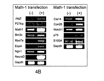
FIG. 4B is a gel showing the results of gene expression in cells transfected
with Atohl followed by treatment of the cells with NT3, FGF and BDNF. The
results
indicate that this protocol gave rise to progenitor cells that subsequently
matured into
cells expressing hair cell genes, including espin, myosin Vila, jagged 2, and
Brn3c,
and p27Kip, in addition to the proneural genes, Ngtil and NeuroD.
FIG. 4C is a gel showing the results of further genetic analysis of the cells
under the differentiating conditions described in 4B; the results showed that
the cells
rth,
also expressed SIO0A, p75 claudin 14, connexin 26, and Notch!, consistent with
some cells having a supporting cell phenotype.
FIG. 4D is a photomicrograph of an MSC cell line selected in Zeocin; the cells
had a high percentage of GFP expression when cultured in serum (green in
original).
FIG. 4E is a row of 4 photomicrographs of cells stained for Myo7a (first
panel), Mathl/Atohl (second panel), or DAN (third panel); the last panel is a
merged
image. After differentiation, the number of hair cell-like cells per DAPI
nucleus rose
and these cells stained for myosin Vila (shown in red in the first panel) and
Atohl
(shown in green in the second panel; arrows in the second and last panels).
FIG. 4F is two rows of 4 photoinicrographs of an Atohl expressing cell line
differentiated to cells with nuclei that were immunopositive for Brn3c (second
column, green in original; indicated by arrowheads) and cytoplasm positive for
6
CA 02669693 2009-05-14
WO 2008/076556
PCT/US2007/084654
myosin Vila (first column, red in original; indicated by arrows). Nuclei were
stained
with DAPI (third column, blue in orignal).
FIG. 4G is a row of three photomicrographs showing that the differentiated
cells were positive for F-actin which protruded from the apex of the cell in
the shape
of a stercocilia bundle (arrow).
FIG. 4H is a row of three photomicrographs showing that 17-actin staining was
arranged in a characteristic V pattern on the apical surface.
FIG. 5A is a gel showing the results of genetic analysis of bone marrow MSC
derived progenitors were co-cultured for 21 days with chick otocyst cells that
had
been treated with mitomycin C (Mito C); the results showed that expression of
jagged 2, p27Kip, Atohl, Brn3c, myosin Vila and espin was increased, whereas
the
expression of these genes in chick cells was undetectable. Chick otocyst cells
that
had been fixed by incubation with parafortnaldehyde were less effective (PFA)
than
the unfixed cells but did cause diftbrentiation of the progenitors.
Conditioned
medium from the chick cells (Cud Med) had no effect (levels of expression of
these
markers similar to previously shown data for differentiating conditions).
FIG. 5B is a set of three photomicrographs showing that expression of Atohl
(Math-1, middle panel, green in original) and myosin Vila (top panel, red in
original)
in cells from a Atohl-GFP mouse showed green fluorescence corresponding to the
induction of this marker in the nucleus and had expression of myosin Vila in
the
cytoplasm.
FIG. 6A is a set of four photomicrographs showing an increase in fluorescence
(green in original) indicating the conversion of bone marrow cells to cells
expressing
Atohl. The cells stained for Atohl (Math!, bottom left, green in original),
myosin
V i la (top left, red in original) and DAPI (top right, blue in original). A
merged image
is shown in on the bottom right panel.
FIG. 6B is a photomicrograph showing that Atohl -expressing cells were
found incorporated into the tissue of the chick otic epithelium. The hair
cells of the
chick were stained with the chick-specific marker, HCA (white in original) and
myosin Vila (red in original), whereas the Atoh- I expressing mouse cells were
green
due to expression of OFF (arrows).
FIG. 6C is a set of four photomicrographs showing a lack of cell fusion,
demonstrated by the presence of HCA (arrowhead, lower panels) in cells that
did not
7
CA 02669693 2009-05-14
WO 2008/076556
PCT/US2007/084654
have green fluorescence and of Atohl-GFP (arrow, right column) exclusively in
cells
that did not stain for HCA, a marker for chicken cells. No cells with both GFP
and
HCA were observed in these experiments. Scale bars are 100 pm.
FIG. 7 is a gel showing the results of genetic analysis of cells after
inhibition
of Notch signaling with an inhibitor of y-secretase increases expression of
hair cell
markers. Gene expression in MSCs treated with a y-secrctasc inhibitor showed
that
loss of Notch signaling increased Atohl expression. The timing of inhibition
was
critical: y-secretasc inhibitor added at dl of differentiation in vitro for a
total of 10
days led to an increase in hair cell markers, myosin Vila and espin, whereas
inhibitor
to added at d3 did not induce hair cell markers.
DETAILED DESCRIPTION
Although stem cells are present in the inner car (Li et al., Trends Mol Med
10,
309-315 (2004); Li et at, Nat Med 9, 1293-1299 (2003); Rask-Andersen et at,
Hear
Res 203, 180-191(2005)), hair cells do not regenerate after damage, and,
therefore, a
source of cells that could potentially be used for cell transplantation in a
therapeutic
replacement of these sensory cells has important implications for treatment of
sensorineural hearing loss. Bone marrow has been harvested and used
extensively in
clinical applications and is a highly desirable sourcc, because cells from a
patient's
bone marrow could potentially be transplanted without the problem of immune
rejection. The present methods include a treatment regimen for hearing loss
including transplantation of hair cells obtained by methods described herein.
By a combination of growth factor stimulation and expression of the
transcription factor, Atohl, that is required for hair cell formation in the
inner ear, the
present inventors demonstrate herein that stem cells, e.g., mesenchymal stem
cells
derived from bone marrow, can be induced to differentiate into hair cells. In
addition,
the neurosensory progenitors obtained from bone marrow can be converted to
sensory
cells by co-culture with cells of the developing sensory epithelium, even in
the
absence of Atoll 1 expression.
Stem cells in bone marrow are known to be the precursors for all lymphoid.
and erythroid cells, but mesenchymal stem cells in bone marrow also act as
precursors
to bone, cartilage, and fat cells (Colter et al., Proe Natl Acad Sci U S A 97,
3213-3218
(2000); Pittenger et al., Science 284, 143-147 (1999)). En addition to
mesenchymal
8
CA 02669693 2009-05-14
WO 2008/076556 PCT/US2007/084654
tissues, these stem cells have been shown to give rise to cells of other
lineages
including pancreatic cells (Hess et al., Nat Biotechnol 21, 763-770 (2003)),
muscle
cells (Doyonnas et al., Proc Natl Acad Sci U S A 101, 13507-13512 (2004)) and
neurons (Dezawa et al., 3 Clin Invest 113, 1701-1710 (2004); Hermann et al., J
Cell
Sci 117, 4411-4422 (2004); Jiang et al., Proc Natl Acad Sci U S A 100 Suppl I,
11854-11860 (2003)). The evidence provided herein demonstrates an extended
range
of cell fates available for these bone marrow-derived cells that includes
cells of the
neurosensory lineage, even including differentiation to inner ear hair cells.
Methods for Generating Cells of the Inner Ear
Methods of generating cells of the inner ear are provided, including
progenitor
cells and differentiated inner ear cells including hair cells and supporting
cells. Stem
cells arc unspecialized cells capable of extensive proliferation. Stem cells
arc
pluripotent and are believed to have the capacity to differentiate into most
cell types
in the body (Pedersen, Scientif. Am. 280:68 (1999)), including neural cells,
muscle
cells, blood cells, epithelial cells, skin cells, and cells of the inner ear
(e.g., hair cells
and cells of the spiral ganglion). Stem cells are capable of ongoing
proliferation in
vitro without differentiating. As they divide, they retain a normal karyotypc,
and they
retain the capacity to differentiate to produce adult cell types.
Hematopoietic stem cells resident in bone marrow arc the source of blood
cells, but in addition to these hematopoietic stem cells, the bone marrow
contains
mesenchymal stem cells (MSCs) that can differentiate into cell types of all
three
embryonic germ layers (Colter et al., Proc Natl Acad Sci U S A 97, 3213-3218
(2000);
Doyonnas et al., Proc Natl Acad Sci US A 101, 13507-13512 (2004); Herzog ct
at.,
Blood 102, 3483-3493 (2003); Hess et al., Nat Biotechnol 21, 763-770 (2003);
Jiang
et al., Nature 418, 41-49 (2002); Pittenger et al., Science 284, 143-147
(1999)). This
has been demonstrated in vivo in studies that track transplanted bone marrow
cells to
specific tissues where they differentiate into the resident tissue type (Mezey
et al.,
=
Proc Nati Acad Sci U S A 100, 1364-1369 (2003); Weimann et al., Proc Nati Acad
Sci
U S A 100, 2088-2093 (2003)).
Many of these cells have been used for transplantation and are a preferred
source of new cells for therapies because the transplanted cells are
immunologically
matched when harvested from a patient to be treated and because they have been
extensively used in clinical applications so that their safety is known.
9
CA 02669693 2009-05-14
WO 2008/076556
PCT/US2007/084654
Stein cells can differentiate to varying degrees. For example, stem cells can
form cell aggregates called cmbryoid bodies in hanging drop cultures. The
cmbryoid
bodies contain neural progenitor cells that can be selected by their
expression of an
early marker gene such as Soxl and the nestin gene, which encodes an
intermediate
filament protein (Lee et al., Nat. Biotech. 18:675-9, 2000).
Neurogenic Stem Cells
Inner car cells or inner ear cell progenitors can be generated from mammalian
stem cells. As described herein, stem cells suitable for use in the present
methods can
be any stem cell that has neurogenic potential, i.e., any stem cell that has
the potential
to differentiate into a neural cell, e.g., neurons, glia, astrocytes, retinal
photoreceptors,
oligodendrocytes, olfactory cells, hair cells, supporting cells, and the like.
Neurogenic stem cells, including human adult stem cells such as bone marrow
mesenchymal stem cells, can be induced to differentiate into inner ear
progenitor cells
that are capable of giving rise to mature inner car cells including hair cells
and
supporting cells. Neurogenic stem cells useful in the methods described herein
can be
identified by the expression of certain neurogenic stem cell markers, such as
nestin,
sox 1, sox2, and musashi. Alternatively or in addition, these cells express
high levels
of helix-loop-helix transcription factors NeuroD, Atohl, and ncurogeninl.
Examples of neurogenie stem cells include embryonic stem cells or stem cells
derived from mature (e.g., adult) tissue, such as the car (e.g., inner ear),
central
nervous system, blood, skin, eye or bone marrow. In some embodiments, the stem
cells are mesenchymal stem cells. Any of the methods described herein for
culturing
stem cells and inducing differentiation into inner car cells (e.g., hair cells
or
supporting cells) can be used.
Stem cells useful for generating cells of the inner ear can be derived from a
mammal, such as a human, mouse, rat, pig, sheep, goat, or non-human primate.
For
example, stem cells have been identified and isolated from the mouse utricular
macula
(Li et al., Nature Medicine 9:1293-1299, 2003).
Generation of Neural Progenitor Cells
There arc a number of induction protocols known in the art for inducing
differentiation of stein cells with neurogenic potential into neural
progenitor cells,
including growth factor treatment (e.g., treatment with Ea', FGF, and 1GF, as
CA 02669693 2009-05-14
WO 2008/076556
PCT/US2007/084654
described herein) and neurotrophin treatment (e.g., treatment with NT3 and
BDNF, as
described herein). Other differentiation protocols are known in the art; see,
e.g.,
Corrales et al., J. Neurobiol. 66(13):1489-500 (2006); Kim et al., Nature 418,
50-6
(2002); Lee et al., Nat Biotechnol 18, 675-9 (2000); and Li et al., Nat
Biotechnol 23,
215-21 (2005).
As one example of an induction protocol, the stem cells are gown in the
presence of supplemental growth factors that induce differentiation into
progenitor
cells. These supplemental growth factors are added to the culture medium. The
type
and concentration of the supplemental growth factors is be adjusted to
modulate the
growth characteristics of the cells (e.g., to stimulate or sensitize the cells
to
differentiate) and to permit the survival of the differentiated cells such as
neurons,
glial cells, supporting cells or hair cells.
Exemplary supplementary growth factors are discussed in detail below, and
include, but are not limited to basic fibroblast growth factor (bFGF), insulin-
like
growth factor (1GF), and epidermal growth factor (EGF). Alternatively, the
supplemental growth factors can include the neurotrophic factors neurotrophin-
3
(NT3) and brain derived ncurotrophic factor (BDNF). Concentrations of growth
factors can range from about 100 ng/mL to about 0.5 ng/mL (e.g., from about 80
np/mL to about 3 ng/mL, such as about 60 ng/mL, about 50 ng/mL, about 40
ng/mL,
about 30 ng/mL, about 20 nWmL, about 10 ng/mL, or about 5 ng/mL).
Neural progenitor cells produced by these methods include inner ear
progenitor cells, i.e., cells that can give rise to inner ear cells such as
hair cells and
supporting cells. Inner ear progenitor cells can be identified by the
expression of
marker genes such as ncstin, sox2, and musashi, in addition to certain inner-
ear
specific marker genes Brn3C, Pax2, and Atohl. The invention includes purified
populations of inner ear progenitor cells expressing nestin, sox2, musashi,
Brn3C,
Pax2, and Atoh I . These inner car progenitor cells are lineage committed, and
can be
induced to further differentiate into hair cells and supporting cells by a
method
described herein.
Progenitor cells prepared by a method described herein can optionally be
frozen for future use.
ti
CA 02669693 2014-10-03
60412-4104
Cell Culture Methods
In general, standard culture methods are used in the methods described herein.
Appropriate culture medium is described in the art, such as in Li ct al.
(supra). For
example, stem cells can be cultured in serum free DMEM/high-glucose and F12
media (mixed 1:1), and supplemented with N2 and B27 solutions and growth
factors.
Growth factors such as EGF, IF-1, and bFGF have been demonstrated to augment
sphere formation in culture. In vitro, stem cells often show a distinct
potential for
forming spheres by proliferation of single cells. Thus, the identification and
isolation
of spheres can aid in the process of isolating stem cells from mature tissue
for use in
to making differentiated cells of the inner ear. The growth medium for
cultured stem
cells can contain one or more or any combination of growth factors. This
includes
leukemia inhibitory factor (L1F) which prevents the stem cells from
differentiating.
To induce the cells (and the cells of the spheres) to differentiate, the
medium can be
exchanged for medium lacking growth factors. For example, the medium can be
scrum-free DMEM/high glucose and F12 media (mixed 1:1) supplemented with 1l2
and 1327 solutions. Equivalent alternative media and nutrients can also be
used.
Culture conditions can bo optimized using methods known in the art.
Differentiation by Expression of Atohl
As described herein, expression of Atohl in stem-cell derived progenitor cells
was sufficient to drive them into adopting hair cell markers. Studies of Atohl
expression in the ear have indicated that this helix-loop-helix transcription
factor
occupies a key place in the hierarchy of inner car transcription factors for
differentiation of hair cells.
Atohl nucleic acids and polypeptides arc known in the art, and described in,
for example, U.S. Pat. Nos. 6,838,444 and 7,053,200, and P.G. PUB. Nos.
2004/0237127 and 2004/0231009, all to Zoghbi et al.
In some embodiments, the Atohl is, or is at least 80%, 85%, 90%,
93%, or 95% identical to, human atonal homolog 1 (ATOH1); ATH1; and HATH1 (for
additional information sec Ben-Arie et al., Molcc. Genet. 5: 1207-1216 (1996);
Bermingham at al., Science 284: 1837-1841 (1999); OM11v1 *601461; UniGene
Ils.532680; GenBank Accession Nos. NM_005172.1 (nucleic acid) and NP_005163.1
(polypeptide)). Other species can also be used, e.g., Mouse Atolil (also known
as
12
El
19199=5= 5q9=15619 3=3531936 6163,91== 9161949=e 91V1613E1E It6T
6916191610 1.3=3=101 100E199103 11030314E0 3113136E96 10660E6691 'MI
13E6119E60 9936,9336a3 5=9616616 =156131= 991=33393 5woolulte tett
0/96163=0 6996563606 3691=1=3 0619e2=e9 396963399e 951=31359 t9at
=93669966 9=59=559 5196=1=6 3593911931 910E909399 3313696966 ton G5
=61699693 9=31969= 3=193999p 3593993966 2662=1613 3693613=9 tptt
365555336 13531=360 q613=6599 69=366496 39606=991 3306669395 1801
696311=69 0=110E064 1=5396623 59361663= 9=6556613 1=1=6e= taot
3569=== 53=965=6 3.133666333 Spoopeloos, 5953=6565 5.9565=305 196
933693=66 56=691653 6=1=6366 5363669669 26191=1= 100690E063 106 09
1339/1903 9=961seve 9361==o6 eoe=o6336 =6=9=69 5956315399 tp8
3=1=6936 136=69663 i6131363912 =939=196 933356a959 =33339696 teL
19199=316 1369969939 6=9=9=1 =16011191 16/9906163 0600106011 M..
=509=995 =6663=61 9669353553 9956691=6o 2993693661 3669669u= t99
59969=266 66199615E9 399936en= 1=3369E35 9336336936 =65113696 108 St
=55159161 16.56636569 96=699361 6=6939956 539:1699933 6336663o3 tps
3696993693 1=6636636 13E636995e 66E9155136 n6'1666936 9395=5695 t8p
3968633336 36,636365e 6=1=6166 6=5963333 =1=6=61 =9059=06 Tap
=636=356 31=6=1=6 6693612139 3=1396136 5=3636393 99E63390.612 tat
3966==3 .6=99590 563=9131S 33311=969 5953569361 =09=63= to Op
9=5=9=5 395=6=63 =1639=93 5336e3=a e=6/39=e =955656= tvz
6969999366 v6=666159 69969361vo 6136=363o 3161963635 9/51.39=69 tat
6=103=65 3939931= 1=9369=6 =5661=96 1353=== =31355663 tat
1351361361 161111165n 9669339696 92E9361133 9693199999 9999669e55 19
9.56e593156 5958666959 516963 66=15363 9=35=153 6090=9601 SE
:SM0110.1
orn snuanbos ondoctAiod put (1gzi - 961 =s03) vmpu igow snow ata
(ou axOas) svga
SCISAHSHdai9GSUH9USIXSN320AdOlIGOdrISdS2MOVNWVVIV OE
saaaa,sairDiCrICIASIO5VSVcIVS.321.1110SOddIclOSMS11001fOttlati
NOVDOSASVVIEIHHHCISXOSVddddel3DOSdIMISSIVNIAIOVWCY1
IMSMINCINNaSdIANIMOCLIVHITIDHWE'UlialnalVV74110MOADNAOX
SSdVHOUSDDIECIAAADDX7M0q0311AXAdOdSMSSSVDOSSEW120110
0A2011d1YVV2StfOrlScISHTIAOVVWVIDIDOSIAITIMVWdaISGTISZad SZ
dAAdHSEVOIlleddeddciddOTIHHdOdOHHH(1073XAMIMUTMIUSW
(I:cm ax us) 62116 22:166,26126 6313961693 9319=331e 3=3=1111 tun
9966663953 6996939=3 15531=6= =99993590 Eu99669659 35369=6= 196
5131103696 6601011/1* 11=1115= 19969999/6 /66196296a 699533=6 106 Ol
3620956953 11339533= =906=106 =65=5236 16631=366 696E835131 tp8
3369333362 n1311363= v66=61159 6563=5=3 3633653E9 369666966 18L
=1=661=6 9=36555= 5=63396z6 3e93665363 5665699619 3=1=6636 1at.
=93631= 933933903e 53599-9936 3333D69=1 03603630E0 3693996666 T.88
9663593336 ouravoul36 369531611 =6399=93 9==peo= 6E19593530 109
3=696qeqe 993316=59 95993G639p =9=25335 opoqeqq639 93536=590 tps
ov6=23353 9339961355 5393512569 oB36636966 6=36399= 5=691356o T9,
v696936995 9362666519 nEa6693999 35,==== 3666=9=5 3359353366 tap
5135963e5e 1663561556 6366999613 6993636=6 =99565352 59996165= 19t
66533=595 9=6935=6 9=5166356 3693696695 59.9.451359 665E56=65 IOC Ol
=6515696o 9.666333363 6=5=5696 93=361666 =69653=3 11=913610 TtZ
193693a363 3636393663 9361319356 693613=93 =3366=66 13363639= 181
3953393593 96613=3 16=6961= 5=093=63 =39359696 963E5=5= IZI
1399361=9 n35936=53 36=6=6= 99363=131 931936=69 3333593363 18
.3933933959 556=69569 9516996=5 5515969059 363936=61 3160331610 I
:SMOHOJ
se soouonbus oppdocigod puu (59ot - = SOD) viiNui iqopi umunq otu,
*(1.Z6Z,L9tr1st .0N. '00V )funguoD
!Iwos umouN osie) 14olv uolov `(TOOSLOO '01=1 .33V 3tuneua0
tS9t80/LOOZS11/I3c1 9S9L0/800Z OAX
P1-S0-600Z 696990 IZO
CA 02669693 2009-05-14
WO 2008/076556
PCT/US2007/084654
1501 gctgctaaaa tgtcgtatct ctgcctctgg tctgggtttc acttatttta taccttggga
1561 gttcatcctt gcgtgttgcg ctcactcaca aataagggag ttagtcaatg aagttgtttc
1621 cccaactgct tgagacccgc attgggtact ttactgaaca cggactattg tgttgttaaa
1681 atgcaggggc agataagagt atctgtagag cttagacacc aagtgtgtcc agcagtgtgt
1741 ctagcggacc cagaatacac gcacttcatc actggccgct gcgccgcctt gaagaaactc
1801 aactgccaat gcagagcaac ttttgatttt aaaaacagcc actcataatc attaaactct
1861 ttgcaaatgt ttgtttttgc aaatgaaaat taaaaaaaaa catgtagtgt caaaggcatt
1921 tggtcaattt tattttgctt tgttaacatt agaaaagtta tttattattg cgtatttgga
1981 cccatttcta cttaattgcc ttttttttac attttctact cgagatcgtt ttattttgat
2041 ttagcaaatc cagttgccat tgctttatgt atgtatgctc ttttacaaat gataaaataa
2101 actcggaaaa aaaaaaaaaa aaaaaaaaaa aaaaaaaaaa aaaa (SEQ ID NO:3)
MSRLLHAEEWAEVKELGDHHRHPQPRHVPPLTPQPPATLQARDLLVRRSC
CGGLSKSPGPVEVREOLCKLKGGVVVDELGCSRQRAPSSKQVNGVQKQRR
LAANARERRRMHGLNHAFDQLRNVIPSFNNDKKLSKYETLQMAQIYINAL
SELLQTPNVGASSGGYSVQLDALHFPAFEDRALTAMMAQKDLSPSLPGGI
LQPVQEDNSKTSPRSHRSDGEFSPHSHYSDSDEAS (SEQ ID N0:4)
The chicken Cathl inRNA (CDS 1 ¨ 717) and polypeptide sequences are as
follows:
1 atggccccag gaggtagcga gtgttgttgc agtgatgccg cgcacatcac ttggaggcag
61 tgggagtaca cgcacgagaa ccaactgtgc gtggcaggaa ctgtcagcag gatgaggccc
121 aggacgtggg tctgcaccgg atctttgtgg gaccaggaag cgggaattac tttgatgggc
181 ccccaaatac ccaaagtgga tgaggcagga gtgatgaccc acccggcaag gtcgctttgc
241 agcactgggg cacatccgtg tcccggggtg gtcgtgctgc ccacgggtgg gatagggcag
301 ccttcaaaga agctctccaa gtacgagacg ctscagatgg cgcaaatcta catcagcgcc
361 cccgccgagc ttctgcacgg gccgcccgcg ccccccgagc cgcccgccaa ggccgagctc
421 cgcggggccc ccttcgagcc tcccccgccg ccccctcctc cgccgccccg cgcctcgccc
481 cccgcgcccg ccaggactcg cttccccccg gcggcggccg cgggcggttt cgcggcgctt
541 ctcgagccgc tgcgcttccc ttctttcccg gcgcagaaag cgccttctcc cgcgctgctc
601 ctggggccgc ccgcgccgca gcagcccgag aggagcaaag cgccgccgcg ctctcaccgc
661 agegacggsg agttctcgcc gcgctcccac tacagtgact cggacgaggc cagctag
(SEQ ID NO:5)
MAPGGSECCCSDAAHITWRQWEYTHENQLCVAGTVSRMRPRTWVCTGSLWDQEAGI
TLMGPQIPKVDEAGVMTHPARSLCSTGAHPCPOVVVLPTGGIGUSEKLSKYETLQ
MAQIYISALAELLHGPPAPPEPPARAELRGAPFEPPPPPPPPPPRASPPAPARTRF
PPAAAAGGFAALLEPLRFPSFPAQICAPSPALLLGPPAPQQPERSKASPRSHRSDGE
FSPRSHYSDSDEAS (SEQ ID NO:6)
To determine the percent identity of two amino acid sequences, or of two
nucleic acid sequences, the sequences arc aligned for optimal comparison
purposes
(e.g., gaps can be introduced in one or both of a first and a second amino
acid or
nucleic acid sequence for optimal alignment and non-homologous sequences can
be
disregarded for comparison purposes). The length of a reference sequence
aligned for
comparison purposes is at least 80% of the length of the reference sequence,
and in
some embodiments is at least 90% or 100%. The amino acid residues or
nucleotides
at corresponding amino acid positions or nucleotide positions arc then
compared.
When a position in the first sequence is occupied by the same amino acid
residue or
nucleotide as the corresponding position in the second sequence, then the
molecules
are identical at that position (as used herein amino acid or nucleic acid
"identity" is
14
CA 02669693 2009-05-14
WO 2008/076556
PCT/US2007/084654
equivalent to amino acid or nucleic acid "homology"). The percent identity
between
the two sequences is a function of the number of identical positions shared by
the
sequences, taking into account the number of gaps, and the length of each gap,
which
need to be introduced for optimal alignment of the two sequences.
For purposes of the present invention, the comparison of sequences and
determination of percent identity between two sequences can be accomplished
using a
Blossum 62 scoring matrix with a gap penalty of 12, a gap extend penalty of 4,
and a
frameshi ft gap penalty of 5.
In some embodiments, the methods include expressing in the cells a Atohl
polypeptide encoded by a nucleic acid that hybridizes to the human Atohl mRNA
under stringent conditions. As used herein, the term "stringent conditions"
describes
conditions for hybridization and washing. Stringent conditions as used herein
are
0.5M sodium phosphate, 7% SDS at 65 C, followed by one or more washes at 0.2X
SSC, 1% SDS at 65 C. See, e.g., Current Protocols in Molecular Biology, John
Wiley
& Sons, N.Y. (2006).
In some embodiments, the methods include expressing exogenous Atohl in a
stem cell. This can be achieved, for example, by introducing an expression
vector in
the cell. As used herein, the term "vector" refers to a nucleic acid molecule
capable of
transporting another nucleic acid to which it has been linked and can include
a
plasmid, cosmid or viral vector. The vector can be capable of autonomous
replication
or it can integrate into a host DNA. Viral vectors include, e.g., replication
defective
retroviruses, adenoviruses and adeno-associated viruses.
A vector can include a Atoh I nucleic acid in a form suitable for expression
of
the nucleic acid in a host cell. Generally, the expression vector includes one
or more
regulatory sequences operatively linked to the nucleic acid sequence to be
expressed.
The term "regulatory sequence" includes promoters, enhancers and other
expression
control elements (e.g., polyadenylation signals). Regulatory sequences include
those
which direct constitutive expression of a nucleotide sequence, as well as
tissue-
specific regulatory and/or inducible sequences. The design of the expression
vector
can depend on such factors as the choice of the host cell to be transformed,
the level
of expression of protein desired, and the like. The expression vectors can be
introduced into host cells using methods known in the art, including calcium
phosphate or calcium chloride co-precipitation, DEAE-dextran-mediated
transfection,
CA 02669693 2009-05-14
WO 2008/076556
PCT/US2007/084654
lipofection, or electroporation. See, e.g., Current Protocols in Molecular
Biology,
John Wiley & Sons, N.Y. (2006).
In the present methods, the Atohl polypeptide expressed in the stem cells will
have the ability to induce differentiation of mesenchyrnal stem cells to hair
cells
and/or supporting cells, as described herein.
Differentiation by Culturing with Chick Otocysts
Also as described herein, the stem cell-derived progenitor cells also
responded
to physical contact with developing otocyst cells from the chicken embryo by
differentiating into sensory epithelial cells, without the requirement for
exogenous
Atohl. This was evidenced by nGFP expression from a Atohl enhancer-GFP
reporter
construct and co-expression of myosin Vila after co-culture and
differentiation, as
described herein. Neurons that express markers of sensory cells have been
induced
from bone marrow MSCs in previous work by incubation with otocyst and
hindbrain-
conditioned medium (Kondo et al., Proc Natl Acad Sci U S A 102, 4789-4794
(2005))
from embryonic mice.
Thus, the methods described herein can include contacting progenitor cells
with otocyst cells, e.g., cells isolated from E3 embryonic chicks, as
described herein.
In some embodiments, the methods include culturing the progenitor cells with
the otocyst cells in a ratio of about 50,000 cells per confluent layer of
otocyst cells, or
by injection of 100,000 cells into an intact otocyst (sec Examples, below).
Alternatively, the stem cells can be cultured in the presence of chick otocyst-
conditioned media, which can be produced using methods known in the art, e.g.,
using
media that has been in contact with a culture of chick otocysts for at about
four days.
Differentiation by Inhibition of Notch Signalling
Notch is a plasma membrane receptor, and the Notch pathway consists of
Notch and its ligands, as well as intracellular proteins that transmit the
Notch signal to
the nucleus. Included in the Notch pathway are the transcription factors that
bear the
effector function a the pathway.
Notch signaling plays a role in lateral inhibition, in which one cell is
singled
out from a cell cluster for a given fate (e.g., differentiation into a hair
cell, for
example). Differentiation is inhibited in those cells not selected to
differentiate,
resulting in the prevention of a specified fate commitment on the part of most
of the
16
CA 02669693 2009-05-14
WO 2008/076556
PCT/US2007/084654
cells of a cluster. Lateral inhibition occurs repeatedly during development.
Central to
this process is binding to the Notch receptor of one of several ligands,
including
Delta, Scabrous and Serrate. Ligand binding to Notch ligand triggers a chain
of
intracellular events resulting in lateral inhibition. A review of the Notch
pathway can
be found at Artavanis-Tsakonas etal., Science 268: 225-232 (1995). As
described
herein, inhibition of Notch in the inner ear progenitor cells described herein
results in
differentiation of the cells into hair cells and supporting cells.
Thus, in some embodiments of the methods described herein, progenitor cells
are grown in the presence of a Notch signalling pathway inhibitor. Exemplary
Notch
pathway inhibitors include y-secretase inhibitors, of which a number are known
in the
art (e.g., arylsulthnamides (AS), dibenzazepines (DBZ), benzodiazepines (BZ),
N-[N-
(3,5-difluorophenacety1)-L-alanyl]-(S)-phenylglycine t-butyl ester (DAPT), L-
685,458 (Sigma-Aldrich), and MK0752 (Merck). A useful concentration will
depend
on the inhibitor chosen.
Other Notch inhibitors include inhibitory nucleic acids (e.g., small
interfering
RNAs, antisense oligonucleotides, and morpholino oligos; methods for
designing,
making, and using them are known in the art, e.g., gene walk methods for
selecting
and optimizing inhibitory sequences, see, e.g., Engelke, RNA Interference
(RNAi):
The Nuts & Bolts of siRNA Technology, (DNA Press, 2004); Mol, Antisense
Nucleic
Acids and Proteins, (CRC, 1994); Sioud, Ribozymes and Sirna Protocols (Methods
in
Molecular Biology), (Humana Press; 2nd edition 2004); and Philips, Antisense
Therapeutics (Methods in Molecular Medicine), (Humana Press 2004)) targeting
Notch (see, e.g., Presente et al., Proc. Nat. Acad. Sci. 101(6):1764-1768
(2004);
Ivanov etal., Proc. Nat. Acad. Sci. 101(46):16216-1622 l (2004)) or its
ligands, i.e.,
Delta or Jagged (see, e.g., Patzel et al., Nature Biotechnology 23, 1440 -
1444 (2005);
Purow et al., Cancer Research 65:2353-2363 (2005); or Stallwood et al., J.
Immunol.
177:885-895 (2006)). Alternatively, the cells can be modified to express m-
Numb
(GcnBank Ace. No. NP_001005743.1) or disheveled (Dv1; the human homologs are
at
GenBank Ace. No. NM 004421.2 (variant 1); NM_004422.2 (variant 2); and
NM 004423.3 (variant 3), both endogenous inhibitors of Notch signalling.
Assaying Differentiation
A variety of methods can be utilized to determine that a stem cell has
differentiated into a progenitor cell, or into a cell of the inner ear, e.g.,
a hair cell or
17
CA 02669693 2009-05-14
WO 2008/076556
PCT/US2007/084654
'supporting cell. For example, the cell can be examined for the expression of
a cell
marker gene. Hair cell marker genes include myosin Vila (myoVIla), Atohl, ot9
acetylcholine receptor, cspin, parvalbumin 3, and Bm3c. Supporting cell
markers
include claudin14, connexin 26, p75Trk, Notch 1, and S100A. Pluripotent stem
cells
generally do not express these genes. A stem cell that propagates and produces
a cell
expressing one or more of these genes, has produced a hair cell, i.e., the
stem cell has
differentiated at least partially into a hair cell. A stem cell that has
differentiated into
an inner car progenitor cell (a precursor of hair cells) expresses early car
marker genes
such as nestin, sox2, musashi, Bm3C, Pax2, and Atohl. A progenitor cell can
express
one or more of these genes. The progenitor cells can be propagated in serum-
free
medium in the presence of growth factors. Removal of growth factors and
expression
of Atohl, or co-culture with chick otocysts, will induce the cells to
differentiate
further, such as into hair cells and supporting cells.
Identification of a hair cell or hair cell progenitor (e.g., a hair cell,
supporting
cell, or progenitor cell that differentiated from a stem cell) can be
facilitated by the
detection of expression of marker genes as described herein. Detection of the
products of' gene expression can be by immunocytochemistry.
Immunocytochemistry
techniques involve the staining of cells or tissues using antibodies against
the
appropriate antigen. In this case, the appropriate antigen is the protein
product of the
tissue-specific gene expression. Although, in principle, a first antibody
(i.e., the
antibody that binds the antigen) can be labeled, it is more common (and
improves the
visualization) to use a second antibody directed against the first (e.g., an
anti-IgG).
This second antibody is conjugated either with fluorochromes, or appropriate
enzymes for colorimetric reactions, or gold beads (for electron microscopy),
or with
the biotin-avidin system, so that the location of the primary antibody, and
thus the
antigen, can be recognized. The protein marker can also be detected by flow
cytometry using antibodies against these antigens, or by Western blot analysis
of cell
extracts.
Alternatively or in addition, gene expression can be analyzed directly, e.g.,
= using PCR methods known in the art, including quantitative PCR, e.g.,
quantitative
RT-PCR, which can be used to detect and compare levels of expression.
18
CA 02669693 2009-05-14
WO 2008/076556
PCT/US2007/084654
Methods of Treatment
The methods described herein can be used to generate cells for therapeutic
use. Treatment methods include generating cells of the inner ear (e.g., hair
cells or
supporting cells) from stem cells, using a method described herein, for
transplantation
into an car of a human in need thereof. Transplantation of the cells into the
inner car
of a subject can be useful for restoring or improving the ability of the
subject to hear,
or for decreasing the symptoms of vestibular dysfunction. Inner ear cells
derived
from stem cells according to the methods described herein need not be fully
differentiated to be therapeutically useful. A partially differentiated cell
that improves
any symptom of a hearing disorder in a subject is useful for the therapeutic
compositions and methods described herein.
A human having a disorder of the inner ear, or at risk for developing such a
disorder, can be treated with inner ear cells (hair cells or supporting cells)
generated
from stem cells using a method described herein. In a successful engraftment,
at least
some transplanted hair cells, for example, will form synaptic contacts with
spiral
ganglion cells, and integrate into the sensory epithelium of the inner car. To
improve
the ability of the cells to engraft, the stein cells can be modified prior to
differentiation. For example, the cells can be engineered to overexpress one
or more
anti-apoptotic genes in the progenitor or differentiated cells. The Fak
tyrosine kinase
or Akt genes are candidate anti-apoptotic genes that can be useful for this
purpose;
overexprcssion of FAK or Akt can prevent cell death in spiral ganglion cells
and
encourage engraftment when transplanted into another tissue, such as an
explanted
organ of Corti (see for example, Mangi et al., Nat. Med. 9:1195-201 (2003)).
Neural
progenitor cells overexpressing integrin may have an enhanced ability to
extend
neurites into a tissue explant, as the integrin has been shown to mediate
neurite
extension from spiral ganglion neurons on laminin substrates (Aletsce et al.,
Audiol.
Neurootol. 6:57-65 (2001)), In another example, ephrinB2 and ephrinB3
expression
can be altered, such as by silencing with RNAi or ovcrexpression with an
exogenously expressed cDNA, to modify EphA4 signaling events. Spiral ganglion
neurons have been shown to be guided by signals from EphA4 that are mediated
by
cell surface expression of ephrin-B2 and -B3 (Brors et al., J. Comp. Neurol.
462:90-
100 (2003)). Inactivation of this guidance signal may enhance the number of
neurons
that reach their target in an adult inner ear. Exogenous factors such as the
19
CA 02669693 2009-05-14
WO 2008/076556
PCT/US2007/084654
neurotrophins BDNF and NT3, and LIF can be added to tissue transplants to
enhance
the extension of neurites and their growth towards a target tissue in vivo and
in ex
vivo tissue cultures. Neurite extension of sensory neurons can be enhanced by
the
addition of neurotrophins (BDNF, NT3) and L1F (Gillespie et al., NeuroReport
12:275-279 (2001)). A Sonic hedgehog (Shh) polypeptide or polypeptide fragment
(e.g., SI-1H-N), can also be useful as an endogenous factor to enhance ncurite
extension. Shh is a developmental modulator for the inner ear and a
chemoattractant
for axons (Charron et al., Cell 113:11 23 (2003)).
Any human experiencing or at risk for developing a hearing loss is a candidate
for the treatment methods described herein. For example, the human can receive
a
transplant of inner car hair cells or supporting cells generated by a method
described
herein. A human having or at risk for developing a hearing loss can hear less
well
than the average human being, or less well than a human before experiencing
the
hearing loss. For example, hearing can be diminished by at least 5, 10, 30,
50% or
more. The human can have sensorineural hearing loss, which results from damage
or
malfunction of the sensory part (the cochlea) or the neural part (the auditory
nerve) of
the car, or conductive hearing loss, which is caused by blockage or damage in
the
outer and/or middle ear, or the human can have mixed hearing loss, which is
caused
by a problem in both the conductive pathway (in the outer or middle ear) and
in the
nerve pathway (the inner ear). An example of a mixed hearing loss is a
conductive
loss due to a middle-car infection combined with a sensorineural loss due to
damage
associated with aging.
The subject can be deaf or have a hearing loss for any reason or as a result
of
any type of event. For example, a human can be deaf because of a genetic or
congenital defect; for example, a human can have been deaf since birth, or can
be deaf
or hard-of-hearing as a result of a gradual loss of hearing due to a genetic
or
congenital defect. In another example, a human can be deaf or hard-of-hearing
as a
result of a traumatic event, such as a physical trauma to a structure of the
ear, or a
sudden loud noise, or a prolonged exposure to loud noises. For example,
prolonged
exposures to concert venues, airport runways, and construction areas can cause
inner
car damage and subsequent hearing loss. A human can experience chemical-
induced
ototoxicity, wherein ototoxins include therapeutic drugs including
antineoplastic
agents, salicylates, quinines, and aminoglycoside antibiotics, contaminants in
foods or
CA 02669693 2009-05-14
WO 2008/076556
PCT/US2007/084654
medicinals, and environmental or industrial pollutants. A human can have a
hearing
disorder that results from aging, or the human can have tinnitus
(characterized by
ringing in the ears).
The cells can be administered by any suitable method. For example, to restore
hearing, inner car cells generated by a method described herein can be
transplanted,
such as in the form of a cell suspension, into the ear by injection, such as
into the
luminac of the cochlea. See, e.g., the methods described in Corrales et al.,
J.
Neurobiol. 66(13):1489-500 (2006) and Hu et al., Experimental Cell Research
302:40-47 (2005), Injection can be, for example, through the round window of
the
to ear or through the bony capsule surrounding the cochlea. The cells can
be injected
through the round window into the auditory nerve trunk in the internal
auditory
meatus or into the scale tympani. In a preferred embodiment, the cells are
administered into or near the sensory epithelium of the subject, e.g., into a
fluid
(perilymph)-filled space above or below the sensory epithelium, i.e., the
scala media,
scala tympani, or scala vestibuli.
Alternatively, a human suitable for the therapeutic compositions and methods
described herein can include a human having a vestibular dysfunction,
including
bilateral and unilateral vestibular dysfunction. Vestibular dysfunction is an
inner ear
dysfunction characterized by symptoms that include dizziness, imbalance,
vertigo,
nausea, and fuzzy vision and may be accompanied by hearing problems, fatigue
and
changes in cognitive functioning. Vestibular dysfunction can be the result of
a genetic
or congenital defect; an infection, such as a viral or bacterial infection; or
an injury,
= such as a traumatic or nontraumatic injury. Vestibular dysfunction is
most commonly
tested by measuring individual symptoms of the disorder (e.g., vertigo,
nausea, and
fuzzy vision). In these embodiments, the inner ear cells generated by a method
described herein can be transplanted, such as in the form of a cell
suspension, e.g., by
injection, into an organ of the vestibular system, e.g., the utricle, ampulla
and
sacculus. The cells would generally be injected into the perilymph of these
organs or
into the vestibule (which connects the 3 organs).
Following treatment with an inner ear cell or inner ear cell progenitor as
described herein, the human can be tested for an improvement in hearing or in
other
symptoms related to inner ear disorders. Methods for measuring hearing are
well-
known and include pure tone audiometry, air conduction, and bone conduction
tests.
21
CA 02669693 2009-05-14
WO 2008/076556
PCT/US2007/084654
These exams measure the limits of loudness (intensity) and pitch (frequency)
that a
human can hear. Hearing tests in humans include behavioral observation
audiometry
(for infants to seven months), visual reinforcement orientation audiometry
(for
children 7 months to 3 years) and play audiometry for children older than 3
years.
Oto-acoustic emission testing can be used to test the functioning of the
cochlear hair
cells, and electro-cochleography provides information about the functioning of
the
cochlea and the first part of the nerve pathway to the brain.
The therapeutic compositions and methods described herein can be used
prophylactically, such as to prevent hearing loss, deafness, or other auditory
disorder
associated with loss of inner car function. For example, a composition
containing a
differentiation agent can be administered with a second therapeutic, such as a
therapeutic that may effect a hearing disorder. Such ototoxic drugs include
the
antibiotics neomycin, kanamycin, amikacin, viomycin, gentamycin, tobramycin,
erythromycin, vancomycin, and streptomycin; chcmotherapeutics such as
cisplatin;
nonstcroidal anti-inflammatory drugs (NSAIDs) such as choline magnesium
trisalicylate, diclofenac, diflunisal, fcnoprofen, flurbiprofen, ibuprofcn,
indomethacin,
ketoprofen, meclofenamate, nabumetone, naproxen, oxaprozin, phenylbutazone,
piroxicam, salsalate, sulindac, and tolmetin; diuretics; salicylates such as
aspirin; and
certain malaria treatments such as quinine and chloroquine.
For example, a human undergoing chemotherapy can also be administered an
inner ear cell or inner car cell progenitor as described herein, by a method
described
herein. The chemotherapeutic agent cisplatin, for example, is known to cause
hearing
loss. Therefore, a composition containing a differentiation agent can be
administered
with cisplatin therapy to prevent or lessen the severity of the cisplatin side
effect. An
inner ear cell or inner ear cell progenitor as described herein can be
administered
before, after and/or simultaneously with the second therapeutic agent. The two
treatments generally will be administered by different routes of
administration.
The compositions and methods featured in the invention arc appropriate for
the treatment of hearing disorders resulting from scnsorincural hair cell loss
or
auditory neuropathy. For example, patients with sensorineural hair cell loss
experience the degeneration of cochlear hair cells, which frequently results
in the loss
of spiral ganglion neurons in regions of hair cell loss, and may also
experience loss of
supporting cells in the organ of Corti, and degeneration of the limbus, spiral
ligament,
22
CA 02669693 2009-05-14
WO 2008/076556
PCT/US2007/084654
and stria vascularis in the temporal bone material. Such patients may benefit
particularly from administration of supporting cells and/or hair cells into
the inner ear.
EXAMPLES
The invention is further described in the following examples, which do not
limit the scope of the invention described in the claims.
Example 1: Sensory Progenitors from Nlesenehymal Stem Cells
Mesenchymal stem cells were obtained from mouse bone marrow by culturing
adherent cells from the marrow under high serum conditions.
Briefly, cells were obtained from bilateral femurs and tibias of 4 week old
C57BL/6 or Aoh/-)2GFP mice (Helms et al., Development 127, 1185-1196 (2000))
by flushing out the bone marrow with MEM-a (Gibco/BRL) containing 10% fetal
bovine serum (FBS; BioWhittaker, Cambrex, NY) andl mM glutamine (Gibco/BRL).
Pelleted cells were resuspended and mixed with RBC lysis buffer (Gibco/BRL).
Approximately 5x10⁶ cells were cultured on a 10 cm dish overnight in MEM-a
with
9% horse serum, 9% FBS, 1% Gluta-Max (Invitrogen) and 100 units/ml penicillin
and
streptomycin (100 g/ml, Sigma) at 37 C in a 5% CO2 atmosphere. Nonadherent
hematopoietic stem cells were removed, leaving adherent bone marrow stromal
cells.
When the cells became confluent, trypsinization was performed and the cells
were
cultured and passaged three to five times, with media changes every 3-4 days.
These
cells are referred to as mesenchymal stem cells (MSC).
lmmunohistochemistry was performed as follows. Cells were fixed for 10 min
with 4% paraformaldehyde in PBS, lmmunostaining was initiated by rehydrating
and
blocking the sections for 1 h with 0.1% Triton X-100 in PBS supplemented with
1%
BSA and 5% goat serum (PBT1). Fixed and permeabilized cells or rehydrated
sections were incubated overnight in P311. CD34, CD44, CD45, Sea-1 antibodies
(BD Biosciences) diluted 1: 40 were used for the characterization of extracted
bone
marrow cells. Hair cells and bone marrow progenitors were characterized using
monoclonal antibody to chick hair cell specific antigen diluted 1:500 (gift
from Guy
Richardson (Bartolami et al., .1 Comp Neurol 314, 777-788 (1991)); polyclonal
antibody to myosin Vila, 1:500 (Oshima et al.,1 Assoc Res Otolaryngol. 8(1):18-
3
(2007)); monoclonal antibody to ncstin, 1,000 (Developmental Studies Hybridoma
Bank, Iowa City, IA); polyclonal antibody to parvalbumin 3, 1:2,000 (Heller et
al., 1
23
CA 02669693 2009-05-14
WO 2008/076556
PCT/US2007/084654
Assoc Res Otolaryngol 3, 488-498 (2002)); monoclonal antibody to Atoh I, 1:100
(Developmental Studies Hybridoma Bank); monoclonal antibody to neurofilament
M,
1:200 (Chcmicon); Polyclonal antibody to collagen type II, 1:40 (Chemicon);
polyclonal antibody to Bm3e (Covance, Princeton); Cy-5 conjugated F-actin
1:1000
(Molecular probe). Samples were washed three times for 20 min each with PBS.
Anti-rabbit, anti-guinea pig and anti-mouse secondary antibodies conjugated
with
FITC-, TR1TC-, and Cy-5- (Jackson ImmunoRcsearch) were used to detect primary
antibodies. The samples were eounterstained with DAPI for 10 min (Vector
Laboratories) and viewed by epifluorescence microscopy (Axioskop 2 Mot
Axiocam,
Zeiss) or confocal microscopy (TCS, Leica). The counting of immunopositivc
cells
was performed by counting 300 cells in 20 randomly selected microscopic fields
and
significance was calculated by Student's t-test.
Flow cytometric analysis was also performed. MSC were incubated with
antibodies to CD34, CD44, CD45 or Sea-I (BD Biosciences) and further incubated
with secondary anti-mouse antibody conjugated to TRITC. Data were acquired and
analyzed using an Agilent 2100 Bioanalyzer system and flow cytometry chips
(Agilent Technology Inc., Palo Alto, CA). The reference window was set so that
fluorescence from the secondary antibody alone was less than 2%.
The MSCs were negative for CD34 and CD45, markers for hematopoietic
stem cells in bone marrow (Jiang et al., Nature 418, 41-49 (2002); Pittenger
et al.,
Science 284, 143-147 (1999)) and positive for CD44 and Sea-I, markers for MSCs
(Dczawa etal., J Clin Invest 113, 1701-1710 (2004)). Sea-1 was present on 5.2%
of
the cells and CD44 was present on 11.5% of the cells based on
immunohistochemistry
and the percentages determined by flow cytometry were similar (Fig. lA and ID
and
Table 1). We detected co-expression of CD44 and nestin as well as Sea-1 and
nestin
on a small percentage of the cells (Fig. 1B and 1C).
Table 1. Co-Expression of CD44 and Sca-1 with Nestin in Mesenchymal
Stem Cells
pre-induction (%) post-induction (%)
Nestin (+) cells 4.7 0.8 14.2 2.0
CD44 (+) cells 11.5 1.6 11.9 1.8
Sea-1 (+) cells 5.2 1.5 5.0 0.4
CD 44 & nestin (+) cells 3.4 0.9 9.9 0.9
Sea-1 & nestin (+) cells 2.8 1.2 4.3 0.5
Positive cells were counted in relation to total nuclei stained by DAPI. Data
are mean th
SE for 10 separate experiments. The increase in cells staining with nestin was
significant
24
CA 02669693 2009-05-14
WO 2008/076556
PCT/US2007/084654
(p <0.001) as was the increase in the cells staining for both nestin and CD44
(p <0.001)
and =stilt and Sca-1 (p < 0.05).
We confirmed the previously reported capacity of MSCs to be converted to
chondrocytes (Pittenger et al., Science 284, 143-147 (1999)) and neurons
(Dezawa et
al., J Clin Invest 113, 1701-1710(2004)). For chondrogenic differentiation,
MSC
were formed into a micropellet and cultured in DMEM with 10 ng/ml TGFbeta I,
6.25
ug/ml transferrin and 6.25 ug/ml insulin for 2 weeks. Their potential to
differentiate
into chondrocytes is demonstrated in Fig. 1E. For neuronal differentiation,
MSC were
cultured in DMEM/F12 1:1 containing N2/827 supplement with bFGF (10 ng/ml) for
14 days and for 7 days without FGF. This resulted in differentiation to
neurons
(Dezawa et al., J Clin Invest 113, 1701-1710 (2004)) as shown by neuronal
markers
(Fig. IF).
To determine whether otic vesicle gowth factors that are important in the
early development of inner ear progenitor cells could have a similar effect on
MSCs,
we removed the scrum from the MSCs after 3-5 passages and cultured the cells
in
serum-free medium containing 1GF-I, EGF and bFGF. -
For the induction of progenitor cells, passage 3-5 MSC were trypsinized and
transferred to 6-well plates or 4 well plates (BD Bioscience) coated with poly-
L-
omithine and gelatin or fibronectin (Sigma) at 5 x 10⁴ cells/ml. Cells were
cultured
for 5-7 days, and then cultured in serum-free medium composed of DMEM/FI2 1:1
containing N2/B27 supplements (Invitrogen). For progenitor cell induction, we
used
a combination of EGF (20 ng/ml) and IGF (50 ng/ml; R&D Systems, Minneapolis,
MN) for 2 weeks followed by the addition of bFGF (10 ng/m1) plus the other
growth
factors for an additional 2 weeks, or a combination of NT3 (30 ng/ml) and bFGF
(10
ng/ml) for 4-5 days followed by NI3 (30 ng/ml) and BDNF (10 ng/ml) for 7 days.
Semiquantitative RT-PCR was performed as follows. Total RNA was
extracted with the RNAeasy minikit (Qiagen, Valencia, CA) according to the
manufacturer's instructions. For reverse transcription, 61.1g of total RNA was
used
with SuperScript III transcriptasc (Invitrogen) and oligo-dT primers. The PCR
cycling conditions were optimized in pilot experiments. Specific cycling
parameters
were: initial denaturation step at 94 C for 2 minutes, denaturation 94 C for
30
seconds, annealing temperature optimized between 56-60 C for 30 seconds,
extension
72 C for 60 seconds, extension 72 C for 60 seconds, and followed by 7 minutes
of
terminal extension at 72 C after the last cycle. The number of cycles was
optimized
CA 02669693 2009-05-14
WO 2008/076556 PCT/US2007/084654
between 30 and 35, and conditions were kept constant for each primer. The
presented
data are from experiments repeated at least 5 times. Control PCR without
reverse
transcriptase did not produce specific bands. The primer pairs and cDNA
product .
lengths were as follows:
Table 2: RT-PCR - Primer Pairs and cDNA Product Length
cONA target Forward SEQ ID NO: Reverse SEQ ID NO; Expected
primer primer product length
OcI4 ATG GCT 7. TTA ACC 8. 1033 bp
GGA CAC CCA AAG .
do OCT CTC CAG
____________ TCA G arr c __
01x2 CCA TGA 9. GAA OCT 10. 211 bp
CCT ATA CCA TAT
CTC AGG CCC TGG
CU CAG G GTG GAA
AG
Sox2 CAC CCG 11. '1'CC CCT 12. 414 bp
GGC CTC TCT CCA
AAC OCT GTT CGC
CAC G AGT CCA
Pa.x2 CCA AAG G ¨ - 13. GGA TAG 14. 544 bp
TOG TOG GAA GGA
ACA AGA CGC TCA
rrG cc , AAG AC
Pox6 AGA CTT 15. TAG CCA 16. 589 bp
TAA CCA GGI"I'GC
AGO GCG GAA GAA
____________ 01 CT
?Quin AAC AGA 17. CTT CAG 18. 392 bp
GAT TOG AAA GGC
AAG CI CC TOT CAC
GCT GGC AGO AG
Musashi ATG GAG 19. Arc 'FCC 20. 332 bp
ACT GAC TTC arc
GCG CCC CGA GTG
CAG AC
GA TA3 CCT CCG 71. ACC GTA 22. 319 bp
ACG GCA GCC CTG
GGA GTC ACG GAG
Trr
Mat/ti AGA TCT 23. ACT GGC 24. 449 bp
ACA TCA crc ATC
ACG CTC AGA GTC
TOT C ACT G ,
Neurogenin-I TOG TOT 75. AAG GCC 26. 400 bp
COT COG GAC CTC
GGA AC CAA ACC TC
NeuroD ACG GGC 27. TGA AAG 28. 513 bp
TGA ACG AGA AGT .
COG cac 'roc CAT
roc AC TGA TO
Brok _
GCC ATG 29. Ara GCG 30. 714 bp
CGC CGA CCT AGA
OTT TGT C TGA TGC
Espin CAG CCT 31. TGA CCT 32. 475 bp
26
CA 02669693 2009-05-14
WO 2008/076556 PCT/US2007/084654
GAG TCA OTC OCT
CCG CAG GCC AGO
ccr C _________________________ GCG CG
Aiyo7n CIC CCI 33. AAG CAC 34. 628 bp
CIA CAT CTG CTC
CGC ToT CTG CTC
GTT CO GTC CAC G =
Zic/ GGC CAA 35. GAG AGC 36. 425 bp
CCC CAA. TOG GOT
AAA GTC GCG TOT
GIG AGG A
Zic2 GGC GGC 37. 'TTG CCA 38. 405 bp
GCA GCT CAG CCC
CCA CAA 000 AAA
CCA GTA GGA CAG
Irk]) TTG CCC 39. CGC TTG 40. 46 bp
CU CCC CTC GCT
____________ CU TTA T _________ CTC GT .
TrkC ACC CGC 41. TCC COG 42. ¨ 521 bp
Arc CCA TOT ACA
GTC AT AAG TGC
P27Kip CTG GAG 43. COT Cli.3 44. 525 BP
COG ATG CTC CAC
GAC GCC AoT GCC
____________ AGA C ___________ AGC
Jag2 GTC CIT 45. GTL TCC 46.
CCC ACA ACC TTG
TGG GAG TI ACC ToG GT ________________
Notch! AGA GAT 47. CAC ACA 48. 306 BP
GTG GGA COG AAC
we, AGO 'ITC ACC CT
AC
P75 GTC GIG 49, CTG TGA 50.
GGC CU Gil' CAC
GTG CCC ACT 000
S/00 GCC AAC 51. ACG TCG 52, 423 bp'
COT GTG AGA CTG
cm um GGC AAG __
Cla 14 CCA GCA 53. COG GCA 54. . 664 bp
CAG COG CGG 'ITG
TCC AG TCC 1TG
________________________________ TAG
Con26 COG AAC 55. CTA AGC 56. 824 bp
CAG AGA ACG GGT
"TAG GAC TGC CTC
CTA C ATC C
Gapdh AAC GGG 57. CAG CCI` 58. 442 bp
AAG CCC TOG CAG
=
ATC ACC CAC CAG
When the expression of neural progenitor cell markers in the resulting
cultures
was assessed, 01x2, nesiin, Sox2, and Masashi were expressed in increased
amounts
in these cells, which are subsequently referred to herein as progenitor cells,
relative to
MSCs based on RT-PCR (Fig. 2A). Pax6 was found in the progenitor cells but not
in
the MSCs (Fig. 2A). Pax2 was not expressed. A low level of Pax5 was detected
but
27
CA 02669693 2009-05-14
WO 2008/076556
PCT/US2007/084654
Pax8 was not expressed (data not shown). A similar pattern of expression was
seen
for the stem cell marker, 0c14, which was expressed in the progenitor cells
but
interestingly, given its role in maintaining the pluripotency of stem cells,
was not
found in the MSCs. The increase in expression of nestin in the progenitor
cells
relative to the MSCs (Fig. 2A) was confirmed by immunohistochemistry (Figs. 2B
and 2C and Table 1) and was significant (p <0.001). Additional markers of the
hair
cell and neural lineages (iltoh I, Brn3c, GATA3) and neuronal markers (TrkB
and
TrkC) were also expressed in the progenitors (Fig. 2A).
Because of the expression of TrkB and TrkC in the progenitor cell populations,
to we tested whether incubation with NT-3 and BDNF, the neurotrophins that
bind to
these receptors, would increase the yield of progenitor cells or alter the
expression of
genes for hair cell or neuronal fate. We found an increase in expression of
Otx2,
Sox2, nestin, and Musashi under these conditions as well as an increase in
Oct4
expression (Fig. 3A), indicating that the cells may have adopted a neural
progenitor
cell fate. The neurotrophin-mediated conversion to progenitor cells had a more
rapid
time course that we found for EGF, IGF-1 and bFGF alone. The expression of
proneural transcription factors, NcuroD and Ngn I, as well as neural and hair
cell
lineage markers, GATA 3, Atoh I , and Brn3c, were also increased and the
expression of
Ngn1 and NeuroD, which select for a neural over a hair cell fate in the inner
ear (Kim
et al., Development 128, 417-426 (2001); Matei et al., Dev Dyn. 234(3):633-50
(2005)) were higher when NT-3 and BDNF were included in the differentiation
medium. Other transcription factors expressed in the otic precursors during
development, Zic2 and Pax6, were elevated in the progenitor cells relative to
the
MSCs, and Zic/ expression was not observed. This suggests that NT-3 and BDNF
induced the formation of cells of a neural lineage that were potentially
destined to
become both neurons and hair cells. However, the cells were not converted to
hair
cells or neurons because markers for these cells were not found (Fig. 3A, hair
cell
markers myosin Vila and espin). We also tested for the expression of genes
characteristic of other epithelial cells in the cochlea such as supporting
cells, because
the progenitors for hair cells can include or give rise to these cells and
found that the
progenitors expressed SI 00A, p75, claudin 14. connexin 26, and Notch].
The observation of supporting cell markers from the MSC-derived progenitor
cells after growth factor induction may be correlated to their origin from a
common
28
CA 02669693 2009-05-14
WO 2008/076556
PCT/US2007/084654
progenitor during in vivo development (Maid i et al., Dev Dyn. 234(3):633-50
(2005);
Satoh and Fekete, Development 132, 1687-1697 (2005)). Since hair cells can be
induced to develop from supporting cells after introduction of the Atohl gene
(lzumikawa et al., Nat Med 11, 271-276 (2005); Zheng and Gao, Nat Neurosci 3,
580-
586 (2000)), the role of supporting cells as potential progenitors for hair
cells via
transdiffcrentiation has been discussed azumikawa et al., Nat Med 11, 271-276
(2005)). The expression of supporting cell genes may reflect an intermediate
or
accompanying stage on the way to becoming hair cells; in Atohl knockout mice
undifferentiated cells with markers of supporting cells have been observed to
activate
to the Atoh/ gene (Fritzsch et al., Dev Dyn 233, 570-583 (2005); Woods et
al., Nat
Neurosci 7, 1310-1318(2004)). Alternatively, supporting cells could be induced
by
the developing hair cells: ectopic hair cells in the greater epithelial ridge
induced
supporting cell markers in surrounding cells (Woods et at., Nat Neurosci 7,
1310-1318
(2004)). The MSCs could be induced to become hair cell progenitors by bFGF,
EGF
and IGF-1, factors that potentially stimulate the in vivo formation of these
progenitors
(Leon et al.,. Endocrinology 136, 3494-3503 (1995); Pauley et al., Dev Dyn
227, 203-
215 (2003); Zheng et al., J Neurosci 17, 216-226 (1997)), and these
progenitors were
able to give rise to hair cells after overexpression of Atohi . An increase in
expression
of neural progenitor markers could be caused by expansion of the cells that
express
these markers or by differentiation of MSCs to the neural progenitor
phenotype.
As described herein, MSC-derived progenitor cells expressed neurotrophin
receptors. BDNF and NT-3 play important roles in maturation of inner ear
neurons
(Fritzsch et al., J Neurosci 17, 6213-6225 (1997); Pirvola and Ylikoski, Curr
Top Dev
Biol 57, 207-223 (2003)), and in differentiation of neural stern cells to
neurons (Ito ct
al., .1 Neurosci Res 71, 648-658 (2003)), and we therefore tested whether the
fate of
the progenitors could be modulated by neurotrophins. Incubation with these
factors
resulted in enrichment of progenitors that could be converted to hair cells by
subsequent Atoh I overexpression (Izumikawa et al., Nat Med 11, 271-276
(2005);
Zheng and Gao, Nat Neurosci 3, 580-586 (2000)) or co-culture with chick
otocyst
cells. Since NT-3 and BDNF were found to increase both Atohl expression and
differentiation in neural stem cells (Ito et al., J Neurosci Res 71, 648-658
(2003)),
neurotrophins could directly increase differentiation of MSCs or could
increase their
competence to respond to overexpressed Atoh/.
29
CA 02669693 2009-05-14
WO 2008/076556
PCT/US2007/084654
Analysis of the progenitor cells obtained from the MSCs revealed parallels
with natural development of the inner ear sensory epithelia. The MSC-derived
progenitors expressed Sox2, which must be present for subsequent hair cell
differentiation in the developing otocyst (Kiernan et al., Nature 434, 1031-
1035
(2005)). The expression of kohl in cells that did not have myosin Vila and the
appearance of myosin Vila at later time points is consistent with the order of
their
expression during development based on immunohistochemistry (Chen et al.,
Development 129, 2495-2505 (2002)). The lack of Pax2 expression was surprising
since the paired box transcription factor is ubiquitously expressed in the
otocyst
(Burton et al., Dev Biol 272, 161-175 (2004); Li et al., 1 Neurobiol 60, 61-70
(2004)).
This may suggest that Pax2 is not required or that it can be replaced by
another factor
for the conversion of MSCs to hair cells. Pax5 was detected and may substitute
for
Pax2 based on their functional equivalence (Bouchard et al., Development.
127(5):1017-28 (2000)). This is consistent with the analysis of the Pax2 null
mouse
(Burton et al., Dev Biol. 272, 161-175 (2004)), which appears to develop hair
cells
despite severe disruption of the normal morphology of the cochlea. The lack of
Zir/
expression relative to Z1c2 is also found during development of a hair cell
phenotype
as compared to sensory neurons in the otocyst (Warner et al., Dev Dyn 226, 702-
712
(2003)) and is thus consistent with the development of a hair cell phenotype.
The
identification of inductive molecules on chick otocyst cells that are not
present in
conditioned media will provide further insights into hair cell
differentiation.
The isolation of progenitor cells that can give rise to the tissue of origin,
as
observed in the inner car (Li et al., Trends Mol Med 10, 309-315 (2004); Li et
al., Nat
Mcd 9, 1293-1299 (2003a)), might be predicted and yet the cells do not
regenerate
after damage, possibly because of the decrease in number of inner ear stem
cells after
birth (Oshima et al., J Assoc Res Otolaryngol. 8(1):18-31 (2007)). Therefore,
a
source of cells to provide replacements for these sensoiy cells is highly
desirable. The
in vivo role of MSCs in regeneration generally remains uncertain although bone
marrow could act as a source of new cells in organs with few progenitors.
Despite the
demonstration that cells from bone marrow migrate into the brain and heart in
adults
(Mezey et al., Proc Natl Acad Sci U S A 100, 1364-1369 (2003); Weimann et al.,
Proc
Nat] Acad Sci U.S A 100, 2088-2093 (2003)) and differentiate into neurons in
the
brain, hematopoietic stem cells from bone marrow were not converted to
CA 02669693 2009-05-14
WO 2008/076556 PCT/US2007/084654
cardiomyocytes after injection (Murry et al., Nature 428, 664-668 (2004)) and
conversion to neurons was extremely rare (Wagers et al., Science 297, 2256-
2259
(2002); Weimann et al., Proc Nat! Acad Sci U S A 100, 2088-2093 (2003)). The
most
=
successful attempts at regeneration by adult stem cells from other tissues
have been
obtained after a lesion Doyonnas et al., Proc Nat! Acad Sci U S A 101, 13507-
13512
(2004); Edge, Transplant Proc 32, 1169-1171(2000); Hess et al., Nat Biotechnol
21,
763-770 (2003); Pagani et al., J Am Coll Cardiol 41, 879-888 (2003)) and
tissue
damage may be required to see cell replacement by bone marrow-derived cells.
Whether bone marrow-derived cells play any regenerative role in the sensory or
peripheral nervous system in a spontaneous response to damage in vivo is an
unanswered question, but, although low-level replacement of hair cells by bone
marrow cells in vivo cannot be ruled out, spontaneous replacement of sensory
cells is
unlikely to be significant given the lack of hair cell regeneration seen in
the adult
cochlear and vestibular systems (Hawkins and Lovett, Hum Mot Genet 13(Spcc No
2):R289-296 (2004); White et al., Nature 441, 984-987 (2006)).
Example 2: Transfection with an Atoh 1 Expression Plasmid Converts
Progenitors to Hair Cells
To test whether the progenitor cells could act as inner ear precursor cells,
it
was evaluated whether overexpression of Atohl , a transcription factor that is
known
to push competent progenitors to a hair cell fate azumikawa et al., Nat Med
II, 271-
276 (2005); Zhcng and Gao, Nat Neurosci 3, 580-586 (2000)), would increase the
expression of hair cell markers.
The efficiency o Atoh I transfection was tested by counting green fluorescent
cells after transfection with a vector coding for GFP expression in addition
to Atoh 1 .
We constructed a vector containing the Atohl coding sequence under EFIalpha-
promotor control in the pTracer-EF vector (Jnvitrogen) that has a GFP-Zeocin
fusion
sequence under the CMV promoter. Gene transfection was done in the progenitor
cell
state or as MSC using LIPOFECTAMINETm transfection reagent (Sigma). Cells were
cultured in Zeocin (Invitrogen) to obtain stable transfectants. Transfected
MSC were
cultured in the serum-free conditions with combinations of growth factors.
When MSCs were transfected, as many as 2% of the cells were GFP positive
at 24 hours (Fig. 4A). RT-PCR at day 14 showed that the transfected cell
population
expressed markers of developing sensory epithelia, such as p27Kip, Brn3c and
31
CA 02669693 2009-05-14
WO 2008/076556
PCT/US2007/084654
jagged2, and mature hair cells markers, myosin Vila and espin (Fig. 4B) as
well as
increased expression of Ngn1 and NeuroD. We also detected expression of
supporting cell markers, S100A, p757t*, claudin 14, conne.,vin 26, and Notch!,
indicating that the progenitor cells could give rise to hair cells and
supporting cells
(Fig. 4C). Selection of MSC transfectants with stable Atohl expression
increased the
percentage of GFP-positive cells (Fig. 4D). Incubation of these cells in the
growth
factors described above followed by immunohistochemistry yielded cells with
expression of Atohl and myosin Vila respectively in 7.7% and 7.1% of the total
cells
(Fig. 4E). Differentiation under growth factor stimulation gave rise to cells
with
Brn3e in the nucleus and myosin Vila in the cytoplasm (Fig. 4F). These cells
were
positive for both markers in the same cells, with 92% of the Atohl-positive
cells
showing staining for myosin Vila, and 77% of the Bm3c-positive cells showing
staining for myosin Vila. Examination of the myosin Vila positive cells for F-
actin
(Fig. 40 and H) indicated that some of the cells (4.9% of the myosin
Vila¨positive
cells) had developed protrusions at their apical poles. These protrusion had
the
polarized appearance of stereociliary bundles and were positive for espin
(Fig. 4G).
/Lohl expression led to strong expression of helix-loop-helix transcription
factors, Ngnl and NeuroD. Several previous studies have indicated that Atohl
expression can increase these transcription factors. In mouse cerebellum Atohl
expression leads to overexpression of NeuroD (Helms etal., Mol Cell Neurosci
17,
671-682 (2001)). In zebrafish NeuroD is not expressed in the absence ofAtohl
(Sarrazin et al., Dev Biol 295, 534-545 (2006)) and is required for hair cell
formation.
The related mouse achaete-scute (Mash 1) upregulates Ngn 1 (Cau et al.,
Development
124, 1611-1621(1997)). However, Ngnl was downregulated by overexpression of
Atohl in chick neural tube (Gowan et al., Neuron 31, 219-232 (2001)).
These data demonstrate that overexpression of Atohl in growth-factor induced
progenitor cells induces the differentiation of a percentage of those cells to
hair cells.
Example 3: Conversion of Progenitors to Hair Cells is Stimulated by
Developing Otoeyst Cells
To test whether the developing otocyst produced factors that would increase
the differentiation of MSCs to hair cells, co-culture experiments of E3 chick
otocyst
cells with MSCs were performed.
CA 02669693 2009-05-14
WO 2008/076556
PCT/US2007/084654
Embryos of the white leghorn strain (Charles River) were harvested 72 hours
after placing fertilized eggs onto rocking platforms in a humidified incubator
maintained at 38 C. The dissection of otocysts from the extracted embryos was
done
in cooled PBS, pH 7.2, after removal of periotie mesenchymal tissues. The
otocysts
were trypsinized and dissociated to single cells for plating and 2 x 10⁴ cells
were
cultured overnight in four-well plates in 10% FBS. One day after plating, the
otocyst
cells were fixed with 4% paraformaldchyde for 20 minutes, or inactivated with
mitomicin C (10 gimp for 3 hours, then washed 4 times with PBS. Conditioned
medium from the cultured cells was collected and frozen prior to use on
progenitors
cells. Progenitor cells (5 x 10⁴ cells/ml) induced in scrum-free medium with
growth
factors, were overlaid on the chick otocyst cells and cultured for 5-7 days
with
EGF/IGF, followed by 10 days with EGF/1GF/FGF and withdrawal of growth factors
for 5-10 more days. The cells were analyzed by RT-PCR or immunohistochemistry
as described herein.
After culture in the presence of the chick otocyst cells for 21 days,
increased
expression of myosin Vila, jaggccI2, p27Kip, Brn3c and Atoll by RT-PCR was
found
(Fig. 5A). The factor(s) was unlikely to be a secreted molecule because
fixation of
the cells did not diminish their ability to promote differentiation after
exposure for 14
days, while conditioned medium was ineffective in 14 days (Fig. 5A).
Conversion of
the stem cells to hair cells could be followed by appearance of green
fluorescence in
the cultures using MSCs derived from transgenic Alai -nGFP mice that express a
nuclear version of enhanced GFP when /Rohl enhancer elements are activated
(Chen
et al., Development 129, 2495-2505 (2002); Lumpkin et al., Gene Expr Patterns
3,
389-395 (2003)). These green cells were observed in the co-cultures with chick
otocyst cells (Fig. 5B) and the cells were co-labeled with antibody to myosin
Vila.
The otocyst from E3 chick embryos were used for injection of progenitor
cells. The dissected otocysts were transferred into 7 ml of serum-free
DMEM/F12 1:1
containing N2 and B27 on a gelatin-coated tissue culture dish. After
attachment of
intact otocysts, progenitor cells from MSC (5 x 10⁷ cells/n.11) were injected
into the
otocyst with a micropipette in 2 I of medium. The left otic vesicles did not
receive
cell grafts and served as controls. The otocysts were harvested after 10-14
days, fixed
30 min in paraformaldehyde (4% in PBS), cryoprotected overnight in sucrose
(30% in
33
CA 02669693 2009-05-14
WO 2008/076556
PCT/US2007/084654
PBS), embedded in TissucTek (EMS) and serially sectioned (16 gm) with a
cryostat
(CM3050, Leica, Nussloch, Germany).
When the progenitor cells were injected into chick otocysts obtained at E3,
conversion of progenitors to cells with hair cell properties (5% of the myosin
Vila-
positive cells were positive for nGFP) was observed (Fig. 6A). The murine hair
cells
were seen to incorporate into the hair cell bearing epithelia of the
developing chicken
otocyst as detected by expression of GFP (Fig. 6B). One possible explanation
for the
expression of hair cell genes by the MSC-derived cells in co-culture is fusion
with
chick cells. To rule this out we labeled the cells with an antibody to chick
hair cell
antigen (Bartolami et al., 1 Comp Neurol 314, 777-788 (1991)). Native chick
hair
cells could be detected lining the internal cavity of the otocyst (51% of
1,352 cells
from 15 otocyst injections that stained for myosin Vila were positive for
chick hair
cell antigen), and the cells that expressed nGFP and hair cell markers did not
co-
express chick hair cell antigen (Fig. 6C) and were therefore of mouse origin
and not
the product of cell fusion.
These experiments, performed in an attempt to understand how contact of the
MSCs with developing otocyst cells provided a signal that induced their
differentiation to hair cells, demonstrated that the inductive effect was
through a cell
surface molecule as opposed to a secreted factor. Injection of the MSC into
the
developing otocyst in vitro indicated that hair cells that differentiated from
the stem
cells were integrated into the chick otocyst epithelium, demonstrating that
the
environment provided by developing chicken otocyst cells could guide
differentiation
and integration of suitable progenitor cells. The instructive influence has
also been
seen previously with inner car-derived stem cells and murine ES cell-derived
progenitor cells (Li et al., Trends Mel Med 10, 309-315 (2004);, Li et al.,
Nat Med 9,
1293-1299 (2003); Li et al., Proc Nati Acad Sci U S A 100, 13495-13500 (2003).
The effect of the co-incubation with otocyst cells may be simply to activate
Atohl expression and a sufficient amount ofAtohl may be required to allow hair
cell
differentiation since the MSCs had low levels of Aloh I but did not have
detectable
sensory epithelial cell markers. This type of high level expression could be
needed
for Atohl to overcome the level of preexisting endogenous inhibitors that
interact
with Atohl protein. The murine cells could be clearly distinguished from the
chick
hair cells that differentiated at the same time by their expression of nGFP
and by
34
CA 02669693 2009-05-14
WO 2008/076556
PCT/US2007/084654
immunolabeling of the chick hair cells with a species-specific antibody. The
cells
were never co-stained (based on examination of 1,352 cells), indicating that
the
mouse hair cells had differentiated from stem cells and did not arise from
cell fusion.
Example 4: Inhibition of Notch Signaling Induces Differentiation of Hair
Cells
The Notch pathway maintains the alternating pattern of hair cells and
supporting cells in vivo by suppressing the differentiation of hair cells from
supporting cells and activation of Notch in the embryo appears to block
development
of hair cells from progenitors.
To examine the effect of the Notch pathway on the differentiation of hair
cells,
the NT3/BDNF treated progenitors were incubated with a y-secretase inhibitor.
Analysis of gene expression in the progenitors made by incubation with NT3,
BDNF,
FGF and subsequently treated with the y-secretase inhibitor demonstrated that
loss of
the notch signaling increased Atohl expression. Atoh I levels rose compared to
the
treatment with growth factors alone based on RI-PCR when the inhibitor was
used at
1 M (Fig. 7). The timing of the addition of the inhibitor was essential with
inhibition at a later stage (after 3 days of differentiation in vitro) causing
less
induction of hair cell markers than inhibition starting at day 0 and
continuing for 10
days. At the low concentration, y-seeretase inhibitor activates ngnl and
NeuroD and
causes no increase in Atohl or hair cell markers. At higher concentration, the
y-
secretase inhibitor increases Atohl and Brn3c expression. The increased Atoll
1
appeared to be able to produce hair cells as the cells expressed markers for
the hair
cells such as myosin7a, p27Kip, As HLH transcription factors mediate the
effects of
the Notch pathway, this result is consistent with the role of Notch and
suggests a
mechanism for preventing hair cell differentiation under normal conditions.
Example 5: Inhibition of Hair Cell Differentiation in Human Stem Cells
To determine if human mesenehymal stem cells (hMSCs) can be differentiated
into inner ear cell types including hair cells or sensory neurons, human bone
marrow
cells from healthy adults were evaluated.
The human bone marrow cells were harvested and cultured as plated on tissue
culture plastic for 16 hours, and nonadherent hematopoictic stem cells were
aspirated.
CA 02669693 2009-05-14
WO 2008/076556 PCT/US2007/084654
First, the adherent cells were cultured in aMEM containing 9% horse serum
and 9% fetal bovine scrum and were negative for blood-forming cell markers,
CD34
and CD45. These cells gave rise to chondrocytes expressing type H and IV
collagen
after culture in the presence of TGFI3, transfcrrin and insulin.
Culture of hMSCs in DMEM/F12 medium containing N2 and 1327 without
serum in the presence of NT-3, BDNF, Sonic hedgehog and retinoic acid for 10
days
gave rise to cells that expressed neurosensory progenitor markers detected by
RT-
PCR, Musashi, nestin, Pax6, Bm3a, NeuroD, Ngnl, and GATA3, and sensory neuron
markers, peripherin and TrkC. These differentiated hMSCs were positive for ft-
111
tubulin (2.1% of the total cells were positive based on immunohistoehemistry)
and, of
=
these cells, 28% co-stained for peripherin and 31% co-stained for Brn3a.
For the differentiation to hair cells, hMSCs were transfected with human
Atoh I in an expression vector with a selectable marker for eukaryotic cells.
The
selected progenitor cells expressed Atohl and, after differentiation in
DMEM/F12
medium containing N2 and B27 with NT-3 and BDNF for 10 days, expressed hair
cell
markers, Atohl, myosin Vila, p27Kip, Jag2 and espin based on RT-PCR.
The ability of these cells to engraft in an organ of Corti, the Atohl-
transfected
cells were co-cultured with an ex vivo organ of Corti from mouse. This gave
rise to
cells expressing myosin Vila and espin that were detected by immunostaining,
i.e.,
differentiated hair cells. When the ex vivo mouse organ of Corti was treated
with
toxins to induce hair cell degeneration, co-cultured bone marrow-derived cells
were
observed to engraft in the mouse sensory epithelium, thus demonstrating the
ability of
cells obtained.
Thus, human MSCs are a potential alternative for cell-based treatment of
hearing loss, as they can be differentiated into inner ear cell types
including hair cells
or sensory neurons, and can be successfully engrafted into structures of the
inner car.
Example 6: Mathl-Estrogen Receptor (ER) Fusion Constructs
One alternative to constitutive expression of Math I is to use a conditional
or
inducible system of gene expression, to upregulate Mathl with an inducer that
is
added to the cell medium or cochlear environment. An inducible model is
particularly
useful when investigating the temporal effects of gene expression.
This Example describes a system in which administration of tamoxifen, a
synthetic estrogen agonist, induces expression of Mathl. A Math 1-estrogen
receptor
36
CA 02669693 2009-05-14
WO 2008/076556
PCT/US2007/084654
(ER) fusion protein, where the ER has been mutated so that it selectively
binds to
tamoxifen rather than estrogen, is constitutively expressed. In the absence of
tamoxifen, the Mathl-ER construct remains quiescent within the cytosol where
it is
inactivated by heat shock proteins. The addition of tamoxifen to the
transfected cells
results in a dose-dependent localization of the Math -ER construct to the
nucleus
where it is transcribed leading to increased expression of Math!. The sequence
of
Mathl is given above.
The sequence of ER used is as follows (SEQ ID NO:59):
ATGTCCAATTTACTGACCGTACACCAAAAMGCCTGCATTACCGGTCGATGCAACGAGTGAT
GAGGTTCGCAAGAACCTGATGGACATOTTCAGGGATCGCCAGGCGTTTTCTGAGCATACCTGGAAAATG
CTTCTGTCCGTTTGCCGGTCGTGGGCGGCATGGTGCAAGTTGAATAACCGGAAATGGTTTCCCGCAGAA
CCTGAAGATGTTCGCGATTATCTTCTATATCTTCAGGCGCGCGGTCTGGCAGTAAAAACTATCCAGCAA
CATTTGGGCCAGCTAAACATGCTTCATCGTCGGTCCGGGCTGCCACGACCAAGTGACAGCAATGCTGTT
TCACTGGTTATGCGGCGGATCCGAAAAGAAAACGTTGATGCCGGTGAACGTGCAAAACAGGCTCTAGCG
TTCGAACGCACTGATTTCGACCAGGTTCGTTCACTCATGGAAAATAGCGATCGCTGCCAGGATATACGT
AATCTGGCATTTCTGGGGATTGCTTATAACACCCTGTTACGTATAGCCGAAATTGCCAGGATCAGGGTT
AAAGATATCTCACGTACTGACGGTGGGAGAATGTTAATCCATATTGGCAGAACGAAAACGCTGGTTAGC
ACCGCAGGTGTAGAGAAGGCACTTAGCCTGGGGGTAACTAAACTGGTCGAGCGATGGATTTCCGTCTCT
GGTGTAGCTGATGATCCGAATAACTACCTGTTTTGCCGGGTCAGAAAAAATGGTGTTGCCGCGCCATCT
GCCACCAGCCAGCTATCAACTCGCGCCCTGGAAGGGATTTTTGAAGCAACTCATCGATTGATTTACGGC
GCTAAGGATGACTCTGGTCAGAGATACCTGGCCTGGTCTGGACACAGTGCCCGTGTCGGAGCCGCGCGA
GATATGGCCCGCGCTGGAGTTTCAATACCGGAGATCATGCAAGCTGGTGGCTGGACCAATGTAAATATT
GTCATGAACTATATCCGTAACCTGGATAGTGAAACAGGGGCAATGGTGCGCCTGCTGGAAGATGGGGAT
CAGGCTGGTGCCATGGGCGATCCACGAAATGAAATGGGTGCTTCAGGAGACATGAGGGCTGCCAACCTT
TGGCCAAGCCCTCTTGTGATTAAGCACACTAAGAAGAATAGCCCTGCCTTGTCCTTGACAGCTGACCAG
ATGGTCAGTGCCTTGTTGGATGCTGAACCGCCCATGATCTATTCTGAATATGATCCTTCTAGACCCTTC
AGTGAAGCCTCAATGATGGGCTTATTGACCAACCTAGCAGATAGGGAGCTGGTTCATATGATCAACTGG
GCAAAGAGAGTGCCAGGCTITGGGGACTTGAATCTCCATGATCAGGTCCACCTTCTCGAGTGTGCCTGG
CTGGAGATTCTGATGATTGGTCTCGTCTGGCGCTCCATGGAACACCCGGGGAAGCTCCTGTTTGCTCCT
AACTTGCTCCTGGACAGGAATCAAGGTAAATGTGTGGAAGGCATGGTGGAGATCTTTGACATGTTGCTT
GCTACGTCAAGTCGGTTCCGCATGATGAACCTGCAGGGTGAAGAGTTTGTGTGCCTCAAATCCATCATT
TTGCTTAATTCCGGAGTGTACACGTTTCTGTCCAGCACCTTGAAGTCTCTGGAAGAGAAGGACCACATC
CACCGTGTCCTGGACAAGATCACAGACACTTTGATCCACCTGATGGCCAAAGCTGGCCTGACTCTGCAG
CAGCAGCATCGCCGCCTAGCTCAGCTCCTTCTCATTCTTTCCCATATCCGGCACATGAGTAACAAACGC
ATGGAGCATCTCTACAACATGAAATGCAAGAACGTGGTACCCCTCTATGACCTGCTCCTGGAGATGTTG
GATGCCCACCGCCTTCATGCCCCAGCCAGTCGCATGGGAGTGCCCCCAGAGGAGCCCAGCCAGACCCAG
CTGGCCACCACCAGCTCCACTTCAGCACATTCCTTACAAACCTACTACATACCCCCGGAAGCAGAGGGC
TTCCCCAACACGATCTGA
ADDITIONAL REFERENCES
Kicie et al., J Neurosei 23, 7742-7749 (2003).
Ma et al., J Assoc Res Otolaryngol 1, 129-143 (2000).
Wang et al., Nature 422, 897-901 (2003).
OTHER EMBODIMENTS
It is to be understood that while the invention has been described in
conjunction with the detailed description thereof, the foregoing description
is intended
37
to illustrate and not limit the scope of the invention, which is defined by
the scope of
the appended claims. Other aspects, advantages, and modifications are within
the
scope of the following claims.
SEQUENCE LISTING IN ELECTRONIC FORM
In accordance with Section 111(1) of the Patent Rules, this description
contains
a sequence listing in electronic form in ASCII text format (file: 81637035
Seq 27-SEP-17 vl.txt).
A copy of the sequence listing in electronic form is available from the
Canadian
Intellectual Property Office.
=
.. = ,
38
CA 2669693 2017-10-04