Note: Descriptions are shown in the official language in which they were submitted.
CA 02669712 2014-06-03
EXTERNAL FIXATION ASSEMBLY AND METHOD OF USE
Field of the Invention
[0001] The present invention relates to external fixation, and particularly
to fixator
pins and devices used in treating bone fractures and deformities with the use
of sub-
atmospheric pressure.
Background
[0002] External fixation is a common technique used to treat a variety of
conditions,
including bone fractures, dislocations, and deformities. Although different
techniques are
used, external fixation generally involves the use of threaded fixator pins
that are screwed
into bone. For bone fractures, two or more fixator pins are inserted into the
bone on each
side of the fracture. Compression and distraction forces are applied to the
fixator pins to
correctly position and align the bone. External fixation may be applied over
several
months for complicated fractures, during which time the pin remains in the
bone. Long
term use of external fixator pins involves risks and complications that can
delay the
patient's recovery and further aggravate the patient's condition. In some
patients, the pin
may result in infection within the pin tract in the bone. In addition, the
skin around the
pin/skin interface can become irritated or infected. The pin may also become
unstable and
loosened in the bone. Therefore, there is a need for improved implements and
devices
that reduce the risks and complications associated with external fixation.
-1-
CA 02669712 2009-05-13
WO 2008/079550 PCT/US2007/084962
Summary of the Invention
[0003] Based on the foregoing, an external fixation assembly includes a
plurality of hollow
fixator pins for insertion into a patient's bone. Each pin has a hollow shaft
with an insertion
end that may be advanced through a tissue opening and into the patient's bone.
The shaft has
an interior passageway or conduit such as a bore that extends generally along
the longitudinal
axis of the shaft. At least one vent aperture, and optionally a plurality of
vent apertures,
extend through the shaft in fluid communication with the bore. The pin may be
removably
connected to a source of vacuum pressure operable to draw fluid or gas through
the aperture
of the pin and apply reduced pressure in the tissue surrounding the pin. The
reduced pressure
may be used to stimulate blood circulation around the tissue opening, reduce
the potential for
inflammation and infection, and stabilize the fixator pin in the bone.
[0004] The shaft may include a first inner section or insertion end, such as a
threaded
section, for securing the fixator pin in the bone. In addition, the shaft may
include a second
outer section, such as a non-threaded section. A connection port is provided
on the shaft, for
example, along or at an end of the outer section to fluidly connect to or
communicate with the
bore inside the pin. The port may be connected to the source of vacuum
pressure by a
suitable connection such as a flexible tube. A cover is removably disposed
around the pin
and surrounds the tissue opening to form a generally fluid-tight enclosure
that is sufficient to
enable sub-atmospheric pressure, i.e., negative pressure, to be maintained
beneath the cover.
A pressure distribution element, such as a porous screen, may additionally be
placed at or
around the pin and between the tissue opening and the cover to permit sub-
atmospheric
pressure to be distributed beneath the cover and at the tissue opening and,
optionally, to
substantially prevent direct contact between the tissue opening and the cover.
[0005] If a plurality of vent apertures are utilized, the apertures may be
located on one or
more sections of the shaft to apply reduced pressure to different selected
locations along the
shaft and optionally to different tissue areas. For example, the apertures may
be formed in
the outer or non-threaded section of the shaft and adapted to apply reduced
pressure at the
epidermis or external to the epidermis. In addition, the apertures may be
formed in the inner
-2-
CA 02669712 2009-09-29
or threaded section and adapted to apply a reduced pressure in the pin tract
in the bone.
Alternatively, the apertures may be formed in two separate areas on the non-
threaded section
of the shaft to apply reduced pressure for example, to one or more of a sub-
cutaneous layer or
organ, the epidermis and/or a tissue layer in the dermis. As yet a further
alternative, apertures
may be provided in the inner or threaded section as well as the outer or non-
threaded section,
as well as along different areas of the outer section, to supply reduced
pressure at any one or
all of the bone, sub-cutaneous tissue or organs, the dermis, the epidermis,
and to areas beneath
the cover and outside of the epidermis, or any other selected tissues or
organs enclosed and
sealed within the cover.
100061 A method for applying external fixation using the hollow fixator
pins described
above includes the step of inserting each pin through a skin opening and into
bone. The pin is
positioned so that the apertures are in substantial alignment with selected
tissue. For example,
the apertures could be aligned with the epidermis, or positioned inside the
pin tract in the
bone or at other desired locations. Once the pins are placed, the skin opening
around each pin
is covered with a sealed enclosure. The hollow pins are connected to a source
of vacuum
pressure. The source of vacuum pressure functions to create reduced pressure
that is supplied
from the pin apertures in the patient's bone tissue or any soft tissues
outside of the bone as
desired.
10006.11 As described herein, there is provided a use of a hollow fixator pin,
a fixator
device, a sealed enclosure, and a source of vacuum pressure, for applying
external fixation to
a bone defect; said pin comprising an inner fluid passageway and a plurality
of apertures
extending through a sidewall of the pin in communication with the passageway;
said pin
being for insertion through a skin opening and into bone and positionable in
the bone so that
the apertures align with selected tissue; said fixator device being for
connection with the pin;
said sealed enclosure being for covering the skin opening around the pin; and
said source of
vacuum pressure being for connection with the fluid passageway in the hollow
pin for
applying a reduced pressure beneath the sealed enclosure through the pin to
thereby apply
external fixation to a bone defect.
Description of the Drawings
[0007] The foregoing summary as well as the following description will be
better
understood when read in conjunction with the Figures in which:
-3-
CA 02669712 2009-09-29
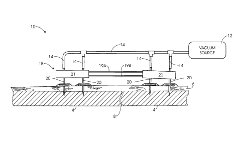
[0008] Figure 1 is a schematic view of an external fixation assembly in
accordance with
the present invention.
[0009] Figure 2A is a cross-sectional view of components used in accordance
with the
present invention, featuring a first embodiment of a fixator pin.
[0010] Figure 2B is a cross-sectional view of components used in accordance
with the
present invention, featuring an implantable pin portion.
-3a-
CA 02669712 2009-05-13
WO 2008/079550 PCT/US2007/084962
[0011] Figure 3 is a cross-sectional view of components used in accordance
with the
present invention, featuring a second embodiment of a fixator pin.
[0012] Figure 4 is an enlarged cross-sectional view of a threaded section of
the fixator pin
of Fig. 3, broken away at one end for clarity.
[0013] Figure 5 is a cross-sectional view of components used in accordance
with the
present invention, featuring a third embodiment of a fixator pin.
[0014] Figure 6 is a cross-sectional view of components used in accordance
with the
present invention, featuring a fourth embodiment of a fixator pin.
[0015] Figures 7A and 7B are cross-sectional view of components used in
accordance with
the present invention, featuring a fourth embodiment of a fixator pin.
[0016] Figure 8 is a cross-sectional view of components used in accordance
with the
present invention, featuring a guide pin used in conjunction with a fixator
pin.
Detailed Description of the Preferred Embodiment
[0017] Referring now to the drawing figures in general, and to Fig. 1
specifically, an
external fixator assembly 10 is shown in accordance with the invention. In
general, the
fixator assembly may include four hollow fixator pins 20 inserted into bone
tissue 4 in the
patient on opposite sides of a fracture or other deformity 8 so that suitable
compression or
distraction forces can be applied. Each fixator pin 20 is positioned at a pin
site and
connected to a source of vacuum pressure 12. Negative or reduced pressure,
e.g., sub-
atmospheric pressure, is applied at each pin site to stimulate blood
circulation to the pin site,
to reduce the potential for inflammation and infection, and to stabilize the
fixator pin. While
each of the fixator pins 20 shown in Fig. 1 is a cannulated pin for supplying
reduced pressure,
other fixator arrangements could be utilized in which one or more fixation
pins are
cannulated while one or more other pins are not cannulated. The non-cannulated
pins may be
used at pin sites where the application of reduced pressure is contra-
indicated or is not
desired or needed.
[0018] Each cannulated fixator pin 20 has a hollow shaft and sidewall 23 that
forms an
-4-
CA 02669712 2009-05-13
WO 2008/079550 PCT/US2007/084962
internal bore 25. The fixator pin 20 may be cannulated from an outer end 24 to
provide an
access port 28 at the outer port end that leads to the internal bore that
extends from the outer
end 24 to the inner or tip end 27 of the pin. To preserve the integrity of the
tip, the bore 25
may terminate before extending through the tip end. The fixator pin 20 is
removably
connected to the source of vacuum pressure 12 by suitable connectors or tubing
14, such as
flexible tubes, removably coupled to the port end 24 of the pin 20. One or
more vent
apertures 34 extend through the sidewall 23 of the fixator pin 20 and
communicate with the
bore in the shaft. The source of vacuum pressure 12 is operable to draw fluid
or gas through
the apertures 34 and bore 25 to create negative pressure at the interface
between the pin and
tissue around the pin.
[0019] Referring now to Figs. 1-2, the external fixator assembly 10 will be
described in
more detail. For purposes of clarity, the fixator assembly 10 is shown in
simplified form with
a fixator device 18 having two fixator pins 20 on each side of a bone fracture
or other
deformity 8. It will be appreciated that more than two fixator pins 20 may be
inserted on
each side of the bone fracture 8, depending on the location and nature of the
fracture. In
addition, it will be appreciated that the fixator assembly 10 is not strictly
intended for bone
fractures, and may be applied to other conditions, including for example,
dislocations and
deformities. The assembly 10 may incorporate a variety of fixator devices, and
the specific
type of fixator is not critical. For example, the fixator assembly 10 may be
used with flexible
or rigid fixators. In addition, the fixator assembly 10 may be applied to
different fracture
types and fracture locations, including for example, femural fractures and
tibial fractures.
[0020] The fixator 18 includes a pair of retainers 21, with each retainer
positioned on one
side of the bone fracture 8. One or more bars connect between the retainers 21
and are
operable to apply compression and distraction forces on the fixator pins. In
Figure 1, the
retainers 21 are connected, for example, by a compression/distraction bar 19A,
19B.
[0021] Referring now to Fig. 2A, a fixator pin 20 is shown having a hollow or
cannulated
shaft 23 with an attachment end 24 (the port end) and an insertion end 26 (the
tip end). The
bore 25 extends through the hollow shaft 23 of the cannulated fixator pin 20
and provides
fluid communication from the attachment end 24 to the insertion end 26. A
vacuum port 28
-5-
CA 02669712 2009-05-13
WO 2008/079550 PCT/US2007/084962
is formed at or through the attachment end 24 of the shaft 23 and is in fluid
communication
with the bore 25. The attachment end 24 is adapted to receive an end of
flexible tubing 14 in
a sealed, snug fit as the tube slides over the attachment end 24 to provide a
fluid flow path to
the vacuum port 28, as shown in Fig. 1. The flexible tubing 14 has an interior
lumen with a
diameter substantially equal to the outer diameter of the fixator pin 20. As
such, the flexible
tubing 14 is configured to slide over the attachment end 24 of the fixator pin
20 and form a
substantially fluid-tight seal. The flexible tube 14 connects the fixator pin
20 with a source
of vacuum pressure 12. A variety of vacuum pressure sources may be used with
the fixator
assembly 10, including, for example a Gast Vacuum pump (Fischer Scientific).
[0022] The pin 20 has a first threaded section 30 that may taper to form a
sharp point or tip
27, and a second non-threaded section 33. The pin 20 may have other
configurations wherein
the tip end does not taper to a point or does not taper at all. The threaded
section 30 is
configured to penetrate into the bone 4 to securely anchor the fixator pin 20
into the pin tract
in the bone 4. For this purpose, the pin may include a self-tapping threaded
tip 27 for tapping
into bone 4. Alternatively, the fixator pin may be provided in the from of a
transfixing pin
420, 520 for positioning through a limb, Figs. 7A and 7B. In such a use, the
pin 420 may
have a threaded middle portion 430 with smooth end portions or the entire pin
520 may be
threaded 530, and the pin 420, 520 may be provided with a Trocar tip. Vent
apertures 435,
535 may be provided in the middle portion of the pin 420, 520 or may be
provided
peripherally, e.g., vent apertures 434, 534.
[0023] As shown schematically in Fig. 2A, the pin 20 screws into the bone 4 to
hold the pin
firmly in place. For this purpose, the pin 20 may be screwed into the bone a
desired depth
greater than that specifically depicted in Fig. 2A so that the non-tapered
portion 33 extends
into the bone to anchor the pin in place. Optionally, a portion of the pin 20,
such as tip 27,
may be detachable to provide an implant that may be left in the patient, as
shown in Fig. 2B.
In such a configuration the pin 20, or the implant portion, e.g. tip 27, may
comprise a bone
substitute material. For example, the pin 20 or tip 27 may comprise a natural,
synthetic, or
natural-synthetic hybrid porous material, and may comprise a material to
support or direct
osteoconduction or a material to induce differentiation of stem cells to
osteogenic cells, i.e.
osteoinductive agents, or materials which provide stem cells, e.g. bone marrow
aspirate.
-6-
CA 02669712 2009-05-13
WO 2008/079550 PCT/US2007/084962
[0024] For example, the pin 20 or tip 27 may be a bioglass, ceramic material,
or other
natural or synthetic porous material, such as calcium sulphate or calcium
phosphate. One
suitable calcium sulphate bone substitute is OSTEOSETO Bone Graft Substitute,
a product
of Wright Medical Technology, Inc. of Arlington TN. Another class of
suitable
materials is one comprising various derivates of calcium phosphate, which can
be used to
provide a structural matrix for osteoconduction, such as hydroxyapatite (coral
based or
chemically derived synthetic ceramic), fluorapatite, tri-calcium phosphate,
bioglass ceramics
and combinations thereof. One suitable calcium phosphate bone substitute is
OsteoGraftTM
Bone Graft Substitute, a product of Millenium Biologix of Kingston, Ontario,
Canada. In
addition, the pin 20 or tip 27 need not comprise a bone substitute material
and may comprise
a metal or other suitable materials.
[0025] In addition, a guide pin 636 may be used in conjunction with the open-
ended fixator
pin 424 to aid in guiding placement of the fixator pin 424, Fig. 8. For
instance, a narrow
guide pin 636 having a cross-sectional dimension less than that of the bore
425 may be
placed in the bone 4 prior to placement of the fixator pin 424, allowing the
physician to first
verify that the guide pin 636 has been placed in the correct location. The
location of the
guide pin 636 may be determined by an x-ray or other suitable imaging
modality. After the
guide pin location has been verified, the fixator pin 424 may be inserted in
the bone 4 by
placing the fixator pin 424 over the guide pin 636 so that the guide pin 636
is located within
the bore 425 of the fixator pin.
[0026] Returning now to Fig. 2A, a plurality of apertures 34 may be formed
through the
non-threaded section 33 of the shaft 23 to form a vent section 35 along the
length of the shaft.
The apertures 34 extend through the wall of the shaft 23. When the vacuum pump
12 is
activated, the vacuum pump draws air or gas through the apertures 34 to create
negative
pressure through and along the apertures. The apertures 34 may be positioned
at various
locations relative to the tip 27 to apply reduced pressure at specific areas
within the pin tract.
For example, the apertures 34 may be positioned where the pin intersects with
the epidermis
("skin/pin interface"), as shown in Fig. 2. In this arrangement, the apertures
may form a
vent section 35 at a location at and above the epidermis to supply negative
pressure through a
reduced-pressure distribution element or screen 50. Alternatively, the
apertures 34 may be
-7-
CA 02669712 2009-09-29
positioned where the pin intersects deeper tissue layers in the dermis 6.
Apertures may be
concentrated at one section of the shaft 23 to treat a specific tissue layer,
or may be formed at
multiple sections of the shaft to supply reduced pressure to multiple layers
or tissues. In Fig.
6, a second embodiment of a fixator pin 320 is shown with apertures 334
positioned at one
section of the shaft to supply reduced pressure at the skin/pin interface and
apertures 335
positioned at another section of the shaft to supply reduced pressure at
deeper tissue layers in
the dermis 6. As shown in Fig. 3, the tip end of the fixator pin 120 may also
include
apertures 134 to supply reduced pressure to bone 4 at the pin/bone interface.
The pins 20,
120, 220, 320, 420, 520 may also be used intermittently or continuously to
effect delivery of
medication, such as antibiotics, local anesthetic, and biopharmaceuticals, to
the various
tissue/pin interfaces by introducing medication into the bore for delivery
through the vent
apertures.
[00271 A fluid-tight enclosure or cover 60, such as OpSite or TEGADERM, is
positioned
over the pin 20 to cover the pin site. The cover 60 is configured to form a
fluid-tight seal
around the pin site to maintain the reduced pressure that is applied at the
tissue/pin interface.
The cover 60 includes an inner face that faces into the pin site, and an outer
face that faces
outwardly and away from the pin site when the cover is placed over the pin 20.
The inner
face may include an adhesive backing 61 that adheres to the patient's skin
around the
periphery 63 of the pin site. Alternatively, or in addition, other adhesives
or sealers may be
applied. The adhesive backing has sufficient adhesive properties to form a
fluid-tight
enclosure around the periphery of the pin site and to hold the cover 60 in
sealed contact with
the patient's skin when reduced pressure is applied beneath the cover. The
cover may be
impermeable or semipermeable depending on the level of permeability needed or
desired for
a particular application as long as the desired level of reduced pressure is
maintained beneath
the cover for a desired amount of time to effect the desired treatment.
[0028] A hole or opening 37 is formed through a central or interior portion of
the cover 60
and is adapted to fit over the attachment end 24 of the fixator pin 20 as the
attachment end 24
of the pin is inserted through the hole 37. The cover 60 engages the outer
circumference of
the pin in a fluid tight seal to substantially prevent leakage of pressure
through the hole
around the pin. Optionally, the cover 60 may incorporate an 0-ring seal 64 at
the hole 37 in
*Trade-mark
-8-
CA 02669712 2009-05-13
WO 2008/079550 PCT/US2007/084962
the cover that is adapted to squeeze around and seal onto the outer periphery
of the pin. The
0-ring 64 engages the exterior of the fixator pin 20 when the cover is placed
over the pin.
The 0-ring 64 has an inner diameter substantially equal to the outer diameter
of the fixator
pin 20 and is configured to frictionally engage the outer surface of the pin.
The 0-ring 64
may be affixed to the cover 60 around the hole 37 by an adhesive or other
bonding.
Alternatively, the 0-ring may be embedded within the cover or heat sealed into
the cover.
For example, the cover 60 may include two plies that form a pocket in which
the 0-ring 64 is
embedded. The frictional engagement between the 0-ring 64 and pin 20 forms a
fluid-tight
seal between the exterior of the pin and the cover.
[0029] It may be desirable to stabilize the 0-ring axially on the pin 20.
Referring to Fig.
2A, the pin 20 includes a pair of circumferential ridges 36 on the outer
section of the pin that
form a seat for the 0-ring 64. The ridges 36 form a narrow groove having a
thickness and
diameter suitable to seat the 0-ring 64. The groove is adapted to receive the
0-ring when the
cover is placed over the pin 20. As a result, the seat formed by the ridges 36
limits the axial
displacement of the 0-ring 64 and cover 60 along the length of the pin 20. The
0-ring may
be formed of any flexible elastomeric material that permits the 0-ring to be
stretched. In this
way, the 0-ring 64 can be stretched to temporarily expand the inside diameter
of the 0-ring
to allow it to be slipped over the top ridge and into the seat allowing the 0-
ring to slide into
and become properly seated within the seat groove.
[0030] A reduced-pressure distribution element such as a porous screen 50 may
surround
the apertures 34 on the fixator pin 20 as shown in Fig. 2A. The screen 50 is
positioned
beneath the cover 60 and over the pin site to help distribute reduced pressure
across its
surface area and to optionally help keep the cover out of direct contact with
the skin around
the pin 20. The screen 50 has sufficient porosity to permit the flow of gases
into the
apertures of the pin when reduced pressure is applied by the vacuum pump. The
screen 50
may also absorb exudate and other liquids that may aspirate from the tissue
around the pin
site. Preferably, the screen 50 is formed out of an open cell polymer foam,
such as
polyurethane foam. Other porous or perforated materials may also be used.
Foams may be
used with a wide range of pore sizes and densities. Since the fixator assembly
10 usually
rests on top of the patient's extremity, it may be desirable to select a light-
weight low density
-9-
CA 02669712 2009-05-13
WO 2008/079550 PCT/US2007/084962
foam or sponge that is less noticeable to the patient. It may optionally be
desirable to form
large perforations or other flow paths in the screen 50 to reduce the weight
of the screen or to
increase the flow of gas drawn by the vacuum pump. In Fig. 2A, the screen 50
and cover 60
are cut to fit over a single pin site. Other screen and cover configurations
may be used,
however, and the configurations illustrated in the drawing figures are not
intended to be the
only workable configurations. For example, it may be desirable to use a single
screen 50 and
cover 60 over multiple pin sites. This may be desirable where pins are spaced
close together
in a relatively small area.
[0031] The fixator assembly 10 may be used in the following manner. After the
pin
locations are selected, small incisions are made through the skin at the pin
locations, and the
fixator pins 20 are placed into the patient's bone. The desired pin location
may include a
fracture or a joint to be immobilized. In such a case where the pin 20 is
inserted in the
fracture or joint, the pin 20 may desirably include an implantable portion
which may
optionally comprises a bone substitute material. The pins 20 are advanced into
the bone until
the pin apertures are positioned at a desired axial locations relative to the
tissue/pin interface.
For example, as shown in Fig. 2A, the apertures 34 may be positioned at the
skin/pin
interface in substantial alignment at, with or above the epidermis 5 or in
communication with
the screen 50. Alternatively, as shown in Fig 6, the apertures 335 may also be
positioned
adjacent to tissue in the dermis 6 either exclusively or in conjunction with
apertures at
another location such apertures 334 at or above the epidermis 5. Apertures may
be provided
at other locations as well. Screens 50 are secured over the pins around the
apertures and over
the incisions. Covers 60 are then placed over the pins 20, and the adhesive
surfaces on the
inner faces of the covers are pressed firmly against the patient's skin to
form a fluid tight
enclosure around the pin sites. Many types of suitable covers may be used. The
fixator 18 is
then assembled and connected with the fixator pins. Once the fixator 18 is
assembled,
flexible tubes 14 are connected to the attachment ends of the fixator pins 20
and to the
suction port of the vacuum pump 12.
[0032] The vacuum pump 12 is activated to apply reduced pressure within the
space 70
beneath the cover 60 as shown in Fig. 2A. The amount of pressure reduction
applied at the
pin sites is dependent on the desired course of treatment, the location of the
pins, the density
-10-
CA 02669712 2009-05-13
WO 2008/079550 PCT/US2007/084962
of the screen material, and other variables. For example, the reduced pressure
may be
between 10 mm Hg below atmospheric pressure and 300 mm Hg below atmospheric
pressure. In the embodiment shown in Fig. 3, reduced pressure is supplied to
the pin/bone
interface at apertures 134 while the cover and optional screen help to
maintain the negative
pressure at that site.
[0033] Thus far, the fixator pins have been described primarily with apertures
that are
positioned to apply reduced pressure at the epidermis and/or dermis. It will
be appreciated
that reduced pressure may be applied at deeper levels in the pin incision and
need not be
limited to the dermis or epidermis. For example, reduced pressure may be
applied, as shown
in Figure 3, at the interface between the fixator pin and bone ("bone/pin
interface") by
apertures 134 positioned at the bone 4 of Figure 3. Application of reduced
pressure in bone
tissue is intended to reduce the occurrence of pin tract infection and
inflammation in the
bone. In addition, the application of reduced pressure in bone tissue is
intended to increase
bone growth and bone ingrowth in the pin tract, which increases stability of
the pin.
[0034] Referring now more specifically to Fig. 3, a third embodiment of a
fixator pin 120 is
shown. The fixator pin 120 is configured to apply reduced pressure at the
bone/pin interface
in a pin tract. The fixator pin 120 is substantially similar to the pins
described above, having
a hollow shaft 131 with a central bore 125, an insertion end 126, a threaded
section 130 on
the insertion end, a non-threaded section 133, and a plurality of apertures
134. The apertures
134 are formed in the threaded section 130 of the insertion end 126 as opposed
to the non-
threaded section 133 of the shaft. In this way, the reduced pressure is
applied through bore
125 to the pin tract inside the bone 4. Referring to Fig. 4, the apertures 134
are preferably
recessed in the groove formed by the thread on the threaded section at the tip
137. The
groove provides additional void space around the apertures to reduce the
potential for
clogging caused by bone fragments that may become lodged in the apertures.
[0035] In some cases, it may be desirable to locate the vacuum port as a side
port on the
side of the pin, rather than at the attachment end. For example, the fixator
appliance may
have retainers that connect over the top of the fixator pins, covering the
attachment ends of
the pins and preventing connection of flexible tubing to the attachment ends.
Therefore,
-11-
CA 02669712 2014-06-03
locating the vacuum port on the side of the pin can avoid problems that occur
when the
attachment end is obstructed or inaccessible. In Fig. 5, a fourth embodiment
of a fixator
pin 220 is shown in accordance with the invention. The fixator pin 220 is
connected to a
retainer 221 that covers the end of the fixator pin. A vacuum port 228 is
formed through
the side wall of the pin 220 and connects with a flexible tube 214. A
cylindrical hub 229
surrounds the vacuum port 228 and projects radially outwardly from the side
wall of the
pin 220. The flexible tube 214 is adapted to slide over the hub 229 to connect
the port
228 to a vacuum pump or other source of reduced pressure. The hub 229 has an
outer
diameter that is substantially equal to the inner diameter of the flexible
tube 214. In this
way, the flexible tube slides over the hub in frictional engagement to form a
fluid-tight
seal around the port 228.
[0036] The terms and expressions which have been employed are used as terms
of
description and not of limitation. There is no intention in the use of such
terms and
expressions of excluding any equivalents of the features shown and described
or portions
thereof. The scope of the claims should not be limited by the preferred
embodiments set
forth herein, but should be given the broadest interpretation consistent with
the
description as a whole.
-12-