Note: Descriptions are shown in the official language in which they were submitted.
CA 02669879 2009-05-15
WO 2008/061542 PCT/EP2006/011148
ION SENSOR FOR FLUID AND METHOD FOR ITS MANUFACTURE
Description
Field of the invention
The invention relates to sensors of charged species in biological, chemical,
industrial or
environmental samples. In particular, the invention relates to a method and a
sensor for
measuring charged species concentrations, in particular ion concentrations,
for example
lithium ion concentrations, in samples, such as blood. The invention also
relates to a method
for the production of such a sensor.
Background and related art
Inorganic ions are an essential requirement for life and are found in large
amounts in drinking
water, blood and every cell of an organism as well as in the environment. For
example, the
concentration of many ions i.e. sodium, potassium, magnesium, and calcium
inside and
outside of cells is essential for any living organism. Consequently, the ion
concentration in the
blood and blood cells of animals and human beings also is of high importance
for a large
variety of body functions.
Normally lithium is a trace element present in the blood plasma, but it is
used as a drug to
treat bipolar mood disorder. It is estimated that worldwide over one million
people take
lithium on a daily basis. A disadvantage in the use of lithium is the very low
therapeutic
index, i.e., the ratio between the toxic concentration and the therapeutic
concentration. Most
patients respond well to a blood plasma concentration of 0.4-1.2 mmol/L
lithium while toxic
effects can occur at a lithium concentration of above 1.6 mmol/L. A prolonged
high blood
lithium level can even result in permanent damage to the nervous system and
even death.
Monitoring of the lithium concentration during treatment is therefore
essential, with regular
checks every couple of months to keep the lithium level at desired level.
1
CA 02669879 2009-05-15
WO 2008/061542 PCT/EP2006/011148
To avoid extensive operator handling, ion-selective electrodes (ISEs) are
routinely used for
measurements of blood parameters in an automated fashion. These ISEs are fast
and offer a
large dynamic range; however, their response is logarithmic and the required
high selectivity
for lithium can be a problem. Additionally, in case of lithium intoxication a
fast procedure for
blood analysis is required. Currently, a venous blood sample must be withdrawn
from the
patient by specially trained personnel and transported to the central
laboratory and the blood
cells need to be removed before the measurement is made. This procedure can
take up to 45
minutes. To minimize sample throughput time and enable measurements on
location,
miniaturized devices employing ion-sensitive field-effect transistors are
available to
i0 determine the concentration of potassium and sodium in whole blood even as
a hand-held
analyzer. However, such analyzers are not used for lithium determination,
because of the high
background concentration of other charged species, in particular sodium ions,
compared to to
the much smaller concentration of lithium ions.
The direct measurement of lithium in whole blood and the determination of
inorganic cations
in blood plasma have been described and demonstrated by E. Vrouwe et al. in
Electrophoresis
2004, 25, 1660-1667 and in Electrophoresis 2005, 26, 3032-3042. Using
microchip capillary
electrophoresis (CE) with defined sample loading and applying the principles
of column
coupling, alkali metals were determined in a drop of whole blood. Blood
collected from a
finger stick was transferred onto the chip without extraction or removal of
components. The
lithium concentration can be determined in the blood plasma from a patient on
lithium therapy
without sample pre-treatment. Using a microchip with conductivity detection, a
detection
limit of 0.1 mmol/L has been obtained for lithium in a 140 mmol/L sodium
matrix.
In these disclosures, the components of the blood sample are separated
electrophoretically
inside a micro-channel. A double T injection geometry is used to select the
ion components
of interest and to guide them to detection electrodes.
In these systems, the sampling loading has to be well defined in order to
ensure the correct
separation of blood plasma components in the double T geometry. In addition,
the double T
geometry is complicated to apply and not well suited for easy to use
applications.
Summary of the invention
2
CA 02669879 2009-05-15
WO 2008/061542 PCT/EP2006/011148
The invention provides a method for the measurement of a concentration of a
charged species
in a sample, the sample having a plurality of types of charged species and at
least one
insoluble component, the method comprising: providing the sample on a surface
of a partly
permeable layer; allowing components of the sample to pass through the partly
permeable
layer into a channel; and separating the components into sections, such that
each at least one
of the sections substantially comprises a single type of the plurality of the
types of charged
species, and determining the charge concentration in the at least one of the
sections.
Thus, the invention provides a method for dividing a sample, in particular a
biological sample
such as blood plasma into sections, each section comprising substantially one
or a one group
of charged species and subsequently determining the concentration of charged
species in this
section.
The invention also provides an apparatus for the measurement of a
concentration of a charged
species in a sample, the sample comprising a plurality of types of charged
species and at least
one insoluble component, the apparatus comprising at least one channel with at
least one
opening, a partly permeable layer covering the at least one opening, at least
two
electrophoresis electrodes arranged along the at least one channel on each
side of the opening,
and at least one sensor for measuring at least one type of charged species in
the at least one
channel.
The method and the apparatus are particularly useful for the measurement of
ion
concentrations of biological samples, for example blood plasma. The ions
measured include
but are not limited to sodium, potassium magnesium, calcium and the like. In
one application
of the invention, the sample may also contain lithium. In this case, the
preferred ion to be
measured is lithium but may be any other ion present in the sample. The
invention is equally
applicable to other charged species such as lipids, DNA or other
polyelectrolytes or electric
charge carrying polymers.
The concentration of a first one of the plurality of type of charged species
may be determined
relative to a second one of the plurality of the types of charged species. The
first type of
charged species may be lithium ions and the second type of charged species
may.be sodium
ions; thus the ratio between lithium and sodium ions in the sample can be
determined.
3
CA 02669879 2009-05-15
WO 2008/061542 PCT/EP2006/011148
The at least one channel may have a single opening covered by a partially
permeable layer.
Using the single opening for sample application, electro-osmotic pressure or
hydrodynamic
pressure and any hydrodynamic flow inside the channel can be advantageously
avoided. In
that way, diffusion is the main or only transport mechanism.
In one embodiment, the at least one channel may have two openings in the
otherwise sealed
channel system. Using hydrodynamic pressure sample injection is realized by
convective flow
form one opening towards the other. In this specific case one opening is
covered with the
sample while the other opening is not.
The partially permeable layer may be a membrane separating the sample from the
at least one
channel. The membrane may be permeable to ions or other charged species while
the
membrane may be impermeable to larger components. In particular, the membrane
may be
impermeable to the insoluble component. The membrane may also be a gas-
permeable
membrane that is impermeable to liquids. The partially permeable layer may be
a separate
layer placed on top or below the at least one opening of the first cover
layer.
A membrane holder may be used on the first cover layer for placing the
membrane on the first
cover layer. The membrane holder may be attached, i.e. glued the first cover
layer or formed
directly in the first cover layer.
The permeable layer may also be a region of the first cover layer that is made
partially
permeable. The permeable layer may comprise at least one region with a
hydrophilic surface.
Additionally, the permeable layer or the first cover layer may comprise at
least one region
with a hydrophobic surface.
The permeable layer may also consist of one or more holes in the channel. The
sample may
thus come into direct contact with a solution inside the channel.
The sample also comprises at least one insoluble component, i.e. in the case
of a biological
sample such as blood, red blood cells, white blood cells, platelets and the
like that are usually
present in the blood. Thus the present invention advantageously allows for the
determination
of an ion concentration in whole blood without prior purification or treatment
thus avoiding
any laboratory pre-treatment of the sample. The invention is therefore
particularly useful for
4
CA 02669879 2009-05-15
WO 2008/061542 PCT/EP2006/011148
the application in patient operated system that do not require a specially
trained physician or
medical care taker.
The at least one sensor comprises one or more pairs of conductivity electrodes
for
determining the charge concentration in the at least one of the sections
substantially
comprising the single type of the plurality of the types of charged species.
For example, a first
pair of conductivity electrodes may be arranged in or nearby the channel at
some distance
from the at least one opening for measuring the concentration of charged
species of a first
polarity. A second pair of conductivity electrodes may be arranged at the
opposite end of the
channel for determining the concentration of a second charged species of
opposite polarity to
the first polarity.
The invention also provides a method for the manufacture of an apparatus for
measuring the
concentration of charged species in a sample, the method comprising providing
a substrate,
forming a channel into the substrate, placing a first cover layer on the
substrate, such that the
first cover layer covers the channel, whereby the first cover layer comprises
at least one
opening providing access to the channel, and placing a partly permeable layer
on the at least
one opening.
Using this method for the production of the apparatus, the partly permeable
layer may be
placed on the at least one opening prior to, after or simultaneously with
placing the first cover
layer on the substrate.
Prior to use of the apparatus, the at least one channel may be filled with an
electrolyte. In one
embodiment the filling of the channel comprises evacuating air and sucking
electrolyte into
the channel. The electrolyte may be filled into the at least one channel prior
to covering the
channel with a second cover layer.
DESCRIPTION OF THE DRAWINGS
The invention may be better understood with respect to the figures and the
detailed
description of preferred embodiments, which is illustrative only and not
limiting to the
invention and wherein:
5
CA 02669879 2009-05-15
WO 2008/061542 PCT/EP2006/011148
Figs. 1 a to 1 d show main components of an apparatus according to the
invention in a top view
and Fig. I e shows a side view of the components of Figs. 1 a to 1 d assembled
to an apparatus
according to the invention.
Fig. 2 shows a section of Fig. l e in greater detail
Figs. 3a to3f show main steps for providing a sample to be measured to the
micro channel in
the enlarged and detailed view of Fig. 2.
Fig. 4a and 4b show an example of an apparatus according to the invention in
top view and in
side view, respectively, Fig 4c and Fig 4d show electrode configurations for
conductivity
detection, both, contactless (Fig 4c) and in contact conductivity detection
(Fig 4c and Fig 4d)
are possible realizations. Fig. 4e shows two possible background measurement
signals at for
example two different measurement temperatures.
Figs. 5a and 5b show alternative embodiments of the present invention and Fig.
5c shows
examples of corresponding measurement signals.
Fig. 6a shows another embodiment of the apparatus with a substantially U-
shaped channel.
Fig. 6b shows a further embodiment with two opening in a single channel.
Fig. 7 shows a further embodiment of the invention with a membrane holder.
Figs. 8a and 8b show an embodiment of the invention with an extra electrode.
Figs. 9a to 9d illustrate a method of the invention in which the fluid is
inserted into the
channel by vacuum.
Fig. 10 shows a further embodiment of the invention in which the fluid is
inserted into the
channel by use of second opening in the channel.
In the figures same reference numerals describe the same or similar objects.
6
CA 02669879 2009-05-15
WO 2008/061542 PCT/EP2006/011148
DETAILED DESCRIPTION OF THE 1NVENTION
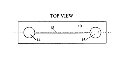
Figs. 1 a to 1 d show the components of an apparatus according to the
invention in a top view.
The apparatus comprises a substrate 10 into which a channel 12 is formed, as
shown in Fig.
1 a. The substrate 10 may be made from glass or plastics material. Any other
material allowing
for the fabrication of channels 12 may be used. In case of glass as the
substrate material, the
channel 12 is etched into the substrate 10 between a first reservoir 14 and a
second reservoir
16 and the side walls of the channel 12 are coated with a polymer. The channel
12 may be of
sub-centimetre dimensions, in particular the channel 12 may be less than 1 cm
in width and
less than 100 m in depth. The first reservoir 14 and the second reservoir 16
may be
considerably larger in size than the width of the channel 12 (e.g. 100 m to 1
cm), but may
have substantially the same depth. The channel 12 and the first reservoir 14
and the second
reservoir 16 may be filled with an electrolyte prior to use. This can be done,
for example, by
evacuating the channel 12, the first reservoir 14 and the second reservoir 16
and then allowing
the electrolyte to be sucked into the channel 12 and the first reservoir 14
and the second
reservoir 16. The first reservoir 14 and the second reservoir 16 can for
example serve for
equilibrating pressure differences to ensure that the channel 12 is always
filled with the
electrolyte.
The channel 12 may also be made of a plurality of nanochannels having a width
of between 1
and 500 nm. The small size of the nanochannels suppresses hydrodynamic and
electro-
osmotic pressure within the channel 12.
The apparatus further comprises a first layer 20 shown in Fig. lb as a cover
layer for covering
in use the substrate 10 and for closing the channel 12 to prevent in use any
fluid like the
electrolyte and the sample inside the channel 12 from evaporation or leaking
out of the
channel 12. The first layer 20 may be made for example, from glass, a
polypropylene film or
hydrophobic membrane, such as those supplied by the Pall Corporation under the
designation
Supor Membrane Disk Fillers (hydrophilic polyether sulfone) or Millipore
Durapor
(polyvinylidene - PVDE) and may have a thickness of less than 1 mm, in
particular less than
1 m. The first layer 20 is non permeable. The first layer 20 provides a first
opening 22 to be
arranged on top of the channel 12 in order to provide access for the sample to
the channel 12.
7
CA 02669879 2009-05-15
WO 2008/061542 PCT/EP2006/011148
The access opening 22 may have the form of a circle but any form suitable for
inserting liquid
into the channel may be used.
In addition, according to the invention a membrane 30 is provided, shown in
Fig. 3c. In the
example shown, the membrane 30 is in use arranged on top or below of the
opening 22 of the
first layer 20. The membrane 30 may be made of a permeable hydrophilic and/or
biocompatible polymer of 1 to 100 m thickness that is semipermeable, for
example,
nitrocellulose. It is possible that the membrane 30 be placed on the channel
12 prior to the
first layer 20. Thus the membrane 30 may also be arranged between the first
layer 20 and the
substrate 10. The membrane 30 may also be integrated into the first layer 20.
In any case, the
membrane 30 is hydrophilic and can be made, for example, from nitrocellulose.
The size and the properties of the membrane 30 may be adapted to allow for
diffusion of
species or transfer of a specified volume of a sample from the sample side to
the inside of the
channel 12 in order to enable comparable measurements.
According to one aspect of the invention, the membrane 30 is permeable to
blood plasma and
its components in the sample but filters out larger insoluble components such
as cell material
in the sample or the like. In this way, cell material like red blood cells,
white blood cells,
platelets or the like are filtered out and only blood plasma enters the
channel 12 for further
examination. Other components may also be filtered out.
According to another aspect of the invention, the membrane 30 is permeable to
charged
species inside the blood plasma and the membrane 30 covered first opening 22
is the only
opening to the channel. It may also be the only opening enabling convective
flow into the
channel 12. In that way convective flow is suppressed and at least the blood
plasma and all
kinds of cell material are prevented from entering the channel while only the
charged species,
in particular the ions diffuse into the channel 12 for further examination.
In a further embodiment of the invention, the membrane 30 and the first layer
20 might be
made in a single step in which the first layer 20 is a polymer film which is
made to act locally
as a membrane or the first layer 20 is a polymer film in which the full
polymer film is a
membrane in which the hydrophobicity is altered. In the latter case, the
hydrophobicity of the
8
CA 02669879 2009-05-15
WO 2008/061542 PCT/EP2006/011148
film is changed such that the film is hydrophilic at the position at which the
sample is to be
injected.
More than one access opening 22 may be made in the first layer 20. This is
useful, for
example, for allowing the sample to enter into the channel 12 at multiple
entry points. This
allows for multiple measurements to be made and averages to be taken. One
further advantage
of more than one access opening 22 is to allow convective flow from one
opening towards
another opening and thus providing an alternative transport mechanism through
the opening
22 into the channel 12.
The membrane 30 can also be provided with microneedles on its surface to
puncture the skin
to obtain the sample more easily. Furthermore the membrane 30 could itself be
punctured to
realize, alter or improve its porosity.
A second polymer film 40 shown in Fig. 1 d is provided for covering the first
layer 20 and the
semipermeable membrane 30 in order to protect the first layer 20 and the
semipermeable
membrane 30 from contamination, to keep them sterile and/or clean prior to use
and to
prevent leakage of fluid from the channel. Should the semipermeable membrane
30 have
microneedles, these microneedles are also protected by the second polymer film
40. The
second polymer film 40 is made of, for example, polypropylene. The second
polymer film 40
may be removed immediately prior to use and a blood sample, i.e. a droplet of
whole blood
may in use be placed on top of the semipermeable membrane 30. The second
polymer film 40
may have a loose end so that it can be easily gripped to be removed prior to
use of the
apparatus 2.
Fig. 1 e shows a side view of the components of Figs. 1 a to 1 d assembled as
an apparatus 2
according to the invention. The first layer 20 is placed in top of the
substrate 10 thus covering
the top side of the channel 12. The first layer 20 has an opening 22 arranged
on top of the
channel 12. The opening 22 is covered by the membrane 30. In the case shown in
Fig. ld the
apparatus 2 is covered by the second polymer layer 40 covering the whole or
part of surface
of the apparatus 2 and thus protecting the apparatus 2 from damage, dust,
evaporation, etc.
The first layer 20 may also include hydrophobic membranes permeable to gas.
The function
of the gas permeable hydrophobic membrane is to prevent over pressure which
might build up
9
CA 02669879 2009-05-15
WO 2008/061542 PCT/EP2006/011148
in the channel 12 as will be explained later. The gas permeable hydrophobic
membrane might
be applied separately but also embedded in the first layer 20.
Fig. 2 shows an exploded view of the area marked by a circle in Fig.le in
greater detail. The
membrane 30 is placed on top of the opening 22 in the first layer 20. The
first layer 20 covers
the channel 12 in the substrate 10 leaving an access to the channel 12 via
opening 22. The
opening 22 is covered by the membrane 30, thus, in use, only components that
can diffuse or
pass otherwise through the membrane 30 can access the channel 12. For
protection and for
preventing unwanted access to or contamination of the membrane 30, the
membrane 30 is
covered by a second polymer film 40. The membrane may be glued or otherwise
fixed on,
under or in the first layer 20. It would be possible to mount the membrane 30
in a holder and
insert this holder in the opening 22 of the first layer 20. An example of a
holder is described
below with respect to Fig. 7.
The channel 12 may be coated with polymers in order to suppress electro
osmosis flow as is
known in the art.
Figs. 3a to 3f show the main steps for providing the sample to be measured to
the channel 12
in the enlarged and detailed view of Fig. 2.
Fig. 3a illustrates a detailed view of the apparatus of Fig. 2, whereby the
channel 12, the
opening 22 and the membrane 30 are filled with a background solution (shown as
grey areas
in the Fig.). For the detection of lithium, the background solution can be a
background
electrolyte (BGE) solution containing for example 50 mmol/L 2-(N-
morpholino)ethanesulfonic acid and 50 mmol/L histidine at pH 6.1. Glucose may
be added,
for example about 200mmol/L for adjusting the osmotic strength of the
background solution.
Other background solutions may be used depending on the charged spieces, i.e.
the ion to be
measured. The second polymer film 40 protects the apparatus 2 and the solution
and prevents
the solution from being contaminated prior to use. Fig. 3a illustrates the
form in which the
apparatus 2 may be shipped to a user.
Fig. 3b shows the removal of the second polymer film 40 prior to use of the
apparatus 2. The
second polymer film 40 serves as a protecting layer for protecting the
membrane 30 and the
first polymer layer 20 during shipping and storage of the apparatus 2. As
shown in Fig. 3b, the
CA 02669879 2009-05-15
WO 2008/061542 PCT/EP2006/011148
second polymer film 40 is removed from the apparatus 2 in order to provide
access for the
sample to the membrane 30. The second polymer film 40 is provided with a quick
release
mechanism, such as a pull-tab, to allow easy removal of at least part of the
second polymer
film 40.
Prior to placing the sample on the membrane illustrated in Fig. 3c, one or
more apparatus
parameters, such as the conductivity of the electrolyte or temperature may be
measured, for
calibration or as a system check. A conductivity measurement of the pure
electrolyte may also
be performed as a system check, i.e. to check that electrolyte is present in
the channels and
that the measurement system is working correctly. It is advisable to flush the
channel 12
electrokinetically prior to carrying out the measurement. This is to get rid
of the first diffused
parts of the sample in the channel 12. The conductivity measurement might be
used for
temperature measurements. The conductivity measurement might also be used as
an internal
check of the condition of the apparatus 2. The later might be realized with
another
temperature measurement method implemented somewhere in or around apparatus 2.
Heating elements may be placed inside or around the channel 12 or around the
apparatus to
alter the temperature of the liquid in the channel 12. The change of
conductivity as a function
of temperature may be used for control or calibration.
In Fig. 3c a sample 50, i.e. an untreated whole blood sample, is placed on the
upper surface of
the membrane 30. The membrane 30 is hydrophilic and permeable. Thus the sample
50 will
be absorbed and pass through the membrane 30 as shown in Fig. 3d, whereby cell
material
such as red blood cells, white blood cells, etc are filtered out. This is done
as the cell material
might break down inside the channel 12 and alter the concentration inside the
channel 12. The
size of the pores of the membrane 30 might also be adjusted to filter out, for
example, lipids
or other larger components so that only electrolytes pass into the channel 12.
Diffusing
through the membrane 30, the filtered sample 50 will come in contact with the
first layer 20
and enter into the opening 22.
As illustrated in Figs. 3d and 3e, the filtered sample 50 diffuses through the
opening 22 into
channel 12 of the substrate 10. The amount of the filtered sample 50 reaching
the channel 12
is determined by the size of the opening 22, the properties of the membrane
30, the properties
of the sample 50 as well as the electrolyte present in channel 12.
11
CA 02669879 2009-05-15
WO 2008/061542 PCT/EP2006/011148
Fig. 3f illustrates how a portion of the filtered sample 50 that diffused into
the channel is
electrophoretically separated in the channel 12 when an electrical field is
applied along the
channel 12. The electrical field will separate all of the charged species in
the filtered sample
and move the charged species towards the reservoirs 14 and 16 at the end of
the channel 12.
Electrodes for providing an electrical field along the channel 12 may be
imbedded or inserted
in the first reservoir 14 and the second reservoir 16. It is also possible
that a plurality of
electrodes are placed along the channel 12 to create extra strong fields at
those locations
where the separation of the ions is necessary, by switching the electric field
from one area to
another. It was explained above that gas permeable hydrophobic membranes are
used in the
apparatus to prevent overpressure off gas. This overpressure may occur at the
electrodes
because of electrolysis.
The measurement may be performed repeatedly.
Fig. 4a and 4b show an example of an apparatus 2 according to the invention in
top view and
in side view, respectively, wherein the first reservoir 14 comprises a first
electrophoresis
electrode 64 and the second reservoir 16 comprises a second electrophoresis
electrode 66. By
applying an electrical voltage to the electrophoresis electrodes 64, 66,
charged particles inside
the channel 12 may be separated or moved along the channel 12. The
electrophoresis
electrodes 64, 66 may be made of any conducting material. Examples of
electrodes used
include, but are not limited to, titanium electrodes with a chrome layer or
silver/silver chloride
electrodes The electrophoresis electrodes 64, 66 can be integrated in the
substrate 10 or may
be otherwise mounted into the reservoirs 14 and 16 or any other place in
channel 12.
In an alternative embodiment, the electrophoresis electrodes 64, 66 and / or
the conductivity
electrodes 72, 74 may be mounted to a measurement device on which the
apparatus 2 can be
mounted for measurement.
The electrodes 72, 74 are not limited to a solely two-way electrode
arrangement but can exist
of multiple electrode arrangement.
12
CA 02669879 2009-05-15
WO 2008/061542 PCT/EP2006/011148
A voltage may be applied to the electrophoresis electrodes 64, 66 by a power
supply or any
means known in the art.
Fig. 4c shows an exploded top view of the area marked by a circle in Fig. 4a
and Fig. 4d
shows an exploded side view of the same area in a side view, as also marked by
a circle in
Fig. 4b. In this area two conductivity electrodes 72 and 74 are provided in
close proximity to
or inside the channel 12 for measuring the conductivity of the fluid across
the channel 12 at
the position of the conductivity electrodes 72, 74. The conductivity
electrodes 72 and 74 may
be integrated in the substrate 10 and at least partially extend into the
channel 12. As shown in
Fig. 4d, the conductivity electrodes 72, 74 may be arranged on the bottom of
the channel 12
but any other position at the channel 12 is possible. The conductivity
electrodes 72, 74 may be
connected to conductivity measurement known in the art.
In one embodiment of the invention, two pairs of conductivity electrodes 72
and 74 are used.
One of the pairs of conductivity electrodes measures positive ions and the
other one of the
pairs of conductivity electrodes measures negative ions in the channel 12. The
two pairs of
conductivity electrodes 72 and 74 are placed on either side of the opening 22
through which
the sample enters into the channel 12.
Placement of the conductivity electrodes 72 and 74 as well as the
electrophoresis electrodes
64, 66 may be carried out during or after the manufacture of the apparatus 2.
For example, the
conductivity electrodes 72 and 74 and the electrophoresis electrodes 64, 66
may be pushed
through the surface of the polymer cover 20 or the substrate 10 into the
channel 12; thus
costly implementation of the conductivity electrodes 72 and 74 and the
electrophoresis
electrodes 64, 66 in the chip can be avoided.
The conductivity in the channel 12 between conductivity electrodes 72 and 74
can be
monitored over time. In case no charged component or an equal distribution of
charged
particles is present inside the channel, for example the BGE solution, a
constant or relatively
slowly varying conductivity will be measured_and monitored as illustrated in
Fig. 4e.
In case of the insertion of charged species, such as ions or the like, into
the channel 12 using
the method described with respect to Fig. 3, the charged species are moved
along the channel
12 by an electric field applied between the electrophoresis electrodes 64 and
66. The charged
13
CA 02669879 2009-05-15
WO 2008/061542 PCT/EP2006/011148
species will be separated electrophoretically while travelling along the
channel 12. For
example, Na-ions of a blood sample 50 will move faster than Li-ion that may
also be present
in the blood sample 50. Thus, two peaks will be measured consecutively by the
conductivity
electrodes 72 and 74. A first peak represents the faster moving Na-ions
passing the
conductivity electrodes 72 and 74 and a second peak represents the slower
moving Li-ions
passing the conductivity electrodes 72 and 74. It is obvious to the person
skilled in the art that
more than two types of ions can be measured and that any charged component
that may be
separated by electrophoresis means can be monitored in that way.
The invention may be applied to measure absolute ion concentrations or for the
measurement
of relative ion concentrations, i.e. for the measurement of Na / Li-
concentration ratios.
Further measurement electrodes or other types of sensors, i.e. optical sensors
such as
fluorescence sensors as known in the art may be added to measure the
concentration or
presence of further species in the sample within the same measurement.
Capacitative sensors
can also be used.
Prior to the measurement of the concentration of the charged species, such as
ions, it is useful
to measure the conductivity of the electrolyte in combination with the
temperature of the
apparatus to ensure that apparatus is performing correctly.
Figs. 5a and 5b show alternative embodiments of the present invention. These
embodiments
may for example be used for calibration purposes.
Fig. 5a shows an apparatus 102 according to the invention and based on the
apparatus 2
described above. In this embodiment of the invention a channel 112 between a
first reservoir
114 and a second reservoir 116 branches into a first channel branch 111 and a
second channel
branch 113. Both the first channel branch 111 and the second channel branch
113 of the
channel 112 are reunited before the second reservoir 116. The first channel
branch 111 is
considerably longer than the second channel branch 113. Both the first channel
branch 111
and the second channel branch 113 have an opening 122 and 123, respectively.
The openings
122 and 123 are each covered with a membrane 130 and 131, respectively.
14
CA 02669879 2009-05-15
WO 2008/061542 PCT/EP2006/011148
If two different samples 150 and 151 are each placed on separate ones of the
membranes 130
and 131 and an electric field is applied along the channel 112, the ions of
each of the samples
will be separated and moved along the channel 112. As the first channel branch
111 is longer
than the second channel branch 113, the charged species, i.e. ions, of the
second sample 151
will arrive first at channel 112 while the charged species of the first sample
150 take
somewhat longer. Thus both of the charged species can be measured
independently one after
the other with the same pair of conductivity electrodes (not shown) resulting
in a signal as
illustrated in the top line of Fig 5c.
This embodiment may also be used for calibration by providing a known sample
150 to
membrane 130 resulting in a corresponding first signal that can be used for
calibration. The
second signal from an unknown sample 151 provided to membrane 131 will arrive
later in
time due to the longer channel branch 111. The strength of the second signal
can than be
compared to the first calibration signal and the concentration of the charged
components in
the unknown sample can be determined as known in the art.
This embodiment might also be used with same sample provided to membrane 130
and to
membrane 131 to realize higher accuracy by for instance averaging.
Fig. 5b shows an alternative embodiment of the invention where two channels
212 and 213
are arranged in parallel. Each of the channels 212 and 213 are basically
identical to the
embodiment of Figs 1- 4 with the advantage that two samples 250 and 251 are
placed in
parallel on membranes 230 and 231, respectively, so that both samples are
measured in
parallel. As both channels 212 and 213 are identical, the measurements can be
compared.
Examples are shown in the lower lines of Fig. 5c
For calibration purposes, one sample, for example a first sample 250 can be a
known sample
with known ion concentrations. Thus the signal of first sample 250 can be used
for calibration
and compared to a signal from a second sample 251 und second channel 213 and
the
concentration of charged particles can be determined in a way known in the
art.
It is obvious, that a plurality of channels can be arranged in parallel, for
example to perform
multiple measurements to accelerate throughput or to increase measurement
statistics.
CA 02669879 2009-05-15
WO 2008/061542 PCT/EP2006/011148
Fig. 6a shows yet another embodiment of an apparatus for the measurement of a
concentration of an ion in a sample wherein a channe1312 is substantially
curved and a first
reservoir 314 comprising a first electrophoresis electrode 364 is place at the
same side of a
substrate as a second reservoir 316 comprising a second electrophoresis
electrode 366.
Contacts for both of the electrophoresis electrodes 364 and 366 may be guided
to the side of
the apparatus for easy contact to the side of the apparatus. In addition the
conductivity
electrodes 372 and 374 are provided in proximity to the second reservoir 366
for measuring
the conductivity of charged component in the channe1312 at this position. The
conductivity
electrodes 372 and 374 may connected via contacts that are arranged at the
same side of the
apparatus or substrate as the contacts for the conductivity electrodes. In
that way, only the
part of the apparatus with the contacts needs to be placed into contact with a
measurement
device and free access to the membrane 330 placed in opening 322 can be
ensured. With such
an apparatus it is possible to have easy access, for example with a finger tip
to the membrane
330, while the apparatus is inserted or contacted to a measurement and/or
control device. The
channel 312 is further straight between the opening 322 and the conductivity
electrodes 372,
374 so that no bending of the channel 312 containing the sample is necessary
which might
influence measurement accuracy or make measurement otherwise difficult.
Fig. 6b shows a modification of the embodiment shown in Fig 6a, further
providing a second
opening 423 in channe1412 that is covered by the same membrane 430 as a first
opening 422.
Thus a sample on membrane 430 will diffuse substantially in the same time
through both of
the openings 422 and 423 in the channe1412. Applying an electrical field to
electrophoresis
electrodes 464 and 466, will, depending on the sign of the voltage, cause for
example the
positively charged species or ions to move into a first channel section 411
towards a second
electrophoresis electrode 466. Similarly negatively charged species are moved
into a channel
section 412 towards a first electrophoresis electrode 464. The conductivity
electrodes 472,
474 and 471, 473 allow for measurement of both of the positively charged
species and the
negatively charged species. Thus the charged species of both electrical
charges can be
measured in parallel.
Fig. 7 shows a modification of the apparatus according to the invention shown
in Fig.2. A
membrane holder 32 is mounted on top of the second layer 20. The membrane 30
is mounted,
for example glued, onto or in the membrane holder 32. Thus the membrane can be
assembled
on the membrane holder before the membrane holder is mounted on the apparatus.
16
CA 02669879 2009-05-15
WO 2008/061542 PCT/EP2006/011148
The membrane holder 32 may be made from plastics material.
In the embodiment shown the membrane holder 32 forms a "cup"-like or a ring
like structure
providing a receiving section for the membrane 30. The upper surface of the
membrane is
substantially planar with the upper rim of the "cup"-like structure of the
membrane holder.
The membrane holder provides thus a frame for the membrane 30 with a well
defined surface
area of the membrane being left for contact with the sample. In that way, the
amount of
sample coming in contact with the membrane can be controlled in a simple and
efficient way,
even when the sample is much bigger, than the membrane.
The walls of the membrane holder may also be higher than the thickness of the
membrane,
thereby providing a "cup"-like or ring-like structure for the sample (not
shown) with the
membrane at the bottom of the "cup". The cup may be used to collect the sample
on the
membrane.
The membrane holder 32 may enable a fast and easy exchange or replacement of
the
membrane 30. By exchanging the membrane 30, the apparatus can be easily
adapted to
different measurements, e.g. by using membranes with different pore sizes, the
size of
components that are filtered out or let into the channel can be adjusted to
the needs of the
particular measurement.
The membrane holder 32 can furthermore enable easy fixation of the membrane 30
on the
first cover layer by for instance a click-and-fix method.
The membrane holder 32 can have the second cover layer 40 on top to prevent
leakage,
evaporation, etcetera. .
Figs. 8a and 8b show an embodiment of the present invention with an additional
anti-tailing
electrode 65 for preventing tailing of the sample or components inside the
channel 12. The
anti-tailing electrode 65 is shown in between first cover layer 20 and
membrane 30. The anti-
tailing electrode 65 may, however, also be arranged differently on the top
side of or at the
opening 22 of first cover layer 20. Fig 8a shows the apparatus with the anti
tailing electrode
65 in the same state as the apparatus shown and described with respect to Fig.
3e. The
17
CA 02669879 2009-05-15
WO 2008/061542 PCT/EP2006/011148
apparatus and the method described with respect to Figs. 1 to 3 apply
accordingly and the
filtered sample may thus diffuse through membrane 30 and first opening 22 into
channel 12 as
described above.
Prior or simultaneously to applying the electrical field along the channel 12
for
electrophoretically separating the portion of the filtered sample illustrated
and described with
respect to Fig. 3f, voltage is applied additionally to anti-tailing electrode
65. Thereby a
portion of the sample component is also driven backwards through the first
opening 22
towards the membrane 30 as indicated by arrow 800 in Fig. 8b. The electrical
field separates
the charged species in the filtered sample and move the charged species
towards the reservoirs
14 and 16 at the end of the channel 12 and towards the membrane 30. Therefore,
no sample
component enters the channel after starting seperation. This effect increases
measurement
accuracy.
The extra electrode 65 may also consist of a plurality of electrodes and might
also be used for
parameter detection prior or during measurement.
Figs. 9a to 9d show how a fluid such as the background electrolyte solution
(BGE) or any
other solution may be inserted into the channel 12 of the apparatus described
with respect to
Figs. 1, to 3 using only one opening 22 in the channel 12. Fig. 9a illustrates
the apparatus of
Fig.2 before any fluid is inserted. A droplet of fluid 14 is put on the
membrane 30 as shown in
Fig. 9b. The fluid 14 will then flow into the membrane 30 until it covers
opening 22 of
channel 12. At this point, illustrated in Fig. 9c, the fluid does not enter by
itself further into
the channel 12 because of the air or gas being inside the channel 12. The air
of gas inside the
channel 12 can only exit the channel 12 through the single opening 22, which
is covered by
fluid 14. Fig. 9d shows that by the application of a vacuum (indicated by
arrow 900) the air or
gas inside the channel 12 can be sucked out of the so that the fluid 14 enters
into the channel
12.
Fig. 10 shows a further method for sampling a fluid such as blood or any other
sample into
the micro-channel 12. A second opening 23 may be provided at some distance of
first opening
22. Both openings 22 and 23 are connected by the channel 12. Preferably, the
channel 12 has
no further openings that said openings 22 and 23 and is otherwise sealed. The
second opening
18
CA 02669879 2009-05-15
WO 2008/061542 PCT/EP2006/011148
23, however, is not covered by a membrane. Fluid inside the channel 12 may
exit through the
second opening 23, when the sample is applied on the first opening 22.
The second opening 23 may be covered by a polymer layer or otherwise closed,
after the fluid
has been filled into the channel 12, to prevent evaporation of the fluid.
During sampling the
second opening 23 has to be connected in any way to air and might not be
covered by the
sample directly.
Connections to the electrodes can be also arranged on one side of the
apparatus allowing for
easy attachment and connection to a measurement device. Easy access is
especially important
when the apparatus is in form of a disposable chip that can be inserted for
one measurement
into a measurement device that may be operated by the patient.
The apparatus 2 can be packaged inside a packaging with suitable interfaces to
allow
connection to electronics for measurement and controls, communications
interfaces and
display interfaces as well as for power electronics.
The openings 22 have been described as being made in the upper surface of the
substrate 10.
However, the openings 22 can also be realized at any other location of the
apparatus 2 for
instance in the side..
The apparatus 2 can be easily used by a patient to measure the concentration
of ions in blood.
For example, for those patients suffering from bipolar mood disorder, the
patient can measure
the concentration of lithium ions in the blood on a regular basis. Should the
concentration go
below a critical level (e.g. 0.4 mmol/L) then the patient can take extra
lithium. Should the
concentration go above a critical level (1.0 mmol/L), then the patient can
stop or lower
medication and if necessary be hospitalised.
The use of the apparatus 2 has been described with respect to the measurement
of lithium
ions. The apparatus 2 could also be used for the measurement of potassium
and/or phosphate
ions to observe the functioning of a kidney or sodium and/or potassium ions to
determine
dehydration.
19
CA 02669879 2009-05-15
WO 2008/061542 PCT/EP2006/011148
The apparatus of the invention has applications outside of the medical field.
For example, it
would be desirable when using the apparatus in the environmental and other
fields to be able
to use the same apparatus over the course of a period of time. In this case,
the apparatus might
be provided with a plurality of openings 22, each of which had its own cover.
The own cover
would be periodically removed from different ones of the plurality of openings
22 to allow
repeated measurements.
The invention has been described with respect to several embodiments. It will,
however, be
clear to those skilled in the art that the invention is not limited thereto.
Rather the cope of the
invention is to be interpreted in conjunction with the following claims.