Note: Descriptions are shown in the official language in which they were submitted.
CA 02670261 2009-05-14
WO 2008/063494 PCT/US2007/023826
APPARATUS AND METHODS OF COMPENSATING FOR ORGAN
DEFORMATION, REGISTRATION OF INTERNAL STRUCTURES TO IMAGES,
AND APPLICATIONS OF SAME
This application is being filed as PCT International Patent application in the
name of
Vanderbilt University, a U.S. national corporation, Applicant for all
countries except the
U.S., and Michael I. Miga, Logan Clements and Robert L. Galloway Jr., each a
U.S.
resident, Applicants for the designation of the U.S. only, on 15 November
2007.
STATEMENT OF FEDERALLY-SPONSORED RESEARCH
The present invention was partially made with Government support awarded by
the
National Institute of Health under Contract Nos. R21 CA91352, 5R33 CA091352-04
and
4R44 CA115263, respectively. The United States Government has certain rights
to this
invention pursuant to these grants.
CROSS-REFERENCE TO RELATED PATENT APPLICATION
This application claims the benefit, pursuant to 35 U.S.C. 119(e), of U.S.
provisional patent application Serial No. 60/859,438, filed November 16, 2006,
entitled
"APPARATUS AND METHODS FOR COMPENSATING FOR ORGAN
DEFORMATION, APPARATUS AND METHODS FOR REGISTRATION OF
INTERNAL STRUCTURES TO IMAGES, AND APPLICATIONS OF SAME," by Michael
I. Miga, Logan Clements and Robert L. Galloway Jr., which is incorporated
herein by
reference in its entirety.
Some references, which may include patents, patent applications and various
publications, are cited and discussed in the description of this invention.
The citation and/or
discussion of such references is provided merely to clarify the description of
the present
invention and is not an admission that any such reference is "prior art" to
the invention
described herein. All references cited and discussed in this specification are
incorporated
herein by reference in their entireties and to the same extent as if each
reference was
individually incorporated by reference. In terms of notation, hereinafter,
"[n]" represents the
nth reference cited in the reference list. For example, [67] represents the
67th reference cited
in the reference list, namely, D. M. Cash, T. K. Sinha,W. C. Chapman, H.
Terawaki, B. M.
1
CA 02670261 2009-05-14
WO 2008/063494 PCT/US2007/023826
Dawant, J. Robert L. Galloway, and M. I. Miga, "Incorporation of a laser range
scanner into
image-guided liver surgery: Surface acquisition, registration, and tracking,"
Med. Phys. 30,
pp. 1671-1682, 2003.
FIELD OF THE INVENTION
The present invention generally relates to image-guided surgery, and in
particular to
apparatus and methods of compensation for soft tissue deformation in relation
to image-
guided surgery in one aspect, and apparatus and methods of compensation for
registration of
internal structures to images in relation to image-guided surgery in another
aspect.
BACKGROUND OF THE INVENTION
Image-guided surgery (IGS) involves patient-specific anatomical images pre-
operatively acquired that spatially localize pathology, digitization
technology that allows the
identification and tracking of targeted points of interest in a patient's
physical space in an
operating room (OR), and alignments of the patient-specific images to the
patient's physical
space in the OR such that the digitization technology can be referenced to the
patient-specific
images and used for guidance during surgery. Central to the IGS is the method
of registering
an image space (a coordinate system corresponding to the pre-operative images)
to a physical
space (a coordinate system corresponding to the intra-operative anatomy of the
patient).
Once the registration is performed, all pre-operative planning and acquired
data related to the
patient's anatomy could be displayed intra-operatively to a surgeon and used
for assistance in
surgical guidance and treatment.
In the recent years, there have been several detailed studies that have
illustrated the
need for soft-tissue deformation correction within image-guided neurosurgery
[1-5]. With
respect to the extracranial environment quantitative data is limited,
nevertheless, there is a
growing acceptance that to translate image-guided interventions to the
extracranial
environment (e.g. abdominal organs such as the liver), the need to correct for
soft tissue
deformation may be essential. While intraoperative magnetic resonance (iMR)
and
computed tomography (iCT) are available, these approaches are somewhat
cumbersome and
are not economically scalable to mid-level medical centers.
On the other hand, hepatic tumors represent a major health care problem in the
U.S.
Along with hepatocellular cancer, many primary neoplasms metastasize to the
liver. In fact,
the most common tumor treated in the liver is metastatic colorectal carcinoma,
a condition
2
CA 02670261 2009-05-14
WO 2008/063494 PCT/US2007/023826
where hepatic metastasectomy can result in long-term survival in properly
selected patients.
This is a frequent problem and the incidence of hepatic resection for
colorectal cancer
metastasis is increasing [6, 7]. For example, it is estimated that 150,000 new
cases of
colorectal cancer will present each year in the United States, from which 50%
will develop
metastatic disease [8]. Of patients with colorectal metastases, 60% will have
hepatic
involvement. Liver metastases occur in many patients with other primary
malignancies (e.g.
breast cancer) and are a frequent cause of cancer-related deaths.
Unfortunately, it is
estimated that there are only 6,000 to 12,000 patients who would be deemed
resectable using
current techniques [9]. These figures do not include the nearly 19,000
patients with primary
intrahepatic malignant tumors [10]. Interestingly, in reviewing the patient
base with
potentially resectable colorectal metastases, the number of candidates who
actually undergo
resection is surprisingly low. This discrepancy may result from many factors,
but is most
likely influenced by the magnitude and complexity of hepatic resections as it
iscurrently
performed. Surgical therapy does improve survival for patients with hepatic
colorectal
metastasis and is largely considered to be more effective than chemotherapy
[8, 9].
In these procedures, a large incision through the abdomen is created to expose
the
anterior surface of the liver. Either wedge or segmental liver resections are
performed to
remove one or more hepatic metastases. In wedge resections, the tumor and a 2-
3cm
surrounding region of the liver is removed, while in segmental resection, an
entire anatomic
segment of the liver is removed. Each of the eight segments of the liver is
supplied by its
own portal venous and hepatic arterial pedicle [11]. Fig. 1 shows the anatomy
of the liver.
Because liver metastases are likely blood-borne via the portal vein, for many
years it
was assumed that there was no advantage in wedge over segmental resection; the
type of
procedure chosen depended greatly on the location and number of metastases
[12].
However, a better understanding of liver anatomy and improvements in
radiologic imaging
and anesthetic techniques has led to the widespread use of segmental liver
resections.
Segmental hepatectomy (i.e. segmentectomy) based on the anatomic descriptions
of
Couinaud is appealing for several reasons. First, blood flow to a segment is
often stopped
prior to transection of the liver. The resultant change of the liver color
indicates the
boundaries of the segment and ensures an adequate margin of normal tissue
throughout the
procedure. Second, segmentectomy may be used to preserve liver substance in
cases that
3
CA 02670261 2009-05-14
WO 2008/063494 PCT/US2007/023826
would otherwise require a resection of the entire right or left lobe. For this
reason, segmental
resection is suitable for the treatment of colorectal liver metastases.
Removal of the tumor
with an adequate margin is sufficient because intrahepatic metastases (i.e.
tumor satellites)
from an established colorectal liver metastasis are rare [13]. Two factors
contribute to
inadequate tumor clearance following non-anatomic wedge resections. First,
traction on the
specimen during division of the liver parenchyma can produce a fracture at the
interface of
the soft liver tissue and the hard colorectal metastasis. Second, lack of
vascular control
during a wedge resection can cause bleeding at the base of the wedge
resection. This
bleeding may obscure the transection and compromise the final margin [14].
When colorectal metastases are confined to the liver, five year survival rates
after
resection range from 30-35%. While morbidity is affected by blood loss, OR
time, and
residual liver volume, the prognosis is dependent on the presence or absence
of adequate
margins, regardless of the type of resection chosen. If positive resection
margins are present
following surgery (i.e., tumor cells are still present in the remaining liver
after the tumor was
excised), five year survival rates range from 0-18%, and few patients remain
disease-free
beyond 20 months. Five year survival rates in patients with negative margins
of less than
1cm range from 1826%, significantly worse than the 44-50% survival rate seen
in patients
with negative margins greater than 1 cm. In further comparing wedge vs.
segmental
resection in one study with over 260 patients, the rate of positive margins
present following
wedge removal was 8 times higher than with segmentectomy [14].
As more liver is removed in a multi-segment or lobectomy procedure, however,
there
are complications. Although liver resection has shown promising survival rates
and a
perioperative mortality rate of less than 5%, a significant increase in
postoperative morbidity
due to hepatic dysfunction and infection has been reported, even by
specialized centers [15-
17]. It was suspected that as more major liver resections were being
conducted, removal of
more significant amounts of liver tissue could cause complications. The
paradigm that at
least one third of healthy liver volume should be left to avoid hepatic
failure following
resection has been a standard for many years, but until recently, data did not
exist to support
this claim. In a study of over 100 patients that studied hepatic dysfunction
and infection after
major liver surgery [18], Schindl et al. used a regression analysis to
demonstrate that
dysfunction increases significantly when the relative residual liver volume
(RLV) was below
4
CA 02670261 2009-05-14
WO 2008/063494 PCT/US2007/023826
26.6%. Body mass index (BMI) was also a highly significant factor; note that
although
prolonged operating time and blood loss were not as significant, they did
improve the
regression model fit. In the 103 patients reviewed for this study, 69%
underwent either a
standard right lobectomy (48) or an extended right lobectomy (23). On average,
these
patients had an RLV value of 33.3%, which is above but within one standard
deviation of the
26.6% cutoff. The authors believe that calculating a specific % RLV before
major liver
surgery from a virtual resection on segmented CT scans provided useful
information for
planning hepatic surgery. They also note that it should not be a barrier to
undertake major
liver resection when the chance for cure outweighs the risks. Interestingly,
most patients
who developed severe postoperative hepatic dysfunction also had additional
predictive
factors, including significant blood loss and long operating time.
Based on this comprehensive study, it is evident that image-guided liver
surgery is
potentially beneficial in several ways. Most importantly, it would allow
surgeons to perform
more specific procedures that they currently only perform in a virtual manner.
By
interactively viewing the accurate location of vasculature and tissue
surrounding the tumor as
they operate, surgeons can more confidently perform segmental and wedge
resections and
avoid the removal of extraneous healthy liver volume. Segment-oriented
anatomical
resection will be more hemostatic as surgeons will be more specific in the
control of blood
vessels due to enhanced anatomical information. Finally, as surgeons become
more efficient
at using 3-D imaging interactively during surgery, operating time will
potentially decrease.
Through an efficacy clinical trial to be conducted by PTI, IGLS will
potentially be shown to
positively affect the ability to perform specific operations with optimized
RLV, decreased
blood loss, and decreased overall operating time. These factors impact hepatic
dysfunction
following major liver transection and can be improved by more specific surgery
as provided
using IGLS.
It is known that determination of an accurate image-to-physical space
registration is a
fundamental step in providing accurate guidance information to surgeons via
image-guided
surgery. Since the use of rigid, point-based landmarks is not feasible in
image-guided liver
surgery (IGLS) applications, surface-based techniques were proposed to
determine the
registration between the preoperative images and the intraoperative patient
space.
Specifically, the iterative closest point (ICP) algorithm, developed by Besl
and McKay, has
5
CA 02670261 2009-05-14
WO 2008/063494 PCT/US2007/023826
traditionally been used to determine the transformation between the image
space surface of
the liver, derived from preoperative image segmentations, and the
intraoperative liver
surface. Intraoperative data were initially acquired using an optically
tracked probe while
more recent efforts have utilized laser range scanner (LRS) technology to
provide spatially
dense, textured delineations. The current protocol for the performance of a
surface-based
image-to-physical space registration first involves the selection anatomical
fiducial points in
the preoperative images sets prior to surgery. The homologous physical space
location of
these anatomical fiducials is then digitized during the surgery such that a
point-based initial
alignment registration can be performed. The point-based registration serves
to provide a
reasonable initial pose for the ICP algorithm. Being that the surface
alignment provided by
the ICP algorithm is highly dependent on the initial pose of the surfaces,
gross errors in the
initial alignment provided by the point-based registration can result in
erroneous surface
alignments. A failed surface-based registration not only compromises the
guidance
information relayed to the surgeon but also impairs deformation correction
efforts due to
inaccurate surface displacement data that are used to drive mathematical
models. In IGLS,
the quality of the initial alignment registration can be compromised by the
large fiducial
localization errors (FLE) inherent in using anatomical landmarks that undergo
non-rigid
deformation relative to the preoperative images. Additionally, gravity and the
effects of the
liver mobilization and packing performed prior to open liver resections can
lead to liver
deformations that compromise the results of a rigid ICP surface registration.
Clinical data
shows a poor initial alignment registration, due to high FLE of the anatomical
fiducials, and
large liver deformations resulted in the convergence of the rigid ICP
algorithm to a gross
misalignment.
Therefore, a heretofore unaddressed need exists in the art to address the
aforementioned deficiencies and inadequacies.
SUMMARY OF THE INVENTION
The present invention provides apparatus and methods of compensation for intra-
operative organ shift of a living subject, being a human being or animal,
which are cost-
effective, clinically translatable, scalable to medical centers and
facilities, and tractable. The
present invention also provides apparatus and methods of surface registration
(increasing the
percentage of correct registrations) and allows registrations in situations
where only a
6
CA 02670261 2009-05-14
WO 2008/063494 PCT/US2007/023826
fraction of the surface may be seen.
In one aspect, the present invention relates to a method of compensation for
intraoperative deformations of an organ of interest of a living subject. The
organ of interest
of the living subject can be a liver, heart, kidney, lung, stomach, brain, or
soft tissues. In one
embodiment, the method includes the steps of:
a. preoperatively acquiring an image of the organ of interest of the living
subject;
b. segmenting the preoperatively acquired image;
c. tessellating the segmented image to obtain a three-dimensional (3D) surface
of
the organ of interest;
d. generating a tetrahedral volumetric mesh from the tessellated 3D surface of
the organ of interest;
e. modeling deformations of the organ of interest from the generated
tetrahedral
volumetric mesh with a finite element model (FEM) having a set of mesh
nodes;
f. intraoperatively acquiring range scan data of the organ of interest,
wherein the
range scan data is associated with the intraoperatively deformed surface of
the
organ of interest;
g. registering the intraoperatively acquired range scan data to the
tessellated 3D
surface of the organ of interest by weighting regions of the intraoperatively
acquired range scan data that are minimally deformed, wherein closest point
distances between mesh nodes of interest and the intraoperatively deformed
surface are calculated;
h. constructing boundary conditions from a preoperatively surgical plan and
the
registered intraoperative range scan data, wherein the boundary conditions
comprise initial deformations of the organ of interest associated with the set
of
mesh nodes;
i. obtaining model solutions of the FEM corresponding to the boundary
conditions iteratively in an incremental fashion, wherein fractions of the
closest point distances are used to prescribe a displacement boundary
condition on the mesh nodes;
7
CA 02670261 2009-05-14
WO 2008/063494 PCT/US2007/023826
j. updating the locations of the mesh nodes by corresponding model solutions
of
the FEM, which triggers calculation of new registrations and boundary
condition values; and
k. repeating steps (i) and (j) until the root mean square (RMS) closest point
distances for all mesh nodes have reached a predetermined value.
The model solutions of the FEM are used to align the preoperatively acquired
image
to the intraoperatively acquired range scan data for compensating for
deformations of the
organ of interest of the living subject.
The step of intraoperatively acquiring range scan data of the organ of
interest is
performed by swabbing an optical stylus on the surface of the organ of
interest or by
scanning the surface of the organ of interest.
In one embodiment, the preoperatively acquired image of the organ of interest
comprises a computer tomography (CT) image, a positron emission tomography
(PET)
image, a magnetic resonance (MR) image, or a functional magnetic resonance
(fMR) image.
The tessellated 3D surface of the organ of interest is represented as a set of
polygons and
serves as input for generating a tetrahedral volumetric mesh.
In another aspect, the present invention relates to a method of compensation
for
intraoperative deformations of an organ of interest of a living subject. In
one embodiment,
the method includes the steps of:
a. preoperatively acquiring an image of the organ of interest of the living
subject;
b. intraoperatively acquiring an image of the organ of interest of the living
subject;
c. generating a first geometric surface of the organ of interest from the
intraoperatively acquired image of the organ of interest;
d. constructing an atlas, [A], of deformations of the organ of interest from
the
pre-operatively acquired image of the organ of interest, wherein the atlas [A]
is in the form of an n x m matrix with n, m being positive integers;
e. generating a second geometric surface of the organ of interest from the
constructed atlas [A] of deformations of the organ of interest;
f. aligning the second geometric surface of the organ of interest to the first
8
CA 02670261 2009-05-14
WO 2008/063494 PCT/US2007/023826
geometric surface of the organ of interest to determine at least one
difference
between a point of the first geometric surface and a corresponding point of
the
second geometric surface of the organ of interest;
g. feeding back the at least one difference to step (e) to generate a new
second
geometric surface of the organ from the atlas [A] of organ deformations of the
organ of interest;
h. iteratively repeating steps (e)-(g) for a predetermined number of
iterations or
until an error tolerance related to the at least one difference between a
point of
the first geometric surface and a corresponding point of the second geometric
surface of the organ of the living subject is no greater than a predetermined
threshold; and
i. compensating for the intraoperative organ deformation.
In one embodiment, the pre-operatively acquired organ images of the living
subject
comprise image data with respect to the organ surface geometry. The image data
with
respect to the organ surface geometry is obtained through the use of at least
one of positron
emission tomography device, electroencephalography device, computer tomography
device,
functional magnetic resonance imaging device, magnetic resonance imaging
device, and
ultrasound imaging device.
In one embodiment, the step of constructing the atlas [A] of deformations of
the
organ of interest comprises the steps of generating a geometric volume of the
organ of
interest from the preoperatively acquired image; modeling deformations of the
organ of
interest based on the geometric volume of the organ of interest with a
computational model;
obtaining m model solutions of the computational model corresponding to the
geometric
volume of the organ of interest, wherein each model solution, A, represents a
solution of
deformation for n variables and corresponding to a set of parameters; and
generating the atlas
[A] in the form of an n x m matrix with each model solution, A, which is in
the form of a
n x 1 matrix, forming a column of the matrix.
The geometric volume of the organ of interest is generated through
segmentation of
the pre-operatively acquired image of the organ of interest, and represented
by a tetrahedral
mesh with n surface nodes.
In one embodiment, the model solutions are obtained by solving at least one
partial
9
CA 02670261 2009-05-14
WO 2008/063494 PCT/US2007/023826
differential equation modeled to represent the relationship between a
deformation of the
organ of interest and at least one force causing the deformation, and wherein
the at least one
partial differential equation is solved with boundary conditions corresponding
to specific
structures of the organ of interest, body force, material properties,
vascularization,
physiological changes related to tissue of the organ of the living subject,
physical conditions
or any combinations of them. The at least one partial differential equation is
solved with the
boundary conditions iteratively.
In one embodiment, the step of intra-operatively acquiring the image of the
organ of
the living subject is performed with an optical device that is capable of
obtaining frequency,
intensity and geometric data with respect to the surface of the organ of
interest of the living
subject simultaneously. The optical device is a laser range scanner, or an
optical stylus. In
another embodiment, the step of intra-operatively acquiring the image of the
organ of interest
of the living subject is performed with stereo pair technology.
In one embodiment, the step of aligning the second geometric surface of the
organ of
interest to the first geometric surface of the organ of interest of the living
subject is
performed with a point-based registration, or a weighted point-based
registration, where the
weighted point-based registration comprises the step of using a salient-
feature weighted
correspondence, and wherein when the organ is a liver, the salient-feature
comprises a
falciform ligament region.
In another embodiment, the step of aligning the second geometric surface of
the organ
of interest to the first geometric surface of the organ of interest of the
living subject is
performed with a registration that provides a surface-to-surface
correspondence using at least
one characteristic feature of the organ of interest of the living subject.
In yet another aspect, the present invention relates to a method of
compensation for
intraoperative deformations of an organ of interest of a living subject. In
one embodiment,
the method has the steps of generating a first geometric surface of the organ
of interest of the
living subject from an intraoperatively acquired image of the organ of
interest of the living
subject; constructing an atlas of deformations of the organ of interest of the
living subject
from a preoperatively acquired organ image; generating a second geometric
surface of the
organ of interest of the living subject from the atlas of deformations;
aligning the second
geometric surface of the organ of interest to the first geometric surface of
the organ of
CA 02670261 2009-05-14
WO 2008/063494 PCT/US2007/023826
interest of the living subject to determine at least one difference between a
point of the first
geometric surface and a corresponding point of the second geometric surface of
the organ of
the living subject, which is related to organ deformation; and compensating
for the intra-
operative organ deformation.
In one embodiment, the intraoperative deformations of the organ of interest
are
corresponding to distributed loading conditions that are associated with
gravity, edema-
induced swelling, mannitoi-induced shrinking, or any combination of them. In
another
embodiment, the intraoperative deformations of the organ of interest are
corresponding to
surface-based loading conditions that are associated with tissue retraction,
tissue resection, or
any combination of them.
In a further aspect, the present invention relates to a system of compensation
for
intraoperative deformations of an organ of interest of a living subject. The
organ of interest
of the living subject is a liver, heart, kidney, lung, stomach, brain, or soft
tissues. In one
embodiment, the system has a first imaging acquiring device for pre-
operatively acquiring an
image of the organ of interest of the living subject; a second imaging
acquiring device for
intra-operatively acquiring an image of an organ of interest of the living
subject; and at least
one computer coupled with the first image acquiring device and the second
image acquiring
device and adapted for performing the steps of:
i). generating a first geometric surface of the organ of interest from the
intraoperatively acquired image of the organ of interest of the living
subject;
ii). constructing an atlas of deformations of the organ of interest from the
preoperatively acquired organ image;
iii). generating a second geometric surface of the organ of interest from the
atlas
of deformations;
iv). aligning the second geometric surface of the organ of interest to the
first
geometric surface of the organ of interest of the living subject to determine
at
least one difference between a point of the first geometric surface and a
corresponding point of the second geometric surface of the organ of interest
of
the living subject, which is related to organ deformation; and
v). compensating for the intraoperative organ deformation.
The system further has a display device coupled to the at least one computer
for
11
CA 02670261 2009-05-14
WO 2008/063494 PCT/US2007/023826
displaying the deformation of the organ of interest dynamically to facilitate
a diagnostic or
surgical procedure.
In one embodiment, the first imaging acquiring device comprises at least one
of
positron emission tomography device, electroencephalography device, computer
tomography
device, functional magnetic resonance imaging device, magnetic resonance
imaging device,
and ultrasound imaging device. The second imaging acquiring device comprises a
laser
range scanner that is capable of obtaining frequency, intensity and geometric
data with
respect to the cortical surface of the living subject simultaneously. The
second imaging
acquiring device can also be an ultrasound imaging device, or an optical
stylus.
In yet a further aspect, the present invention relates to a method of surface
registration
in image guided surgery. In one embodiment, the method includes the steps of:
a. preoperatively acquiring an image of an organ of interest of a living
subject;
b. generating a first surface of the organ of interest of the living subject
from the
preoperatively acquired image;
c. designating a set of target points, T={tn}, n= 1, 2, 3, ..., NT, from the
first
surface of the organ of interest, wherein the set of target points T contains
one
or more patch points corresponding to homologous anatomical features of the
first surface of the organ of interest, and wherein each of the set of target
points tn, is indexed with a binary index, pTm, such that when pTn, = 0, the
target point tm is a non-patch point, and when pTm = 1, the target point tm is
a
patch point;
d. intraoperatively acquiring an image of the organ of interest of the living
subject;
e. generating a second surface of the organ of interest of the living subject
from
the intraoperatively acquired image;
f. designating a set of source points, S={sm}, m = 1, 2, 3, ..., Ns, from the
second surface of the organ of interest, wherein the set of source points S
contain one or more patch points corresponding to homologous anatomical
features of the second surface of the organ of interest, which is related to
the
corresponding homologous anatomical features of the first surface of the
organ of interest, and wherein each of the set of source points sm is indexed
12
CA 02670261 2009-05-14
WO 2008/063494 PCT/US2007/023826
with a binary index, psm, such that when p s m = 0, the source point sm is a
non-
patch point, and when psm = 1, the source point sm is a patch point;
g. determining a point correspondence between the set of source points S and
the
set of target points T, whereby a closest point operator, C={Cm} is
determined;
h. biasing the point correspondence between the one or more source patch
points
and the one or more target patch points by a weighting factor, wpc, so as to
bias the closest point operator Cm, via the following relationship:
_
WPC Sm - tn 11 lf pS m - pT n - 1
dm.n
tilSm - tn 11 otherwise
wherein 0 < wpc << 1, dm, is Euclidian distance between a source point sm
and a target point t,,;
i. aligning the second surface of the organ of interest to the first surface
of the
organ of interest using a weighted point based registration (PBR) between the
set of source points S and the set of target points T by finding a rigid-body
transformation (SZ ) between the set of source points and the set of target
points
that minimizes an objective function of:
NM
J l~! E Wm Cm(Sm~T)-n(Sm~Z ~
m=1
wherein {wm} is a set of weights letting wm = 1 for psm = 0 and wm = WPBR for
p Sm = 1, and wherein WPBR is a function of iteration, i, in the form of:
WPBR \t/ = WPBR,base + [WPBR,max - WPBR,base ] exp[- a(i - 1)],
wherein i = 1, 2, 3, ...N, N being the number of iteration, WPBR,,,,ax is a
maximum patch weight factor and corresponds a patch weight at a first
iteration, WpBR,base is a baseline patch weight factor, with 1<_WpBR,base ~
WPBR,max5 and a is a relaxation constant with 0<_cx <_1, wherein the aligned
second surface of the organ of interest up; and
j. repeating steps (f)-(i) using the aligned second surface of the organ of
interest
until the root mean square (RMS) closest point distances between the source
and target points have reached a predetermined value.
13
CA 02670261 2009-05-14
WO 2008/063494 PCT/US2007/023826
In one embodiment, the pre-operatively acquired image of the organ of interest
of the
living subject comprises image data with respect to the surface geometry of
the organ of
interest, where the image data with respect to the surface geometry of the
organ of interest is
obtained through the use of at least one of positron emission tomography
device,
electroencephalography device, computer tomography device, functional magnetic
resonance
imaging device, magnetic resonance imaging device, and ultrasound imaging
device.
In one embodiment, the step of generating a first surface of the organ of
interest of
the living subject comprises the step of extracting details of the first
surface using at least one
of the following features: user identification of surface features or visible
structures; surface
curvature; surface shape; surface orientation within the living subject;
distribution of surface
normal orientation; and potential for deformation of the anatomic structure.
In one embodiment, the step of intra-operatively acquiring images of the organ
of
interest of the living subject is performed with one of the following methods:
swabbing the
surface of the anatomic structure with a tracked instrument; laser range
scanning of the
anatomic structure for surface determination; intra-operative ultrasound
scanning of the
anatomic structure; imaging of the anatomic structure using a tracked
laparoscope or
endoscope; and surface extraction of the anatomic structure from a binocular
scene such as
provided by a binocular laparoscope or endoscope.
In one aspect, the present invention relates to a method of surface
registration in
image guided surgery. In one embodiment, the method includes the steps of:
a. pre-operatively acquiring an image of an anatomic structure of a living
subject;
b. generating a first surface of the anatomic structure of the living subject
from
the preoperatively acquired image of the anatomic structure of the living
subj ect;
c. constructing one or more first patches corresponding to the first surface
of the
anatomic structure, wherein each first patch contains a surface subset of
physical space data related to a salient anatomical feature of the first
surface
of the anatomic structure;
d. intra-operatively acquiring an image of the anatomic structure of the
living
subject;
14
CA 02670261 2009-05-14
WO 2008/063494 PCT/US2007/023826
e. generating a second surface of the anatomic structure of the living subject
from the intraoperatively acquired image of the anatomic structure of the
living subject;
f. constructing one or more second patches corresponding to the second surface
of the anatomic structure, wherein each second patch contains a surface subset
of physical space data from the second surface related to a corresponding
salient anatomical feature of the first surface of the anatomic structure;
g. aligning the one or more second patches to the corresponding one or more
first patches, wherein each second patch is dynamically biased to a
corresponding first patch; and
h. completing a registration of the second surface to the first surface with
physical space data not contained in the first and second patches to indicate
surgical position in both image space and physical space.
The anatomic structure of the living subject comprises a liver, heart, kidney,
lung,
stomach, or brain, where when the anatomic structure comprises a liver, the
salient-feature
comprises a falciform ligament region.
In one embodiment, the pre-operatively acquired images of the anatomic
structure
comprise image data with respect to the surface geometry of the anatomic
structure, where
the image data with respect to the surface geometry of the anatomic structure
is obtained
through the use of at least one of positron emission tomography device,
electroencephalography device, computer tomography device, functional magnetic
resonance
imaging device, magnetic resonance imaging device, and ultrasound imaging
device.
In one embodiment, the step of generating a first surface of the anatomic
structure of
the living subject comprises the step of extracting details of the first
surface using at least one
of the following features: user identification of surface features or visible
structures; surface
curvature; surface shape; surface orientation within the living subject;
distribution of surface
normal orientation; and potential for deformation of the anatomic structure.
The step of intra-operatively acquiring images of the anatomic structure of
the living
subject is performed with one of the following methods: swabbing the surface
of the
anatomic structure with a tracked instrument; laser range scanning of the
anatomic structure
for surface determination; intra-operative ultrasound scanning of the anatomic
structure;
CA 02670261 2009-05-14
WO 2008/063494 PCT/US2007/023826
imaging of the anatomic structure using a tracked laparoscope or endoscope;
and surface
extraction of the anatomic structure from a binocular scene such as provided
by a binocular
laparoscope or endoscope.
The step of aligning the one or more second patches to the corresponding one
or more
first patches comprises the step of giving more weight to the patches than to
the rest of the
surfaces.
The step of completing a registration of the second surface to the first
surface with
physical space data not contained in the first and second patches comprises
the step of
increasing weight to the rest of the surfaces when final adjustments in the
registration are
made with all available surface information.
The step of completing a registration of the second surface to the first
surface with
physical space data not contained in the first and second patches is performed
with a point-
based registration, where the point-based registration comprises a weighted
point-based
registration, and wherein the weighted point-based registration comprises the
step of using a
salient-feature weighted correspondence.
In another aspect, the present invention relates to a method of surface
registration in
image guided surgery. In one embodiment, the method has the steps of
generating a first
surface of an anatomic structure of a living subject from pre-operatively
acquired images of
the anatomic structure of the living subject; generating a second surface of
the anatomic
structure of the living subject from intra-operatively acquired images of the
anatomic
structure of the living subject; constructing one or more second patches
corresponding to the
second surface of the anatomic structure, wherein each second patch contains a
surface
subset of physical space data from the second surface related to a
corresponding salient
anatomical feature of the first surface of the anatomic structure; aligning
the one or more
second patches to the corresponding one or more first patches, wherein each
second patch is
dynamically biased to a corresponding first patch; and completing a
registration of the
second surface to the first surface with physical space data not contained in
the first and
second patches to indicate surgical position in both image space and physical
space.
In one embodiment, the step of completing a registration of the second surface
to the
first surface with physical space data not contained in the first and second
patches is
performed with a point-based registration, where the point-based registration
comprises a
16
CA 02670261 2009-05-14
WO 2008/063494 PCT/US2007/023826
weighted point-based registration, and wherein the weighted point-based
registration
comprises the step of using a salient-feature weighted correspondence.
In yet another aspect, the present invention relates to a system of surface
registration
in image guided surgery. In one embodiment, the system has a first imaging
acquiring
device for pre-operatively acquiring images of an anatomic structure of a
living subject; a
second imaging acquiring device for intra-operatively acquiring images of at
least part of the
anatomic structure of a living subject; and at least one computer coupled with
the first image
acquiring device and the second image acquiring device and adapted for
performing the steps
of:
i). generating a first surface of the anatomic structure of the living subject
from
the pre-operatively acquired images of the anatomic structure of the living
subject;
ii). constructing one or more first patches corresponding to the first surface
of the
anatomic structure, wherein each first patch contains a surface subset of
physical space data related to a salient anatomical feature of the first
surface
of the anatomic structure;
iii). generating a second surface of the anatomic structure of the living
subject
from the intra-operatively acquired images of the anatomic structure of the
living subject;
iv). constructing one or more second patches corresponding to the second
surface
of the anatomic structure, wherein each second patch contains a surface subset
of physical space data from the second surface related to a corresponding
salient anatomical feature of the first surface of the anatomic structure;
v). aligning the one or more second patches to the corresponding one or more
first patches, wherein each second patch is dynamically biased to a
corresponding first patch; and
vi). completing a registration of the second surface to the first surface with
physical space data not contained in the first and second patches to indicate
surgical position in both image space and physical space.
In one embodiment, the system further comprising a display device coupled to
the at
least one computer for displaying the registration dynamically to facilitate a
diagnostic or
17
CA 02670261 2009-05-14
WO 2008/063494 PCT/US2007/023826
surgical procedure.
In one embodiment, the first imaging acquiring device comprises at least one
of
positron emission tomography device, electroencephalography device, computer
tomography
device, functional magnetic resonance imaging device, magnetic resonance
imaging device,
and ultrasound imaging device. In one embodiment, the second imaging acquiring
device
includes a laser range scanner that is capable of obtaining frequency,
intensity and geometric
data with respect to the cortical surface of the living subject
simultaneously. In another
embodiment, the second imaging acquiring device includes an ultrasound imaging
device. In
an alternative embodiment, the second imaging acquiring device comprises a
tracked
instrument, an optical stylus, a tracked laparoscope or endoscope, or a
binocular laparoscope
or endoscope.
These and other aspects of the present invention will become apparent from the
following description of the preferred embodiment taken in conjunction with
the following
drawings, although variations and modifications therein may be affected
without departing
from the spirit and scope of the novel concepts of the disclosure.
BRIEF DESCRIPTION OF THE DRAWINGS
The patent or application file contains at least one drawing executed in
color. Copies
of this patent or patent application publication with color drawing(s) will be
provided by the
Patent and Trademark Office upon request and payment of the necessary fee.
Fig. 1 shows anatomy of a liver of a living subject, with eight Couinaud
segments and
key ligaments labeled, where segment I is located posterior to the liver.
Fig. 2 shows (a) a digital photograph taken of the OR scene from the view of
the
range scanner; (b) range scanner setup in the OR; and (c) laser range scanner
output showing
a textured point cloud from the liver surface.
Fig. 3 shows physical-to-image registration results for four surgical cases
using the
LRS.
Fig. 4 shows a snapshot of a graphical user interface to facilitate the
segmentation of
the liver from tomographic images.
Fig. 5 shows (a) a liver phantom deformed by an anterior translation of the
medial
lobe, and (b) an iterative closest point registration between 2 surfaces
(surfaces derived from
taken by CT).
18
CA 02670261 2009-05-14
WO 2008/063494 PCT/US2007/023826
Fig. 6 shows (a) surface overlay of registered LRS point cloud and segmented
preoperative CT surface (grey), (b) preoperative CT slice with contour overlay
of registered
LRS point cloud (arrow indicates approximate 1cm difference between surfaces),
and (c)
overlay of deformed liver onto combined preoperative-LRS data tomogram show in
Fig. 7b.
Fig. 7 shows an incremental strategy, (a) a portion of the closest point
distance on a
boundary is prescribed in a normal-to-the-boundary direction while allowing
point to have
lateral freedom along surface, and (b) visualized in three-dimensions.
Fig. 8 shows (a) an application of closest point distance as Cartesian
displacement
vector, and (b) incremental application with normal displacement but free
lateral shift along
the surface.
Fig. 9 shows results of model-updating approach for 4 clinical cases with:
(red)
deformed image volume, (CT) preoperative scan, and (white contour),
intraoperatively
acquired LRS scan.
Fig. 10 shows clinical results showing visualizations of (a), (b) ICP
registration, and
(c), (d) the salient-feature weighted registration, where Falciform regions
are highlighted.
Fig. 11 shows (a) and (b) two clinical cases showing LRS-to-preoperative image
registration, and (c) and (d) mapped closest point distance (color bar range
is 0-16 mm for
both (c), (d)).
Fig. 12 shows (a) closest point distances from undeformed and deformed
surfaces for
deformation 1, and (b) deformation 2, and (c) and (d) same after ICAt
registration, where
color bar range for (a)-(d) is from 0 to 20, 18, 8, 6 mm, respectively.
Fig. 13 shows (a) microCT of gel-tissue, (b) FEM mesh, (c) mechanical testing
of
gel-tissue, and gel control, and (d) indentation of a mouse liver.
Fig. 14 shows a segmented CT surface of the liver phantom with subsurface
tumors,
where the labels for each tumor is used as a reference for the results.
Fig. 15 shows corresponding CT slices from (a) nondeformed and (b) deformed
image. The plastic object placed below the liver produces the deformation
observed in Fig.
6a. The height of the object is approximately 38 mm.
Fig. 16 shows a histogram of signed distance distribution for rigid alignments
between a nondeformed surface and the surface deformed at (a) the left lobe
and (b) the
inferior ridge. The solid line indicates that the alignment is performed by
registering the
19
CA 02670261 2009-05-14
WO 2008/063494 PCT/US2007/023826
entire surface with the ICP algorithm. The dashed line is acquired with the
same registration
method, but this time only regions of the surface that were visually
identified as "minimally
deformed" are used in the registration.
Fig. 17 shows an example set of boundary conditions for the liver phantom
mesh.
The dark grey represents the boundary nodes that obey the mixed "closest
point" boundary
condition formulation. The medium grey value denotes fixed regions, while the
light grey
boundary conditions are stress free.
Fig. 18 shows implementation of "closest point" boundary conditions using
norrnal
tangential space. The closes point distance for the boundary node at increment
1, Ild, = dcp,,
is determined. From dcp,;, a normal distance, 11h, II =a dcp,;, is computed
for use in the "closest
point" boundary condition where a is the solution scale fraction that scales
the closest point
distance in the incremental framework. The new position of the boundary node,
denoted by
the small circle, lies on a plane that is a distance of a dcp; along the
direction en, away from
its original location. The mixed formulation of the "closest point" boundary
condition shown
in equation (14) gives the node freedom to slide along this plane through the
stress-free
boundary conditions imposed on the two tangential axes. The same process is
repeated for
the second and subsequent increments.
Fig. 19 shows various alignments of intraoperative range scan of phantom with
the
preoperative surface: (a) Using the surrounding extrinsic fiducials, (b) Using
the complete
surface data with ICP registration, and (c) using only the manually identified
minimally
deformed regions for the ICP. (c) serves as the ground truth for experiments
testing the
DIRR algorithm.
Fig. 20 shows tumor error after modeling the first deformation case with
varying
solution scale. The mean error is plotted along with the minimum and maximum
errors
labeled with the tumor number from Fig. 16 where they occurred.
Fig. 21 shows EM tumor errors with respect to the initial alignment. The
solution
scale used for these experiments is 0.2. The five alignment methods are listed
in Table 2.
The dotted lines indicate the tumors experiencing the most deformation.
Fig. 22 shows tumor errors from FEM model while varying implementation of
boundary conditions and the construction method of the preconditioner and
stiffness matrix.
CA 02670261 2009-05-14
WO 2008/063494 PCT/US2007/023826
The initial rigid alignment used here was based on the external fiducials.
Fig. 23 shows tumor errors from FEM model while varying implementation of
boundary conditions and the construction method of the preconditioner and
stiffness matrix.
The initial rigid alignment used here was based on ICP of the range scan
surface.
Fig. 24 shows a quantitative comparison of closest point boundary conditions
using
(a) the normal-tangential local coordinate frame and (b) the Cartesian
coordinate frame. The
cartesian boundary conditions result in a clear boundary between the closest
point nodes and
nodes that prescribe a different type of boundary condition. This boundary is
highlighted by
the red arrows in (b).
Fig. 25 shows a graphical depiction of the weighted point correspondence
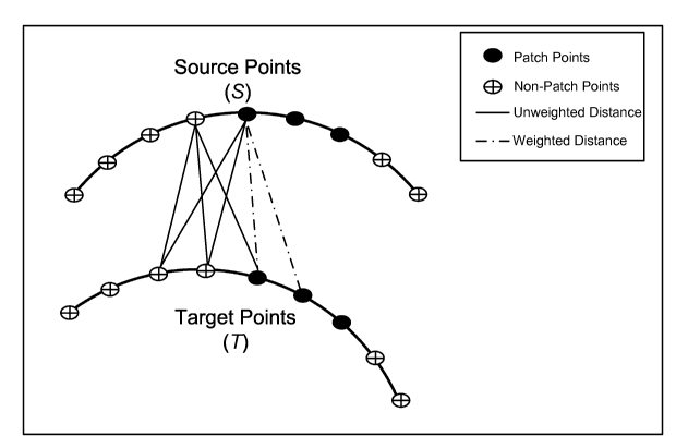
method.
Only the Euclidean distances computed from source patch point to target patch
point are
biased by the weighting factor wPc (i.e., dashed lines). The point
correspondence
determination for non-patch points is not effected by the weighting (i.e.,
solid lines).
Fig. 26 shows plots of dynamic PBR weighting factor function with various
relaxation parameter (a) values. Decreasing the value of a increases the
length of time that
the patch region dominates the PBR at each iteration. As the value of a
approaches to zero
the PBR weighting scheme becomes more akin to that proposed by Maurer et al.
[59].
Fig. 27 shows digital photograph (a), raw LRS scan (b) and sample CT slice (c)
of
imaging phantom. The silicon liver model, located in the center of the
phantom, is
surrounded by a set of seven white Teflon spheres. These spheres, which can be
localized in
both LRS and CT image spaces, are used in the determination of the "gold
standard" ICP
registration and serve as targets in the robustness studies.
Fig. 28 shows phantom LRS simulated falciform patch selected from full scan
(a) was
used to delineate the homologous region in the CT image surface (b). To more
accurately
simulate the typical LRS surface field of view obtained during surgery, a
subregion of the
LRS was manually selected for use in the robustness trials.
Fig. 29 shows traditional ICP registration results (a) and overlaid image and
falciform
patch regions (b) for the clinical data used in the robustness trials (Patient
3). Note the large
contrast in the accuracy of the alignment in this case than that shown in Fig.
1. The RMS
residual for this registration was 3.43 mm. Note that for this data set the CT
image and LRS
surfaces contained 57,873 points and 19,863 points, respectively. The CT image
and LRS
21
CA 02670261 2009-05-14
WO 2008/063494 PCT/US2007/023826
falciform regions consisted of 3,148 and 594 points, respectively.
Fig. 30 shows histogram representations of the RMS residual (a) and TRE (b)
results
from the phantom robustness trials. Note that for these trials the "gold
standard" RMS
residual and TRE values were found to be 0.621mm and 2.31mm, respectively.
Note the far
greater number of ICP RMS residual results that fell >2mm and TRE results that
were >5mm
as compared to the weighted patch ICP results.
Fig. 31 shows clinical results for Patient 1 showing visualizations of the ICP
registration (a), (b) and the patch ICP registration (c), (d). The LRS and ICP
patches are
highlighted for the ICP and patch ICP registrations in (b) and (d),
respectively. Note that the
ICP alignment causes an extremely erroneous registration. The LRS scan of the
anterior
liver surface becomes registered to the posterior of the liver. The patch ICP
registration
provides a much more reasonable alignment. Note that for this data set the CT
image and
LRS surfaces contained 48,843 points and 16,293 points, respectively. The CT
image and
LRS falciform regions consisted of 1,397 and 728 points, respectively.
Fig. 32 shows clinical results for Patient 2 showing visualizations of the ICP
registration (a), (b) and the patch ICP registration (c), (d). The LRS and ICP
patches are
highlighted for the ICP and patch ICP registrations in (b) and (d),
respectively. The ICP
registration shows an apparent misalignment which is corrected via the
proposed method.
Note that for this data set the CT image and LRS surfaces contained 53,459
points and
16,675 points, respectively. The CT image and LRS falciform regions consisted
of 1,632 and
1,507 points, respectively.
Fig. 33 shows a histogram representation of the RMS residual data obtained
from
robustness test performed on clinical data (Patient 3). The "gold standard"
ICP registration is
shown in Fig. 29 and this registration yielded an RMS residual of 3.43mm. Note
that only
one patch ICP trial yielded a significantly greater RMS residual than the
"gold standard".
Fig. 34 shows a comparison of an original slice (a), a manual segmentation (b)
and a
level set segmentation (c) of a liver of a patient.
Fig. 35 shows a surface model generation from the segmented contours. The
initial
surface mesh (a) is generated using the marching cubes method, and refined (b)
with a
surface fitting technique that employs Radial Basis Functions [81], providing
a smoother
surface with less vertices, potentially increasing the speed and accuracy of
the registration.
22
CA 02670261 2009-05-14
WO 2008/063494 PCT/US2007/023826
Fig. 36 shows a surface data acquisition in the OR. In the left image, the
surgeon is
digitizing points on the liver surface with the optically tracked probe. The
right image shows
the range scanner in position to acquire surface data of the liver
intraoperatively.
Fig. 37 shows a screen shot of range scan module in ORION surgical navigation
software. From the top-left panel clockwise: the native tomogram, two
different perspectives
of the three-dimensional liver and the vasculature as segmented by MeVis, and
a
tomographic slice of the segmented liver.
Fig. 38 shows a time plot of respiratory data. The data is aligned according
to the
axes provided by the primary component analysis. The origin is the mean of the
original
respiration data.
Fig. 39 shows ICP registration results. For each case, the registered range
scan data is
overlaid on top of three tomographic slices from the volume.
Fig. 40 shows ICP registration results. For each case, the registered probe
data is
overlaid on top of three tomographic slices from the volume.
Fig. 41 shows a comparison of surface registrations using tracked probe (left
column)
and range scan (right column). Both data sets are overlaid on the identical
slice from the
image volume.
Fig. 42 shows (left column) original rigid registration of range scan data
overlaid on
to mograms, (right column) the deformed liver volume from the finite element
model is
overlaid in red. In the areas where the point cloud was used for the boundary
conditions,
there is improved agreement between the range scan surface and the deformed
image surface.
Fig. 43 shows the relatively planar range scan data results in misalignments
during
the surface-based registration, in case 7, and the qualitatively identified
landmark, where the
falciform ligament resided before surgery is rotated clockwise, as indicated
by the white
arrows.
DETAILED DESCRIPTION OF THE INVENTION
The present invention is more particularly described in the following examples
that
are intended as illustrative only since numerous modifications and variations
therein will be
apparent to those skilled in the art. Various embodiments of the invention are
now described
in detail. Referring to the drawings, like numbers indicate like parts
throughout the views.
As used in the description herein and throughout the claims that follow, the
meaning of "a,"
23
CA 02670261 2009-05-14
WO 2008/063494 PCT/US2007/023826
"an," and "the" includes plural reference unless the context clearly dictates
otherwise. Also,
as used in the description herein and throughout the claims that follow, the
meaning of "in"
includes "in" and "on" unless the context clearly dictates otherwise.
Moreover, titles or
subtitles may be used in the specification for the convenience of a reader,
which has no
influence on the scope of the invention. Additionally, some terms used in this
specification
are more specifically defined below.
DEFINITIONS
The terms used in this specification generally have their ordinary meanings in
the art,
within the context of the invention, and in the specific context where each
term is used.
Certain terms that are used to describe the invention are discussed below, or
elsewhere in the specification, to provide additional guidance to the
practitioner in describing
various embodiments of the invention and how to practice the invention. For
convenience,
certain terms may be highlighted, for example using italics and/or quotation
marks. The use
of highlighting has no influence on the scope and meaning of a term; the scope
and meaning
of a term is the same, in the same context, whether or not it is highlighted.
It will be
appreciated that the same thing can be said in more than one way.
Consequently, alternative
language and synonyms may be used for any one or more of the terms discussed
herein, nor
is any special significance to be placed upon whether or not a term is
elaborated or discussed
herein. Synonyms for certain terms are provided. A recital of one or more
synonyms does
not exclude the use of other synonyms. The use of examples anywhere in this
specification,
including examples of any terms discussed herein, is illustrative only, and in
no way limits
the scope and meaning of the invention or of any exemplified term. Likewise,
the invention
is not limited to various embodiments given in this specification.
As used herein, "around", "about" or "approximately" shall generally mean
within 20
percent, preferably within 10 percent, and more preferably within 5 percent of
a given value
or range. Numerical quantities given herein are approximate, meaning that the
term
"around", "about" or "approximately" can be inferred if not expressly stated.
As used herein, the term "living subject" refers to a human being such as a
patient, or
an animal such as a lab testing pig.
As used herein, "registration", "map" and "alignment" are synonyms in the
specification, unless the context therein clearly indicates otherwise.
24
CA 02670261 2009-05-14
WO 2008/063494 PCT/US2007/023826
As used herein, "organ shift" and "organ deformation" are synonyms in the
specification, unless the context therein clearly indicates otherwise.
OVERVIEW OF THE INVENTION
The description will be made as to the embodiments of the present invention in
conjunction with the accompanying drawings. In accordance with the purposes of
this
invention, as embodied and broadly described herein, this invention, in one
aspect, relates to
a method of compensation for intraoperative deformations of an organ of
interest of a living
subject, where the organ of interest of the living subject is a liver, heart,
kidney, lung,
stomach, brain, or soft tissues.
In one embodiment, the method includes the following steps: At step (a), an
image of
the organ of interest of the living subject is preoperatively acquired. The
pre-operatively
acquired organ image of the living subject comprises image data with respect
to the organ
surface geometry. The image data with respect to the organ surface geometry is
obtained
through the use of at least one of positron emission tomography device,
electroencephalography device, computer tomography device, functional magnetic
resonance
imaging device, magnetic resonance imaging device, and ultrasound imaging
device.
At step (b), an image (or intraoperative shape) of the organ of interest of
the living
subject is intraoperatively acquired by, for example, an optical device
capable of obtaining
frequency, intensity and geometric data with respect to the surface of the
organ of interest of
the living subject simultaneously. The optical device is a laser range
scanner, or an optical
stylus. The intraoperative shape of the organ of interest of the living
subject may also be
acquired by stereo pair technology. It would be also advantageous if the
geometric surface is
also referenced to the field of view or is textured by a digital image of the
field of view.
At step (c), a first geometric surface of the organ of interest is generated
from the
intraoperatively acquired image of the organ of interest in step (b). At step
(d), an atlas, [A],
of deformations of the organ of interest is constructed from the pre-
operatively acquired
image of the organ of interest. The atlas [A] is in the form of an n x m
matrix with n, m
being positive integers. Step (c) is performed by generating a geometric
volume of the organ
of interest from the preoperatively acquired image in step (a); modeling
deformations of the
organ of interest based on the geometric volume of the organ of interest with
a computational
model; obtaining m model solutions of the computational model corresponding to
the
CA 02670261 2009-05-14
WO 2008/063494 PCT/US2007/023826
geometric volume of the organ of interest, where each model solution, A,
represents a
solution of deformation for n variables and corresponding to a set of
parameters; and
generating the atlas [A] in the form of an n x m matrix with each model
solution, A, which is
in the form of a n x 1 matrix, forming a column of the matrix.
The geometric volume of the organ of interest is generated through
segmentation of
the pre-operatively acquired image of the organ of interest, and represented
by a tetrahedral
mesh with n surface nodes.
The model solutions are obtained by solving at least one partial differential
equation
modeled to represent the relationship between a deformation of the organ of
interest and at
least one force causing the deformation. The at least one partial differential
equation is
solved with boundary conditions corresponding to specific structures of the
organ of interest,
body force, material properties, vascularization, physiological changes
related to tissue of the
organ of the living subject, physical conditions or any combinations of them.
The at least
one partial differential equation is solved with the boundary conditions
iteratively.
At step (e), a second geometric surface of the organ of interest is generated
from the
constructed atlas [A] of deformations of the organ of interest.
At step (f), the second geometric surface of the organ of interest is aligned
to the first
geometric surface of the organ of interest to determine at least one
difference between a point
of the first geometric surface and a corresponding point of the second
geometric surface of
the organ of interest. The alignment of the second geometric surface of the
organ of interest
to the first geometric surface of the organ of interest of the living subject
is performed with a
point-based registration, or a weighted point-based registration, where the
weighted point-
based registration comprises the step of using a salient-feature weighted
correspondence, and
wherein when the organ is a liver, the salient-feature comprises a falciform
ligament region.
The alignment of the second geometric surface of the organ of interest to the
first geometric
surface of the organ of interest of the living subject may also be performed
with a registration
that provides a surface-to-surface correspondence using at least one
characteristic feature of
the organ of interest of the living subject.
At step (g), the at least one difference is fed back to step (e) to generate a
new second
geometric surface of the organ from the atlas [A] of organ deformations of the
organ of
interest. At step (h), steps (e)-(g) are iteratively repeated for a
predetermined number of
26
CA 02670261 2009-05-14
WO 2008/063494 PCT/US2007/023826
iterations or until an error tolerance related to the at least one difference
between a point of
the first geometric surface and a corresponding point of the second geometric
surface of the
organ of the living subject is no greater than a predetermined threshold.
Then, the
intraoperative organ deformation is compensated. Note, here, as elsewhere in
the
specification, while the method is given in a number of steps in an order, it
is understood that
these steps do not have to be performed in that given order.
In another aspect, the present invention relates to a method of surface
registration in
image guided surgery. In one embodiment, the method includes the following
steps:
At first, a surface of an anatomic structure (or an organ of interest) of the
living
subject is extracted from a tomographic scan, for example, CT, MRI, PET or
SPECT images
preoperatively acquired by any one of a number of conventional image
processing
algorithms. The pre-operatively acquired image of the anatomic structure
comprises image
data with respect to the surface geometry of the anatomic structure.
Then, details of the surface of the anatomic structure are extracted by any
one of the
following features: (a) user identification of surface features or visible
structures, (b) surface
curvature, (c) surface shape, (d) surface orientation within the patient, (e)
distribution of
surface normal orientation, and (f) potential for deformation. Areas of these
details are
collected and grouped into surface subsets called "patches". In other words,
one or more first
patches corresponding to the first surface of the anatomic structure are
constructed, where
each first patch contains a surface subset of physical space data related to a
salient
anatomical feature of the first surface of the anatomic structure.
An intraoperative determination of an internal anatomic structure is obtained
by one
of the following methods: (i) swabbing the surface with a tracked instrument,
(ii) laser range
scanning for surface determination, (iii) intraoperative ultrasound, (iv)
imaging using a
tracked laparoscope or endoscope, (v) surface extraction from a binocular
scene such as
provided by a binocular laparoscope or endoscope.
Once the intraoperative surface has been obtained, details corresponding to
the
surface parameters extracted from the preoperative images are obtained. These
may include:
user identification of surface features or visible structures, surface
curvature, surface shape,
surface orientation within the patient, distribution of surface normal
orientation, and/or
potential for deformation. The procedure constructs one or more second patches
27
CA 02670261 2009-05-14
WO 2008/063494 PCT/US2007/023826
corresponding to the second surface of the anatomic structure, wherein each
second patch
contains a surface subset of physical space data from the second surface
related to a
corresponding salient anatomical feature of the first surface of the anatomic
structure.
The one or more second patches are aligned to the corresponding one or more
first
patches, where each second patch is dynamically biased to a corresponding
first patch. The
alignment of the one or more second patches to the corresponding one or more
first patches
comprises the step of giving more weight to the patches than to the rest of
the surfaces.
Then, a registration of the second surface to the first surface with physical
space data not
contained in the first and second patches to indicate surgical position in
both image space and
physical space is performed, by increasing weight to the rest of the surfaces
when final
adjustments in the registration are made with all available surface
information, using a point-
based registration, preferably, a weighted point-based registration. The
weighted point-based
registration comprises the step of using a salient-feature weighted
correspondence.
There is no expectation that extraction of these parameters will lead to
exactly
homologous patches in both spaces. However, the alignment of the patches will
be initially
weighted more heavily than that of the rest of the surface in the cost
function of the
alignment algorithm. As the alignment progresses, using techniques such as
iterative closest
point (ICP), chamfer matching or other techniques, the weight of the general
surface
alignment rises relative to the patch weight until final adjustments in the
registration use all
available surface information.
One advantage of the patch registration over standard surface ICP is that it
captures
additional information beyond the surface shape description and uses that to
improve the
registration. This means that patch techniques should work when only part of
the organ or
structure of interest is visible. This may prove critically important as one
moves to
minimally invasive techniques.
Because surface-based registrations are search processes, it is customary to
provide
an initial alignment to reduce the space that has to be searched. The second
advantage of the
patch registration is that the early high weighting of the patches can provide
the initial
alignment, thus removing a first step.
Biological surfaces tend to be smooth and rotationally syrnmetric about at
least one
axis. The third advantage of patch registration is that because of the weight
applied to a
28
CA 02670261 2009-05-14
WO 2008/063494 PCT/US2007/023826
patch, the patch counts more than a similar zone and thus tends to "anchor"
the registration
where without the patch it might find a solution with similar metrics but
incorrect alignment.
Yet another aspect of the present invention relates to a system of surface
registration
in image guided surgery. In one embodiment, the system has a first imaging
acquiring
device for pre-operatively acquiring images of an anatomic structure of a
living subject; a
second imaging acquiring device for intra-operatively acquiring images of at
least part of the
anatomic structure of a living subject; and at least one computer coupled with
the first image
acquiring device and the second image acquiring device and adapted for
performing the steps
of:
i). generating a first surface of the anatomic structure of the living subject
from
the pre-operatively acquired images of the anatomic structure of the living
subject;
ii). constructing one or more first patches corresponding to the first surface
of the
anatomic structure, wherein each first patch contains a surface subset of
physical space data related to a salient anatomical feature of the first
surface
of the anatomic structure;
iii). generating a second surface of the anatomic structure of the living
subject
from the intra-operatively acquired images of the anatomic structure of the
living subject;
iv). constructing one or more second patches corresponding to the second
surface
of the anatomic structure, wherein each second patch contains a surface subset
of physical space data from the second surface related to a corresponding
salient anatomical feature of the first surface of the anatomic structure;
v). aligning the one or more second patches to the corresponding one or more
first patches, wherein each second patch is dynamically biased to a
corresponding first patch; and
vi). completing a registration of the second surface to the first surface with
physical space data not contained in the first and second patches to indicate
surgical position in both image space and physical space.
In one embodiment, the system further comprising a display device coupled to
the at
least one computer for displaying the registration dynamically to facilitate a
diagnostic or
29
CA 02670261 2009-05-14
WO 2008/063494 PCT/US2007/023826
surgical procedure.
In one embodiment, the first imaging acquiring device has at least one of
positron
emission tomography device, electroencephalography device, computer tomography
device,
functional magnetic resonance imaging device, magnetic resonance imaging
device, and
ultrasound imaging device. In one embodiment, the second imaging acquiring
device
includes a laser range scanner that is capable of obtaining frequency,
intensity and geometric
data with respect to the cortical surface of the living subject
simultaneously. In another
embodiment, the second imaging acquiring device includes an ultrasound imaging
device. In
an alternative embodiment, the second imaging acquiring device comprises a
tracked
instrument, an optical stylus, a tracked laparoscope or endoscope, or a
binocular laparoscope
or endoscope.
These and other aspects of the present invention are more specifically
described
below.
IMPLEMENTATIONS AND EXAMPLES OF THE INVENTION
Without intent to limit the scope of the invention, exemplary methods and
their
related results according to the embodiments of the present invention are
given below. Note
that titles or subtitles may be used in the examples for convenience of a
reader, which in no
way should limit the scope of the invention. Moreover, certain theories are
proposed and
disclosed herein; however, in no way they, whether they are right or wrong,
should limit the
scope of the invention so long as the invention is practiced according to the
invention without
regard for any particular theory or scheme of action.
EXAMPLE 1:
COMPENSATION FOR INTRAOPERATIVE ORGAN SHIFT
A. Image-Guided Liver Surgery System: The current image-guided liver surgery
(IGLS) system is developed through collaborative efforts between researchers
at Vanderbilt
University, Washington University in St. Louis, and Pathfinder Therapeutics
Inc. (PTI), and
has been used to collect data in more than 15 clinical cases. This working
prototype has been
in development for over eight years. Preoperative CT and MR images are
acquired from the
liver of a patient one to two weeks before the procedure. The liver of the
patient is
segmented from these preoperatively acquired image volumes. This segmentation
can be
performed by manually outlining the contour of the liver surface. However, the
manual
CA 02670261 2009-05-14
WO 2008/063494 PCT/US2007/023826
procedure is often very slow and tedious and may require hours to complete. An
alternative
and more expedient semi-automatic method was developed by Dr. Benoit Dawant at
Vanderbilt University which is based on the level-set technique [30, 31]. This
method takes
5-15 minutes to run, and then up to 15 minutes of user interaction after the
segmentation.
From the segmented images, a 3D surface is tessellated using the Marching
Cubes Method
[32]. The tessellated 3D surface includes a mesh of connected triangles and is
used for the
surface registration. These methods are currently integrated into the current
system and are
used when making data available to the applicants.
Intra-operative surface data of the exposed liver of the patient is obtained
with a laser
range scanner (LRS). Laser range scanning uses the principle of triangulation
to determine
3D points in space. A laser light is emitted from the LRS and strikes the
surface of the liver.
The reflected light is received by a CCD camera. Based on the reflected light
pattern and the
known trigonometric relationship between the laser emitter and the camera, the
3D location
is computed. According to this system, a line of laser light sweeps across the
surface of
interest, as shown in Fig. 2a, for acquiring a dense point cloud (shown in
Fig. 2b) to represent
the surface of interest. In addition, a digital image of the liver is captured
of the operative
field and mapped to the point cloud thus generating a textured point cloud, as
shown in Fig.
2c. For OR use, the laser range scanner is optically tracked and brought into
the operative
field when needed for acquiring a scan.
The range scan data is then registered with the preoperative surface using the
iterative
closest point (ICP) method, modified to use k-d trees for quicker closest
point searches [33-
36]. A point-based registration based on rough anatomic points serves as an
input for the
ICP. This initial registration restricts the search range of the ICP solution,
thus improving
time-to-solution and reducing the chance of falling into local minima.
Retrospective
registration results from four of the clinical cases are shown in Fig. 3,
where three
tomographic slices from the preoperative data are laid out in each row, and
the registered
range scan data is overlaid in white. Under careful inspection, the presence
of deformation
can be observed in these preliminary results.
B. Segmentation ofLiver Images for Model-Building and Determining Residual
Liver
Volume: In one embodiment, an FEM of the segmented liver is generated. Non-
rigid organ-
based registration provides more specific therapy for liver tumors over rigid
based techniques
31
CA 02670261 2009-05-14
WO 2008/063494 PCT/US2007/023826
by determining the amount of tissue removed during the surgery through a
pre/post
segmented liver volume comparison. This is then compared to what is predicted
by the intra-
operative surgical plan as initially digitized during the case. A critical
component in this
analysis is the accurate segmentation of the liver from the pre and post-
operative CT and MR
images. In one embodiment, the current method is using an innovative
segmentation
technique that has been evaluated in clinical practice and was developed by
Dr. Benoit
Dawant and researchers at Vanderbilt University. This level set-based method
uses a new
speed function that reduces leakage problems that are known to affect level
set based
methods [40], which has been evaluated at the Catholic University of Louvain
in Belgium.
The accuracy and repeatability of the method is compared with those of manual
segmentation
for determining liver volume in living liver transplant donors from MR images.
For 18 living
donors, two observers each performed two semi-automatic and two manual
segmentations.
Each measurement was timed and the average number of slice/volume is
approximately 20.
Actual graft weight was measured during surgery. The times to perform manual
and
automatic segmentation were compared. Accuracy and repeatability were
evaluated with the
Bland-Altam method. The key findings of this study were that (1) mean
interaction time was
reduced from 25 minutes with manual segmentation to 5 minutes with Dawant's
method; (2)
differences between the actual volume and the estimated volume ranged from -
223 to +123
mL for manual segmentation and from -124-to +86 mL with the method; (3)
automatic
segmentation improved weight estimation in 15 out of 18 cases; and (4) inter
and intra-
observer repeatability (reliability) was improved using the semi-automatic
method. The
method developed is not fully automatic as it requires placing initial seeds
within the
structures to be segmented. In difficult cases, i.e., when boundaries between
the liver and
adjacent structures, heart, and intestine, are ill-defined, editing the
contours according to the
algorithm is also required, where placement of the seeds and contour editing
is included in
the total interaction time. To facilitate user interaction, a graphical user
interface is
developed to be shown in Fig. 4. This interface permits the placement of seed
points, editing
of contours, and visualization of tomographic volumes in the coronal,
transverse, and sagittal
directions. 3D segmented surfaces can be rendered and superimposed on the
tomographic
volumes. While the clinical validation study was conducted on MR images, which
has been
extended to CT images [30] and is also extendable to other types of images.
Although a
32
CA 02670261 2009-05-14
WO 2008/063494 PCT/US2007/023826
clinical study comparable to the one performed to test the algorithm on MR
images has not
yet been conducted on CT images, the preliminary results indicate a very good
agreement
between the method and manual delineation. In an exemplary embodiment, 6 CT
volumes
with an average number of slices equal to 196 (slice thickness ranging from
1mm to 2mm)
were selected and the liver contours were delineated manually and with the
method.
Semiautomatic and manual contours were compared using a similarity index
commonly used
to compare segmentations defined as:
S 2N(C, n C2 ) (1)
= N(CJ u N(C2 )
in which N is the number of voxels within a contour and Cl and C2 are the
manual and the
semi-automatic contours respectively. Values of S above 0.8 indicate a very
good agreement
between delineations. The mean S value for the six volumes was 0.94 (min=
0.92,
max=0.95). It is expected that the method is successful and more useful within
the CT
context. Indeed, because of the large number of slices in CT volumes,
segmenting the liver
by hand in this modality is a daunting task. The average time required to
segment 20 MR
slices was 25 minutes. Extrapolating this number to 200 slices in CT (-4
hours) becomes
quite alarming. Interaction time for the semiautomatic segmentation method
does not grow
linearly with the number of slices and is orders of magnitude faster than the
manual.
C. Non-rigid Organ-based Registration: One object of the invention is to
determine
the extent of improvement by deformation correction both in the quantitative
targets and in
registration robustness. This can be achieved by the deformation compensation
computational node according to the invention. It should be noted that the
data necessary for
the computational node is not unusual and should be available in most surgical
guidance
systems. At most, an added cost to other surgical systems would be the
addition of a
commercial laser range scanner which is quite reasonable when compared to the
cost of an
overall system.
The challenge for IGLS is more difficult than that of its neurosurgical
counterpart.
Undoubtedly, soft-tissue deformation is present but the nature of loading, the
information
available, and the tolerable margins for error are quite different. As opposed
to
neurosurgery, exploratory liver surgery does not have the confines of the
cranium. In
addition, surgical presentation of the organ often requires separation from
the surrounding
33
CA 02670261 2009-05-14
WO 2008/063494 PCT/US2007/023826
ligamenture and a presentation that differs considerably from its anatomical
orientation
within the body during preoperative scanning. While in neurosurgery data can
be taken
immediately post-durotomy, which allows for minimal differences between
preoperative and
intraoperative initialization, this is not the case for the liver whereby
deformations induced
by organ presentation are already in place when intraoperative data can be
taken. This
dramatic difference in organ shape is a specific challenge for image-guided
liver surgery.
Fortunately, the tolerance for localization is not quite as constrained due to
the regenerative
nature of the liver.
Fig. 5 is an example of the challenges associated with deformations during
organ
presentation. Fig. 5a is a liver phantom with a possible deformation
associated with organ
presentation, and Fig. 5b shows the results of a rigid surface registration
between the
deformed and undeformed surfaces using the ICP method [41]. What is
interesting about this
result is that a standard surface registration method introduces error in
areas that are
minimally deforming while reducing errors in the deforming region (white arrow
shows error
introduction while gray shows error reduction). In the clinical example [39],
as shown in
Fig. 2, an equivalent registration is performed and the result is shown in
Fig. 6a, where the
LRS partial surface is aligned atop the respective patient's CT liver surface
following ICP
registration. The discrepancy in the registration is due to deformation and is
designated by
the white arrow in Fig. 6b. According to embodiments of the present invention,
the inventors
implemented a very basic approach [39] using the closest point correspondence
after surface
registration to provide a boundary condition mapping from one partial surface
to the other
and then allow the finite element model to generate remaining deformations, as
shown in Fig.
6c. It can be seen from Fig. 6c how the deformed liver does match visually
better the LRS
contour which represents the intraoperative state. While the initial results
in Fig. 6c are
satisfying, more experience in the OR began to shape the challenges that
needed to be
addressed if the approach was to be more general and robust. It was evident
that the initial
alignment was very important. It has been experimented with a deformation
identifying rigid
registration (DIRR) which was based on phantom work [38], as described in
Example 2
below. The DIRR is designed to generate an initial registration that was
representative of
Fig. 6a as opposed to Fig. 6b with the goal of allowing the FEM model to
enforce
deformation only on those surfaces experiencing it [38]. The hypothesis was
that starting the
34
CA 02670261 2009-05-14
WO 2008/063494 PCT/US2007/023826
non-rigid registration process from an initial condition that preferentially
aligned minimally
deforming regions was better than using one that minimized the error over the
entire surface.
In one embodiment, a novel cost function for the DIRR was developed in the
form of:
NS
f (S) exp(- ds;1(2r2 )) (2)
where for each point `i' in the Ns points of intraoperative data, a signed
distance, ds,; to the
non-deformed surface is calculated. The term T is based on the standard
deviation of a
Gaussian function and is similar to the fuzzy correspondence work by Chui and
Rangarajan
[42]. Once this registration had been established, a series of incremental
deformations are
applied to the finite element model to align the preoperative and
intraoperative state. Fig. 7
illustrates the incremental framework which was specifically designed to
better account for
geometric nonlinearities. Additionally, a linear elastic finite element model
using a specially
formulated normal-tangential boundary condition framework was utilized which
allowed for
displacements to be imposed that were normal to the organ surface but also
allowed for
lateral (i.e. tangential) degrees of freedom. This allowed for a more natural
deformation to
be applied to the organ. For example, in Fig. 8, a clear discrepancy can be
seen where
different boundary conditions are applied, i.e., vector displacement and
stress free. In Fig.
8b, no distinction is shown due to the nature of the incremental solution and
boundary
condition formulation thus producing a more natural organ deformation.
This approach was tested in a set of phantom experiments, which some of the
data is
shown in Fig. 5 [38], whereby an initial pre-deformation CT image volume was
acquired and
2 realistic lobe deformations were applied to the phantom with subsequent
repeat CT
scanning. The phantom contained 6 subsurface tumor-like targets that could
easily be
tracked in repeat scanning and were used for validation and error assessment.
Upon
initialization by either ICP or the DIRR, the incremental model-correction
framework was
applied and average target registration error was tabulated. Table 1 lists the
findings
regarding the target registration error for subsurface targets after the full
correction. When
comparing to rigid ICP registration alone, the study demonstrates smaller
target errors with
model-correction and with either initialization method, i.e., the DIRR or ICP
initialization.
The study indicated improvement using the DIRR over the ICP method of
initialization. It
should also be noted that apart from these results, there were other important
findings within
CA 02670261 2009-05-14
WO 2008/063494 PCT/US2007/023826
[38]. For example, various strategies were employed within the FEM framework
to account
for large deformations that were of varying levels of computational
efficiency. In addition to
these phantom experiments, the method has also been preliminary tested on
clinical cases in
[43]. Fig. 9 illustrates an overlay of (1) the corrected image volumes in red,
(2) the
underlying preoperative CT scans, and (3) the intraoperatively acquired LRS
data registered
to the CT volume as shown by the white contour. In this study, the
initialization was
performed with ICP and the conformity of shape is encouraging given the better
agreement
between the red image volume and the white contour. Unfortunately, post-
operative CTs
were not taken in these cases or anatomical targets during the case for a more
quantitative
comparison. This however can be corrected by the methods of the present
invention.
Table 1. Results of phantom deformation experiments showing target error in
millimeters for
(a) using the complete surfaces of the target and source for initialization,
and (b) using a
realistic partial surface for to represent LRS data.
ICP -Complete DIRR -Complete
No Model With Model No Model With Model
1 St Deformation 5.7 2.7 9.9 1.7
2" Deformation 3 2.5 3 2.3
ICP -Complete DIRR -Complete
No Model With Model No Model With Model
1S Deformation 7.3 4.3 11.8 3.8
2" Deformation 3.8 3.5 3.4 2.4
The genesis of the DIRR was to try to secure alignment initializations that
would
enhance model-updating as evidenced in Table 1. While aspects of the DIRR were
interesting, it became evident as OR experience continued that organ shape
change was a
more global event in many instances, as shown in Figs. 8 and 9. The automatic
delineation
of a particular minimally-deforming region may not be possible in many
clinical
presentations. However, one feature that was continually noted from case to
case was the
identification of the falciform region on the liver surface which is
reasonably identifiable on
CT image volume renderings. In light of the sensitivity of the approaches to
initialization, a
newly proposed registration method is being currently investigated which is
based on the
concept of increasing the weighting of salient features such as the falciform
region [44].
While the DIRR or standard ICP may be applicable in certain instances, more
robust
registration methods to handle the range of problems that can occur during
clinical
36
CA 02670261 2009-05-14
WO 2008/063494 PCT/US2007/023826
presentation would be desirable. Another aspect that became evident in OR
experiences was
that often the liver would deform in such a way that curvature would decrease.
As a result,
many of the registration methods would become confounded by local minima.
Figs. l0a and
l Ob demonstrates a clinical result whereby ICP has failed due to a lack of
geometric features.
In this case, the DIRR was not appropriate due to a more global shape change.
Figs. lOc-lOd
shows the corrected alignment using the new weighted salient-feature
registration method.
Figs. l Ob and l Od are the same as Figs. l0a and l Oc respectively, where the
visualization has
been modified to allow easy identification of the falciform regions.
D. Registration and Deformation Correction: One underpinning problem with
image-guided techniques that continuously confounds large-scale adoption is
the difficulty in
accounting for soft tissue deformation. While pristine anatomical information
and an
abundance of functional imaging methods (flVIR, PET, SPECT, etc.) are
routinely acquired
preoperatively, the translation of that information into a dynamic OR
environment has been
stymied. In recent years, the ability to co-register all of these forms of
preoperative
information has had significant advances e.g., [45-47]. However, sensitive
anatomical
landmarks, e.g., large subsurface vessels, could shift from their preoperative
position due to
intra-operative surgical deformations and injury could ensue. With respect to
neurosurgery,
rigid cranial constraints allow IGS to still have functional use within
today's ORs despite
deformations (although compensation for deformation is needed to improve IGS
in this
application also). With abdominal organs such as the liver, IGS application is
considerably
more challenging. The lack of landmarks, rigid or otherwise, for patient-to-
image
registration is one particular challenge. Additionally, in exploratory liver
surgery, the
surgical presentation of the organ is significantly deformed from its
preoperative state. As a
result, even if landmarks were available on the liver, the organ is already
significantly
distorted prior to patient-to-image registration. The methods underpinning the
deformation
correction strategy is to combine rigid and non-rigid techniques for organ-to-
organ
registration, i.e. segmented liver volume - to - acquired laser range scanned
liver.
In this embodiment, data that characterizes the shape of the visible organ
surface
during surgery will be obtained using the laser range scanning methodology.
This data will
be used to analyze the nature of soft tissue liver deformations, and to
generate novel methods
to correct for organ shift thus allowing for accurate patient-to-image
registration. Figs. 11a-
37
CA 02670261 2009-05-14
WO 2008/063494 PCT/US2007/023826
11d show examples of how cases vary. Figs. 11 a and I lb show two example
registrations
between the liver surface as generated by the CT (gray) and the LRS surface
(red). Figs. 11 c
and 11 d quantify the closest point distance between these two surfaces post-
surface
registration and demonstrate the variability in deformation that can occur.
While the results
from Fig. 12c may be acceptable, the results from 12d are more deleterious to
guidance. This
variability in organ registration is a significant hurdle for translating IGS
techniques to the
extracranial environment.
In previous work, the general approach that was investigated was to address
this
problem in five steps: (1) generation of a patient-specific computer model of
the liver from
preoperative CT images, (2) acquisition of the intraoperative organ surface
using LRS, (3)
application of an initial "best"-rigid alignment (e.g. DIRR from above), (4)
non-rigid
mapping of the preoperative organ to the intra-operative state using a finite
element model of
soft-tissue deformation, and finally, (5) the deforming of all preoperative
images based on
the deformation field as calculated by the model. In step (3), the term "best"-
rigid is still
being defined. For example, it is not clear that starting the non-rigid
alignment of step (4)
from an initial registration that minimizes the sum of the squared signed
distance error is
better than some other registration transformation (e.g. ICP, DIRR, or
weighted salient-
feature will all provide different initializations) [38, 48]. With respect to
the DIRR, it was
found that in many cases the amount of surface information needed to constrain
the DIRR
exceeded that which could be routinely acquired in the OR. Additionally, often
the
deformation was more global resulting in a reduction in geometric feature that
would
confound the DIRR with local minima, as well as ICP (see, for example, in
Figs. I Oa and
l Ob). While investigation of this methodology is ongoing, expansion towards
more robust
initialization and computational efficiency is desired.
Accordingly, a weighted point-based registration approach was generated that
uses a
relatively novel weighted correspondence method based on salient features [44]
and is
presented in Example 3 below. The impetus for the development was the routine
identification of the falciform ligament area within CT and LRS data. Briefly
stated, the
algorithm weighs salient features (such as the falciform) on both source and
target surfaces
more prominently for correspondence. The weights are exclusive in that only
when a
correspondence is determined between two salient features is the weight non-
zero. This
38
CA 02670261 2009-05-14
WO 2008/063494 PCT/US2007/023826
operation allows for a more aggressive initialization. In addition, the
weights are dynamic
and are described by the following expression,
WPBR ll/ - WPBR,base + (A WPBR )exp[- a(Z -1)] (3)
Where WPBR,base is a baseline weight between salient features, AWPBR is an
initial
amplification factor to more aggressively align these features in early
iterations, and a is a
decay rate for the amplification factor. Initial results using this approach
have been
encouraging and can be seen in Figs. lOc-IOd. The details of the salient
feature weighting
algorithm are described in Example 3 below [49] and continue to evolve as more
experience
is gained from the OR environment.
While preliminary results with the incremental FEM approach, as shown in Figs.
8, 9
and 10, have been encouraging and computational times associated with the
framework are
somewhat tractable, the need for a shift correction strategy that is more
automated, more
robust, faster, and equally as accurate would be desirable. To accomplish
this, a novel
approach is being investigated that regresses a deformed liver shape from an
atlas of shapes
such that it best fits LRS intra-operative surface data while simultaneously
registering to that
surface using the salient-feature weighted registration method. This approach
can only be
successful if an atlas of sufficient resolution can be generated. Based on OR
experiences, it
is hypothesized that an atlas of deformations could be pre-computed prior to
surgery and
would be sufficient to capture the dynamic OR events that affect liver shape.
As part of that
hypothesis, it is proposed that gravity may play an important role in
correcting for liver
deformations and needs to be included in the atlas of deformations. In Fig. l
Oc, a clinical
registration result is shown and illustrates that the curvature of the
intraoperative liver surface
in (red) has become more planar than the preoperative segmented liver
counterpart. While
gravitational loading is experienced similarly both preoperatively during
imaging, and
intraoperatively during surgery, there is change to the supportive forces
intraoperatively and
the orientation of the liver with respect to gravity. During surgery, the
liver is separated from
its supportive constraints which allows for more degrees of freedom that would
inevitably
result in deformation. To correct this, the atlas of deformed liver shapes
will contain
solutions with varying surgical constraints and changes in the direction of
gravitational
loading. Based on OR experiences, dorsal regions of segments V-VIII are areas
one would
expect minimal deformation and would prescribe fixed or constrained
displacements. Other
39
CA 02670261 2009-05-14
WO 2008/063494 PCT/US2007/023826
regions would remain largely stress free with medial and lateral edges of the
organ allowed
to move along the abdominal wall surface but constrained to minimally deform
in a direction
normal to the surface. Similar approaches are being pursued within the
neurosurgery context
with considerable success [50-52].
As experience grows in the OR, other manipulations may be built into the atlas
such
as distinct lobe deformations similar to that in Fig. 5a. Another natural
addition to the atlas
will be variability in mechanical properties. Given the state of disease and
other mitigating
factors, it is possible that liver may contain varying degrees of fibrosis. As
a result, the
inclusion of mechanical parameters within the atlas may be necessary. This
range will be
determined in specific aim #3 and is anticipated to yield 3-4 values to
capture the range.
In one embodiment, an initial atlas includes approximately 30-35 different
organ
orientations with respect to gravity, up to 5 different boundary condition
realizations for each
orientation, and up to 3-4 values of mechanical properties. This constitutes
an atlas of
approximately 700 solutions. On an 8-processor blade with a reasonably
discretized FE
mesh (about 100,000-150,000 elements), each solution takes approximately 45
seconds with
the entire atlas being built in approximately 9 hours. Given the preoperative
preparation
time, this is sufficient; however, this will almost certainly change with the
faster CPUs
continually on the horizon. The approach to deformation compensation is a
combined
alignment and organ-to-organ shape conforming approach and is inspired by work
from [53-
58]. The algorithm begins with equally weighted regression coefficients that
represent the
weighting of each deformed shape from the atlas. From this shape called the
source,
correspondence between the source and the target (intra-operative laser range
scanner data of
the liver) is established using the salient-feature weighted closest point
metric and a weighted
point-based alignment [59] of the target is performed. Once achieved, a second
closest
Euclidean distance operation is calculated to establish correspondence. A new
set of
regression coefficients is calculated with
[A]T [A]{a}+ [A] = [A]T {s} (4)
where [A] represents the an n x m deformed shape atlas (where n is the number
of points on
the matching surface and m is the number of shapes within the atlas), {a} are
the regression
coefficients, [A] is a regularization matrix (typically a diagonalized matrix
based on the
properties of [A] or other shape constraints), and {s} is the reference shape
as defined by the
CA 02670261 2009-05-14
WO 2008/063494 PCT/US2007/023826
source and the closest point correspondence. While this equation is relatively
simple,
constraints to the range of regression coefficients, alternative
regularization methods, and
different correspondence methods are under investigation. Once the new shape
is calculated,
the process proceeds iteratively by performing another salient-feature
registration, followed
by surface fitting, and so forth. This specific formulation is called an
iterative closest atlas
(ICAt) method. The unique combination of techniques, such as weighted
registration,
regression, regularization, closest point, etc., embeds the ICAt method as a
deformation
correction strategy for image-guided liver surgery using sparse intra-
operative data is
significantly innovative. A preliminary result demonstrating the approach
using the same
phantom experiments of Figs. 5-8, and Table 1 is shown in Fig. 12. The target
registration
error for the 6 tumor-like targets was 3.9 and 2.8 mm for deformations 1 and
2, respectively,
comparing to Table lb. While the results are comparable to the DIRR, the
registration times
have dramatically improved from 15-25 minutes for Table 1 results to 15-25
seconds in Fig.
12. Of note with these experiments is that the salient-feature weighting was
not used in this
result. It is anticipated that incorporation of the salient-feature weighting
will add further
improvement.
The rationale for pursuing atlas based techniques is that it allows one to
deform the
liver under a variety of deformation conditions that are more complicated than
simply
"pushing" on the organ. For example, deformation due to gravity represents a
distributed
load on the organ as opposed to a surface loading condition, i.e. applying a
direct
deformation to the organ surface. The subsurface deformation behavior
associated with a
distributed load is very different than a surface loading as shown in [60].
Building an atlas
allows a range of deformed liver shapes under many different loading
scenarios; and, given
that information regarding material properties is unknown, atlas-based methods
allow this
information to be added relatively easily. Additionally, since information
regarding posterior
constraints on the liver is difficult, an atlas-based approach allows us to
incorporate
variability in these conditions quite easily which may in fact vary from
surgeon to surgeon
depending on their procedures. The last distinct advantage is that all FEM
calculations are
done preoperatively which makes model-compensation a reality for today's OR
environment.
One aspect not mentioned above with respect to deformation is that during a
procedure, the liver is in constant motion due to respiration. The liver
motion is accounted in
41
CA 02670261 2009-05-14
WO 2008/063494 PCT/US2007/023826
clinical experiments by suspending respiration during brief apneic periods.
These apneic
periods were performed at the same location in the respiratory cycle, so that
the liver would
reside approximately in the same location for every registration. This
technique, which is
controlled by the anesthesiologist, is most frequently utilized during
thoracic procedures
when lung expansion impedes the operative procedure. The level of anesthesia
is monitored
with continuous computerized EEG and computerized bispectral index (BIS),
which provides
a correlation between the depth of anesthesia and the possibility of the
patient being awake
during surgery. There are 2-5 brief apneic periods during a procedure, each
lasting no more
than four minutes.
E. Separate Compute Node Strategy: In order to achieve deformation correction
capabilities for image-guided surgical systems in the liver, the initial
realization will be
performed on a blade configuration with 8 64-bit AMD Opteron 800 Series Dual-
Core
Processors as provided in the PRIMERGY BX630 eight socket AMD Opteron
Processor
server blade of Fujitsu. Since the blade contains 8 sockets and with each
socket capable of
supporting a dual-core processor, the unit has the capability to become a 16
core machine.
This compact single blade system should allow for rapid calculation of highly
resolved FEM
meshes as well as image deforming capabilities. Although some may view a
separate
compute node strategy as cumbersome, the compact nature of a blade system
allows its
placement on the most surgical platforms thus eliminating a remote computing
scenario and
concerns over data transmission security. All data is ported to the correction
node which
includes the segmented liver image volume and LRS data. Currently PTI's system
performs
an iterative closest point registration on the main processor to provide
physical-to-image
registration. One of the objectives of the invention is to revite novel non-
rigid registration
methods on the correction node to compensate for organ deformation during
surgical
presentation. Accordingly, the general preoperative procedure entails that at
least one day
prior to surgery: (1) a CT liver image volume is acquired, (2) a preoperative
planning session
is performed (15-20 minutes) where segmentation and salient features are
designated (salient
feature will predominantly reflect the location of the falciform ligament
region), and (3) once
complete, automatic mesh generation, boundary condition deployment, and atlas
solution
generation are performed prior to the case. In one embodiment, within the
operative setting,
the organ is presented, laser range scan data is acquired, and the non-rigid
registration
42
CA 02670261 2009-05-14
WO 2008/063494 PCT/US2007/023826
method is performed followed by an updated (i.e. deformed) image set for the
surgeon. The
providing of LRS data through a secure socket connection to the node would
initiate
correction. Once completed, a new deformed image set is loaded into the
visualization and
guidance modules automatically.
F. Clinical Trial: In one embodiment, each of three early-adopter sites of the
system
is tasked with conducting a 15-20 patient study with the following guidelines.
Patients are
screened and given written consent before participating in this clinical
trial. Subjects of
interest are recruited based on the severity of their disease, and only those
patients with
single or multiple tumors requiring at least two anatomic segment resections
are included in
this particular trial. Prior to receiving any treatment, the patients undergo
a physical exam,
laboratory tests, and a chest x-ray to determine their overall medical
condition as well as their
ability to undergo surgery. The laboratory tests, physical exams and pre-
operative treatment
procedures (e.g., arteriography, portal venous embolization, etc.) utilized in
this study are
scheduled based on specific tumor type, disease setting and surgeon
preference. These are all
considered standard of care. Tomographic imaging (CT, MR, and/or PET) of the
liver is the
only imaging study that is necessary, which is also part of standard care for
patients with
liver tumors undergoing surgical resection. The abdominal CT scan is a
triphase study of
uncontrasted acquisition, venous phase contrast, and arterial phase contrast.
The venous
phase contrasted study is used for the image-guided surgery study, and its
slice thickness is
approximately 2.5mm. The other phases have slice thickness of about 5.0mm.
For participating patients, the surgeon localizes anatomic landmarks from the
scans.
These landmarks are also physically localized at the time of surgery. The
liver's surface is
segmented by the semi-automatic segmentation techniques discussed previously.
The results
from this method are reviewed by the surgeon (i.e., investigator at the
clinical site) and
modified if necessary. An ultrasound is done during the surgery to confirm the
location of all
tumors. All procedures above are standard therapy for a patient prior to
hepatic resection.
The preoperative segmented liver surface extracted from the CT is used in
several stages of
this investigation. From the scans, total functional liver volume is
determined by subtracting
tumor volume from total liver volume. The segmented scans are also used as
part of the
image to physical space registration process detailed below.
A standard laparotomy surgical procedure is performed, followed by liver
43
CA 02670261 2009-05-14
WO 2008/063494 PCT/US2007/023826
mobilization. These are all standard procedures for major liver surgery. As
previously
mentioned, planned periods of apnea are used to decrease liver motion. With
the addition of
apneic periods and guidance, it is estimated that the entire operative
procedure itself would
potentially be lengthened a minimal amount (maximum 20 minutes duration).
Liver ablation
and resection procedures generally require at least 4-6 hours, so this would
represent a very
minimal alteration in the standard operative procedure. Collection of physical
space points
on the liver's surface takes place during the apneic periods. Before any
surfaces are acquired,
there is an apneic period to acquire anatomic landmarks with the optically
localized surgical
probe. Typically, 4 to 7 anatomic landmarks are acquired. The landmarks are
chosen based
on the ability to localize them accurately within both image and physical
space. Landmarks
may be anatomic (i.e. infrahepatic/suprahepatic vena cava, tip of the gall
bladder, tip of the
right lobe, portal vein bifurcation) or geometrically unique (inferior edge at
segments IV and
V, groove underneath falciform ligament, junction between segment III and IV).
These
landmarks are also identified and localized on the CT and/or MR image volumes
or 3D
reconstructions and used as targets to verify the accuracy of the registration
process. The
next apneic period is used for surface acquisition and image registration,
which involves the
laser range scanner and the other components of the IGLS device under
investigation. The
range scanner is mounted to a cart and swung into the sterile field, one to
two feet away from
the liver. Measures to protect the sterile field are under the surgeon's
discretion. Note that
this method does not require any contact with the patient. There is a brief
period of
positioning and calibration of the scanner, which takes less than one minute
and does not
require an apneic period. Once the scanner is in place, the apneic period
begins and a laser
range scan is acquired. The time required for the surface scanning process is
8-15 seconds
and approximately 20,0001iver surface points are acquired during this time.
The laser used
is low power, considered eye-safe by the FDA and orders of magnitude below the
Maximum
Permissible Exposure level for skin as stated in the American National
Standard for Safe Use
of Lasers (ANSI Z136.1). Once a surface is acquired in the operating room, it
is registered
with the segmented CT and/or MR liver surface using the iterative closest
point method.
Once the mapping has been determined, the surgeon will use the preoperatively
acquired
images and corresponding surface and volume renderings to resect the tumor and
surrounding tissue. This image-guidance augments the standard surgical
procedure that is
44
CA 02670261 2009-05-14
WO 2008/063494 PCT/US2007/023826
conducted. After the resection procedure is completed, a CT is acquired within
5 days. The
parameters for these scans are the same as above. The liver is segmented from
these scans
using the semi-automatic methods, and results from this segmentation method
will be
reviewed by the surgeon and modified if necessary. Residual liver volume is
calculated from
these scans and expressed as a percentage of total functional liver volume
calculated from the
preoperative scans.
The data described above is used for a retrospective evaluation of the
correction
approach. Anatomical targets are designated by each site and used in
validation of PTI's
current approach. These same targets are available for retrospective analysis.
In addition,
information regarding salient features and their correspondence will be
quantified, i.e.,
specifically closest point distances, and curvature assessments. The results
shown in Figs. 9-
12 suggest the value of this evaluation strategy. In addition, at one site
(St. Louis site),
additional data is taken regarding the initiation of resection on the liver
surface. More
specifically, the optical stylus is used to designate spatially on the OR
liver surface where a
resection begins. This allows for two predicted resective volumes to be
calculated based on a
rigid registration approach and the novel correcting approach. The volumes are
then
compared to the OR volume collected as well as the one predicted by analyzing
the pre-post
CT image volumes. Given that exploratory liver surgery is not performed within
an intra-
operative imaging unit, the collection of this data and the analysis proposed
provides
sufficient validation for the approach.
G. Model Improvements & Mechanical Property Assessment: As mentioned above,
it would be desirable to build in a realistic range of mechanical properties
to the atlas. As
Fig. 11 suggests, the extent of deformation can be different for two similar
cases. While the
discrepancy could be related to differences in organ presentation, the
possibility that
mechanical properties vary from patient-to-patient must not be neglected as a
potential cause
especially given the extensive range of functional tasks of the liver and the
affects that
lifestyle can have on the pliability of the organ.
As a result, interrogating liver ex vivo specimens from the OR at Vanderbilt
University is proposed. In one embodiment, measurements of soft-tissue
properties are
performed using an experimental gel-tissue mechanical assay with the Enduratec
ELF 3100
mechanical tester. The assay involves the following steps: (1) samples are
immediately
CA 02670261 2009-05-14
WO 2008/063494 PCT/US2007/023826
suspended post-resection within a polyacrylamide gel doped with CT contrast
agent, (2) a
microCT image volume is obtained, (3) mechanical testing in the ELF 3100 is
conducted,
and, (4) finite element model analysis is conducted. Fig. 13a shows the
microCT of a gel-
tissue suspension, where the tissue shown is a mouse liver. Fig. 13b shows the
computational model built from the CT, which is sliced through part of the
boundary so that
the internal soft-tissue mass can be observed. Fig. 13c shows an example of
mechanical
testing with the ELF3 100. In addition, a reference gel made from the same
polyacrylamide
stock is also made for independent tests to obtain background gel properties
(one of the
traces in Fig. 13c). Upon completion of testing, the computer model is used to
simulate the
compression and the mechanical properties of the tissue specimen are
determined. It is
important to note that viscoelastic effects have been observed in this data;
but currently,
analysis is performed after a dwell period to factor out transient effects. It
is expected that in
the future developing more sophisticated models which could include these
effects as well as
geometric nonlinearities. Fig. 13d shows a recent development towards
indentation testing of
tissue samples. While the model is used to determine properties for the gel-
tissue assay,
elastic indentation analysis theory is being used for determining properties
with respect to
Fig. 13d [62]. Correlation of these testing methods increases the certainty of
measurements.
EXAMPLE 2:
COMPENSATING FOR INTRAOPERATIVE SOFT-TISSUE DEFORMATIONS
USING INCOMPLETE SURFACE DATA AND FINITE ELEMENTS
The IGLS requires the ability to identify and compensate for soft tissue
deformation
in the organ. The predeformed state is represented as a complete three-
dimensional surface
of the organ, while the intraoperative data is a range scan point cloud
acquired from the
exposed liver surface. The first step is to rigidly align the coordinate
systems of the
intraoperative and preoperative data. Most traditional rigid registration
methods minimize an
error metric over the entire data set. In this Example, a new deformation-
identifying rigid
registration (DIRR) is reported that identifies and aligns minimally deformed
regions of the
data using a modified closest point distance cost function. Once a rigid
alignment has been
established, deformation is accounted for using a linearly elastic finite
element model (FEM)
and implemented using an incremental framework to resolve geometric
nonlinearities.
Boundary conditions for the incremental formulation are generated from
intraoperatively
46
CA 02670261 2009-05-14
WO 2008/063494 PCT/US2007/023826
acquired range scan surfaces of the exposed liver surface. A series of phantom
experiments is
presented to assess the fidelity of the DIRR and the combined DIRR/FEM
approaches
separately. The DIRR approach identified deforming regions in 90% of cases
under
conditions of realistic surgical exposure. With respect to the DIRR/FEM
algorithm,
subsurface target errors were correctly located to within 4 mm in phantom
experiments.
METHODS
A. Overview: The details of the individual steps of model-updating image-
guided
liver surgery (MUIGLS) are described in the subsequent sections. Preoperative
image data is
acquired of the patient's abdomen using CT or MR scans. From these
preoperative scans, the
liver is segmented, and a three-dimensional surface is tessellated. This
surface is used to
determine a rigid alignment with respect to the intraoperative range scan
data. Rather than
perform this registration using the traditional ICP method, a new form of
alignment that
weights regions of the data that are minimally deformed is developed. The
tesselated surface
also serves as the input for generation of a tetrahedral volumetric mesh that
will be the basis
for a finite element model. Before running the FEM, boundary conditions are
constructed
based on the rigidly registered intraoperative data. The closest point
distance between a
boundary node of interest and the intraoperative, deformed surface is
calculated. Execution
of the model is repeated in an incremental fashion. Rather than using the
entire closest point
distance, a fraction of this value is used to prescribe the displacement
boundary condition on
the node. Each successive solution of the model updates the location of the
mesh nodes,
which triggers the calculation of new correspondences and boundary condition
values. The
model is repeated until the root mean square (RMS) closest point distances for
all boundary
nodes using the closest point boundary condition has reached some
predetermined value.
The results from the FEM are used to warp the preoperative image to match the
intraoperative presentation.
Before explaining the methods used in MUIGLS, it is necessary to state some of
the
assumptions regarding image-guided liver surgery. From observing procedures in
the OR,
the liver is assumed to be an elastic substance. Unlike neurosurgery, there
are no apparent
fluid effects in the organ, so there is no shrinking or swelling and volume is
preserved. The
most obvious feature of the deformation appears to be a shape change, where
one region of
the liver surface changes relative position with respect to another region.
Often the
47
CA 02670261 2009-05-14
WO 2008/063494 PCT/US2007/023826
deformation can be concentrated in a central region of the liver whereby a
significant amount
of semi-rigid translation and rotation is experienced by areas in the organ
periphery, i.e., a
lever-arm effect occurs due to significant deformations located more central
to the organ.
Translational effects due to forces such as diaphragm motion have been
discussed in previous
research [24] and are taken into account by employing breath-hold protocols
[36]. During
the surgery, the liver may change shape because of manipulation by the surgeon
or resection,
and this will warrant a new registration. At that point, the laser scanner
will acquire a new
intraoperative surface, so that the registration and deformation compensation
will be
recomputed. "Minimally deformed" areas are considered to be those which
undergo
deformation no greater than a few millimeters as determined by visual
inspection. The goal
in MUIGLS is to reduce the amount of error from large scale deformations (1-4
cm) below
the previously stated 1 cm level of target registration accuracy while not
causing additional
error seen in the minimally deformed regions.
B. Data Representation: Phantom studies were performed on a poly (dimethyl)
siloxane (rubber silicone) model of the liver, which.is attached to a
plexiglass base. Two sets
of point-based landmarks are used for the study. Surrounding the outskirts of
the phantom
are vertical cylinders also attached to the vertical base, where seven white
Teflon spheres
have been placed in machined holders at the cylinder tops to serve as
fiducials for a point-
based registration. Inside the liver are mock tumors made of styrofoam, which
are spherical
with a radius on the order of about 1-1.5 cm. The intensity of these tumors is
approximately
20 times lower than the surrounding phantom, allowing for the tumors to be
easily segmented
with a simple region growing algorithm. The centroids of these tumors will
serve as
subsurface targets for accuracy studies. The position of the targets within
the phantom is
shown in Fig. 14. To induce a deformation in the phantom, an object of height
about 38.0
mm is placed underneath a region of the model. A large nylon screw pinned down
other
regions of the phantom and kept them stationary. Two different sites were
chosen for
deformation. These locations were chosen to mimic some of the physical
manipulations that
a surgeon may perform during a procedure. The first deformation occurred under
the left
lobe, where tumors 1 and 2 experienced the most shift, while the second site
was at the
middle of the inferior ridge, underneath segments III, IV, and V. For this
case, the largest
shift occurred at tumors 4 and 6. CT scans and range scans were taken while
the phantom
48
CA 02670261 2009-05-14
WO 2008/063494 PCT/US2007/023826
was in the nondeformed state and for each one of the deformed states.
Corresponding slices
from the nondeformed tomogram and one of the deformed volumes are shown in
Fig. 15,
where corresponding CT slices are acquired from (a) nondeformed and (b)
deformed image.
The plastic object placed below the liver produces the deformation. The height
of the object
is approximately 38 mm.
C. Identification of Deformation: According to one embodiment of the present
invention, the first step in MUIGLS is the rigid alignment between the
preoperative and
intraoperative coordinate systems. Although conventional rigid registrations
are relatively
easy to implement, they are also susceptible to misalignment caused by
deformation. It is
possible to reduce the effects of deformation on rigid registration by
identifying areas that are
minimally deformed and using only landmarks in these regions for the
registration. The
effects of identifying minimally deformed regions are illustrated with the ICP
algorithm that
is a common method of registering two surfaces. The ICP algorithm relies on
the closest
point distance metric. For the i-th point in data set X, the closest point
distance dcp; is
defined as the minimum distance from this point to a landmark in the other
data set Y
dcP,; =d(X;,Y)=argmin ,E, dis(X;,y) (5)
In the ICP algorithm, the RMS residual of closest point distances over the
entire
surface is the cost function that is minimized through the iterative process.
However, more
information can be obtained when examining a histogram of the signed distance
value
distribution at a given alignment. As shown in Fig. 16, a histogram of the
signed distance
distribution for rigid alignments between a nondeformed surface and the
surface deformed at
(a) the left lobe and (b) the inferior ridge. The solid line indicates that
the alignment is
performed by registering the entire surface with the ICP algorithm. The dashed
line is
acquired with the same registration method, but this time only regions of the
surface that
were visually identified as "minimally deformed" are used in the registration.
The signed distance indicates how far a point is outside of the surface
(positive) or
inside (negative). In this figure, the alignments were obtained using ICP: one
scenario used
the entire surface in the registration, while the other used only areas that
were visually
identified as minimally deformed. When using the entire surface in the
registration, the
signed distance histogram has a narrow band of values distributed in a
relatively uniform
fashion, as displayed by the solid lines in Fig. 16. When only the minimally
deformed
49
CA 02670261 2009-05-14
WO 2008/063494 PCT/US2007/023826
regions are registered using ICP, the histogram of signed distances has a much
sharper peak
at the histogram bins closest to zero, as indicated by the dashed lines. This
alignment also
produces a larger range of distance values that are associated with the
deformed areas shown
in Fig. 4a and play no role in this selective ICP alignment.
Often, information about the surface regions which are minimally deformed is
not
available a priori. A DIRR algorithm that aligns two surfaces according to the
minimally
deformed areas without any manual identification of these regions is
developed. For each
point i in the Ns points of intraoperative data, a signed distance, ds,;, to
the nondeformed
surface is calculated. These distance values are then used in the following
cost function:
NS
f (5) eXp 2,r 2 (6)
The Gaussian term is similar to one used for fuzzy correspondence in the work
of
Chui et al. [42]. As more points approach a closest point distance of zero,
the output value
of the cost function will increase. At the same time, this cost function does
not cause
significant penalties for points which have large signed distances to the
target surface that are
associated with deformation. The parameter that controls the behavior of the
cost function is
the standard deviation of the gaussian function, i. This parameter usually
ranges between
about 0.5 - 2.0 mm. Currently, the cost function is optimized using Powell's
method as
implemented in the VXL library. The parameters for the optimization are the
six degrees of
freedom that represent the rigid transformation that is applied to the
intraoperative data. Unit
quaternions represent the rotation.
In many cases, it is necessary to speed up calculations and provide smoother
objective
functions. The underlying surfaces of segmented preoperative data and
intraoperative data
were represented by radial basis functions (RBFs). A biharmonic RBF was used
to
interpolate the signed distance between any point in three dimensional space
and the surface.
The zero isocontour from the resulting RBF function represents the fitted
surface. To make
this method computationally efficient for large data sets, a special
implementation which
provides for the fast evaluation and solution of RBFs was used, developed by
FarField
Technology (FastRBF, Far Field Technology, Christchurch, NZ).
To test the DIRR algorithm, points sets from the deformed range scan and CT
data
were registered to the minimally deformed surfaces. These areas were manually
designated
CA 02670261 2009-05-14
WO 2008/063494 PCT/US2007/023826
from the surface by visual inspection and knowledge regarding the location of
the object
deforming the phantom. The minimally deformed areas were the only points used
in an ICP
registration, which served as a "ground truth" alignment to produce the same
effect in the
closest point histogram distribution as observed in Fig. 16. Then, the DIRR
was performed
without the aid of identifying the deformed surfaces. The results were
compared to the
ground truth using the six subsurface targets representing the tumor
centroids. Like many
registration algorithms, the DIRR needs an initial guess that roughly aligns
the two surfaces.
The initial alignment is achieved by identifying four landmarks on the phantom
surface to
serve as fiducials in a point-based registration.
To test the sensitivity of the DIRR to initial alignment, the position of each
fiducial
was randomly perturbed up to 1 cm away from its original position for 1000
trials. The
results from the DIRR were compared against the ground truth and categorized
as either a
success or a failure. A success was defined as any registration where all
tumor errors were
less than 5.0 mm, which was confirmed by visually inspecting the resulting
alignment.
D. Deformation Correction Using Finite Element Modeling: After the rigid
alignment between the two coordinate systems has been established, the next
step is to model
the deformation using a finite element model. The mesh used in the model is
constructed
from the preoperative tomographic volume, which represents the nondeformed
state of the
organ. The first step in mesh generation is to segment the liver from the rest
of the abdomen.
Segmentation is performed either manually or using a semi-automatic method
that is a
modification of the level-set method. The manual segmentation requires many
hours to
perform, while the level-set method can usually be completed in 30 min to 1 h.
From the
segmented organ volume, a surface is tesselated using either the marching
cubes method or
the aforementioned surface fitting algorithm using RBFs. The surface is
represented as a set
of polygons and serves as input to the mesh generation software. This software
uses the
boundary description to generate a tetrahedral grid volume of the entire liver
shape.
The deformation of the liver is modeled using a linear stress-strain
relationship for an
isotropic, three-dimensional solid. If static equilibrium is assumed, then
v = 6 = B (7)
where 6 is the stress tensor and B is the body force vector. Stress can be
related to strain by
the following relationship:
51
CA 02670261 2009-05-14
WO 2008/063494 PCT/US2007/023826
6 = Cs (8)
where C represents the material stiffness matrix. For a Hookean solid, C
depends on two
properties, Young's Modulus, E, and Poisson's ratio, v. The displacement
vector, u, is the
value that will be solved for, and it is related to normal strain 6 and the
shear strain y by
E= Sx s= S'uy, s= S'uZ (9)
x y ~ ~ Z ~ 1
y-Y =+ S~y yXi = ~PX + S'Z yn = Suy + (10)
SY &x ~ & cFz ' bi 8Y
where u={Px, Py, f.cZ } is the cartesian displacement. By combining equations
(7)-(10), a
system of partial differential equations can be expressed in terms of the
displacement vector,
u, to form the Navier equation
E V E 0(0=u)=B (11)
2(l + v) 2(l + vXl - 2v)
The partial differential equation is solved using the Galerkin weighted
residual
technique with linear basis functions. The system of equations that solves for
the
displacement vectors at every node in the mesh can be written as
[K] {u}= {b} (12)
One fundamental component to employing the finite element method is the
prescription of boundary conditions. These boundary conditions are derived
from knowledge
of the forces applied to the liver within surgery as well as information from
the intraoperative
data. There are three different types of boundary conditions implemented in
the model. The
first set of boundary conditions are categorized as "fixed," a set of
Dirichlet conditions
representing immobile regions of the organ. Typically, obscured regions of the
right lobe
that rest against other parts of the viscera belong to the fixed category.
"Stress-free"
boundary conditions are the second category, which represent regions
unrestricted by force.
The final type will be referred to as "closest point" boundary conditions.
These nodes play
the most significant role in modeling the deformation and are considered a
mixed boundary
condition, in that, of the 3 vector components, one component is Dirichlet
while the
remaining two are Neumann conditions. An example of how the different regions
of the
organ are classified according to boundary condition type is illustrated in
Fig. 17, where the
52
CA 02670261 2009-05-14
WO 2008/063494 PCT/US2007/023826
dark grey represents the boundary nodes that obey the mixed "closest point"
boundary
condition formulation. The medium grey value denotes fixed regions, while the
light grey
boundary conditions are stress free.
The details in implementing the "closest point" boundary conditions are
critical to
successfully recovering the deformation in the approach. It should be noted
that the initial
DIRR is also integral to the prescription of boundary conditions; i.e., at the
initiation of
deformation, the closest point distances are directly related to the DIRR
registration.
Furthermore, with such a large amount of deformation present intraoperatively,
improper
correspondence can lead to boundary conditions that would cause improper
nonrigid
alignments and unrealistic distortions of the organ shape.
In this embodiment, two measures are taken to avoid improper correspondence
when
setting the displacements for the boundary conditions: 1) manipulation of the
finite element
equations such that the equations are sensitive to the organ surface geometry;
2)
implementation of incremental solutions with a moving grid. The first involves
modifying
the conventional finite element method such that the weighted residual vector
equations at
the boundary are expressed in a coordinate reference that is designated to
have one
coordinate axis normal to the organ surface and the remaining two being
tangent to that
surface (as opposed to traditional Cartesian coordinate references). With
respect to modeling
anatomical organs and their deformations, there are some aspects to the
application of
boundary conditions that are particularly challenging to traditional Cartesian
representations.
For example, in the application of displacement boundary conditions to the
liver, it is often
desirable to express the movement of the boundary in a direction that is
relative to the
geometric shape, i.e., the coordinate system associated with directions that
are approximately
normal and tangential to the organ surface. One strategy is to take the
desired normal
displacement and convert this to its Cartesian counterparts, i.e.,
dn x=n y=n z=n dx
dt, = =i, y i, z=t', dy (13)
dt2 iZ y= iZ z- i2 dz
where n, t', and t2 represent an orthogonal coordinate system with the normal
(to the organ
surface) and tangential axes, respectively. In this case, the inverse
relationship in equation
(13) would be used since the transformation shown is from Cartesian to normal-
tangential
53
CA 02670261 2009-05-14
WO 2008/063494 PCT/US2007/023826
space (n-t space). In these equations, the application of a displacement
normal to an organ
surface can be achieved; however, the ability to relate mixed boundary
conditions within the
n-t space framework is not possible using equation (13). For example, it may
be desirable to
allow an organ surface to slide along a supporting wall yet not deform in a
direction normal
to the wall, i.e., through the wall. In another situation, the deformation may
need to be
applied in a direction normal to the organ surface yet allow for sliding of
tissue along the
displacing surface (e.g., depressing the organ with a smooth object like a
retractor). This
type of boundary condition requires stress-free conditions tangent to the
direction of
constraint/motion and restricted normal displacements, e.g.,
6r, =6t2 =0, uõ =0 (ora*dcp;) (14)
where 6t, and 6t2 are stresses applied tangent to the organ surface, and u,,
is a displacement
normal to the surface. In this instance, the framework described in (13)
cannot achieve these
degrees of freedom in organ movement behavior.
A better approach than using equation (13) is to rotate the equations of
equilibrium
for nodes concerned with the boundary into an n-t space coordinate reference.
This process
usually involves the use of rotational matrices (sensitive to the organ
boundary) being
applied at the local element assembly level
[RI. [KI. [RT t {u }j = [RI. {b}; (15)
where the premultiplication by [RI on the left and right-hand side rotates the
equilibrium
equation and body force components ([RI is the matrix shown in equation (13)
and would be
associated with the normal and tangential coordinates reference of the i-th
node), and the
LRT t. multiplication rotates the displacement coefficients from Cartesian to
the n-t space (i, j
refer to i-th weighted residual equation, and j-th displacement coefficient,
respectively).
Careful attention must be paid to the determination of the rotational matrix,
[R], and to the
arrangement of rotational multiplications (note, that [R"1] is orthogonal and
equivalent to
[RT]). This approach to n-t space calculation has been reported by Engelman et
al. [63].
Based on the experience with realistic anatomical deformations in the brain
and liver, this
type of boundary condition formulation has great utility in prescribing tissue-
mimicking
deformations [48]. With respect to the approach, the "closest point" boundary
condition is of
the form expressed in equation (14) and is only possible through the
formulation described
54
CA 02670261 2009-05-14
WO 2008/063494 PCT/US2007/023826
by equation (15). More specifically, in these surface regions, the liver is
prescribed to
deform normal to the organ surface a designated amount (based on a fraction,
(X, of the
closest point distance, dp,;) and is also allowed to slide tangentially to
accommodate that
motion.
The second measure to improve correspondence involves an incremental approach
for
the model-updating process. The technique uses an incremental application of
the
displacement boundary conditions in conjunction with a moving grid. The
displacement
increment size is not fixed; but rather, it is based on an attenuation of the
value obtained from
the closest point operator, which is recalculated before each incremental
solution of the
model. The advantages of this approach are that it avoids geometric
nonlinearities and
provides more realistic deformations by recalculating the surface normals
based on the
current deformed grid. Others have used similar approaches in the brain and
have found the
incremental approach to moderately improve the fidelity of the deformation
modeling [72].
According to the invention, one of unique features is that the moving grid is
being used
within the updates to calculate new surface normals and closest point
distances. In one
embodiment, a stopping criterion used to halt the incremental updates is
defined as the RMS
distance to intraoperative data from all closest point boundary nodes. Once
the average
closest point distance is within about 1-2 mm, a final increment is calculated
and applied.
Fig. 18 shows the relationship for the first and second increments of the
approach. The
closest point distance between the boundary node position at the time of the
first increment
and the intraoperative data is calculated I d, I= dcp, . The normal distance,
n, = a * dCp, * en, is calculated where a is the fraction by which the closest
point distance is
scaled, and en, is the unit vector associated with n, . The "closest point"
boundary conditions
are then set with the attenuated closest point distance designated as a
Dirichlet condition
along the normal direction and stress-free conditions for the two tangential
axes, as described
by equation (14). After the finite element model is calculated, the new
position of the node
(small circle) will lie on a plane formed by the tangential axes. This plane
is a distance Iln, 11
away from the node's original plane. However, the node is not confined to
reside along the
normal due to the tangential stress-free conditions; therefore, the point to
point distance is not
CA 02670261 2009-05-14
WO 2008/063494 PCT/US2007/023826
required to equal 11h, II . After the position has been updated, a new closest
point distance
IId2 11 = dcP Z and normal distance for the boundary condition n2 = a * dcp z*
enz are calculated.
With respect to the solution of equation (13), a sparse format and iterative
solver were
implemented using the Portable, Extensible Toolkit for Scientific Computation
(PETSc)
package [73], which is capable of solving large linear systems in parallel.
For these
experiments, the matrix was preconditioned using an incomplete LU
factorization and an
iterative solver based on the generalized minimal residual (GMRES) method
[74].
Experiments were performed on the phantom data in order to examine the effects
of
various parameters involved in the incremental approach. For every finite
element
experiment, the partial surface from a deformed range scan data set was used
to drive the
model. Target registration error (TRE), as defined in Fitzpatrick et al. [75],
was calculated
using the subsurface tumors. The target positions from the nondeformed mesh
were updated
through the model and compared to the actual positions obtained from the CT
volume of the
deformed organ.
The implementation of "closest point" boundary conditions is an important
factor
with regards to accurately localizing targets. The cartesian representation
for this category of
boundary conditions was tested against rotating the node into a local normal-
tangential
coordinate system and prescribing the mixed boundary conditions as described
above.
Another factor affecting the closest point calculations was the initial
alignment that was used
to transform the intraoperative data. As a result, five separate registration
methods were used
to provide the initial alignment prior to performing FEM model-based
compensation. The
methods are shown in Table 2.
TABLE 2: Initial Alignment Methods for Intraoperative Range Scan Date In The
FEM
Alignment Method Non-deformed Deformed Surface
Surface
FID Fiducials N/A N/A
ICP-WHOLE ICP COMPLETE CT Complete CT
ICP-PARTIAL ICP COMPLETE CT Partial Range Scan
DIRR-WHOLE DIRR COMPLETE CT Complete CT
DIRR-PARTIAL DIRR COMPLETE CT Partial Range Scan
Since idle time is undesirable during surgery, the incremental finite element
approach
must be designed to be as expedient as possible. Computation time can be
reduced through
56
CA 02670261 2009-05-14
WO 2008/063494 PCT/US2007/023826
two measures. First, the number of increments can be decreased, which is
achieved by
increasing the solution scale constant responsible for attenuating the closest
point distances
before setting the boundary condition values, i.e., as the solution scale
approaches unity, the
number of increments decreases. The second method for reducing computation
time is to
make every incremental execution of the model faster. Within each successive
solution of
(12), the majority of computation is devoted to rebuilding the stiffness
matrix and
recalculating the preconditioner, which are necessary due to the dynamic grid.
These steps
can be completely avoided after the first increment by using the original mesh
for every
iteration, updating the boundary nodes and conditions separately, similar to
the approach of
Platenik et al. [72]. In this case, only the right-hand side is affected, and
the individual
solutions from each increment can be summed to determine the final
displacements. When
solving the model multiple times, the quality of the dynamic grid could
degrade. To avoid a
problem with mesh quality, the original mesh is used for each iteration, but
the normals and
boundary nodes are separately maintained and updated after each iteration.
Since the
normals vary, the rotation matrices will change and the stiffness matrix must
be rebuilt.
Thus, more computation time is likely required per incremental solution, but
the original
mesh structure is preserved, possibly enhancing performance of the solver by
improving the
condition number of the stiffness matrix in equation (12).
RESULTS
A. Deformation Identification: The "ground truth" for the rigid alignment
involves
manually identifying minimally deformed regions on the CT surface through
visual
inspection, and then using these regions in an ICP registration. When
registering the
minimally deformed region of the surface to the original volume, the RMS of
the closest
point distances was 0.9 mm (max closest point distance = 3.3 mm) for the first
deformation
case using approximately 10,000 points, and 1.0 mm (max = 3.4) for the second
deformation,
where the partial surface contained approximately 8800 points. As a
comparison, the RMS
of the closest point distances was 4.2 mm (max = 14.3) and 2.6 mm (max = 10.8
mm) for the
two data sets when using the whole surface in the ICP registration. The
differences between
the fiducial registration, the whole surface ICP, and the "ground truth"
alignment based on
ICP using only the minimally deformed regions are displayed in Fig. 19, (a)
using the
surrounding extrinsic fiducials, (b) using the complete surface data with ICP
registration, and
57
CA 02670261 2009-05-14
WO 2008/063494 PCT/US2007/023826
(c) using only the manually identified minimally deformed regions for the ICP.
Fig. 19c
displaying the ground truth indicates a better alignment of the left side of
the surface
compared to the alignment obtained from the external fiducials, and will serve
as the ground
truth for experiments testing the DIRR algorithm.
To initialize the DIRRs optimization, four points representing landmarks on
the
surface were used for a fiducial-based registration. The fiducial registration
error (FRE) is
defined by the following equation [75]
FRE= 1 ~IRx; +t-y; z (16)
N
where N is the number of landmarks in point sets x and y, and R and t are the
rotation and
translation parameters that represent the rigid registration. The FRE for
these initial
registrations were 5.8 and 5.3 mm for the two cases using deformed CT data and
5.8 and 8.5
mm for range scan data. Since it is difficult to localize surface landmarks
with a high degree
of precision or accuracy, the position of the landmarks was perturbed by a
distance of up to 1
cm. From these random perturbations, the initial alignment given to the DIRR
was varied
over 1000 trials. Table 3 shows the results of the DIRR registration
experiments, with trials
classified as a "success" or "failure" based on the definition given above. In
all trials, the
complete non-deformed surface from the CT data was used. For the deformed
surface,
columns two and three indicate the results when using complete surface data
from CT
volumes, and columns four and five display the results when using only the
partial surface
acquired from the range scanner.
TABLE 3: Results from the DIRR Algorithm. These Values are the Mean Distance,
mm,
from the Centroid Location Using the DIRR Alignment to the Corresponding
Centroids at
Ground Truth
Deformation 1 Complete CT Incomplete Range Scan
DIRR DIRR DIRR DIRR
Success Failure Success Failure
Trials 999 1 903 97
Tumor 1 1.6 26.4 3.6 19.2
Tumor 2 1.2 139.2 2.9 15.0
Tumor 3 0.5 11.2 1.8 7.6
Tumor 4 0.7 7.2 0.9 5.0
Tumor 5 0.5 9.7 2.5 4.1
Tumor 6 0.8 9.4 1.4 4.3
58
CA 02670261 2009-05-14
WO 2008/063494 PCT/US2007/023826
Mean 0.9 13.9 2.2 9.2
Deformation 2 Com lete CT Incomplete Range Scan
DIRR DIRR DIRR DIRR
Success Failure Success Failure
Trials 1000 0 972 28
Tumor 1 1.4 N/A 4.1 22.9
Tumor 2 1.2 N/A 3.5 16.4
Tumor 3 1.3 N/A 2.4 10.8
Tumor 4 1.8 N/A 3.9 7.7
Tumor 5 1.4 N/A 2.2 11.1
Tumor 6 1.9 N/A 3.2 7.0
Mean 1.5 N/A 3.2 12.7
B. Finite Element Modeling Experiments: The two material properties that
describe
a linearly elastic surface are Young's modulus, E, and Poisson's ratio, v.
Both properties
were varied to determine their effect on the model. Young's modulus did not
affect the
model while varying it between 30 and 400 kPa. This material property would
have an
impact if there was heterogeneity in the model, such as incorporating
different material
properties for stiff tumors. When varying Poisson's ratio between 0.3 and
0.495, the model
did exhibit some change. The RMS distance between boundary nodes was 0.7 mm
over the
range of parameter values, with some individual nodes moving as much as 8.0 mm
between
solutions. The largest movement in the subsurface targets was 0.5 mm. Although
varying
the properties did not significantly affect these modeling studies, it could
play a larger role as
the model becomes more advanced.
The solution scale, a, of equation (15), is a constant that represents the
fraction of the
closest point distance used for the boundary condition values. Before every
increment, the
updated closest point distance is calculated for each node and then scaled by
this constant.
The incremental FEM model was tested with six different values for the
solution scale
constant, ranging from 0.05 to 1Ø Fig. 20 shows the effects of the solution
scale on the
model for the first deformation case when aligning the intraoperative range
scan data with
each of the initial rigid alignments listed in Table 2 on the intraoperative
data. In the second
deformation set, varying the solution scale produced no significant effect on
the model. The
relationship between solution scale and the number of increments is shown in
Table 4. To
better understand the effects of the approaches highlighted in Table 4 with
respect to
individual targets, Fig. 21 reports target error associated with each mock
tumor shown in Fig.
59
CA 02670261 2009-05-14
WO 2008/063494 PCT/US2007/023826
14 (a solution scale of 0.2 was used for all results shown). The next set of
experiments
focused on the implementations of the boundary conditions and the stiffness
matrix. While
holding the solution scale fixed at 0.2 and using an initial alignment based
on fiducials or
ICP, the model was run, testing the normal-tangential description of boundary
conditions
against the cartesian boundary conditions. For both of these implementations,
the model was
tested using both a moving grid and a static grid. For the normal-tangential
method, an
additional test was performed to use the original static mesh, but to use
updated closest points
and normals. Ultimately, this requires rebuilding of the stiffness matrix with
each increment.
The results are shown in Fig. 22 (where the FID alignment was used) and Fig.
23 (ICP-
PARTIAL alignment).
TABLE 4: Relationship of the Solution Scale with the Number of FEM Increments
Data Set Registration 0.05 0.1 0.2 0.4 0.6 1.0
Deformation FID 52 27 14 8 5 1
1 ICP-WHOLE 27 14 8 5 4 1
ICP-PARTIAL 29 15 9 5 4 1
DIRR-WHOLE 45 23 12 7 5 1
DIRR-PARTIAL 44 23 12 7 5 1
Deformation FID 41 21 11 6 5 1
2 ICP-WHOLE 13 7 5 4 3 1
ICP-PARTIAL 12 7 5 3 3 1
DIRR-WHOLE 16 9 6 4 3 1
DIRR-PARTIAL 18 10 6 4 3 1
It is important to understand how the various components of the MUIGLS
approach
affect the localization of tumors.
Table 5 summarizes the effects of both registration and finite element
modeling on
target accuracy. The finite element modeling results come from the best
scenario, where
normal-tangential boundary conditions are used on a moving grid. In the column
for the
ICP-WHOLE alignment, the first deformation case yields a difference in the
mean error
when comparing the before and after model application. When employing the DIRR-
WHOLE method, a more marked reduction occurs in the regions where the greatest
amount
of shift has occurred (tumors 1 and 2), as it has been identified by the DIRR.
There are still
improvements in the second deformation case, where the deformation is less
significant.
TABLE 5: Improvement of Tumor Error, MM, as a Result of Finite Element
Modeling
(a) Deformation 1
CA 02670261 2009-05-14
WO 2008/063494 PCT/US2007/023826
Tumor ICP WHOLE DIRR ICP PARTIAL DIRR
WHOLE PARTIAL
Before After Before After Before After Before After
Model Model Model Model Model Model Model Model
1 6.6 1.9 32.6 2.5 7.0 2.8 33.3 5.6
2 4.9 1.0 21.3 2.6 6.8 2.3 22.0 4.2
3 8.0 3.9 2.0 1.4 8.4 5.5 4.2 3.1
4 6.3 1.7 1.6 1.3 8.1 3.5 6.5 5.3
2.5 2.3 1.2 0.9 5.4 4.9 0.6 0.4
6 6.0 5.2 0.6 1.2 8.0 6.9 3.9 4.2
Mean 5.7 2.7 9.9 1.7 7.3 4.3 11.8 3.8
5 (b) Deformation 2
Tumor ICP WHOLE DIRR ICP PARTIAL DIRR
WHOLE PARTIAL
Before After Before After Before After Before After
Model Model Model Model Model Model Model Model
1 2.9 2.1 1.9 1.9 3.5 3.1 3.3 2.9
2 1.8 1.3 1.6 1.0 2.0 1.2 1.6 1.1
3 1.3 1.5 1.7 1.2 1.9 1.4 1.2 1.2
4 4.3 3.5 4.6 3.6 5.3 5.3 4.2 3.8
5 3.8 3.5 2.8 2.3 4.1 4.5 3.3 1.4
6 4.2 3.0 5.8 3.6 6.1 5.5 6.8 4.0
Mean 3.0 2.5 3.0 2.3 3.8 3.5 3.4 2.4
DISCUSSION
A. Deformation Identifying Rigid Registration: The most common form of
determining correspondence is based on the closest point distance operator.
For most
surfaces and correspondence strategies, closest point distances are used as
initial estimates of
correspondence, allowing the iterative alignment of images to naturally bring
points to their
true one-to-one correspondence. With the presence of deformation, the closest
point operator
becomes less reliable as a means of determining correspondence. Many groups
have
proposed modifications to the closest point operator in order to achieve a
more accurate
correspondence estimate [42, 75-79].
Establishing correspondence with a closest point distance can be inaccurate
when a
large deformation is present. Rather than establish correspondence, the DIRR
algorithm
computes the signed distance to the underlying target surface, often
represented by RBFs.
The signed distance values are used to drive the Gaussian term in the cost
function (2), which
rewards transformations where there are many points with small signed
distances. When the
cost function is at a maximum, it is associated with minimally deformed
regions that are
61
CA 02670261 2009-05-14
WO 2008/063494 PCT/US2007/023826
well-aligned. At the same time, the cost function does not penalize large
signed distances
associated with deformation.
The DIRR algorithm performs better when given a complete representation of the
deformed surface. When perturbing each of the fiducials in the deformed set by
1 cm, there
was only one failure in 1000 trials for the first deformation case while there
were no failures
for the second case. Both sets of trials came within 2 mm from the ground
truth alignment.
The partial surfaces from range scan surface data reach a successful alignment
90% of the
time or greater. One strategy to improve success could be to use a priori
information
regarding the extent of deformation. Similar to the manually delineated
deformation results,
this information could be incorporated into DIRR semiautomatically, by
manually classifying
regions of the surface according to the confidence that deformation is or is
not taking place.
This confidence measure could be used to weight each point in the cost
function accordingly.
Other sources of error regarding the DIRR include inaccuracies due to surface
acquisition. These errors more than likely arise from range data acquisition
and to a lesser
extent the surface extracted from the segmentation of the tomograms. A
discussion on the
sources of error in range scans and how they pertain to image-guided surgery
can be found in
[36]. While the surface fit using the RBF data gets rid of some of the input
noise, detail is
lost as well. There is also the possibility that small regions of deformation
(1-3 mm) are not
being accounted for in either the partial ICP or DIRR algorithms, which is not
in the scope of
this study.
B. Modeling Considerations: In most cases, the FEM model provides significant
improvement over results from rigid registrations alone, as indicated by Table
5. The largest
improvement in accuracy comes from rotating the boundary nodes into a normal-
tangential
coordinate system. By implementing mixed boundary conditions, which allow the
nodes to
move along the plane tangent to the surface, the results suggest that organ
shift is better
accommodated. When using the Cartesian boundary conditions, the lack of
interaction is
observed by a distinct delineation where a transition of boundary condition
types occurs,
which is illustrated in Fig. 24. Specifying the displacement in the direction
of the normal is
intuitive if one were to examine the deformation from these experiments as a
series of small
increments. At every increment, the actual displacement should closely align
with surface
normals. Allowing the node to freely move in the tangential direction resolves
any
62
CA 02670261 2009-05-14
WO 2008/063494 PCT/US2007/023826
discrepancies between the true direction of deformation and the one specified
by the surface
nonmal. However, unlike these phantom experiments, there might be a situation
in which the
deformation is not in the direction of the normals for the acquired
intraoperative surface. At
this point, the DIRR becomes more important. By identifying the deformation
through the
rigid registration, it would be possible to determine the vector that
describes the orientation
of the deformation with respect to the finite element mesh which could then
serve in place of
the mesh's surface normal.
Due to the incomplete nature of the intraoperative data acquisition, the
initial rigid
alignment used to set up the closest point boundary conditions also plays a
significant role.
In both deformation cases, the transformation obtained from the DIRRWHOLE
alignment
provided very good results. However, when using an incomplete surface in the
DIRR-
PARTIAL alignment, small misalignments arise, especially rotations that were
not recovered
by the model and led to larger inaccuracies. The rigid fiducials from the
images also
provided good results, although the errors were lower in the first case, since
there was little
difference between the resulting registration and the one determined using
DIRR. In both
cases, the ICP algorithm did not perform as accurately as other alignments,
since the
alignment misregisters minimally deformed surfaces and eliminates the meaning
of holding
these areas fixed in the boundary conditions.
Given the time sensitive nature in the operating room and the significant
costs that
can be associated with running the finite element model numerous times, the
selection of
parameters for this model must focus on limiting the number of increments
while
maximizing the accuracy. One of the quickest ways to limit the computational
intensity is to
keep the solution scale as high as possible, i.e., large increments. The
incremental approach
has the greatest effect when geometric nonlinearities are more significant. In
the first
deformation case, there was a significant effect with the FID alignment, where
the model
must resolve the rigid registration between Figs. 19a and 19c in addition to
the large shift,
which is on the order of 3 cm in some areas. There are limited effects from
the solution scale
for ICP-WHOLE, ICP-PARTIAL, and DIRR-WHOLE, since there is less shift to
resolve. In
each of these three alignments, the lowest mean tumor error occurred at a
solution scale less
than 1. While the mean tumor error for these alignments varied less than 0.5
mm over the
full range of solution scale values, there were some instances of individual
tumor errors
63
CA 02670261 2009-05-14
WO 2008/063494 PCT/US2007/023826
improving 1-2 mm. Finally, there were adverse effects for the DIRR-PARTIAL
alignment.
In this case, as mentioned above, there was a slight misalignment, which
compounded with
the incremental approach. The solution scale had very little effect on the
smaller
deformations observed in the second deformation case, as there are less issues
with
determining correspondence.
Another way to reduce computation time is to eliminate the steps where the
stiffness
matrix was rebuilt. From the results of Fig. 22, there was a decrease in
targeting accuracy
when implementing this time-saving tactic, mainly at the tumors where the most
shift was
present. The error was also higher when using a static grid and rebuilding the
stiffness
matrix with updated surface normals to preserve mesh quality. However, when
aligning the
surface with ICP, these measures became more effective.
Since the model is primarily driven by intraoperative data, the method by
which
boundary conditions are chosen for each node can play a significant role in
the resulting
accuracy. If nodes that are specified to have closest point boundary
conditions are located
where there is minimal coverage provided by intraoperative data, inaccurate
values for
boundary conditions could result. One way to limit these inaccuracies is to
use RBF fitting
to construct a distance map associated with the intraoperative data, providing
a more
complete representation of the data and accurate closest point distance
calculations for the
boundary conditions at the cost of greater preprocessing time. In this
example, it is
determined if the deformation could be identified and corrected from partial
surface data
alone, which has the capability to acquire subsurface information
intraoperatively using
coregistered ultrasound [75], which could improve the accuracy of this method.
Considering the numerous amount of nonrigid registration algorithms available,
it
might seem more intuitive to implement one of these methods instead. In fact,
deformable
algorithms that use feature and geometric information are being considered in
future studies
as a means of comparison. The main challenge that arises with many of these
methods is
how to deform the preoperative mesh in regions where there is no
intraoperative data present
to provide corresponding features to drive the nonrigid algorithm. Using fixed
boundary
conditions to hold these regions immobile does not accurately represent the
deformation that
is occurring in the operating room. In fact, most of the boundary on the
underside of the
phantom or the liver is allowed to deform and is prescribed stress-free
boundary conditions.
64
CA 02670261 2009-05-14
WO 2008/063494 PCT/US2007/023826
This method appears to be more intuitive than to modify a nonrigid
transformation to
simulate stress-free boundary conditions in areas where the intraoperative
data is incomplete.
Arguably though, the advantage of FEM-based compensation is that the
deformation
behavior can be grounded within an analysis of the continuum as relayed within
a partial
differential equation describing elastic mechanics. As a result, compensation
is based on the
physics of deformation rather than a process of polynomial interpolation.
While it is true that
polynomial basis functions are often at the core of FEM, the process of
prescribing the
correct boundary conditions for modeling deformation has a distinct link to
physical
quantities such as displacement, strain, force, and stress.
C. The Role of Surface Coverage: The incomplete surface data seems to provide
the
largest challenge for developing the model-updated framework. If the partial
coverage of the
range scanner is uneven and does not capture enough points over the minimally
deformed
region, then the cost function (2) will result in values different from those
acquired with a
complete, uniformly sampled description, as obtained from the CT data. This
uneven
coverage could lead to a shift in the location of the desired minimum. As a
result, this
alignment could have inaccuracies with regards to identifying deformation and
establishing
accurate correspondence. The same effect is also observed when using only the
minimally
deformed regions of the partial range scan surface in an ICP registration.
In the first deformation case, the DIRR-PARTIAL algorithm results in a slight
rotation normal to the deformation in the first case. This rotation places the
ridge of the
intraoperative data over the wrong area of the surface. As a result, the
correspondences are
incorrect and improper values are used for the boundary conditions, leading to
higher
inaccuracies than other initial alignments. The second deformation case shows
another
challenge regarding intraoperative data acquisition that involves accurately
capturing the
deformation. Both range scans were acquired from the top view of the phantom,
while much
of the deformation in the second case is occurring at the inferior ridge. If
range scan data
would have been more focused on the site of deformation, the algorithms would
have
performed better.
While partial surface data can have a significant effect on identifying and
subsequently correcting for deformation, the uneven coverage is a more
important issue.
Simulated range scans were created by taking the CT data from deformed sets
and
CA 02670261 2009-05-14
WO 2008/063494 PCT/US2007/023826
eliminating the points representing the bottom region of the phantom. Initial
studies using
these data sets show good convergence with the DIRR. For the first deformation
case, a suc-
cessful registration, as defined in Section II, was 99.6% over 1000 trials
using the simulated
range scans, and 96.5% for the second case. Both data sets were closer to the
results
provided by the complete CT sets than the range scan surfaces. This data can
be used in the
future to determine the effects of coverage on the DIRR and deformable models.
Among other things, the present invention recites a method for identifying and
compensating shift using only surface data. The goal of the DIRR was to
provide the same
rigid registration that would occur if only the minimally deformed regions of
the surface
were used. The DIRR accomplished this objective to within 2 mm when using a
complete
description of a deformed surface and 4 mm for a partial surface. The finite
element model
resulted in improvements over the rigid registration when closest point
boundary conditions
were represented in a normal-tangential framework. The incremental approach
had a modest
effect for cases of large deformations. The model achieved the best accuracies
when initial
alignments were provided from complete descriptions of the deformed surface
(ICP-
WHOLE, DIRR-WHOLE). However, the FEM also performed better when aligned using
DIRR compared to ICP alignment for both representations of the deformed
surface (complete
CT and partial range scan).
EXAMPLE 3:
ROBUST SURFACE REGISTRATION USING SALIENT ANATOMICAL
FEATURES - A PATCH APPROACH
This example relates to how to implement a surface based registration method
that
utilizes the homologous, salient anatomical features to ensure convergence to
reasonable
solutions under conditions of poor initial alignment. Additionally, the
robustness and
feasibility of the implemented algorithm are demonstrated, relative to the
traditional ICP
based method, using both phantom and clinical data.
METHOD
A. Overview: The implemented weighted patch ICP algorithm in this embodiment
is
effectively a novel, non-obvious extension of the WGF algorithm proposed by
Maurer et al.
The homologous anatomical features, or patches, will be used to both bias
point
correspondence determination as well as play a more significant role in the
PBR performed at
66
CA 02670261 2009-05-14
WO 2008/063494 PCT/US2007/023826
each iteration of the algorithm. The weighting scheme used to bias the PBR is
dynamic over
the course of the algorithm where the homologous patch regions play an
overwhelming role
early in the registration process to ensure the patches are initially aligned
and a more
supportive role at later iterations in the algorithm.
For the following explanation, let S={sm} for m = 1, ...,Ns be the source
point set
and T={tõ} for n = 1, ...,NT be the target point set. Assume that the point
sets S and T
each contain a single set of patch points that describe a homologous
anatomical feature used
to drive the registration. Further, let {psm} and {pT,} be binary arrays,
where an array value
of 0 describes the non-patch point indices and a value of 1 describes the
patch point indices.
Let {wm} be a set of weights where w,,, = 1 for psm = 0 and wm = WPBR, a
dynamicweighting
factor used to bias the PBR at each iteration, for psm = 1.
B. Point Correspondence Determination: In order to bias the point
correspondence
determination for the patch point sets, a weighting factor, wPc, is
introduced, where 0 < wpc
1. The weighting factor is used to bias the closest point operator, Cm, by
significantly
decreasing the Euclidian distances (d) between patch point pairs via the
following
relationship:
d = [WPCHSm-tn 11 ifP~,=Pn =1 (17)
'"" tilsm -tn 11 otherwise
In other words, Euclidean distances identified as being between source and
target patch
points are multiplied by the fractional weighting factor (wpc). Since the
weighting factor is
presumably, a very small fraction, the corresponding point found for a source
patch point will
primarily be contained within the target patch point set. Fig. 25 shows a
pictorial
representation of the weighted point correspondence method. Biasing the point
correspondence determination alone, however, will not be enough to facilitate
a robust
surface alignment under conditions of poor initial pose and soft tissue
deformation. As
described in the next section, biasing the rigid PBR performed at each
iteration will provide
increased robustness according to this embodiment of the present invention.
C. Weighted Point Based Registration: Once point correspondence has been
determined, the weighted rigid PBR method described by Maurer et al. [59] is
implemented.
This method seeks to find the rigid-body transformation (SZ ) that minimizes
the following
objective function (f):
67
CA 02670261 2009-05-14
WO 2008/063494 PCT/US2007/023826
m(Sm,T)-n(Sm)Z (18)
m l.
f(~) r=1
where { wm } is a set of weights letting wrõ = 1 for ps,n = 0 and w,,, = WPBR,
where wPBR _l,
for ps,n = 1. The weighting factor (wPBR) serves to increase the role of the
patch points within
the determination of the transformation, Sl . A closed form solution for the
special case of wm
= 1/N,n for m = 1, . . . , Nm has been presented by Arun et.al. [70]. The
solution is based on
the singular value decomposition of the covariance matrix of the position
vectors in the two
spaces. The closed form solution presented by Maurer et al., which is valid
for all wm > 0 is
an extension of the aforementioned solution.
In the WGF algorithm presented by by Maurer et al. [59], the weights used
within the
PBR for the geometrical features used in the registration (i.e. wpBR) remain
constant
throughout the registration process. This implementation is modified by
creating a dynamic
scheme by which the patch point weight, WPBR, is dynamic as the algorithm
progresses.
D. Dynamic Weighting Scheme: Being that FLE and soft tissue deformation, the
initial alignment provided by the anatomical fiducial based PBR can be quite
poor. In order
to circumvent incorrect, local minima convergence issues, the alignment of the
homologous
patch is made to play a very strong role early in the weighted patch ICP
algorithm. However,
due to segmentation inaccuracies and the fact that a one-to-one correspondence
between
source and target patch regions most likely will not exist, it is important
that the bias in the
PBR towards the patch regions to be less significant as the registration
continues. In other
words, since patch region identified in the source data will not likely
contain the entire target
patch point set and by biasing the registration too heavily throughout the
registration process
could lead to convergence to an incorrect local minima. In order to circumvent
these
problems, the remainder of the surface data is allowed to play a more
significant role as the
registration proceeds. By employing this dynamic weighting, the patch regions
serve as an
anchor at later iterations within the algorithm such that deformation will not
cause a
divergence in the final registration result. The following equation describes
the behavior of
the dynamic weighting scheme, where wPBR is described as a function of
iteration (i, i>l):
WPBR ll/ WPBR,base +`WPBR,max - WPBR,base ] exp[- a(l -1)] (19)
In the above equation, WPgR,,na,, is the maximum patch PBR weight factor and
corresponds to
the patch weight at the very first iteration of the algorithm. The weight
factor WPBR
68
CA 02670261 2009-05-14
WO 2008/063494 PCT/US2007/023826
approaches WPBR, base, the baseline patch weight where WPBR, max -WPBR, max
?l, as i
becomes significantly large. The rate at which WPBR approaches WPBR,, ax is
determined by
the relaxation constant a, where a E [0, 1]. A graphical representation of
Equation (19) and
the effects of the relaxation constation a are graphically described in Fig.
26.
E. Validation: Quantitative robustness experiments and qualitative visual
assessments were performed using both phantom and clinical data were performed
to
compare the proposed weighted patch ICP algorithm with a traditional ICP
implementation.
F. Phantom Experiments: In order to quantitatively compare the developed
weighted
patch ICP algorithm with the traditional ICP method, the imaging phantom shown
in Fig. 27
was constructed. Poly (dimethyl) siloxane (rubber silicone) was used to
fabricate a liver
model. The liver model was surrounded by seven Teflon spheres (Small Parts
Inc., Miami
Lakes, FL), imageable using both CT and LRS modalities. The sphere points were
localized
in the LRS scan using a least squares sphere fitting method described by Ahn
et.al. and
served as point based fiducials for determining the "gold standard" ICP
registration and
targets for the robustness experiments. Once the fiducials and surfaces were
extracted from
both CT and LRS images, a PBR was computed using the seven sphere fiducial
points. This
PBR served as an initial alignment registration from which the "gold standard"
ICP
registration was computed using the entire LRS surface. A simulated falciform
patch region
was manually selected from the full LRS data as shown in Fig. 30. The "gold
standard" ICP
registration was then used to extract the analogous region on the CT image
surface of the
liver phantom. The CT image falciform region contained all the points within a
3mm radius
of each of the LRS surface falciform points. This was done to simulate
segmentation errors
in accurately delineating the homologous patch region on the image surface
(shown in Fig.
28). Additionally, only a sub region of the LRS data was used in the
robustness studies since
the LRS scans acquired intraoperatively very rarely contain the amount of
surface
information shown in the complete LRS scan. The region was selected based on
the
inventors' experience of the most scanned regions during the observed surgical
procedures
(shown in Fig. 28). The number of points contained within the CT image liver
surface and
simulated falicform region were 106,661 and 2,936, respectively. The number of
points
contained within the full and partial liver LRS scans was 34,546 and 12,367
with 1,125 and
802 falciform points in each of the respect full and partial scans.
69
CA 02670261 2009-05-14
WO 2008/063494 PCT/US2007/023826
The robustness trials involved perturbing the LRS data from the "gold
standard" ICP
alignment with a random six degree-of-freedom, rigid-body transformation to
simulate a
variety of initial alignments. The transformations were computed by generating
a set of six
random parameters (three translation and three rotation). The rotation
parameters (B", By, BZ)
were generated by a uniformly distributed random number generator and set to
obtain values
between -45 and 45 . The translation parameters (t", ty, tZ) were also
generated using a
uniformly distributed random number generator and set to obtain values between
-50mm and
50mm. The robustness trials were run over 250 perturbations per registration
method and the
data compared in terms of sphere target registration errors (TRE) and surface
RMS residuals.
The parameters used for the ICP implementation for these trials were a maximum
iteration number of 1000 and convergence criterion of 1 e 4mm RMS residual
difference
between iterations. The parameters used for the weighted patch ICP
registration were as
follows: 1000 maximum iterations, WpBR,maX = 150, WPBR,base = 25, wpc = 1 e 4,
a = 0.01, and
a convergence criterion of 1 e 4 mm RMS residual difference between
iterations.
G: Clinical Examples: Three sets of clinical data obtained from liver
resections
performed at Barnes-Jewish Hospital in St. Louis, MO were used for this
portion of the
validation experiments. The first two sets of patient data were used to
visually determine the
effectiveness of the proposed patch ICP registration algorithm. In each of
theses cases, poor
initial alignment conditions and significant soft tissue deformation let to
incorrect ICP
alignments. A third set of clinical data for a Patient 3 was used to perform
robustness tests
similar to those described for the phantom experiments. The ICP registration
for the third set
of clinical data was determined as a successful registration based on visual
alignment
evaluation, as shown in Fig. 29. For all the clinical registrations, the same
parameters
(outlined previously) were used for both the ICP and weighted patch ICP
registration
algorithms. Additionally, the same robustness study parameters were used for
the
perturbation transformations. The robustness data is reported in terms the RMS
residual
relative to the "gold standard" ICP registration.
RESULTS
A: Phantom Experiments: The PBR calculated between the CT and LRS sphere
point sets yielded an FRE of 1.358mm. The "gold standard" ICP registration
based off this
PBR gave a TRE of 2.31mm and an RMS residual of 0.612mm. The histogram results
for
CA 02670261 2009-05-14
WO 2008/063494 PCT/US2007/023826
the 250 perturbation trials with respect to both RMS residual and sphere TRE
values are
shown in Fig. 30. The mean RMS residual over all trials was found to be 2.58
2.81mm for
the ICP trials and 0.856 1.60mm for the patch ICP trials. The mean TRE values
were found
to be 57.32 76.54mm for the ICP trials and 8.093 37.14mm. Based on the TRE
distributions, the trials that resulted in TRE values of >5mm were considered
failed
registrations (the mean TRE values >5mm for the ICP trials was found to be
149.68 46.102mm and 267.10 17.952mm). Given this criterion, it was found that
the
traditional ICP registration provided an incorrect registration in 93 of the
250 trials, which
the patch ICP algorithm failed on only 5 trials.
B: Clinical Examples: The qualitative clinical results for patients 1 and 2
are shown
in Figs. 31 and 32, respectively. For both data sets, the patch ICP
registration provides a
much more reasonable alignment. Most noticeably, for patient 1, as shown in
Fig. 31 the ICP
registration resulted in a gross misalignment of the surfaces where the LRS
scan of the
anterior liver surface was aligned with the posterior surface. The improvement
in the surface
alignment provided by the weighted patch ICP algorithm is also visible in Fig.
32.
Specifically, the alignment near the umbilical fissure is significantly
improved relative to the
ICP registration result.
Additionally, the robustness results, shown in Fig. 33, provide further
evidence of the
increased robustness of the proposed method. While the final RMS residual
between the two
surfaces is not the most objective measure of registration accuracy, it is
highly unlikely that
registrations that result in large RMS residuals correspond with reasonable
alignments.
While it is possible that incorrect alignments may still provide small RMS
residuals, based
on the data provided it is reasonable to conclude that the proposed algorithm
is much more
robust to different initial poses based on the fact that only a
single registration out of the 250 trials resulted in a RMS residual
significantly larger than the
baseline ICP registration.
DISCUSSION
The preliminary data presented from both the phantom and clinical studies
provide
strong evidence that the invented weighted patch ICP algorithm is more robust
to poor initial
alignment than the traditional ICP method. Additionally, the patch ICP
registration
algorithm provided much improved registrations for two of the sets of clinical
data where the
71
CA 02670261 2009-05-14
WO 2008/063494 PCT/US2007/023826
traditional ICP method resulted in large misalignments.
One of the primary advantages of the disclosed algorithm is the use of the
dynamic
PBR weighting scheme described by Equation (19). This dynamic weight factor
allows for
the registration to be significantly biased towards patch alignment at early
iterations, while
utilizing this patch alignment as an anchor at later iterations. By lowering
the PBR weight
factor of the patch points at later iterations the remaining surface
information is utilized to
provide a more unbiased alignment of the surfaces. Additionally, the dynamic
weighting
scheme also compensates for segmentation errors in the delineation of exactly
homologous
patch regions. Since the non-patch regions of the surfaces play a more
significant role later
in the registration process, the registration is given the opportunity to
refine to a more
globally correct alignment. Based on the current implementation, more
favorable results can
be facilitated by being a little more conservative in the segmentation of the
LRS anatomical
patches which being a bit more liberal in the preoperative anatomical feature
delineation. As
long as homologous target patch points exist for all source patch points (the
opposite does
not have to be true), the current implementation will not cause a bias towards
an incorrect
registration.
Another favorable quality of the disclosed method is the ease at which the
method
could be incorporated into current IGLS procedures. The additional time
required to
delineate the pertinent anatomical features from either LRS data or via
digitization with a
tracked probe would represent only a modest increase in the amount of time
required to
perform the surface based registrations during surgery. Additionally,
implementing the
algorithm with K-d-trees to perform the closest point searches allows the
algorithm to nearly
as fast as a traditional ICP registration. Preliminary results suggest that
increase in time
required to perform the weighted patch ICP registration is modest in
comparison to the
traditional approach.
While the preliminary data is promising, a number of caveats exist with the
proposed
algorithm in its current form. In contrast to the ease of accurately
delineating the falciform
region within the LRS data, the ability to accurately segment the falciform
region, based on
the surface groove, is highly dependant on patient anatomy, image quality, and
the quality of
segmentation. Additionally, the current implementation is not robust to
outlier points which
significantly limit the effectiveness of the algorithm. Future work will be
geared towards
72
CA 02670261 2009-05-14
WO 2008/063494 PCT/US2007/023826
determining the optimal weighting parameters with respect to convergence and
robustness as
well as an exploration of the utility of the disclosed weighted patch ICP
method to
deformation compensation schemes.
Accordingly, the preliminary results of the disclosed weighted patch ICP
algorithm
suggest that this method is more robust to poor initial alignments than the
traditional ICP
based approach. Additionally, the incorporation of the disclosed algorithm
would require
little additional effort over the current technique. Future work may involve
determining the
optimal algorithm parameters with respect to convergence time and robustness.
Additionally, the algorithm should be validated, relative to the ICP
based method, with respect to providing relevant displacement data for model
updating
purposes.
EXAMPLE 4:
IMAGE-GUIDED LIVER SURGERY: CONCEPTS AND INITIAL CLINICAL
EXPERIENCES
Image guided surgery (IGS) provides navigational assistance to the surgeon by
displaying surgical probe position on a set of preoperative tomograms in real-
time. In this
study, the feasibility of implementing IGS concepts into liver surgery was
examined during
eight hepatic resection procedures. Preoperative tomographic image data was
acquired and
processed. Accompanying intraoperative data on liver shape and position was
obtained
through optically tracked probes and laser range scanning technology. The
preoperative and
intraoperative representations of the liver surface were aligned using the
Iterative Closest
Point (ICP) surface matching algorithm. Surface registrations resulted in mean
residual
errors from 2-6 mm, with errors of target surface regions being below a stated
goal ofl cm.
Issues affecting registration accuracy include liver motion due to
respiration, the quality of
the intraoperative surface data, and intraoperative organ deformation.
Respiratory motion
was quantified during the procedures as repeatably cyclical, primarily along
the cranial-
caudal direction. The resulting registrations were more robust and accurate
when using laser
range scanning to rapidly acquire thousands of points on the liver surface and
when capturing
unique geometric regions on the liver surface, such as the inferior edge.
Finally, finite
element models recovered much of the observed intraoperative deformation,
further
decreasing errors in the registration. Image-guided liver surgery has shown
the potential to
73
CA 02670261 2009-05-14
WO 2008/063494 PCT/US2007/023826
provide surgeons with important navigation aids that could increase the
accuracy of targeting
lesions and the number of patients eligible for surgical resection.
MATERIALS AND METHODS
A. Image Acquisition and Segmentation: Preoperative image volumes were
acquired
by Computed Tomography (CT) or Magnetic Resonance (MR). Both modalities used
tri-
phase studies which produce an uncontrasted image volume, a volume with
arterial phase
contrast, and a third volume where the contrast has washed out of the arteries
and provides
more emphasis on the venous vasculature. This imaging protocol is standard for
patients
undergoing liver tumor resection. The pixel spacing for these images ranged
from 0.6 to 1.0
mm. The preferred slice thickness was 2.0 mm, though in these studies, the
acquired
volumes ranged from 0.8-5.0 mm. Forth is study, it is highly desirable that
the tomographic
slices do not overlap.
From the resulting tomograms, the liver was segmented from the surrounding
abdominal viscera. Two methods of segmentation were performed. The first
involved the
authors manually outlining the contour of the liver, which can take 4 hours or
longer. To
greatly reduce user interaction, the group has developed a semi-automatic
method [31, 34]
that is based on the level set technique [80]. This method was specifically
designed to
identify the edges of the liver, which can be difficult to discern near the
ribs and heart. After
segmentation is completed, there is a brief review and user interaction phase
with the surgeon
to further refine the segmentation. Corresponding results from an example
manual and
automatic segmentation of a CT slice are shown in Fig. 34. The segmented
contours are used
to generate a three-dimensional surface model using the marching cubes methods
[34].
Further refinement is performed using surface fitting software (FastRBF
Toolkit, Far Field
Technology, Christchurch, NZ) involving radial basis functions (RBFs). This
method
provides a smoother representation with less points, as illustrated in Fig.
35.
B. Intraoperative Data: To digitize individual points in three-dimensional
space, the
OPTOTRAK3020 (Northern Digital, Waterloo, Ontario) optical localization system
was
used. The system consists of an infrared camera, which determines the position
and
orientation of specialized probes embedded with infrared diodes (IREDs).
Points are
digitized by placing them in contact with the probe tip. The OPTOTRAK system
is capable
of acquiring single points with a root mean square accuracy of 0.1 mm. Surface
data is
74
CA 02670261 2009-05-14
WO 2008/063494 PCT/US2007/023826
generated by sweeping the probe across the entire organ, allowing the tracking
system to
rapidly collect digitized points on the surface. For this study, the rate that
position was
updated was set to 40Hz. Fig. 36 displays the OPTOTRAK system in use,
acquiring points
on the liver surface.
Dense surface representations were acquired intraoperatively with a
commercially
available laser range scanner (RealScan 200C, 3D Digital Corp, Bethel,
CT).This method
serves as a complimentary means to acquire surface data. The range scanner
uses the
principle of optical triangulation to rapidly capture thousands of three-
dimensional points in a
noncontact fashion. The laser used is very low power, a class I eye-safe
laser, orders of
magnitude below the Maximum Permissible Exposure level for skin as stated in
the
American National Standard for Safe Use of Lasers (ANSIZ136.1). Fig. 38 shows
the range
scanner acquiring data in the operating room. In addition to collecting three-
dimensional
surface data, the scanner simultaneously acquires a video image of the scene,
and texture
maps the appropriate color information onto each three-dimensional point. The
texture
mapped point data is extremely useful in identifying the exposed liver surface
from the
resulting range scans and segmenting them from the rest of the intraoperative
scene. Fig. 2
shows the video image acquired by the scanner, along with the three-
dimensional point
cloud, and how this data is combined to form the texture mapped data.
To have relevance in the surgical suite, the output points of the range
scanner must be
reported in reference to the OPTOTRAK localization system. To that end,
individual
infrared diodes (IREDs) that are tracked by the OPTOTRAK camera are rigidly
attached to
the scanner. A calibration procedure was developed to link the position of the
IREDs with
the ranges canner system, and tracking studies were performed [32]. A more
robust method
of IRED placement on the range scanner was developed, allowing for tracking
with
submillimetric errors [35].
C. Rigid Registration: The surface of the liver has been chosen as the feature
for
registration. Intraoperative surface data is acquired using the range scanner
or the tracked
probe. This data is then registered with the surface model generated from the
preoperative
tomographic image volume using the Iterative Closest Point (ICP) method [14].
To make the
searching process more efficient, k-d trees were used [34].
The ICP registration method can be susceptible to gross misalignment if a
suitable
CA 02670261 2009-05-14
WO 2008/063494 PCT/US2007/023826
initial estimate is not provided. Anatomical landmarks on or near the liver is
identified and
used to obtain an initial registration. Before the procedure, a set of four to
five landmarks are
identified in the image volume by the surgeon and their three-dimensional
image coordinates
are recorded. Typical land marks include the inferior tip of the liver, the
lateral tip of the
right lobe, the portal vein bifurcation, and the junction of the inferior vena
cava with the
liver. In some instances, unique geometric features on the exposed liver
surface are used.
Then, the corresponding position of these landmarks is identified
intraoperatively by
touching them with the tracked probe and recording the probe's position. Once
the position
of each anatomical landmark has been acquired, a point-based registration is
computed that
minimizes the root mean square distance between corresponding anatomical
landmarks. Due
to the possibility of deformation and the difficulty in localizing landmarks,
the resulting
transformation is not accurate enough for guidance, but it usually can provide
an acceptable
guess that is close enough to result in ICP reaching a suitable minimum.
This registration method was used to test the quality of the semiautomatic
segmentation. If the semiautomatic segmentation can provide nearly the same
alignment as a
meticulous manual segmentation, then it would reduce pre-processing time
significantly.
Both segmented surfaces are used to calculate a registration on the same
intraoperative data.
As a result, the registrations generate two intraoperative surfaces, which
differ only in
position and orientation. The distance between these two surfaces was
calculated and used to
determine how similar registrations these two segmentations provide.
D. Intraoperative Deformation: The liver is a soft tissue that can undergo
deformation due to a number of surgical loads (resection, immobilization,
repositioning).
Deformation could compromise the accuracy of targeting lesions if only a rigid
mapping is
used to register between the intraoperative data and the preoperative images.
Thus, a
biomechanical model of the liver using the finite element method (FEM) is
implemented to
handle deformation. FEM analysis provides a powerful tool for modeling soft
tissue
deformation and has been applied to the brain shift problem in neurosurgical
procedures [39].
Efforts to implement finite element modeling in liver resections have been
limited to virtual
reality and surgical simulation, where accuracy of the deformation is
sacrificed to achieve
realistic deformations at real-time frame rates for the purposes of training
and planning. To
begin the analysis, a volumetric mesh is generated from the patient's
preoperative images and
76
CA 02670261 2009-05-14
WO 2008/063494 PCT/US2007/023826
it serves as the model used to solve a system of partial differential
equations that simulates
the patient's liver undergoing a deformation. The simulation is driven by
boundary
conditions that describe the forces interacting with the liver surface. Some
regions of the
liver are held fixed, while other move freely. The third and most important
category of
boundary condition deforms points on the liver surface to order to match them
with
intraoperative representation.
E. Surgical Navigation Software: The Operating Room Image-Oriented Navigation
(ORION) system was created at Vanderbilt University to handle the tasks
required for an
image-guided surgical procedure. ORION was developed using Microsoft Visual
C++ 6.0
and Win 32 API. Under the current framework, ORION is capable of rendering
updates at a
rate of 30-40 frames per second. For this study, new components were developed
in ORION
that involved fast surface registration, communication with the laser range
scanner, and
three-dimensional rendering of the liver surface. In addition, the group has
collaborated with
MeVis (Center for Medical Diagnostic Systems and Visualization Bremen,
Germany) to
incorporate their vascular segmentation and representation capabilities for
surgical planning
into ORION, so that it can display the probe position with respect to their
models of the
vasculature, tumors, and resection planes. A screen shot from ORION during one
of the
procedures is shown in Fig. 37.
P. Clinical Acquisition: Institutional Review Board (IRB) approval was
obtained at
both Vanderbilt University Medical Center and Washington University School of
Medicine
for the intraoperative acquisition of liver surface data. Informed consent was
obtained from
eight patients (5 at Vanderbilt University, 3 at Washington University)
undergoing standard
liver tumor resection procedures. Of these 8 cases, only one patient was
undergoing
resection for a primary tumor; the other seven cases presented with metastatic
liver tumors.
Three of the patients were female, five of the patients were male, and their
mean age was
59.4 9.2 years. The results presented from case 6 of this group have been
previously
published by the group in [35]. To determine the extent of liver motion due to
respiration,
the surgeon placed the tracked probe on the liver surface of the patient. The
probe is held at
the same location on the liver surface for 30-60 seconds, which corresponds to
4-10
breathing cycles. For the entire duration, the three-dimensional location of
the probe tip is
recorded at a rate of 40Hz and used to analyze the behavior of motion due to
respiration.
77
CA 02670261 2009-05-14
WO 2008/063494 PCT/US2007/023826
First, noise was removed from the data points using a moving average filter.
Then, the three-
dimensional path representing the liver point's motion during these
respiratory cycles was
examined using principal component analysis (PCA). PCA reorganizes the
coordinate
system so that it is aligned with the three axis where the variance is the
greatest. If PCA
indicates that the variance along one of these axes is greater than the other
two, it signifies
the path that the point travels during respiration is primarily along one-
dimension.
For the purpose of registration, planned periods of apnea were used to
decrease
respiratory related liver motion. These apneic periods were part of the
approved IRB
protocol and each occurred at the same point in the respiratory cycle, so that
the liver would
reside approximately in the same location for every registration. There were 2-
5 brief apneic
periods, each lasting no more than four minutes, over the course of the
procedure. During
each apneic period, physical space data was acquired for the registration
process. First, point
based landmarks were digitized with a sterilized, tracked probe for the
purposes of
determining an initial estimate of the registration that served as input to
the ICP algorithm.
After the initial alignment, surface data was captured, either with the probe
or the range
scanner. The probe was placed in contact with the liver and swept across the
surface. The
range scanner attaches to a surgical arm that stays out of the operating field
while not in use.
When ready to scan, the surgical arm is swiveled into the intraoperative
scene, as shown in
Fig. 36. After a brief setup for positioning the scanner and determining the
correct
parameters, the surface is scanned. The scanner has the potential to acquire
anywhere from
15,000 to 45,000 points on the liver surface. The number of points acquired is
dependent on
the organ size and the area of liver surface visible to the scanner. In four
of the eight cases,
range scan data of the liver surface was available. In all but one case,
surface data was
acquired using an optically tracked probe.
RESULTS
A. Respiratory Motion: Table 6 shows the results from the principle component
analysis of respiratory motion. No respiratory data is available for case 2.
Two sets of data
from different time points during surgery were available for case 8. For each
case, the
fraction of motion that is attributed to the primary axis is shown, along with
the average
motion in mm between peak inhalation and peak exhalation that the liver moves
along the
primary axis. Fig. 38 shows time plots of respiratory data from cases 4 and 8.
The three
78
CA 02670261 2009-05-14
WO 2008/063494 PCT/US2007/023826
plots represent each of the three primary axes as determined by the PCA. The
origin
represents the mean position of this data.
Table 6: Principle Component Analysis of respiratory motion data. No
respiratory data
was acquired in case 2, and two separate sets of respiratory data were
acquired on case 8.
Case Primary Axis Motion Fraction Motion along primary axis
1 87% 12.5f1.2 n=5)
3 97% 11.2f3.5 n=4
4 91% 17.1f1.4 n=21
5 74% 6.8f1.8 n=13
6 80% 14.1f1.7 n=7)
7 80% 11.9f2.0 n=6)
8a 96% 24.6 1.9 (n=8)
8b 98% 29.7 1.2 n=8
B. Surface Registration: The segmented surfaces used for registration studies
contained 45,000 to 80,000 vertices. However, differences between manual and
level set
segmented surfaces for an individual subject were no greater than 3,000
vertices. Tables 6
and 7 show the results from registration experiments. The cases were split
into two
categories based on the type of intraoperative data used. The second column
provides the
number of intraoperative surface points, rounded to the nearest 100. The
values in the 3rd
and 4th columns represent the root mean square surface residual error (with
the maximum
closest point distance given in parentheses) of the registrations from the
manual and level set
segmented surfaces, respectively. The final column contains the root mean
square and
maximum point distances between the two registrations. From Tables 6 and 7, it
is clear how
many more points are acquired when using the range scanner. While the
individual surface
registration errors are not significantly different, the differences between
the two registrations
are much higher when using a tracked probe, indicating that there are
noticeable differences
between the registrations obtained by using different segmentation methods.
Table 7: Surface registration experiments with cases with range scan data. The
number of
physical space points was rounded to nearest 100. The 3rd and 4th columns
provide the root
mean square residual and maximum closest point distance (in parentheses) for
the
registrations. The difference between the registrations is displayed in the
final column.
Case Scan Points Manual Level Set Difference
1 19000 6.2 (18.7) 6.4 (19.8) 1.8 (3.2)
2 20000 5.0 18.4 5.0 16.7 2.2(3.3)
79
CA 02670261 2009-05-14
WO 2008/063494 PCT/US2007/023826
6 29000 2.3 11.9 2.3 11.5 1.4(2.
7
7 48000 5.5 (19.2) 5.2 (18.5) 3.5 (5.7)
Table 8: Surface registration experiments with cases with tracked probe data.
The number of
physical space points was rounded to nearest 100. The 3rd and 4th columns
provide the root
mean square residual and maximum closest point distance (in parentheses) for
the
registrations. The difference between the registrations is displayed in the
final column.
Case Probe points Manual Level Set Difference
2 1600 6.5 24.9) 6.7 23.4) 2.3 5.2)
3 500 5.7 20.9 5.0 19.4 19.5 35.6
4 1500 5.0 14.6 4.9 19.8 5.6(7.7)
5 700 6.0 17.1 5.9 17.1 6.5 10.0
6 2400 3.0 20.0 3.0 21.2 1.2(l.9)
7 1900 6.4 (24.8) 6.4 (26.4 2.9 (5.5)
8 2200 6.5 20.5 6.0 17.9 3.7(5.5)
Figs. 39 (range scan data) and 40 (tracked probe data) show a graphical
representation
of the registration results. In these figures, the intraoperative data is
overlaid onto the
corresponding tomographic slices. In three cases, both tracked probe data and
range scan
data of the liver surface were available, and each modality was used for a
surface
registration. A comparison of the resulting registrations is shown in Fig. 41,
where both data
sets are overlaid on the same tomographic slice using the respective
registrations. These
slices indicate how sparse the tracked probe data is in relation to the
range scanner, and how there are greater errors in localizing and registering
the tracked probe
surfaces.
For three of the cases, the inferior edge of the liver could be manually
identified in
the range scan data, and it was broken into three regions for initial
targeting studies. While
serving as a target, these regions were removed from the registration process.
The error
metric for the target surface was computed by taking the root mean square
distance between
each point on the target and the point on the segmented surface which
intersected with the
target point's normal vector. The results of the targeting studies are found
in Table 9. Since
the stated goal of this research project is to provide targeting with
errorsless than 1 cm, the
number of points on the target ridge that exceeded this limit are listed in
the last column.
Table 9: Targeting results using the inferior ridge
CA 02670261 2009-05-14
WO 2008/063494 PCT/US2007/023826
Case Target Number Mean Normal Points _lcm
of Points Residual Distance
1 Right 1700 5.1 3.5 4.7 2.5 18 (1.0%0)
Middle 1500 5.1 3.5 5.6 2.7 47 3.0%
Left 2700 4.8 3.5 9.3 3.7 1171 (44.4%)
2 Right 3000 5.0 3.7 4.5 3.7 304 10.1%
Middle 1900 4.9 3.6 7.9 4.5 603 31.5%)
Left 1500 4.9 3.7 9.0 5.1 554 38.0%)
7 Right 1000 4.5 2.9 5.2 4.3 57 5.6%
Middle 1600 4.4 2.9 5.5 5.2 365 22.3%
Left 1300 4.5 3.0 2.6 2.5 15 1.0%
C. Finite Element Modelling: Much of the error arising from the rigid
registration
can be attributed to non-rigid deformation that occurs in the intraoperative
setting. For each
case where range scan data was available, a tetrahedral mesh was generated
from the image
data for the purposes of the finite element model. Using the rigidly
registered intraoperative
data to determine the boundary conditions, the displacements were solved using
the FEM
model that was generated from the patient's preoperative data. Fig. 42 shows
the results
from the patient model, where the displacements at each node have been used to
warp the
preoperative image. The deformed image is fused with the preoperative data and
the
registered point cloud to show the difference between the registration before
and after
implementation of the finite element model.
DISCUSSION
In this example, the framework for applying image guided surgery concepts to
liver
resections is provided. It is shown how the framework has been applied during
initial clinical
settings and analyzes some of the most significant issues that could affect
the surface
registration. With a successful registration, the ORION system can provide
powerful
navigation aids to the surgeon as illustrated by Fig. 36. It can display the
position of a
tracked surgical instrument in relation to preoperative tomographic volumes
and rendered
surfaces, including important subsurface vasculature and tumors. This will
allow the surgeon
to have real-time quantitative information regarding the proximity of critical
vascular and
biliary structures as well as preoperative resection plans.
Other researchers have focused their efforts on phantom studies [63] and
percutaneous studies [63], but this work is unique in that it concentrates on
acquiring and
81
CA 02670261 2009-05-14
WO 2008/063494 PCT/US2007/023826
registering data from open abdominal hepatic tumor resections. The initial
work was also
based on phantom studies, which resulted in registration errors of 2.9 mm and
targeting
errors of 2.8 mm [21]. The updated system used the laser range scanner to
reduce
registration errors and target errors in phantom studies to under 0.8 and 2.0
mm respectively
[35]. The clinical findings result in higher registration errors, due to the
presence of a
number of factors that can be eliminated during idealized phantom studies. The
most
important aspects are the decrease in the exposed surface region that can be
acquired by the
range scanner and the presence of intraoperative deformation. Other factors
include the
inaccuracies of the segmentation, and the introduction of added noise to the
range scan data
caused by surrounding structures and surgical instruments located in the
scanner's field of
view.
This study also examined the amount of respiratory motion in the liver
observed
during a procedure. The group first examined respiratory motion when Herline
et al. [63] did
some initial studies in two human patients. The results indicated the average
motion of the
liver was 10.3 2.5 mm. These results are consistent with the amplitude of
respiratory motion
in the findings. In addition, a principal component analysis is employed to
determine how
much of the motion is along one dimension, as has been done in related non-
invasive
imaging studies [63]. Their results indicate periodic one-dimensional motion
along the
cranial-caudal axis on the order of about 10-30 mm. However, in the
intraoperative data,
there is some misalignment present when the primary axis of the motion is
transformed into
image space and compared to the imaging axis that corresponds to the cranial-
caudal
direction. This misalignment could be caused by registration errors or patient
positioning on
the imaging gantry, but another significant cause could be the repositioning
of the liver
during surgery. Thus, the intraoperative orientation with respect to the
cranial-caudal axis
has been modified. This information will be valuable for future studies to
account for this
motion and lower the number of apneic periods.
The results from the registration experiments indicate that the ranges canner
provides
a better likelihood of an accurate, robust registration than the pen probe.
The range scanner
only requires 15-30 seconds to acquire a surface, which contains20 to30 times
more points
than the probe can acquire in the same time frame. In addition, the range
scanner provides
uniformly sampled data using a non-contact method. Both of these features
limit the amount
82
CA 02670261 2009-05-14
WO 2008/063494 PCT/US2007/023826
of error in surface acquisition compare to the tracked probe. These
differences are
showcased in Fig. 41. As a result, the range scanner provides data for a
surface registration
that is independent of segmentation method, as indicated in Tables 6. Table 7
shows the
large differences in registration results with respect to segmentation method
when using the
tracked probe. As semiautomatic segmentation becomes a non-factor in the
registration,
hours of user interaction time can be saved before the procedure.
While the overall number of points is important to the performance of the
registration,
so is the information that it contains. If the range scanner captures a region
that is relatively
planar, then the ICP algorithm could determine multiple alignments that
provide equally
suitable matches. As a result, a misalignment could be determined to be
equally as desirable
as the correct registration. However, when geometrically unique regions of the
liver are
captured, many of the false matches are eliminated. The most practical feature
in terms of
exposure is the inferior edge of the liver, near the junction of the left and
right lobes at
segments III, IV, and V. In case 7, there was very little information about
the ridge present
in the range scan, which causes a visible misalignment, shown in Fig. 43. In
this figure, the
notch where the falciform ligament usually resides serves as a qualitative
landmark. The
misalignment causes this landmark to rotate clockwise, as indicated by the
arrows. Also,
Table 6 indicates that Case 7 has the highest difference in registration
between the two
segmentation methods among the cases with range scan data. This is another
indicator that
relatively planar surfaces do not produce a unique alignment and are
susceptible to
misregistration. To confirm the assertion that the ridge produces robust
surface registrations,
multiple registrations were performed on the same data while perturbing the
initial
alignment. The registration converged to the correct alignment over a higher
range of
perturbations when a pronounced ridge was present. As a result, the range
scanner is now
oriented at more of angle rather than an overhead perspective of the operating
field, and in
some cases, the liver is repositioned to make the ridge more accessible. This
increases the
likelihood that unique surface features are acquired from the liver.
In all cases, a significant component of the rigid registration error can be
attributed to
non-rigid deformation. The intraoperative forces and manipulation cause
noticeable shape
changes in the liver compared to the preoperative images. When deformation is
encountered
by the rigid ICP registration, it interprets this non-rigid motion as
registration error. In some
83
CA 02670261 2009-05-14
WO 2008/063494 PCT/US2007/023826
cases, such as case 7, the change in shape may be one of the factors inducing
a misalignment.
In each of the four cases displayed in Fig. 10, there is strong agreement
between
intraoperative data and the preoperative image surface after being deformed by
the finite
element model. This outcome is the direct result of the boundary conditions
explicitly
driving the boundary nodes to the intraoperative data. Since only an
incomplete region of the
liver surface is available during surgery, boundary conditions from these
regions must
recover most of the intraoperative deformation. The finite element model is
desirable for this
application because it determines a deformation that is based on the
underlying
biomechanics. In phantom studies, the FEM was able to recover deformations on
the order
of 3-4 cm to with in a subsurface target error of 4.0 mm [35]. Currently, the
finite element
studies are conducted retrospectively, and future studies will determine the
logistics of
incorporating the required computational resources into the operating room
system.
While accuracy for image-guided systems is paramount, the amount of time
required
by this technology also plays a role in feasibility. Increased time under
anaesthesia could
provide a health risk to the patient. In the framework, most of the time-
consuming tasks are
part of preoperative preparation, and often take place several days before the
procedure.
None of the intraoperative tasks take more than a few minutes, and only
surface acquisition
and registration evaluation require apneic periods. Since all apneic periods
are initiated at
the same point of the respiratory cycle, a single surface registration should
hold over many
apneic periods. Major events, such as readjustment of the liver or resection,
may require
another registration. A summary of the events in image-guided surgery along
with the time
required to perform each task is located in Table 10.
Table 10: Approximate time requirements for the tasks in image-guided liver
surgery. *
indicates that these tasks need to be performed during an apneic period:
(a) Preoperative Tasks
Task Approximate Time
Manual Segmentation 3-4 hrs.
Automatic Segmentation 15 min.
Marchin Cubes 5 min.
RBF Fitting 5 min.
Range Scan Calibration 5 min.
84
CA 02670261 2009-05-14
WO 2008/063494 PCT/US2007/023826
(b) Intraoperative Tasks
Task Approximate Time
* Landmark Localization and Registration 30 sec.
* Surface Acquisition with Tracked Probe 1-2 min.
Range Scan Setup (not o timized 1-2 min.
* Surface Acquisition with Range Scanner 15-20 sec.
ICP Registration using k-d trees 1-5 min.
FEM Model 2-3 min.
Image Deformation 2-3 min.
Validation of the registration with respect to subsurface structures will be a
difficult
task. In three of the cases, the ridges of the liver were able to serve in a
manner similar to
targets. In these cases, the mean error was less than 1.0 cm, which is the
stated accuracy goal
this study. However, in a few cases, there were a significant amount of points
that were over
this value. Inaccuracies in calculating the normals for the intraoperative
point cloud, along
with issues of correspondence and deformation caused these errors. A more
rigorous set of
validation experiments are currently being developed, which will provide an
accurate
assessment of subsurface target registration error. These studies involve
implanting external
markers (barium, microcoils, 1 mm stainless steel beads) by guiding a delivery
mechanism to
the centroid of the target as indicated by the results of the surface
registration. The target and
surrounding area will then be resected, and imaging studies will determine the
location of the
implanted marker with respect to the target.
Another avenue of future work is the fusion of data from multiple
intraoperative
modalities to provide a more accurate intraoperative description of the liver.
The range scan
data provides a wealth of information about the exposed surface of the liver,
the fusion of
additional intraoperative modalities could provide a more accurate
registration. While
localization with the tracked probe is much slower and more prone to error, it
could be used
to enhance the range scan surface by specifically acquiring points on the
inferior edge and
regions which are not visible to the scanner. Intraoperative ultrasound (IOUS)
is another
important tool for navigation during liver resection cases, and it provides a
wealth of
subsurface information that optical tracking and range scanning cannot.
Subsurface data
could be valuable in the finite element model as well as targeting studies.
the lab has
previously reported on incorporating ultrasound into other image-guided
surgical
CA 02670261 2009-05-14
WO 2008/063494 PCT/US2007/023826
applications.
In brief, this example shows some initial data regarding intraoperative
surface
registration for open abdominal hepatic tumor resection procedures.
Respiration motion has
been quantified as one-dimensional and periodic. This motion is primarily
aligned in the
cranial-caudal direction, although the liver is slightly reoriented during the
surgical process.
Registrations were robust and accurate when using dense surface data acquired
intraoperatively from the range scanner. Additionally, these registrations
performed better
when the range scan data accurately acquired the unique geometric information
from the
ridges on the liver surface. Using the ridge as a target surface, the error
calculated from
average normal distance was less than 1 cm.
The foregoing description of the exemplary embodiments of the invention has
been
presented only for the purposes of illustration and description and is not
intended to be
exhaustive or to limit the invention to the precise forms disclosed. Many
modifications and
variations are possible in light of the above teaching.
The embodiments were chosen and described in order to explain the principles
of the
invention and their practical application so as to enable others skilled in
the art to utilize the
invention and various embodiments and with various modifications as are suited
to the
particular use contemplated. Alternative embodiments will become apparent to
those skilled
in the art to which the present invention pertains without departing from its
spirit and scope.
Accordingly, the scope of the present invention is defined by the appended
claims rather than
the foregoing description and the exemplary embodiments described therein.
LIST OF REFERENCES
[1] T. Hartkens, D. L. G. Hill, A. D. Castellano-Smith, D. J. Hawkes, C. R.
Maurer, A. J. Martin, W.
A. Hall, H. Liu, and C. L. Truwit, "Measurement and analysis of brain
deformation during
neurosurgery," IEEE Transactions on Medical Imaging, vol. 22, pp. 82-92, Jan
2003.
[2] A. Nabavi, P. M. Black, D. T. Gering, C. F. Westin, V. Mehta, R. S.
Pergolizzi, M. Ferrant, S. K.
Warfield, N. Hata, R. B. Schwartz, W. M. Wells, R. Kikinis, and F. A. Jolesz,
"Serial intraoperative
magnetic resonance imaging of brain shift," Neurosurgery, vol. 48, pp. 787-
797, 2001.
[3] A. Nabavi, C. T. Mamisch, D. T. Gering, D. F. Kacher, R. S. Pergolizzi, W.
M. Wells, R. Kikinis,
P. M. Black, and F. A. Jolesz, "Image-guided therapy and intraoperative MRI in
neurosurgery,"
Minimally Invasive Therapy & Allied Technologies, vol. 9, pp. 277-286, Aug
2000.
[4] C. Nimsky, O. Ganslandt, M. Buchfelder, and R. Fahlbusch, "Intraoperative
magnetic resonance
tomography - experiences in neurosurgery," Nervenarzt, vol. 71, pp. 987-994,
2000.
[5] C. Nimsky, O. Ganslandt, S. Cerny, P. Hastreiter, G. Greiner, and R.
Fahlbusch, "Quantification
of, visualization of, and compensation for brain shift using intraoperative
magnetic resonance
imaging," Neurosurgery, vol. 47, pp. 1070-9; discussion 1079-80., 2000.
[6] A. L. Bradley, W. C. Chapman, J. K. Wright, J. W. Marsh, S. Geevarghese,
T. K. Blair, and C.
86
CA 02670261 2009-05-14
WO 2008/063494 PCT/US2007/023826
W. Pinson, "Surgical experience with hepatic colorectal metastasis," Am.
Surg., vol. 65, pp. 560-567,
1999.
[7] J. Scheele, R. Stang, A. Altendorf-Hofmann, and e. al., "Resection of
colorectal liver metastasis,"
World J. Surg., vol. 19, pp. 59-71, 1995.
[8] Y. Fong and L. H. Blumgart, "Surgical options in the treatment of hepatic
metastases from
colorectal cancer," Curr. Prob. Surg., vol. 35, pp. 336-413, 1995.
[9] M. D. Stone, B. Cady, and R. L. Jenkins, "Surgical therapy for recurrent
liver metastases from
colorectal cancer," Arch Surg, vol. 125, pp. 718-722, 1990.
[10] A. C. Society, "Cancer Facts and Figures," American Cancer Society,
Atlanta 2004.
[11] J. O. Garden and H. Bismuth, "Anatomy of the liver," in Hepatobiliary and
Pancreated
Surgery, 5th Edition, S. D. Carter, Ed. London: Chapman and Hall Medical,
1996, pp. 1-4.
[12] P. A. Sheiner and S. T. Brower, "Treatment of metastatic cancer to the
liver," Seminars in
Iliver Disease, vol. 14, pp. 169-177, 1994.
[13] J. Yamamoto, K. Sugihara, T. Kosuge, and e. al., "Pathologic support for
limited
hepatectomy in the treatment of liver metastases from colorectal cancer," Ann.
Surg., vol. 221, pp. 74-
78, 1995.
[14] R. P. DeMatteo, C. Palese, W. R. Jarnagin, and e. al., "Anatomic
segmental hepatic resection
is superior to wedge resection as an oncologic operation for colorectal liver
metastates," J.
Gastrointest. Surg., vol. 4, pp. 178-184, 2000.
[15] T. U. Cohnert, H. G. Rau, E. Butler, and e. al., "Preoperative risk
assessment of hepatic
resection for malignant disease," World J. Surg., vol. 21, pp. 396-400, 1997.
[16] W. R. Jarnagin, M. Gonen, Y. Fong, and e. al., "Improvement in
peroperative outcome after
hepatic resection: analysis of 1803 consecutive cases over the past decade,"
Ann. Surg., vol. 236, pp.
397-406, 2002.
[17] C. Laurent, A. Sa Cunha, P. Couderc, and e. al., "Influence of
postoperative morbidity on
long-term survival following liver resection for colorectal metastases," Br.
J. Surg., vol. 90, pp. 1131-
1136, 2003.
[18] M. J. Schindl, D. N. Redhead, K. C. H. Fearon, and e. al., "The value of
residual liver volume
as a predictor of hepatic dysfunction and infection after major liver
resection," Gut, vol. 55, pp. 289-
296, 2005.
[19] T. Sunthau, M. Vetter, P. Hassenpflug, H.-P. Meinzer, and O. Hellwich, "A
concept for
augmented reality visualization based on a medical application in liver
surgery," in Proc. of the
ISPRS Commision V Symposium, Corfu, Greece, 2002, pp. 274-280.
[20] Y. Masutani and K. Fumihiko, "Modally controlled free form deformation
for non-rigid
registration in image-guided liver surgery," in Medical Image Computing and
Computer-Assisted
Inverventions 2001. vol. 2208, S. Verlag, Ed. Berlin, 2001, pp. 1275-1278.
[21] N. Ayache, "Epidaure: a research project in medical image analysis,
simulation, and robotics
at INRIA," IEEE Trans Med Imaging, vol. 22, pp. 1185-1201, 2003.
[22] INRIA, "Project-team epidaure: Epidaure, project images, diagnostic,
automatique, robotique
medical imaging, & robotics," INRIA 2003.
[23] J. M. Blackall, A. P. King, G. P. Penney, A. Adam, and D. J. Hawkes, "A
statistical model of
respiratory motion and deformation of the liver," in Medical Image Computing
And Computer
Assisted Interventions 2001. vol. 2208, S. Verlag, Ed. Berlin: Springer
Verlag, 2001, pp. 1338-1340.
[24] G. P. Penney, J. M. Blackall, M. S. Hamady, T. Sabharawal, A. Adam, and
D. J. Hawkes,
"Registration of freehand 3D ultrasound and magnetic resonance liver images,"
Medical Image
Analysis, vol. 8, pp. 81-91, 2004.
[25] P. Bao, J. Warmath, R. L. Galloway, and A. J. Herline, "Ultrasound-to-
computer-tomography
registration for image-guided laparoscopic liver surgery," Surg. Endosc., p.
electronic publication,
2005.
[26] B. B. Frericks, F. C. Caldarone, B. Nashan, and e. al., "3D CT modeling
of hepatic vessel
architecture and volume calculation in living donated liver transplantation,"
Eur JRadiol, vol. 14, pp.
326-333, 2004.
[27] H. Lang, A. Radtke, C. Liu, N. R. Fruhauf, H. 0. Peitgen, and C. E.
Broelsch, "Extended left
87
CA 02670261 2009-05-14
WO 2008/063494 PCT/US2007/023826
hepatectomy - modified operation planning based on three-dimensional
visualization and liver
anatomy," Langenbecks Arch Surg., vol. 389, pp. 306-310, 2004.
[28] D. Selle, B. Preim, A. Schenk, and H. O. Peitgen, "Analysis of
vasculature of liver surgical
planning," IEEE Trans Med Imaging, vol. 21, pp. 1344-1257, 2002.
[29] D. Knaus, E. Friets, J. Bieszczad, R. C. Chen, M. I. Miga, R. L.
Galloway, and D. Kynor,
"System for laparoscopic tissue tracking," in International Symposium on
Biomedical Imaging 2006
Washington, D. C.: IEEE, 2006.
[30] Z. Cao, "Segmentation of medical images using level set-based methods,"
in Electrical
Engineering, Computer Engineering, and Computer Science Nashville: Vanderbilt
University, 2004.
[31] L. Hermoye, I. Laamari-Azjal, Z. J. Cao, L. Annet, J. Lerut, B. M.
Dawant, and B. E. Van
Beers, "Liver segmentation in living liver transplant donors: Comparison of
semiautomatic and
manual methods," Radiology, vol. 234, pp. 171-178, Jan 2005.
[32] W. Lorensen and H. E. Cline, "Marching cubes: A high resolution 3d
surface construction
algorithm," ACM Computer Graphics, vol. 21, pp. 163-169, 1987.
[33] J. L. Bentley, "Multidimensional binary search trees used for associative
searching,"
Communications of the ACM, vol. 18, pp. 509-517, 1975.
[34] Z. Zhang, "Iterative point matching for registgration of free-form curves
and surfaces," Int. J.
of Computer Vision, vol. 13, pp. 119-152, 1994.
[35] D. M. Cash, T. K. Sinha, W. C. Chapman, R. L. Galloway, and M. I. Miga,
"Fast, accurate
surface acquisition using a laser range scanner for image-guided liver
surgery," Medical Imaging
2002: Visualization, display, and image-guided procedures: Proc. of the SPIE
2002, vol. 4681, pp.
100-110, 2002.
[36] D. M. Cash, T. K. Sinha, W. C. Chapman, H. Terawaki, B. M. Dawant, R. L.
Galloway, and
M. I. Miga, "Incorporation of a laser range scanner into image-guided liver
surgery: Surface
acquisition, registration, and tracking," Medical Physics, vol. 30, pp. 1671-
1682, Jul 2003.
[37] D. M. Cash, "Surface registration and deformation compensation in image-
guided liver
surgery," in Biomedical Engineering Nashville: Vanderbilt University, 2004, p.
183.
[38] D. M. Cash, M. I. Miga, T. K. Sinha, R. L. Galloway, and W. C. Chapman,
"Compensating
for intraoperative soft-tissue deformations using incomplete surface data and
finite elements," IEEE
Transactions on Medical Imaging, vol. 24, pp. 1479-1491, Nov 2005.
[39] M. I. Miga, D. M. Cash, Z. Cao, R. L. Galloway, B. M. Dawant, and W. C.
Chapman,
"Intraoperative registration of the liver for image-guided surgery using laser
range scanning &
deformable models," in SPIE Medical Imaging 2003: Visualization, Display, and
Image-Guided
Procedures, San Diego, CA, 2003.
[40] B. M. Dawant, S. Pan, and R. Li, "Robust segmentation of medical images
using geometric
deformable models and a dynamic speed function," in Medical Image Computing
and Compter-
Assisted Intervention 2001. vol. 2208, N. a. Viergever, Ed.: Springer Verlag,
2001.
[41] P. J. Besl and N. D. McKay, "A Method for Registration of 3-D Shapes,"
IEEE Transactions
on Pattern Analysis and Machine Intelligence, vol. 14, pp. 239-256, 1992.
[42] H. Chui and A. Rangarajan, "A new point matching algorithm for non-rigid
registration,"
Computer Vision and Image Understanding, vol. 89, pp. 114-141, 2003.
[43] D. M. Cash, S. C. Glasgow, L. W. Clements, M. I. Miga, B. M. Dawant, Z.
Cao, R. L.
Galloway Jr., and W. C. Chapman, "Image-guided liver surgery: Concepts and
initial clinical
experiences," Journal of Gastrointestinal Surgery, vol. (in press), 2006.
[44] L. W. Clements, D. M. Cash, W. C. Chapman, R. L. Galloway, and M. I.
Miga, "Robust
surface registration using salient anatomical features in image-guided liver
surgery," in SPIE Medical
Imaging 2006: Visualization, Image-guided Procedures, and Display San Diego,
CA: SPIE, 2006.
[45] J. P. W. Pluim, J. B. A. Maintz, and M. A. Viergever, "Image registration
by maximization of
combined mutual information and gradient information," IEEE Transactions on
Medical Imaging,
vol. 19, pp. 809814, Aug 2000.
[46] J. Rosenman, "Incorporating functional imaging information into radiation
treatment,"
Seminars in Radiation Oncology, vol. 11, pp. 83-92, 2001.
[47] S. Sgouros, S. Seri, and K. Natarajan, "The clinical value of
electroencephalogram/magnetic
88
CA 02670261 2009-05-14
WO 2008/063494 PCT/US2007/023826
resonance imaging co-registration and three-dimensional reconstruction in the
surgical treatment of
epileptogenic lesions," Childs Nervous System, vol. 17, pp. 139-144, 2001.
[48] M. I. Miga, D. M. Cash, Z. Cao, R. L. Galloway Jr., B. M. Dawant, and W.
C. Chapman,
"Intraoperative registration of the liver for image-guided surgery using laser
range scanning and
deformable models," in Medical Imaging 2003: Visualization, Image-guided
Procedures, and
Display, San Diego, 2003, pp. 350-359.
[49] L. W. Clements, D. M. Cash, W. C. Chapman, R. L. Galloway, and M. I.
Miga, "Robust
surface registration using salient anatomical features in image-guided liver
surgery," Proc. of SPIE:
Medical Imaging 2006, vol. 6141, 2/11-16/2006 2006.
[50] P. Dumpuri and M. I. Miga, "Model-updated image guidance: A statistical
approach to
gravity-induced brain shift," in Medical Image Computing and Computer-Assisted
Intervention -
Miccai 2003, Pt 1. vol. 2878 Berlin: SPRINGER-VERLAG BERLIN, 2003, pp. 375-
382.
[51] P. Dumpuri, R. C. Thompson, B. M. Dawant, A. Cao, and M. I. Miga, "An
atlas-based
method to compensate for brain shift: Preliminary results," Medical Image
Analysis, p. (in press),
2006.
[52] P. Dumpuri, R. C. Thompson, T. K. Sinha, and M. I. Miga, "Automated brain
shift correction
using a pre-computed deformation atlas," Proc. of SPIE: Medical Imaging 2006,
vol. 6141, 2/11 -
16/2006 2006.
[53] C. Davatzikos, "Measuring biological shape using geometry-based shape
transformations,"
Image and Vision Computing, vol. 19, pp. 63-74, 2001.
[54] C. Davatzikos and J. L. Prince, "Convexity analysis of active contour
problems," Image and
Vision Computing, vol. 17, pp. 27-36, 1999.
[55] C. Davatzikos, D. G. Shen, A. Mohamed, and S. K. Kyriacou, "A framework
for predictive
modeling of anatomical deformations," IEEE Transactions on Medical Imaging,
vol. 20, pp. 836-843,
2001.
[56] S. K. Kyriacou and C. Davatzikos, "A biomechanical model of soft tissue
deformation, with
applications to non-rigid registration of brain images with tumor pathology,"
Medical Image
Computing and Computer-Assisted Intervention - Miccai'98, vol. 1496, pp. 531-
538, 1998.
[57] S. K. Kyriacou, C. Davatzikos, S. J. Zinreich, and R. N. Bryan,
"Nonlinear elastic registration
of brain images with tumor pathology using a biomechanical model," IEEE
Transactions on Medical
Imaging, vol. 18, pp. 580-592, 1999.
[58] S. K. Kyriacou, D. G. Shen, and C. Davatzikos, "A framework for
predictive modeling of
intra-operative deformations: A simulation-based study," Medical Image
Computing and Computer-
Assisted Intervention - Miccai 2000, vol. 1935, pp. 634-642, 2000.
[59] C. R. Maurer, G. B. Aboutanos, B. M. Dawant, R. J. Maciunas, and J. M.
Fitzpatrick,
"Registration of 3D images using weighted geometrical features," IEEE
Transactions on Medical
Imaging, vol. 15, pp. 836-849, 1996.
[60] M. I. Miga, J. M. Fitzpatrick, R. L. Galloway, and K. D. Paulsen,
"Incorporation of surface-
based deformations for updating images intraoperatively," in SPIE Medical
Imaging 2001:
Visualization, Display, and Image-guided Procedures San Diego, CA: SPIE, 2001.
[61] F. J. Carter, T. G. Frank, P. J. Davies, D. McLean, and A. Cuschieri,
"Measurements and
modelling of the compliance of human and porcine organs," Medical Image
Analysis, vol. 5, pp. 231-
236, Dec. 2001.
[62] M. Stevanovic, M. Yovanovich, and J. R. Culham, "Modeling contact between
rigid sphere
and elastic layer bonded to rigid substrate," IEEE Trans on Components and
Package Technologies,
vol. 24, pp. 207-212, 2001.
[63] A. J. Herline, J. D. Stefansic, J. P. Debelak, S. L. Hartmann, C. W.
Penson, R. L. Galloway,
and W. C. Chapman, "Image-guided surgery: Preliminary feasibility studies of
frameless stereotactic
liver surgery,"Arch. Surg. 134, pp. 644-650, 1999.
[64] A. J. Herline, J. L. Herring, J. D. Stefansic, W. C. Chapman, J. Robert
L. Galloway, and B.
M. Dawant, "Surface registration for use in interactive, image-guided liver
surgery," Comput. Aided
Surg. 5, pp. 11-17, 2000.
[65] P. J. Besl and N. D. McKay, "A method for registration of 3-d shapes,"
IEEE Trans. Pattern
89
CA 02670261 2009-05-14
WO 2008/063494 PCT/US2007/023826
Anal. Machine Intell. 14(2), pp. 239-255, 1992.
[66] D. M. Cash, T. K. Sinha, W. C. Chapman, J. Robert L. Galloway, and M. I.
Miga, "Fast
accurate surface acquisition using a laser scanner for image-guided surgery,"
SPIE: Med. Imag.,
2002.
[67] D. M. Cash, T. K. Sinha,W. C. Chapman, H. Terawaki, B. M. Dawant, J.
Robert L.
Galloway, and M. I. Miga, "Incorporation of a laser range scanner into image-
guided liver surgery:
Surface acquisition, registration, and tracking," Med. Phys. 30, pp. 1671-
1682, 2003.
[68] D. M. Cash, M. I. Miga, T. K. Sinha, R. L. Galloway, and W. C. Chapman,
"Compensating
for intraoperative soft-tissue deformations using incomplete surface data and
finite elements," IEEE
Trans. Med. Imaging 24(11), pp. 1479-1491, 2005.
[69] C. R. Maurer, G. B. Aboutanos, B. M. Dawant, R. J. Maciunas, and J. M.
Fitzpatrick,
"Registration of 3-d images using weighted geometrical features," IEEE Trans.
Med. Imaging 15(6),
pp. 836-849, 1996.
[70] K. S. Arun, T. S. Huang, and S. D. Blostein, "Least-squares fitting of
two 3-d point sets,"
IEEE Trans. Pattern Anal. Mach. Intell. 9(5), pp. 698-700, 1987.
[71] S. J. Ahn, W. Rauh, H. S. Cho, and H.-J. Warnecke, "Orthogonal distance
fitting of implicit
curves and surfaces," IEEE Trans. Pattern Anal. Mach. Intell. 24(5), pp. 620-
638, 2002.
[72] L. A. Platenik, M. I. Miga, D. Roberts, K. E. Lunn, F. E. Kennedy, A.
Hartov, and K. D.
Paulsen, "In vivo quantification of retraction deformation modeling for
updated image-guidance
during neurosurgery," IEEE Trans. Biomed. Eng., vol. 49, no. 8, pp. 823-835,
Aug. 2002.
[73] S. Balay, W. D. Gropp, L. C. McInnes, and B. F. Smith, Efficienct
Management of
Parallelism in Object Oriented Numerical Software Libraries. Cambridge, MA:
Birkhauser, 1997, pp.
163-202.
[74] Y. Saad and M. H. Schultz, "GMRES: A generalized minimal residual
algorithm for solving
nonsymmetric linear systems," in SIAMJ. Sci. Statist. Comput., vol. 7, 1986,
pp. 856-869.
[75] J. Fitzpatrick, J. West, and C. Maurer, "Predicting error in rigid-body
point-based
registration," IEEE Trans. Med. Imag., vol. 17, no. 5, pp. 694-702, Oct. 1998.
[76] A. E. Johnson and S. B. Kang, "Registration and integration of textured
3D data," Image Vis.
Comput., vol. 17, no. 2, pp. 135-147, Feb. 1999.
[77] G. Sharp, S. Lee, and D. Wehe, "ICP registration using invariant
features," IEEE Trans.
Pattern Anal. Mach. Intell., vol. 24, no. 1, pp. 90-102, Jan. 2002.
[78] G. Godin, D. Laurendeau, and R. Bergevin, "A method for the registration
of attributed range
images," in Proc. 3DIM 2001, May 2001, pp. 179-186.
[79] P. Bao, J. Warmath, R. L. Galloway, and A. Herline, "Ultrasound to CT
registration for
image-guided laparoscopic liver surgery," Surgical Endosc., 2004, to be
published.
[80] R.Malladi,J.A.Sethian,andB.C.Vemuri, "Shapemodeling withfrontpropagation:
A level set
approach," IEEE Transactions of Pattern Analysis and Machine Intelligence,
vol. 17, pp. 158-175,
1995.
[81] J. Carr, B. Beatson, R.K. adn McCallum, W. Fright, T. McLennan, and T.
Mitchell, "Smooth
surface reconstruction from noisy range data," in ACM GRAPHITE 2003,
Melbourne, Austraila,
2003, pp. 119-126.