Note: Descriptions are shown in the official language in which they were submitted.
CA 02670615 2013-12-12
ASSAY MEMBRANE AND METHOD OF USE THEREOF
BACKGROUND OF THE INVENTION
FIELD OF THE INVENTION
10001] The present invention relates to a method for the colorimetric
detection of at least
one analyte in a sample, preferably multiple analytes in a sample, and
membranes for use in
the method.
BACKGROUND INFORMATION
10002] Biomarkers can identify disease prior to exhibition of clinical
symptoms by a
subject, and therefore provide the ability to treat the molecular basis of
disease using targeted
therapies. Thus, biomarkers allow the pharmacodynamic effects of targeted
therapeutics to
be evaluated before clinical signs and symptoms become evident.
100031 New biomarkers are being discovered by means of a large number of
proteomic
and genomic studies using low throughput, labour intensive technologies such
as nass
spectrometry and high performance liquid chromatography. These technologies
are great
discovery tools for identifying novel biomarkers in small numbers of samples
from subjects.
However, biomarkers must be qualified by testing large numbers of samples from
subjects
before being accepted as a clinically valid biomarker, Currently available
technologies for
screening large numbers of samples from subjects are expensive and cumbersome.
100041 Colorimetric immunoassays are often considered the accepted standard
for single
protein measurement. These assays typically involve a primary antigen-specific
antibody to
bind the target antigen from the sample, with antigen binding detected using a
secondary
antibody linked to a colorimetric detection system. The most widely used
format is enzyme-
linked immunosorbent assays (ELISA), having well-established protocols for the
measurement of single proteins in solutions.
100051 There is a need for simple, rapid and cost-effective diagnostic
tests that can be
used to detect biomarkers in biological samples. The increasing interest in
the simultaneous
measurement of multiple proteins in samples has lead to the development of
multiplexed
immunoassays in a microarray format. Protein-based microarrays are currently
used for a
variety of applications. However, this technology has yet to be adopted as a
routine method
CA 02670615 2009-05-26
WO 2008/067091 PCT/US2007/082732
2
for diagnostic testing, due to technical challenges surrounding the
sensitivity, specificity and
cross-reactivity of the assay reagents and the need for expensive
instrumentation.
Accordingly, technologies are needed that can rapidly develop and implement
assay methods
adapted for high throughput, scalable and cost-effective screening to validate
the utility of
biomarkers across large segments of the population.
SUMMARY OF THE INVENTION
100061 One aspect of the invention relates to a microporous membrane for
detecting at
least one target analyte in a sample, the membrane comprising an array that
comprises at least
one capture element and optionally a plurality of control elements spotted,
printed or the like,
onto the membrane surface, the at least one capture element corresponding to
and being able
to bind a target analyte. The plurality of control elements, when optionally
included, may
comprise:
i) at least one fiduciary marker,
ii) at least one negative control to monitor background signal,
iii) at least one negative control to monitor assay specificity,
iv) at least one positive colourimetric control,
v) at least one positive control to monitor assay performance or any
combination thereof.
100071 While the present invention is described with reference to use of
colourimetry and
colourimetric controls, it should be understood that other detection systems
may be
employed. For example, fluorescent dyes such as Texas Red and enzyme
substrates that
generate a chemiluminescent signal may be used.
100081 Another aspect of the invention relates to a microporous membrane
for detecting
a plurality of target analytes in a sample, the membrane comprising an array
of capture
elements printed on the membrane surface, each capture element corresponding
to and being
able to bind a target analyte. Optionally, the array may further include a
plurality of control
elements comprising
i) at least one fiduciary marker,
ii) at least one negative control to monitor background signal,
iii) at least one negative control to monitor assay specificity,
iv) at least one positive colourimetric control,
CA 02670615 2009-05-26
WO 2008/067091 PCT/US2007/082732
3
v) at least one positive control to monitor assay performance, or any
combination
thereof.
100091 In one embodiment the array of capture elements comprises a
plurality of groups
of capture elements, each capture element within a group being able to bind
the same target
analyte, and each group of capture elements being able to bind a different
target analyte than
any other group of capture elements. In an alternative embodiment the array of
capture
elements comprises a plurality of pairs of capture elements, each capture
element in a pair
being able to bind the same target analyte, each pair of capture elements
being able to bind a
different target analyte than any other pair of capture elements.
100101 In one embodiment, the plurality of groups of capture elements are
able to bind a
plurality of target analytes, wherein the plurality of target analytes
comprises one or more
panels of target analytes indicative of one or more human diseases or
conditions as set forth
in Table 1. In an alternative embodiment, the plurality of groups of capture
elements are able
to bind a plurality of target analytes, wherein the plurality of target
analytes comprises one or
more panels of target analytes determines the efficacy of one or more
treatments as set forth
in Table 2. In another alternative embodiment, the plurality of groups of
capture elements are
able to bind a plurality of target analytes, wherein the plurality of target
analytes comprises
one or more panels of target analytes for animal testing as set forth in Table
3. In another
alternative embodiment, the plurality of target analytes represent a
combination of any two or
more panels as set forth in Tables 1-3.
100111 In one embodiment the target analyte is selected from a protein, a
protein
fragment, a peptide, a polypeptide, a polypeptide fragment, an antibody, an
antibody
fragment, an antibody binding domain, an antigen, an antigen fragment, an
antigenic
determinant, an epitope, a hapten, an immunogen, an immunogen fragment, a
metal ion, a
metal ion-coated molecule, biotin, avidin, streptavidin, an inhibitor, a co-
factor, a substrate,
an enzyme, a receptor, a receptor fragment, a receptor subunit, a receptor
subunit fragment, a
ligand, a receptor ligand, a receptor agonist, a receptor antagonist, a
signalling molecule, a
signalling protein, a signalling protein fragment, a growth factor, a growth
factor fragment, a
transcription factor, a transcription factor fragment, an inhibitor, a
monosaccharide, an
oligosaccharide, a polysaccharide, a glycoprotein, a lipid, a cell, a cell-
surface protein, a cell-
CA 02670615 2009-05-26
WO 2008/067091 PCT/US2007/082732
4
surface lipid, a cell-surface carbohydrate, a cell-surface glycoprotein, a
cell extract, a virus, a
virus coat protein, a hormone, a serum protein, a milk protein, an
oligonucleotide, a
macromolecule, a drug of abuse, or any combination of any two or more thereof.
[0012] In one embodiment the capture element is selected from a protein, a
protein
fragment, a binding protein (BP), a binding protein fragment, an antibody, an
antibody
fragment, an antibody heavy chain, an antibody light chain, a single chain
antibody, a single-
domain antibody (a VHH for example), a Fab antibody fragment, an Fc antibody
fragment,
an Fv antibody fragment, a F(ab')2 antibody fragment, a Fab' antibody
fragment, a single-
chain Fv (scFv) antibody fragment, an antibody binding domain, an antigen, an
antigenic
determinant, an epitope, a hapten, an immunogen, an immunogen fragment, a
binding
domain; a metal ion, a metal ion-coated molecule; biotin, avidin,
streptavidin; a substrate, an
enzyme, an abzyme, a co-factor, a receptor, a receptor fragment, a receptor
subunit, a
receptor subunit fragment, a ligand, an inhibitor, a hormone, a binding site,
a lectin, a
polyhistidine, a coupling domain, an oligonucleotide, or a combination of any
two or more
thereof.
[0013] In one embodiment the capture element is an antibody or antibody
fragment and
the target analyte is an antigen. In another embodiment the capture element is
an antigen and
the target analyte is an antibody or antibody fragment.
100141 In one embodiment the target analyte is an antigen associated with a
disease or
disorder, such as an infectious disease, allergic disease, autoimmune disease,
cardiac disease,
cancer or graft versus host disease.
[0015] In another embodiment the target analyte is a blood contaminant for
testing blood
bank samples, a compatibility determinant for assessing transplant rejection,
an analyte
indicative of pregnancy (such as human chorionic gonadotropin, hCG) or
fertility, a drug or
hormone present in a body fluid, a cell activation marker such as a growth
factor, a cytokine
or a chemokine.
100161 In another embodiment the target analyte is an antibody associated
with a disease
or disorder, such as an infectious disease, allergic disease, autoimmune
disease, cardiac
disease, cancer, graft versus host disease, or organ transplant rejection.
CA 02670615 2009-05-26
WO 2008/067091 PCT/US2007/082732
[0017] In one embodiment the membrane is a nitrocellulose, nylon,
polyvinylidene
di fluoride, polyester, polystyrene, polyethersulfone, cellulose acetate,
mixed cellulose ester or
polycarbonate membrane.
100181 In one embodiment the membrane is removably attached to a bottomless
microtiter plate.
100191 In one embodiment the fiduciary marker is a dye, dye-conjugated
protein or
chromogenic protein, hapten-conjugated protein or enzyme-conjugated protein;
for example,
Coomassie Blue, colloidal gold, Ponceau S, a peroxidase enzyme such as
horseradish
peroxidase (HRP), or a dyed molecular weight marker. Preferably the fiduciary
marker
permits orientation and gridding of the array.
100201 The array contained on the membrane of the invention, whether
through spotting,
printing or other methods known to those of skill in the art, typically
comprises a plurality of
controls including, for example, at least one negative control to monitor
background signal, at
least one negative control to monitor assay specificity, at least one positive
colourimetric
control, and at least one positive control to monitor assay performance.
[00211 In one embodiment the negative control to monitor background signal
is print
buffer.
100221 In one embodiment the negative control to monitor assay specificity
comprises
one or more antibody isotypes, a corresponding antibody or antibody isotype
from a different
animal species or a closely related ligand.
[0023] In one embodiment the positive colourimetric control is an enzyme
capable of
reacting with a substrate to generate a detectable result. In one embodiment
the enzyme label
comprises horseradish peroxidase, alkaline phosphatases, I3-D-galactosidase or
glucose
oxidase. In one embodiment the colourimetric control comprises the same
colourimetry
system used to resolve positive capture element-target analyte binding.
100241 In one embodiment the positive control to monitor assay performance
comprises
one binding partner of a complementary binding pair, wherein the other binding
partner is a
sample component or an assay reagent. The assay performance control is
preferably selected
CA 02670615 2009-05-26
WO 2008/067091
PCT/US2007/082732
6
from a target analyte, a binding partner corresponding to and able to bind a
non-target analyte
that will be present in the sample, a binding partner corresponding to and
able to bind an
assay reagent, and a colourimetric enzyme label, or any combination of any two
or more
thereof.
100251 In another embodiment the array comprises 1, 2, 3 or 4 positive
controls to
monitor assay performance. In a preferred embodiment the array comprises at
least 3
positive controls to monitor assay performance.
100261 In one embodiment each element on the array is printed as a discrete
area of
between 100 gm to 500 gm in diameter. Preferably each discrete area is between
350 gm to
400 gm in diameter.
100271 In one embodiment the discrete areas of the array are printed in a
5X5 grid but
any array format is useful within the scope of the invention. It is not
necessary that the array
be symmetrical.
100281 In one embodiment four or more different capture elements are
printed in the
array. In another embodiment, at least two replicates of each capture element
are printed in
the array.
100291 Another aspect of the invention relates to a microporous membrane
for detecting
a plurality of target antigens or antibody ligands in a sample, the membrane
comprising an
array of capture elements printed on the membrane surface, each capture
element
corresponding to and being able to bind a target antigen or antibody ligand,
the capture
elements comprising an antibody or antibody fragment. The array may further
comprise a
plurality of control elements including
i) at least one fiduciary marker,
ii) at least one negative control to monitor background signal,
iii) at least one negative control to monitor assay specificity,
iv) at least one positive colourimetric control,
v) at least one positive control to monitor assay performance or a
combination thereof.
100301 Another aspect of the invention relates to a microporous membrane
for detecting
a plurality of target antibodies in a sample, the membrane comprising an array
of capture
CA 02670615 2009-05-26
WO 2008/067091 PCT/US2007/082732
7
elements printed on the membrane surface, each capture element corresponding
to and being
able to bind a target antibody, the capture elements comprising an antigen or
antibody ligand.
Optionally, the array may further include a plurality of control elements
including:
i) at least one fiduciary marker,
ii) at least one negative control to monitor background signal,
iii) at least one negative control to monitor assay specificity,
iv) at least one positive colourimetric control,
v) at least one positive control to monitor assay performance or a
combination thereof.
10031] Another aspect of the invention relates to a microporous membrane
for detecting
a plurality of target ligands in a sample, the membrane comprising an array of
capture
elements printed on the membrane surface, each capture element corresponding
to and being
able to bind a target ligand, the capture elements comprising a receptor or a
receptor subunit.
The array optionally further includes a plurality of control elements
including
i) at least one fiduciary marker,
ii) at least one negative control to monitor background signal,
iii) at least one negative control to monitor assay specificity,
iv) at least one colorimetric control,
v) at least one positive control to monitor assay performance or any
combination thereof.
100321 Another aspect of the invention relates to a microporous membrane
for detecting
a plurality of target receptors or receptor subunits in a sample, the membrane
comprising an
array of capture elements printed on the membrane surface, each capture
element being able
to bind a target receptor or receptor subunit, the capture elements comprising
a receptor
ligand or receptor subunit ligand. The array optionally further includes a
plurality of control
elements including:
i) at least one fiduciary marker,
ii) at least one negative control to monitor background signal,
iii) at least one negative control to monitor assay specificity,
iv) at least one positive colourimetric control,
v) at least one positive control to monitor assay performance or any
combination thereof.
CA 02670615 2009-05-26
WO 2008/067091
PCT/US2007/082732
8
[0033] Another aspect of the invention relates to a kit for detecting of a
plurality of target
analytes in a sample including
(a) at least one membrane as described above, and optionally one or more of
(b) a background reducing reagent (otherwise known as a blocking solution),
(c) a wash solution,
(d) one or more antibodies (including antibody-binding protein (BP)
conjugates or
antibody-enzyme label conjugates or both) for detection of antigens, ligands
or
antibodies bound to the capture elements or for detection of the positive
controls,
(e) a colorimetric detection system,
(0 software for determination of signal intensity at each spot and analysis
of results, and
(g) a protocol for measuring the presence of analytes in samples and any
combination
thereof.
100341 In one embodiment the background reducing agent is a protein
blocking agent
selected from the group comprising skim milk, casein, bovine serum albumin,
gelatins from
fish, pigs or other species and dextran. The blocking agent may be
supplemented with a
detergent such as Tween 20, Triton X-100 and CHAPS.
[0035] In one embodiment the colourimetrie detection system comprises an
enzyme label
selected from the group comprising horseradish peroxidase, alkaline
phosphatases, 13-D-
galactosidase or glucose oxidase and a substrate selected from the list
comprising 3, 3', 5, 5'-
tetramethylbenzidine, diaminobenzidine, metal-enhanced diaminobenzidine, 4-
ehloro-1-
naphthol, colloidal gold, nitro-blue tetrazolium chloride, 5-bromo-4-chloro-3"-
indolylphosphate p-toluidine salt and naphthol AS-MX phosphate + Fast Red TR
Salt.
100361 In another aspect the invention relates to a method of processing a
microarray or
detecting an analyte in a sample comprising
(a) providing a membrane described above,
(b) adding at least one sample to the membrane, and
(c) processing the membrane such that a detectable result is given by two
or more of
i) at least one fiduciary marker,
ii) at least one positive colourimetric control, and
iii) at least one positive control to monitor assay performance.
CA 02670615 2013-12-12
9
100371 In one embodiment the step of processing the membrane comprises a
blocking
step during which available protein binding sites on the membrane arc blocked,
an optional
wash step, contacting the membrane with the sample containing one or more
analytes to be
measured, a wash step to remove non-bound material from the membrane,
contacting the
membrane with one or more secondary antibodies that correspond to and will
bind one or
more target analytes and non-target analyte that is bound to an assay
performance control, an
optional wash stepõ and contacting the membrane with one or both of an enzyme
conjugate
or an enzyme substrate to generate a detectable result
100381 It is intended that reference to a range of numbers disclosed herein
(for example,
1 to 10) also incorporates reference to all rational numbers within that range
(for example, I,
1.1, 2, 3, 3.9, 4, 5, 6, 6.5, 7, 8, 9 and 10) and also any range of rational
numbers within that
range (for example, 2 to 8, 1.5 to 5.5 and 3.1 to 4.7) and, therefore, all sub-
ranges of all
ranges expressly disclosed herein are hereby expressly disclosed. These are
only examples of
what is specifically intended and all possible combinations of numerical
values between the
lowest value and the highest value enumerated are to be considered to be
expressly stated in
this application in a similar manner.
100391 The invention may also be said broadly to consist in the parts,
elements and
features referred to or indicated in the specification of the application,
individually or
collectively, in any or all combinations of two or more of said parts,
elements or features, and
where specific integers are mentioned herein that have known equivalents in
the art to which
the invention relates.
BRIEF DESCRIPTION OF THE DRAWINGS
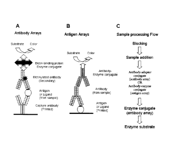
100401 Figures 1A-1C are pictorial diagrams summarizing (IA) the processing
of an
antibody array of the invention for antigen detection, (1B) an antigen array
of the invention
for antibody detection, and (IC) the steps for processing a printed array of
the invention.
100411 Figure 2 is a pictorial diagram summarizing the function and
processing of the
controls spots present in an array printed on an assay membrane of the
invention.
CA 02670615 2009-05-26
WO 2008/067091
PCT/US2007/082732
[0042] Figure 3 is a graphical diagram summarizing the results of Example 5
obtained
from processing human serum spiked with cytokines. The hash line represents
the threshold
signal above which the result is considered positive. The threshold is set at
two times the
signal intensity of the negative control spot (the Buffer spot).
100431 Figure 4 is a graphical diagram summarizing the results Example 6
obtained from
processing human serum sample spiked with antibodies to Hepatitis B surface
antigens. The
hash line represents the threshold signal above which the result is considered
positive. The
threshold is set at two times the signal intensity of the negative control
spot (the Buffer spot).
100441 Figure 5 is a graphical diagram summarizing the results of Example 7
that
assesses the efficacy of a fiduciary marker of one embodiment of the
invention. The x-axis
units identify the tube number and thus the dilution factor.
100451 Figure 6 is a graphical diagram showing the results of Example 10
that
demonstrates upper respiratory viral pathogen arrays for detection of
antibodies in serum
samples. Six human serum samples were tested at a dilution of 1 in 800 for
presence of
antibodies to each of the six viral antigens on arrays. A signal intensity
threshold of 100000
was set for a positive test. Based on this threshold the results shown in
Table 20 were
obtained.
100461 Figure 7 is a graphical diagram showing the results of Example 11
showing the
results from ten human serum samples that were tested at a dilution of 1 in
4000 for presence
of antibodies to each of the four Hepatitis B antigens on arrays.
DETAILED DESCRIPTION OF THE INVENTION
100471 The present invention relates to an assay membrane for detecting of
at least one
target analyte or a plurality of target analytes in a sample, as well as kits
for detecting said
target analytes and a method of processing the assay membrane.
[0048] Biomarkers can identify disease prior to exhibition of clinical
symptoms by a
subject, and therefore provide the ability to treat the molecular basis of
disease using targeted
therapies. The basis for subject stratification lies in correlation of
molecular heterogeneity of
disease with heterogeneity of response to therapy. As such, the target of the
drug must be
CA 02670615 2009-05-26
WO 2008/067091 PCT/US2007/082732
11
present and have a role in maintaining or worsening the disease state of the
subject in order
for the drug to be effective. This target would therefore serve as a biomarker
to determine
whether the subject is a candidate for treatment with that particular therapy.
For example, the
presence of Her2/neu in tumors is required for effective treatment with anti-
Her2 antibodies,
such as Herceptin. Biomarkers can also be used for retrospective sample
analysis after
clinical trials have been completed or after post-marketing analysis of new
drugs to perform
subgroup analysis to identify covariates that were expected to account for
differences in
response.
100491 Accordingly, the present invention contemplates a plurality of
capture agents
arranged to detect one or more (i.e., a panel) target analytes (i.e.,
biomarkers) that may be
used for a variety of assays. For example, a panel of biomarkers may be
monitored during
clinical trials to determine effectiveness of therapy while simultaneously
ensuring lack of side
effects or any other adverse events. Thus, a panel of biomarkers may be used
to test a variety
of conditions and/or to further validate one or more potential biomarkers.
Exemplary
conditions include, but are not limited to, human diseases or allergies,
pregnancy detection,
animal diseases, and animal testing performed prior to export. It should be
understood that
the panel of biomarkers can be used during all phases of clinical trials to
obtain a greater
understanding of drug mechanism in a population prior to approval and general
administration of the drug. Furthermore, biomarkers can support clinical
outcome results
from efficacy studies and help to measure real clinical benefit to the
subject.
100501 Biomarkers can also be used to determine which subjects are likely
to respond to
a particular therapeutic. Biomarkers can also be used to monitor disease
progression and
treatment efficacy by measuring levels of various disease parameters
simultaneously, thereby
increasing the benefit of treatment to the subject. These biomarker panels aim
to identify the
right drug for the right subject at the right time.
[00511 Biomarker validation for prediction of a particular disease;
disorder, or condition
refers to the confirmation of accuracy, reproducibility and effectiveness of
biomarkers in
detecting the disease, disorder or condition. The major challenge for
biomarker validation is
the high level of variability of biomarker levels across the human population
and the
considerable molecular heterogeneity of specific diseases, even from a single
tissue. As a
CA 02670615 2009-05-26
WO 2008/067091
PCT/US2007/082732
12
newly discovered biomarker makes the transition from the research setting to
the clinical
diagnostic laboratory, it should progress through defined stages of
confirmation. The first
task of biomarker validation is evaluation of research technology,
performance, and
specifications (analytical validation). However, the ultimate goal is initial
validation of the
biomarker to identify early stage diseases, disorders, or conditions (clinical
validation). Upon
technical and clinical confirmation, assays involving the biomarker are moved
systematically
toward a standardized, reproducible, high-throughput format for clinical
diagnostic
implementation. With laboratory performance rigorously established, the
clinical variables
can subsequently be analyzed to define limitations, applications, and clinical
utility.
Definitions
100521 Unless defined otherwise, all technical and scientific terms used
herein have the
same meaning as commonly understood by one of ordinary skill in the art to
which this
invention belongs. Although any methods and materials similar or equivalent to
those
described herein can be used in the practice or testing of the invention, the
preferred methods
and materials are now described.
[00531 As used in this specification and the appended claims, the singular
forms "a",
"an", and "the" include plural references unless the context clearly dictates
otherwise. Thus,
for example, references to "the method" includes one or more methods, and/or
steps of the
type described herein which will become apparent to those persons skilled in
the art upon
reading this disclosure and so forth.
100541 The term "biomarker" refers to any substance used as an indicator of
a biologic
state. Thus, a biomarker can be any substance whose detection indicates a
particular disease
state (for example, the presence of an antibody may indicate an infection).
Furthermore, a
biomarker can be indicative of a change in expression or state of a protein
that correlates with
the risk or progression of a disease, or with the susceptibility of the
disease to a given
treatment. Once a proposed biomarker has been validated, it can be used to
diagnose disease
risk, presence of disease in an individual, or to tailor treatments for the
disease in an
individual (e.g., choices of drug treatment or administration regimes). In
evaluating potential
drug therapies, a biomarker may be used as a surrogate for a natural endpoint
such as survival
or irreversible morbidity. If a treatment alters the biomarker, which has a
direct connection
CA 02670615 2009-05-26
WO 2008/067091 PCT/US2007/082732
13
to improved health, the biomarker serves as a "surrogate endpoint" for
evaluating clinical
benefit.
10055] As used herein, the term "assay element" refers to any of a number
of different
elements for use in an array of the invention. Exemplary assay elements
include, but are not
limited to, capture elements and control elements.
10056] The term "capture element" refers to a molecule that is able to bind
to a target
analyte. Examples of useful capture elements include proteins, protein
fragments, binding
proteins, binding protein fragments, antibodies (polyclonal, monoclonal, or
chimeric),
antibody fragments, antibody heavy chains, antibody light chains, single chain
antibodies,
single-domain antibodies (a VHH for example), Fab antibody fragments, Fc
antibody
fragments, Fv antibody fragments, F(ab')2 antibody fragments, Fab' antibody
fragments,
single-chain Fv (scFv) antibody fragments, antibody binding domains, antigens,
antigenic
determinants, epitopes, haptens, immunogens, immunogen fragments, binding
domains; a
metal ion, a metal ion-coated molecule, biotin, avidins, streptavidins;
substrates, enzymes,
abzymes, co-factors, receptors, receptor fragments, receptor subunits,
receptor subunit
fragments, ligands, inhibitors, hormones, binding sites, lectins,
polyhistidines, coupling
domains, and oligonucleotides. Useful capture elements will correspond to and
be able to
bind a specific target analyte, such as a molecule or class of molecules that
are present in a
sample to be tested.
100571 Equally, the term "control capture element" refers to a capture
element that
functions as a control, either a negative control that should not bind any
analyte or a positive
control that will bind a non-target analyte.
100581 The term "control element" refers to an element that is used to
provide
information on the function of the assay, for example binding specificity, the
level of non-
specific background binding, the degree of binding cross-reactivity, and the
performance of
assay reagents and the detection system. Preferred controls useful herein
include at least one
negative control to monitor background signal, at least one negative control
to monitor assay
specificity, at least one positive colourimetric control, and at least one
positive control to
monitor assay performance.
CA 02670615 2009-05-26
WO 2008/067091 PCT/US2007/082732
14
100591 The term "control to monitor assay performance" refers to an element
that forms
one part of a complementary binding interaction during an assay and is
intended to provide
information on the accuracy of the assay result. In one embodiment the
positive control to
monitor assay performance comprises one binding partner of a complementary
binding pair,
where the other binding partner is a sample component or an assay reagent. The
assay
performance control is preferably selected from a target analyte, a binding
partner
corresponding to and able to bind a non-target analyte that will be present in
the sample, a
binding partner corresponding to and able to bind an assay reagent, and a
colourimetric
enzyme label, or any combination of any two or more thereof. An example of a
binding
partner corresponding to and able to bind a non-target analyte that will be
present in the
sample is an anti-Ig antibody that will bind an immunoglobulin present in a
serum sample,
therefore confirming a sample has been added. An example of a binding partner
corresponding to and able to bind an assay reagent is an anti-Ig antibody that
will bind a
secondary immunoglobulin that is used to process the assay, such as
biotinylated anti-target
analyte antibody. Another example of a binding partner corresponding to and
able to bind an
assay reagent is a biotinylated antibody that will bind a streptavidin-
peroxidase conjugate that
is used to process the assay.
[0060] The term "control to monitor assay specificity" refers to an element
that is closely
related to at least one binding partner of a complementary binding pair
present in the assay
and is intended to provide information of the specificity of the complementary
binding. This
control is a negative control that is not expected to generate a detectable
result during normal
assay processing. For example, in an antibody array for antigen detection, the
assay
specificity control would comprise an antibody that should not bind any
antigen in the
sample. Alternatively, in an antigen array for antibody detection, the assay
specificity control
would comprise an antigen that should not bind any antibody in the sample.
100611 The term "fiduciary marker" refers to a coloured marker or label
that will always
be detectable on the membrane, preferably irrespective of the performance of
the assay or
processing of the membrane. The fiduciary marker acts therefore as a "true"
positive control.
100621 The term "microporous membrane" refers to a membrane with protein
binding
characteristics and a narrow pore-size distribution. In one embodiment the
porosity of the
CA 02670615 2009-05-26
WO 2008/067091
PCT/US2007/082732
membrane may determine the exposure time of reagents with membrane bound
components
by controlling the flow rate through the membrane. Microporous membranes for
use in the
present invention comprise nitrocellulose, nylon, polyvinylidene difluoride,
polyester,
polystyrene, polyethersulfone, cellulose acetate, mixed cellulose esters and
polycarbonate.
100631 The term "negative control" refers to an element comprising print
buffer or an
unrelated protein to which no complementary binding partner is intended to be
present in the
assay. Any detectable signal from the negative control can be used to
determine the
background threshold of the assay and the accuracy of any positive results. In
one
embodiment the negative control to monitor background signal is print buffer.
The print
buffer is a solution used to carry and print the capture elements and control
elements onto the
membrane and may comprise buffered saline, glycerol and a surfactant,
preferably a
polysorbate surfactant such as Tween 20. The blocking solution is used to
reduce non-
specific protein biding to the membrane surface and preferably comprises skim
milk, casein,
bovine serum albumin, gelatins from fish, pigs or other species, dextran or
any mixture of any
two or more thereof, preferably in a solution of phosphate buffered saline and
a surfactant
such as Tween 20.
100641 The term "positive colourimetric control" as used herein refers to
an enzyme or
enzyme conjugate that provides a detectable signal upon addition of the enzyme
substrate.
100651 The term "printing" as used herein refers to the placement of the
assay elements
(control and capture elements) on the membrane surface, with or without an
adapter molecule
between the membrane and the element. Preferably the assay elements bind to
the membrane
by covalent or non-covalent interaction. One of skill in the art will
recognize that methods of
placing assay elements on the membrane include printing, spotting or other
techniques known
in the art. For purposes of the present application, the term "printing" can
be used to include
any of the methods for placing the assay elements on the membrane.
100661 The terms "sample" and "specimen" as used herein are used in their
broadest
sense to include any composition that is obtained and/or derived from
biological or
environmental source, as well as sampling devices (e.g., swabs) which are
brought into
contact with biological or environmental samples. "Biological samples" include
those
obtained from an animal (including humans, domestic animals, as well as feral
or wild
CA 02670615 2013-12-12
16
animals, such as ungulates, bear, fish, lagamorphs, rodents, etc.), body
fluids such as urine,
blood, plasma, fecal matter, milk, nipple exudate, cerebrospinal fluid (CSF),
semen, sputum,
and saliva, as well as solid tissue. Biological samples also include a cell
(such as cell lines,
cells isolated from tissue whether or not the isolated cells are cultured
after isolation from
tissue, fixed cells such as cells fixed for histological and/or
immunohistochemical analysis),
tissue (such as biopsy material), cell extract, tissue extract, and nucleic
acid (e.g,, DNA and
RNA) isolated from a cell and/or tissue, and the like. Also included are
materials obtained
from food products and food ingredients such as dairy items, vegetables, meat,
meat by-
products, and waste. "Environmental samples" include environmental material
such as
surface matter, soil, water, and industrial materials, as well as material
obtained from food
and dairy processing instruments, apparatus, equipment, disposable, and non-
disposable
items. In one embodiment, the biological sample is a cell, tissue, and or
fluid obtained from a
mammal, including from the upper respiratory tissues (such as nasopharyngeal
wash,
nasopharyngeal aspirate, nasopharyngeal swab, and oropharyngeal swab), from
the lower
respiratory tissues (such as bronchiolar lavage, tracheal aspirate, pleural
tap, sputum), blood,
plasma, serum, stool, milk, nipple exudate, and tissue from any organ such as,
without
limitation, lung, heart, spleen, liver, brain, kidney, and adrenal glands.
These examples are
illustrative, and are not to be construed as limiting the sample types
applicable to the present
invention.
100671 The term "antibody" as used herein includes naturally occurring
antibodies as
well as non-naturally occurring antibodies, including, for example, single
chain antibodies,
chimeric, bifunctional and humanized antibodies, as well as antigen-binding
fragments
thereof. Such non-naturally occurring antibodies can be constructed using
solid phase
peptide synthesis, can be produced recombinantly or can be obtained, for
example, by
screening combinatorial libraries consisting of variable heavy chains and
variable light chains
(see Huse et al., Science 246:1275-1281, 1989).
These and other methods of making, for example, chimeric, humanized, CDR-
grafted, single
chain, and bifunctional antibodies are well known (Winter and Harris, Immunol.
Today
14:243-246, 1993; Ward et al., Nature 341:544-546, 1989; Harlow and Lane,
Antibodies: A
laboratory manual (Cold Spring Harbor Laboratory Press, 1999); Hilyard et al.,
Protein
Engineering: A practical approach (IRL Press 1992); Borrabeck, Antibody
Engineering,
2d ed. (Oxford University Press 1995)). In
CA 02670615 2009-05-26
WO 2008/067091
PCT/US2007/082732
17
addition, modified or derivatized antibodies, or antigen binding fragments of
antibodies, such
as pegylated (polyethylene glycol modified) antibodies, can be useful for the
present
methods. As such, Fab, F(ab')2, Fd and Fv fragments of an antibody that retain
specific
binding activity are included within the definition of an antibody.
100681 The term "secondary antibody" refers to an antibody that will bind a
target
analyte and that is conjugated with either an adaptor molecule such as biotin
or an enzyme
label such as horseradish peroxidase (HRP). Antibody-adaptor conjugates are
processed to
give a detectable result by contacting the antibody-adaptor conjugate with an
adaptor-enzyme
conjugate and then the enzyme substrate; for example, antibody-biotin
conjugates will bind
streptavidin-HRP conjugates. Antibody-enzyme label conjugates include antibody-
HRP
conjugates. Use of secondary antibodies is discussed and exemplified below.
100691 The term "binds specifically" or "specific binding activity" or the
like, means that
two molecules form a complex that is relatively stable under physiologic
conditions. The
term is also applicable where, an antigen-binding domain is specific for a
particular
epitope, which is carried by a number of antigens, in which case the antibody
carrying the
antigen-binding domain will be able to bind to the various antigens carrying
the epitope.
Specific binding is characterized by a high affinity and a low to moderate
capacity.
Typically, the binding is considered specific when the affinity constant is
about 1 x 10⁻⁶ M,
generally at least about 1 x 10⁻⁷ M, usually at least about 1 x 10⁻⁸ M, and
particularly at least
about 1 x 10⁻⁹ M or 1 x 10⁻¹⁰ M or less.
Array Design
100701 As described above, one aspect of the invention relates to a
microporous
membrane for detecting a plurality (i.e., a panel) of target analytes (e.g.,
biomarkers) in a
sample, the membrane comprising an array that comprises at least one capture
element and a
plurality of control elements printed on the membrane surface, the at least
one capture
element corresponding to and being able to bind a target analyte. When
optionally included,
the plurality of control elements include
i) at least one fiduciary marker,
ii) at least one negative control to monitor background signal,
CA 02670615 2009-05-26
WO 2008/067091 PCT/US2007/082732
18
iii) at least one negative control to monitor assay specificity,
iv) at least one positive colourimetric control,
v) at least one positive control to monitor assay performance or any
combination thereof.
100711 The choice of membrane is dependent on three main membrane
characteristics:
protein-binding capacity, porosity, and strength. The ability of the membrane
to immobilize
macromolecules, in particular proteins is crucial as the membrane serves as
the solid phase
used in the assay. However, this ability must be balanced with the
availability of appropriate
reagents (i.e., blockers) for blocking non-specific interactions on the
membrane. Similarly, in
a flow-through configuration the porosity of the membrane may determine the
exposure time
of reagents with membrane bound components by controlling their flow rate
through the
membrane. However, porosity must be balanced with the degree of array spot
spreading
during array manufacture, which can result in decreased signal intensity or
cross
contamination between adjacent spots. The strength of the membrane is
important for the
manufacture and eventual use of a device. A wide range of membranes are
available with
differing characteristics, allowing a particular membrane to be chosen
depending on the
requirements of an assay.
[0072] In preferred embodiments, microporous membranes for use in the
present
invention comprise nitrocellulose, nylon, polyvinylidene difluoride,
polyester, polystyrene,
polyethersulfone, cellulose acetate, mixed cellulose esters and polycarbonate.
100731 While some membranes such as cellulose acetate may have insufficient
binding
capacities for diagnostic immunoassays, the characteristics of such membranes
may be
applicable for assays where lower levels of accuracy or sensitivity are
sufficient.
100741 The microporous membrane is preferably removably attachable to a
bottomless
microtiter plate. Accordingly, the membrane can be divided into individual
microtiter wells
that are separated from each other by a physical barrier, to prevent sample
mixing between
wells. Moreover, different assays can be conducted in separate wells,
requiring smaller
volumes of assay reagents.
100751 Capture elements specific for a target analyte are used to detect
the presence or
absence of the analyte in a sample. A wide range of complementary binding or
coupling
CA 02670615 2009-05-26
WO 2008/067091
PCT/US2007/082732
19
partners are known, with the choice of capture elements determined by the
analytes to be
detected, the requirement for adapter molecules and the level of specificity
required for the
assay.
100761 In one embodiment the target analyte is selected from a protein, a
protein
fragment, a peptide, a polypeptide, a polypeptide fragment, an antibody, an
antibody
fragment, an antibody binding domain, an antigen, an antigen fragment, an
antigenic
determinant, an epitope, a hapten, an immunogen, an immunogen fragment, a
metal ion, a
metal ion-coated molecule, biotin, avidin, streptavidin, an inhibitor, a co-
factor, a substrate,
an enzyme, a receptor, a receptor fragment, a receptor subunit, a receptor
subunit fragment, a
ligand, a receptor ligand, a receptor agonist, a receptor antagonist, a
signalling molecule, a
signalling protein, a signalling protein fragment, a growth factor, a growth
factor fragment, a
transcription factor, a transcription factor fragment, an inhibitor, a
monosaccharide, an
oligosaccharide, a polysaccharide, a glycoprotein, a lipid, a cell, a cell-
surface protein, a cell-
surface lipid, a cell-surface carbohydrate, a cell-surface glycoprotein, a
cell extract, a virus, a
virus coat protein, a hormone, a serum protein, a milk protein, an
oligonucleotide, a
macromolecule, a drug of abuse, or any combination of any two or more thereof.
100771 In one embodiment the capture element is selected from a protein, a
protein
fragment, a binding protein, a binding protein fragment, an antibody, an
antibody fragment,
an antibody heavy chain, an antibody light chain, a single chain antibody, a
single-domain
antibody (a VHH for example), a Fab antibody fragment, an Fc antibody
fragment, an Fv
antibody fragment, a F(ab')2 antibody fragment, a Fab' antibody fragment, a
single-chain Fv
(scFv) antibody fragment, an antibody binding domain, an antigen, an antigenic
determinant,
an epitope, a hapten, an immunogen, an immunogen fragment, a binding domain;
metal ion,
or metal ion-coated molecule, biotin, avidin, streptavidin; a substrate, an
enzyme, an abzyme,
a co-factor, a receptor, a receptor fragment, a receptor subunit, a receptor
subunit fragment, a
ligand, an inhibitor, a hormone, a binding site, a lectin, a polyhistidine, a
coupling domain, an
oligonucleotide, or a combination of any two or more thereof.
100781 In another embodiment, the complementary binding partners comprise
antibody-
antigen interactions or antibody-ligand interactions.
CA 02670615 2009-05-26
WO 2008/067091 PCT/US2007/082732
100791 In another embodiment, the capture elements may comprise antibodies
or
fragments thereof that are immobilised on the membrane surface and are
specific for different
antigens or ligands that may be present in a sample.
100801 In another embodiment, the capture elements may comprise antigens or
ligands
and the assay involves the detection of specific antibodies that may be
present in a sample.
10081] In further embodiments, the capture elements may comprise of a
receptor or a
subunit of a receptor that binds a specific ligand.
100821 In one embodiment the target analyte is associated with an
infectious disease,
allergic disease, autoimmune disease, cardiac disease, cancer or graft versus
host disease.
100831 In one embodiment the target analyte is selected from the list
comprising
angiogenesis factors such as Ang-2, FGF basic, HB-EGF, HGF, KGF, PDGF-BB, TIMP-
1,
TIMP-2, TPO and VEGF; Biomarkers such as A-SAA, Acrp-30 (Adiponectin), AR
(Amphiregulin), Apo A-1, Apo B-100, C-peptide, sCD14, sCD30 (TNFRSF8), CD4OL,
CRP
(C-reactive protein), ErbB2, FasL, Fibrinogen, Fibronectin, IGFBP-1, IGFBP-3,
Leptin, LIF,
MPO (Myeloperoxidase), NT-proBNP, OPG (Osteoprotegrin), OPN (Osteopontin), PAI-
1
Active, PAI-1 Total, PAPP-A, P1GF (Placental Growth Factor), Prolactin, RANK,
RANKL,
Resistin, Tissue Factor and TRAIL; Cell Adhesion Molecules such as E-Cadherin,
E-
Selectin, ICAM-1, L-Selectin, P-Selectin and VCAM-1, Chemokines such as ENA-
78,
Eotaxin, Eotaxin-2, Exodus-2, GROa, GROy, HCC-4 (CCL-16), 1-309, IP-10, ITAC,
Lymphotactin, MCP-1, MCP-2, MCP-3, MCP-4, MDC, MIF, MIG, MIP-la, MIP-1 0, MIP-
16, MIP-3a, MIP-313, MIP-4 (PARC), MPIF-1, NAP-2, RANTES, SDF-1 p and TARC;
Cytokines such as GM-CSF, G-CSF, IFNa, IFNy, IL-la, IL-
lra, IL-2, IL-3, IL-4, IL-
5, IL-6, IL-7, IL-8, IL-9, IL-10, IL-11, IL-12p40, IL-12p70, IL-13, IL-15, IL-
16, IL-17, IL-18
and TNFa; Cytokine receptors such as IL-2R, IL-2Ry, IL-6R, TNF-RI and TNF-RII;
Growth
Factors such as, EGF, HGH, TGFa and TGFf3; Immunoglobulins such as IgA, IgD,
IgE, IgG
and IgM; Matrix Metalloproteinases such as MMP-1, MMP-2, MMP-3, MMP-7, MMP-8,
MMP-9, MMP-10 and MMP-13; and Neurotrophic Factors such as P-NGF, BDNF, CNTF
and NT3, or any combination of any two or more thereof.
CA 02670615 2009-05-26
WO 2008/067091
PCT/US2007/082732
21
100841 In another embodiment the target analyte is an antigen from a
family, genus,
species, subtype or individual microorganism. Exemplary microorganisms
include, but are
not limited to, Mycobacterium, BruceIla, Bacillus, Treponema, Clostridium,
Staphylococcus,
Enterococcus, Streptococcus, Haemolyticus, Pseudomonas, Campylobacter,
Enterobacter,
Neisseria, Proteus, Salmonella, Simonsiella, Riemerella, Escherichia,
Neisseria,
Meningococcus, Moraxella, Kingella, Chromobacterium and Branhamella, or from a
virus
such as adenovirus, influenza, cytomegalovirus, hepatitis, human
immunodeficiency virus,
avian influenza virus, respiratory syncytial virus, herpex simplex virus,
parainfluenza virus,
pestivirus, porcine parvovirus, peudorabies virus, rotavirus, calicivirus,
canine distemper
virus, or from other microorganisms such as Leptospira, Toxoplasma,
Trypanosoma, or
Plasmodium, or any combination of any two or more thereof.
100851 Other exemplary target analytes include, but are not limited to,
human chorionic
gonadotropin, growth hormone, insulin, glucagon, adrenocorticotropic hormone,
thyroid
stimulating hormone, a-fetoprotein, human placental lactogen, leptin, inhibin
A, activin A,
pregnancy-associated plasma protein A, placenta growth factor, pregnancy-
specific beta-1
glycoprotein; steroids such as testosterone, oestriol, cortisol, progesterone,
corticosterone,
aldosterone; thyroid hormones such as thyroxine, triiodothyronine; thyroid
binding globulin
(TBG); active peptides such as bradykinin, gastrin, angiotensin, thyroid
hormone-releasing
hormone, luteinising hormone-releasing hormone; physiologically active amines
such as
epinephrine, norepinephrine, histamine, serotonin; prostaglandins, such as
PGF2a, PGE,
thromboxanes and prostacyclins, or any combination of any two or more thereof.
100861 In another embodiment the target analyte is an allergen. Exemplary
allergens
include, but are not limited to, indoor allergens such as Mites, Tyr. put,
Lep. dest. or .mayrei,
Felis, Bos, Albumine, Pen. cit., Pen. not., Asp. fumigatus, Alt. alt.,
Malassezia furfur, Latex,
Plodia, Blatella; outdoor allergens such as Betula, Juniperus, Phleum,
Parietaria and judicea;
representative allergens from cats, dogs, mouse, rat, pig, a sheep, chicken,
rabbit, a hamster, a
horse and pigeon, food allergens such as celery, carrot, peanut, apple, shrimp
and fish; venom
allergens such as bee or wasp, auto-allergens such as liver membrane antigens,
ssDNA
antigens and antigens in or on skeleton muscle cells, and any combination of
any two or more
thereof.
CA 02670615 2009-05-26
WO 2008/067091 PCT/US2007/082732
22
[0087] In another embodiment, one or more capture agents are arranged to
detect one or
more (i.e., a panel) target analytes (i.e., biomarkers) that would be
indicative of particular
human conditions or diseases. Such tests could consist of any combination of
the panels
listed in Table 1, depending upon local requirements.
Table 1: Human Condition/Disease Panels
Infectious disease screening for epidemiological Human immunodeficiency
virus (HIV)-1, HIV2,
studies in developing nations Hepatitis A virus, Hepatitis B virus,
Hepatitis C virus,
Herpes simplex virus (HSV)-1, HSV-2, Treponema
pallidum, Mycobacterium tuberculosis, Neisseria
gonorrhoeae, Plasmodium falciparum.
Upper respiratory viral infections Adenovirus, Cytomegalovirus (CMV),
Influenza A,
Influenza B, Parainfluenza 1, Parainfluenza 2,
Parainfluenza 3 and Respiratory Syncytial Virus
(RSV), Group A Streptococci.
Acute lower respiratory infections Streptococcus pneumoniae, Haemophilus
influenzae,
Mycoplasma pneumoniae, Chlamydia pneumoniae,
Moraxella catarrhalis, C-reactive protein,
procalcitonin.
Gastrointestinal pathogen panel Salmonella, Shigella, Campylobacter,
Vibrio.
Panel for liver viral disease testing Hepatitis virus A, B, C, D, E and G
surface and core
antigens, anti-Hepatitis A, B, C, D, E and G serum
antibodies.
Sexually transmitted disease panel Human immunodeficiency virus (HIV)-1,
HIV2,
Treponema pallidum, Neisseria gonorrhoeae,
Chlamydia trachomatis.
Blood borne disease panel Plasmodium falciparum (malaria),
Trypanosoma cruzi
(Chagas disease), Brucella spp (Brucellosis), Human
immunodeficiency virus (HIV)-1, HIV2, Hepatitis A
virus, Hepatitis B virus.
ToRCH panel Toxoplasma gondii, Rubella virus,
Cytomegalovirus
and Herpes simplex virus I and HSV2.
Biosecurity panel Bacillus anthracis (anthrax),
Clostridium botulinum
(botulism), Clostridium perfringens, Yersinia pestis
(plague), Coxiella burnetii (Q fever), Staphylococcal
enterotoxin B, Vibrio cholerae (Cholera).
Fertility panel Estradiol, follicle stimulating hormone,
human
chorionic gonadotrophin, lutenizing hormone,
progesterone, prolactin, testosterone, parathyroid
hormone.
Drugs of abuse panel Acetaminophen, Amphetamines,
Barbiturates,
Cannabinoids, cocaine metabolites, methadone,
opiates, salicylate and tricyclic anti-depressants.
Panel for cardiovascular disease testing Brain natriuretic pepetide (BNP),
N-terminal proBNP
(Nt-proBNP), creatine kinase (CK)-MB, myoglobin,
cardiac Troponin I, cardiac Troponin T, High-
sensitivity C-reactive protein.
Panel for autoimmune disease testing Rheumatoid factor, C-reactive protein,
soluble human
leukocyte antigen (HLA)-DR, antibodies against
double stranded DNA, citrullinated peptides, small
nuclear ribonucleoproteins, neutrophil cytoplasmc
(ANCA) and nuclear antigens (ANA).
CA 02670615 2009-05-26
WO 2008/067091 PCT/US2007/082732
23
Panel for measuring hormone levels Insulin, Leptin, Thyroxin3 and 4 ,
Thyroid Stimulating
Hormone (TSH), growth hormone, testosterone,
estrogen, leutenizing hormone.
Panel for general cancer testing Free and total Prostate specific antigen
(PSA),
Carcinoembryonic antigen (CEA), CA125, CA15-3,
CA19-9, CA24-2, CA72-4, alpha fetoprotein (AFP).
Markers of Inflammation Interleukin (IL)-la and p, ILI receptor
antagonist,
IL2, IL4, IL6, IL8, ILI 0, IL12, IL13, IFNy, TNFa,
MIPla and 13, MCP1, RANTES, soluble VCAM, C-
reactive protein, soluble TNFa receptor I and II.
Allergen panel to screen for serum IgE binding Allergens obtained by
recombinant methods or derived
from dust mites, grass and tree pollen, animal dander,
moulds, insect venoms and foods such as soy protein,
milk proteins, proteins derived from varieties of nuts,
cereals and legumes, proteins from seafood such as
shrimp, abalone and lobsters.
100881 In another embodiment, one or more capture agents are arranged to
detect one or
more (i.e., a panel) target analytes (i.e., biomarkers) that would be useful
in determining
treatment efficacy for the indicated diseases. Such tests could consist of any
combination of
the panels listed in Table 2, depending upon local requirements.
Table 2: Panels to Determine Efficacy of Treatments
Autoimmune diseases Cytokines and chemokines including
CTLA4,
tumor necrosis factor alpha (TNFa), bLyS
(BAFF), interferon gamma (IFNy), eotaxin,
CXCL10 or IP I 0, osteopontin, osteoprotegerin
and RANKL.
Other biomolecules such as pyridinoline,
deoxypyridinoline, cartilage oligomeric matrix
protein.
Antibodies against double stranded DNA, small
nuclear ribonucleoproteins, nuclear proteins such
as Sjogren's Syndrome antigen (SS)-A and SS-B,
nuclear antigen, Sm antigen, ribosomal P
proteins, cardiolipin and topoisomerase I.
Antibodies against therapeutic proteins such as
abatacept (an immunoglobulin fused to the
ectodomain of CTLA4), rituximab (a chimeric
anti-CD20 antibody), toclizumab (anti-1L6
receptor antibody), etanercept (recombinant
soluble human TNFa fused to IgG), infliximab (a
chimeric anti-TNFa antibody), adalimumab (a
humanized anti-TNFa antibody), anakinra (a
human IL6 receptor antagonist protein) and
others.
CA 02670615 2009-05-26
WO 2008/067091
PCT/US2007/082732
24
Cancer Human chorionic gonadotrophin,
Tyrosinase,
HMGB1, S100-beta, melanoma inhibitory
activity (MIA), soluble HLA-DR, matrix
metalloproteinases (MMP)) such as MMP-1 and
MM P-9, cytokines including Interleukin (IL)-6,
IL8 and ILIO, high molecular weight melanoma-
associated antigen (HMW- MAA), haptoglobin,
osteopontin, moiesin, transferrin, FK506,
haptoglobin precursor protein, progesterone
receptor, estrogen receptor, serine protease
urokinase-type plasminogen activator,
plasminogen activator inhibitor Type I, human
papilloma virus, Epstein-Barr virus, Glutathione
S-transferase PI.
Antibodies against therapeutic proteins such as
rituximab (a chimeric anti-CD20 antibody),
cetuximab (a chimeric anti-EGF antibody),
trastuzumab (humanized anti-Her2/ neu
antibody), tositumomab (mouse monoclonal anti-
CD20 antibody), gemtuzumab (humanized anti-
CD33 antibody), bevacimumab (a humanized
anti-VEGF antibody), alemtuzumab (a humanized
anti-CD52 antibody) and Ibritumomab tiuxetan
(mouse anti-CD20 antibody).
Cardiovascular diseases and stroke risk Interleukin (IL)-1, IL-6, IL-10,
ischemia-
modified albumin, monocyte chemoattractant
assessment
protein (MCP)-1, plasminogen activator-1,
TNFa, von Willebrand factor, soluble CD40
ligand, myeloperoxidase, placental growth factor,
fibrinogen, and heart-type fatty acid binding
protein (H-FABP), matrix metalloproteinase
(MMP)-9, B-type neurotrophic growth factor
(BNGF), serum amyloid A, fibrinogen, sICAM
and S-100b.
100891 In
another embodiment, one or more capture agents are arranged to detect one or
more (i.e., a panel) target analytes (i.e., biomarkers) that would be useful
in animal testing.
Such tests could consist of any combination of the panels listed in Table 3,
depending upon
local requirements.
Table 3: Animal Testing Panels
Avian Avian influenza virus, Avian
pneumovirus, Avian
reovirus, avian rhinotracheitis virus,
Chicken anemia virus.
CA 02670615 2009-05-26
WO 2008/067091 PCT/US2007/082732
Bovine Bovine Adenovirus, Bovine Coronavirus,
Leptospira spp, Bovine leukosis Virus, Bovine
respiratory syncytial virus, bovine spongiform
encephalopathy, bovine viral diarrhoea virus,
BruceIla abortus , Neospora caninum,
Mycoplasma bovis, Bovine babesiosis, Rotavirus,
contagious bovine pleuropneumonia, bovine
Herpes Virus Type I and II, bovine parainfluenza
3.
Canine Canine distemper virus, canine
coronavirus,
canine herpes virus, canine parvovirus, Borrelia
burgdorferii, Rickettsia rickettsii, Ehrlichia canis,
Rickettsia conori, canine rheumatoid factor, dog
erythrocyte antigen, canine Hepatitis virus 1 and
2, canine parainfluenza 1, Borrelia afzelii,
Leishmania donovani, Ehrlichia equi, Rickettsia
conorii.
Equine Equine arteritis virus, equine
infectious anemia
virus, equine herpesvirus Type I, equine
adenovirus, equine influenza virus, Babesia equi,
Babesia caballi, Borrelia burgdorferii, Borrelia
afzelii, Ehrlichia equi, Leishmania donovani.
Feline Feline coronavirus, feline calicivirus,
feline
leukemia virus, feline herpesvirus, Feline
immunodeficiency virus, feline infectious
peritonitis virus, feline panleukopaenia virus,
feline viral rhinotracheitis virus, Feline Enteric
Corona Virus.
Porcine pathogens Porcine influenza A virus, porcine
parvovirus,
porcine reproductive and respiratory syndrome
virus, Pseudorabies virus, porcine rotavirus,
porcine BruceIla suis, transmissible
gastroenteritis (TGE) virus, classical swine fever
virus, porcine respiratory coronavirus.
Ovine pathogens Ovine Herpes virus, BruceIla ovis,
pseudorabies
virus (Aujesky's).
Protein and endocrine panel for all species Estrone sulfate, progesterone,
growth hormone,
serum cortisol, testosterone, thyroxine (T)-3, T-4,
Serum albumin, serum globulin, insulin,
parathyroid hormone, thyroid stimulating
hormone, leutenizing hormone.
Panel of pathogens to test for export Bovine viral diarrhoea virus,
enzootic bovine
leucosis virus, bovine Herpes Virus Type I,
Maedi visna virus, Brucella ovis, Mycobacterium
paratuberculosis (Johne's disease),
Campylobacter fetus, Trichomonas foetus,
Leptospira spp, Streptococcus equi, Infectious
bovine rhinotracheitis virus.
CA 02670615 2009-05-26
WO 2008/067091
PCT/US2007/082732
26
Panel for Mastitis testing of cattle
Streptococcus agalactiae, Streptococcus uberis,
Staphylococcus aureus, Mycoplasma spp,
Eschericia colt, Klebsiella spp, Pseudomonas spp,
Prototheca spp., Haptoglobin, serum amyloid A,
immunoglobulins, lactoferrin, serum albumin.
Markers of Inflammation Interleukin (IL)-la and 13, IL1 receptor
antagonist, IL2, 1L4, IL6, IL8, IL10, IL12, IL13,
IFNy, TNFa, MIPla and 13, MCP1, RANTES,
soluble VCAM, C-reactive protein, soluble TNFa
receptor I and II.
100901 After array manufacture and prior to sample addition, all available
protein-
binding sites on the membrane surface are blocked by addition and incubation
with one or a
combination of reagents. These reagents are called "Blockers" and serve to
decrease or at
best eliminate non-specific protein binding from the sample on the membrane
surface thereby
decreasing overall background signal. This increases the ratio of signal to
noise, thereby
increasing the overall sensitivity of the assay. Blockers play no active part
in the subsequent
reactions between the sample and other assay reagents and the immobilized
proteins on the
membrane. Exemplary blockers include, but are not limited to, bovine serum
albumin,
casein, non-fat dry milk, gelatin derived from fish, pigs and other sources,
dextran, serum
derived from sources other than the sample being analysed such as from
steelhead salmon,
guinea pigs, hamsters, rabbit and other sources, polyethylene glycol,
polyvinyl pyrrollidone,
and commercial preparations including HeteroBlock (Omega Biologicals, Bozeman,
MT),
SuperBlock, StartingBlock, SEA BLOCK (Pierce, Rockford, IL). Typically,
blockers are
made up in buffer solutions such as, for example, phosphate buffer, phosphate
buffered
saline, Tris buffer, acetate buffer and others. The blockers may also be
supplemented with
detergents such as, for example, Tween 20, Tween 80, Nonidet P40, sodium
dodecyl sulfate
and others.
[0091] The
membrane of the invention comprises at least one fiduciary marker that will
always be detectable on the membrane, preferably detectable irrespective of
the performance
of the assay or processing of the membrane.
100921 In
preferred embodiments the fiduciary marker is a dye, dye-conjugated protein
or a chromogenic protein such as haemoglobin.
CA 02670615 2009-05-26
WO 2008/067091 PCT/US2007/082732
27
[0093] The use of at least one fiduciary marker will obviate the necessity
of this element
being detected based on successful array processing, in comparison to the
positive
colourimetric controls. The fiduciary marker is therefore a "true" positive
control that would
always be detectable regardless of array processing, and can be used to orient
and help to grid
the array.
[0094] The membrane of the invention also comprises at least one control to
monitor
assay specificity. The control is intended to provide information of the
specificity of binding
between the capture element and the target analyte, or between the binding
partners of the
assay detection steps.
100951 In one embodiment the assay specificity control comprises one or
more antibody
isotypes, a corresponding antibody or antibody isotype from a different animal
species or a
closely related ligand. For example, in human antibody arrays, human IgM and
anti-human
IgM can be used as controls to monitor assay specificity.
100961 The membrane of the invention also comprises at least one control to
monitor
assay performance. The control is intended to provide information of the
efficiency of the
complementary binding interactions or the quality or performance of the
reagents used.
100971 In one embodiment the assay performance control comprises one
binding partner
of a complementary binding pair, wherein the other binding partner is an assay
reagent. The
assay performance control is preferably selected from the list comprising the
target analyte, a
non-specific binding partner or a colourimetric enzyme label.
[0098] In one embodiment the positive colourimetric control is an enzyme
label
conjugate capable of reacting with a colourimetric substrate, comprising an
enzyme selected
from the list comprising horseradish peroxidase, alkaline phosphatases, 13-D-
galactosidase or
glucose oxidase.
[00991 The identity of the assay controls will be dependent on the type of
array, the
identity of the target analyte, and the type of sample to be analyzed.
[0100] For example, either anti-human IgG-HRP or anti-mouse IgG-HRP may be
used in
arrays printed with antigens and antibodies, respectively. The final detection
antibody in
CA 02670615 2009-05-26
WO 2008/067091 PCT/US2007/082732
28
antigen arrays will often be anti-human IgG-HRP, while for antibody arrays it
will often be a
biotinylated mouse IgG. These controls can provide a positive control in
addition to
providing information on the performance or quality of the HRP substrate.
101011 Mouse IgG, human IgG and anti-human IgG present on antigen or
antibody
arrays can act either as positive or negative controls depending on the array
format, in
addition to providing information of assay specificity. For example, mouse IgG
should
provide the positive signal in antibody arrays, while the latter two should
provide a positive
signal in antigen arrays. In allergen arrays, human IgM and anti-human IgM may
be replaced
as controls with human IgE and anti-human IgE. These controls can also serve
as controls
for overall assay performance.
[01021 In preferred embodiments the elements on the array are printed in
discrete areas
of between 100 gm to 500 gm in diameter. More preferably, the discrete areas
are between
350 gm to 400 gm in diameter.
101031 In preferred embodiments, the discrete areas of the array are
printed in a 5 x 5
grid. Preferably the array comprises up to nine control elements and two
replicates of each of
eight different capture elements.
101041 In preferred embodiments the capture elements are printed in two or
more
replicates of four different capture elements and multiples thereof.
Detection of Target Analytes
101051 The assay techniques used in conjunction with the membranes of the
present
invention include any of a number of well known colourimetric enzyme-linked
assays.
Examples of such systems are well known in the art. The assay techniques are
based upon
the formation of a complex between a complementary binding pair, followed by
detection
with a colourimetric detection system comprising an enzyme-conjugate label and
a
colourimetric substrate. In the present invention, the solid phase carrier or
substrate is a
microporous membrane. The detection system will be described with reference to
enzyme-
linked immunosorbent assays (ELISA), though a skilled person would appreciate
that such
techniques are not restricted to the use of antibodies but are equally
applicable to any
colourimetric assay.
CA 02670615 2009-05-26
WO 2008/067091 PCT/US2007/082732
29
101061 Figure 1 shows a schematic representation of assay formats and
sample
processing flow. Panel (A) shows the processing steps of an antibody array for
detection of
antigens or ligands from biological test samples. Panel (B) shows the
processing of an
antigen or ligand array for detection of antibodies in biological test
samples. Panel (C) shows
the general sample processing flow in which each of the reagents described
below and in the
Examples are added to the array printed according to Example 1.
101071 Figure 2 shows a schematic representation of function of control
elements and
their binding to various reagents added during processing of an antibody assay
for antigen
detection. The addition of various reagents is shown on the left of the Figure
with sequential
additions being made from bottom to top. Color is developed only if the
appropriate
functional reagent binds to the control element.
101081 In one embodiment the ELISA is in the "sandwich" assay format. In
this format
the target analyte to be measured is bound between two antibodies ¨ the
capture antibody and
the detection antibody. In another embodiment the ELISA is a non-competitive
assay, in
which an antibody binds to the capture antigen and the amount of bound
antibody is
determined by a secondary detection antibody.
101091 In another embodiment the ELISA is a competitive assay, where a
labelled
antigen is used instead of a labelled antibody. Unlabelled antigen and the
labelled antigen
compete for binding to the capture antibody and the amount of target analyte
bound can be
determined by the proportion of labelled antigen detected.
10110] Either monoclonal or polyclonal antibodies may be used as the
capture and
detection antibodies in sandwich ELISA systems. Monoclonal antibodies have an
inherent
monospecificity toward a single epitope that allows fine detection and
quantitation of small
differences in antigen. A polyclonal antibody can also be used as the capture
antibody to
bind as much of the antigen as possible, followed by the use of a monoclonal
antibody as the
detecting antibody in the sandwich assay to provide improved specificity. A
monoclonal
antibody can also be used as the capture antibody to provide specific analyte
capture,
followed by the use of a polyclonal antibody as the detecting antibody in the
sandwich assay.
CA 02670615 2009-05-26
WO 2008/067091 PCT/US2007/082732
[OM] An important consideration in designing an array is that the capture
and detection
antibodies of each binding pair must recognise two non-overlapping epitopes so
that when
the antigen binds to the capture antibody, the epitope recognised by the
detection antibody
must not be obscured or altered. A large number of complementary binding pairs
have
already been developed for ELISA and can be used in the present invention.
[0112] For multiplexed assays it is also important that there is no overlap
between each
of the binding pairs to eliminate crossreactivity. A number of multiplexed
ELISAs have been
developed and it is anticipated other combinations of binding pairs could be
configured
through testing.
10113] In one embodiment the enzyme-conjugate label comprising an enzyme
selected
from the list comprising horseradish peroxidase, alkaline phosphatase, 13-D-
galactosidase or
glucose oxidase.
[0114] In one embodiment the enzyme label may be conjugated directly to a
primary
antibody or introduced through a secondary antibody that recognises the
primary antibody. It
may also be conjugated to a protein such as streptavidin if the primary
antibody is biotin
labelled.
101151 In one embodiment the assay detection system comprises a detection
colourimetric substrate selected from the list comprising 3, 3', 5, 5'-
tetramethylbenzidine,
diaminobenzidine, metal-enhanced diaminobenzidine, 4-chloro- I-naphthol,
colloidal gold,
nitro-blue tetrazolium chloride, 5-bromo-4-chloro-3 '-indolylphosphate p-
toluidine salt and
naphthol AS-MX phosphate + Fast Red TR Salt.
[0116] In preferred embodiments the colourimetric reaction can be detected
and
optionally quantified and analysed using an image capture device such as a
digital camera or
a desktop scanner attached to a computer. Known methods for image analysis may
be used.
For example, the density values of known standard elements can be used to
generate standard
curves. Density values for unknown analytes can be analysed using the standard
curve for
each analyte to calculate actual concentrations. Values for each analyte can
be identified
based on the spotting position of each capture element within the array.
CA 02670615 2009-05-26
WO 2008/067091 PCT/US2007/082732
31
[0117] Membranes of the present invention are particularly amenable to use
in kits for
the detection of target analytes. Such kits may comprise the membranes
together with
instructions and any assay consumables required. Different kits are envisaged
for different
target analytes and types of array. Accordingly, in one aspect the invention
relates to a kit
comprising a membrane of the invention and optionally, one or more processing
reagents.
For example, a kit of the invention optionally includes one or more of, or any
combination of
any two or more of
(a) a background reducing reagent (otherwise known as a blocking solution),
(b) a wash solution,
(c) one or more antibodies (including antibody-binding protein conjugates
or antibody-
enzyme label conjugates or both) for detection of antigens, ligands or
antibodies
bound to the capture elements or for detection of the positive controls,
(d) a colourimetric detection system,
(e) software for determination of signal intensity at each spot and
analysis of results, and
(0 a protocol for measuring the presence of analytes in samples.
[0118] In another aspect the invention also relates to a method of
processing a membrane
of the invention. Such a method comprises
(a) providing a membrane of the invention as described above,
(b) adding at least one sample to the membrane, and
(c) processing the membrane such that a detectable result is given by two
or more of
i) at least one fiduciary marker,
at least one positive colourimetric control, and
iii) at least one positive control to monitor assay performance.
101191 In one embodiment the step of processing the membrane comprises a
blocking
step during which available protein-binding sites on the membrane are blocked
with a
blocker, an optional wash step, contacting the membrane with the sample
containing the one
or more analytes to be measured, a wash step to remove non-bound material from
the
membrane, contacting the membrane with one or more secondary antibodies that
correspond
to and will bind one or more target analytes and non-target analyte that is
bound to an assay
performance control, a wash step, and contacting the membrane with one or both
of an
CA 02670615 2009-05-26
WO 2008/067091 PCT/US2007/082732
32
enzyme conjugate or an enzyme substrate to generate a detectable result.
Examples of
processing a membrane of the invention are described below.
101201 In another embodiment, the microporous membranes of the invention
can be used
for the simultaneous detection of at least one target analyte in a sample, and
preferably a
plurality of different target analytes in a sample, and have utility in
diagnostic and screening
assays.
101211 Thus, the microporous membranes of the invention provide the
advantage that
they can be adapted to high throughput (or ultra high throughput) analysis
and, therefore, any
number of samples (e.g., 96, 1024, 10,000, 100,000, or more) can be examined
in parallel,
depending on the particular support used. A particular advantage of adapting
the
microporous membranes to high throughput analysis is that an automated system
can be used
for adding or removing reagents from one or more of the samples at various
times, for adding
different reagents to particular samples, or for subjecting the samples to
various heating
cycles.
101221 For example, the automated system may consist of one or more
temperature-
controlled chambers and one or more robotic arms mounted on a deck that has
platforms
configured to hold 96-well plates. The movement of the robotic arms and the
temperature in
the chambers are controlled by a central computer unit. The array plates are
stacked on the
deck of the instrument. In one embodiment, the plates containing samples to be
analysed are
stacked in a chamber with temperature of 4 C. One robotic arm then
sequentially transfers
each individual array plate on one platform while the other arm sequentially
transfers each
individual sample plate on the second platform. A nozzle containing 96
disposable tips then
aspirates a predetermined volume of sample from each well of the sample plate
and transfers
the sample to the corresponding wells of the array plate. The array plate
containing the
sample is then transferred to a chamber with temperature of 37 C. This process
is repeated
until sample has been added to all the array plates stacked on the deck. The
array plates are
incubated for a predetermined time followed by transfer of each plate to the
platform for
addition of wash buffer with the nozzle containing 96 disposable tips. The
wash buffer is
aspirated after a predetermined time and this wash process is repeated
multiple (i.e., two or
more) times. Each array plate then receives the secondary antibody followed by
transfer to a
CA 02670615 2009-05-26
WO 2008/067091
PCT/US2007/082732
33
chamber with temperature of 37 C. The array plates are incubated for a
predetermined time
followed by transfer of each plate to the platform for addition of wash buffer
with the nozzle
containing 96 disposable tips. The wash buffer is aspirated after a
predetermined time and
this wash process is repeated multiple (i.e., two or more) times. Each array
plate then receives
the detection reagent followed by incubation for a predetermined time followed
by transfer of
each plate to the platform for addition of wash buffer with the nozzle
containing 96
disposable tips. The wash buffer is aspirated after a predetermined time and
the plate
transferred to the 37 C chamber for drying. The plates are transferred back to
the deck after a
predetermined period and manually processed for analyses of data.
101231 In
addition to the convenience of examining multiple test agents and/or samples
at the same time, such high throughput assays provide a means for examining
duplicate,
triplicate, or more aliquots of a single sample, thus increasing the validity
of the results
obtained, and for examining control samples under the same conditions as the
test samples,
thus providing an internal standard for comparing results from different
assays.
[0124]
Various aspects of the invention will now be illustrated in non-limiting ways
by
reference to the following examples.
EXAMPLES
Example 1
General Procedures for Array Manufacture
[0125]
Membranes or films attached to a bottomless 96-well polystyrene plate (such as
nylon, Nalge Nunc International, USA,) were used for printing microarrays.
Various
methods may be used to attach the membranes to the 96-well bottomless plates.
In this
example, a rubber sheet with dimensions of 128mm length and 86mm and lmm
thickness
was used. 96 round holes were stamped on the sheet with a diameter of 6.35mm
and a center
to center distance of 9mm. The sheet was then coated with adhesive on both
sides and glued
to one side of the 96-well bottomless plate such that the second adhesive
layer was still
available for binding to a membrane. A nylon membrane was then cut to the
dimensions of
128mm length and 86mm width and attached to the other side of the rubber sheet
to create a
gasket that creates leak proof wells.
CA 02670615 2009-05-26
WO 2008/067091 PCT/US2007/082732
34
[0126] Another method for attaching the membranes to the 96-well bottomless
plates
involves use of an adhesive that is applied to one side of the plate.
Thereafter, a nylon
membrane cut to the dimensions of 128mm length and 86mm width is attached
using
pressure such that there is no leakage between the wells.
101271 Microarrays were printed by mixing proteins in a print buffer
solution containing
phosphate buffered saline, glycerol and Tween 20 to give a final concentration
of 10%
glycerol and 0.005% Tween 20.
101281 Arrays were printed by using a benchtop contact microarrayer
(LabNext Inc,
USA) using quill pins designed to give uniform spots with an average diameter
of 350 gm
(ChipMaker II, Telechem International Inc, USA). Arrays were printed at
ambient
temperature and humidity of 60% ( 10%).
[0129] Up to ninety-six replicate arrays were printed on the membrane. Each
array had
up to 25 spots printed in 5X5 grids (Number of Columns X Number of Rows).
Arrays with
less than 25 spots were printed such that they contained 5, 10, 15 or 20 spots
in patterns of
5X1, 5X2, 5X3 and 5X4 spots.
101301 Each array had a series of control spots that were printed in Column
1 and Row 5.
These control spots included a fiduciary marker (a dye-conjugated protein such
as
BlueRanger Prestained Protein Molecular Weight Marker, Pierce Biotechnology
Inc, USA,
Catalog Number 26681), negative control (phosphate buffered saline containing
20%
glycerol and 0.005% Tween 20 and a non-specific antibody), positive controls
(enzyme-
conjugated protein such as streptavidin conjugated horseradish peroxidase,
Pierce, Catalog
Number 21126) and sample specific controls to monitor the overall performance
of the assay.
[0131] Test proteins were printed at concentrations ranging from 0.05 mg/
ml to 1.0
mg/ml, usually 0.5 mg/ml, determined by the binding affinity of the specific
protein to the
analyte being measured.
10132] After printing the arrays were kept at 4 C for at least 8 hours
before use.
CA 02670615 2009-05-26
WO 2008/067091 PCT/US2007/082732
Example 2
General Procedure for array processing and analysis
[0133] The arrays were incubated at 37 C for 60 min after adding 100 !al of
Blocker [1%
casein (Vector Labs, USA) in phosphate buffered saline containing 0.1% Tween
20 (PBS-T)]
to each well. The Blocker was then aspirated off.
[0134] Samples containing analytes were added by diluting in Blocker at a
volume of 50
ul in each well and the membrane incubated at 37 C for 60 min. The membrane
was washed
3X with PBS-T to remove excess non-bound analytes.
101351 For antibody arrays (for antigen detection), adapter-conjugated
detection
antibodies were added to the wells at concentrations either recommended by the
manufacturer
or empirically determined by experimentation. Examples of adapter-conjugated
antibodies
include hapten-conjugated antibodies such as biotin-conjugated antibodies. The
membrane
was incubated at 37 C for 60 min and washed 3X with PBS-T. The membrane was
then
incubated with an anti-adapter antibody (such as an anti-biotin antibody)
conjugated to an
enzyme or an adapter-conjugated enzyme (such as a streptavidin-conjugated
enzyme) for
37 C for 60 min. An example of an enzyme is horseradish peroxidase. The
membrane was
washed 3X with PBS-T.
101361 Alternately, for antigen arrays (for antibody detection), after
washing excess
analytes, the membrane was incubated with anti-immunoglobulin antibodies
conjugated with
an enzyme such as horseradish peroxidase. The membrane was incubated for 37 C
for 60
min. The membrane was washed 3X with PBS-T.
101371 In either case, the bound enzyme was detected and measured using an
enzyme
substrate that results in a colored precipitate deposited on the protein spot.
An example of a
substrate used is metal enhanced diaminobenzidine (Pierce, USA) that gives a
brown
precipitate with horseradish peroxidase. Alternatively the bound anti-analyte
antibodies can
be detected using a second antibody or adapter such as streptavidin conjugated
to colloidal
gold.
CA 02670615 2009-05-26
WO 2008/067091 PCT/US2007/082732
36
101381 The membrane was dried for 60 min at ambient temperature and scanned
at 600
dpi resolution or photographed using a digital camera with a resolution of at
least 4
megapixels and the image saved in the TIFF format.
101391 The color intensity at each spot was determined using gridding
software that
placed grids on all the arrays using the fiduciary marker to align the grid at
the appropriate
position. The intensity values were obtained in a Microsoft EXCe1TM
spreadsheet file, which
can then be used for analysis of results.
Example 3
Antibody Arrays
101401 Arrays for detection of antigens such as protein markers of
autoimmune diseases,
cardiovascular diseases, cancer and infectious agents, or ligands such as
growth factors,
hormones, cytokines and chemokines are created by printing panels of
antibodies as capture
elements for specific capture of the antigen. A series of control antibodies
and control
proteins are also printed. These controls serve a variety of functions
including controls for
monitoring assay performance including performance of individual reagents,
controls for
monitoring the specificity of the capture antibodies and fiduciary markers for
gridding the
arrays after sample processing for determination of signal intensity at each
spot in the array.
Table 4 summarises the reagents that may be used to print and process antibody
arrays. The
assay performance control numbering relates to the numbering in Figure 2.
Table 4: Antibody array reagents
Reagent Function Example Comments
Protein or antibody Fiduciary marker to enable
BlueRanger dye The fiduciary marker will
with a chromogenic the gridding software to conjugated always be
detectable.
dye locate and place grids on protein marker
each spot of the array
Print Buffer Negative control for
determining the background
signal in the array
Antibody Control to monitor the Anti-mouse IgG
conjugated to a function of enzyme- conjugated to
hapten (e.g. biotin) conjugated Streptavidin or biotin
any other biotin-binding
protein(BP) (assay
performance (3))
CA 02670615 2009-05-26
WO 2008/067091 PCT/US2007/082732
37
Hapten binding Positive colourimetric Streptavidin-
protein- enzyme control to monitor the horseradish
conjugate performance of the enzyme peroxidase
substrate
Anti-IgG antibody Control to demonstrate
the Addition of serum will
addition of sample result in binding of
serum
(assay performance (1)) IgG to this spot;
If non-human sample is to
be tested the antibody will
be replaced with one
appropriate for capture of
IgG from the species being
tested
IgG for capture of Control to demonstrate the Anti-
mouse IgG The secondary biotinylated
detection antibodies addition of secondary monoclonal antibodies
will
conjugated to detection antibody mix bind to the anti-mouse
IgG
hapten (assay performance (2))
Non-specific Negative control for Hamster IgG IgG from a species not
antibody determining antigen capture represented in the array
specificity panel and detection
(assay specificity) reagents
Panel of test Diagnostic tests for disease Anti-cytokine Each of
the test antibodies
antibodies for or wellness markers antibody may either be printed in
capture of antigens (capture elements)
duplicate or as single spots
or ligands (for a panel of 16
tests)
[0141] The arrays are printed in 5X5 grids as shown in Table 5 below. Test
and control
antibodies are printed at concentrations ranging from 0.1 mg/ml to lmg /ml
depending upon
the affinity of the antibody for its antigen and the signal obtained from the
control antibodies.
Table 5: Antibody array design for antigen detection
Fiduciary marker Test antibody 1 Test antibody 1
Test antibody 2 Test antibody 2
Print Buffer
Test antibody 3 Test antibody 3 Test antibody 4
Test antibody 4
(negative control)
Hapten-conjugated
antibody (assay Test antibody 5 Test antibody 5
Test antibody 6 Test antibody 6
performance (3))
Print Buffer
Test antibody 7 Test antibody 7 Test antibody 8
Test antibody 8
(negative control)
CA 02670615 2009-05-26
WO 2008/067091 PCT/US2007/082732
38
HRP-hapten BPAnti-mouse
Anti-IgG antibody Non-specific
conjugate antibody
(assay performance antibody Fiduciary
marker
(colourimetric (assay performance
(1)) (assay specificity)
control) (2))
101421 Printed arrays are used for measuring the presence of marker
proteins by initially
incubating with Blocker at 37 C for 60 min.
101431 Up to ninety-six different samples to be tested such as serum,
plasma or any other
biological material are added to their own well. Samples may be added without
dilution or
may be diluted in Blocker prior to addition to the test well. The membrane is
incubated at
37 C for 60 min, and non-bound material is washed off with PBS-T.
101441 Antigens or ligands bound to arrayed antibodies are detected by
sequential
incubations with biotinylated secondary antibodies and a biotin-binding
protein-conjugated to
an enzyme. The amount of enzyme at each spot is then measured by using a
substrate that
results in a colored precipitate deposited at the spot.
101451 In this example, positive controls are processed and detected as
follows. The
colourimetric control is processed to generate a colour result by addition of
the enzyme
substrate. The assay performance controls are processed as follows. Assay
performance
control (1), an anti-IgG antibody, will bind IgG (a non-target analyte)
present in the serum
sample. IgG binding will be detected using a secondary antibody, either an
antibody-adapter
conjugate (e.g. an anti-IgG antibody-biotin conjugate) or an antibody-enzyme
conjugate (e.g.
an antibody-HRP conjugate). Assay performance control (2), an anti-mouse
antibody, will
bind the biotinylated secondary antibody. This interaction will then be
detected by addition
of the biotin binding protein-enzyme conjugate and enzyme substrate or a
biotin-binding
molecule conjugated to a colored moiety such as colloidal gold. Assay
performance control
(3), an antibody-biotin conjugate, will bind the biotin binding protein-enzyme
conjugate and
this interaction will be detected by addition of the enzyme substrate.
CA 02670615 2009-05-26
WO 2008/067091
PCT/US2007/082732
39
Example 4
Antigen Arrays for Antibody Detection
[0146]
Arrays for detection of antibodies to antigens of interest such as protein
markers
of autoimmune diseases, cardiovascular diseases, cancer and infectious agents,
or ligands
such as growth factors, hormones, cytokines and chemokines are created by
printing panels
of antigens or ligands as capture elements for specific capture of antibodies.
A series of
control antibodies and control proteins are also printed. These controls serve
a variety of
functions including controls for monitoring assay performance, including the
performance of
individual reagents, controls for monitoring the specificity of the assay and
fiduciary markers
for gridding the arrays after sample processing for determination of signal
intensity at each
spot in the array. Table 6 summarizes the reagents that may be used to print
and process
antigen arrays.
Table 6: Antigen array reagents
Reagent Function Example Comments
Protein or antibody Fiduciary marker to enable BlueRanger dye
with a chromogenic the gridding software to conjugated
protein
dye locate and place grids on marker
each spot of the array
Print Buffer Negative control for
determining the
background signal in the
array
Anti-IgM antibody Control to demonstrate the Anti-human IgM Addition of
serum will
addition of sample result in binding of
serum
(assay performance (1)) IgM to this spot;
If non-human sample is to
be tested the antibody will
be replaced with one
appropriate for capture of
IgG from the species being
tested
Hapten binding Positive colourimetric Streptavidin-
protein- enzyme control to monitor the horseradish
conjugate performance of the enzyme peroxidase
substrate
CA 02670615 2009-05-26
WO 2008/067091
PCT/US2007/082732
Anti- IgG antibody Control to demonstrate
the Addition of serum will
addition of sample result in binding of
serum
(assay performance (1)) IgG to this spot;
If non-human sample is to
be tested the antibody will
be replaced with one
appropriate for capture of
IgG from the species being
tested
IgG Control to demonstrate the Human IgG The secondary
antibody
addition of secondary will bind the IgG; If
non-
detection antibody human sample is to be
(assay performance (2)) tested the antibody
will be
replaced with one
appropriate for capture of
IgG from the species being
tested
Non-specific Negative control for Hamster IgG IgG from a species not
antibody determining antigen represented in the
array
capture specificity panel and detection
(assay specificity) reagents
Panel of test Diagnostic tests for disease Influenza A Each of the test
antigens
antigens or ligands or wellness markers antigen may
either be printed in
for capture of (capture elements) duplicate or as single
spots
antibody (for a panel of 16
tests)
[0147] The arrays are printed in 5X5 grids as shown in Table 7 below.
Control
antibodies are printed at concentrations ranging from 0.1 mg/ml to 1 mg/ml
depending upon
the signal obtained from the control antibodies. Test antigens or ligands are
printed at
concentrations ranging from 0.05 mg/ml to 1 mg/ml depending on the affinity of
the antigen
to test antibodies from positive control biological samples.
Table 7: Antigen array design for antibody detection
Fiduciary marker Test antigen I Test antigen 1 Test
antigen 2 Test antigen 2
Print Buffer
Test antigen 3 Test antigen 3 Test antigen 4 Test
antigen 4
(negative control)
CA 02670615 2009-05-26
WO 2008/067091 PCT/US2007/082732
41
Anti-IgM
antibody
Test antigen 5 Test antigen 5 Test antigen 6 Test
antigen 6
(assay
performance (1))
Print Buffer
Test antigen 7 Test antigen 7 Test antigen 8 Test
antigen 8
(negative control)
HRP-conjugated Anti-IgG antibody Antibody from
protein IgG (assay non-crossreactive
(assay Fiduciary marker
(colourimetric performance (2)) species
performance (1))
control) (assay specificity)
[0148] Printed arrays are used for measuring the presence of antibodies by
initially
incubating with Blocker at 37 C for 60 min.
101491 Up to ninety-six different samples to be tested such as serum,
plasma or any other
biological material are added to their own well. Samples may be added without
dilution or
may be diluted in Blocker prior to addition to the test well. The membrane is
incubated at
37 C for 60 min, and non-bound material is washed off with PBS-T.
101501 Antibodies bound to arrayed antigens are detected by incubations
with enzyme
conjugated secondary antibodies or secondary antibodies conjugated to a
colored molecule
such as colloidal gold. The amount of enzyme at each spot is then measured by
using a
substrate that results in a colored precipitate deposited at the spot.
101511 Positive controls are detected as follows. The colourimetric control
was detected
by addition of enzyme substrate. The assay performance (1) control will bind
IgG in the
serum sample and is detected by addition of a secondary antibody-enzyme
conjugate and the
enzyme substrate. The assay performance (2) control will bind the secondary
antibody-
enzyme conjugate and is detected by addition of the enzyme substrate. The
assay
performance (1) control will bind IgM in the serum sample and is detected by
addition of a
secondary antibody-enzyme conjugate and the enzyme substrate.
CA 02670615 2009-05-26
WO 2008/067091
PCT/US2007/082732
42
Example 5
Antibody Arrays for detection of serum cytokines
101521 The reagents listed in Table 8 were used for manufacturing and
processing of
cytokine arrays.
Table 8: Cytokine array reagents
Reagent Vendor Catalog No. Function
Mouse anti-human IFNy antibody BioLegend 507501
Mouse anti-human TNFa antibody BioLegend 502801 Capture elements
Mouse anti-human IL4 antibody BioLegend 500701
Goat anti-human IgG antibody Pierce 31119 Detect sample
addition.
Mouse anti-human IgM antibody BioLegend 314501 (assay performance
(1))
Biotin anti-human IFNy antibody BioLegend 502503
Biotin anti-humanTNFa antibody BioLegend 502903
Secondary antibodies
Biotin anti-human IL4 antibody BioLegend 500803
Biotin anti-human IgG antibody Pierce 31774 Detect IgG and IgM
Biotin anti-human IgM antibody BioLegend 314503 binding to the assay
performance (1) control
Streptavidin- horseradish peroxidase Pierce 21126 Colour
reaction
Goat anti-human IgG horseradish Pierce 31412
Fiduciary marker and
peroxidase colour reaction
Human IgG Pierce 31154
Assay specificity
Human IgM Pierce 31146
101531 The
arrays were printed in 5X5 grids as shown in Table 6 below. Anti-cytokine
antibodies were printed at 0.2 mg/ml and the control antibodies were printed
at the
concentrations indicated in Table 9. An assay performance control for the
secondary
antibody was not used.
Table 9: Antibody array detection for cytokine detection
Anti-human IgG- Anti-human IgG Anti-human IgG Anti-human IgM
Anti-human IgM
peroxidase (50 antibody (assay antibody (assay
antibody (assay antibody (assay
jig/m1) (fiduciary performance (1))
performance (1)) performance (1) performance (1)
marker) (200 ug/m1) (200 ug/m1) (2001,ig/m1) (200
ug/m1)
Anti-human IFNy Anti-human IFNy Anti-human Anti-
human
Print Buffer antibody (capture antibody (capture TNFa
antibody TNFa antibody
(negative control) element) element) (capture element) (capture
element)
(200 vtg/m1) (200 g/ml) (200 g/ml) (200
g/m1)
CA 02670615 2009-05-26
WO 2008/067091
PCT/US2007/082732
43
Biotinylated anti-
Anti-human IL4 Anti-human IL4
human IgG-
antibody (capture antibody (capture Print Buffer Print
Buffer
(assay
element) element) (negative control) (negative
control)
performance (3))
50 ig/m1
(200 g/m1) (200 Kg/m1)
p
Print Buffer Print Buffer Print Buffer Print
Buffer Print Buffer
(negative control) (negative control) (negative control) (negative control)
(negative control)
Streptavidin-
Anti-human IgG-
peroxidase Human IgG (50 Human IgG (50 Human IgM (50
peroxidase (50
(colourimetric [tg/m1) (assay Ag/m1) (assay
ug/m1) (assay
tig/m1) (fiduciary
control) (400 performance (2)) performance (2)) performance
(2))
marker)
lag/m1)
101541 Printed arrays were used for measuring the amount of cytokines by
initially
incubating with Blocker at 37 C for 60 min.
101551 Human serum spiked with known amounts of cytokines was added to each
of the
ninety-six arrays on the membrane plate. The membrane was incubated at 37 C
for 60 min,
and non-bound material was washed off with PBS-T.
10156] Cytokines bound to arrayed antibodies were detected by sequential
incubations
with biotinylated anti-cytokine antibodies and streptavidin-conjugated
horseradish
peroxidase. Positive controls were detected by incubating with biotinylated
anti-humanIgG
and anti-humanIgM antibodies before streptavidin-HRP was added. The amount of
peroxidase in each spot was then measured by using metal enhanced
diaminobenzidine.
101571 In this experiment, assay performance (2) antibodies on the array
detected the
secondary antibodies for resolving the assay performance (1) control (i.e. the
biotinylated
anti-human IgG and anti-human IgM antibodies), rather than being antibodies
that detected
the biotinylated anti-human cytokine antibodies preferred in Example 3.
101581 A representative example of the results obtained from processing a
human serum
sample spiked with TNFoi at 1.25 pz/m1 and IFNI, at 0.32 ng/ml is shown in
Figure 3. The
hash line represents the threshold signal above which the result is considered
positive. The
CA 02670615 2009-05-26
WO 2008/067091 PCT/US2007/082732
44
=
threshold is based on the signal intensity of negative control spots and is
two times the signal.
The data is also shown in Table 10 for the samples tested.
Table 10: Results of Example 5
Sample Number Test Result Comment
1 IgG Positive Sample was serum
IgM Positive Sample was serum
IFN7 Positive Cytokine present
TNFa Positive Cytokine present
IL4 Negative Not present
Example 6
Antigen Arrays for detection of antibodies
in samples reactive to infectious disease antigens
[0159] The reagents listed in Table 11 were used for manufacturing and
processing of
antigen arrays.
Table 11: Antibody detection assay reagents
Reagent Vendor Catalog No. Function
Anti-mouse antibody peroxidase Pierce 31430 Colour reaction
Recombinant Hepatitis B core antigen BiosPacific J44400352
Capture elements
Recombinant Hepatitis B surface antigen, BiosPacific J44050228
ad
Recombinant Hepatitis B surface antigen, BiosPacific J44030228
ay
Mouse anti-Hepatitis B antibody, reactive BiosPacific A34060259P
to ad and ay subtype
CMV E 1 A IgG antigen BiosPacific J43010230
CMV E 1 A IgM antigen BiosPacific J43020230
Influenza A antigen BiosPacific J43610149
Influenza B antigen BiosPacific J43620149
Goat anti-human IgG horseradish Pierce 31412 Fiduciary marker and
peroxidase colour reaction
Goat anti-human IgM horseradish Pierce 31415 Colour reaction
peroxidase
Goat anti-human IgG antibody Pierce 31119 Detect sample
addition.
Mouse anti-human IgM antibody BioLegend 314501 (assay performance
(1))
Detect IgG and IgM
binding to the assay
performance (1) control
Human IgG Pierce 31154 Detect detection
antibody
addition. (assay
performance (1))
Goat IgG Pierce 31212 Assay specificity
CA 02670615 2009-05-26
WO 2008/067091
PCT/US2007/082732
[0160] The
arrays were printed in 5X5 grids. Control antibodies and the recombinant or
purified antigens were printed at various concentrations as shown in Table 12.
Table 12: Antigen assay design for antibody detection
Anti-human IgG- CMV E IA IgG CMV El A IgG CMV ElA IgM CMV
ElA IgM
peroxidase Ag Ag Ag Ag
jig/m1 0.5 mg/ml 0.5 mg/ml 0.5 mg/ml
0.5 mg/ml
Hepatitis B Hepatitis B
Hepatitis B Hepatitis B
Print Buffer surface Ag ad surface Ag ad surface Ag ay
surface Ag ay
0.25 mg/ml 0.25 mg/ml 0.25
mg/1 0.25 mg/ml
Anti-human IgM Influenza A Ag Influenza A Ag
Influenza B Ag Influenza B Ag
antibody 0.25 mg/ml 0.25 mg/ml 0.2
5mg/m1 0.25 mg/ml
Hepatitis B core Hepatitis B core
Print Buffer Ag Ag Print Buffer
Print Buffer
0.25 mg/ml 0.25 mg ml
Anti-human IgG- Anti-human IgG Human IgG Goat IgG Anti-
human IgG-
peroxidase antibody
peroxidase
50 jig/m1 0.2 mg/ml 0.01 mg/ml 0.2 mg/ml
50 ug/m1
[0161] Printed arrays were initially incubated with Blocker at 37 C for 60
min.
101621 Human
serum spiked with lOug/ ml of anti-Hepatitis B antibodies reactive to ad
and ay subtypes were added to Row A of the plate containing the arrays and
diluted 2-fold in
Blocker from Row A to G. Row H contained only the Blocker solution. The
membrane was
incubated at 37 C for 60 min, and non-bound antibodies were washed off with
PBS-T.
[0163]
Antibodies bound to arrayed antigens were detected by incubation with a
mixture
of horseradish peroxidase-conjugated anti-mouse IgG antibody, and anti-human
IgG and IgM
antibodies. The amount of peroxidase in each spot was then measured by using
metal
enhanced diaminobenzidine.
[0164] As shown in Figure 4, the assay returned a positive signal for the
presence of anti-
Hepatitis B antibodies reactive to ad and ay subtypes. The signal from the
other antigens was
below the threshold for a positive tests result, highlighting the specificity
of the array.
CA 02670615 2009-05-26
WO 2008/067091 PCT/US2007/082732
46
Example 7
Optimization of print concentration of fiduciary marker
101651 50 ul of the Print buffer was added to each of three tubes of
lyophilized
BlueRanger Prestained Protein Molecular Weight Marker (Pierce, Catalog No.
26681). The
three protein solutions were pooled and transferred to a microfuge tube
labeled Tube 1. Four
other microfuge tubes were labeled from 2 to 5 and 62 IA of Print buffer was
added to each
tube.
101661 The prestained protein was diluted 2-fold from tubes 1 to 5 and used
for printing
arrays. The array configuration is shown in Table 13.
Table 13: Array configuration
Undiluted prestained protein Undiluted prestained protein
in 2 dilution of protein 1 in 2 dilution of protein
in 4 dilution of protein 1 in 4 dilution of protein
in 8 dilution of protein 1 in 8 dilution of protein
in 16 dilution of protein 1 in 16 dilution of protein
101671 The arrays were scanned and analyzed and the results are shown in
Figure 5,
where the x-axis units identify the tube and thus the dilution factor.
Example 8
Allergen Arrays for anti-IgE Antibody Detection
101681 Arrays for detection of IgE antibodies to allergens of interest such
as those
obtained by recombinant methods or derived from, e.g., dust mites, grass and
tree pollen,
animal dander, moulds, insect venoms and foods such as soy protein, milk
proteins, proteins
derived from varieties of nuts, cereals and legumes, proteins from seafood
such as shrimp,
abalone and lobsters and others are created by printing panels of allergens as
capture
elements for specific capture of IgE antibodies. A series of control
antibodies and control
proteins are also printed. These controls serve a variety of functions
including controls for
monitoring assay performance, including the performance of individual
reagents, controls for
monitoring the specificity of the assay and fiduciary markers for gridding the
arrays after
CA 02670615 2009-05-26
WO 2008/067091
PCT/US2007/082732
47
sample processing for determination of signal intensity at each spot in the
array. Table 14
summarizes exemplary reagents that may be used to print and process allergen
arrays.
Table 14: Allergen array reagents
Reagent Function Example Comments
Protein or antibody Fiduciary marker to enable BlueRanger dye
conjugated with a the gridding software to conjugated protein
chromogenic dye or locate and place grids on marker or
enzyme each spot of the array antibody-horse
radish peroxidase
conjugate
Print Buffer Negative control for
determining the
background signal in the
array
Anti-IgE antibody Determine presence of total Mouse anti-human Presence of
IgE in serum
(Assay performance IgE in sample IgE antibody will result in binding
of
control 1) IgE to this spot;
If non-human sample is to
be tested the antibody will
be replaced with one
appropriate for capture of
IgE from the species being
tested
Hapten binding Positive colorimetric Streptavidin-
protein- enzyme control to monitor the horseradish
conjugate performance of the enzyme peroxidase
substrate
Anti- human IgG Control to demonstrate the Addition of serum will
antibody addition of sample result in binding of
serum
(assay performance IgG to this spot;
control 1) If non-human sample is
to
be tested the antibody will
be replaced with one
appropriate for capture of
IgG from the species being
tested
IgE Control to demonstrate the Human IgE The secondary
antibody
(Assay performance addition of secondary will bind the IgE; If
non-
control 2) detection antibody human sample is to be
tested the antibody will be
replaced with one
appropriate for capture of
anti-IgE for the species
being tested
Non-specific Negative control for Hamster IgG IgG from a species not
antibody determining antigen represented in the
array
capture specificity panel and detection
(assay specificity) reagents
CA 02670615 2009-05-26
WO 2008/067091 PCT/US2007/082732
48
Panel of test Diagnostic tests to Dust mite extract Each of the test
allergens
allergens for capture determine specificity of may either be printed
in
of antibody patient IgE to allergen duplicate or as single
spots
(capture elements) (for a panel of 16
tests)
101691 The arrays are printed in 5X5 grids as shown in Table 15. Control
antibodies are
printed at concentrations ranging from 0.1 mg/ml to 1.0 mg/ml depending upon
the signal
obtained from the control antibodies. Test allergens are printed at
concentrations ranging
from 0.05 mg/ml to 1.0 mg/ml depending on the affinity of the antigen to test
antibodies from
positive control biological samples.
Table 15: Allergen array design for IgE antibody detection
Fiduciary marker Test allergen 1 Test allergen 1
Test allergen 2 Test allergen 2
Print Buffer
Test allergen 3 Test allergen 3 Test allergen 4
Test allergen 4
(negative control)
Anti-IgE
antibody
Test allergen 5 Test allergen 5 Test allergen 6
Test allergen 6
(assay
performance (1))
Print Buffer
Test allergen 7 Test allergen 7 Test allergen 8
Test allergen 8
(negative control)
Anti-human Antibody from
HRP-conjugated IgG antibody IgE non-
protein (assay (assay crossreactive Fiduciary
(colorimetric performance performance species marker
control) (1)) (2)) (assay
specificity)
101701 Printed arrays are used for measuring the presence of antibodies by
initially
incubating with Blocker at 37 C for 60 min.
101711 Up to ninety-six different samples to be tested (e.g., serum or
plasma) may be
added to the individual wells. Samples may be added without dilution or may be
diluted in
CA 02670615 2009-05-26
WO 2008/067091
PCT/US2007/082732
49
Blocker prior to addition to the test well. The membrane is incubated at 37 C
for 60 min, and
non-bound material is washed off with PBS-T.
[0172] Antibodies bound to arrayed antigens are detected by incubations
with enzyme
conjugated secondary antibodies or secondary antibodies conjugated to a
colored molecule
such as colloidal gold. The amount of enzyme at each spot is then measured by
using a
substrate that results in a colored precipitate deposited at the spot.
Positive controls are
detected as follows: The colorimetric control was detected by addition of
enzyme substrate.
The assay performance control (1) will bind IgG in the serum sample and is
detected by
addition of a secondary antibody-enzyme conjugate and the enzyme substrate.
The assay
performance (2) control will bind the secondary antibody-enzyme conjugate and
is detected
by addition of the enzyme substrate. The assay performance (1) control will
bind IgE in the
serum sample and is detected by addition of a secondary antibody-enzyme
conjugate and the
enzyme substrate.
Example 9
Composite Antibody and Antigen Arrays
101731 Arrays for simultaneous detection of presence of antibodies and
antigens such as
cytokines and other protein biomarkers in serum are created by printing panels
of
corresponding cognate antigens and antibodies as capture elements for specific
capture of
analytes. A series of control antibodies and control proteins are also
printed. These controls
serve a variety of functions, such as monitoring assay performance, including
the
performance of individual reagents, monitoring the specificity of the assay,
and fiduciary
markers for gridding the arrays after sample processing for determination of
signal intensity
at each spot in the array. Table 16 summarizes exemplary reagents that may be
used to print
and process these composite arrays.
Table 16: Composite Antibody and Antigen Array Reagents
Reagent Function Example Comments
Protein or antibody Fiduciary marker to enable BlueRanger dye
conjugated with a the gridding software to conjugated
protein
chromogenic dye or locate and place grids on marker or
enzyme each spot of the array antibody-horse
radish peroxidase
conjugate
CA 02670615 2009-05-26
WO 2008/067091
PCT/US2007/082732
Print Buffer Negative control for
determining the
background signal in the
array
Anti-secondary Monitor performance of Goat anti-mouse
Addition of secondary
antibody IgG secondary detection IgG antibody antibody will result
in
(Assay performance antibodies binding of IgG to this
spot;
control 3) the antibody spotted
will
vary depending upon the
contents of the secondary
antibody cocktail.
Hapten binding Positive colorimetric Streptavidin-
protein- enzyme control to monitor the horseradish
conjugate performance of the enzyme peroxidase
substrate
Anti- human IgG Control to demonstrate the Addition of serum will
antibody addition of sample result in binding of
serum
(assay performance IgG to this spot;
control 1) If non-human sample is
to
be tested the antibody will
be replaced with one
appropriate for capture of
IgG from the species being
tested
IgG Control to demonstrate the Human IgG The secondary
antibody
(Assay performance addition of secondary will bind the IgG; If
non-
control 2) detection antibody human sample is to be
tested the antibody will be
replaced with one
appropriate for capture of
IgG from the species being
tested
Non-specific Negative control for Hamster IgG IgG from a species not
antibody determining antigen represented in the
array
capture specificity panel and detection
(assay specificity) reagents
Panel of test Diagnostic tests to anti-nuclear Each of the test
antigens
antigens for capture determine specificity of antigen
(ANA) may either be printed in
of antibody patient IgG or IgM to duplicate or as single
spots
antigen (for a panel of 16
tests)
(capture elements)
Panel of test Diagnostic tests to C-reactive protein Each of the test
antibodies
antibodies for determine presence of may either be printed
in
binding to antigens antigens in patient
samples duplicate or as single spots
(capture elements) (for a panel of 16
tests)
101741 The
arrays are printed in 5X5 grids as shown in Table 17. Control antibodies are
printed at concentrations ranging from 0.1 mg/ml to 1.0 mg/ml depending upon
the signal
obtained from the control antibodies. Test antigens and antibodies are printed
at
CA 02670615 2009-05-26
WO 2008/067091
PCT/US2007/082732
51
concentrations ranging from 0.05 mg/ml to 1.0 mg/ml depending on the affinity
of the
antigen to test antibodies from positive control biological samples.
Table 17: Composite antibody and antigen arrays
Fiduciary marker Test antigen 1 Test antigen 1 Test
antigen 2 Test antigen 2
Print Buffer
Test antigen 3 Test antigen 3 Test antigen 4 Test
antigen 4
(negative control)
Anti-secondary
antibody IgG
Test antibody 1 Test antibody 1 Test antibody 2
Test antibody 2
(assay
performance (3))
Print Buffer
Test antibody 3 Test antibody 3 Test antibody 4
Test antibody 4
(negative control)
Anti-human IgG Antibody from
H RP-conjugated
antibody IgG non-crossreactive
protein
(assay (assay species Fiduciary marker
(colorimetric
control) performance (1)) performance (2)) (assay
specificity)
101751 Printed arrays are used for measuring the presence of antigens and
antibodies by
initially incubating with Blocker at 37 C for 60 min.
101761 Up to ninety-six different samples to be tested such as serum or
plasma are added
to individual wells. Samples may be added without dilution or may be diluted
in Blocker
prior to addition to the test well. The membrane is incubated at 37 C for 60
min, and non-
bound material is washed off with PBS-T.
101771 Antigens and Antibodies bound to arrayed reagents are detected by
incubations
with enzyme conjugated secondary antibodies or secondary antibodies conjugated
to a
colored molecule such as colloidal gold. The amount of enzyme at each spot is
then measured
by using a substrate that results in a colored precipitate deposited at the
spot. Positive controls
CA 02670615 2009-05-26
WO 2008/067091 PCT/US2007/082732
52
are detected as follows. The colorimetric control was detected by addition of
enzyme
substrate. The assay performance control (1) will bind IgG in the serum sample
and is
detected by addition of a secondary antibody-enzyme conjugate and the enzyme
substrate.
The assay performance (2) control will bind the secondary antibody-enzyme
conjugate and is
detected by addition of the enzyme substrate. The assay performance (3)
control will bind
secondary IgG (detection antibodies) and is detected by addition of a
detection antibody-
enzyme conjugate and the enzyme substrate.
Example 10
Upper Respiratory Viral Pathogen Arrays
for Detection of Antibodies in Serum Samples
[0178] The reagents listed in Table 18 were used for manufacturing and
processing of
antigen arrays.
Table 18: Antibody detection assay reagents
Reagent Vendor Catalog No. Function
Anti-mouse antibody peroxidase Pierce 31430 Colour reaction
Adenovinis antigen Virion 1121 Capture elements
Cytomegalovirus CF antigen Virion 1130
Influenza A antigen Microbix EL-13-02
Influenza B antigen Microbix EL-14-02
Parainfluenza 3 antigen Microbix EL-10-02
Respiratory Syncytial Virus antigen Virion 1124
Cytomegalovirus IgG antigen BiosPacific J43010230
Influenza A antigen BiosPacific J43610149
Goat anti-human IgG horseradish Pierce 31412 Fiduciary marker and
peroxidase colour reaction
Goat anti-human IgG antibody Pierce 31119 Detect sample
addition.
Mouse anti-human IgM antibody BioLegend 314501 (assay performance
(1))
Detect IgG and IgM
binding to the assay
performance (1) control
Human IgG Pierce 31154 Detect detection
antibody
addition. (assay
performance (2))
Mouse IgG Pierce 31202 Assay specificity
Goat anti-mouse IgG Pierce 31164 Assay specificity
[0179] The arrays were printed in 5X5 grids. Control antibodies and the
recombinant or
purified antigens were printed at various concentrations as shown in Table 19.
CA 02670615 2009-05-26
WO 2008/067091 PCT/US2007/082732
53
Table 19: Upper Respiratory Viral Pathogen assay design for antibody detection
Anti-mouse
Adenovirus Ag Adenovirus Ag CMV CF Ag CMV CF Ag
IgG-peroxidase
2-fold dilution 2-fold dilution 2-fold dilution
2-fold dilution
0.1 mg/ml
Influenza A Ag Influenza A Ag
Influenza B Ag Influenza B Ag
Print Buffer (Microbix) (Microbix)
0.40 mg/ ml 0.40 mg/ ml
0.50 mg/ml 0.50 mg/ml
Anti-mouse IgG Parainfluenza 3 Parainfluenza 3
RSV Ag RSV Ag
antibody Ag Ag
2-fold dilution 2-fold dilution
0.05 mg/ ml 1.0 mg/ml 1.0 mg/ml
CMV ElA IgG CMV ElA IgG Influenza A Ag Influenza A Ag
Print Buffer Ag Ag (Biospacific) (BiosPacific)
0.40 mg/ml 0.40 mg/ml 0.40 mg/ml 0.40
mg/ml
Anti- Human Anti-mouse IgG-
human IgG mouse IgG Anti- Human IgG
IgM peroxidase
0.01 mg/ml 0.01 mg/ml 0.025 mg/ml
0.1 mg/ml 0.1 mg/ml
[0180] Printed arrays were initially incubated with Blocker at 37 C for 60
min. Human
serum samples were added at a 1 in 100 dilution to Row A of the plate
containing the arrays
and diluted 2-fold in Blocker from Row A to G. Row H contained only the
Blocker solution.
The membrane was incubated at 37 C for 60 min, and non-bound antibodies were
washed off
with PBS-T.
101811 Antibodies bound to arrayed antigens were detected by incubation
with
horseradish peroxidase-conjugated anti-mouse IgG antibody. The amount of
peroxidase in
each spot was then measured by using metal enhanced diaminobenzidine.
101821 Figure 6 shows the results of six human serum samples at a dilution
of 1 in 800
for presence of antibodies to each of the six viral antigens on arrays. A
signal intensity
threshold of 100000 was set for a positive test. The assay returned a positive
signal for some
of the antigens on the array. The signal from the other antigens was below the
threshold for a
positive tests result, highlighting the ability of the arrays to determine the
presence of serum
antibodies to the arrayed antigens. Based on this threshold the results shown
in Table 20
were obtained.
Table 20
Sample Adeno CMV Flu A Flu B Para3 RSV
1 Negative Negative Negative Negative Negative
Positive
2 Negative Positive Positive Positive Positive
Positive
3 Negative Negative Negative Negative Negative
Positive
CA 02670615 2009-05-26
WO 2008/067091 PCT/US2007/082732
54
4 Negative Positive Negative Negative Negative
Positive
Negative Negative Negative Negative Negative Negative
12 Positive = Negative Negative Negative Negative Positive
Example 11
Hepatitis B Antigen Arrays for Detection
of Anti-Hepatitis B Antibodies in Serum Samples
101831 The reagents listed in Table 21 were used for manufacturing and
processing of
antigen arrays.
Table 21: Anti-Hepatitis B antibody detection assay reagents
Reagent Vendor Catalog No. Function
Anti-mouse antibody peroxidase Pierce 31430 Colour reaction
Hepatitis B surface antigen "ad" BiosPacific J44010031 Capture
elements
Hepatitis B surface antigen "ay" BiosPacific J44020031
Hepatitis B "E" antigen BiosPacific J44200352
Hepatitis B core antigen BiosPacific J44400352
Goat anti-human IgG horseradish Pierce 31412 Fiduciary marker and
peroxidase colour reaction
Goat anti-human IgG antibody Pierce 31119 Detect sample
addition.
(assay performance (1))
Detect IgG and IgM
binding to the assay
performance (1) control
Human IgG Pierce 31154 Detect detection
antibody
addition. (assay
performance (2))
Goat anti-mouse IgG Pierce 31164 Assay specificity
101841 The arrays were printed in 5X3 grids. Control antibodies and the
recombinant or
purified antigens were printed at various concentrations as shown in Table 22.
Table 22: Antigen assay design for anti-Hepatitis B antibody detection
Anti-mouse IgG- Hepatitis B surface Hepatitis B surface
peroxidase antigen "ad" antigen "ad"
0.1 mg/ml 0.2 mg/ ml 0.2 mg/ ml
Hepatitis B surface Hepatitis B surface
Print Buffer antigen "ay" antigen "ay"
0.2 mg/ ml 0.2 mg/ ml
Anti-mouse IgG Hepatitis B "E" Hepatitis B "E"
antibody antigen antigen
0.2 mg/ml 0.2 mg/ ml 0.2 mg/ ml
CA 02670615 2013-12-12
Hepatitis B core Hepatitis B core
Print Buffer antigen antigen
0.2 mg/ ml 0.2 mg/ ml
Anti-human IgG = Anti-mouse IgG-
Human IgG
antibody peroxidase
0.05 mg/ml
0.2 mg/ml 0.1 mg/nil
1.0185] Printed arrays were initially incubated with Blocker at 37 C for 60
min. Human
serum samples were added at a 1 in 100 dilution to Row A of the plate
containing the arrays
and diluted 2-fold in Blocker from Row A to G. Row H contained only the
Blocker solution.
The membrane was incubated at 37 C for 60 min, and non-bound antibodies were
washed off
with PBS-T.
101861 Antibodies bound to arrayed antigens were detected by incubation
with
horseradish peroxidase-conjugated anti-human IgG antibody. The amount of
peroxidase in
each spot was then measured by using metal enhanced diaminobenzidine, Figure 7
shows the
results of ten human serum samples tested at a dilution of 1 in 4000 for
presence of
antibodies to each of the four Hepatitis B antigens on arrays.
101871 Although the invention has been described with reference to the
above examples,
the scope of the claims should not be limited by the preferred embodiments set
forth in the
examples, but should be given the broadest interpretation consistent with the
description as a
whole.