Note: Descriptions are shown in the official language in which they were submitted.
CA 02670804 2009-05-27
WO 2007/136763 PCT/US2007/011924
IMMUNOLOGICAL COMPOSITION
Related Applications
This application claims priority to U.S. Ser. No. 60/801,853 filed May 19,
2006.
Field of the Invention
' The disclosure relates to immunological compositions for vaccinating human
beings against infection by the Human Immunodeficiency Virus (HIV).
Background of the Invention
Globally, by the end of 2001 40 million people were estimated to be infected
with HIV (UNAIDS 2001). AIDS killed 2.3 million African people in 2001 and is
now the fourth commonest cause of death worldwide. Over 90% of HIV infections
occur in developing countries, with the majority of infections found in sub-
Saharan
Africa (28.1 million) and Asia and the Pacific (7.1 million). Because of the
high cost
of antiretroviral therapy, treatment of HIV infection is not a realistic
approach in these
countries nor is likely to be in the foreseeable future. There is an urgent
need to
explore other approaches to control the epidemic, in particular preventative
measures
such as health education, treatment of sexually transmitted diseases, vaccines
and
topical microbicides.
There is a broad scientific consensus that a successful vaccine to prevent HIV-
1 transmission must be able to elicit HIV-specific CD8+ cytotoxic T-
lymphocytes
(CTL) and also antibodies capable of neutralising primary HIV isolates (Nab).
Major
approaches toward this end include live, attenuated vaccines; inactivated
viruses with
adjuvants; subunit vaccines with adjuvants; live-vector based vaccines; and
DNA
vaccines. Major concerns regarding safety issues have been raised for the use
of live,
attenuated vaccines in humans. The protective immunity generated in monkeys
immunized with inactivated viruses with adjuvants is not virus-specific.
Subunit
vaccines, such as highly purified recombinant monomeric HIV-1 envelope
proteins
elicit neither virus-specific CTL nor antibody responses that can neutralize
primary
patients isolates of HIV-1, even when adjuvanted with potent immunostimulants.
CA 02670804 2009-05-27
WO 2007/136763 PCT/US2007/011924
At the present, combining DNA vaccines and live-vector based vaccines in
prime-boost regimens appears to be the most promising vaccine strategies. For
instance, in one study, macaques primed with NYVAC-HIV 1 env or NYVAC-HIV
env/gag pol and boosted with HIV-1 gp120 or peptide were protected against
HIV2
challenge. In another study, macaques primed with NYVAC-HIV-2 envlgag pol or
NYVAC-HIV-2env and boosted with HIV-2 envelope have been protected against
i.v.
HIV-2 challenge. Ongoing studies in humans include a Phase I trial using DNA-
prime (1mg or 2mg) and MVA-boost in 120 volunteers. There is a clear need in
the
art for effective immunological compositions and methods for immunizing humans
against HIV. Such compositions and methods are provided by this disclosure.
Brief Description of the Drawings
Figure 1. Nucleotide sequence of NYVAC-HIV C plasmid
(pMA60gp 120C/gagpolnef-C-14.
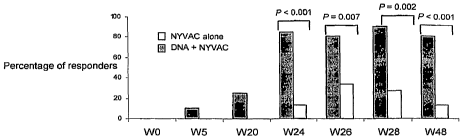
Figure 2. Percentage of responders following administration of NYVAC alone or
DNA following by NYVAC (prime-boost).
Figure 3. Measurement of INF-y-secreting T cells following administration of
NYVAC alone or DNA following by NYVAC (prime-boost).
Figure 4. Difference in the magnitude of the immune following administration
of
NYVAC alone or DNA following by NYVAC (prime-boost).
Figure 5. Representative flow cytometry profiles of env-specific INF-y-
secreting T
cells following administration of NYVAC alone or DNA following by NYVAC
(prime-boost).
Figure 6. Correlation between the frequencies of INF-y-secreting T cells
measured
by flow cytometry and ELISPOT.
Figure 7. Flow cytometry profiles of CD4 and CD8 T cells recognizing various
peptides following administration of NYVAC alone or DNA following by NYVAC
(prime-boost).
Figure 8. IgG antibody levels at different time points following
administration of
NYVAC alone or DNA following by NYVAC (prime-boost).
Figure 9. Analysis of the immune response 72 weeks following administration of
NYVAC alone or DNA following by NYVAC (prime-boost).
2
CA 02670804 2009-05-27
WO 2007/136763 PCT/US2007/011924
Summary of the Invention
Disclosed herein are methods for immunizing human beings against infectious
or other agents such as tumor cells by inducing or enhancing a dominant CD4 T
cell
response against that agent. In one embodiment, a method of administering to a
host
a first form of an immunogen and subsequently administering a second form of
the
immunogen, wherein the first and second forms are different, and wherein
administration of the first form prior to administration of the second form
enhances
the immune response resulting from administration of the second form relative
to
administration of the second form alone, is provided. Also provided are
compositions
for administration to the host. For example, a two-part immunological
composition
where the first part of the composition comprises a first form of an immunogen
and
the second part comprises a second form of the immunogen, wherein the first
and
second parts are administered separately from one another such that
administration of
the first form enhances the immune response against the second form relative
to
administration of the second form alone, is provided. The immunogens, which
may
be the same or different, are preferably derived from the infectious agent or
other
source of immunogens. Other embodiments are shown below.
Detailed Descriution
The present invention provides compositions and methodologies useful for
treating and / or preventing conditions relating to an infectious or other
agent(s) such
as a tumor cell by stimulating an immune response against such an agent. In
general,
the immune response results from expression of an immunogen derived from or
related to such an agent following administration of a nucleic acid vector
encoding the
immunogen, for example. In certain embodiments, multiple immunogens (which may
be the same or different) are utilized. In other embodiments, variants or
derivatives
(i.e., by substitution, deletion or addition of amino acids or nucleotides
encoding the
same) of an immunogen or immunogens (which may be the same or different) may
be
utilized.
As used herein, an "immunogen" is a polypeptide, peptide or a portion or
derivative thereof that produces an immune response in a host to whom the
immunogen has been administered. The immunogen is typically isolated from its
3
CA 02670804 2009-05-27
WO 2007/136763 PCT/US2007/011924
source (i.e., an infectious agent) of which it forms a part (i.e., a protein
normally
found within a cell). The immune response may include the production of
antibodies
that bind to at least one epitope of the immunogen and / or the generation of
a cellular
immune response against cells expressing an epitope of the immunogen. In
certain
cases the immunogen may be the epitope per se. Where different forms of
immunogen are utilized, the immunogens may be the same or different. The
immunogen may stimulate a de novo response or enhance an existing response
against
the immunogen by, for example, causing an increased antibody response (i.e.,
amount
of antibody, increased affinity / avidity) 'or an increased cellular response
(i.e.,
increased number of activated T cells, increased affinity / avidity of T cell
receptors).
In certain embodiments, the ininune response is protective, meaning the immune
response is capable of preventing infection of or growth within a host and /
or by
eliminating an agent (i.e., HIV) from a host.
The immunological compositions of the present inventions may include one or
more immunogen(s) from a single source or multiple sources. For instance, in
certain
embodiments the present invention relates to the induction or enhancement of
an
immune response against human immunodeficiency virus (HIV). Immunological
compositions may include one or more immunogens expressed by cells infected
with
HIV and / or displayed on the HIV virion per se. With respect to HIV, the
immunogens may be selected from any HIV isolate. As is well-known in the art,
HIV
isolates are now classified into discrete genetic subtypes. Subtype B has been
associated with the HIV epidemic in homosexual men and intravenous drug users
worldwide. Most immunogens, laboratory adapted isolates, reagents and mapped
epitopes belong to subtype B. In sub-Saharan Africa, India and China, areas
where the
incidence of new HIV infections is high, subtype B accounts for only a small
minority
of infections, and subtype C appears to be the most common infecting subtype.
Thus,
in certain embodiments, it may be preferable to select immunogens from HIV
subtypes B and / or C. It may be desirable to include immunogens from multiple
HIV
subtypes (i.e., HIV subtypes B and C) in a single immunological composition.
Suitable HIV immunogens include ENV, GAG, POL, NEF, as well as variants,
derivatives, and fusion proteins thereof, for example.
The present invention relates in certain embodiments to immunological
compositions capable of inducing or enhancing a dominant CD4 T cell immune
response against an immunogen. A dominant CD4 T cell immune response is
4
CA 02670804 2009-05-27
WO 2007/136763 PCT/US2007/011924
typically characterized by observing high proportion of immunogen-specific CD4
cells within the population of total responding T cells following vaccination
as
determined by an IFN-y ELISPOT assay. For example, this response may be
characterized by the presence of up to 55; 100; 250; 500; 750; or 1,000 or
more spot-
forming units (SFUs) by IFN-y ELISPOT assay per one million (106) blood
mononuclear cells. A dominant CD4 T cell immune response also typically but
not
necessarily provides a high proportion of responders (i.e., up to 50%, 60%,
70%,
80%, 85%, 90%, 95% or 100% of subjects tested) as compared to responders
demonstrating a CD8 T cell immune response. A dominant CD4 T cell immune
response is also typically but not necessarily polyfunctional, meaning that
the
majority of responding CD4 T cells secret both IL-2 and IFN- y. A dominant CD4
T
cell immune response also typically but not necessarily encompasses several
epitopes
(i.e., several populations of clonal CD4 T cells) within or between
responders, as
compared to mono-epitopic CD8 T cell responses. A dominant CD4 T cell response
may include one, more than -one or all of the characteristics described above.
Surprisingly, it has been found that the immunological compositions and
methods
presented herein induce a dominant CD4 T cell response in human beings.
. In preferred embodiments of the present invention, vectors are used to
transfer
a nucleic acid sequence encoding a polypeptide to a cell. A vector is any
molecule
used to transfer a nucleic acid sequence to a host cell. In certain cases, an
expression
vector is utilized. An expression vector is a nucleic acid molecule that is
suitable for
transformation of a host'cell and contains nucleic acid sequences that direct
and / or
control the expression of the transferred nucleic acid sequences. Expression
includes,
but is not limited to, processes such as transcription, translation, and
splicing, if
introns are present. Expression vectors typically comprise one or more
flanking
sequences operably linked to a heterologous nucleic acid sequence encoding a
polypeptide. As used herein, the term operably linked refers to a linkage
between
polynucleotide elements in a functional relationship such as one in which a
promoter
or enhancer affects transcription of a coding sequence. Flanking sequences may
be
homologous (i.e., from the same species and / or strain as the host cell),
heterologous
(i.e., from a species other than the host cell species or strain), hybrid
(i.e., a
combination of flanking sequences from more than one source), or synthetic,
for
example.
5
CA 02670804 2009-05-27
WO 2007/136763 PCT/US2007/011924
In certain embodiments, it is preferred that the flanking sequence is a
trascriptional regulatory region that drives high-level gene expression in the
target
cell. The transcriptional regulatory region may comprise, for example, a
promoter,
enhancer, silencer, repressor element, or combinations thereof. The
transcriptional
regulatory region may be either constitutive, tissue-specific, cell-type
specific (i.e.,
the region is drives higher levels of transcription in a one type of tissue or
cell as
compared to another), or regulatable (i.e., responsive to interaction with a
compound
such as tetracycline). The source of a transcriptional regulatory region may
be any
prokaryotic or eukaryotic organism, any vertebrate or invertebrate organism,
or any
plant, provided that the flanking sequence functions in a cell by causing
transcription
of a nucleic acid within that cell. A wide variety of transcriptional
regulatory regions
may be utilized in practicing the present invention.
Suitable transcriptional regulatory regions include, for example, the
synthetic
E/L promoter; the CMV promoter (i.e., the CMV-immediate early promoter);
promoters from eukaryotic genes (i.e., the estrogen-inducible chicken
ovalbumin
gene, the interferon genes, the gluco-corticoid-inducible tyrosine
aminotransferase
gene, and the thymidine kinase gene); and the major early and late adenovirus
gene
promoters; the SV40 early promoter region (Bemoist and Chambon, 1981, Nature
290:304-10); the promoter contained in the 3' long terminal repeat (LTR) of
Rous
sarcoma virus (RSV) (Yamamoto, et al., 1980, Cell 22:787-97); the herpes
simplex
virus thymidine kinase (HSV-TK) promoter (Wagner et al., 1981, Proc. Natl.
Acad..
Sci. U.S.A. 78:1444-45); the regulatory sequences of the metallothionine gene
(Brinster et al., 1982, Nature 296:39-42); prokaryotic expression vectors such
as the
beta-lactamase promoter (Villa-Kamaroff et al., 1978, Proc. Natl. Acad. Sci.
U.S.A.,
75:3727-31); or the tac promoter (DeBoer et al., 1983, Proc. Natl. Acad. Sci.
U.S:A.,
80:21-25). Tissue- and / or cell-type specific transcriptional control regions
include,
for example, the elastase I gene control region which is active in pancreatic
acinar
cells (Swift et al., 1984, Cell 38:639-46; Ornitz et al., 1986, Cold Spring
Harbor
Symp. Quant. Biol. 50:399-409 (1986); MacDonald, 1987, Hepatology 7:425-515);
the insulin gene control region which is active in pancreatic beta cells
(Hanahan,
1985, Nature 315:115-22); the immunoglobulin gene control region which is
active in
lymphoid cells (Grosschedl et al., 1984, Cell 38:647-58; Adames et al., 1985,
Nature
318:533-38; Alexander et al., 1987, Mol. Cell. Biol., 7:1436-44); the mouse
mammary
tumor virus control region in testicular, breast, lymphoid and mast cells
(Leder et al.,
6
CA 02670804 2009-05-27
WO 2007/136763 PCT/US2007/011924
1986, Cell 45:485-95); the albumin gene control region in liver (Pinkert et
al., 1987,
Genes and Devel. 1:268-76); the alpha-feto-protein gene control region in
liver
(Krumlauf et al., 1985, Mol. Cell. Biol., 5:1639-48; Hammer et al., 1987,
Science
235:53-58); the alpha 1-antitrypsin gene control region in liver (Kelsey et
al., 1987,
Genes and Devel. 1:161-71); the beta-globin gene control region in myeloid
cells
(Mogram et al., 1985, Nature 315:338-40; Kollias et al., 1986, Cell 46:89-94);
the
myelin basic protein gene control region in oligodendrocyte cells in the brain
(Readhead et al., 1987, Cell 48:703-12); the myosin light chain-2 gene control
region
in skeletal muscle (Sani, 1985, Nature 314:283-86); the gonadotropic releasing
hormone gene control region in the hypothalamus (Mason et al., 1986, Science
234:1372-78), and the tyrosinase promoter in melanoma cells (Hart, I. Semin
Oncol
1996 Feb;23(1):154-8; Siders, et al. Cancer Gene Ther 1998 Sep-Oct;5(5):281-
91),
among others. Other suitable promoters are known in the art.
In certain embodiments, a substitution of one amino acid for another may be
made in the sequence of an immunogen. Substitutions may be conservative, or
non-
conservative, or any combination thereof. Conservative amino acid
modifications to
the sequence of a polypeptide (and the corresponding modifications to the
encoding
nucleotides) may produce polypeptides having functional and chemical
characteristics
similar to those of a parental polypeptide. For example, a "conservative amino
acid
substitution" may involve a substitution of a native amino acid residue with a
non-
native residue such that there is little or no effect on the size, polarity,
charge,
hydrophobicity, or hydrophilicity of the amino acid residue at that position
and, in
particlar, does not result in decreased inununogenicity. Suitable
substitutions may be
selected from the following Table I:
Table I
Original Exemplary Substitutions Preferred
Residues Substitutions
Ala Val, Leu, Ile Val
Arg Lys, Gln, Asn Lys
Asn Gln Gln
Asp Glu Glu
Cys Ser, Ala Ser
Gln Asn Asn
Glu Asp Asp
Gly Pro, Ala Ala
His Asn, Gln, Lys, Arg Axg
Ile Leu, Val, Met, Ala, Phe, Norleucine Leu
7
CA 02670804 2009-05-27
WO 2007/136763 PCT/US2007/011924
Leu Norleucine, Ile, Val, Met, Ala, Phe Ile
Lys Arg, 1,4 Diamino-butyric Acid, Gln, Asn Arg
Met Leu, Phe, Ile Leu
Phe Leu, Val, Ile, Ala, Tyr Leu
Pro Ala Gly
Ser Thr, Ala, Cys Thr
Thr Ser Ser
Trp Tyr, Phe Tyr
Tyr Trp, Phe, Thr, Ser Phe
Val Ile, Met, Leu, Phe, Ala, Norleucine Leu
In other embodiments, it may be advantageous to combine a nucleic acid
sequence encoding an immunogen with one or more co-stimulatory component(s)
such as cell surface proteins, cytokines or chemokines in a composition of the
present
invention. The co-stimulatory component may be included in the composition as
a
polypeptide or as a nucleic acid encoding the polypeptide, for example.
Suitable co-
stimulatory molecules include, for instance, polypeptides that bind members of
the
CD28 family (i.e., CD28, ICOS; Hutloff, et al. Nature 1999, 397: 263-265;
Peach, et
al. J Exp Med 1994, 180: 2049-2058) such as the CD28 binding polypeptides B7.1
(CD80; Schwartz, 1992; Chen et al, 1992; Ellis, et al. J. Immunol., 156(8):
2700-9)
and B7.2 (CD86; Ellis, et al. J. Immunol., 156(8): 2700-9); polypeptides which
bind
members of the integrin family (i.e., LFA-1 (CD11a / CD18); Sedwick, et al. J
Immunol 1999, 162: 1367-1375; Wulfing, et al. Science 1998, 282: 2266-2269;
Lub,
et al. Immunol Today 1995, 16: 479-483) including members of the ICAM family
(i.e., ICAM-1, -2 or -3); polypeptides which bind CD2 family members (i.e.,
CD2,
signalling lymphocyte activation molecule (CDw150 or "SLAM"; Aversa, et al.
J Immunol 1997, 158: 4036-4044) such as CD58 (LFA-3; CD2 ligand; Davis, et al.
Immunol Today 1996, 17: 177-187) or SLAM ligands (Sayos, et al. Nature 1998,
395: 462-469); polypeptides which bind heat stable antigen (HSA.or CD24; Zhou,
et
al. Eur Jlmmunol 1997, 27: 2524-2528); polypeptides which bind to members of
the
TNF receptor (TNFR) family (i.e., 4-1BB (CD137; Vinay, et al. Semin Immunol
1998, 10: 481-489)), OX40 (CD134; Weinberg, et al. Semin Immunol 1998, 10: 471-
480; Higgins, et al. J Immunol 1999, 162: 486-493), and CD27 (Lens, et al.
Semin
Immunol 1998, 10: 491-499)) such as 4-IBBL (4-IBB ligand; Vinay, et al. Semin
Irnmunol 1998, 10: 481-48; DeBenedette, et al. J Immunol 1997, 158: 551-559),
TNFR associated factor-1 (TRAF-1; 4-1BB ligand; Saoulli, et al. J Exp Med
1998,
187: 1849-1862, Arch, et al. Mol Cell Biol 1998, 18: 558-565), TRAF-2 (4-1BB
and
8
CA 02670804 2009-05-27
WO 2007/136763 PCT/US2007/011924
OX40 ligand; Saoulli, et al. J Exp Med 1998, 187: 1849-1862; Oshima, et al.
Int
Immunol 1998, 10: 517-526, Kawamata, et al. J Biol Chern 1998, 273: 5808-
5814),
TRAF-3 (4-1BB and OX40 ligand; Arch, et al. .Mol Cell Biol 1998, 18: 558-565;
Jang, et al. Biochem Biophys Res Commun 1998, 242: 613-620; Kawamata S, et al.
J
Biol Chem 1998, 273: 5808-5814), OX40L (OX40 ligand; Gramaglia, et al. J
Immunol 1998, 161: 6510-6517), TRAF-5 (OX40 ligand; Arch, et al. Mol Cell Biol
1998, 18: 558-565; Kawamata, et al. JBiol Chem 1998, 273: 5808-5814), and CD70
(CD27 ligand; Couderc, et al. Cancer Gene Ther., 5(3): 163-75). CD154 (CD40
ligand or "CD40L"; Gurunathan, et al. J. Immunol., 1998, 161: 4563-4571; Sine,
et
al. Hum. Gene Ther., 2001, 12: 1091-1102) Other co-stimulatory molecules may
also
be suitable for practicing the present invention.
One or more cytokines may also be suitable co-stimulatory components or
"adjuvants", either as polypeptides or being encoded by nucleic acids
contained
within the compositions of the present invention (Parmiani, et al. Immunol
Lett 2000
Sep 15; 74(1): 41-4; Berzofsky, et al. Nature Immunol. 1: 209-219). Suitable
cytokines include, for example, interleukin-2 (IL-2) (Rosenberg, et al. Nature
Med. 4:
321-327 (1998)), IL-4, IL-7, IL-12 (reviewed by Pardoll, 1992; Harries, et al.
J. Gene
Med. 2000 Jul-Aug;2(4):243-9; Rao, et al. J. Immunol. 156: 3357-3365 (1996)),
IL-
15 (Xin, et al. Vaccine, 17:858-866, 1999), IL-16 (Cruikshank, et al. J. Leuk
Biol.
67(6): 757-66, 2000), IL-18 (J. Cancer Res. Clin. Oncol. 2001. 127(12): 718-
726),
GM-CSF (CSF (Disis, et al. Blood, 88: 202-210 (1996)), tumor necrosis factor-
alpha
(TNF-a), or interferon-gamma (INF-y). Other cytokines may also be suitable for
practicing the present invention.
Chemokines may also be utilized. For example, fusion proteins comprising
CXCL10 (IP-10) and CCL7 (MCP-3) fused to a tumor self-antigen have been shown
to induce anti-tumor immunity (Biragyn, et al. Nature Biotech. 1999, 17: 253-
258).
The chemokines CCL3 (MIP-1 a) and CCL5 (RANTES) (Boyer, et al. Vaccine, 1999,
17 (Supp. 2): S53-S64) may also be of use in practicing the present invention.
Other
suitable chemokines are known in the art.
It is also known in the art that suppressive or negative regulatory immune
mechanisms may be blocked, resulting in enhanced immune responses. For
instance,
treatment with anti-CTLA-4 (Shrikant, et al. Irnmunit,y, 1996, 14: 145-155;
Sutmuller,
et al. J. Exp. Med., 2001, 194: 823-832), anti-CD25 (Sutmuller, supra), anti-
CD4
9
CA 02670804 2009-05-27
WO 2007/136763 PCT/US2007/011924
(Matsui, et al. J. Immunol., 1999, 163: 184-193), the fusion protein IL13Ra2-
Fc
(Terabe, et al. Nature Immunol., 2000, 1: 515-520), and combinations thereof
(i.e.,
anti-CTLA-4 and anti-CD25, Sutmuller, supra) have been shown to upregulate
anti-
tumor immune responses and would be suitable in practicing the present
invention.
An immunogen may also be administered in combination with one or more
adjuvants to boost the immune response. Adjuvants may also be included to
stimulate or enhance the immune response against PhtD. Non-limiting examples
of
suitable adjuvants include those of the gel-type (i.e., aluminum
hydroxide/phosphate
("alum adjuvants"), calcium phosphate), of microbial origin (muramyl dipeptide
(MDP)), bacterial exotoxins (cholera toxin (CT), native cholera toxin subunit
B
(CTB), E. coli labile toxin (LT), pertussis toxin (PT), CpG oligonucleotides,
BCG
sequences, tetanus toxoid, monophosphoryl lipid A (MPL) of, for example, E.
coli,
Salmonella minnesota, Salmonella typhimurium, or Shigella exseri), particulate
adjuvants (biodegradable, polymer microspheres), immunostimulatory complexes
(ISCOIVIs)), oil-emulsion and surfactant-based adjuvants (Freund's incomplete
adjuvant (FIA), microfluidized emulsions (MF59, SAF), saponins (QS-21)),
synthetic
(muramyl peptide derivatives (murabutide, threony-MDP), nonionic block
copolymers (L121), polyphosphazene (PCCP), synthetic polynucleotides (poly
A:U,
poly I:C), thalidomide derivatives (CC-4407/ACTIMID)), RH3-ligand, or
polylactide
glycolide (PLGA) microspheres, among others. Fragments, homologs, derivatives,
and fusions to any of these toxins are also suitable, provided that they
retain adjuvant
activity. Suitable mutants or variants of adjuvants are described, e.g., in WO
95/17211
(Arg-7- Lys CT mutant), WO 96/6627 (Arg-192-Gly LT mutant), and WO 95/34323
(Arg-9-Lys and Glu-129-Gly PT mutant). Additional LT mutants that can be used
in
the methods and compositions of the invention include, e. g., Ser-63-Lys, Ala-
69-
Gly,Glu-110-Asp, and Glu-112-Asp mutants. Other suitable adjuvants are also
well-
known in the art.
As an example, metallic salt adjuvants such alum adjuvants are well-known in
the art as providing a safe excipient with adjuvant activity. The mechanism of
action
of these adjuvants are thought to include the formation of an antigen depot
such that
antigen may stay at the site of injection for up to 3 weeks after
administration, and
also the formation of antigen/metallic salt complexes which are more easily
taken up
by antigen presenting cells. In addition to aluminium, other metallic salts
have been
used to adsorb antigens, including salts of zinc, calcium, cerium, chromium,
iron, and
CA 02670804 2009-05-27
WO 2007/136763 PCT/US2007/011924
berilium. The hydroxide and phosphate salts of aluminium are the most common.
Formulations or compositions containing aluminium salts, antigen, and an
additional
immunostimulant are known in the art. An example of an immunostimulant is 3-de-
0-acylated monophosphoryl lipid A (3D-MPL).
Any of these components may be used alone or in combination with other
agents. For instance, it has been shown that a combination of CD80, ICAM-1 and
LFA-3 ("TRICOM") may potentiate anti-cancer immune responses (Hodge, et al.
Cancer Res. 59: 5800-5807 (1999). Other effective combinations include, for
example, IL-12 + GM-CSF (Ahlers, et al. J. Immunol., 158: 3947-3958 (1997);
Iwasaki, et al. J. Immunol. 158: 4591-4601 (1997)), IL-12 + GM-CSF + TNF-a
(Ahlers, et al. Int. Immunol. 13: 897-908 (2001)), CD80 + IL-12 (Fruend, et
al. Int.
J. Cancer, 85: 508-517 (2000); Rao, et al. supra), and CD86 + GM-CSF + IL-12
(Iwasaki, supra). One of skill in the art would be aware of additional
combinations
useful in carrying out the present invention.In addition, the skilled artisan
would be
aware of additional reagents or methods that may be used to modulate such
mechanisms. These reagents and methods, as well as others known by those of
skill
in the art, may be utilized in practicing the present invention.
Other agents that may be utilized in conjunction with the compositions and
methods provided herein include anti-HIV agents including, for example,
protease
inhibitor, an HIV entry inhibitor, a reverse transcriptase inhibitor, and / or
or an anti-
retroviral nucleoside analog. Suitable compounds include, for example,
Agenerase
(amprenavir), Combivir (Retrovir / Epivir), Crixivan (indinavir), Emtriva
(emtricitabine), Epivir (3tc / lamivudine), Epzicom, Fortovase / Invirase
(saquinavir),
Fuzeon (enfuvirtide), Hivid (ddc / zalcitabine), Kaletra (lopinavir), Lexiva
(Fosamprenavir), Norvir (ritonavir), Rescriptor (delavirdine), Retrovir / AZT
(zidovudine), Reyatax (atazanavir, BMS-232632), Sustiva (efavirenz), Trizivir
(abacavir / zidovudine / lamivudine), Truvada (Emtricitabine / Tenofovir DF),
Videx
(ddI / didanosine), Videx EC (ddI, didanosine), Viracept (nevirapine), Viread
(tenofovir disoproxil fumarate), Zerit (d4T / stavudine), and Ziagen
(abacavir). Other
suitable agents are known to those of skill in the art. Such agents may either
be used
prior to, during, or after administration of the compositions and / or use of
the
methods described herein.
11
CA 02670804 2009-05-27
WO 2007/136763 PCT/US2007/011924
Nucleic acids encoding immunogens may be administered to patients by any
of several available techniques. Various viral vectors that have been
successfully
utilized for introducing a nucleic acid to a host include retrovirus,
adenovirus, adeno-
associated virus (AAV), alphavirus, herpes virus, and poxvirus, among others.
It is
understood in the art that many such viral vectors are available in the art.
The vectors
of the present invention may be constructed using standard recombinant
techniques
widely available to one skilled in the art. Such techniques may be found in
common
molecular biology references such as Molecular Cloning: A Laboratory Manual
(Sambrook, et al., 1989, Cold Spring Harbor Laboratory Press), Gene Expression
Technology (Methods in Enzymology, Vol. 185, edited by D. Goeddel, 1991.
Academic Press, San Diego, CA), and PCR Protocols: A Guide to Methods and
Applications (Innis, et al. 1990. Academic Press, San Diego, CA).
Preferred retroviral vectors are derivatives of lentivirus as well as
derivatives
of murine or avian retroviruses. Examples of suitable retroviral vectors
include, for
example, Moloney murine leukemia virus (MoMuLV), Harvey murine sarcoma virus
(HaMuSV), murine mammary tumor virus (MuMTV), SIV, BIV, HIV and Rous
Sarcoma Virus (RSV). A number of retroviral vectors can incorporate multiple
exogenous nucleic acid sequences. As recombinant retroviruses are defective,
they
require assistance in order to produce infectious vector particles. This
assistance can
be provided by, for example, helper cell lines encoding retrovirus structural
genes.
Suitable helper cell lines include `Y2, PA317 and PA12, among others. The
vector
virions produced using such cell lines may then be used to infect a tissue
cell line,
such as NIH 3T3 cells, to produce large quantities of chimeric retroviral
virions.
Retroviral vectors may be administered by traditional methods (i.e.,
injection) or by
implantation of a"producer cell line" in proximity to the target cell
population
(Culver, K., et al., 1994, Hum. Gene Tlier., 5 (3): 343-79; Culver, K., et
al., Cold
Spring Harb. Symp. Quant. Biol., 59: 685-90); Oldfield, E., 1993, Hum. Gene
Ther., 4
(1): 39-69). The producer cell line is engineered to produce a viral vector
and
releases viral particles in the vicinity of the target cell. A portion of the
released viral
particles contact the target cells and infect those cells, thus delivering a
nucleic acid
encoding an immunogen to the target cell. Following infection of the target
cell,
expression of the nucleic acid of the vector occurs.
12
CA 02670804 2009-05-27
WO 2007/136763 PCT/US2007/011924
Adenoviral vectors have proven especially useful for gene transfer into
eukaryotic cells (Rosenfeld, M., et al., 1991, Science, 252 (5004): 431-4;
Crystal, R.,
et al., 1994, Nat. Genet., 8 (1): 42-5 1), the study eukaryotic gene
expression (Levrero,
M., et al., 1991, Gene, 101 (2): 195-202), vaccine development (Graham, F. and
Prevec, L., 1992, Biotechnology, 20: 363-90), and in animal models (Stratford-
Perricaudet, L., et al., 1992, Bone Marrow Transplant., 9(Suppl. 1): 151-2 ;
Rich, D.,
et al., 1993, Hum. Gene Ther., 4 (4): 461-76). Experimental routes for
administrating
recombinant Ad to different tissues in vivo have included intratracheal
instillation
(Rosenfeld, M., et al., 1992, Cell, 68 (1): 143-55) injection into muscle
(Quantin, B.,
et al., 1992, Proc. Natl. Acad. Sci. U.,S A., 89 (7): 2581-4), peripheral
intravenous
injection (Herz, J., and Gerard, R., 1993, Proc. Natl. Acad. Sci. U.S:A., 90
(7): 2812-
6) and stereotactic inoculation to brain (Le Gal La Salle, G., et al., 1993,
Science, 259
(5097): 988-90), among others.
Adeno-associated virus (AAV) demonstrates high-level infectivity, broad host
range and specificity in integrating into the host cell genome (Hermonat, P.,
et al.,
1984, Proc. Natl. Acad. Sci. U.S.A., 81 (20): 6466-70). And Herpes Simplex
Virus
type-1 (HSV-1) is yet another attractive vector system, especially for use in
the
nervous system because of its neurotropic property (Geller, A., et al., 1991,
Trends
Neurosci., 14 (10): 428-32; Glorioso, et al., 1995, Mol. Biotechnol., 4 (1):
87-99;
Glorioso, et al., 1995, Annu. Rev. Microbiol., 49: 675-710).
Alphavirus may also be used to express the immunogen in a host. Suitable
members of the Alphavirus genus include, among others, Sindbis virus, Semliki
Forest virus (SFV), the Ross River virus and Venezuelan, Western and Eastern
equine
encephalitis viruses, among others. Expression systems utilizing alphavirus
vectors
are described in, for example, U.S. Pat. Nos. 5,091,309; 5,217,879; 5,739,026;
5,766,602; 5,843,723; 6,015,694; 6,156,558; 6,190,666; 6,242,259; and,
6,329,201;
WO 92/10578; Xiong et al., Science; Vol 243, 1989, 1188-1191; Liliestrom, et
al.
Bio/Technology, 9: 1356-1361, 1991. Thus, the use of alphavirus as an
expression
system is well known by those of skill in the art.
Poxvirus is another useful expression vector (Smith, et al. 1983, Gene, 25
(1):
21-8; Moss, et al, 1992, Biotechnology, 20: 345-62; Moss, et al, 1992, Curr.
Top.
Microbiol. Immunol., 158: 25-38; Moss, et al. 1991. Science, 252: 1662-1667).
The
most often utilized poxviral vectors include vaccinia and derivatives
therefrom such
13
CA 02670804 2009-05-27
WO 2007/136763 PCT/US2007/011924
as NYVAC and MVA, and members of the avipox genera such as fowlpox,
canarypox, ALVAC, and ALVAC(2), among others.
An exemplary suitable vector is NYVAC (vP866) which was derived from the
Copenhagen vaccine strain of vaccinia virus by deleting six nonessential
regions of
the genome encoding known or potential virulence factors (see, for example,
U.S. Pat.
Nos. 5,364,773 and 5,494,807). The deletion loci were also engineered as
recipient
loci for the insertion of foreign genes. The deleted regions are: thymidine
kinase
gene (TK; J2R); hemorrhagic region (u; B 13R+B 14R); A type inclusion body
region
(ATI; A26L); hemagglutinin gene (HA; A56R); host range gene region (C7L-K1L);
and, large subunit, ribonucleotide reductase (14L). NYVAC is a genetically
engineered vaccinia virus strain that was generated by the specific deletion
of
eighteen open reading frames encoding gene products associated with virulence
and
host range. NYVAC has been show to be useful for expressing TAs (see, for
example, U.S. Pat. No. 6,265,189). NYVAC (vP866), vP994, vCP205, vCP1433,
placZH6H4Lreverse, pMPC6H6K3E3 and pC3H6FHVB were also deposited with the
ATCC under the terms of the Budapest Treaty, accession numbers VR-2559, VR-
2558, VR-2557, VR-2556, ATCC-97913, ATCC-97912, and ATCC-97914,
respectively.
Another suitable virus is the Modified Vaccinia Ankara (MVA) virus which
was generated by 516 serial passages on chicken embryo fibroblasts of the
Ankara
strain of vaccinia virus (CVA) (for review see Mayr, A., et al. Infection 3, 6-
14
(1975)). It was shown in a variety of animal models that the resulting MVA was
significantly avirulent (Mayr, A. & Danner, K. [1978] Dev. Biol. Stand. 41:
225.34)
and has been tested in clinical trials as a smallpox vaccine (Mayr et al.,
Zbl. Bakt.
Hyg. I, Abt. Org. B 167, 375-390 (1987), Stickl et al., Dtsch. med. Wschr. 99,
2386-
2392 (1974)). MVA has also been engineered for use as a viral vector for both
recombinant gene expression studies and as a recombinant vaccine (Sutter, G.
et al.
(1994), Vaccine 12: 1032-40; Blanchard et al., 1998, J Gen Virol 79, 1159-
1167;
Carroll & Moss, 1997, Virology 238, 198-211; Altenberger, U.S. Pat. No.
5,185,146;
Ambrosini et al., 1999, J Neurosci Res 55(5), 569).
ALVAC-based recombinant viruses (i.e., ALVAC-1 and ALVAC-2) are also
suitable for use in practicing the present invention (see, for example, U.S.
Pat. No.
5,756,103). ALVAC(2) is identical to ALVAC(1) except that ALVAC(2) genome
comprises the vaccinia E3L and K3L genes under the control of vaccinia
promoters
14
CA 02670804 2009-05-27
WO 2007/136763 PCT/US2007/011924
(U.S. Pat. No. 6,130,066; Beattie et al., 1995a, 1995b, 1991; Chang et al.,
1992;
Davies et al., 1993). Both ALVAC(1) and ALVAC(2) have been demonstrated to be
useful in expressing foreign DNA sequences, such as TAs (Tartaglia et al.,
1993 a,b;
U.S. Pat. No. 5,833,975). ALVAC was deposited under the terms of the Budapest
Treaty with the American Type Culture Collection (ATCC), 10801 University
Boulevard, Manassas, Va. 20110-2209, USA, ATCC accession number VR-2547.
Another useful poxvirus vector is TROVAC. TROVAC refers to an attenuated
fowlpox that was a plaque-cloned isolate derived from the FP-1 vaccine strain
of
fowlpoxvirus which is licensed for vaccination of 1 day old chicks. TROVAC was
likewise deposited under the terms of the Budapest Treaty with the ATCC,
accession
number 2553.
"Non-viral" plasmid vectors may also be suitable in practicing the present
invention. Plasmid DNA molecules comprising expression cassettes for
expressing an
immunogen may be used for "naked DNA" immunization. Preferred plasmid vectors
are compatible with bacterial, insect, and / or mammalian host cells. Such
vectors
include, for example, PCR-II, pCR3, and peDNA3.1 (Invitrogen, San Diego, CA),
pBSII (Stratagene, La Jolla, CA), pET15 (Novagen, Madison, WI), pGEX
(Pharmacia
Biotech, Piscataway, NJ), pEGFP-N2 (Clontech, Palo Alto, CA), pETL (B1ueBaclI,
Invitrogen), pDSR-alpha (PCT pub. No. WO 90/14363) and pFastBacDual (Gibco-
BRL, Grand Island, NY) as well as Bluescript plasmid derivatives (a high copy
number COLEI-based phagemid, Stratagene Cloning Systems, La Jolla, CA), PCR
cloning plasmids designed for cloning Taq-amplified PCR products (e.g., TOPOTM
TA cloning kit, PCR2.1 plasmid derivatives, Invitrogen, Carlsbad, CA).
Bacterial vectors may also be used with the current invention. Tliese vectors
include, for example, Shigella, Salmonella, Vibrio cholerae, Lactobacillus,
Bacille
calmette guerin (BCG), and Streptococcus (see for example, WO 88/6626; WO
90/0594; WO 91/13157; WO 92/1796; and WO 92/21376). Many other non-viral
plasmid expression vectors and systems are known in the art and could be used
with
the current invention.
Additional nucleic acid delivery techniques include DNA-ligand complexes,
adenovirus-ligand-DNA complexes, direct injection of DNA, CaPO4 precipitation,
gene gun techniques, electroporation, and colloidal dispersion systems, among
others.
Colloidal dispersion systems include macromolecule complexes, nanocapsules,
CA 02670804 2009-05-27
WO 2007/136763 PCT/US2007/011924
microspheres, beads, and lipid-based systems including oil-in-water emulsions,
micelles, mixed micelles, and liposomes. The preferred colloidal system of
this
invention is a liposome, which are artificial membrane vesicles useful as
delivery
vehicles in vitro and in vivo. RNA, DNA and intact virions can be encapsulated
within the aqueous interior and be delivered to cells in a biologically active
form
(Fraley, R., et al., 1981, Trends Biochem. Sci., 6: 77). The composition of
the
liposome is usually a combination of phospholipids, particularly high-phase-
transition-temperature phospholipids, usually in combination with steroids,
especially
cholesterol. Other phospholipids or other lipids may also be used. The
physical
characteristics of liposomes depend on pH, ionic strength, and the presence of
divalent cations. Examples of lipids useful in liposome production include
phosphatidyl compounds, such as phosphatidylglycerol, phosphatidylcholine,
phosphatidylserine, phosphatidylethanolamine, sphingolipids, cerebrosides, and
gangliosides. Particularly useful are diacylphosphatidylglycerols, where the
lipid
moiety contains from 14-18 carbon atoms, particularly from 16-18 carbon atoms,
and
is saturated. Illustrative phospholipids include egg phosphatidylcholine,
dipalmitoylphosphatidylcholine and distearoylphosphatidylcholine.
Strategies for improving the efficiency of nucleic acid-based immunization
may also be used including, for example, the use of self-replicating viral
replicons
(Caley, et al. 1999. Vaccine, 17: 3124-2135; Dubensky, et al. 2000. Mol. Med.
6:
723-732; Leitner, et al. 2000. Cancer Res. 60: 51-55), codon optimization
(Liu, et al.
2000. Mol. Ther., 1: 497-500; Dubensky, supra; Huang, et al. 2001. J. Virol.
75:
4947-4951), in vivo electroporation (Widera, et al. 2000. J. Immunol. 164:
4635-
3640), incorporation of CpG stimulatory motifs (Gurunathan, et al. Ann. Rev.
Immunol., 2000, 18: 927-974; Leitner, supra), sequences for targeting of the
endocytic or ubiquitin-processing pathways (Thomson, et al. 1998. J. Virol.
72:
2246-2252; Velders, et al. 2001. J. Immunol. 166: 5366-5373), prime-boost
regimens
(Gurunathan, supra; Sullivan, et al. 2000. Nature, 408: 605-609; Hanke, et al.
1998.
Vaccine, 16: 439-445; Amara, et al. 2001. Science, 292: 69-74), and the use of
mucosal delivery vectors such as Salmonella (Darji, et al. 1997. Cell, 91: 765-
775;
Woo, et al. 2001. Vaccine, 19: 2945-2954). Other methods are known in the art,
some of which are described below.
Administration of a composition of the present invention to a host may be
accomplished using any of a variety of techniques known to those of skill in
the art.
16
CA 02670804 2009-05-27
WO 2007/136763 PCT/US2007/011924
The composition(s) may be processed in accordance with conventional methods of
pharmacy to produce medicinal agents for administration to patients, including
humans and other mammals (i.e., a "pharmaceutical composition"). The
pharmaceutical composition is preferably made in the form of a dosage unit
containing a given amount of DNA, viral vector particles, polypeptide,
peptide, or
other drug candidate, for example. A suitable daily dose for a human or other
mammal may vary widely depending on the condition of the patient and other
factors,
but, once again, can be determined using routine methods. The compositions are
administered to a patient in a form and amount sufficient to elicit a
therapeutic effect,
i.e., to induce a dominant CD4 T cell response. Amounts effective for this use
will
depend on various factors, including for example, the particular composition
of the
vaccine regimen administered, the manner of administration, the stage and
severity of
the disease, the general state of health of the patient, and the judgment of
the
prescribing physician. The dosage regimen for immunizing a host or otherwise
treating a disorder or a disease with a composition of this invention is based
on a
variety of factors, including the type of disease, the age, weight, sex,
medical
condition of the patient, the severity of the condition, the route of
administration, and
the particular compound employed. Thus, the dosage regimen may vary widely,
but
can be determined routinely using standard methods.
In general, recombinant viruses may be administered in compositions in an
amount of about 104 to about 109 pfu per inoculation; often about 104 pfu to
about 106
pfu. Higher dosages such as about 104 pfu to about 1010 pfu, e.g., about 105
pfu to
about 109 pfu, or about 106 pfu to about 148 pfu, or about 107 pfu can also be
employed. Another measure commonly used is DICC50; suitable DICC50 ranges for
administration include about 10', about 102, about 103, about 104, about 105,
about
106, about 107 , about 108, about 109, about 1010 DICC50. Ordinarily, suitable
quantities of plasmid or naked DNA are about 1 g to about 100 mg, about 1 mg,
about 2 mg, but lower levels such as 0.1 to 1 mg or 1-10 g may be employed.
Actual
dosages of such compositions can be readily determined by one of ordinary
skill in
the field of vaccine technology.
The pharmaceutical composition may be administered orally, parentally, by
inhalation spray, rectally, intranodally, or topically in dosage unit
formulations
containing conventional pharmaceutically acceptable carriers, adjuvants, and
vehicles.
The term "pharmaceutically acceptable carrier" or "physiologically acceptable
17
CA 02670804 2009-05-27
WO 2007/136763 PCT/US2007/011924
carrier" as used herein refers to one or more formulation materials suitable
for
accomplishing or enhancing the delivery of a nucleic acid, polypeptide, or
peptide as
a pharmaceutical composition. A "pharmaceutical composition" is a composition
comprising a therapeutically effective amount of a nucleic acid or
polypeptide. The
terms "effective amount" and "therapeutically effective amount" each refer to
the
amount of a nucleic acid or polypeptide used to induce or enhance a dominant
CD4 T
cell r'esponse.
Injectable preparations, such as sterile injectable aqueous or oleaginous
suspensions, may be formulated according to known methods using suitable
dispersing or wetting agents and suspending agents. The injectable preparation
may
also be a sterile injectable solution or suspension in a non-toxic
parenterally
acceptable diluent or solvent. Suitable vehicles and solvents that may be
employed
are water, Ringer's solution, and isotonic sodium chloride solution, among
others. For
instance, a viral vector such as a poxvirus may be prepared in 0.4% NaCI or a
Tris-
HCl buffer, with or without a suitable stabilizer such as lactoglutamate, and
with or
without freeze drying medium. In addition, sterile, fixed oils are
conventionally
employed as a solvent or suspending medium. For this purpose, any bland fixed
oil
may be employed, including synthetic mono- or diglycerides. In addition, fatty
acids
such as oleic acid find use in the preparation of injectables.
Pharmaceutical compositions comprising a nucleic acid, immunogen(s), or
other compound may take any of several forms and may be administered by any of
several routes. In preferred embodiments, the compositions are administered
via a
parenteral route (intradermal, intramuscular or subcutaneous) to induce an
immune
response in the host. Alternatively, the composition may be administered
directly into
a lymph node (intranodal) or tumor mass (i.e., intratumoral administration).
Preferred embodiments of administratable compositions include, for example,
nucleic acids, viral particles, or polypeptides in liquid preparations such as
suspensions, syrups, or elixirs. Preferred injectable preparations include,
for example,
nucleic acids or polypeptides suitable for parental, subcutaneous,
intradermal,
intramuscular or intravenous administration such as sterile suspensions or
emulsions.
For example, a naked DNA molecule and / or recombinant poxvirus may separately
or
together be in admixture with a suitable carrier, diluent, or excipient such
as sterile
water, physiological saline, glucose or the like. The composition may also be
provided in lyophilized form for reconstituting, for instance, in isotonic
aqueous,
18
CA 02670804 2009-05-27
WO 2007/136763 PCT/US2007/011924
saline buffer. In addition, the compositions can be co-administered or
sequentially
administered with one another, other antiviral compounds, other anti-cancer
compounds and/or compounds that reduce or alleviate ill effects of such
agents.
As previously mentioned, while the compositions of the invention can be
administered as the sole active pharmaceutical agent, they can also be used in
combination with one or more other compositions or agents (i.e., other
immunogens,
co-stimulatory molecules, adjuvants). When administered as a combination, the
individual components can be formulated as separate compositions administered
at
the same time or different times, or the components can be combined as a
single
composition.
A kit comprising a composition of the present invention is also provided. The
kit can include a separate container containing a suitable carrier, diluent or
excipient.
The kit can also include an additional components for simultaneous or
sequential-
administration. In one embodiment, such a kit may include a first form of an
immunogen and a second form of the immunogen. Additionally, the kit can
include
instructions for mixing or combining ingredients and/or administration. A kit
may
provide reagents for performing screening assays, such as one or more PCR
primers,
hybridization probes, and / or biochips, for example.
All references cited within this application are incorporated by reference. A
better understanding of the present invention and of its many advantages will
be had
from the following examples, given by way of illustration.
19
CA 02670804 2009-05-27
WO 2007/136763 PCT/US2007/011924
EXAMPLES
Example
Materials and Methods
The recombinant vectors DNA C and NYVAC-HIV C expressed HIV genes
derived from the Chinese R5 clade C virus (97CN54; Su, et al. J. Virol. 2000.
74:
11367-76; WO 01/36614). This clone has been shown to be representative of
clade C
strains circulating in China and India. All HIV genes have been optimised for
codon
usage since it has recently been shown that humanization of synthetic HIV gene
codons allowed for an enhanced and REV/RRE- independent expression of env and
gag-pol genes in mammalian cells. Genes were optimised for both safety and
translation efficiency. The env gene has been designed to express the secreted
gpl20
form of the envelope proteins and contain an optimal synthetic leader sequence
for
enhanced expression. The gag, pol and nef genes were fused to express a GAG-
POL-
NEF polyprotein. An artificial -1 frameshift introduced in the natural
slippery
sequence of the p7-p6 gene junction results in an in-frame GAG-POL-NEF fusion
protein due to the absence of ribosomal frameshift. An N-terminal Gly - Ala
substitution in gag prevents the formation and release of virus-like particles
from
transfected cells. This strategy allows for an equimolar production of GAG,
POL and
NEF proteins and an enhanced MHC Class-I restricted presentation of their CTL
epitopes. For safety and regulatory reason, the packaging signal sequence has
been
removed; the integrase gene deleted; and the reverse transcriptase gene
disrupted by
insertion of a scrambled nef gene at the 3' end of the DNA sequence coding for
the
RT active site known to be associated with an immunodominant CTL epitope. The
nef
gene has been dislocated by fusing its 5' half to its 3' half without losing
its
inununodominant CTL epitopes.
A. NYVAC-HIV-C (vP2010)
1. Donor plasmid pMA60gp120C/gagpolnef-C-14.
Donor plasmid pMA60gp120C/GAG-POL-NEF-C-14 was constructed for
engineering of NYVAC or MVA expressing HIV-1 clade C gpl20 envelope and
GAG-POL-NEF proteins. The plasmid is a pUC derivative containing TK left and
right flanking sequences in pUC cloning sites. Between two flanking sequences
two
synthetic early/late (E/L) promoters in a back to back orientation
individually drive
codon-optimized clade C gp 120 gene and gag-pol-nef gene. The locations of the
TK
CA 02670804 2009-05-27
WO 2007/136763 PCT/US2007/011924
flanking sequences, E/L promoters, transcriptional termination signal, gp120
and gag-
pol-nef genes as described in Table II below:
Table II
p1VlA60gp120C/gagpolnef-C-14
Left flanking sequence Nt. 1609-2110 corn lementar
Right flanking sequence Nt. 4752-5433 com lementary)
E/L promoter for 120 Nt. 12-51
Gp 120 gene (ATG-TGA) Nt 61-1557
Terminal signal for 120 Nt.1586-1592
E/L promoter for gagpolnef Nt. 9794-9833 (complementary)
gagpolnef gene (ATG-TAA) Nt. 5531-9784 (com lement
Terminal signal for gagpolnef Nt.5422-5416 (com lernentary)
2. Construction of pMA60gp120C/gagpolnef-C-14 DNA origin:
a. pMA60: This plasmid is a pUC derivative containing TK right and left
flanking sequences in pUC cloning sites. Between the two flanking sequences
there is
a synthetic E/L promoter. The left flanking sequence is located at 37-550 and
right
flanking sequence is at 610-1329. The E/L promoter
(AAAATTGAAATTTTATTTTTTTTTTTTGGAATATAAATA) is located at 680-
569.
b. pCR-Script clade C-syngp120: The plasmid contained a codon-optimized
clade C HIV-1 gp120 gene. The gp120 gene is located at nucleotides 1-1497 (ATG-
TAA).
c. pCRs-cript clade C-syngaapolnef The plasmid containing a codon-optimized
clade C HIV-1 gagpolnef gene was provided by Hans Wolf and Walf Wagner
(Regensburg University, Germany). The gagpolnef gene was located between
nucleotides 1-4473 (ATG-TAA).
d. pSE1379.7: The plasmid is a Bluescript derivative containing a synthetic
E/L
promoter. The E/L promoter is located at nucleotides 1007- 968.
3. Construction of pMA60 gp120C/gagpolnef-C-14:
21
CA 02670804 2009-05-27
WO 2007/136763 PCT/US2007/011924
a. Construction of pMA60-T5NT-24: pMA60 has a synthetic E/L promoter but
has no transcriptional termination signal for the promoter. To insert a
terminal signal
T5NT for the promoter, a DNA fragment composed of a pair of oligonucleotides,
5'-
CCGGAATTTTTATT-3'(7291) / 3'- TTAAAAATAAGGCC-5' (7292), was inserted
into Xma I site on pMA60. The resulted plasmid was designated pMA60-T5NT-24
(notebook 1959, p54, Lisa Murdin, Aventis Canada). A Vector-NTI file for the
plasmid was included. In the file the E/L promoter was located at nt.3356-3395
and
the T5NT sequence is at nt 3417-3423.
b. Construction of pMA60gpl20C-10: To generate a clade C gp120 gene
without extra sequence between promoter and start codon ATG a Kpnl-Kpnl
fragment (nt. 4430-1527) containing the gpl20 gene was isolated from pCR-
Script
clade C-syngp120 and used as template in a PCR. In the PCR, primers 7490/ 7491
(7490: 5 ' -TTGAATTCTCGAG CATGGACAGGGCCAAGCTGCTGCTGCTGCTG
and 7491: 5'-TGCTGCTCACGTTCCTGCACTCCAGGGT) were used to amplify a
-370 bp 5'-gp120 fragment. The fragment was cut with EcoRl and AatII
generating
an EcoRI-AatII fragment (- 300 bp). The EcoRt-AatII fragment was used to
replace a
corresponding EcoR1-Aat II fragment (nt. 4432-293) on pCR-Script clade C-
syngpl20 resulting in a plasmid pCR-Script clade Cgpl20-PCR-19. A XhoI-XhoI
fragment containing a gp120 gene was isolated from pCR-Script cladeCgpl20- PCR-
19 and cloned into Xhol site on pMA60-T5NT-24 generating pMA60gpl 20C-1 0.
c. Construction of pMA60gp120C/gagpolnef-C-14: To create a clade C
gagpolnef gene without extra sequence between promoter and stat codon of the
gene a
KpnI-KpnI (nt 7313-4352) fragment containing the gagpolnef gene was isolated
from
pCRscript-Syngagpolnef and used as template in a PCR reaction. The primers
were
oligonucleotides (7618:
5'TTTCTCGAGCATGGCCGCCAGGGCCAGCATCCTGAGG / 7619: 5'-
ATCTGCTCCTGCAGGTTGCTGGTGGT). A fragment (- 740 bp) amplified in the
PCR was cloned into Sma I site on pUC18 resulting in a plasmid designated
pATGgpn-740. The -740 bp fragment in pATGgpn-740 was confirmed by DNA
sequencing., The pATGgpn-740 was cut with Xhol and Stul generating an Xhol-
Stul
fragment (- 480 bp). In addition, pCRScript-syngagpolnef was cut with Stul and
KpnI
generating a StuI-KpnI fragment (nt. 479-4325). Meanwhile pSE1379.7, a
Bluescript
22
CA 02670804 2009-05-27
WO 2007/136763 PCT/US2007/011924
derivative containing an E/L promoter, was linealized with XhoI and Kpnl
generating
an XhoI-KpnI receptor fragment (- 3 kb). The two fragments (XhoI-Stu I and
Stui-
KpnI) and the receptor fragment (XhoI-KpnI) were ligated together generating a
plasmid pATGgagpolnef-C-2. Finally, the pATG-gagpolnef-C-2 was cut with Sall
generating a SaII-SaII fragment that contained an E/L-gagpolnef cassette. The
SaII-
SaII fragment was cloned into a SaII site on pMA60gp120C-10 generating
pMA60gp120C/gagpolnef -C-14.
4. Generation of NYVAC-HIV-C recombinant (vP2010)
The IVR was performed by transfection of 1 CEF cells (Merial product) with
pMA60gp120C/gagpolenef C-14 using calcium phosphate method and
simultaneously infection of the cells with NYVAC as rescue virus at MOI of 10.
After -14 hr, the transfected-infected cells were harvested, sonicated and
used for
recombinant virus screening. Recombinant plaques were screened based on plaque
lift hybridization method. A 1.5 kb clade C gpl20 gene that was labeled with
p32
according to a random primer labeling kit protocol (Promega) was used as
probe. In
the first round screening, -11700 plaques were screened and three positive
clones
designated vP2010-1,vP 2010-2, vP2010-3, were obtained. After sequential four
rounds of plaque purification, recombinants designated vP2010-1-2-1-1, vP2010-
1-2-
2-1, vP2010-1-4-1-1, vP2010-1-4-1-2 and vP2010-1-4-2-1 were generated and
confirmed by hybridization as 100% positive using the gpl20 probe. P2 stocks
of
these recombinants were prepared. A P3 (roller bottle) stock with a titer 1.2x
109 was
prepared.
5. Stability of vP2010
To verify that the NYVAC-HIV-C (vP2010) recombinant could be passaged
without lost of transgene expression, a stability test was performed. The
recombinant
was passaged from P2 stock to P10 in CEF cells with moi of 0.1 and 0.01.
Plaques
generated in CEF cells with the plO stocks were analyzed with anti-gp120
monclonal
antibody K3A (Virogenetics) and anti-clade C p24 anti serum (Aventis Pasteeur
France). The results show that in moi of 0.1, 84 % plaques are positive to
gp120
antibody. In moi of 0.01, 76% plaques are positive to gp 120 antibody and 100%
23
CA 02670804 2009-05-27
WO 2007/136763 PCT/US2007/011924
plaques are positive to p24 antiserum. There was some loss (16-24%) of clade C
gp120 expression, even though the virus is relatively stable over 10 passages
with a
low MOI.
Expression of clade C gp 120 and gagpolnef from various passaged-vP2010
were verified by Western blot. The CEF cells were infected with various
passaged-
vP2010 viruses. Cell culture media and cell lysates after the infection were
analyzed
with anti-gp120 monoclonal antibody K3A (Virogenetics) and anti-p24 serum
(Aventis Pasteur in France). Expression of gp120 and gagpolnef from P10
viruses was
shown in Fig.1 and Fig.2. The expected gp120 band and GAG-POL-NEF fusion
protein band with molecular weight 120-190 kd were observed. Successful
expression
of gp 120 and GAG-POL-NEF from vP2010 was also demonstrated by immunoplaque
assay as mentioned above.
B. DNA. C
The DNA C vector was engineered to contain the components listed above
using the pORT system first described by Canenburgh, et al. (Nucleic Acid Res.
2001. 29: e26) (Cobra Biomanufacturing Plc; United Kingdom).
EXAMPLE 2
Immunization of Human Beings Against HIV-C
A. Immunological Compositions
1. DNA Vaccine ("DNA C")
DNA C is maintained in liquid form, with an extractable volume of 2 ml to 2.2
ml in 5 ml vials stored at -20 C. The appearance is clear and the composition
contains
the following components per ml of DNA HIV-C: DNA C (1.05 mg), Tris-HCl (1.57
mg), EDTA (0.372 mg), NaCI (9 mg). These components are brought to one ml with
water for injections.
2. NYVAC-HIV C (vP2010)
The presentation is in a liquid form, with an extractable volume of 1 ml to
1.1ml in single dose 3m1 vials stored at -200C. The composition contains 107
BaDICCso
NYVAC-HIV C(vP2010), 0.25 ml 10 mM Tris-HCI buffer; pH 7.5, 0.25 ml Virus
stabilizer (lactoglutamate); and, 0.5 ml freeze-drying medium.
24
CA 02670804 2009-05-27
WO 2007/136763 PCT/US2007/011924
B. Clinical Trial Design
The data provided herein reflects the results of a clinical trial in which 40
volunteers were randomised to receive DNA C (naked DNA) or nothing at weeks 0
and 4, followed by NYVAC C at weeks 20 and 24. Administration Regimens 1
(DNA C - NYVAC C prime boost) and 2 (NYVAC C only) are shown in Table III
below:
Table III
Immunization Regimens
Regimen Week 0 Week 4 Week 20 Week 24
1 DNA C 2x2m1 IM* DNA C NYVAC C IM NYVAC C
Unprimed right and left vastus 2x2ml IM non-dominant IM
N=20 lateralis right and left deltoid non-
vastus dominant
lateralis deltoid
2 Nothing Nothing NYVAC C IM NYVAC C
Prime-boost non-dominant IM non-
n=20 deltoid dominant
deltoid
* IM denotes intramuscular administration
The main objectives of this trial were to evaluate the safety and
immunogenicity of the prime boost regimen (DNA C + NYVAC C) compared to
NYVAC C alone. The design was open for participants and clinical
investigators,
without a placebo control, and 40 volunteers (see description of trial
population
below) were randomized to receive DNA C or nothing on day 0 and at week 4
followed by NYVAC C at weeks 20 and 24. The participants received two IM
injections (right and left vastus lateralis), with each injection containing
two ml DNA
C in liquid form (1.05 mg per ml and a total dose of 4.2 mg). NYVAC C was
administered as a one ml (107=7CCID50 NYVAC C per ml) in the deltoid. The
laboratory investigators undertaking and interpreting the assays were blind to
the
allocation. The primary endpoints were safety (local and systemic side
effects) and
immunogenicity. The protocol was determined to be safe and immunogenic, as
described below.
The primary immunogenicity endpoints were measured at week 26 and 28 by
the quantification of T-cell responses using the IFN-y ELISPOT assay following
a
conventional over night stimulation of the blood mononuclear cells with the
panel of
peptide pools encompassing env, gag, po1 and nef of HIV-1 CN54 clade C. The T-
CA 02670804 2009-05-27
WO 2007/136763 PCT/US2007/011924
cell responses were also measured on day 0 and at weeks 5, 20, 24 and 48. A
positive
ELISPOT assay was defined as exhibiting > 4-fold more spots than the negative
control and > 55 SFU/106 cells (i.e., a "responder"). Individual assays were
considered "valid" if the negative control < 50 SFU/106 cells and the positive
control
(SEB) > 500 SFU/106 cells.
Forty healthy male and female participants in London and Lausanne at low
risk of HIV infection were entered into the study. Fifty percent of the
enrolled
volunteers were females and fifty percent were males. The majority (90%) of
volunteers were Caucasians having a median age of 32 years. As a result of
preserving the integrity of the randomization, an imbalance between the two
groups
emerged with 23 participants allocated to receive DNA C and NYVAC C, and 17
allocated to NYVAC C alone. After the first DNA vaccination, two participants
were
withdrawn from the vaccination scheme due to adverse events, and the second
DNA
vaccination was given to 21 participants only. The two withdrawn participants
did not
receive NYVAC C but attended all visits. A further three participants also
received no
NYVAC: one female received two DNA C imrnunizations but decided that she did
not wish to receive the two NYVAC C immunizations and attended some visits;
another two participants were lost to follow-up. The remainder (n=35) received
the
full vaccination scheme shown in Table III and have completed tlie study (all
have
reached the 48 week timepoint).
C. Clinical Trial Results
A significant difference in the proportion of subjects with positive vaccine-
induced T-cell responses within the two study groups was observed. The
proportion
of responders was 90% (18/20) in the DNA C+NYVAC C group compared to 40 %
(6/15) in the NYVAC C alone group (P=0.003). One of the six responders in the
NYVAC C alone group had a very week response just above background (in the
range
of 60 SFU/106 cells) at weeks 26 and 28 but also at weeks 0, 5 and 20 prior to
vaccination. Although due to the study design, this subject had to be
considered
positive at weeks 26 and 28, the T-cell response observed was clearly non-
specific
and for these reasons it was not further considered in the additional
analyses. It was
thereby concluded that the proportion of subjects with vaccine-induced
specific T-cell
26
CA 02670804 2009-05-27
WO 2007/136763 PCT/US2007/011924
responses was 33% (5 out of 15) in the group vaccinated with NYVAC C alone.
The
proportion of responders after the DNA C vaccination was very low after two
vaccinations (2/18 or 12.5% at week 5, 4/18 or -22% at week 20) (Fig. 2).
Furthermore, the proportion of responders in the DNA C + NYVAC C group mostly
peaked (17 out of 20) after the first NYVAC C boost and the proportion of
responders
was still 75% at week 48, i.e. 6 months after the completion of the
vaccination. Only
two subjects within the NYVAC C alone group had still positive vaccine-induced
T-
cell responses at week 48.
Vaccine-induced T-cell responses were also assessed using the IFN-y
ELISPOT assay following stimulation of blood mononuclear cells with a panel of
464
peptides (15-mers overlapping by 11 amino acids) grouped in 8 pools (50-60
peptides
per pool). The peptides encompassed the env, gag, pol and nef proteins of HIV-
1 and
were designed based on the sequence of the immunogens expressed by the DNA and
NYVAC that were derived from the CN54 clade C isolate. Vaccine-induced T-cell
responses were predominantly directed against env in both DNA C + NYVAC C and
NYVAC C alone groups. Env-specific responses were observed in 22 out of 23
responders in both groups while gag, pol and nef vaccine-induced T-cell
responses
were only observed in 20% of volunteers (data not shown). The responses
against
gag, pol and nef were generally transient and substantially lower in magnitude
compared to the env-specific responses. The env-specific T-cell responses
following
DNA C + NYVAC C vaccination were significantly greater compared to the NYVAC
alone group. At the time of peak response (week 26), the mean measurement of
IFN-y
secreting T-cells was 450 SFU/106 cells in the DNA C + NYVAC C group and 110
SFU/106 cells within NYVAC C alone group (Fig. 3). The differences in the
magnitude of T-cell response between the two groups were significant
(P=0.016).
Consistent with the substantial difference in the magnitude of the T-cell
response
between the two groups, the 5 responders within the NYVAC C alone group had
most
(4 out of 5) of the T-cell response below 200 SFU/106 cells while nine of the
18
responders within the DNA C+NYVAC C group had T-cell responses greater than
300 SFU/106 cells (Fig. 4).
The distribution of vaccine-induced T-cell responses in CD4 and CD8 T-cell
populations was assessed in three of the five responders in the NYVAC C alone
group
and in 16 of 18 responders in the DNA C + NYVAC C group. Only responders with
27
CA 02670804 2009-05-27
WO 2007/136763 PCT/US2007/011924
more than 100 SFU/106 blood mononuclear cells measured in the IFN-y ELISPOT
assay were characterised using polychromatic flow cytometry. The vaccine-
induced
T-cell responses were mediated by CD4 T-cells in all the investigated 19
responders
(three in the NYVAC alone and 16 in the DNA C + NYVAC C groups). Vaccine-
induced CD8 T-cell responses were additionally observed one of the three
responders
in the NYVAC C alone group and in seven of 16 responders in the DNA C + NYVAC
C groups. Representative flow cytometry profiles of env-specific IFN-y
secreting CD4
and CD8 T-cell responses in responder #11 vaccinated with DNA C+NYVAC C are
shown in Fig. S. The characterization of vaccine-induced CD4 and CD8 T-cell
responses was performed mostly for env-specific 'responses since the frequency
and
the magnitude of the T-cell responses observed against gag, pol and nef was
very low
and generally below 100 SFU/106 cells. Of note, the polychromatic flow
cytometry
analysis allowed us to provide an independent confirmation of the responses
assessed
using the IFN-y ELISPOT assay. The frequencies of IFN-y secreting T-cells
measured
by both assays were compared in 19 responders. It is important to underscore
that
there was a very high correlation between the frequencies of IFN-y secreting T-
cells
measured by the ELISPOT assay and flow cytometry (Fig. 6).
The panel of T-cell functions analyzed included IL-2, TNF-a and IFN-y
secretion and proliferation for both CD4 and CD8 T-cells and also
degranulation
activity for CD8 T-cells. Env-specific CD4 and CD8 T-cells functions were
analysed
using polychromatic flow cytometry. T-cell functions were analysed after
stimulation
with env peptide pools. For example, responder #11 (vaccinated with DNA C +
NYVAC C) had both env-specific CD4 and CD8 T-cell responses. On the basis of
the
analysis of IL-2 and IFN-y secretion, three distinct env-specific CD4 T-cell
populations were identified: a) single IL-2, b) dual IL-2/IFN-y and single IFN-
y. The
three functionally distinct populations of env-specific CD4 T-cells were
equally
represented. Env-specific CD4 T-cells were also able to secrete TNF-a and we
identified two populations, i.e. single TNF-(x and dual TNF-a/IFN-y secreting
CD4 T-
cell populations which were equally represented. Furthermore, vaccine-induced
CD4
T-cells efficiently proliferated after stimulation with the env peptide pools.
Similar to CD4 T-cells, the analysis of IL-2 and IFN-y secretion in CD8 T-
cells identified two distinct env-specific CD8 T-cell populations: a) dual IL-
2/IFN-y
and single IFN-y secreting cell populations. It was found that the majority
(70%) of
28
CA 02670804 2009-05-27
WO 2007/136763 PCT/US2007/011924
env-specific CD8 T-cells were single IFN-y secreting cells and the remaining
cells
were dual IL-2/IFN-y. Almost the totality of IFN-y secreting CD8 T-cells were
also
able to secrete TNF-a and were therefore dual TNF-a/IFN-y secreting cells. A
substantial proportion of env-specific CD8 T-cells had degranulation activity
following antigen-specific stimulation as indicated by the expression of
CD107.
Finally, vaccine-induced CD8 T-cells were endowed with proliferation capacity
following env-specific stimulation. Similar functional profiles of vaccine-
induced
CD4 and CD8 T-cell responses were confirmed in six additional vaccinees. Taken
together, these results indicated that vaccination with DNA C + NYVAC C
induced
polyfunctional env-specific CD4 and CD8 T-cell responses.
Phenotypic analysis of vaccine-induced T-cell responses was performed in
volunteer #26 vaccinated with DNA C + NYVAC C. Both env-specific CD4 and CD8
T-cells were induced following vaccination. Blood mononuclear cells of
volunteer
#26 were collected at different time points (week 24, 26 and 48) and were
stimulated
with env derived peptide pools for 16 hours and stained with CD4, CD8, CD45RA,
CCR7, IL-2 and IFN-y antibodies. It has been previously demonstrated that
CD45RA
and CCR7 define functionally distinct populations of memory antigen-specific
CD4
and CD8 T-cells. The totality (single IL-2+dual iL-2/IFN-y+single IFN-y) of
env-
specific CD4 T-cells were CD45RA-CCR7- and the phenotypic profile and
percentage of env-specific CD4 T-cells remained unchanged over time.
In volunteer #26, Env-specific CD8 T-cells (dual IL-2/IFN-y+ single IFN-y)
were almost equally distributed within CD45RA-CCR7- and CD45RA+CCR7- cell
populations at week 24. However, there was a progressive loss of the CD45RA-
CCR7- env-specific CD8 T-cell population over time and about 90% of the
vaccine-
induced CD8 T-cells were CD45RA+CCR7- at week 48. The changes in phenotype
and in the percentage of env-specific CD8 T-cells were observed only for
vaccine-
induced CD8 T-cells since the phenotype and the percentage of EBV/CMV-specific
CD8 T-cell responses assessed in blood samples collected at the same time
points in
volunteer #26 remained unchanged. Similar results were obtained in three
additional
volunteers.
Identification of peptides/epitopes recognized by vaccine-induced CD4 and
CD8 T-cell populations was performed in nine volunteers, eight belonging to
the
DNA C + NYVAC C and one to the NYVAC C alone groups. Peptides/epitopes
29
CA 02670804 2009-05-27
WO 2007/136763 PCT/US2007/011924
characterization was limited to the env-specific responses. After the initial
screening
using env derived peptide pools, identification of the peptides/epitopes
recognized
was performed by testing the reactivity of blood mononuclear cells against the
relevant peptides in a matrix setting using the IFN-y ELISPOT assay. Following
this
analysis, 19 different peptides/epitopes were identified in the nine
volunteers studied
and further characterization of the vaccine-induced CD4 and CD8 T-cell
populations
recognizing these peptides/epitopes was performed using polychromatic flow
cytometry (Table 4).
TABLE 4
Type of Peptide sequence HIV
Antigen Region
Class II VGNLWVTVYYGVPVW C1/C2
WVTVYYGVPVWKGAT C1/C2
GATTTLFCASDAKAY CI/C2
TTLFCASDAKAYDTE C1/C2
THACVPADPNPQEMV C1/C2
ENVTENFNMWKNEMV C1/C2
ENFNMWKNEMV-NQMQ C1/C2
EMVNQMQEDVISLWD C1/C2
CVKLTPLCVTLECRN CI/C2
NCSFNATTVVRDRKQ V1/V2
NATTVVRDRKQTVYA V1/V2
VYALFYRLDIVPLTK C3
FYRLDIVPLTKKNYS C3
INCNTSAITQACPKV C3
PKVTFDPIPIHYCTP C3
FDPIPIHYCTPAGYA C3
TGDIIGDIRQAHCNI V3/C4
SSSIITIPCRIKQII V4/V5
ITIPCRIKQIINMWQ C5
CRIKQIINMWQEVGR C5
VGRAMYAPPIKGNIT C5
MYAPPIKGNITCKSN C5
PIKGNITCKSNITGL C5
ETFRPGGGDMRNNWR C5
ELYKYKVVEIKPLGV C5
YKVVEIKPLGVAPTT C5
EIKPLGVAPTTTKRR C5
Class I LGVAPTTTKRRVVER C5
HLA-A*O1 YSENSSEYY V 1/V2
A variable number of peptide/epitopes, ranging from two to eight, were
recognized in each volunteer with a mean of 4.2 peptides / epitope. Ten out of
19
CA 02670804 2009-05-27
WO 2007/136763 PCT/US2007/011924
peptides/epitopes identified in the nine volunteers have similarities to those
previously
identified in subjects with chronic HIV-1 infection or in vaccine studies
performed in
mice and humans.
Representative flow cytometry profiles of vaccine-induced env-specific CD4
and CD8 T-cells recognizing individual peptides/epitopes are shown in Fig. 7.
Fine
epitope mapping of the peptide LTKKNYSENSSEYYR recognized by CD8 T-cells in
seven volunteers (six belonging to the DNA C+ NYVAC C and one to the NYVAC
C alone groups) was performed. Using a set of overlapping peptides, it was
determined that the epitope recognized by vaccine-induced CD8 T-cells
corresponded
to the sequence YSENSSEYY (two representative examples).
Vaccine-induced IgG antibodies against gp140 CN54 were assessed at
different time points during the vaccination regimen (Figs. 8A and 8B). The
induction
of IgG anti-gpl40 CN54 was assessed in an ELISA assay. Only a small number of
volunteers (25%) had a measurable antibody response at week 26, i.e. 2 weeks
after
the second NYVAC C immunization, in the NYVAC C alone group. No responders
were present at week 48. A large percentage (75%) of volunteers had measurable
IgG
anti-gp140 antibodies at week 26 in the DNA C+NYVAC C group. No antibody
response was detected after the DNA immunization and only 10% of volunteers
responded after the first NYVAC C immunization. However, similar to the NYVAC
C alone group, the vaccine-induced antibody response was transient and only 5%
of
volunteers had measurable antibody response at week 48. In addition to the
significant differences in the percentage of responders between the two study
groups,
the magnitude of the antibody response was also significantly greater in the
DNA C +
NYVAC C group compared to the NYVAC C alone group.
The neutralization activity of the vaccine-induced antibodies was assessed in
three different assays including a) a multiple rounds neutralization assay on
blood
mononuclear cells using the homologous primary isolate CN54, b) a
neutrlization
assay in a single cycle infection of primary isolate Bx08 in the engineered
cell line
TZMbI, and c) a neutralization assay using Bal replication in macrophages. The
vaccine-induced antibodies failed to show any neutralizing activity.
The duration of the study in the original protocol was 48 weeks. However, in
order to have insights on the long-term durability of the vaccine-induced T-
cell
response, the protocol was subsequently amended to assess the T-cell responses
at
week 72, i.e. one year after the last immunization. The protocol was amended
only in
31
CA 02670804 2009-05-27
WO 2007/136763 PCT/US2007/011924
Lausanne and, after IRB approval, blood was collected at week 72 only in those
volunteers that were originally enrolled in Lausanne and had a positive IFN-y
ELISPOT assay at week 48. Thirteen volunteers (11 belonging to the DNA
C+NYVAC C group and 2 to the NYVAC C alone group) were analyzed at week 72
(Fig. 9A and 9B). None of the two volunteers belonging to the NYVAC C alone
group had a positive IFN-y T-cell response at week 72. Nine out of the 11
volunteers
belonging to the DNA C+NYVAC C group had a positive IFN-y T-cell response at
week 72. Of interest, the magnitude of the IFN-y T-cell response observed at
week 72
was unchanged compared to that measured in the 9 volunteers at week 28 and 48.
While the present invention has been described in terms of the preferred
embodiments, it is understood that variations and modifications will occur to
those
skilled in the art. Therefore, it is intended that the appended claims cover
all such
equivalent variations that come within the scope of the invention as claimed.
32