Note: Descriptions are shown in the official language in which they were submitted.
CA 02671721 2012-06-13
TITLE
Method and System for Providing Sensor Redundancy
FIELD
[0001] This invention relates to sensor systems in closed loop or
semi-closed loop
applications and more specifically to systems for predicting sensor values and
detecting the
failure of a sensor.
BACKGROUND
[0002] Over the years, body characteristics have been determined by
obtaining a sample
of bodily fluid. For example, diabetics often test for blood glucose levels.
Traditional blood
glucose determinations have utilized a finger prick using a lancet to withdraw
a small blood
sample. These systems are designed to provide data at discrete points and do
not provide
continuous data to show the variations in the characteristic between testing
times. These discrete
measurements are good to give some idea on how one's blood glucose values are
at a point in
time, and thus, enough information for a diabetic to give "correction" amounts
of insulin to
reduce their current blood glucose reading. However, these discrete readings
are not able to
provide enough information for any type of automatic or semi-automatic system
of giving insulin
based on blood glucose values.
100031 Recently, a variety of implantable electrochemical sensors
have been developed
for detecting and/or quantifying specific agents or compositions in a
patient's blood or interstitial
fluid. For instance, glucose sensors are being developed for use in obtaining
an indication of
blood glucose levels in a diabetic patient. These glucose sensors connected
(wired or wirelessly)
to a blood glucose monitor can provide continuous glucose readings over a
period of time such as
3 to 5 days. Such readings are useful in monitoring and/or adjusting a
treatment regimen which
typically includes the regular administration of insulin to the patient. Thus,
blood glucose
readings improve medical therapies with semi-automated medication infusion
pumps of the
external type, as generally described in U.S. Patent Nos. 4,562,751;
4,678,408; and 4,685,903; or
1
CA 02671721 2012-06-13
automated implantable medication infusion pumps, as generally described in
U.S. Patent No.
4,573,994. Typical thin film sensors are described in commonly assigned U.S.
Patent Nos.
5,390,671; 5,391,250; 5,482,473; and 5,586,553. See also U.S. Patent No.
5,299,571. In
addition, characteristic glucose monitors used to provide continuous glucose
data are described in
commonly assigned U.S. Patent Publication No. 20060202859 entitled
"Telemetered
Characteristic Monitor System and Method of Using the Same" published
September 14, 2006.
In addition, infusion pumps receiving sensor data is described in commonly
assigned U.S. Patent
Publication No. 20050065464 entitled "System for Providing Blood Glucose
Measurements to an
Infusion Device" published March 24, 2005.
[0004] As sensor technology improves, there is greater desire to use the
sensor values to
control the infusion of drugs and medicine, like insulin in a closed loop or
semi-closed loop
system. Specifically, a closed loop system for diabetes would entail a glucose
sensor and an
insulin infusion pump attached to a patient, where the delivery of insulin
would be automatically
administered by the controller of the infusion pump based on the sensor's
glucose value readings.
A semi-closed system would typically include a patient intervention step where
the amount of
insulin to be infused as calculated by the controller of the infusion pump
would require a patient
acceptance before delivery. However, given the ramifications of over-delivery
and/or under
delivery of medication, no one has yet to develop a viable way to actually
create a working
closed loop/semi-closed loop system where obtained sensor values can be
trusted enough to be
used to control the delivery of medication such as insulin with sufficient
safeguards to operate on
its own or even with a patient confinn/decline step.
SUMMARY
[0005] According to an embodiment of the invention, a closed loop infusion
system and
method for controlling blood glucose concentration in the body of a user is
described.
Embodiments of the present invention include obtaining a first glucose reading
from a first
glucose sensor located at a first site and obtaining a second glucose reading
from a second
glucose sensor located at a second site. In preferred embodiments, the system
and method
corroborate the signals generated by the first and second sensors. In an
embodiment, the
2
CA 02671721 2012-06-13
corroboration is performed by deriving a first predictive value to the first
glucose reading using
the second glucose reading as an input and deriving a second predictive value
to the second
glucose reading using the first glucose reading as an input. A first error
between the first
predictive value and the first glucose reading and a second error between the
second predictive
value and the second glucose reading are determined. By comparing a sum of the
absolute error
values of the first and second errors to a threshold, a failing sensor can be
identified.
100061 According to another embodiment of the invention, the system
and method
determine whether the first glucose sensor or second glucose sensor has the
least error in the
sensor signal and calculates a reported blood glucose value based on the
glucose sensor having
the least error in the sensor signal. In further embodiments, a comparison to
a meter glucose
value can be used to determine if the first or second glucose sensor is
failing.
[00071 Other features and advantages of the invention will become
apparent from the
following detailed description, taken in conjunction with the accompanying
drawings which
illustrate, by way of example, various features of embodiments of the
invention.
BRIEF DESCRIPTION OF THE DRAWINGS
100081 A detailed description of embodiments of the invention will be
made with
reference to the accompanying drawings, wherein like numerals designate
corresponding parts in
the several Figures.
100091 FIG. 1 is a block diagram of a closed loop glucose control
system in accordance
with an embodiment of the present invention.
[00101 FIG. 2 is a front view of closed loop hardware located on a
body in accordance
with an embodiment of the present invention.
100111 FIG. 3 (a) is a perspective view of a glucose sensor system for use
in an
embodiment of the present invention.
[00121 FIG. 3 (b) is a side cross-sectional view of the glucose
sensor system of FIG. 3 (a).
100131 FIG. 3 (c) is a perspective view of a sensor set of the
glucose sensor system of
FIG. 3 (a) for use in an embodiment of the present invention.
[0014] FIG. 3 (d) is a side cross-sectional view of the sensor set of FIG.
3 (c).
3
CA 02671721 2012-06-13
[0015] FIG. 4 is a cross sectional view of a sensing end of the
sensor of FIG 3 (d).
[0016] FIG. 5 is a perspective view illustrating another preferred
embodiment of the
subcutaneous sensor insertion set and telemetered characteristic monitor
transmitter device when
mated together in relation to the characteristic monitor system.
[0017] FIG. 6 is a top view of the subcutaneous sensor insertion set and
telemetered
characteristic monitor transmitter device when separated.
[0018] FIG. 7 is a top view of an infusion device with a reservoir
door in the open
position, for use in an embodiment of the present invention.
[0019] FIG. 8 is a side view of an infusion set with the insertion
needle pulled out, for
[0020] FIG. 9 (a) and (b) are block diagrams of a closed loop glucose
control system in
accordance with embodiments of the present invention.
[0021] FIG. 10 is a block diagram of auto blood withdrawal and return
in accordance
with an embodiment of the present invention.
[0022] FIG. 11 is a cross-sectional view of a sensor set and an infusion
set attached to the
body in accordance with an embodiment of the present invention.
[0023] FIG. 12(a) is a model describing the relationship between
glucose in interstitial
fluid and plasma glucose in accordance with an embodiment of the present
invention.
[0024] FIG. 12(b) is a plot of a plasma glucose step in comparison
with the resulting
[0025] FIG. 13 illustrates a block diagram of two glucose sensors
simultaneously
attached to the body at different locations in accordance with an embodiment
of the present
invention.
[0026] FIG. 14 is a plot of the two glucose sensors of FIG. 13 over
time compared to a
[0027] FIG. 15 describes the adaptive filter arrangement used to
provide sensor
corroboration and fault checking between the two sensors in accordance with
embodiments of the
present invention.
4
CA 02671721 2012-06-13
[0028] FIG. 16 is a plot of the unprocessed sensor signals with the
corresponding
prediction traces calculated with adaptive filters in accordance with
embodiments of the present
invention.
[0029] FIG. 17 is a plot of the performance of each prediction of the
sensor values in
accordance with embodiments of the present invention.
[0030] FIG. 18 is a plot of the fault detection in accordance with
embodiments of the
present invention.
[0031] FIG. 19 is a flowchart illustrating the steps used by the
adaptive filter arrangement
of FIG. 15 in accordance with embodiments of the present invention.
[0032] FIG. 20 is a flowchart illustrating the steps in the fault handling
process of FIG.
16 in accordance with embodiments of the present invention.
DETAILED DESCRIPTION
[0033] As shown in the drawings for purposes of illustration, the
invention is embodied
in a closed loop infusion system for regulating the rate of fluid infusion
into a body of a user
based on feedback from an analyte concentration measurement taken from the
body. In
particular embodiments, the invention is embodied in a control system for
regulating the rate of
insulin infusion into the body of a user based on a glucose concentration
measurement taken
from the body. In preferred embodiments, the system is designed to model a
pancreatic beta cell
(13-cell). In other words, the system controls an infusion device to release
insulin into a body of a
user in a similar concentration profile as would be created by fully
functioning human 13-cells
when responding to changes in blood glucose concentrations in the body.
[0034] Thus, the system simulates the body's natural insulin response
to blood glucose
levels and not only makes efficient use of insulin, but also accounts for
other bodily functions as
well since insulin has both metabolic and mitogenic effects. However, the
algorithms must
model the n-cells closely, since algorithms that are designed to minimize
glucose excursions in
the body, without regard for how much insulin is delivered, may cause
excessive weight gain,
hypertension, and atherosclerosis. In preferred embodiments of the present
invention, the system
is intended to emulate the in vivo insulin secretion pattern and to adjust
this pattern consistent
with the in vivo 13-cell adaptation experienced by normal healthy individuals.
The in vivo 13-cell
5
CA 02671721 2012-06-13
response in subjects with normal glucose tolerance (NGT), with widely varying
insulin
sensitivity (Si), is the optimal insulin response for the maintenance of
glucose homeostasis.
[0035] Preferred embodiments include a glucose sensor system 10, a
controller 12 and an
insulin delivery system 14, as shown in FIG. 1. The glucose sensor system 10
generates a sensor
signal 16 representative of blood glucose levels 18 in the body 20, and
provides the sensor signal
16 to the controller 12. The controller 12 receives the sensor signal 16 and
generates commands
22 that are communicated to the insulin delivery system 14. The insulin
delivery system 14
receives the commands 22 and infuses insulin 24 into the body 20 in response
to the commands
22. In an alternative semi-closed loop embodiment, the commands 22 would have
to be
confirmed by the user before the insulin delivery system 14 would infuse
insulin.
[0036] Generally, the glucose sensor system 10 includes a glucose
sensor, sensor
electrical components to provide power to the sensor and generate the sensor
signal 16, a sensor
communication system to carry the sensor signal 16 to the controller 12, and a
sensor system
housing for the electrical components and the sensor communication system.
[0037] Typically, the controller 12 includes controller electrical
components and software
to generate commands for the insulin delivery system 14 based on the sensor
signal 16, and a
controller communication system to receive the sensor signal 16 and carry
commands to the
insulin delivery system 14.
[0038] Generally, the insulin delivery system 14 includes an infusion
device and an
infusion tube to infuse insulin 24 into the body 20. In particular
embodiments, the infusion
device includes infusion electrical components to activate an infusion motor
according to the
commands 22, an infusion communication system to receive the commands 22 from
the
controller 12, and an infusion device housing to hold the infusion device.
[0039] In preferred embodiments, the controller 12 is housed in the
infusion device
housing and the infusion communication system is an electrical trace or a wire
that carries the
commands 22 from the controller 12 to the infusion device. In alternative
embodiments, the
controller 12 is housed in the sensor system housing and the sensor
communication system is an
electrical trace or a wire that carries the sensor signal 16 from the sensor
electrical components to
the controller electrical components. In other alternative embodiments, the
controller 12 has its
own housing or is included in a supplemental device. In another alternative
embodiment, the
6
CA 02671721 2012-06-13
controller is located with the infusion device and the sensor system all
within one housing. In
further alternative embodiments, the sensor, controller, and/or infusion
communication systems
may utilize a cable, a wire, fiber optic lines, RF, IR, or ultrasonic
transmitters and receivers, or
the like instead of the electrical traces.
System Overview
[0040] Preferred embodiments of the invention include a sensor 26, a
sensor set 28, a
telemetered characteristic monitor transmitter 30, a sensor cable 32, an
infusion device 34, an
infusion tube 36, and an infusion set 38, all worn on the body 20 of a user,
as shown in FIG. 2.
The telemetered characteristic monitor transmitter 30 includes a transmitter
housing 31 that
supports a printed circuit board 33, batteries 35, antenna (not shown), and a
sensor cable
connector (not shown), as seen in FIG. 3 (a) and 3 (b). A sensing end 40 of
the sensor 26 has
exposed electrodes 42 and is inserted through skin 46 into a subcutaneous
tissue 44 of a user's
body 20, as shown in FIG. 3 (d) and 4. The electrodes 42 are in contact with
interstitial fluid
(ISF) that is present throughout the subcutaneous tissue 44. The sensor 26 is
held in place by the
sensor set 28, which is adhesively secured to the user's skin 46, as shown in
FIGs. 3 (c) and 3 (d).
The sensor set 28 provides for a connector end 27 of the sensor 26 to connect
to a first end 29 of
the sensor cable 32. A second end 37 of the sensor cable 32 connects to the
transmitter housing
31. The batteries 35 included in the transmitter housing 31 provide power for
the sensor 26 and
electrical components 39 on the printed circuit board 33. The electrical
components 39 sample
the sensor signal 16 and store digital sensor values (Dsig) in a memory and
then periodically
transmit the digital sensor values Dsig from the memory to the controller 12,
which is included in
the infusion device.
[0041] As shown in FIGs. 3(a)-(b), the telemetered characteristic
monitor transmitter 30
is coupled to a sensor set 28 by a sensor cable 32. In alternative
embodiments, the cable 32 may
be omitted, and the telemetered characteristic monitor transmitter 30 may
include an appropriate
connector for direct connection to the connector portion 26 of the sensor set
28 or the sensor set
28 may be modified to have the connector portion 26 positioned at a different
location. For
example, FIGs. 5 and 6 show a possible alternative embodiment where
characteristic monitor
transmitter 100' and the sensor set 10' can be modified to allow a side-by
side direct connection
between the characteristic monitor transmitter 100' and the sensor set 10'
such that the
7
CA 02671721 2012-06-13
characteristic monitor transmitter 100' detachable from the sensor set 10', as
seen in FIG. 6.
Another possible embodiment (not shown) can modify the top of the sensor set
10' to facilitate
placement of the telemetered characteristic monitor transmitter 100' over the
sensor set 10'.
[0042] The controller 12 processes the digital sensor values Dsig and
generates
commands 22 for the infusion device 34. Preferably, the infusion device 34
responds to the
commands 22 and actuates a plunger 48 that forces insulin 24 out of a
reservoir 50 located inside
the infusion device 34, as shown in FIG. 7. In particular embodiments, a
connector tip 54 of the
reservoir 50 extends through the infusion device housing 52 and a first end 51
of the infusion
tube 36 is attached to the connector tip 54. A second end 53 of the infusion
tube 36 connects to
the infusion set 38. Insulin 24 is forced through the infusion tube 36 into
the infusion set 38 and
into the body 16. The infusion set 38 is adhesively attached to the user's
skin 46, as shown in
FIG. 8. As part of the infusion set 38, a cannula 56 extends through the skin
46 and terminates in
the subcutaneous tissue 44 completing fluid communication between the
reservoir 50 and the
subcutaneous tissue 44 of the user's body 16.
[0043] In alternative embodiments, the closed-loop/semi-closed loop system
can be a part
of a hospital-based glucose management system. Given that insulin therapy
during intensive care
has been shown to dramatically improve wound healing, reduce blood stream
infections, renal
failure, and polyneuropathy mortality, irrespective of whether subjects
previously had diabetes
(See Van den Berghe G. et al. NEJM 345: 1359-67, 2001), the present invention
can be used in
this hospital setting to control the blood glucose level of a patient in
intensive care. In these
alternative embodiments, since an IV hookup is typically implanted into a
patient's arm while the
patient is in an intensive care setting (e.g. ICU), a closed loop glucose
control can be established
which piggy-backs off the existing IV connection. Thus, in a hospital based
system, intravenous
(IV) catheters which are directly connected to a patient vascular system for
purposes of quickly
delivering IV fluids, can also be used to facilitate blood sampling and direct
infusion of
substances (e.g. insulin, anticoagulants) into the intra-vascular space.
Moreover, glucose sensors
may be inserted through the IV line to give real-time glucose levels from the
blood stream.
Therefore, depending on the type of hospital based system, the alternative
embodiments would
not necessarily need the described system components such as the sensor 26,
the sensor set 28,
the telemetered characteristic monitor transmitter 30, the sensor cable 32,
the infusion tube 36,
8
CA 02671721 2012-06-13
and the infusion set 38 as described in the preferred embodiments. Instead,
standard blood
glucose meters or vascular glucose sensors as described in U.S. Patent
Publication No.
20040064086 entitled "Multi-lumen Catheter," published April 1, 2004, can be
used to provide
the blood glucose values to the infusion pump control and the existing IV
connection can be used
to administer the insulin to the patient.
[0044] It is important to appreciate that numerous combinations of
devices in the
hospital-based system can be used with the closed loop controller of the
present invention. For
example, as described in FIG. 9b compared to a subcutaneous sensor system in
FIG. 9a, an auto
blood glucose/intravenous insulin infusion system can automatically withdraw
and analyze blood
for glucose concentration at fixed intervals (preferably 5 ¨ 20 minutes),
extrapolate the blood
glucose values at a more frequent interval (preferably I minute), and use the
extrapolated signal
for calculating an iv-insulin infusion according to the controller described
below. The modified
auto blood glucose/intravenous insulin infusion system would eliminate the
need for
subcutaneous sensor compensation and subcutaneous insulin compensation which
would be
required with a subcutaneous sensor system (as described below when discussing
the delay
problems inherent in a subcutaneous sensor system). The automatic withdrawal
of blood, and
subsequent glucose determination can be accomplished with existing technology
(e.g. VIA or
Biostator like blood glucose analyzer) or by the system described in FIG. 10.
The system in FIG.
10 uses a peristaltic pump 420 to withdraw blood across an amperometric sensor
410 (the same
technology as used in sensor 26) and then return the blood with added flush
(0.5 to 1.0 ml) from
the reservoir 400. The flush can consist of any makeup of saline, heparin,
glucose solution
and/or the like. If the blood samples are obtained at intervals longer than 1
minute but less than
20 minutes, the blood glucose determinations can be extrapolated on a minute-
to-minute basis
with extrapolation based on the present (n) and previous values (n-1) to work
with the logic of
the controller as described in detail below. For blood samples obtained at
intervals greater than
20 minutes, a zero-order-hold would be used for the extrapolation. Based on
these blood glucose
values, the infusion device can administer insulin based on the closed loop
controller described
in greater detail below.
[0045] In other modifications to the system, a manual blood
glucose/intravenous insulin
infusion system can be used where frequent manual entry of blood glucose
values from a
9
CA 02671721 2012-06-13
standard blood glucose meter (e.g. YSI, Beckman, etc) and extrapolate the
values at more
frequent intervals (preferably 1 min) to create a surrogate signal for
calculating IV-insulin
infusion. Alternatively, a sensor blood glucose/intravenous insulin infusion
system can use a
continuous glucose sensor (e.g. vascular, subcutaneous, etc.) for frequent
blood glucose
determination. Moreover, the insulin infusion can be administered
subcutaneously rather than
intravenously in any one of the previous examples according to the controller
described below.
[0046] In still further alternative embodiments, the system
components may be combined
in a smaller or greater number of devices and/or the functions of each device
may be allocated
differently to suit the needs of the user.
Controller
[0047] Once the hardware for a closed loop system is configured, such
as in the preferred
embodiments described above, the affects of the hardware on a human body are
determined by
the controller. In preferred embodiments, the controller 12 is designed to
model a pancreatic beta
cell ([3-cell). In other words, the controller 12 commands the infusion device
34 to release
insulin 24 into the body 20 at a rate that causes the insulin concentration in
the blood to follow a
similar concentration profile as would be caused by fully functioning human 13-
cells responding
to blood glucose concentrations in the body 20. Thus, the controller 22 is
intended to emulate
the in vivo insulin secretion pattern and to adjust this pattern to be
consistent with in vivo 13-cell
adaptation. The in vivo 13-cell response in subjects with normal glucose
tolerance (NGT), with
widely varying insulin sensitivity (Si), is the optimal insulin response for
the maintenance of
glucose homeostasis. The biphasic insulin response of a 13-cell can be modeled
using
components of a proportional, plus integral, plus derivative (PID) controller
along with various
filters. Description of a PID controller to emulate 13-cells can be found in
commonly assigned
U.S. Patent No. 6,558,351. In alternative embodiments, the controller may
simply be the
controller in an infusion pump that calculates the amount of insulin to be
infused by knowing the
insulin sensitivity/carbohydrate ratio of the individual, the target blood
glucose level, amount of
carbohydrates to be ingested and the current blood glucose level supplied by
the sensor. An
example of such a controller is described in commonly assigned U.S. Patent No.
6,554,798
entitled "External Infusion Device with Remote Programming, Bolus Estimator
and/or Vibration
Alarm Capabilities,".
CA 02671721 2012-06-13
Sensor Redundancy
[0048] Regardless of the controller used with the present system,
closed loop/semi-closed
loop algorithms for insulin delivery rely on a continuous glucose sensor to
drive a control
algorithm that determines the optimal insulin dose to administer through a
pump delivery
mechanism. Therefore sensor reliability and fault detection and handling are
crucial to the
dependability and safety of such an application. It is therefore desirable to
have an assessment
mechanism that can evaluate the sensor signal fidelity and initiate the
appropriate action
following detection of a sensor failure. In the event a fault is detected a
request for sensor
replacements should be initiated and a temporary suspension of insulin
delivery or control should
switch to a fixed mode of operation with set basal patterns.
[0049] One method of identifying whether the sensor values are
reliable involves the
measure of other signals by the sensor that may provide information about the
state of the sensor
(such as voltage readings, impedance, etc). This approach has some merit, but
we cannot assure
that we always know the sensor is accurate. Another possibility to assure an
accurate sensor
reading is to use a dual or 3-up sensing system located in a single sensor
site so that the sensors
could be used to check one another. This approach has merit because the system
would continue
in closed-loop mode as long as the sensors were in agreement, and the
likelihood of each sensor
failing in the same way, or at the same time is supposedly small. However,
there exists the
possibility that an interferon affects all sensors the same way, or the sensor
insertion site is
affected so that all sensors misread the glucose in a similar fashion. Thus,
several situations can
arise were two functioning sensors produce dissimilar outputs, or two
dysfunctional sensors
could present similar outputs that are credible of a person's glucose state.
Therefore, even this
technique may have a potential failure mode.
[0050] Consequently, the subject of this present invention relates to
the use of sensor
redundancy, where the sensing method and/or sensor location are different from
one another. For
example, in one embodiment, two subcutaneous sensors located at different
sites would assure
that the potential for common effects due to sensor location or interferences
is negligible.
However, alternative sites may generate different physiological delays that
could result from skin
temperature or pressure variance at the measuring site. For example, when
additional pressure is
applied to one of the sites due to sleep posture, the readings may vary.
Moreover, two identical
11
CA 02671721 2012-06-13
sensors who should exhibit the same readings can exhibit varying time lags,
sensitivities and
offsets leading to confusing signals. Thus, in preferred embodiments, sensors
using different
technology are placed in different body fluids, e.g. one sensor in
subcutaneous tissue and one in
blood. Therefore, although the previous description described various types of
electro-enzymatic
sensors, the system will use other types of sensors, such as chemical based,
optical based or the
like. For example other types of sensors are described in the following
references: U.S. Patent
No. 6,011,984 issued January 4, 2000 to Van Antwerp et al. and entitled
"Detection of Biological
Molecules Using Chemical Amplification"; and U.S. Patent No. 6,766,183 issued
July 20, 2004
to Walsh et al. and entitled "Long Wave Flourophore Sensor Compounds and Other
Fluorescent
Sensor Compounds in Polymers". Other compounds using Donor Acceptor
fluorescent
techniques may be used, such as disclosed in U.S. Patent No. 5,628,310 issued
May 13, 1997 to
Rao et al. and entitled " Method and Apparatus to Perform Trans-cutaeous
Analyte Monitoring";
U.S. Patent No. 5,342,789 issued August 30, 1994 to Chick et al. and entitled
"Method and
Device for Detecting and Quantifying Glucose in body Fluids"; and U.S. Patent
No. 5,246,867
issued September 21, 1993 to Lakowicz et al. and entitled "Determination and
Quantification of
Saccharides by Luminescent Lifetimes and Energy Transfer". The bottom line is
that, use of two
different types of sensors at two different locations, may offer the ideal
redundancy needed to
assure failsafe performance of the system that relies heavily on accurate
sensor readings.
Challenges to Sensor Redundancy
[0051] However, different sensor technologies and different
measurement fluids are
known to have significantly varying time lags. For example, the complexity of
the problem can
be seen with a subcutaneous glucose sensor 26. As described with respect to
FIG. 11, a
physiological delay 422 is due to the time required for glucose to move
between blood plasma
420 and interstitial fluid (ISF). The delay is represented by the circled
double headed arrow 422
in FIG. 11. Generally, as discussed above, the sensor 26 is inserted into the
subcutaneous tissue
44 of the body 20 and the electrodes 42 near the tip of the sensor 40 are in
contact with
interstitial fluid (ISF). But the desired parameter to be measured is the
concentration of blood
glucose. Glucose is carried throughout the body in blood plasma 420. Through
the process of
diffusion, glucose moves from the blood plasma 420 into the ISF of the
subcutaneous tissue 44
12
CA 02671721 2012-06-13
and vice versa. As the blood glucose level 18 changes so does the glucose
level in the ISF. But
the glucose level in the ISF lags behind the blood glucose level 18 due to the
time required for
the body to achieve glucose concentration equilibrium between the blood plasma
420 and the
ISF. Studies show the glucose lag times between blood plasma 420 and ISF vary
between 0 to 30
minutes. Some parameters that may affect the glucose lag time between blood
plasma 420 and
ISF are the individual's metabolism, the current blood glucose level, whether
the glucose level is
rising, or falling, or the like. A model illustrated in FIG. 12a has been
created to describe this
dynamic relationship between ISF and plasma glucose. This model is based on
the assumption
that the capillary 410 separating plasma 420 and ISF in the subcutaneous
tissue 44 compartments
creates a resistance to glucose diffusion into the ISF space (i.e.
subcutaneous space). Glucose is
cleared from the ISF space 44 into Fat/Muscle Cells 440 by a rate proportional
to the
concentration of glucose in that compartment. This mathematical relationship
is described by the
following mass balance equation:
dC2 7, V r,
0µ02 k12 )C2 11'21 1 L'l
(1)
dt V2
[0052] where the rate of glucose clearance from the subcutaneous tissue has
a constant
uptake rate of k02, and constant glucose diffusion rates between the plasma
and subcutaneous
tissue k12 and k21. The plasma 420 and ISF in the subcutaneous tissue 44 have
glucose
concentrations Ci and C2 with corresponding volumes Vj and V2 respectively.
The plasma 120 to
ISF 130 time constant and gradient can be expressed as:
C2 k21 V 1
¨ =
T =
_____________________________________________________________________________
(2)
C, 1(12 + k02 V2 k12 k02
[0053]
where time constant T is the time delay between plasma and ISF glucose.
Equation
(2) assumes steady state conditions where the steady state glucose
concentration in the ISF
compartment (C2) is dependent upon the rate of glucose clearance from this
compartment (42)
and the rate of glucose diffusion to the compartment (k12 and k21). All rate
parameters are
assumed constant therefore the time lag between ISF and plasma glucose
concentration is also
constant, as is the gradient. A theoretical plasma glucose step response is
then illustrated in FIG.
12b with the resulting ISF glucose concentration superimposed with a gradient
of 0.8 and first
order time lag of 10 minutes. It takes approximately 50 minutes or 5 time
constants for the
13
CA 02671721 2012-06-13
transient response from ISF glucose concentration to completely equilibrate.
As illustrated in
FIG. 12a plasma glucose can be estimated from a measurement of ISF glucose
through an
electrochemical sensor 28. A low current in the nA range is measured through
an electrochemical
reaction which is considered to be proportional to ISF glucose. The
electrochemical sensor will
generate a similar transient like transport delay in addition to this
physiologic delay.
100541 In addition, a chemical reaction delay 424 is also introduced
by the sensor
response time, represented by the circle 424 surrounding the tip of the sensor
26 in FIG. 11. The
sensor electrodes 42 are coated with protective membranes that keep the
electrodes 42 wetted
with ISF, attenuate the glucose concentration, and reduce glucose
concentration fluctuations on
the electrode surface. As glucose levels change, the protective membranes slow
the rate of
glucose exchange between the ISF and the electrode surface. In addition, there
is a chemical
reaction delay simply due to the reaction time for glucose to react with
glucose oxidase GOX to
generate hydrogen peroxide, and the reaction time for a secondary reaction,
the reduction of
hydrogen peroxide to water, oxygen and free electrons. Although this sensor
delay can be
identified, different site anomalies could create even greater time lag
variance. This sensor lag
time can also vary slightly between manufacturing batches and often have
different offsets.
Microdialysis sensors are known to have a much greater delay due to the long
diffusion process
across the dialysis membrane. Sensors utilizing florescent and infrared optics
again have
different sets of characteristics.
100551 There are also processing delays as the analog sensor signal Isig is
converted to
digital sensor values Dsig. In preferred embodiments, the analog sensor signal
Isig is integrated
over one-minute intervals and then converted to a number of counts. In essence
this one-minute
integration creates a delay of 30 seconds. In particular embodiments, the one-
minute values are
averaged into 5-minute values before they are sent to the controller. The
resulting average delay
is two and one half minutes. In alternative embodiments, longer or shorter
integration times are
used resulting in longer or shorter delay times. In other embodiments the
analog sensor signal
current Isig is continuously converted to an analog voltage Vsig and a AID
converter samples the
voltage Vsig every 10 seconds. Then six 10-second values are pre-filtered and
averaged to create
a one-minute value. Finally, five 1-minute values are filtered and then
averaged creating a five-
14
CA 02671721 2012-06-13
minute value resulting in an average delay of two and one half minutes. Other
embodiments use
other electrical components or other sampling rates and result in other delay
periods.
Solution to Prior Obstacles When Using Redundant Sensors
[0056] Given the present difficulties of having a single sensor work
effectively to give
reliable sensor readings, the addition of additional sensors have not been
considered in the prior
art. However, the present invention devises a method and system where two
different sensors
with varying site differences and sensor variances can still be used to model
a transfer function
difference between each other that can help corroborate each other's readings
and identify each
other's failures. This transfer function encompasses differences in sensing
site characteristics and
time varying intrinsic sensor dynamics. These models enable each sensor output
to be predicted
based on the other sensor signal. Although the preferred embodiment envisions
two different
types of sensors in different sites, the algorithm described below can
function with two similar
sensors sampling the same space or two sensors of completely different
technologies sampling
different fluid e.g. plasma, whole blood or ISF. The approach adjusts a set of
filter coefficients
based on the difference in each real-time sensor reading. As this is a data
based approach it has
the benefit of not requiring much information about the sensor, sensor site or
sensor
characteristics.
[0057] The block diagram illustrated in FIG. 13 describes two glucose
sensors 800 and
850 simultaneously attached to the body at different locations. For the
specific example of FIG.
13, two sensors 800 and 850 are the same type inserted in the subcutaneous
tissue of (1) the arm
and (2) the abdomen to measure glucose in the interstitial fluid. The values
are compared to a
reference blood glucose value to see actual differences in delay and noise.
However, in other
examples, the sensors 800 and 850 can be the same type or two different type
of sensors. FIG. 13
shows the sources of lag in glucose measurement in both sensors 800 and 850
where the digitized
sensor signal contains a combination of first order lags and gradient effects.
The first lag and
gradient effect 1310 encountered in this process originates from the
measurement site and the
second lag and gradient effect 1320 is a transport lag intrinsic to all
glucose sensors. As this
algorithm is data driven it adapts automatically to either characteristic. The
first sensor site is
characterized by a first order filter Gi(jw) which has the effect of creating
a time lag of some
CA 02671721 2012-06-13
finite duration and signal attenuation similar to the effect illustrated in
FIG 12B. This delay is
proportional to the rate of glucose diffusion into the measuring space.
Following this delay and
attenuation the signal will be further delayed and attenuated by sensor
transport lags and the
diffusion process of the sensor type characterized by a first order filter
H1(jo)). Cascaded together
both filters have a second order effect. The second sensor site is
characterized by the first order
filter G2(jo)) and the second sensor is characterized by H2(je)), which have
similar characteristics
to the first sensor and site but with differing magnitude and delay. Further
to this combined
effect, all sensors contain some degree of electronic noise ni(t) and n2(t).
Two first order effects
provide a second order frequency response the equivalent to having two
cascaded filters. In an
example, the first site Gi(jo)) creates a gradient of 0.1 and a time lag of 10
minutes. This is
followed by an additional lag of 2 minutes with unity gradient from the first
sensor th(jw). The
resultant sensor signal is s1(t) from the first sensor 800 which is a
combination of these effects
with white Gaussian noise added to simulate electronic noise. The second site
G2(jo)) has a time
lag of 5 minutes and gradient of 0.2. The sensor time lag H2(jo)) is 1 minute
with unity gradient.
The second sensor signal s2(t) from the second sensor 850 has additive white
noise of similar
power. FIG. 14 traces sensor signals from both sensors 800 and 850. The
signals at each
processing stage are illustrated in FIG. 14 where it is obvious that the first
trace has the greatest
lag when comparing to the BG signal sampled from plasma. The second sensor
signal s2(t) has
twice the amplitude of the first sensor signal si(t) but only half the time
delay. Noise corruption
is obvious from both traces.
[0058] In order to evaluate sensor reliability, were a divergence
between two filter
residuals will indicate a possible fault in one or both sensors 800 and 850,
the adaptive filter
arrangement of FIG. 15 is used to perform system identification and tuning of
two predictive
filters. Following a sufficient training period, each predictive filter Ai(z)
and A2(z) can predict a
sensor output using the other sensor as input. Either infinite impulse
response (IIR) or finite
impulse response (FIR) filters would suffice. The examples presented in this
document use 32nd
order FIR filters where predictions are described by Equations 3 and 4:
N ¨1
S 11 (k) = (n)s 2 ¨ n)
(3)
n=0
N
S 21 (k) =1 A2 (n) s ¨ n)
(4)
n=0
16
CA 02671721 2012-06-13
[0059] In the above Equations adaptive filter coefficients A1 and A2
are continuously
adjusted to match the combined response of site and sensor filters
Gi(ja)).Hi(jw) and
G2(jw).H2(ja)) which characterize the medium between the glucose and the
acquired sensor
signal. The primes denote a prediction value for sensor signals s1 and s2.
During the tuning
process errors are calculated from each filter output described by Equations 5
and 6, and are fed
back to adapt the corresponding filter coefficients.
el (k) = si (k) ¨ si '(k)
(5)
e2 (k) = S2 (k)- S2 '(k)
(6)
[0060] An adaptive algorithm is used to update the coefficients to
best minimize this
error. The adaptive tuning algorithm utilized in the preferred embodiment is a
recursive least
squares (RLS) algorithm which exponentially weights data to gradually remove
the effects of old
data and thus tracking varying characteristics slowly. This is particularly
important as sensor
characteristics can drift over time since sensitivities may vary, whether
related directly to sensor
stability or the body's natural reaction at the site by the wound healing
process. Nonetheless this
approach should compensate for changing characteristics with periodic update
tuning of the filter
coefficients. In alternative embodiments, other adaptive tuning algorithms can
be used instead of
the RLS algorithm ranging from the simplistic least means squares (LMS)
algorithm to the more
complicated Kalman filtering and the like.
[0061] The unprocessed sensor signals are illustrated in FIG. 16 with
the corresponding
prediction traces calculated with adaptive filters following a short tuning
duration. The first trace
of FIG. 16 shows the second sensor trace s2(t) and the sensor prediction
s2'(t) calculated by
applying the first sensor trace si(t) to filter A2(z). Clearly time delay and
gain has been accurately
accounted for with a small amount of noise still present in the processed
signal. The second trace
of FIG. 16 shows the first sensor trace si(t) and its corresponding prediction
si'(t) by applying the
second sensor signal s2(t) to filter Ai(z). It can be seen that the prediction
not only corrects for
gain and time lag but also predicts the sensor signal with an improved signal-
to-noise (SNR)
ratio. This has additional benefit to sensor fault detection were based on a
secondary signal
sensor noise can be filtered from the primary sensor signal without incurring
an additional delay.
17
CA 02671721 2012-06-13
This is significantly beneficial for closed-loop algorithms in particular that
make fast dosing
decisions. The performance of each prediction is illustrated in FIG. 17 where
the first trace is the
error in predicting the second sensor based on the first sensor. The second
trace shows the error
in predicting the first sensor using the second sensor as input. Evidently the
tuning process is
efficient approximately reaching sufficient performance in less than 2.5
hours. Fault detection as
illustrated in FIG. 18 is based on a combined error calculation expressed by
Equation 7 where a
combined error ofEt< 2 nA indicates that both sensors are functioning
correctly and no fault
action should be taken. An alarm should alert if this threshold is exceeded
and the logic will enter
a fault handling mode (as described in detail below).
Et(t)= lei (01 + le2 (01 (7)
[0062] Under normal working conditions (no fault, combined error E,<
2 nA) the sensor
output with the minimum error expressed by Equation (8) should be used to
drive the control
algorithm for an output y for the nth sample. This will indicate the sensor
with the least noise.
y(n) = {si (n) , el WI e2 (n)l}
(8)
s2 (n) , e (n) > le2 (n)
[0063] FIG. 19 is a flowchart explaining the steps used by the adaptive
filter arrangement
of FIG. 15 in accordance with embodiments of the present invention. The
algorithm starts at
block 1610 where the controller 12 receives sensor values st(t) from the first
sensor 800. At
block 1620, the controller 12 receives sensor values s2(t) from second sensor
850. At block
1630, the first predictive filter At(z) begins to predict the value st'(t) of
the first sensor 850 using
the sensor values s2(t). Similarly, at block 1640, the second predictive
filter A2(z) begins to
predict the value s2'(t) of the second sensor 800 using the sensor values WO.
At block 1650, the
difference between the first sensor value s(t) and the first predicted sensor
value se(t) is
calculated as et(t) and the difference between the second sensor value s2(t)
and the second
predicted sensor value s2t(t) is calculated as e2(t). The total error Et is
then calculated by adding
the absolute values of ei(t) and e2(t). At block 1660, the total error Et is
compared to a threshold
value. hi the preferred embodiment, the threshold is set at 2 nA, but the
value can be increased
or decreased based on the system's tolerance for error. If the total error Et
is greater than the
threshold, the algorithm will indicate a sensor failure and go into a fault
handling mode at block
1670. The fault handling mode will be described in detail with respect to FIG.
20. Otherwise, if
18
CA 02671721 2012-06-13
the total error Et is less than or equal to the threshold, then at block 1680,
the logic will determine
which sensor has the least amount of noise. If the first sensor is showing
less noise, then the
logic goes to block 1690 where the predicted sensor value for the first sensor
st'(t) is used to
calculate the blood glucose value of the individual (i.e. st'(t) * CF), where
CF is a calibration
factor used to calibrate the sensor signal to provide a BG value. On the other
hand, if the second
sensor is showing less noise, the logic goes to block 1700 where the predicted
sensor value for
the second sensor s2'(t) is used to calculate the blood glucose value of the
individual (i.e. s2'(t) *
CF). In alternative embodiments, the actual sensor values of the sensor will
be used to calculate
the blood glucose values rather than the predicted sensor values. Regardlees,
based on the
selected glucose sensor value, the controller 12 can calculate the amount of
insulin that should be
administered at block 1710 using the selected glucose sensor value.
[0064] FIG. 20 is a flowchart explaining the steps in the fault
handling process of FIG. 19
in accordance with embodiments of the present invention. Given the small
likelihood that both
sensors would fail at the same time, the fault handling process of FIG. 20 is
used to determine
which sensor is failing and should not be used further, and provides a
temporary working
solution until the faulty sensor can be replaced. The logic also provides a
method to determine if
both sensors are failing and determine that the closed-loop operation should
immediately cease.
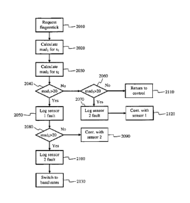
The logic starts at block 2010 where an alarm would be triggered once the
logic enters the fault
handling mode. The alarm would include a request for a current meter value
using the traditional
finger prick method. The current meter value will be used as a current blood
glucose value to
compare against each sensor reading. At block 2020 and 2030, the mean absolute
difference
calculated as a percentage between the current blood glucose value and the
first sensor 800 and
the second sensor 850 will be calculated. The mean absolute difference can be
calculated as
follows:
madi = 100*ICF.st ¨ BGI / BG %
mad2 = 100*ICF.s2¨ BG! / BG %
[0065] At block 2040, the mean absolute difference for the first
sensor 800 will be
compared to a threshold value to determine if the blood glucose value returned
by the first sensor
800 deviates too much from the current blood glucose value returned by the
meter. In the
19
CA 02671721 2012-06-13
preferred embodiment the threshold is set to a 20 % difference. However, in
alternative
embodiments, the threshold can be set to a higher or lower value. If the
threshold is exceeded,
the logic at block 2050 will determine that first sensor 800 is faulty and
report that the first
sensor 800 is failing. After the first sensor 800 values are checked at block
2040, the second
sensor 850 values are checked at blocks 2060 and 2080. At block 2060 and 2080,
the mean
absolute difference for the second sensor 850 will be compared to the same
threshold value to
determine if the blood glucose value returned by the second sensor 850
deviates too much from
the current blood glucose value returned by the meter. If the threshold is
exceeded, the logic at
blocks 2070 and 2100 will determine that second sensor 850 is faulty and
report that the first
sensor 850 is failing. Depending on which sensors are determined to be
failing, the logic defaults
to four different possibilities. The first possibility is found at block 2110.
If both sensors 800
and 850 are do not exceed the threshold (and thus, neither sensor is
determined to be failing), the
logic at block 2110 exits the fault handling mode and returns back to normal
operation of FIG.
19. The second possibility is found at block 2120 where only the second sensor
850 is found to
be failing. In this case, the logic of block 2120 will stop using the signals
from the second sensor
850, and the closed loop system/semi-closed loop system will continue using
only the sensor
values from the first sensor 800 until the second sensor 850 can be replaced.
Similarly, the third
possibility is found at block 2090 where only the first sensor 800 is found to
be failing. In this
case, the logic of block 2090 will stop using the signals from the first
sensor 800, and the closed
loop system/semi-closed loop system will continue using only the sensor values
from the second
sensor 850 until the first sensor 800 can be replaced. The last possibility is
found at block 2130,
where both sensors are found to be failing and need replacement. If the logic
of block 2130 is
triggered a different insulin delivery strategy should be immediately adopted
such as limiting the
insulin delivery only to minimal basal amounts.
100661 In an alternative embodiment, one sensor will act as the primary
sensor, and the
second sensor will act as a watchdog. In an example of this embodiment, the
second sensor 850
will only be used to detect if the first sensor 800 is failing. If the total
error Et exceeds the
threshold at block 1660 of FIG. 19, then the system will automatically
implement a different
insulin delivery strategy such as limiting the insulin delivery only to
minimal basal amounts, and
both sensors would be signaled to be replaced. In addition, if the total error
Et does not exceed
CA 02671721 2012-06-13
the threshold, no error comparison will be made between the two sensors.
Instead, only the
predicted sensor values of the first sensor 800 will be used.
[0067] While the description above refers to particular embodiments
of the present
invention, it will be understood that many modifications may be made. For
example, additional
steps and changes to the order of the algorithms can be made while still
performing the key
teachings of the present invention. In addition, although the preferred
embodiments described
the use of two sensors, in alternative embodiments three or more sensors can
be used with the
present invention.
[0068] The scope of the claims should not be limited by the preferred
embodiments set
forth herein, but should be given the broadest interpretation consistent with
the description as a
whole.
21