Note: Descriptions are shown in the official language in which they were submitted.
CA 02672135 2009-06-09
WO 2008/073916 PCT/US2007/087023
METHOD AND APPARATUS FOR VERIFYING
OCCLUSION OF FALLOPIAN TUBES
Field of the Invention
[0001] The field of the invention generally relates to methods and devices
used to
verify or detect occlusion of a body lumen. More specifically, the field of
the invention
pertains to methods and devices for detecting or verifying fallopian tube
occlusion.
Backaround.og the Invention
[0002] Conventionally, bilateral tubal sterilization (BTS) has been used for
sterilization
in female patients. Typically, BTS is surgically accomplished by ligation of
the fallopian
tubes using one or more surgical approaches. More recently, various non-
operative
methods of achieving sterility have been developed as an altemative to
conventional
BTS procedures. For example, Conceptus, Inc. of San Carlos, California, has
developed the ESSURE micro-insertion device which is deployed
hysteroscopically.
Also, Adiana, Inc. of Redwood City, CA, has developed a hysteroscoPically-
placed
device which uses low level radiofrequency energy to damage the fallopian
tubes. A
soft polymer matrix is left behind in the tube to facilitate closure. In both
of these
processes, sterilization is accomplished by occlusion of the intramural
portion of the
fallopian tubes.
[0003] These new, non-operative methods require some. sort of post-procedure
verification to ensure that the fallopian tube(s) have indeed been occluded.
Typically,
occlusion is verified after the sterilization procedure with the aid of
hysterosalpinography
1
CA 02672135 2009-06-09
WO 2008/073916 PCT/US2007/087023
(HSG). HSG is a radiographic technique in which a contrast media (e.g., oil or
water
soluble fluid containing a radiographically opaque compound of a material such
as
iodine) is injected slowly into the uterine-cavity and fallopian tubes via a
transcervicallly-
placed cannula. Radiographic images are taken to delineate the inside of the
uterus
and fallopian tubes. Tubal occlusion is verified by the lack of contrast media
past a
specific location in the tube (or by lack of contrast media in certain
anatomical spaces
such as the pouch of Douglas). Unfortunately, HSG subjects the patient to
ionizing
radiation and the patient may potentially be sensitive to the contrast medium.
Also,
because HSG involves radiation, the procedure must be performed in a
specialized
suite or room suitable for radioactive procedures.
[0004] More recently, hysterosalpingo-conttast sonography (HyCoSy) has been
developed for imaging the uterus and fallopian tubes. HyCoSy is an ultrasonic
technique that is accomplished transvaginally after the uterus and fallopian
tubes are
filled with contrast media. Tubal occlusion (or lack thereof) is determined by
the
absence of contrast media past a specific location in the fallopian tube or by
the
absence of contrast media in other anatomical spaces (e.g., the pouch of
Douglas).
While HyCoSy does obviate the risks of radiation exposure, the method employs
somewhat complex and expensive equipment. There is a need for a less complex
device and method that can be used to verify and/or detect occlusions within
the
fallopian tube. Preferably the device and method should be able to verify
occlusion in
the intramural portion of the patient's fallopian tubes.
2
CA 02672135 2009-06-09
WO 2008/073916 PCT/US2007/087023
Summary
[0005]' In one embodiment of the invention, a device for verifying occlusion
of the
fallopian tube in a female subject includes an elongate gas delivery member
having a
lumen disposed therein, the elongate gas delivery member adapted for sealing
engagement with the subject's uterus. The device includes a pressurized
insufflation
gas source coupled to the elongate gas delivery member, the insufflation gas
source
being in communication with the lumen of the elongate gas delivery member. The
insufflation gas may include, for example, carbon dioxide. The device includes
a.
pressure gauge interposed between the pressurized insufflation gas source and
a distal
end of the elongate gas delivery member for monitoring insufflation gas
pressure of the
subject's uterine cavity. In an alternative embodiment, a pressure sensor may
be
affixed or otherwise incorporated into'the elongate gas delivery member to
measure
intra-uterine pressure.
[0006] In another embodiment of the invention, a device for verifying
occlusion of the
fallopian tube in a female subject includes an elongate gas delivery member
having a
lumen disposed therein, the elongate gas delivery member adapted for sealing
engagement with the subject's uterus. The device includes a pressurized
insufflation
gas source coupled to the elongate gas delivery member, the insufflation gas
source
being in communication with the lumen of the elongate gas delivery member. A
flow
meter is interposed between the pressurized insufflation gas source and a
distal end of
the elongate gas delivery member.for monitoring the flow rate of the
insufflation gas into
the subject's uterine cavity.
[0007] In still another embodiment of the invention, the device may include
both the
pressure gauge and the flow meter as described above. One or both of the
pressure
3
CA 02672135 2009-06-09
WO 2008/073916 PCT/US2007/087023
gauge and flow meter may be used to detect leakage of the insufflation gas
past the
region of the fallopian tube containing the occlusive device. For example, the
measured
flow rate required to keep a substantially constant pressure within the
uterine cavity may
be used to detect the presence or absence of any leaks across the putative
occlusion.
Alternatively, the pressure gauge may be monitored after charging the uterine
cavity
with a pressurized charge of insufflation gas. The decay or drop on pressure
may be
used to detect any leaks across the occlusion formed within the fallopian
tubes.
[0008]' In still another embodiment of the invention, a method of verifying
the
occlusion of a fallopian tube of a female subject includes the steps of
providing a source
of pressurized insufflation gas, the gas source being coupled to a delivery
member that
can be inserted into the uterine cavity so as to form a seal between the
delivery member
and_the uterus. Pressurized insufflation:gas is then delivered from the source
to the
uterine cavity. The pressure of the insufflation gas contained within the
uterus is
measured over a period of time to detect the presence or absence of,fallopian
tube
occlusion. For example, the pressure drop over a period of time may be used to
determine whether the fallopian tube(s) are indeed occluded. The threshold or
cutoff
levels for leakage rates may be determined experimentally.
[0009] In yet another embodiment of the invention, a method of verifying the
occlusion of a fallopian tube of a female subject includes the steps of
providing a source
of pressurized insufflation gas, the gas source being coupled to a delivery
member that
can be inserted into the uterine cavity so as to form a seal between the
delivery member
and the uterus. Pressurized insufflation gas is then delivered from the
source:to the
uterine cavity. After the uterine cavity has initially been charged, a small
flowof
insufflation gas may be metered into the cavity to maintain a substantially
constant
4
CA 02672135 2009-06-09
WO 2008/073916 PCT/US2007/087023
pressure. The flow rate (or volume) of this metered gas may be monitored to
detect the
presence or absence of fallopian tube occlusion. The threshold or cutoff
levels used to
determine whether or not the fallopian tube(s) are indeed occluded may be
determined
experimentally.
Brief Description of the Drawings
[0010] The drawings illustrate the design and utility of various embodiments
of the
present invention, in which similar elements are referred to by common
reference
numerals. In order to better appreciate how the above-recited and other
advantages
and objects of the present inventions are obtained, a more particular
description of the
present inventions briefly described above will be rendered by reference to
specific
embodiments thereof, which are illustrated in the accompanying drawings.
Understanding that these drawings depict only typical embodiments of the
invention and
are not therefore to be considered limiting of its scope, the invention will
be described
and explained with additional specificity and detail through the use of the
accompanying
drawings in which:
[0011] FIG. 1 is a schematic representation of a device for verifying
occlusion of the
fallopian tube in a female subject according to one embodiment.
[0012] FIG. 2 is a schematic representation of a device for verifying
occlusion of the
fallopian tube in a female subject according -to another embodirrient.
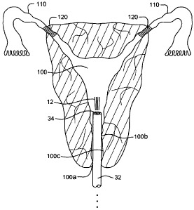
[0013] FIG. 3 is a partial cross-sectional view of the female reproductive
system
showing placement of a gas delivery member according to one embodiment of the
invention.
CA 02672135 2009-06-09
WO 2008/073916 PCT/US2007/087023
[0014] FIG. 4 is a partial cross-sectional view of the female reproductive
system
showing placement of a gas delivery member according to another embodiment of
the
invention.
[0015] FIG. 5 is a partial cross-sectional view of the female reproductive
system
showing placement of a gas delivery member according to still another
embodiment of
the invention.
[0016] FIG. 6 is a flowchart of a method of verifying occlusion of a fallopian
tube of a
female subject according to one embodiment.
Detailed Description
[0017] FIG. 1 illustrates an apparatus 10 for verifying whether or not a
fallopian tube
of a female subject is occluded. The apparatus 10 generally includes a source
of
pressurized insufflation gas 12. The insufflation gas 12 may include a gas
such as, for
example, USP grade carbon dioxide, although other gases may also be used in
the
apparatus 10. In the case of carbon dioxide, the insufflation gas 12 may be
stored as a
liquid and released in gaseous form. The pressurized insufflation gas.12 may
be
contained in a vessel or container 14 such as, for instance, a cylinder or
tank commonly
used in medical applications to store pressurized gases. In other,
embodiments,
however, the apparatus 10 may be coupled to another source of pressurized gas.
For
example, hospitals and other medical facilities often have pressurized gas
ports
integrated into the construction of individual examination rooms.
[0018] The apparatus 10 includes a conduit 16 that is used to connect or
couple the
various components of the apparatus 10. The conduit 16 includes an interior
lumen
through which the pressurized insufflation gas 12 can flow through. The
conduit 46 may
8
CA 02672135 2009-06-09
WO 2008/073916 PCT/US2007/087023
include tubing, piping, hose, or the like. The conduit 16 may be rather rigid
or- stiff in
certain segments or regions while flexible in others. For example, conduit
segment 16b
in FIGS. 1 and 2 is made of a flexible hose or the like to permit manipulation
of the gas
delivery member (described in more detail below).
[0019] The tank 14 of pressurized insufflation gas 12 is coupled via the
conduit 16 to
a shut off valve 18. This shut off valve 18 can be used to stop all gas flow
through the
apparatus 10. The shut off valve 18 may be integrated with the tank 14 or it
may be a
separate component. The shut off valve 18 permits the removal and replacement
of a
tank 14 that may have a low reserve of insufflation gas 12. A downstream
segment of
conduit 16 connects the shut off valve 18 to a pressure gauge 20: The pressure
gauge
20 is used to monitor the level or quantity of insufflation gas 12 remaining
in the
container 14. In addition, the pressure gauge 20 indicates to the operator
when the
main shut of valve 18 has been opened or closed. Downstream of the pressure
gauge
20, another conduit segment 16 connects to a pressure regulator 22. The
pressure
regulator 22 is adjustable by the operator and permits .the occlusion
verification tests
described herein to be performed at a multitude of pressures. In this regard,
the
particular pressure applied to the uterine cavity 100 (shown in FIGS. 1-5) can
be
adjusted by the operator. The pressure regulator 22 may include-a dial or
indicator of
the pressure so that the operator can quickly and accurately adjust the
pressure of the
apparatus 10.
[0020] Still referring to FIG. 1, a conduit 16 connects the downstream gas
flow from
the pressure regulator 22 to a flow control valve 24. The flow control valve
24 is used
control the flow rate of the insufflation gas 12 into the uterine cavity 100.
For example,
FDA standards for hysteroscopic insufflation require flow rates of less than
100
7
CA 02672135 2009-06-09
WO 2008/073916 PCT/US2007/087023
ml/minute. The flow control valve 24 can thus be used to raise or lower the
flow rate of
the -insufflation gas 12 as needed. Gas from the flow control valve 24
continues via
conduit 16 to a valve 26 that modulates the flow through the apparatus 10. The
valve
26 operates in either an "off" state or an "on" state. The valve 26 may
include a
powered solenoid valve that, when energized, permits insufflation gas 12 to
flow into the
uterine cavity 100. In contrast, when the solenoid valve is not energized,
insufflation
gas 12 cannot pass the valve 26. The state of the valve 26 may be controlled
through
electronic circuitry (not shown) that is coupled to switch, button,. or the
like that is used
to trigger gas insufflation. Such circuitry is well known to those skilled in
the art and is
not described herein.
[0021] In certain embodiments of the invention, the valve 26 may be used to
isolate
the apparatus 10. For example, if pressure is being monitored within the
uterine cavity
100 (or within the system as a proxy for uterine cavity pressure), the valve
26 may be
switched to an "off' state after the uterine cavity 100 has been pressurized
with
insufflation gas 12. The decay or loss of pressure within the system can then
be
monitored to detect or verify occlusion of the subject's fallopiari tubes 110.
[0022] Still referring to FIG. 1, a conduit 16 connects the downstream output
of the
valve 26 to a pressure gauge 28 and flow meter 30. The pressure gauge 28 is
used to
measure the pressure within the uterine cavity 100. The actual point of
measurement,
however, may be outside the uterine cavity 100 as is shown in FIGS. 1 and 2.
Generally, it is not expected that there would be a large pressure drop from
the location
of the pressure gauge 28 in FIGS. 1 and 2 and the pressure contained within
the uterine
cavity 100. Consequently, the pressure taken proximally with respect to the
outlet of the
apparatus 10 is thought to be an accurate estimate of the actual pressure
experienced
8
CA 02672135 2009-06-09
WO 2008/073916 PCT/US2007/087023
within the uterine cavity 100. The pressure gauge 28 may be an analog pressure
gauge
or even one with a digital readout or output that could be displayed on
monitor or
computer. In other embodiments, however, the pressure gauge 28 may measure
pressure directly within the uterine cavity 100 using a small semiconductor,
piezoelectric, or Micro-Electro-Mechanical Systems (MEMS) based pressure
sensor. In
this regard, the pressure gauge 26 may be integrated into the gas delivery
member 32
which is described in detail below).
[0023] In certain embodiments, only the pressure gauge 28 is needed to detect
or
verify occlusion of the fallopian tubes 110. For example, as explained above,
the
uterine cavity 100 may be charged with a pressurized volume of insufflation
gas 12.
The solenoid valve 16 can then be turned to the "off' state and the pressure
gauge 28
can be monitored to detect any leaks. Any leaks within the fallopian tube(s)
110 are
detected be a reduction in measured pressure. The reduced pressure is caused
by
insufflation gas 12 passing the region of the fallopian tube 110 containing
the occlusive
device 120 and exiting out of the fallopian tube 110 and into the peritoneum
cavity. For
example, the presence of a leak between the occlusive device 120 and the
fallopian
tube 100 may be determined if the pressure drops above a certain threshold
rate (e.g.,
mmHg/sec). In certain embodiments, some leakage within the system may be
attributed to leakage between the uterine cavity 100-and the gas delivery
member
(described below) if the seal is not complete. Consequently, there may be a
background or baseline level of pressure decay within the system even if the
occlusive
device(s) 120 have completely occluded the fallopian tubes 110. In this case,
the
natural or background rate of leakage may be determined and leakage rates
falling
above this level may be used to verify the presence or absence of any leaks.
9
CA 02672135 2009-06-09
WO 2008/073916 PCT/US2007/087023
[0024] As an alternative to using the pressure gauge 28, the apparatus-10 may
employ a flow meter 30 to verify or detect-occlusion of the fallopian tubes
110. In #his
embodiment, the uterine cavity 100 is charged with pressurized insufflation
gas 12 to a
target or set point pressure. The system 10 then supplies additional
insufflation gas 12
to the uterine cavity 100 to maintain the target pressure. The flow rate of
the additional
insufflation gas 12 needed to maintain a substantially constant pressure
within the
uterine cavity 110 can then be used to verify occlusion of the fallopian tubes
110. For
example, the presence of a leak can be made once the rate of gas flow (or
volume)
exceeds a certain threshold value. For example, there may be some slight
leakage
between the gas delivery member (described below) and uterine cavity 100.
Additional
leakage beyond this baseline level can be detected by additional flow needed
within the
apparatus 10 to maintain the pressure within the uterine cavity 100..
[0025] In this embodiment, the pressure within the uterine cavity 100 may be
determined using the pressure gauge 28 described above, or alternatively, a
pressure
gauge 28 contained on or in the gas delivery member that is used to measure
the
pressure directly within the uterine cavity 100. The flow control valve 24 may
be
arranged in a feedback loop with the pressure gauge 28 (or other pressure
sensor) such
that the flow of insufflation gas 12 can automatically adjusted based on. real
time or near
real time measurements of pressure within uterine cavity 100.
[0026] As seen in FIG. 1, a flexible conduit 16b such as a hose or tubing
connects the
proximal aspects of the device 10 to a gas delivery member 32. The gas
delivery
member 32 may be an elongate tubular member having one or more lumens 34
contained therein that are used as a passageway forthe insufflation gas 12.
The gas
delivery member 32 may be formed as a catheter or cannula that is sized for
insertion
CA 02672135 2009-06-09
WO 2008/073916 PCT/US2007/087023
into the uterine cavity 100. For example, the gas delivery member 32 may take
the form
of a Foley-type catheter. The catheter or cannula may be dimensioned to have
an
external diameter such that a substantially airtight seal is formed between
the gas
delivery member 32 and the uterine cavity 100. The gas delivery member 32 may
form
a seal the external os 100a of the uterus, the internal os 100b of the uterus,
or the
cervical canal 100c or a combination thereof. In one aspect, as seen in FIG:
2, the gas
delivery member 32 may include a sealing member 36 that aids in forming the
seal with
the uterine cavity 100. The sealing member 36 may include a pliable or
resilient
member that is disposed about the periphery of the gas delivery member 32. In
yet
another alternative, the sealing member 36 may including an expandable member
such
as, for instance, an inflatable balloon or the like that is affixed to the gas
delivery
member 32.
[0027] Still referring to FIG. 1, the lumen 34 of the gas delivery member 32
is coupled
to a conduit 16 that communicates with a purge valve 38. Activation.of the
purge valve
38 enables the evacuation of insufflation gas 12 from the uterine-cavity 100.
The purge
valve 38 may take the form of a solenoid valve that is activated
electronically.
Preferably, the conduit 16 connecting to the lumen 34 of the gas delivery
member 32 to
the purge valve 38 is located on the gas delivery member 32 at a~location that
lies
outside the patient. The connecting conduit 16 may even conriect somewhere
further
on the proximal end of the gas delivery system.
[0028] FIG. 2 illustrates an alternative embodiment of the apparatus 10 in
which the
gas delivery member 32 is separate from an evacuation member 40. In FIG. 2,
both the
gas delivery member 32 and the evacuation rnember40 pass through a common
sealing member 36 although separate sealing members 36 could be used for each
11
CA 02672135 2009-06-09
WO 2008/073916 PCT/US2007/087023
member 32, 40. The embodiment in FIG. 2 is different from that disclosed in
FIG. 1 in
there is no common lumen that both delivers and evacuates insufflation gas 12
into and
out of the uterine cavity 100.
[0029] It should be understood that a variety of designs may be employed for
the gas
delivery member 32. For example, FIG. 3 illustrates a view of the deployed gas
delivery
member 32 inside the uterine cavity 100. The gas delivery member 32 includes a
single
lumen 34 that is used for both delivery and evacuation of insufflation gas 12.
FIG. 4
illustrates a dual lumen embodiment of a gas delivery member 32 which has a
first
lumen 34 for insufflation gas delivery and a second lumen 35 for insufflation
gas
evacuation. FIG. 5 illustrates yet another embodiment that uses a separate
evacuation
member 40. The evacuation member 40 includes its own lumen 42 for gas
evacuation.
[0030] FIG. 6 illustrates an exemplary flow diagram showing one embodiment of
the
operation of the device 10. Initially, as seen in step 200, the device 10 is
started by
connecting the various components and ensuring that the same are operational.
Next,
in step 205 the-device 10 undergoes a purge. process to flush the system with
insufflation gas 12 (e.g., carbon dioxide). The gas delivery member 32 is then
inserted
into the uterine cavity 100 transvaginally by the operator. Alternatively, the
purge
process may be initiated after insertion of the device 10 into the patient. In
yet another
alternative, the purge process. may take both before and after placement of
the device
10. During the placement process, the subject may be placed into:the lithotomy
position
with knees raised and the cervix exposed using a standard speculum or the
like. The
gas delivery member 32 can then be advanced within the subject's cervix.
[0031] As seen in step 210, a low pressure test is then run to determine
whether or
not a proper seal has been formed between the gas delivery member 32 and the
uterus.
12
CA 02672135 2009-06-09
WO 2008/073916 PCT/US2007/087023
For example, a low pressure of about 50 mmHg insufflation gas 12 may be
delivered to
check for system leaks. Assuming a leak was detected, as illustrated in
the'pass query
step 215, the operator then adjusts the seal and/or placement of the gas
delivery
member 32 and checks for other sources of leaks within the system (step 220).
The low
ptessure seal test (step 210) is then performed again. After the device 10
passed the
low pressure test, a mid-level pressure is then delivered to the uterine
cavity 100 to
verify occlusion of the fallopian tubes 110 as is shown in step 225 of FIG. 6.
The mid-
level pressure may include an applied pressure of around 120 mmHg. Occlusion
of the
fallopian tubes 110 may be verffied or confirmed using either the pressure or
flow
methods discussed herein.
[0032] Next, as seen in step 230 of FIG. 6, a query is made whether or not the
test
was passed. In this regard, if a leak was detected;. the user would be
notified that
complete occlusion of the fallopian tubes 110 was not verified and the
verification step
failed (step 235). Assuming that the mid-level pressure test was successfully
passed -
thereby indicating that the fallopian tubes were fully occluded when subject
to the mid-
level pressure, the subject is then tested at a higher pressure level as is
shown in step
240 in FIG. 6. The higher pressure level may include a pressure on the order
of around
185 mmHg. It should be understood that the exactpressutes described above with
respect to the seal test and the mid and high pressure tests for fallopian
tube occlusion
may vary and still fall within the scope of the invention. Referring back to
FIG. 6,
another query is performed (step 245) to asses whether leaks were detected at
the
higher applied pressure. If leaks were detected, then the operator would be
notified that
the verification test failed (step 250). However, if no leaks were detected`
at the higher
applied pressure, then the subject is said to have passed the occlusion
verification test
13
CA 02672135 2009-06-09
WO 2008/073916 PCT/US2007/087023
(step 255). In step 255, the patient is assured that the fallopian tubes 110
have indeed
been fully occluded.
[0033] The device 10 described herein has been described in the context of
testing
both fallopian tubes 110 at the same time for determining whether total
occlusion has
occurred. In another embodiment of the invention, it may be possible-to
isolate one of
the two fallopian tubes 110 for testing. For example, an inflatable member
such as an
inflatable balloon or the like may be used to seal off one of the fallopian
tubes 100 such
that the other fallopian tube 110 can be tested at a single time.
[0034] While embodiments of the present invention have been shown and
described,
various modifications may be made without departing from the scope of the
present
invention. The invention, therefore, should not be limited, except to the
following claims,
and their equivalents.
14